Article

Planktonic Pro- and Microeukaryotes of the Kuibyshev Reservoir and Its Bays During the Cyanobacterial Bloom Period

by
Mikhail Yu. Gorbunov
1,
Svetlana V. Bykova
1,
Natalia G. Tarasova
1,2,
Ekaterina S. Krasnova
1 and
Marina V. Umanskaya
1,*
1
Samara Federal Research Center RAS, Institute of Ecology of the Volga Basin RAS, Russian Academy of Science, 10, Komzina str., 445003 Tolyatti, Russia
2
I.D. Papanin Institute for Biology of Inland Waters, Russian Academy of Science, 152742 Borok, Russia
*
Author to whom correspondence should be addressed.
Water 2025, 17(11), 1602; https://doi.org/10.3390/w17111602
Submission received: 20 April 2025 / Revised: 18 May 2025 / Accepted: 22 May 2025 / Published: 25 May 2025
(This article belongs to the Section Biodiversity and Functionality of Aquatic Ecosystems)

Abstract

:
Kuibyshev Reservoir, the largest in the Volga basin, is poorly covered by modern molecular studies. The results of a metabarcoding study of pro- and eukaryotic microbial plankton in its lower section during the summer period are presented. Bacterioplankton composition was typical for most temperate freshwater bodies and characterized by the dominance of cyanobacteria, Pseudomonadota, Bacteroidota, Actinomycetota, and PVC superphylum (Verrucomicrobiota and Planctomycetota), with a somewhat increased proportion of the latter. The protist community was dominated by Cryptista, principally phototrophic, and various ciliates. Several picoeukaryotic groups were newly detected in the reservoir. A relationship between the composition of both bacterioplankton and protist communities and the stage of phytoplankton succession, including the cyanobacterial bloom, was observed. Some inconsistency between the cyanobacterial bloom phase and the structure of other parts of the microbial plankton is obviously due to some temporal delay, spatial station position, and inflow from tributaries. Heterotrophic bacterioplankton indicator species of the main bloom stage include OTUs representing both the phycosphere of colonial cyanobacteria and free-living species. Among the protists, sessile ciliates benefit most from plenty of substrates for colonization, while cyanobacterial grazers and parasites were minor. Overall, the cyanobacterial bloom creates new niches for the plankton community and significantly modifies its structure.

1. Introduction

Anthropogenic eutrophication of freshwater and marine ecosystems is currently one of the most significant challenges facing humanity. This phenomenon is characterized by the excessive growth of planktonic primary producers (phytoplankton) or so-called water “blooms” [1,2,3]. The blooms can be caused by different phytoplankton taxa, including dinoflagellates, prymnesiophycean, and ochrophycean algae, but in freshwater, the most problematic are blooms of cyanobacteria, which are the only group of bacteria that perform oxygenic photosynthesis.
The cyanobacterial species that commonly cause water blooms are diverse. The main causative agents of blooms in eutrophic freshwater reservoirs are colonial chroococcal species with gas vacuoles, mostly Microcystis species; filamentous, nitrogen-fixing, planktonic nostocacean species of the genera Dolichospermum, Aphanizomenon s.l., and some others; and filamentous cyanobacteria without heterocysts of the genera Planktothrix, Planktolyngbya, Geitlerinema, and Pseudanabaena [4,5].
The microplankton community is highly interconnected, and these connections are not limited to predator–prey interactions. Due to their small size and high surface area-to-volume ratio, cells inevitably leak low- and medium-molecular-weight compounds. Many members of the community secrete high-molecular-weight substances to increase hydrodynamic resistance to settling or combine into aggregates/colonies, along with chelators and exoenzymes to obtain resources from the environment. It is not surprising that many planktonic bacteria use all these products and, in particular, are auxotrophic in a number of vitamins and other substances, such as hemes [6].
The structure of both the heterotrophic prokaryotic and eukaryotic planktonic communities in the surface layers of water is relatively stable on the highest phylogenetic levels. However, the proportion of photosynthetic cyanobacteria within the overall freshwater planktonic community fluctuates to a much greater extent than, for example, the proportions of proteobacteria and actinobacteria among the heterotrophic part of the prokaryotic community. These wide fluctuations in the abundance of cyanobacteria, both as a whole class and as individual species or functional groups, inevitably lead to significant alterations in the overall structure of the prokaryotic and microeukaryotic plankton communities. This has been confirmed by a number of published studies indicating that the intensity and duration of freshwater cyanobacterial blooms, as well as their taxonomic and functional composition, affect the relative abundance of many ecologically significant planktonic organisms, including non-phototrophic prokaryotes, protists, crustaceans, and rotifers, e.g., [1,7].
A number of studies have investigated the effect of cyanobacterial blooms on the functional composition of eukaryotic phytoplankton [8] and on the ratio of autotrophic and heterotrophic components of microplankton [9,10]. Laboratory experiments [11] and field observations [12,13] have shown the interdependence between the number and diversity of ciliates and the species structure of cyanobacteria. A study in one of the suburban reservoirs of Champs-sur-Marne (France) showed that the taxonomic structure of free-living bacteria during blooms of Dolichospermum (“Anabaena”) and Microcystis differed significantly [14]. The structure of heterotrophic bacteria in communities dominated by other cyanobacterial species is less studied. Although there are studies on the prokaryote’s structure in the unialgal enrichments of different species of cyanobacteria belonging to the genera Aphanizomenon, Planktothrix, and Microcystis [15], the possibility of extrapolating these results to natural ecosystems is not clear.
The Volga River is the longest river in Europe, with a length of 3531 km and a catchment area of 1,360,000 km2. Its source and upper reaches, the so-called Verkhnyaya (Upper) Volga, are situated in the boreal forest zone. The area between the mouths of the Oka and Kama rivers is located in the mixed and broadleaf forest zone, and the part down to the mouths of the Samara and Syzranka rivers is in the forest-steppe zone. Together, these two parts make up the Srednyaya (Middle) Volga in its broad sense, while the entire course of the river downstream is known as the Nizhnyaya (Lower) Volga. Currently, most of the river has been transformed into a cascade of reservoirs, with the largest being the Kuibyshev Reservoir [16]. The Kuibyshev Reservoir is the third-largest artificial freshwater reservoir in the world and the fifteenth-largest in the world by volume. By both values, it is the largest in Europe.
Since its creation, microscopic monitoring of different groups of microbial plankton has been conducted in the reservoir [17,18,19,20,21,22]. All available data indicate that in the 20th and early 21st centuries, cyanobacterial blooms have been recorded in this reservoir from late June to August and September. During the Initial bloom period, filamentous, heterocystous (nitrogen-fixing) Nostocales species of cyanobacteria predominated, but as the bloom progressed, they may be replaced by Microcystis species [18,19]. However, the information about its ecological features was mostly published locally and so does not influence the understanding of its significant ecological importance on a global scale.
The phylogenetic composition of the microplankton of the Volga reservoirs, especially the Kuibyshev Reservoir, remains virtually unstudied, with the exception of a number of recently published studies [23,24,25,26]. In this work, we attempted to fill this gap and analyze the phylogenetic composition of pro- and eukaryotic microbial plankton of the lower part of the Kuibyshev Reservoir, including Usa, Suskan, and Cheremshan Bays, in the summer of 2023.

2. Materials and Methods

2.1. Study Site and Sampling

The research was carried out in the lower Priplotinny (near-dam) Reach of the Kuibyshev Reservoir and three bays that were formed as a result of the partial flooding of Volga tributaries during the reservoir’s creation. Usa Bay is located adjacent to the near-dam reach and has a significant length (more than 50 km) but a relatively small (~1 km) average width. Cheremshan Bay, the largest in the reservoir, connects with it in the upper part of the Novodevichensky Reach, located upstream of the Priplotinny Reach. With a length only slightly less than that of Usa Bay, its maximum width exceeds 11 km. Suskan Bay is located on the right side of the mouth of the Cheremshan Bay; its upper part is isolated from the reservoir’s water area by a dam and has been long used for fish farming. The sampling in Suskan Bay was performed in its lower part, which is connected both to Cheremshan Bay and the Novodevichensky Reach of the Kuibyshev Reservoir.
Samples were collected during the cyanobacterial bloom period from June to August 2023 at 11 stations, as shown in Figure 1. Their codes, coordinates, and collection dates are listed in Table S1.
At all stations, samples were taken from the upper 0–1 m water layer. The temperature, pH, redox potential, electrical conductivity, and dissolved oxygen concentration in the water column were measured using portable meters at the time of sampling. Chemical analyses were performed in the lab using manual colorimetric methods [27,28,29]. The concentrations of total phosphorus and iron were determined in unfiltered samples; concentrations of dissolved phosphorus, ammonium, and silicate, as well as water color, were measured in samples filtered through glass fiber filters with 1.2 μm nominal retention.
To determine the concentration of chlorophylls, 0.2–1.0 L water samples were filtered through glass fiber filters with a nominal retention of 1.2 μm (FPSV, Vladisart, Vladimir, Russia). The filters were air-dried and stored in a dark container in the refrigerator at 4–10 °C until delivery to the laboratory and then in the freezer at −20 °C. Photosynthetic pigments collected on the filters were extracted with 90% acetone in the dark at 4 °C for 24 h. The spectra of the extracts were recorded on a Specord M-40 spectrophotometer (Carl Zeiss JENA, Jena, Germany) in the range 350–850 nm before and after acidification. Pigment concentrations were calculated by spectral reconstruction or using the three-wavelength equations from [30]. Phaeophytin a concentrations were determined according to [31].
Samples for microscopic phytoplankton identification and enumeration were fixed with Lugol’s solution and concentrated by filtration, as described in [32]. The number and species composition of planktonic ciliates were determined in samples pre-concentrated by gravity filtration through 5–7 µm membrane filters and fixed with a saturated solution of HgCl2 [33]. Species identification of phytoplankton and ciliates was performed based on the morphology of the cells and colonies. The number of heterotrophic bacterioplankton was determined on nitrocellulose membrane filters with a pore diameter of 0.2 µm after DAPI staining [34].

2.2. DNA Extraction, 16S and 18S rRNA Gene Amplification, and Sequencing

DNA isolation and high-performance sequencing were performed at Syntol (Moscow, Russia). For DNA extraction, 100 to 250 mL of water was filtered through 0.2 µm sterile cellulose nitrate (CN) membrane filters (Vladisart, Russia), fixed by 80% ethanol, and stored at −18 °C until further processing. DNA was extracted using Sorb-GMO-B isolation kits (Syntol, Moscow, Russia).
The concentration of the isolated DNA was determined using a Quantus fluorometer (Promega, Madison, WI, USA) with the QuantiFluor® dsDNA kit (E2670). Quality control was carried out using RT-PCR on a CFX real-time PCR system (Bio-Rad, Hercules, CA, USA). For each sample, two libraries were prepared using prokaryotic and eukaryotic primers. The specific primers were 515F, GTGYCAGCMGCCGCGGTAA [35] and 806R, GGACTACNVGGGTWTCTAAT [36] for prokaryotes and Euk574F, CGGTAAYTCCAGCTCYA and Euk897R, TCYDAGAATTYCACCTCT [37] for eukaryotes, and both pairs were used with standard Illumina adapters. The preparation of libraries was performed in accordance with the protocol described in the manual “16S Metagenomic Sequencing Library Preparation” (Part #15044223 Rev B; Illumina, San Diego, CA, USA).
The obtained sample libraries were pooled in equimolar quantities and sequenced on an Illumina MiSeq platform using MiSeq Reagent Kit v2 (2 × 250 cycles) (Illumina, San Diego, CA, USA). The data quality was assessed using FastQC (version 0.12.1) and MultiQC (version 0.4).

2.3. Sequence Data Analysis

The Usearch 11.0.67 program was used to merge paired-end reads, and their further processing and clustering into OTUs were carried out using the standard UPARSE pipeline [38]. Prokaryotic OTUs were further designated as BOTUs, and eukaryotic ones were designated as EOTUs.
The OTUs’ taxonomy was assigned using the SILVA SSU database version 138.1 [39]. In addition, we used a database of 16S rRNA sequences extracted from the Genomic Taxonomy Database (GTDB) v. 214.1 [40] after slight pre-filtering and header conversion to UPARSE format [41], as well as the PR2 database [42]. Taxonomic determination was performed using the SINA online classifier [43] for the SILVA database and the SINTAX classifier included in the USEARCH package for all other cases [41].
We excluded all OTUs of mitochondria and plastids of higher plants (identified by SILVA) from the prokaryotic library, as well as OTUs of Metazoa, higher plants, and nucleomorphs (determined by PR2) from the eukaryotic library. Singletons and doubletons were also excluded from the total OTU list. We considered OTUs with a relative abundance greater than 1% in at least one sample as dominant. Indicator species were identified among dominant heterotrophic prokaryotic and protist OTUs based on IndVal values [44].

2.4. Statistical Analysis

Statistical data processing was carried out in the R v. 4.3.2 platform using the Phyloseq and Vegan packages and in the program PAST v.4.16c [45]. Simple formula calculations, such as diversity indices and regressions, were performed directly in spreadsheet editors (MS Office Excel, Libre Office Calc, etc.).

3. Results

3.1. Abiotic Conditions and Chlorophyll a

Physicochemical characteristics, concentrations of biogenic elements, and chlorophyll a of the water samples from the investigated area are shown in Table 1. The water in the reservoir showed moderate mineralization of ~240 mg L−1. During the observation period, the water column was well aerated; the maximum oxygen concentration (18.75 mg L−1) was recorded in August in the surface water layer at station UB3–8 (at the mouth of the Usa Bay). There was no pronounced thermal stratification: in June, the average temperature gradient in the water column at different stations was 0.01–0.08 °C m−1, and in July–August, it was 0.1–1.0 °C m−1. The water transparency varied from 0.3 to 2.25 m; the highest transparency was recorded in June during the clear water phase. pH values ranged from 7.7 to 9.5, with no pronounced spatiotemporal changes. The concentrations of total phosphorus, ammonium nitrogen, and chlorophyll a correspond to eutrophic conditions (Table 1).
Non-metric multidimensional scaling by chemical parameters and chlorophyll was used to divide all samples into three distinct groups (Figure 2). Three June samples were combined into group 1; two more were combined into group 2; and the last June sample, together with all July and August samples, was combined into group 3.

3.2. Microbial Gene Diversity of the Lower Part of the Kuibyshev Reservoir

The combined libraries contained 2990 operational taxonomic units (OTUs) of prokaryotes, including archaea, bacteria, and algal plastids (1,624,292 reads), and 1131 OTUs of protists and fungi (971,692 reads). Among prokaryotes, there were 2852 OTU of archaea and bacteria (1,341,907 reads).
Among prokaryotes, the dominant phylum by the number of reads was Cyanobacterota (including the heterotrophic classes Vampirivibrionia and Candidatus Sericytochromatia), followed in decreasing order by Pseudomonadota, Bacteroidota, Actinomycetota, Verrucomicrobiota, and Planctomycetota. Together, these phyla accounted for more than 95% of the prokaryotic reads. The highest numbers of OTUs among bacteria were found, in decreasing order, in the phyla Pseudomonadota, Bacteroidota, Verrucomicrobiota, and Planctomycetota. Among protists, Alveolata (including Ciliophora and Myzozoa), Cryptista, and Stramenopiles (represented mostly by Gyrista) dominated, accounting for 79% of all reads. Representatives of the phyla Amorphea and Stramenopiles contributed the most to the total number of OTUs among protists (Table S2).
The lowest diversity of prokaryotic plankton was observed in July at the mouth of Usa Bay (UB4-7), and the highest diversity was observed in June at the upper station of Usa Bay (UB8-6), which is influenced by the Usa River (Table 2). By contrast, the microeukaryotic diversity in the UB8-6 sample was the lowest. The highest diversity of planktonic microeukaryotes was observed in Suskan Bay (SB-7) and at station KR3-8, located opposite the mouth of Usa Bay, but the latter station had a relatively low diversity of prokaryotes (Table 2). Overall, there was no correlation between the diversity indices of pro- and eukaryotic plankton communities.

3.3. Community Structure and Composition of Phototrophic Cyanobacteria

Phototrophic cyanobacteria (class Cyanophyceae = Cyanobacteriia = Oxyphotobacteria), also referred to as Cyanobacteriia, are the only class of bacteria capable of oxygenic photoautotrophy. Therefore, in the surface aerobic layers of water, they do not compete significantly for most resources with other bacteria. Furthermore, we considered Cyanobacteriia separately from all other classes of bacteria, including the heterotrophic classes of the Cyanobacteriota phylum, Vampirovibrionia and Candidatus Sericytochromatia.
A total of 56 OTUs of Cyanobacteriia were found in the combined library. Of these, 51 OTUs belonged to 11 different families, while 5 OTUs were identified only to the class or order level. In terms of the total number of reads of Cyanobacteriia, representatives of the Aphanizomenaceae (10 OTUs) and Cyanobiaceae (18 OTUs) dominated, accounting for 67.9% and 15.5%, respectively. They were accompanied by Microcystaceae (2 OTUs) and Pseudanabaenaceae (6 OTUs), each accounting for 9.7% and 4.8%, respectively. Other families and unidentified cyanobacterial OTUs made up 20 OTUs, accounting for 2.2% of reads.
The fraction of cyanobacterial sequences in the total number of bacterial sequences in four samples was less than 10% (Table 3). In all other samples, the contribution of cyanobacterial sequences varied from 20% to 63%. The ratio of cyanobacterial and chloroplast sequence numbers was used as a rough estimate of the proportion of cyanobacteria in phytoplankton. In three samples collected in June, the proportion of chloroplast sequences was greater than 90%, indicating that phytoplankton was predominantly eukaryotic. Obviously, no cyanobacterial bloom occurred in these samples. Based on similarity in phytoplankton composition, they were grouped into a single cluster (Figure 3). Among the cyanobacterial species, BOTU-13 (Microcystis aeruginosa) and BOTU-1 (Aphanizomenon flos-aquae) were dominant. They were accompanied by BOTU-33 (Pseudanabaena/Limnothrix) and two OTUs belonging to the Cyanobiaceae: BOTU-24 and BOTU-21 (Figure 4).
Close to this group were two other June samples (Figure 3) with a proportion of chloroplast sequences decreased to ~70%, which indicates a somewhat more intense development of cyanobacteria (Table 3 and Figure 4). Based on the composition of chloroplast sequences, these samples are quite similar to the samples of the first group. However, the composition of cyanobacteria had significantly changed; the two main OTUs contributing were BOTU-1 (A. flos-aquae) and BOTU-24 (Cyanobiaceae) (Figure 4). Therefore, we believe these samples could be separated into a special group representing the initial stage of the cyanobacterial bloom.
All other samples were combined into a fairly compact group characterized by an intense bloom of BOTU-1 (A. flos-aquae), which accounted for more than 60% of cyanobacterial sequences on average (Table 3, Figure 3 and Figure 4). It was accompanied by BOTU-7 (Dolichospermum flos-aquae), BOTU-13 (M. aeruginosa), and BOTU-24 (Cyanobiaceae). Other species were minor (Figure 3). It should be noted that this division of samples into groups (Table 3, Figure 3 and Figure 4) fully corresponds to their division according to physicochemical indicators and chlorophyll concentration (Figure 2). Thus, three stages of cyanobacterial development in the plankton of the Kuibyshev reservoir—the period before the bloom (stage A), the initial period (stage B), and the main bloom period (stage C)—were observed from June to August 2023. We further analyzed the composition and structure of heterotrophic prokaryotes and protists at different stages of the Aphanizomenon bloom.

3.4. Heterotrophic Bacteria Community Structure and Composition in Different Parts of the Reservoir

The 2803 isolated OTUs of heterotrophic bacteria (1,341,388 reads) were classified into 48 phyla, 149 classes, and 460 families. A total of 68 OTUs containing 1070 reads (less than 0.1%) remained unclassified, even at the phylum level. The changes in the proportions of macrotaxa at different stations, depending on the conditions, are shown in Figure 5. Before the bloom, Pseudomonadota and Bacteroidota were the most abundant phyla, whereas after the onset of the bloom, the PVC superphylum dominated, followed by Pseudomonadota and Actinomycetota (Figure 5). The proportion of the PVC superphylum was highest during the initial bloom period; it decreased slightly during the main bloom period, while the proportions of Pseudomonadota and Bacteroidota increased (Figure 5).
More pronounced changes occurred at the level of individual dominant OTUs, with some representatives of the same phylum being replaced by others (Figures S1 and S2). Overall, 101 dominant OTUs of heterotrophic bacteria, whose relative abundance exceeded 1% in at least one of the samples, were determined in the prokaryotic library. Next, we consider the structure of the dominant bacterial complex at different stages of the cyanobacterial bloom.
Stage A. In June, before the onset of the cyanobacterial bloom, heterotrophic plankton was dominated by 27 OTUs, which accounted for ~75% of all sequences. Among these, 11 OTUs, representing a total of 45–52% of reads per sample, were from proteobacteria. This was largely due to the sharp dominance of Oxalobacteraceae (Betaproteobacteria), particularly two OTUs: BOTU-16 (Massilia, up to 30.6% in sample) and BOTU-34 (closely related to Noviherbaspirillum, up to 16.4%). These were accompanied by BOTU-32 (Bacteroidia: Flavobacteriaceae: Flavobacterium), BOTU-53 (γPRO: Pseudomonadaceae, Pseudomonas), BOTU-151 (Bacteroidia: Crocinitomicaceae: Fluviicola), BOTU-22 (γPRO: Alteromonadaceae: Rheinheimera), BOTU-8 (αPRO: Pelagibacteraceae: Fonsibacter), and BOTU-48 (γPRO: Moraxellaceae: Alkanivorax) (Figure S2).
Stage B. During the initial period of the bloom, the number of dominant OTUs was 29, but their total relative abundance decreased to 48-53%. This decline was due to a significant reduction in the proportion of Pseudomonadota, primarily stemming from the vanishing of the Oxalobacteraceae. Additionally, other proteobacteria, namely BOTU-53 (Pseudomonas) and BOTU-48 (Alkanindiges), also disappeared from the dominant group. In their place, BOTU-36 (Limnohabitans) emerged as a dominant OTU, while the proportion of BOTU-8 (Fonsibacter) remained almost unchanged (Figure S2). Among Planctomycetota BOTU-31 (Planctomycetaceae)—closely related to the Planctopirus/Schlesneria clade—and BOTU-15 (Pirellulaceae: Roseimaritima) were the main contributors. These two OTUs, along with BOTU-79 (BAC: Chitinophagaceae: Sediminibacterium) reached their maximum contributions only in the initial period and, exceptionally, in sample UB7-6, which was collected simultaneously (on the same date) with samples UB6-6 and UB8-6 (Table S1 and Figure S1). Other dominant OTUs of stage B decreased in abundance in July and August (stage C, the main bloom period) but remained in the dominant group (Figure S2).
Stage C. During the main bloom period, the complex of dominant OTUs was the most numerous and diverse, and their composition varied greatly depending on the sample (Figure S2). In general, during stage C, the main dominant groups were BOTU-4 (ACT: Nanopelagicaceae: Candidatus Planktophila), BOTU-29 (βPRO: Usitatibacteraceae: UKL13-2), and BOTU-8 (Fonsibacter). BOTU-29, along with BOTU-233 (BAC: Sphingobacteriales: NS11-12), BOTU-98 (αPRO: Acetobacteraceae: Roseomonas), BOTU-3509 (VER: Chthoniobacteraceae JACTMZ01), and BOTU-59 (αPRO: Sphingomonadaceae: Sphingorhabdus), contributed significantly to heterotrophic bacterioplankton only during this phase (Figure S2).
Only three OTUs, BOTU-8 (αPRO: Pelagibacteraceae: Candidatus Fonsibacter 2.5-3.8%), BOTU-4 (ACT: Nanopelagicaceae: Candidatus Planktophila, 1.4-5.0%), and BOTU-12 (ACT: Nanopelagicaceae: Candidatus Nanopelagicus limnes, 1.6-4.0%), were present in the dominant complex throughout the observation period. BOTU-22 (γPRO: Alteromonadaceae: Rheinheimera) constituted 4% of the total in stage A, and then its abundance promptly decreased at the beginning of the bloom; however, during the main phase, it was restored, reaching 1.9% on average, and even 6.5% in some samples.
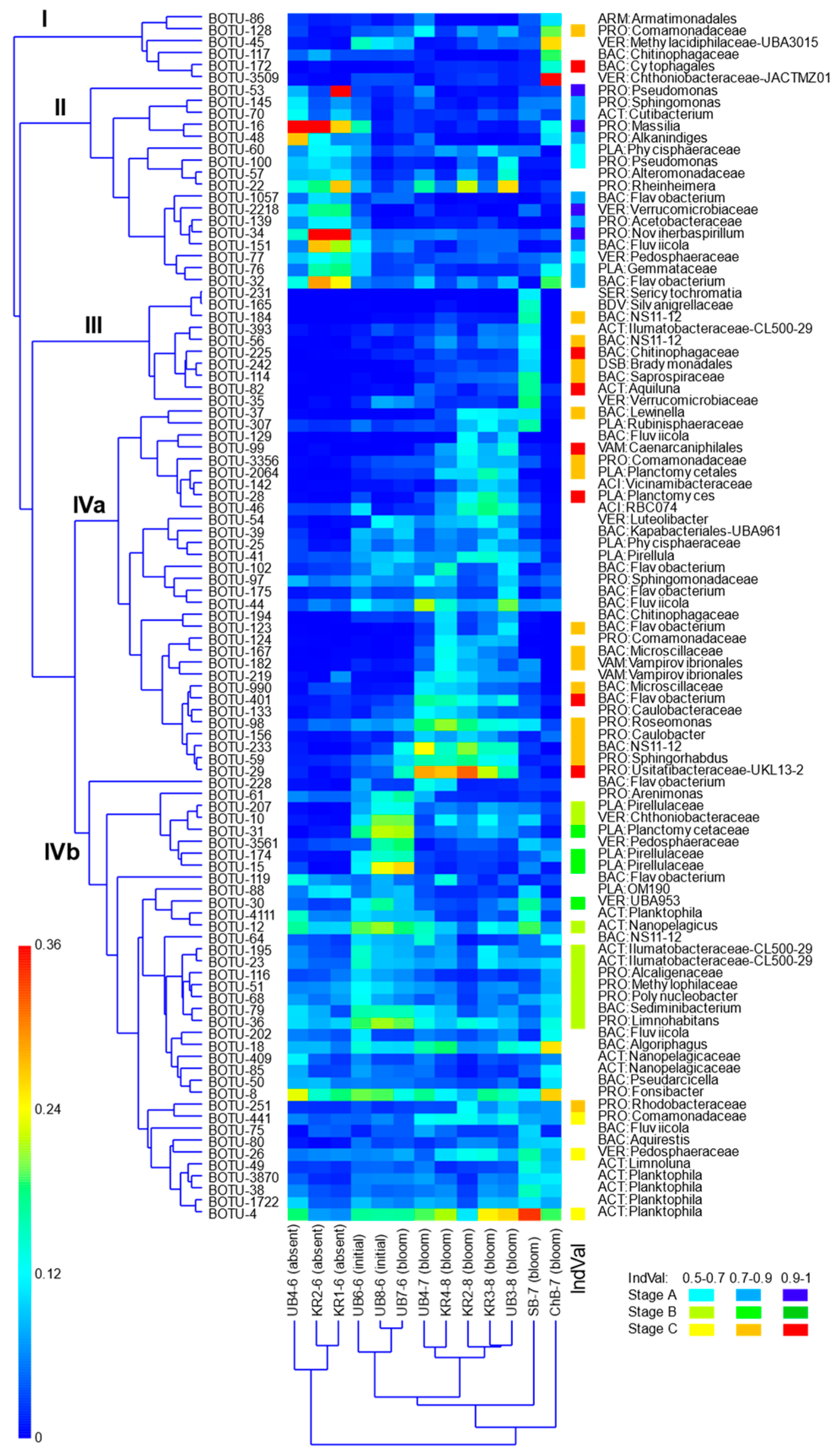
Two-way UPGMA clustering of square-root-transformed relative abundances of dominant OTUs using the cosine distance as a distance measure (Figure 6) revealed that the most separated group consisted of three samples of the clear water phase. However, among main bloom stage samples, the Suskan Bay (SB-7) and Cheremshan Bay (ChB-7) samples were most different from other stations, along with sample UB7-6, also with a strong bloom, which clustered instead with its two spatiotemporal neighbors, UB6-6 and UB8-6, which were in the early bloom stage.
Dominant OTUs were separated into four clusters—I to IV—the last of which was clearly divided into IVa and IVb subclusters. Of these, cluster II was clearly associated with the clear water phase, while cluster IVa was associated with the main bloom phase, and IVb united OTUs that developed in samples independently of the bloom stage. Two other clusters were characteristic of two aberrant samples: OTUs of cluster I dominated in the ChB-7 sample, and those of cluster III dominated in SB-7.
IndVal analysis [44] clearly confirmed this; most OTUs in cluster II were strong indicators of the clear water phase (stage A), those in clusters III and IVa were strong indicators of the main bloom (stage C), and OTUs in cluster IVb were mostly weak indicators of the early (stage B) or main bloom phase (Figure 6). Most of the dominant OTUs of this cluster, such as Nanopelagicales, were not indicators of any stage, and they remained significant throughout the whole observation period. Only BOTU-30 (Verrucomicrobiota: Opitutales: UBA953), planctomycetes BOTU-31 (Planctomycetaceae), and two Pyrellulaceae—BOTU-15 and BOTU-174—had an indicator value of 70–90% for stage B (Figure 6).
Archaea were quite an insignificant component of the prokaryotic plankton community, with only 49 OTUs and 519 reads. Woesearchaeales dominated in all samples, except for the UB4-7 sample (Figure 7). In the latter, only three archaeal sequences and two OTU belonging to Methanofastidiosales (2 reads) and Woesearchaeales (1 read) were found. The entire variety of archaea and the largest number of reads (433 out of 519 total reads) were detected in June at the upper stations of Usa Bay, in samples UB8-6 and UB7-6 (Figure 7a). No archaea were found in the Cheremshan Bay (ChB7 sample). At all other stations combined, 26 archaeal OTUs and 86 reads were registered.
In June, the number of archaeal sequences in Usa Bay decreased almost monotonously from the upper part of the bay to its mouth (Figure 7a). At the stations of the main part of the reservoir and in the Suskan Bay, archaea were scarce in terms of both the OTU number (1–5) and the number of reads (no more than 12), and no patterns were found in their spatial and temporal distribution (Figure 7b).
Among Woesearchaeales, 36% of dominant OTUs exactly matched or were close to clones from freshwater plankton; 14% were from freshwater sediment, 9% were from groundwater, 32% were from the iron-rich hot spring Jinata Onsen (Japan), and 4.5% each were from marine sediment and hydrothermal vent sediments.

3.5. Protist Community Structure and Composition in Different Parts of the Reservoir

At the macrotaxa level, the protist community structure and composition changed significantly depending on the stage of the cyanobacterial bloom. Before its onset, the community was dominated by Cryptophyceae, phototrophic Stramenopiles, and Archaeplastida; the latter was represented almost exclusively by Chlorophyta. Eukaryotic algae accounted for more than 60% of all protist sequences. With the bloom onset, their proportion decreased due to an increased proportion of Ciliophora, but they were still accompanied by Cryptophyceae and Stramenopiles (Figure 8).
With the onset of the bloom, the proportion of ciliates rose sharply, while the share of phototrophs decreased; the proportion of ciliates was highest in the initial phase of the bloom. The share of heterotrophic flagellates was variable and was lowest in samples from Cheremshan Bay (ChB-7) and one of the two samples from the initial stage of the bloom (UB8-6). The group, combining amoeboids, fungi, and undefined forms, varied in most samples from 5 to 15%, but in the ChB-7 sample, it reached 30%, mostly due to a single OTU, EOTU-21, identified as a member of the uncultured chrysophycean cluster II and distantly related to the mixotrophic “Ochromonastuberculate (94.6% similarity).
Overall, we consider as dominant 102 protist OTUs. When analyzing their distribution in the studied samples, a more complex picture emerges (Figure 9, Figures S3 and S4).
Two-way UPGMA clustering, similar to that described above for heterotrophic prokaryotes, revealed that station ChB-7 was separated at the highest level and was thus highly divergent from all the others (Figure 9). The rest of the samples were combined into two clusters, uniting, respectively, samples with no bloom and samples at all stages of the bloom. In the latter, subclusters were detected, comprising samples at the initial bloom stage, the main phase, and, separately, station SB-7, which deviated most strongly from all the others.
Accordingly, five clusters of OTUs (I–V) were resolved. Cluster I included OTUs, characteristic of station ChB-7; some of them developed in smaller quantities in other samples, mainly during the initial bloom stage. Likewise, cluster V united OTUs developing predominantly at the SB-7 station. Cluster II included a number of OTUs, present in similar quantities in plankton of all samples, while cluster III was confined to samples of the clear water phase, and cluster IV was confined to the main bloom phase. Noteworthy was the absence of a specific cluster associated with the initial bloom stage. OTUs that developed significantly in this stage formed subclusters in clusters I, II, and V.
The IndVal approach shows that most of the OTUs grouped into cluster III are indicator units of stage A (clear water phase), while cluster IV contains mostly indicator species of stage C (the main bloom). A significant part of the indicator species of this phase, however, belong to cluster V, and a somewhat smaller part belongs to cluster I. Most indicators of the initial bloom phase (stage B) belong to cluster II. They include three OTUs of spirotrich ciliates: the tintinnid EOTU-210, the strobilidiid EOTU-12 (Rimostrombidium lacustris), and EOTU-158. Several heterotrophic flagellates—EOTU-115 (Bicosoeca: Stramenopiles), EOTU-112 (Craspedida: Choanoflagellata), and EOTU-15 (Pirsonia: MAST-2: Stramenopiles)—and the dinoflagellates EOTU-69 and EOTU-208 (Haptista: Centroplasthelida) were also indicators of this stage (Figure 9).
However, the most numerous OTUs from this cluster II, the cryptophyte Cryptomonas curvata EOTU-1 (Cryptista: Cryptomonadales) and the katablepharid EOTU-9, a member of LG-E clade [46], which still has no validly described representatives, are not indicator species of any stage, and the centric diatom Stephanodiscus EOTU-7 (Mediophyceae) was a weak indicator of stage A. These OTUs were present in significant quantities in most samples (Figure S4). EOTU-7 cannot be identified to the species level since it has 100% similarity to culture collection strains bona fide identified as five different species of Stephanodiscus and two species of Cyclostephanos. Likewise, EOTU-1 also has about a dozen 100% matches, with isolates mostly identified as Cryptomonas curvata but also as other Cryptomonas species, such as C. erosa, C. gyropyrenoidosa, C. obovata, C. lucens, etc.
Stage A. Most of the OTUs that dominated during the period of clear water belonged to cluster III (Figure 9). The protist community was dominated by algal species (Figures S3 and S4). In addition to the already-mentioned EOTU-7 (Stephanodiscus) and EOTU-1 (Cryptomonas curvata), these included the cryptophyte EOTU-8 (Plagioselmis), the centric diatom EOTU-87 (Skeletonema), and the green alga EOTU-51 ([Chlamydomonas] aff. noctigama). Another centric diatom, EOTU-104 (Cyclotella), two green flagellates, three cryptophytes including EOTU-36 (Pyrenomonadales), two species of Cryptomonas, and two unidentified dinoflagellates averaged 1–3%. A representative of Bolidophyceae, EOTU-346, averaged about 0.7% per sample from this stage. Among the heterotrophic protists, the most numerous were the katablepharid EOTU-9, the ciliate EOTU-70 (Balanion planctonicum), EOTU-114 (Fungi: Chytridiomycetes), and EOTU-68 (Stramenopiles: MAST-12). EOTU-180 (Telonemia) and two heterotrophic Chrysophyceae were also present in the dominant complex.
Stage B. With the onset of bloom, a significant restructuring of the community occurred (Figure 9, Figures S3 and S4). Several ciliate OTUs entered the dominant complex. At this stage, they were represented nearly exclusively by representatives of Choreotrichia and Oligotrichia (Spirotrichea). A single OTU, EOTU-12 (Rimostrombidium) reached 13% of the abundance, and three more OTUs, EOTU-13, 2, and 158 (Strobiliidae), as well as the tintinnid EOTU-4, were less numerous (1–2.5% each). Diatoms decreased somewhat in abundance, and green algae disappeared from the dominant complex. A profound restructuring occurred in the composition of cryptophytes; Pyrenomonadales gave way to OTUs of the genus Cryptomonas.
Only at this stage did EOTU-76, a representative of CRY1, the aplastidic heterotrophic clade of Cryptophyceae, enter the dominant complex; however, it was noticeably inferior in abundance to the katablepharide EOTU-9. Other heterotrophs such as EOTU-131 (Fungi: Chytridiomycota), the flagellate EOTU-119 (Choanoflagellata: Craspedida), EOTU-15 (Stramenopiles: MAST-2: Pirsonia), and EOTU-28 (Cercozoa: Thecofilosea) also made some contribution at this stage.
Stage C. During the main bloom stage, ciliates further increased in abundance, while algae remained at the same level, and heterotrophic flagellates slightly decreased in abundance (Figure S3). Spirotricha remained dominant, but the representatives of Peritrichia (Oligohymenophorea) significantly increased in importance. EOTU-26 (Vorticella campanula) reached an average abundance of 4.5%, and three more OTUs each accounted for 1.2–2.3%. EOTU-22 (Prostomatea: Coleps) became another representative of the dominant complex. Among the algae, diatoms and cryptomonads retained their importance. EOTU-63 (Aulacoseira granulata) was a constant component of plankton during this period; the proportions of other diatoms were significantly lower. Cryptophytes were represented mainly by EOTU-1 (Cryptomonas curvata). The dinoflagellate EOTU-862 (Ceratium hirundinella) was also included in the list of dominant groups. The proportion of green algae increased slightly. They were represented mainly by flagellates: EOTU-79 from the clade “reinhardtii” (Chlorophyta: Chlamydomonadaceae), which could not be unambiguously identified even to the genus level, and two more OTUs, Chlamydomonadaceae and EOTU-98 (Tetraselmis: Chlorodendrales). Non-motile Sphaeropleales and Trebouxiophyceae, previously grouped in traditional systems within Chlorococcaceae, were present in the samples but were minor in number.
However, the ratio of ciliates to cryptophytes at this stage was highly variable (Figure 8, Figures S3 and S4). At stations UB7-6 and UB4-7, cryptophytes predominated, while in samples UB3-8, KR3-8, KR4-8, SB-7, and ChB-7, they were inferior in number not only to ciliates but also to ochrophytes.

3.6. Microscopic Determination of Phytoplankton, Planktonic Ciliates, and Heterotrophic Bacteria in the Lower Part of the Kuibyshev Reservoir

According to the microscopic analysis, the abundance of phytoplankton varied from 3.2·106 to 1.3·109 cells L−1, increasing in the samples from stage A to stage C (Table S3). At stage A, eukaryotic algae predominated in the phytoplankton composition (63–91% of the total phytoplankton abundance); at stage B their share decreased, and the contribution of cyanobacteria was 88–93% of the total phytoplankton abundance. At stage C, the contribution of cyanobacteria reached 97.6–99.8% per sample. It should be noted that the abundance of eukaryotic algae was highest in two June samples (KR1-6 and KR2-6), while in all other samples, their abundance was lower, and on average, it was lower at stage C than at the beginning of the bloom (stage B) (Table S3). The abundance of ciliates ranged from 56 to 2028 ind. L−1, and heterotrophic bacteria abundance ranged from 1.6 ·106 to 8.3 ·106 cells mL−1 (Tables S4 and S5). For both groups, a noticeable trend toward an increase in abundance from June to August and from stage A to stage C was found. However, the correlation of their abundance with the abundance of cyanobacteria was insignificant. In contrast, the abundance of single picocyanobacteria was highest at stage A and significantly decreased in the samples with blooms (Table S5). The dominant species among phytoplankton and planktonic ciliates at different stations are shown in Tables S3 and S4.

4. Discussion

Plankton is an integral system; its composition and dynamics depend on current and previous environmental conditions and relationships within the system. In freshwater bodies of the temperate zone, the composition of plankton undergoes strong intra-annual fluctuations, so-called seasonal succession, determined by both abiotic (hydrology, temperature, availability of biogenic elements, and the intensity and duration of solar insolation) and biotic (syntrophy, auxotrophy, grazing, parasitism, etc.) conditions [47,48]. Cyanobacterial blooms are, in a sense, a normal part of this seasonal cycle under certain conditions. After the completion of spring mixing and temperature increase, dormant stages of cyanobacteria of the genera Microcystis, Dolichospermum, Aphanizomenon, etc., are mobilized into the water column and, if other abiotic and biotic conditions allow, begin their active development [49]. In eutrophic conditions, sufficient nutrients remain in the water even after the end of spring diatom blooms, and the establishment of stable or at least intermittent temperature stratification coincides with the beginning of their mass cyanobacterial development [1,2,50].
Our study on the microbial plankton community in the lower part of the Kuibyshev Reservoir covered three summer months of 2023. Water warming during the entire observation period was slightly higher than the average long-term values [16,20], and the period of intensive spring mixing had already ended by the end of May 2023. Transparency, mineralization, pH, and dissolved oxygen concentration were generally within the range of long-term fluctuations, taking into account spatial and seasonal changes [16]. Concentrations of total phosphorus and ammonium nitrogen also corresponded to long-term values, but total Si and Fe concentrations were slightly higher (Table 1).
A chlorophyll a concentration above 30 µg/L is often used as a criterion for water bloom [51,52]. Based on this threshold value, as well as a set of physicochemical indicators, microscopic data, and metabarcoding results concerning the ratio of cyanobacterial and chloroplast sequences (Table 3 and Table S3; Figure 2 and Figure 3), we divided the obtained samples into three groups.
The most heterogeneous samples were collected in June. Based on microscopy, chlorophyll concentration, and rRNA metabarcoding, we assigned three of them (KR1-6, KR2-6, and UB4-6) to the clear water phase (stage A), preceding the cyanobacterial bloom. Two more samples (UB6-6 and UB8-6) from central Usa Bay had a combination of characteristics intermediate between the clear water phase and a typical cyanobacterial bloom. We consider them to represent the initial bloom (stage B). The last sample (UB7-6) met all parameters consistent with the characteristics of a typical cyanobacterial bloom (stage C). Such heterogeneity is common for large temperate water bodies, e.g., the Great Lakes or the Kuibyshev Reservoir. In these systems, cyanobacterial blooms generally start locally—in individual coastal areas, bays, mouths of tributaries, etc.—where the most favorable combinations of conditions for the recruitment of dormant cyanobacterial cells and their development are formed: low water currents, elevated temperature, and an influx of biogenic elements [20,53,54]. In the Kuibyshev Reservoir, blooms usually begin in small bays, and only later spread to the main water area [18,19,20].
All samples collected in July and August showed typical cyanobacterial blooms (stage C). With one exception, concentrations of chlorophyll a exceeded the bloom threshold [51,52], reaching almost 190 μg/L in one sample. Typical cyanobacterial blooms characterized by water discoloration and visible macroscopic colonies were also visually observed. According to microscopy, Aphanizomenon flos-aquae was the dominant cyanobacterial species at all stations; metabarcoding data confirmed this observation. Cyanobacterial blooms dominated by Aphanizomenon flos-aquae, with several species of Dolichospermum and Microcystis s.l. as subdominants, have been characteristic of the eutrophic Kuibyshev Reservoir for a long time—at least for the last 40-50 years [18,19]. In some years, the dominance shifted toward “Microcystis pulverea” (Wood) Forti, which is still an “unrevised species” [55]. It certainly does not belong to the genus Microcystis s.str., and it is most probably a part of the small-celled planktonic Aphanocapsa collective, whose members are scattered within freshwater Cyanobiaceae.
However, in 2023, there was virtually no significant seasonal succession of cyanobacteria during the bloom period, and a slightly increased proportion of Microcystis among cyanobacteria was recorded only in the initial period at a relatively low total cyanobacterial abundance (Figure 2). It should be noted that the proportion of Microcystis aeruginosa, according to metabarcoding data, was always lower than that obtained by microscopy. This systematic difference can be partly explained by the lower ploidy of Microcystis cells [56] and a smaller number of 16S RNA gene copies in its chromosome compared to typical Nostocales. One way or another, Microcystis aeruginosa did not dominate in any of the studied samples, according to both microscopy and metabarcoding data.
The two dominant clones in Cyanobiaceae, BOTU-24 and BOTU-21, could not be identified with certainty. BOTU-24 was identical to the phycoerythrin-containing Synechococcus sp. MH301 clone from Lake Moidze, which is somewhat separated from any known group of freshwater picocyanoplankton [57]. BOTU-21 was identical to Synechococcus sp. SR-R4S6. Their similarity to colonial strains of Aphanothece elabens, Aphanothece spp., and Cyanodictyon sp. was 98.5–99%. Their abundance was high in samples in which both colonial (“Microcystis pulverea” and Aphanocapsa spp.) and unicellular picocyanobacteria were numerous, according to microscopic data, so it is probably premature to conclude that they are identical to any of these morphospecies.
Throughout the observation period, the dominant heterotrophic bacterial assemblage included typical aquatic actinobacteria of the order Nanopelagicales, Planktophila, and Nanopelagicus [58,59] and of the Luna cluster (Microbacteriaceae), Aquiluna, and Limnoluna [60,61], as well as the proteobacteria Fonsibacter and Limnonabitans [62], which are widely distributed in aquatic habitats. Most of the other dominant OTUs are also typical and widespread representatives of freshwater plankton. Despite sample-to-sample variations, the listed OTUs formed a relatively stable core of the bacterioplankton community of the studied part of the Kuibyshev Reservoir. Two OTUs of Rheinheimera (Alteromonadaceae), BOTU-22 and BOTU-57, may be also included in this group. The latter could not be determined even to the genus level by either SILVA or GTDB, but it exactly matches the sequence of R. chironomi K19414T (NR_043699). Both are regular components of the bacterioplankton community, although they are not among the dominant OTUs in most samples with cyanobacterial blooms. An increased proportion of PVC superphylum bacteria and primarily planctomycetes was observed throughout almost the entire summer period of 2023. This feature was noted earlier, in 2021 [26], so it can be considered a characteristic feature of the studied reservoir.
The core of the protist community consists of several OTUs, most of which are representatives of Cryptista, the phototrophic EOTU-1 (Cryptomonas curvata) and EOTU-188 (Cryptomonas marssonii), and the heterotrophic EOTU-76 (CRY1) and EOTU-9 (Katablepharid LG-E). It has been recently reported that aplastidic cryptophytes of the CRY1 clade are one of the most important groups of small (3–5 µM) freshwater heterotrophic flagellates [63]. The LG-A was described as a group of freshwater heterotrophic picoeukaryotes closely related to Cryptophyceae [46]. They are defined in the SILVA and PR2 databases as “uncultured Katablepharids” but are quite distant from their validly described species. Our data confirm the ubiquitous presence of both groups in the Kuibyshev Reservoir. This list also includes two more phototrophic cryptophytes from the order Pyrenomonadales, EOTU-8 (Plagioselmis nanoplanktica) and EOTU-36 (Teleaulax/Geminigera), as well as the diatom EOTU-7 (Stephanodiscus hantzschii), which were much more abundant at the clear water stage A than after the onset of the bloom but nevertheless remained dominant at all stages.
In June, before the onset of the cyanobacterial bloom, a strong dominance of Massilia and Noviherbaspirillum (Oxalobacteraceae) was observed. Representatives of the Oxalobacteraceae are widely distributed but are mainly found in soils, sediments, and plant samples; only rarely are they observed in water [64,65,66]. The known species of Noviherbaspirillum and Massilia have been described primarily in soils, although, for example, Massilia mucilaginosa and M. frigida [67] have been isolated from stream water. It should be noted that in July–September, Oxalobacteraceae practically disappeared from the bacterioplankton of all samples; by contrast, in Lake George, USA [52], Massilia was one of the dominants during the very short July bloom of Aphanizomenon sp. Along with Oxalobacteraceae, the prokaryotic complex characteristic of this phase included gammaproteobacteria such as BOTU-53 and BOTU-100 (each having 100% similarity with several strains of different Pseudomonas species) and BOTU-48 (PRO: Moraxellaceae: Alkanindiges)—probably the soil species. Although [68] indicated the presence of clusters close to all these genera, they are not obligately present in freshwater bodies and are often confined to coastal zones [69]. Therefore, they probably enter the community with inflow or as a result of sediment resuspension, and for some time, they receive favorable conditions for their development. Other representative OTUs of this phase, such as the alphaproteobacteria Sphingomonas, Bacteroidetes Flavobacterium and Fluviicola, planctomycetes, and Verrucomicrobia, are, however, more typical of freshwater plankton [46,62]. Therefore, its composition probably reflects the development of a specific mix of meroplanktonic and euplanktonic forms.
Among protists, approximately 60% of reads in the clear water phase (stage A) belonged to phototrophic groups, mainly cryptophytes and melosiroid diatoms, which are always present in plankton but often undergo the greatest development in spring [18,19,70,71]. By contrast, the relative abundance of Chlorophyta was low, despite the large number of their OTUs recorded at this stage. The proportion of heterotrophic flagellates was also the highest at this stage, while ciliates were a minor component of the community and absent from the dominant complex, except for EOTU-70 (Balanion: Prostomatea), an effective cryptophyte grazer and a typical component of the late spring ciliate community in other lakes [72]. In general, the protist community in this stage is characterized by a predominance of both typical spring and year-round-developing autochthonous eukaryotic algae, accompanied by heterotrophic flagellates.
The communities of the heterotrophic bacterioplankton and planktonic protists of samples UB6-6 and UB8-6, which belong to the initial bloom period (Stage B) in terms of Chl a concentration and phytoplankton structure (Table 3), differed greatly from the communities of stage A. However, they were very similar to those of another sample, UB7-6, which was close to them geographically and in sampling date, but by the chlorophyll concentration, they already belonged to the main bloom period, stage C. The high similarity of the composition and structure of the microbial plankton of these three samples (Figure 5 and Figure 8, Figures S1–S4) probably indicates that changes in the structure of heterotrophic bacterioplankton and protist communities occur with some delay relative to the onset of cyanobacteria development. It is obvious that this stage is transitional and thus very short-lived, and it has been recorded extremely rarely in other studies; it was only represented in our data set due to fortunate circumstances.
Stage B contained only a few clear indicator species. Among heterotrophic bacteria, OTUs belonging to cluster IVb dominated at this stage (Figure 6), but they were also numerous in other samples, especially in Suskan and Cheremshan Bays. Several planctomycete OTUs and three actinomycetes that reached their maximum abundance during this period remained dominant at stage C (Figures S1 and S2). Only four members of the PVC superphylum were strong indicators of this stage (Figure 6). Among the protists, Cryptophyte algae of the genus Cryptomonas continued to retain their importance, and the main dominant, EOTU-1 (C. curvata), reached its maximum abundance at this stage. However, the abundance of other phototrophic protists decreased (Figures S3 and S4). Ciliates entered the dominant complex and were represented by Spirotrichea, both tintinnids, and strobilidiids. These OTUs, as well as several heterotrophic flagellates of different taxonomic affiliations, one centrochelid, and one dinoflagellate, are indicators of this stage (Figure 9).
Judging by the results of the cluster analysis, this stage is relatively close in the composition of eukaryotes and heterotrophic bacteria to the main bloom phase, but it differs greatly from stage A. Regarding phytoplankton only, in an analysis of seasonal phytoplankton dynamics in the Kuibyshev Reservoir in 1990, Pautova et al. [73] identified several clusters reflecting different stages of succession. Stage B (the initial bloom) in the present work most closely resembles cluster 3 from Pautova et al. [73], which was characterized by strong dominance of cryptophytes, namely Chroomonas acuta (Pyrenomonadaceae), with a notable contribution from cyanobacteria and a low overall abundance of phytoplankton.
According to microscopic analysis, the relative abundance of Chroomonas acuta in some of the 2023 samples was also rather high (Table S3). By contrast, according to metabarcoding, the cryptophytes in our samples were dominated by large Cryptomonas curvata (Cryptomonadaceae) (Figure 9 and Figure S4).
Chroomonas acuta is now regarded as a synonym of Komma caudata (Geitler) Hill = Chroomonas caudata Geitler, and its description emphasizes bright blue-green chloroplast [74]. In our 18S rRNA library, clones matching Komma caudata (AM901358) and Chroomonas caudata were both present but were represented by solitary copies, whereas the dominant OTU among Pyrenomonadales (EOTU-8) was closest in sequence to Rhodomonas minuta and Plagioselmis nannoplanctica = “Rhodomonas minuta var. nannnoplanctica”, which are somewhat similar to Komma caudata in cell shape but have red or brown chloroplasts [75]. The reason for this discrepancy is likely that the use of Lugol’s solution for fixation for microscopic enumeration turns the blue color of chloroplasts reddish or olive. This problem is not limited to the Kuibyshev Reservoir; for example, in the Lake of the Woods (Canada) [76], a similar inconsistency between the metabarcoding and the microscopic identification is evident.
The samples of the main bloom period (stage C) were heterogeneous, with the exception of a fairly close “core” uniting five samples from Usa Bay and the main water area of the Priplotinny Reach of the Kuibyshev Reservoir, collected in July and August. Nevertheless, indicator species with high significance and entire groups of OTUs characteristic of this stage as a whole were distinguished. In heterotrophic bacterioplankton, they belong to cluster IVa (Figure 6). It is notable that members of this cluster such as BOTU-29 (PRO: Usitatibacteraceae: UKL13-2) and BOTU-233 (Bacteroidetes: Sphingobacteriales: NS11-12: UKL13-3) are identical in sequence to the corresponding clones from work [77], which are part of the phycosphere of Aphanizomenon. Other OTUs of this cluster—BOTU-98 (α-PRO: Acetobacteraceae: Roseomonas), BOTU-44 (Bacteroidetes: Fluviicola), BOTU-59 (α-PRO: Sphingomonadaceae: Sphingorhabdus), and BOTU-401 (Bacteroidetes: Flavobacterium)—belong to genera whose representatives have been mentioned in many studies as components of the phycosphere of Microcystis aeruginosa [78,79]. Similarly, the main OTUs of planktonic protists characteristic of this phase were combined into cluster IV (Figure 9). The OTUs of this cluster also make up about half of the reads of the dominant complex in the “core” samples. Almost half of these OTUs belong to ciliates, and, in contrast to the initial stage, in the main stage of bloom, sessile peritrichs predominate. They obviously benefit from a large number of suspended particles, including colonies of cyanobacteria and organic pico- and nanosestons formed during the mechanical fragmentation of colonies and their death [80].
Only a few reads of species and groups capable of feeding on colonial cyanobacteria (such as Collodictyon and closely related representatives of the CruMs lineage or ciliates of the class Nassophorea) or their symbionts and parasites, particularly amoebae [81], were noted. The only exception is EOTU-321 (Copromyxa microcystoides), which made up 1.2% of the total abundance in sample UB4-7 and much less in all other samples. It should be noted, however, that the symbiotic and parasitic protists of Aphanizomenon and Dolichospermum have been studied less thoroughly than Microcystis-associated fauna. The dominant complex in this stage contained three OTUs belonging to Perkinsida, clade A31. The ecology of this group has been poorly studied, but marine representatives mostly include parasites of dinoflagellates and cryptophytes. One indication of the attachment of an unknown Perkinsida to the filaments of Aphanizomenon is known [82], but this does not prove their parasitism. There are also several reports on the parasitism of cyanobacteria by chytridiomycetes [83], another group that more typically parasitizes eukaryotic algae.
It should be noted that the mass development of Rhizaria observed in 2021 [25] was not detected in the 2023 samples; this may be due to interannual differences, a short-lived outbreak of this group in 2021, or differences in the PCR primers used.
Despite the bloom, two samples from Suskan and Cheremshan Bays differed sharply from all the “main” bloom samples. This is probably due to the geographic remoteness of these stations, as both bays are geographically distant from the Priplotinny Reach and have limited hydrological connections to other stations. In the case of Cheremshan Bay, the water runoff from the Bolshoy Cheremshan River can also have a strong effect on the plankton composition, as shown for Lake Superior [54]. The uniqueness of the phytoplankton of Cheremshan Bay has been noted earlier [18,19], and our results extend this conclusion to the microbial plankton community as a whole. A significant number of bacterial and protist OTUs were present only at one of these stations, and they were almost absent in the other samples. For example, OTE-3509 (Verrucomicrobiota: Chtoniobacteraceae), which accounted for 12% of abundance in sample ChB-7, differs in sequence from OTU-10—which was dominant among Chtoniobacteraceae in the other stage C samples—by 3.2%.
Since no species of haptophytes have yet been observed in Volga reservoirs by microscopy [70], it is worth noting that EOTU-200 sequences matching Chrysochromulina parva (Haptophyta) were present among the dominant groups in the ChB-7 sample, with a relative abundance of 3.7%. As in 2021 [25], sequences belonging to Bolidophyceae were found in the 18S rRNA library. The corresponding OTUs were distant from marine species and belonged to the freshwater LG-A group [46].
Archaea in the planktonic community of the studied reservoir site were sparse and represented almost exclusively by Nanoarchaeota, mainly of the order Woesearchaeales (although in the latest version of SILVA’s taxonomy, 138.2, some of them are considered Pacearchaeales). Their highest abundance and diversity were observed in June at the upper stations of Usa Bay, near the mouth of the river. In other samples from the bay and the reservoir, their abundance and diversity were minimal. The dominance of Woesearchaeales is somewhat unusual, as they are mainly described in anaerobic and/or extreme habitats. However, Woesearchaeales also dominated in summer in surface discharge waters in Quebec province (Canada) [84]; they were also among the dominant species in the brackish Pearl River in China [85], where their contribution to plankton increased with decreasing salinity. Several aerophilic freshwater lineages of Woesearchaeales are also known [86,87], but it is difficult to determine whether the OTUs we found belong to them.
Although several methanogenic archaea lineages have been shown to be associated with Microcystis aeruginosa blooms [88], data on other cyanobacterial bloom-causing species are much less clear. In any case, there were virtually no representatives of any methanogenic archaea in our samples, and the numbers of archaea in our samples were too small (and the errors too large) to draw reliable conclusions. This was not our specific goal, but in any case, other methods, particularly specific archaeal primers, are required.
Some features of the bacterioplankton dynamics may be mostly related not to biotic relationships within the community but to external, e.g., hydrological, factors. In particular, the spring outbreak of Oxalobacteraceae may be linked to their influx from bottom sediments during the recently finished stage of complete mixing. The same may also explain the dynamics of archaea, but it is impossible to confirm this assumption based on the available data.

5. Conclusions

The Volga reservoir cascade is the largest system of artificial reservoirs in Europe and has an ecological impact on a significant part of the East European Plain. The Kuibyshev Reservoir is the largest of the Volga and Kama reservoirs. Its hydrobiological studies by classical methods have been carried out since its filling; however, like the entire Volga basin, it has been poorly covered by modern molecular studies. Our study fills this gap and provides a picture of the phylogenetic composition of prokaryotic and eukaryotic microbial plankton of the lower section of the Kuibyshev Reservoir during the summer period of its seasonal succession.
The bacterioplankton of the studied section are typical of freshwater bodies of the temperate zone. They are characterized by the dominance of cyanobacteria, Pseudomonadota, Bacteroidota, Actinomycetota, Verrucomicrobiota, and Planctomycetota. A characteristic feature of the studied reservoir is an increased proportion of PVC superphylum bacteria, primarily planctomycetes. The protists were dominated by Cryptista, mainly phototrophic cryptophytes, and, during the bloom period, by various ciliates. Picoplanktonic Chrysochromulina (Haptophyceae) and members of an uncultured branch of Bolidophyceae (Diatomista: Ochrophyta) were present at some stations and/or at some stage of seasonal succession. Neither group has previously been observed in the Volga basin. Although Rhizaria mass development was observed at several stations for a short period in 2021, it was not recorded in 2023.
Both in the composition of bacterioplankton and in the protist communities, a dependence of their structure on the phase of seasonal phytoplankton succession, and especially on the cyanobacterial bloom intensity, was observed. The dynamics of the changes in the composition of heterotrophic bacterioplankton and protist community were similar to each other and somewhat inconsistent with the bloom stages determined by the composition of phytoplankton and the concentration of chlorophyll a. Obviously, the restructuring of heterotrophic bacterioplankton and protist communities is somewhat delayed relative to the outbreak of cyanobacteria. Additionally, the spatial position of the stations and lateral inflow also influence the structure of communities.
Indicator heterotrophic bacterioplankton species of the main bloom period include many OTUs closely related to the known components of the phycosphere of Aphanizomenon and Microcystis colonies, along with several free-living species. Among the protists, species known to feed on cyanobacteria or their potential parasites were minor. However, sessile peritrichous ciliates, which gained a significant advantage from the appearance of numerous cyanobacterial colonies serving as substrates for colonization, comprised a significant part of the dominant group. Thus, the mass development of cyanobacteria creates various new niches for the rest of the plankton community and significantly modifies its structure. However, a more accurate assessment of its impact requires expanding the scope of research to cover the entire reservoir area and the entire ice-free period.

Supplementary Materials

The following supporting information can be downloaded at: https://www.mdpi.com/article/10.3390/w17111602/s1, Table S1: Study stations and sample codes; Table S2: The structure of the combined ribosomal SSU gene library of the plankton community of the Kuibyshev Reservoir and its bays (June–August 2023) at the level of phyla of prokaryotes and upper taxonomic levels of eukaryotes; Table S3: The structure of the phytoplankton according to the microscopic analysis: total abundance and proportion of dominant species. The first dominant is highlighted in red, the second in orange, and the third in yellow; Table S4: The structure of planktonic ciliates according to the microscopic analysis: total abundance and proportion of dominant species. First dominant is highlighted in red, second—in orange and third—in yellow; Table S5: Abundance of heterotrophic bacteria and single-cell picocyanobacteria according to microscopic analysis; Figure S1: Relative abundance (% total reads) of 101 dominant OTUs of heterotrophic bacteria during the observation period at the phylum level; Figure S2: Relative abundance (% total reads) of dominant OTUs of heterotrophic bacteria during different bloom periods; Figure S3: Structure of dominant OTUs of protists (all 102 OTUs) at the level of functional groups (a) and macrotaxa (b); Figure S4: Relative abundance (% total reads) of dominant OTUs of protists during different bloom periods.

Author Contributions

Conceptualization, M.Y.G. and M.V.U.; Formal analysis, M.Y.G.; Funding acquisition, M.V.U.; Investigation, M.Y.G., S.V.B., N.G.T., E.S.K. and M.V.U.; Visualization, M.Y.G., S.V.B. and M.V.U.; Writing—original draft, M.Y.G. and M.V.U.; Writing—review and editing, M.Y.G., S.V.B., N.G.T., E.S.K. and M.V.U. All authors have read and agreed to the published version of the manuscript.

Funding

This research was funded by the Russian Science Foundation, grant no. 23-14-20005, https://rscf.ru/project/23-14-20005/ (accesed on 15 April 2025).

Data Availability Statement

All the data are provided in the main text and the Supplementary Materials. Raw rDNA sequences have been deposited into GenBank with Accession Number PRJNA1267265.

Conflicts of Interest

The authors declare no conflicts of interest.

References

  1. Havens, K.E. Cyanobacteria blooms: Effects on aquatic ecosystems. In Cyanobacterial Harmful Algal Blooms: State of the Science and Research Needs; Hudnell, H.K., Ed.; Springer: New York, NY, USA, 2008; pp. 733–747. [Google Scholar] [CrossRef]
  2. Huisman, J.; Codd, G.A.; Paerl, H.W.; Ibelings, B.W.; Verspagen, J.M.; Visser, P.M. Cyanobacterial blooms. Nat. Rev. Microbiol. 2018, 16, 471–483. [Google Scholar] [CrossRef]
  3. Wurtsbaugh, W.A.; Paerl, H.W.; Dodds, W.K. Nutrients, eutrophication and harmful algal blooms along the freshwater to marine continuum. WIREs Water 2019, 6, e1373. [Google Scholar] [CrossRef]
  4. Reynolds, C.S.; Huszar, V.; Kruk, C.; Naselli-Flores, L.; Melo, S. Towards a functional classification of the freshwater phytoplankton. J. Plankton Res. 2002, 24, 417–428. [Google Scholar] [CrossRef]
  5. Padisák, J.; Crossetti, L.O.; Naselli-Flores, L. Use and misuse in the application of the phytoplankton functional classification: A critical review with updates. Hydrobiologia 2009, 621, 1–19. [Google Scholar] [CrossRef]
  6. Kim, S.; Kang, I.; Lee, J.W.; Jeon, C.O.; Giovannoni, S.J.; Cho, J.C. Heme auxotrophy in abundant aquatic microbial lineages. Proc. Natl. Acad. Sci. USA 2021, 118, e2102750118. [Google Scholar] [CrossRef] [PubMed]
  7. Moustaka-Gouni, M.; Sommer, U. Effects of harmful blooms of large-sized and colonial cyanobacteria on aquatic food webs. Water 2020, 12, 1587. [Google Scholar] [CrossRef]
  8. Korneva, L.G.; Solovyeva, V.V. Dynamics of morphofuncitonal groups of phytoplankton in the Rybinsk reservoir and assessment of the reservoir water quality by the community index. Water Resour. 2021, 48, 65–72. [Google Scholar] [CrossRef]
  9. Kopylov, A.I.; Kosolapov, D.B.; Mikryakova, I.S. Long-term dynamics of heterotrophic bacterioplankton in a large eutrophic reservoir. Inland Water Biol. 2020, 13, 585–591. [Google Scholar] [CrossRef]
  10. Xu, H.; Liu, W.; Zhang, S.; Wei, J.; Li, Y.; Pei, H. Cyanobacterial bloom intensities determine planktonic eukaryote community structure and stability. Sci. Total Environ. 2022, 838, 156637. [Google Scholar] [CrossRef]
  11. Kosiba, J.; Wilk-Woźniak, E.; Krztoń, W. The effect of potentially toxic cyanobacteria on ciliates (Ciliophora). Hydrobiologia 2019, 827, 325–335. [Google Scholar] [CrossRef]
  12. Tirjaková, E.; Krajčovičová, K.; Illyová, M.; Vďačný, P. Interaction of ciliate communities with cyanobacterial water bloom in a shallow, hypertrophic reservoir. Acta Protozool. 2016, 55, 173–188. [Google Scholar] [CrossRef]
  13. Andreeva, V.A.; Bykova, S.V.; Umanskaya, M.V.; Tarasova, N.G. Free-living ciliates during the period of the greatest cyanobacterial water bloom in the Usinsky bay (Kuibyshev reservoir). Proc. Samara Sci. Center RAS 2021, 23, 127–133. (In Russian) [Google Scholar] [CrossRef]
  14. Louati, I.; Pascault, N.; Debroas, D.; Bernard, C.; Humbert, J.F.; Leloup, J. Structural diversity of bacterial communities associated with bloom-forming freshwater cyanobacteria differs according to the cyanobacterial genus. PLoS ONE 2015, 10, e0140614. [Google Scholar] [CrossRef] [PubMed]
  15. Zhu, L.; Zancarini, A.; Louati, I.; De Cesare, S.; Duval, C.; Tambosco, K.; Bernard, C.; Debroas, D.; Song, L.; Leloup, J.; et al. Bacterial communities associated with four cyanobacterial genera display structural and functional differences: Evidence from an experimental approach. Front. Microbiol. 2016, 7, 1662. [Google Scholar] [CrossRef]
  16. Rozenberg, G.S.; Vykhristyuk, L.A. (Eds.) Kuibyshev Reservoir (Scientific-Information Handbook); IEVB RAS: Tolyatti, Russia, 2008; 123p. (In Russian) [Google Scholar]
  17. Ivatin, A.V. Bacterioplankton and Bacteriobenthos of the Kuibyshev Reservoir; Kassandra: Tolyatti, Russia, 2012; 183p. (In Russian) [Google Scholar]
  18. Pautova, V.N.; Nomokonova, V.I. Dynamics of Phytoplankton of the Lower Volga—From the River to the Cascade of Reservoirs; IEVB RAS: Tolyatti, Russia, 1994; 188p. (In Russian) [Google Scholar]
  19. Tarasova, N.G. Phytoplankton Composition, Seasonal Dynamics and Invasive Species of the Kuibyshev Reservoir. Ph.D. Thesis, IEVB RAS, Tolyatti, Russia, 2005; 146p. (In Russian). [Google Scholar]
  20. Konovalov, S.M.; Pautova, V.N. (Eds.) Phytoplankton Ecology of Kuibyshev Reservoir; Nauka: Leningrad, Russia, 1989; 304p. (In Russian) [Google Scholar]
  21. Zharikov, V.V. Inventory of Free-Living Ciliates in the Volga Reservoirs; IEVB RAS: Togliatti, Russia, 1996. (In Russian) [Google Scholar]
  22. Bykova, S.V. Structure and Spatial Distribution of Planktonic Ciliates from the Middle and Lower Volga Reservoirs. Inland Water Biol. 2021, 14, 377–390. [Google Scholar] [CrossRef]
  23. Fedotova, A.V.; Serkebaeva, Y.M.; Sorokin, V.V.; Dedysh, S.N. Filterable microbial forms in the Rybinsk water reservoir. Microbiology 2013, 82, 728–734. [Google Scholar] [CrossRef]
  24. Kuznetsova, E.V.; Sukhanova, E.V.; Kosolapov, D.B. Taxonomic Diversity and Size-Morphological Structure of Bacterioplankton of the Rybinsk Reservoir. Microbiology 2021, 90, 324–335. [Google Scholar] [CrossRef]
  25. Umanskaya, M.V.; Gorbunov, M.Y.; Bykova, S.V.; Tarasova, N.G. Diversity and Transformation of the Community of Planktonic Freshwater Protists in the Estuarine Tributary Zone of a Large Plainland Reservoir: Metabarcoding of the 18S Ribosomal RNA Gene. Biol. Bull. 2023, 50, 707–723. [Google Scholar] [CrossRef]
  26. Umanskaya, M.V.; Gorbunov, M.Y. Phylogenetic Structure of Bacterioplankton in Water Bodies of the Kuibyshev Reservoir Basin during Cyanobacterial Bloom. Microbiology 2013, 93, 876–890. [Google Scholar] [CrossRef]
  27. Lurie, Y.Y. (Ed.) Unified Methods of Analysis of Waters; Khimija: Moscow, Russia, 1973; 376p. (In Russian) [Google Scholar]
  28. Solorzano, L. Determination of ammonia in natural waters by the phenolhypochlorite method. Limnol. Oceanogr. 1969, 14, 799–801. [Google Scholar] [CrossRef]
  29. Novikov, Y.V.; Lastochkina, K.O.; Boldina, Z.N. Methods of Studying the Quality of Water in Reservoirs; Medicine: Moscow, Russia, 1990; 400p. (In Russian) [Google Scholar]
  30. Jeffrey, S.W.; Humfrey, G.F. New spectrophotometric equations for deter-mining chlorophylls a, b, c, and c2 in higher plants, algae and natural phyto-plankton. Biochem. Physiol. Pflanz. 1975, 167, 191–194. [Google Scholar] [CrossRef]
  31. Lorenzen, C.J. Determination of chlorophyll and pheo-pigments: Spectro-photometric equations. Limnol. Oceanogr. 1967, 12, 343–346. [Google Scholar] [CrossRef]
  32. Mordukhai-Boltovskoy, F.D. (Ed.) Methodology for Studying Biogeocenoses of Inland Water Bodies; Nauka: Moscow, Russia, 1975; 240p. (In Russian) [Google Scholar]
  33. Bereczky, M.C. Fixations-und Färbungsschnellverfahren bei quantitativen ökologischen Untersuchungen von Protozoen in Binnengewässern. Arch. Protistenkd. 1985, 129, 187–190. [Google Scholar] [CrossRef]
  34. Porter, K.G.; Feig, Y.S. The use of DAPI for identifying and counting of aquatic microflora. Limnol. Oceanogr. 1980, 25, 943–948. [Google Scholar] [CrossRef]
  35. Parada, A.E.; Needham, D.M.; Fuhrman, J.A. Every base matters: Assessing small subunit rRNA primers for marine microbiomes with mock communities, time series and global field samples. Environ. Microbiol. 2016, 18, 1403–1414. [Google Scholar] [CrossRef] [PubMed]
  36. Apprill, A.; McNally, S.; Parsons, R.; Weber, L. Minor revision to V4 region SSU rRNA 806R gene primer greatly increases detection of SAR11 bacterioplankton. Aquat. Microb. Ecol. 2015, 75, 129–137. [Google Scholar] [CrossRef]
  37. Hugerth, L.W.; Muller, E.E.; Hu, Y.O.; Lebrun, L.A.; Roume, H.; Lundin, D.; Wilmes, P.; Andersson, A.F. Systematic design of 18S rRNA gene primers for determining eukaryotic diversity in microbial consortia. PLoS ONE 2014, 9, e95567. [Google Scholar] [CrossRef]
  38. Edgar, R. UPARSE: Highly accurate OTU sequences from microbial amplicon reads. Nat. Methods 2013, 10, 996–998. [Google Scholar] [CrossRef]
  39. Quast, C.; Pruesse, E.; Yilmaz, P.; Gerken, J.; Schweer, T.; Yarza, P.; Peplies, J.; Glöckner, F.O. The SILVA ribosomal RNA gene database project: Improved data processing and web-based tools. Nucleic Acids Res. 2013, 41, D590–D596. [Google Scholar] [CrossRef]
  40. Ali, A. DADA2 Formatted 16S rRNA Gene Sequences for Both Bacteria & Archaea (4.4). Zenodo 2023. [Google Scholar] [CrossRef]
  41. Edgar, R.C. SINTAX: A simple non-Bayesian taxonomy classifier for 16S and ITS sequences. Biorxiv 2016, 074161. [Google Scholar] [CrossRef]
  42. Guillou, L.; Bachar, D.; Audic, S.; Bass, D.; Berney, C.; Bittner, L.; Boutte, C.; Burgaud, G.; de Vargas, C.; Decelle, J.; et al. The Protist Ribosomal Reference database (PR2): A catalog of unicellular eukaryote small sub-unit rRNA sequences with curated taxonomy. Nucleic Acids Res. 2013, 41, D597–D604. [Google Scholar] [CrossRef] [PubMed]
  43. Pruesse, E.; Peplies, J.; Glöckner, F.O. SINA: Accurate high-throughput multiple sequence alignment of ribosomal RNA genes. Bioinformatics 2012, 28, 1823–1829. [Google Scholar] [CrossRef]
  44. Dufrêne, M.; Legendre, P. Species assemblages and indicator species: The need for a flexible asymmetrical approach. Ecol. Monogr. 1997, 67, 345–366. [Google Scholar] [CrossRef]
  45. Hammer, Ø.; Harper, D.A. Past: Paleontological statistics software package for educaton and data anlysis. Palaeontol. Electron. 2001, 4, 1. [Google Scholar]
  46. Richards, T.A.; Vepritskiy, A.A.; Gouliamova, D.E.; Nierzwicki-Bauer, S.A. The molecular diversity of freshwater picoeukaryotes from an oligotrophic lake reveals diverse, distinctive and globally dispersed lineages. Environ. Microbiol. 2005, 7, 1413–1425. [Google Scholar] [CrossRef]
  47. Suthers, I.M.; Richardson, A.J.; Rissik, D. The importance of plankton. In Plankton: A Guide to Their Ecology and Monitoring for Water Quality; Suthers, I., Rissik, D., Richardson, A., Eds.; CSIRO Publishing: Clayton, Australia, 2019; pp. 1–13. [Google Scholar]
  48. Matveev, V.F.; Matveeva, L.K. Seasonal succession and long-term stability of pelagic community in a productive reservoir. Mar. Freshw. Res. 2005, 56, 1137–1149. [Google Scholar] [CrossRef]
  49. Cottingham, K.L.; Weathers, K.C.; Ewing, H.A.; Greer, M.L.; Carey, C.C. Predicting the effects of climate change on freshwater cyanobacterial blooms requires consideration of the complete cyanobacterial life cycle. J. Plankton Res. 2021, 43, 10–19. [Google Scholar] [CrossRef]
  50. Reynolds, C.S. Phytoplankton periodicity: The interactions of form, function and environmental variability. Freshw. Biol. 1984, 14, 111–142. [Google Scholar] [CrossRef]
  51. Bruns, N.E.; Heffernan, J.B.; Ross, M.R.V.; Doyle, M. A simple metric for predicting the timing of river phytoplankton blooms. Ecosphere 2022, 13, e4348. [Google Scholar] [CrossRef]
  52. Berman, M.C.; Hrycik, A.R.; Costello, A.; Bai, Y.; Rose, K.C.; Relyea, R.; Dordick, J.S. Spatiotemporal dynamics of microbial communities and cyanobacteria blooms in two North American Lakes using long-read 16S rRNA sequencing. Ecol. Indic. 2024, 159, 111738. [Google Scholar] [CrossRef]
  53. Isles, P.D.; Giles, C.D.; Gearhart, T.A.; Xu, Y.; Druschel, G.K.; Schroth, A.W. Dynamic internal drivers of a historically severe cyanobacteria bloom in Lake Champlain revealed through comprehensive monitoring. J. Great Lakes Res. 2015, 41, 818–829. [Google Scholar] [CrossRef]
  54. Kreiling, R.M.; Givens, C.E.; Baker, A.C.; Kiesling, R.L.; Dantoin, E.D.; Perner, P.M.; Sterner, S.P.; Gierke, K.J.; Reneau, P.C. Role of tributary cyanobacterial and nutrient transport and sediment processes on cyanobacterial bloom initiation in Lake Superior nearshore. J. Great Lakes Res. 2025, 51, 102409. [Google Scholar] [CrossRef]
  55. Komárek, J.; Anagnostidis, K. Cyanoprokaryota 1. Teil: Chroococcales. In Süsswasserflora von Mitteleuropa 19/1; Ettl, H., Gärtner, G., Heynig, H., Mollenhauer, D., Eds.; Gustav Fischer: Jena, Germany; Stuttgart, Germany; Lübeck, Germany; Ulm, Germany, 1998; 548p. [Google Scholar]
  56. Griese, M.; Lange, C.; Soppa, J. Ploidy in cyanobacteria. FEMS Microbiol. Lett. 2011, 323, 124–131. [Google Scholar] [CrossRef]
  57. Crosbie, N.D.; Pöckl, M.; Weisse, T. Dispersal and phylogenetic diversity of nonmarine picocyanobacteria, inferred from 16S rRNA gene and cpcBA-intergenic spacer sequence analyses. Appl. Environ. Microbiol. 2003 69, 5716–5721. [CrossRef]
  58. Jezbera, J.; Sharma, A.K.; Brandt, U.; Doolittle, W.F.; Hahn, M.W. ‘Candidatus Planktophila limnetica’, an actinobacterium representing one of the most numerically important taxa in freshwater bacterioplankton. Int. J. Syst. Evol. Microbiol. 2009, 59, 2864–2869. [Google Scholar] [CrossRef]
  59. Neuenschwander, S.M.; Ghai, R.; Pernthaler, J.; Salcher, M.M. Microdiversification in genome-streamlined ubiquitous freshwater Actinobacteria. ISME J. 2018, 12, 185–198. [Google Scholar] [CrossRef]
  60. Hahn, M.W. Description of seven candidate species affiliated with the phylum Actinobacteria, representing planktonic freshwater bacteria. Int. J. Syst. Evol. Microbiol. 2009, 59, 112–117. [Google Scholar] [CrossRef]
  61. Lipko, I.A.; Belykh, O.I. Environmental Features of Freshwater Planktonic Actinobacteria. Contemp. Probl. Ecol. 2021, 14, 158–170. [Google Scholar] [CrossRef]
  62. Chiriac, M.C.; Haber, M.; Salcher, M.M. Adaptive genetic traits in pelagic freshwater microbes. Environ. Microbiol. 2023, 25, 606–641. [Google Scholar] [CrossRef]
  63. Šimek, K.; Mukherjee, I.; Szöke-Nagy, T.; Haber, M.; Salcher, M.M.; Ghai, R. Cryptic and ubiquitous aplastidic cryptophytes are key freshwater flagellated bacterivores. ISME J. 2023, 17, 84–94. [Google Scholar] [CrossRef]
  64. Lin, S.Y.; Hameed, A.; Arun, A.B.; Liu, Y.C.; Hsu, Y.H.; Lai, W.A.; Rekha, P.D.; Young, C.C. Description of Noviherbaspirillum malthae gen. nov., sp. nov., isolated from an oil-contaminated soil, and proposal to reclassify Herbaspirillum soli, Herbaspirillum aurantiacum, Herbaspirillum canariense and Herbaspirillum psychrotolerans as Noviherbaspirillum soli comb. nov., Noviherbaspirillum aurantiacum comb. nov., Noviherbaspirillum canariense comb. nov. and Noviherbaspirillum psychrotolerans comb. nov. based on polyphasic analysis. Int. J. Syst. Evol. Microbiol. 2013, 63, 4100–4107. [Google Scholar] [CrossRef] [PubMed]
  65. Chaudhary, D.K.; Dahal, R.H.; Hong, Y. Noviherbaspirillum pedocola sp. nov., isolated from oil-contaminated experimental soil. Arch. Microbiol. 2021, 203, 3071–3076. [Google Scholar] [CrossRef]
  66. Wu, X.; Jin, C.Z.; Jin, F.J.; Li, T.; Sung, Y.J.; Oh, H.M.; Lee, H.G.; Jin, L. Lacisediminimonas profundi gen. nov., sp. nov., a member of the family Oxalobacteraceae isolated from freshwater sediment. Antonie Leeuwenhoek 2020, 113, 253–264. [Google Scholar] [CrossRef]
  67. Holochová, P.; Mašlaňová, I.; Sedláček, I.; Švec, P.; Králová, S.; Kovařovic, V.; Busse, H.-J.; Stanková, E.; Barták, M.; Pantůček, R. Description of Massilia rubra sp. nov., Massilia aquatica sp. nov., Massilia mucilaginosa sp. nov., Massilia frigida sp. nov., and one Massilia genomospecies isolated from Antarctic streams, lakes and regoliths. Syst. Appl. Microbiol. 2020, 43, 126112. [Google Scholar] [CrossRef] [PubMed]
  68. Newton, R.J.; Jones, S.E.; Eiler, A.; McMahon, K.D.; Bertilsson, S. A guide to the natural history of freshwater lake bacteria. Microbiol. Mol. Biol. Rev. 2011, 75, 14–49. [Google Scholar] [CrossRef]
  69. Mueller-Spitz, S.R.; Goetz, G.W.; McLellan, S.L. Temporal and spatial variability in nearshore bacterioplankton communities of Lake Michigan. FEMS Microbiol. Ecol. 2009, 67, 511–522. [Google Scholar] [CrossRef]
  70. Korneva, L.G. Phytoplankton of Volga River Basin Reservoirs; Kostroma Printing House: Kostroma, Russia, 2015; 284p. (In Russian) [Google Scholar]
  71. Sakharova, E.G.; Korneva, L.G. Phytoplankton in the littoral and pelagial zones of the Rybinsk Reservoir in years with different temperature and water-level regimes. Inland Water Biol. 2018, 11, 6–12. [Google Scholar] [CrossRef]
  72. Schalch-Schuler, M.; Bassin, B.; Andrei, A.S.; Dirren-Pitsch, G.; Waller, K.; Hofer, C.; Pernthaler, J.; Posch, T. The planktonic freshwater ciliate Balanion planctonicum (Ciliophora, Prostomatea): A cryptic species complex or a “complex species”? J. Eukaryot. Microbiol. 2025, 72, e13084. [Google Scholar] [CrossRef]
  73. Pautova, V.N.; Nomokonova, V.I.; Gorbunov, M.Y. Seasonal succession of phytoplankton in the Kuibyshev Reservoir. Inn. Water Biol. 2001, 3, 29–35. (In Russian) [Google Scholar]
  74. Hill, D.R. Chroomonas and other blue-green cryptomonads. J. Phycol. 1991, 27, 133–145. [Google Scholar] [CrossRef]
  75. Javornicky, P. Taxonomic notes on some freshwater planktonic Cryptophyceae based on light microscopy. Hydrobiologia 2003, 502, 271–283. [Google Scholar] [CrossRef]
  76. Zastepa, A.; Comte, J.; Crevecoeur, S. Prevalence and ecological features of deep chlorophyll layers in Lake of the Woods, a complex hydrological system with strong trophic, physical, and chemical gradients. J. Great Lakes Res. 2023, 49, 122–133. [Google Scholar] [CrossRef]
  77. Driscoll, C.B.; Otten, T.G.; Brown, N.M.; Dreher, T.W. Towards long-read metagenomics: Complete assembly of three novel genomes from bacteria dependent on a diazotrophic cyanobacterium in a freshwater lake co-culture. Stand. Genom. Sci. 2017, 12, 1–16. [Google Scholar] [CrossRef]
  78. Parulekar, N.N.; Kolekar, P.; Jenkins, A.; Kleiven, S.; Utkilen, H.; Johansen, A.; Sawant, S.; Kulkarni-Kale, U.; Kale, M.; Sæbø, M. Characterization of bacterial community associated with phytoplankton bloom in a eutrophic lake in South Norway using 16S rRNA gene amplicon sequence analysis. PLoS ONE 2017, 12, e0173408. [Google Scholar] [CrossRef]
  79. Reignier, O.; Bormans, M.; Hervé, F.; Robert, E.; Savar, V.; Tanniou, S.; Amzil, Z.; Noel, C.; Briand, E. Spatio-temporal connectivity of a toxic cyanobacterial community and its associated microbiome along a freshwater-marine continuum. Harmful Algae 2024, 134, 102627. [Google Scholar] [CrossRef] [PubMed]
  80. Pratt, J.R.; Rosen, B.H. Associations of Species of Vorticella (Peritrichida) and Planktonic Algae. Trans. Am. Microsc. Soc. 1983, 102, 48–54. [Google Scholar] [CrossRef]
  81. Van Wichelen, J.; D’hondt, S.; Claeys, M.; Vyverman, W.; Berney, C.; Bass, D.; Vanormelingen, P. A hotspot of amoebae diversity: 8 new naked amoebae associated with the planktonic bloom-forming cyanobacterium Microcystis. Acta Protozool. 2016, 55, 61–87. [Google Scholar] [CrossRef]
  82. Jobard, M.; Wawrzyniak, I.; Bronner, G.; Marie, D.; Vellet, A.; Sime-Ngando, T.; Debroas, D.; Lepère, C. Freshwater Perkinsea: Diversity, ecology and genomic information. J. Plankton Res. 2020, 42, 3–7. [Google Scholar] [CrossRef]
  83. Gerphagnon, M.; Macarthur, D.J.; Latour, D.; Gachon, C.M.; Van Ogtrop, F.; Gleason, F.H.; Sime-Ngando, T. Microbial players involved in the decline of filamentous and colonial cyanobacterial blooms with a focus on fungal parasitism. Environ. Microbiol. 2015, 17, 2573–2587. [Google Scholar] [CrossRef]
  84. Groult, B.; St-Jean, V.; Lazar, C.S. Linking Groundwater to Surface Discharge Ecosystems: Archaeal, Bacterial, and Eukaryotic Community Diversity and Structure in Quebec (Canada). Microorganisms 2023, 11, 1674. [Google Scholar] [CrossRef] [PubMed]
  85. Lin, X.; Zhang, C.; Xie, W. Deterministic processes dominate archaeal community assembly from the Pearl River to the northern South China Sea. Front. Microbiol. 2023, 14, 1185436. [Google Scholar] [CrossRef] [PubMed]
  86. Liu, X.; Li, M.; Castelle, C.J.; Probst, A.J.; Zhou, Z.; Pan, J.; Liu, Y.; Banfield, J.F.; Gu, J.D. Insights into the ecology, evolution, and metabolism of the widespread Woesearchaeotal lineages. Microbiome 2018, 6, 102. [Google Scholar] [CrossRef] [PubMed]
  87. Lomakina, A.; Bukin, S.; Shubenkova, O.; Pogodaeva, T.; Ivanov, V.; Bukin, Y.; Zemskaya, T. Microbial Communities in Ferromanganese Sediments from the Northern Basin of Lake Baikal (Russia). Microorganisms 2023, 11, 1865. [Google Scholar] [CrossRef]
  88. Li, C.; Hambright, K.D.; Bowen, H.G.; Trammell, M.A.; Grossart, H.P.; Burford, M.A.; Hamilton, D.P.; Jiang, H.; Latour, D.; Meyer, E.I.; et al. Global co-occurrence of methanogenic archaea and methanotrophic bacteria in Microcystis aggregates. Environ. Microbiol. 2021, 23, 6503–6519. [Google Scholar] [CrossRef]
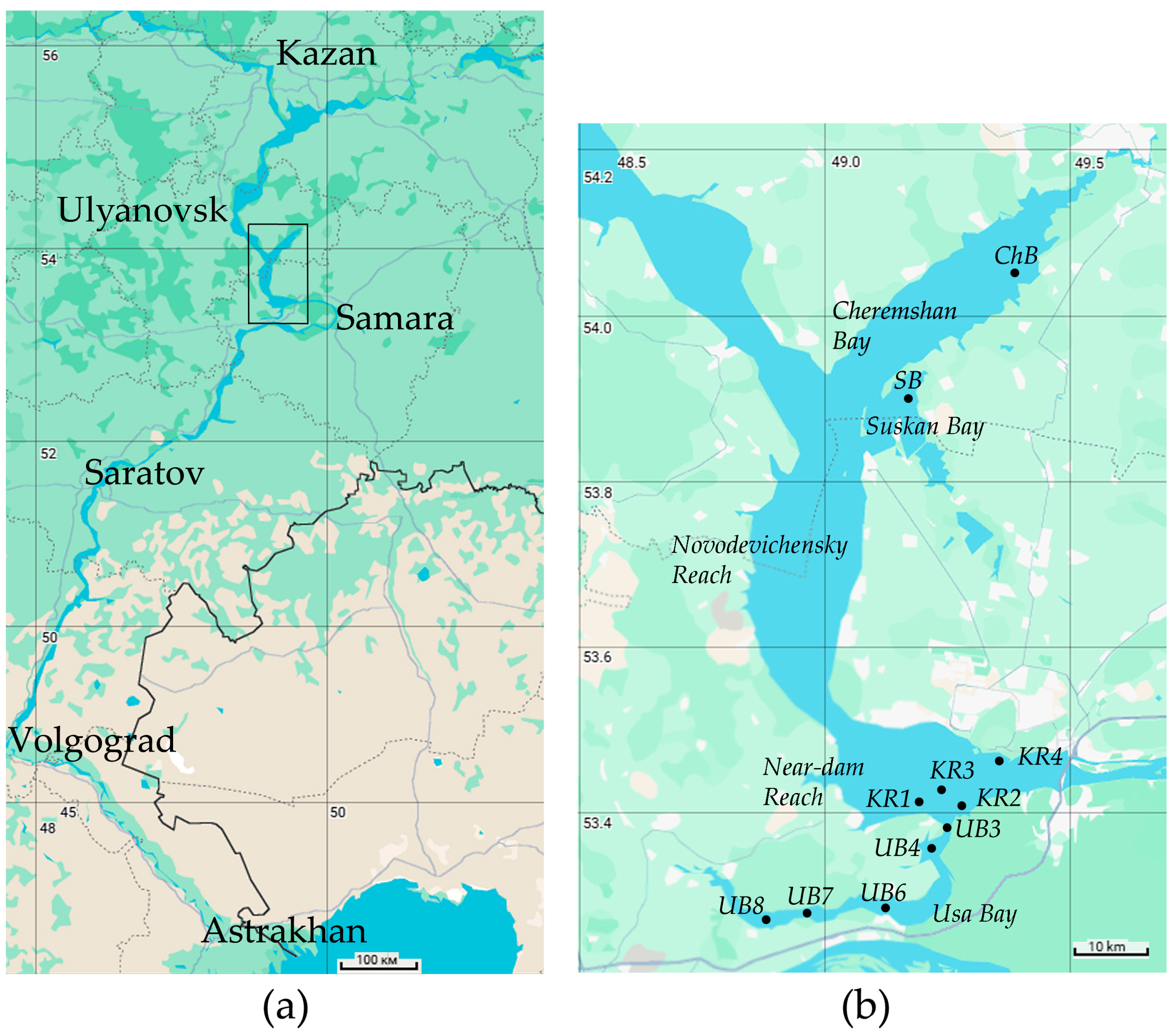
Figure 1. (a) The map of the lower part of the Volga basin; the frame indicates the research area of the Kuibyshev Reservoir. (b) The research area of the Kuibyshev Reservoir: location of sampling stations. Station coordinates, sampling dates, and sample labels are given in Table S1.
Water 17 01602 g001
Figure 2. NMDS plot of studied samples according to physical and chemical conditions and Chl a concentration. Final stress value: STRESS = 0.0201.
Water 17 01602 g002
Figure 3. Network plot of studied samples according to community composition based on Bray–Curtis similarity of relative abundances in the cyanobacterial and chloroplast OTU complex.
Water 17 01602 g003
Figure 4. The composition of the dominant cyanobacterial complex at different stages of bloom. Relative abundance was calculated from the sum of cyanobacterial and chloroplast reads.
Water 17 01602 g004
Figure 5. The phylum-level structure of heterotrophic bacterioplankton before and during two stages of the cyanobacterial bloom.
Water 17 01602 g005
Figure 6. Two-way UPGMA clustering of relative abundances of dominant prokaryotic OTUs using cosine distance measure of sqrt-transformed abundances. Right column—color-coding IndVals; only significant values at a significance level of 0.05 are shown.
Water 17 01602 g006
Figure 7. Composition and structure of archaea in the plankton of studied stations: (a) June samples from Usa Bay; (b) all other samples.
Water 17 01602 g007
Figure 8. Structure of protists at the level of functional groups (a) and macrotaxa (b).
Water 17 01602 g008
Figure 9. Two-way UPGMA clustering of relative abundances of dominant protist OTUs using the cosine distance measure of square-root-transformed abundances. Right column—color-coded IndVals; only significant values at a significance level of 0.05 are shown.
Water 17 01602 g009
Table 1. Physical and chemical conditions in the surface layer of water (average value ± SD).
ParametersThe Whole PeriodJuneJulyAugust
Number of samples13634
Transparency, m1.05 ± 0.541.41 ± 0.550.87 ± 0.150.72 ± 0.40
Temperature, °C22.8 ± 3.919.1 ± 0.726.1 ± 3.026.0 ± 0.7
Conductivity, µS cm−1374 ± 20381 ± 16388 ± 20353 ± 2
pH8.51 ± 0.747.77 ± 0.089.12 ± 0.289.18 ± 0.23
Dissolved oxygen, mg L−112.14 ± 4.06nd 18.91 ± 1.1815.36 ± 2.95
Dissolved oxygen, %152 ± 51nd112 ± 17192 ± 38
Water color, °Pt61 ± 1159 ± 1356 ± 368 ± 6
SO4, mg L−141 ± 933 ± 251 ± 646 ± 5
HCO3, mg L−1203 ± 54252 ± 18181 ± 51147 ± 1
N-NH4, mg L−10.12 ± 0.050.10 ± 0.050.12 ± 0.050.15 ± 0.05
Ptot, mg L−10.10 ± 0.210.16 ± 0.310.06 ± 0.020.03 ± 0.01
Si, mg L−13.28 ± 1.263.70 ± 1.823.03 ± 0.482.85 ± 0.11
Fetot, mg L−10.32 ± 0.080.33 ± 0.070.35 ± 0.110.28 ± 0.07
Chl a, µg L−142.0 ± 48.714.5 ± 10.035.2 ± 13.788.3 ± 69.0
Note: 1 nd—not determined.
Table 2. Indices of diversity and dominance of prokaryotes and microeukaryotes in individual samples and in the entire library. N, OTU number; B-P, Berger–Parker dominance; 1-D, Simpson diversity; 1/D, inverse Simpson index (polydominance); H2, Shannon index (log2-based); E, Pielou evenness.
SampleProkaryotes and ChloroplastsProtists and Fungi
NB-P1-D1/DH2ENB-P1-D1/DH2E
KR2-810960.270.9111.05.620.564810.180.9519.25.800.65
KR3-89630.290.909.95.530.565070.080.9729.86.020.67
UB3-88500.200.9417.26.030.624680.150.9622.55.760.65
KR4-89250.200.9213.25.550.564330.130.9623.75.750.66
KR1-68510.210.9212.05.070.523470.140.9417.35.160.61
KR2-68260.250.909.84.770.493350.150.9416.55.100.61
UB6-614320.130.9729.66.590.634670.190.9518.75.770.65
UB7-617360.250.9313.46.250.585230.260.9111.15.250.58
UB4-68580.250.9212.55.600.572750.220.9313.95.280.65
UB8-619190.070.9851.67.210.663220.280.888.14.470.54
SB-78830.230.9313.75.610.574120.080.9730.05.660.65
UB4-79720.510.743.84.240.435320.250.9212.45.450.60
ChB-77350.210.9212.85.320.563370.180.9416.35.110.61
Sum29900.180.9623.36.710.5811310.120.9734.96.720.66
Table 3. Indicators of the development of phototrophic cyanobacteria. Cya/(Cya + Chl) is the proportion of sequences of Cyanobacteriia in their sum with chloroplasts; Cya/Bac is the proportion of Cyanobacteriia in the total bacterial sequence number.
Samples without Cyanobacterial BloomsSamples with Cyanobacterial Blooms
Initial PeriodMain Period
Stage AStage BStage C
KR2-6UB4-6KR1-6UB8-6UB6-6KR3-8ChB-7SB-7UB7-6UB3-8KR2-8UB4-7KR4-8
Cya/(Cya + Chl), %3.77.09.731.229.575.176.276.479.282.284.690.090.6
Cya/Bac,%4.01.68.85.520.352.147.551.130.038.651.262.958.5
Chl a, μg L−122.43.912.69.28.333.431.323.930.463.8189.350.466.8
Disclaimer/Publisher’s Note: The statements, opinions and data contained in all publications are solely those of the individual author(s) and contributor(s) and not of MDPI and/or the editor(s). MDPI and/or the editor(s) disclaim responsibility for any injury to people or property resulting from any ideas, methods, instructions or products referred to in the content.