Article

Legionella in Urban and Rural Water, a Tale of Two Environments

by
Zandice Faith Mnisi
,
Zaakirah Delair
and
Atheesha Singh
*
Water and Health Research Centre, Faculty of Health Sciences, University of Johannesburg, Doornfontein, P.O. Box 17011, Johannesburg 2028, South Africa
*
Author to whom correspondence should be addressed.
Water 2025, 17(10), 1491; https://doi.org/10.3390/w17101491
Submission received: 9 April 2025 / Revised: 6 May 2025 / Accepted: 14 May 2025 / Published: 15 May 2025
(This article belongs to the Section Water and One Health)

Abstract

:
Legionella pneumophila (L. pneumophila), the bacteria causing Legionnaires’ disease, a severe pneumonia with high morbidity and mortality globally. The underreporting of this disease is marked particularly in low-income African countries, where data on Legionellosis remains extremely limited. Gauteng, South Africa’s most densely populated province, faces challenges such as rapid urbanisation, limited access to sanitary facilities, and ageing infrastructure, which can compromise drinking water quality by increasing the presence of bacteria within the water distribution systems. Although research on Legionella in South Africa has been conducted, no research has compared its prevalence in urban and rural households in the country. This study examines the presence and distribution of L. pneumophila and amoeba-associated strains in water distribution systems in both urban (Hillbrow and Atteridgeville) and rural (Zandspruit and Melusi) areas in Gauteng province in South Africa. In total, 134 water samples were obtained from tap faucets and storage containers, and 260 biofilm samples were obtained from tap faucets, storage containers, and toilet bowls. Water samples were analysed for Escherichia coli (E. coli) using the IDEXX Colilert® and for L. pneumophila using the IDEXX LegiolertTM assay. Both water and biofilm samples were analysed for evidence of amoeba-associated Legionella using the amoeba enrichment technique. The Colilert assay detected total coliforms in 13% of the urban samples and 25% of the rural samples. The LegiolertTM assay detected L. pneumophila in 52% of urban and 78% of rural samples. Amoeba-associated L. pneumophila was confirmed in 35% of urban samples and 25% in rural samples. The conventional PCR confirmed L. pneumophila in 81% of both urban and rural samples, while real-time PCR detected L. pneumophila in 97% of urban and 100% of rural samples. In total, 111 water and 19 biofilm samples tested positive for the presence of L. pneumophila across the four areas. These results revealed that L. pneumophila is prevalent in both urban and rural water systems in Gauteng.

1. Introduction

A worldwide emerging bacterium, Legionella, is the cause of severe pneumonia, Legionnaires’ disease, with a mortality rate ranging from 4% to 18% [1,2]. Globally, it is estimated that 8000 to 18,000 cases occur annually, although the actual number is likely much higher due to underreporting [3,4]. This underreporting is due to irregular sampling, inaccessibility of diagnostic tools in medical centres, and limited surveillance programmes [5]. These concerns are mostly marked in low-income countries in Africa, where data on Legionellosis remains extremely limited [6]. Health systems in African countries such as South Africa primarily focus on the concurrent epidemics of HIV and tuberculosis, leading to sub-optimal treatment of Legionnaires’ disease in some cases [7]. Legionnaires’ disease is typically identified only during public health crises [5], while Legionella species (spp.) often go unnoticed in single households.
Legionella spp. are aerobic, facultative intracellular bacilli, Gram-negative bacteria that are present in a variety of natural and man-made water systems such as lakes, hot water taps, cooling towers, and shower heads [8,9]. These bacteria are considered indigenous to aquatic environments [10], where they thrive in biofilms, stagnant water, protozoan hosts, and warm temperatures ranging from 20 °C to 48 °C [11]. Among the 70 Legionella species documented, L. pneumophila is the most virulent [12,13], causing approximately 90% of the Legionnaires’ disease, in particular, with serogroup 1 accounting for 85% of the cases [13,14].
Inhalation of aerosols carrying Legionella bacteria is the primary route of transmission. Moreover, person-to-person transmission and aspiration have been rarely reported [14,15]. Legionella infection occurs when susceptible individuals inhale contaminated aerosols [16], which then enter the alveolar macrophages, replicate, and cause inflammation and tissue damage [9]. Individuals mostly susceptible to Legionella infection include the elderly, males, smokers, and those with suppressed immunity due to organ transplants or chemotherapy patients [14,17].
These bacteria require cysteine to survive, highlighting their nature as intracellular bacteria of free-living protozoa, because this nutrient is scarce in water [18]. For the bacteria to survive in water environments, they need to interact with specific hosts [19], such as Acanthamoeba spp. and Naegleria spp., which serve as an ecological niche for the bacteria to survive and proliferate [20]. The protozoan host provides the bacteria with protection from unfavourable conditions, such as high temperatures and disinfectants, and nutrients for growth and survival [21].
Despite being the smallest of South Africa’s provinces, Gauteng accommodates over 14.7 million residents, representing 25.4% of the country’s total population. This makes Gauteng the most densely populated province in the nation [22]. Notably, 43% of residents in the province are immigrants from across Africa and constitute 38% of Hillbrow’s population [23]. This in-country migration has contributed to a diverse population, which, along with urbanisation, has led to slum conditions, and limited access to sanitary facilities and quality drinking water [24].
According to South African statistics, piped drinking water facilities are available only to 88.2% of the country’s households [25]. This leads to many households in rural and peri-urban areas relying on stored water for drinking and domestic use. Additionally, improper and irregular cleaning of the storage containers and poor household hygiene often result in biofilm formation inside the containers [26]. The presence of biofilms within the storage water containers contributes to the survival of free-living amoeba, which provides a protective niche for L. pneumophila, enhancing its survival and proliferation [27]. The water storage container material plays a significant role, with polyethylene material mostly used in informal areas promoting bacterial regrowth more than steel, which is more expensive and unaffordable to most people [25].
Gauteng, being a highly populated region with large rural areas and rapid urbanisation, limited access to sanitary facilities and quality drinking water, marks it differently from other regions, making it more vulnerable to the presence of Legionella spp. In 2024, the National Institute for Communicable Diseases (NICD) reported 56 confirmed cases of Legionellosis nationwide, with Gauteng Province accounting for the second highest cases [28]. These factors present an ideal case to investigate the presence and distribution of the bacteria in the region. Previous studies on Legionella bacteria in Gauteng province focused on large buildings, particularly hospital facilities [29] and high-rise buildings. Up to date, there is no research that has compared the prevalence of Legionella in urban and rural area households in the country. This study aimed to address these knowledge gaps by investigating the presence of Legionella bacteria in urban and rural environments in Gauteng, South Africa.

2. Material and Methods

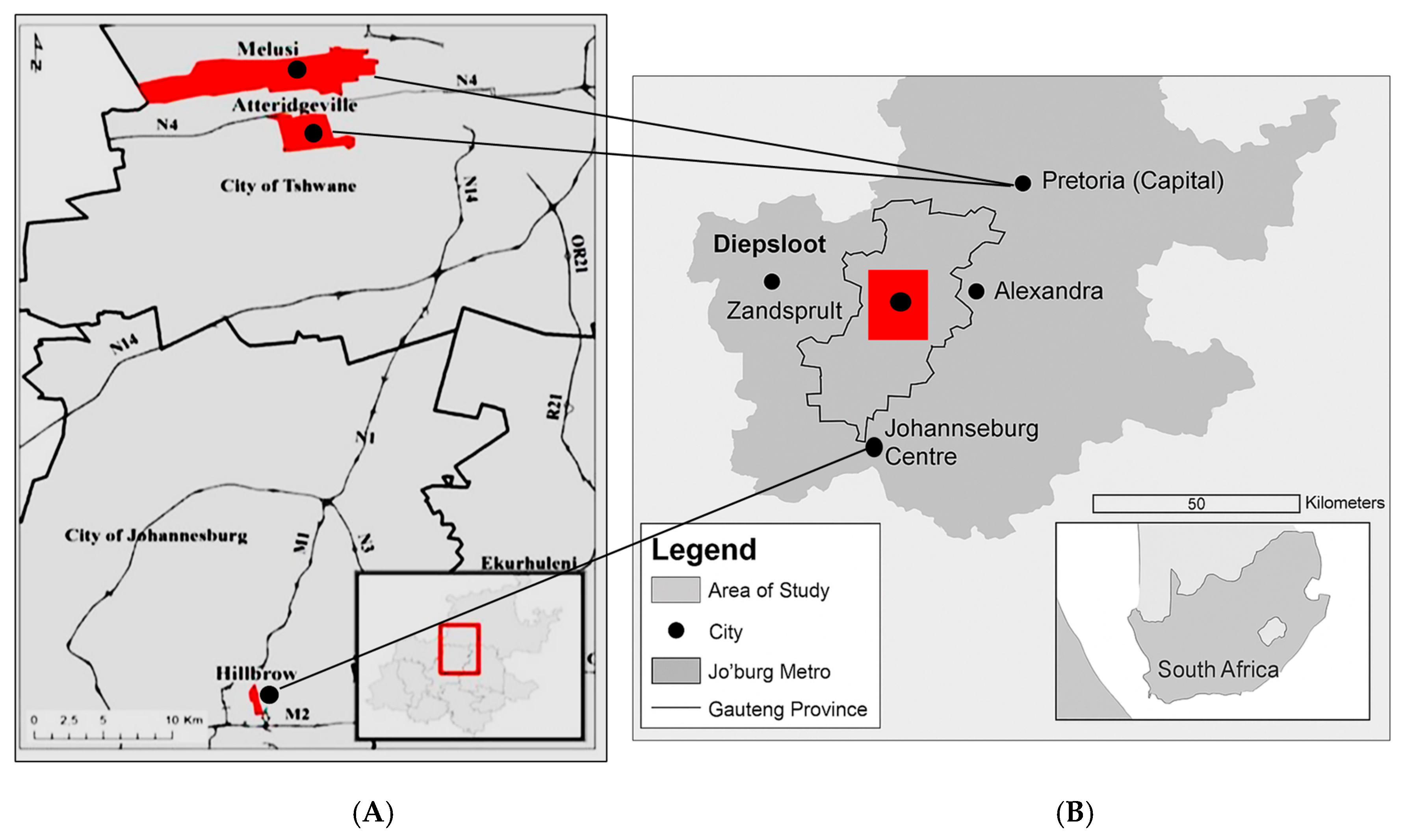
2.1. Study Design and Site Description

This is a cross-sectional, non-randomised study conducted from March 2024 to July 2024 in selected urban and rural areas in Gauteng Province, South Africa. The study sites were selected to incorporate the variation in infrastructure and socioeconomic status, as the growth of Legionella bacteria are influenced by water temperature, stagnation and biofilms which are found in plumbing systems of high-rise buildings in urban areas and rural areas which have limited access to piped infrastructure and therefore rely on water collected and stored in plastic buckets. This diversity allows for a comparative assessment of Legionella contamination in rural and urban households. Sampling was conducted in two urban metropolitans, Hillbrow in Johannesburg Central and Atteridgeville in Pretoria West, and two informal settlements, Zandspruit in Johannesburg and Melusi in Tshwane.
Hillbrow (Figure 1A) is a large, densely populated inner-city residential urban area of Johannesburg (GPS co-ordinates: 26.196871, 28.050063). It is characterised by high-rise apartment buildings and smaller housing units. According to the SANS 10400A:2022 Occupancy or Building Classification, Hillbrow is classified as Building Class H5: Hospitality, which refers to hospitality establishments where unrelated individuals lease furnished accommodations on a short-term basis within a household or private residence.
Atteridgeville (Figure 1A) is an urban housing township in Pretoria (GPS co-ordinates: -25.772121, 28.072406), with diverse building infrastructures, including formal low-rise houses, apartment complexes, and informal settlements located in Pretoria West. According to the SANS 10400A:2022 Occupancy or Building Classification, Atteridgeville is classified as Building Class H4: Dwelling House, which refers to standalone households.
Melusi (Figure 1A) is a rural settlement located in Tshwane municipality (GPS coordinates-25.724338, 28.122957 characterised by freestanding informal housing structures with open spaces between and around them, eliminating the need for consolidating individual structures into large collective compounds. Zandspruit (Figure 1B) is a rural settlement located in approximately 4 km2 in Region C (GPS coordinates-26.048000, 27.920000), City of Johannesburg Metropolitan Municipality. This settlement is characterised by freestanding informal housing structures. According to the SANS 10400A:2022 Occupancy or Building Classification, household structures in both Zandspruit and Melusi could not be classified, and occupancy could not be determined, due to the areas being informal settlements.

2.2. Ethical Clearance

Ethical clearance for sample collection and analysis was granted by the Faculty of Health Research Ethics Committee (REC-2418-2023) in November 2023 at the University of Johannesburg.

2.3. Pre-Sampling Building Site Assessment

A building site assessment was conducted before sample collection to identify potential Legionella spp. exposure risks. A checklist was used to obtain information on water sources, which included availability of taps and storage water containers, water quality, frequency of refill of stored water, plumbing renovations, maintenance and materials, availability of flushing toilets, and hot water.

2.4. Sample Collection

In Hillbrow, water samples were collected from three floors in each building (top, middle, and ground floor) from kitchen and bathtub tap faucets. In Zandspruit, Melusi, and Atteridgeville, water samples were obtained from tap faucets inside and outside of the housing structure as well as stored water containers. In total, 134 water (80 cold, 39 stored, and 15 hot) and 260 biofilm samples were obtained over the sampling period. A total of 129 households were conveniently chosen from the four sites: Hillbrow (27 households, 27 water, 54 biofilm), Zandspruit (30 households, 31 water, 60 biofilm), Melusi (36 households, 36 water, 72 biofilm), and Atteridgeville (36 households, 40 water, 74 biofilm).
Water samples were collected in 1-litre (for hot and stored where available) and 2 litres (for cold water) sterile sampling bottles containing 0.01 g and 0.03 g of 0.1 N sodium thiosulfate, respectively, to neutralise the residual chlorine [29]. Neutralisation was performed to prevent further disinfection and antimicrobial activity of chlorine for accurate detection of bacteria. Residual chlorine levels were measured on-site before sample collection. According to South African National Standard (SANS) 241: 2015 by the South African Bureau of Standards (SANS 241 standards, SABS), the permissible limit of free chlorine in drinking water is ≤5 mg/L. Biofilm samples were obtained by swabbing the inner surfaces of the tap faucets, water storage containers, and toilet bowls using a sterile swab in transport media (Copan’s Faecal Swab Cary-Blair Collection and Transport System, Copan Diagnostic, Inc., Murrieta, CA, USA). Biofilm samples were obtained from toilet bowls based on the evidence that linked Legionella contamination to toilet flushing water [30]. Tap faucets biofilm samples were obtained by allowing the water to run for 60 s to moisten the pipe, after which water was collected, and a sterile swab was inserted beyond the bend of the tap to collect a biofilm sample. All samples were kept in cooler boxes containing ice packs and taken to the University of Johannesburg, Water and Health Research Centre (WHRC) laboratory for analysis.

2.5. In-Field Water Analysis

In-field water analysis (temperature, pH, TDS, and conductivity) was carried out using a portable Combo Tester® (Hanna Instruments, JHB, SA). Residual chlorine was determined using a chlorine photometer (Hanna Instruments, JHB, SA) according to the manufacturer’s instructions. Turbidity measurements were taken upon arrival at the laboratory using the TN100 EUTECH turbidimeter.

2.6. Bacterial Control Strains

Control bacterial strains were obtained from the American Type Culture Collection (ATCC). The L. pneumophila (ATCC 33152) strain was cultured on a selective medium, Buffered Charcoal Yeast Extract (BCYE) agar supplemented with L-cysteine and ferric pyrophosphate at 37 °C for 7 days. For long-term storage, the strains were stored in Lenox broth containing glycerol. Acanthamoeba castellanii (ATCC 30010) was cultured on a Non-Nutrient Agar (NNA) seeded with heat-killed E. coli at 32 °C for up to 21 days. Other bacterial strains used in this study included E. coli (ATCC 25922), Pseudomonas aeruginosa (P. aeruginosa) (ATCC 27853), Klebsiella pneumonia (K. pneumonia) (ATCC 31488), and Enterococcus faecalis (E. faecalis) (ATCC 29212).

2.7. Detection of E. coli and Total Coliforms in Water Samples

The IDEXX Colilert® Quanti-Tray/2000TM most probable method was used to test for the presence of coliforms and E. coli. For each water sample, 100 mL was used for analysis as per the manufacturer’s (IDEXX, USA) instructions. Autoclaved distilled water, K. pneumonia ATCC (31488), P. aeruginosa (ATCC 27853), and E. coli (ATCC 25922) strains were used as a product control, coliform positive control, Colilert negative control, and E. coli positive control, respectively. The Quanti-trays were incubated at 37 °C for 18 h. After incubation, yellow wells were identified as positive for total coliforms. The positive wells were counted to determine the most probable number (MPN) counts, which were then read from the appropriate MPN table supplied by the manufacturer.

2.8. Microbiological Detection of Legionella Species

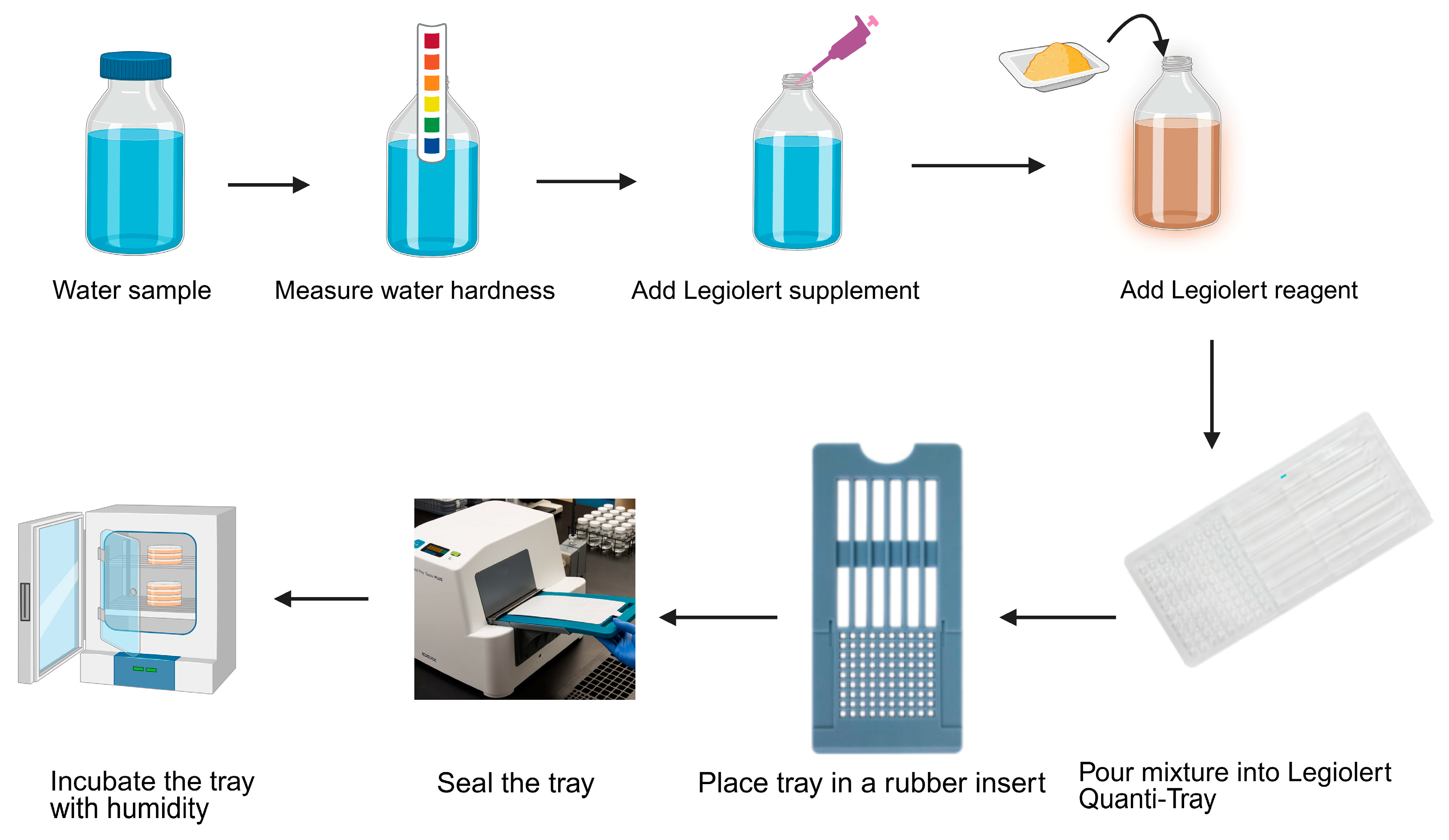
2.8.1. Detection of Legionella pneumophila by IDEXX LegiolertTM

L. pneumophila was detected using the LegiolertTM Quanti-Tray system (IDEXX, USA) with the IDEXX Quanti-Tray sealer plus following the manufacturer’s instructions. Water samples were tested for water hardness with a dip strip supplied with the LegiolertTM system assay. Autoclaved distilled water, E. faecalis (ATCC 29212), and L. pneumophila (ATCC 33152) strains were used as a product control, negative control, and positive control, respectively. The trays were incubated at 39 °C for 7 to 21 days in a humid environment. And positive results were indicated by brown colour change or turbid wells observed in the trays. The positive wells were counted to determine the most probable number (MPN) counts, which were then read from the appropriate MPN table supplied by the manufacturer (Figure 2).

2.8.2. Amoeba Enrichment Technique

The amoeba enrichment technique previously described by [29,31] was used to detect and quantify amoeba-associated Legionella from water and biofilm samples (Figure 3). Briefly, 500 mL of the water sample was mixed with 20 mL Page’s saline (Oxoid, UK) and concentrated by filtering onto a 0.45 µM nitrocellulose membrane filter and placed onto a Non-Nutrient Agar (NNA) (Oxoid, UK) plate seeded with heat-killed E. coli (ATCC 25922) (the grid facing down). For biofilm samples, the swabs were cut and added to 10 mL Page’s saline into individual sterile tubes. For each sample, 200 µL of the solution was inoculated on NNA plates seeded with heat-killed E. coli. The plates were incubated in a humid environment at 32 °C and observed every three days under the 10× objective of a light microscope (Olympus, Japan) for the presence of trophozoites and cysts for up to 21 days. Amoeba positive samples were lysed by passing them through a 21 G × 1, 0.8 × 25 mm needle syringe (Henan Aile Industrial Co. Ltd., Zhengzhou, HA, CN ) ten times. A 100 µL volume of lysate was inoculated onto BCYE agar culture media for L. pneumophila growth and incubated aerobically at 37 °C in a humid environment for up to 14 days. Typical L. pneumophila colonies were tested for cysteine dependency (CD) by incubation on BCYE agar supplemented with L-cysteine and ferric pyrophosphate and nutrient agar plates until growth was observed on the BCYE agar. Colonies growing on BCYE, but not on nutrient agar, were regarded as cysteine dependent (CD+) and were reported as presumptive L. pneumophila (Figure 3).

2.9. Molecular Detection of Legionella Species

2.9.1. DNA Extraction

DNA was extracted from two type of sample, liquid broth from the LegiolertTM Quanti-Trays and L. pneumophila colonies from BCYE agar plates. The liquid broth from the LegiolertTM Quanti-Trays was removed and transferred to 50 mL tubes. The tubes were centrifuged at 8500 rpm speed, 25 °C for 10 min using a Neofuge 15R centrifuge (Heal Force, Vacutec). The supernatant was discarded, and 2 mL of autoclaved distilled water was used to resuspend the pellet. DNA was extracted using the guanidium thiocyanate method [32].
For presumptive L. pneumophila colonies on the BCYE agar, the boiling method was used to extract DNA. A single colony from each plate was suspended in 500 µL of autoclaved distilled water in 2 mL tubes. The tubes were centrifuged at 10,000 rpm speed for 10 min. The supernatant was discarded. One hundred (100) µL of autoclaved distilled water was added to the pellet and resuspended. The tubes were heated at 95 °C for 15 min in a heating block (2 mL Eppendorf ThermoStat plus). Then, they were cooled on ice for 10 min. Thereafter, they were centrifuged at 10000 rpm for 5 min. The supernatant was pipetted out into 1.5 mL Eppendorf tubes as a DNA sample and kept at −20 °C for downstream processing.

2.9.2. Identification of Legionella by Polymerase Chain Reaction (PCR)

Legionella spp. and L. pneumophila were detected by amplification of the extracted DNA using the primers (Table 1) targeting the specific regions of the 16S rRNA and the mip gene, respectively. Each PCR was a 20 µL mixture consisting of 2 µL of the 10 x PCR Buffer (QIAGEN® Hotstart Taq DNA polymerase (QIAGEN GmbH, Hilden, NW, DE) and 15 mM MgCl2), 0.6 µL (20 mM each) dNTP mix, 2 µL primer mix for Legionella spp. (10 µM each LEG 225 forward and LEG 858 reverse) or L. pnuemophila (10 µM each L. pneumophila (F) and L. pneumophila (R)), respectively, 2 µL of template DNA, 0.1 µL HotStar Taq DNA polymerase, 2 µL MgCl2, 1 µL 5 × Q-solution (QIAGEN GmbH, Hilden, NW, DE), and 10.3 µL PCR grade water (QIAGEN GmbH, Hilden, NW, DE).
The PCR was amplified in a T100TM thermocycler (BIORAD) under the following cycling conditions for Legionella spp.: an initial enzyme activation of 95 °C for 15 min, followed by 30 cycles of denaturation at 94 °C for 1 min, annealing at 55 °C for 90 s, and elongation at 72 °C for 1 min with a final extension step of 72 °C for 10 min, allowing the completion of all reactions. The cycling conditions for L. pneumophila were as follows: an initial enzyme activation of 95 °C for 15 min, followed by 35 cycles of denaturation at 95 °C for 30 s, annealing at 65 °C for 45 s, and elongation at 72 °C for 1 min with a final extension step of 72 °C for 5 min, allowing the completion of all reactions.

2.9.3. DNA Electrophoresis

PCR products were separated by gel electrophoresis on a 2% (w/v) agarose gel slab (Hispanger, Spain) prepared in TAE buffer (10 mM Tris; 5 mM EDTA; 5 mM acetic acid; pH 8) with 10 mg/mL ethidium bromide. All samples were electrophoresed at 80 volts for 45–60 min and viewed using a UV transilluminator and digitally photographed (Omega fluor). The size of the bands was compared to a 100 bp molecular marker (Fermentas®, Waltham, MA, USA) as well as the L. pneumophila positive control added with the samples on each gel.

2.9.4. Sequencing

A subset of Hillbrow (2) and Zandspruit samples (4) from the LegiolertTM assay was selected for sequencing to confirm the presence and identity of L. pneumophila in the samples. The PCR products were confirmed on a gel and were outsourced to Inqaba Biotech (SA) for Sanger sequencing. The resulting sequences obtained were analysed on the NCBI BLAST website (http://www.ncbi.nlm.nih.gov/BLAST, accessed on 1 February 2025) to determine the identities of the positive PCR products.

2.9.5. Real-Time PCR

The presence of L. pneumophila was confirmed by real-time PCR using the primers shown in Table 2. The amplification was performed using a 20 µL reaction mixture containing 2 µL of the 10 × PCR Buffer (QIAGEN® Hotstart Taq DNA polymerase and 15 mM MgCl2), 0.6 µL (20 mM each) dNTP mix, 2 µL (10 µM) primer-probe mix, 2 µL of template DNA, 0.1 µL HotStar Taq DNA polymerase, 2 µL MgCl2, and 11.3 µL PCR grade water. The qPCR was amplified in a Rotor-Gene Q (QIAGEN) under the following cycling conditions: an initial Taq polymerase activation at 95 °C for 15 min, followed by 43 cycles of denaturation at 95 °C for 20 s, and finally, an annealing/extension step at 60 °C for 60 s.
Samples were considered positive when the threshold cycle (CT) value was less than 35. A standard curve was generated based on the (CT) values of known concentrations.

2.9.6. Data Analysis

A one-way ANOVA test was used to analyse differences in Legionella presence, with results considered statistically significant at p < 0.05. To evaluate the influence of temperature on Legionella counts, Pearson correlation and linear regression analyses was performed using Microsoft Excel (Office 365), significance set at p < 0.05. Descriptive statistics for the overall sample were determined using SPSS v 28. Data were graphed using GraphPad Prism v10, and images were constructed using Biorender® software accessed during April 2025 (BioRender.com, Toronto, ON, CA), respectively. The Rotor-Gene Q software v2.1.0.9 was used to analyse the qPCR data.

3. Results

3.1. Study Site: Urban and Rural Building Classification

The SANS 10400A:2022 Occupancy or Building Classification was used to categorise the household buildings in the study sites (Hillbrow, Atteridgeville, Melusi, and Zandspruit). Hillbrow (High Rise Buildings) were classified as Building Class H5: Hospitality, where unrelated individuals lease accommodations on a short-term basis in established residences. Atteridgeville household buildings were classified as Building Class H4: Dwelling House, which refers to standalone households. The household buildings in Zandspruit and Melusi could not be classified due to the areas being informal settlements.

3.2. Pre-Sampling Walkthrough Site Assessment for Legionella spp. Exposure

The walkthrough assessment of the four sites revealed that 70% (90/129) of the households collected water for use from taps, while 30% (39/129) collected water from taps and stored it in plastic buckets. The frequency of stored water refill was distributed as follows: 64% (25/39) once a day, 15% (6/39) more than once a day, 15% (6/39) every two days, 3% (1/39) every three days, and 3% (1/39) once a week. A total of 16% (20/129) of households treated the water before use, and 84% (109/129) did not use water treatment. About 98% (127/129) of the households indicated that their water was odourless, with no discoloration and no taste. However, 2% (2/129) reported theirs to be poor, with discernible discoloration and a sour/metallic taste. The majority, 86% (110/129), of the households did not specify the plumbing material used, while 19 households reported using the following plumbing materials: 32% (6/19) iron, 32% (6/19) PVC, 32% (6/19) copper, and 5% (1/19) PEX. In addition, 77% (99/129) of households had no records of plumbing maintenance, while 19% (24/129) employed maintenance annually and 5% (6/129) only when needed. Additionally, 94% (121/129) of the households had no plumbing renovation records, and 8% (8/129) indicated they had renovated their plumbing systems recently. Moreover, 72% (93/129) of the households had flushing toilets; however, only 40% (52/93) had toilet lids. Point of aerosolization was observed in 32% (41/93) of the households. The summary of the walkthrough risk assessment results is tabulated in Table 3.

3.3. In-Field Water Analysis

The recorded water temperature of the four study sites ranged from 22.1 °C to 42 °C (average 25.5 °C) at Hillbrow, 14.8 °C to 18.4 °C (average 16.3 °C) at Zandspruit, 12.7 °C to 33.2 °C (average 18.7 °C) at Melusi, and 15.2 °C to 54.1 °C (average 25.6 °C) at Atteridgeville. The water pH ranged from 6.3 to 8.9 (average 8.0) at Hillbrow, 8.5 to 8.8 (average 8.9) at Zandspruit, 5.8 to 9.6 (average 7.8) at Melusi, and 4.6 to 8.9 (average 7.3) at Atteridgeville. The water TDS ranged from 1.2 ppt to 3.8 ppt (average 1.9 ppt) at Hillbrow, 0.9 ppt to 1.1 ppt (average 1.1 ppt) at Zandspruit, 1.0 to 1.8 ppt (average 1.3 ppt) at Melusi, and 1.0 ppt to 3.4 ppt (average 1.5 ppt) at Atteridgeville. Conductivity ranged from 2.9 mS/m to 8.3 mS/m (average 3.88 mS/m) at Hillbrow, 1.92 mS/m to 3.06 mS/m (average 2.13 mS/m) at Zandspruit, 0.02 mS/m to 0.4 mS/m (average 0.2 mS/m) at Melusi, and 1.35 mS/m to 6.78 mS/m (average 2.59 mS/m) at Atteridgeville. Free chlorine ranged from 0.05 mg/L to 0.94 mg/L (average 0.32 mg/L), 0.2 mg/L to 1.77 mg/L (average 0.73 mg/L) at Zandspruit, too low to be detected to 1.05 mg/L (average 0.06 mg/L) at Melusi, and too low to be detected to 0.32 mg/L (average 0.06 mg/L) at Atteridgeville. Turbidity ranged from 0.01 NTU to 2.29 NTU (average 0.36 NTU) at Hillbrow, 0.0 to 0.62 NTU (average 0.07 NTU) at Zandspruit, 0.0 to 0.55 NTU (average 0.02 NTU) at Melusi, and 0.0 to 1.18 NTU (average 2.29 NTU) at Atteridgeville. The overall results per site are shown in Figure 4 below.

3.4. Detection of E. coli and Total Coliform

Escherichia coli and total coliform serve as indicators of general microbial water quality and potential faecal contamination [35]. The IDEXX Colilert® Quanti-Tray/2000™ most probable method detected total coliforms in 30% (8/27) of Hillbrow samples, with MPN/100 mL values ranging from 1.0 to 10.9. In Zandspruit, about 10% (3/31) of the sample with a range of 4.1 to 14.6 MPN/100 mL. In Melusi, 39% (14/36) of the samples with a range of 1.0 to 648.88 MPN/100 mL, and 3% (1/40) in Atteridgeville with an MPN/100 mL of 1.0. However, there was no E. coli detection at any of the study sites. The summary of the E. coli and total coliform results is tabulated in Table 4.

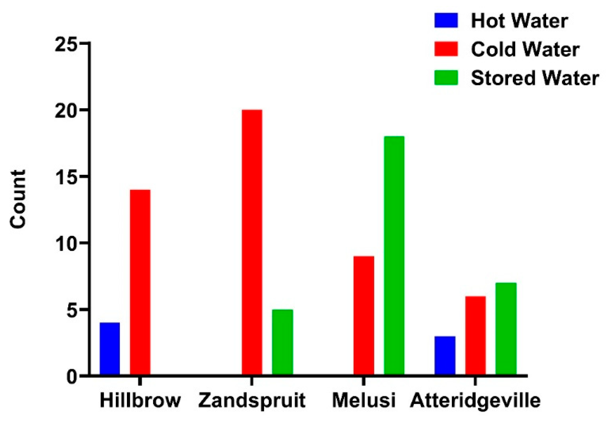
3.5. Legionella pneumophila Detected by IDEXX LegiolertTM

The IDEXX LegiolertTM assay detected L. pneumophila in 4 hot and 14 cold water samples at Hillbrow (average MPN/100 mL: 61.74 for cold and 6.15 for hot). A total of 20 cold and 5 stored water samples tested positive at Zandspruit (average MPN/100 mL: 26.17 for cold and 26.98 for stored). Melusi recorded 9 cold and 18 stored water samples testing positive (average MPN/100 mL: 29 for cold and 10.18 for stored). Moreover, Atteridgeville recorded 3 hot, 6 cold, and 7 stored water samples testing positive for Legionella spp. (average MPN/100 mL: 765.1 for hot, 387.82 for cold, and 1004.6 for stored). Overall, L. pneumophila was detected in 67% (18/27) of Hillbrow, 81% (25/31) of Zandspruit, 75% (27/36) of Melusi, and 43% (17/40) of Atteridgeville water samples. Figure 5 shows the variation in temperature for Legionella detected. The overall detection of Legionella from the study sites is shown in Figure 6.
Linear regression was performed to establish if any relationship existed between water temperature (hot, cold and stored) and Legionella counts across the three study areas (Table 5). Atteridgeville was not included due to the large outliers. A moderate positive association was noticed in Hillbrow (R2 = 0.13) and Melusi (R2 = 0.16), where the Legionella counts increased with a slight increase in temperature. In contrast, Zandspruit showed a low correlation (R2 = 0.03). The slopes of the regression lines ranged from 3.05 to 15.38 between Melusi and Hillbrow, respectively, indicating a variation in growth to temperature responsiveness.

3.6. Microbial Isolation of Free-Living Amoeba and Amoeba-Associated Legionella

Free-living amoeba was detected in 5% (7/134) of water and 13% (34/260) of biofilm samples. Free-living amoeba was detected in three Hillbrow, one Zandspruit, two Melusi, and one Atteridgeville water samples. Additionally, it was also detected in 6 swab samples from Hillbrow, 8 from Zandspruit, 6 from Melusi, and 14 from Atteridgeville. After lysis, the lysate was cultured on BCYE agar, where presumptive L. pneumophila isolates were subjected to cysteine dependency testing and PCR for confirmation. Amoeba-associated L. pneumophila was confirmed in 2 Hillbrow and 1 Melusi water samples and in 3 Hillbrow, 1 Zandspruit, 5 Melusi, and 10 Atteridgeville biofilm samples. Overall, 54% (22/41) with both the cysteine dependency test and PCR.

3.7. Molecular Detection of Legionella Species in Water Samples

The molecular method analysed 134 water samples for the detection of Legionella spp., specifically targeting the 16S rRNA and L. pneumophila by targeting the macrophage infectivity potentiator (mip) gene. This method detected the 16s rRNA gene in 41% (55/134) and the mip gene in 81% (108/134) of the samples. The summary of the positive results per site is tabulated in Table 6.

3.8. Real-Time PCR

A standard curve was used for L. pneumophila qPCR to ensure efficiency while performing qPCRs. To validate the qPCR assays prior to the application of environmental samples, the detection limit and amplification efficiency of each reaction were determined. The nucleic acids were standardised, and a standard curve with 10-fold serial dilutions of DNA controls was prepared and assayed in triplicate. PCR amplification efficiency (E) for each assay was calculated from the slope of the standard curves as −3.5. The experimental points aligned in a straight line with correlation coefficient (R2) of 0.987. The copy numbers for the positive samples are shown in Table 7 below.

3.9. Sequencing

Two (2) Hillbrow and four Zandspruit samples from the LegiolertTM assay that had darker bands on the gel electrophoresis, ensuring sufficient DNA, were selected for Sanger sequencing. These samples were analysed for the presence of L. pneumophila using primers targeting the mip gene. The resulting sequences were subjected to BLAST analysis (NCBI, Bethesda, MD, USA) against the NCBI database accessed during February 2025 to identify sequences with high similarity to known Legionella spp. The results confirmed the identity of the samples as L. pneumophila, with 98% similarity to reference strains. The results of the selected samples are shown in Table 8.

4. Discussion

The current study investigated the detection of Legionella spp., particularly L. pneumophilia and amoeba-associated L. pneumophila, in both urban and rural households in the Gauteng province in South Africa. Water and swab samples were analysed for the detection of L. pneumophila and amoeba-associated L. pneumophila using a combination of microbiological and molecular methods. The classification of Hillbrow as H5: Hospitality suggests that it is a densely populated living environment with multiple occupants sharing water systems. These water systems can cause water stagnation, creating a favourable environment for the bacteria to grow [36]. Atteridgeville classified as H4: Dwelling house, suggests a standalone household with independent water systems, most likely to result in regular water use and less risk of stagnation. In contrast, the lack of classification in Zandspruit and Melusi highlights the informal nature of these areas, where water access is through communal taps and stored water instead of individual piped infrastructure. The inconsistent water supply and biofilm formation within the storage containers may increase microbial contamination [37].
In this study, water storage practices were more prevalent in the rural areas than in the urban sampling areas. Rural areas are dependent upon storage of water since their water source is communal taps. Studies have shown that improper and irregular cleaning of the storage containers often results in biofilm formation inside the containers [26], which contributes to the survival of free-living amoeba that in turn provides a protective niche for L. pneumophila, enhancing its survival and proliferation [27]. The water storage containers are usually plastic, which promotes bacterial regrowth rather than steel buckets, which are more expensive and unaffordable to most people [25]. Stored water was replaced daily in most households, and based on this replacement, no contamination was expected to be detected. However, the evidence of L. pneumophila in the stored water indicates that daily replacement of stored water alone is not sufficient to ensure water cleanliness. Studies have shown that unsealed storage water containers, scoops and unwashed hands can cause contamination to recently collected water [38]. Only urban areas treated water before use, while rural areas had no records of water treatment. Absence of water treatment in rural areas increases the potential risk of microbial contamination, including Legionella spp. This might explain why some households in rural areas reported discoloured water with a sour/metallic taste. Studies have shown that water treatment inactivates and eliminates waterborne pathogens in water distribution systems [39].
Plumbing materials used in urban areas include PVC, PEX, Iron, and copper, while plumbing material in the rural areas were not specified. Studies have shown that some materials can influence the growth of bacteria by providing nutrients and consuming secondary disinfectants [4]. In addition, reports on copper pipes on the growth of L. pneumophila are controversial, as some studies indicate that copper inhibited L. pneumophila growth, while others found the bacteria to growth better on copper than on other plumbing materials [40]. Given that most of the households in both urban and rural areas did not specify their plumbing materials, due to a lack of knowledge of this information, it was impossible to determine whether these findings align with either perspective. Although only urban areas conducted plumbing maintenance and renovations, they were not carried out on a regular basis, thus allowing microbial contamination to accumulate within the water distribution systems. Studies have shown that poorly maintained plumbing systems, particularly in ageing infrastructures, contribute to the formation of biofilms from water stagnation [41].
Aerosolization points from toilets were observed in both areas; however, they were more prevalent in rural areas due to the absence of toilet covers in 87% of the toilets. Although evidence of L. pneumophila from toilet flushing is lacking, a study by [30] shows that aerosolized toilet water may contain the bacteria and might have been the cause of two Legionnaires’ disease cases.
This study recorded water temperatures of the four sites ranging from 12.7 °C to 54.1 °C, with higher temperatures in the urban areas. The low temperatures recorded in the rural areas were due to lack of access to hot water during sampling. Notably, most of the water samples (79%) that tested positive for the bacteria were at 25 °C and below. These results align with a recent study that reported L. pneumophila in 28% of hot and 31% of warm water samples [42]. In addition, the occurrence of the bacteria in a temperature range of 12.7 °C to 54 °C suggest that the bacteria are mesophilic. This is supported by a similar study that reported the bacteria’s ability to survive in water temperatures from 5.7 °C to 63 °C [43]. This study recorded pH values ranging from 4.6 to 9.6. These values were moderately alkaline in rural areas and slightly alkaline in urban areas, with the highest values of 9.6 in rural and 8.9 in urban areas. According to the SANS 241 standard for safe drinking water, the recorded pH values were within the acceptable range (≥5 to ≤9.7). Similar studies have shown L. pneumophila to survive at a pH range of 9.0 to 9.5 [44,45]. The low pH values observed in Melusi and Atteridgeville often cause corrosion within water systems, leading to biofilm formation [46]. This study recorded turbidity ranges of 0.01–2.29 and 0.00–0.18 NTU in Hillbrow and Atteridgeville, respectively. According to SANS 241 standards turbidity in drinking water should not exceed 1 NTU. Turbidity levels in the rural areas fell within the acceptable limit. This is in support of a study that reported inadequate water treatment and plumbing system maintenance to often elevate turbidity levels, promoting L. pneumophila survival [47].
Hydrographic factors such as protozoan hosts, temperature, and water stagnation influence Legionella proliferation. Protozoan hosts protect the bacteria from harsh conditions, including disinfectants and high temperatures thereby, increasing their survival, resistance, and pathogenicity [48]. Stagnant water promotes biofilm accumulation, creating favourable conditions for amoeba and Legionella colonisation [49]. Although chlorination is considered the primary disinfection method in water systems, L. pneumophila has shown resilience to this method [48]. The detection of the bacteria in water samples with free chlorine levels ranging from 0.01 to 1.77 mg/L in this study, emphasise that L. pneumophila is persistent in chlorinated water. A study by Xi et al. [11] detected Legionella bacteria in samples with free chlorine concentrations as low as 0.19–0.22 mg/L. Additionally, Assaidi et al. [49] observed that free chlorine levels between 0.2 and 0.5 mg/L are insufficient in eliminating L. pneumophila. Findings from this study and the literature suggest that both insufficient disinfectant concentrations and the limitations of disinfectants contribute to the persistence and proliferation of the bacteria.
Coliforms are not causative agents of serious illness, but their detection in drinking water indicates the occurrence of other waterborne pathogens, including bacteria and protozoa [35]. The detection of coliforms in this study is supported by the study conducted in three rural areas in the Eastern Cape province that detected total coliform and E. coli in storage water containers [50], although no E. coli was detected in this study. According to SANS 241 standards, the permissible limit for total coliform is ≤10 MPN/100 mL and for E. coli, it is 0 MPN/100 mL in drinking water. Total coliform detections in urban areas were near or within this permissible limit, while levels in rural areas exceeded it with up to 648.88 MPN/100 mL detected in Melusi. This could be attributed to hygiene practices within these households mostly using stored water for their daily needs. The absence of E. coli suggests low likelihood of faecal contamination in the water systems. Although L. pneumophila is not a faecal indicator microorganism, reports on its detection in treated and untreated wastewater suggests that faecal environments serve as reservoirs for the bacteria [51,52]. This study used both biochemical (culture-based) and molecular (PCR-based) techniques to detect L. pneumophila. Notably, discrepancies were observed between these techniques. Conventional PCR detected the bacteria’s DNA in samples that were negative with the LegiolertTM culture technique. Studies have reported that, in complex environmental samples, false-negative or inconclusive results such as too numerous to count (TNTC) can occur due to the overgrowth of non-Legionella bacteria, which can mask the growth of L. pneumophila [53].
The IDEXX LegiolertTM assay showed a greater prevalence of L. pneumophila (78%) in the rural areas compared to urban areas (52%). This assay has reported sensitivity for L. pneumophila of >99% [54]. However, the molecular detection method in this study proved to be more sensitive with a detection rate of 81%, which was equal in both the rural and urban areas. In comparison, the conventional PCR detected the 16S rRNA in 41% and mip gene in 81% of the samples. These results indicate that the 16S rRNA gene is highly conserved among all bacteria, making it difficult to differentiate between Legionella bacteria and other bacteria present in a sample. Studies have shown negative results for the 16S rRNA gene but detected the mip gene in water samples [55]. The urban areas showed a greater prevalence of amoeba-associated L. pneumophila 68% (15/22) compared to rural areas 32% (7/22), indicating that free-living amoeba plays a crucial role in the bacteria’s survival in water systems [20].
Results obtained by real-time PCR for L. pneumophila showed 23% (31/134) samples positive. The Ct values for samples ranged between 8.15 and 27.40. Each qPCR sample with the threshold cycle (Ct) value less than 35 was considered positive. Samples from Hillbrow and Melusi did not yield positive results for L. pneumophila; however, there were 23% (7/31) samples from Atteridgeville and 71% (22/31) samples from Zandspruit that were positive. The detection of the bacterial DNA in these areas suggest that it is prevalent in both urban and rural areas. Rural areas, Zandspruit in particular, showed the highest bacterial load, with Ct value of 8.15 and copy numbers of up to 47.32 × 10⁸ copies/µL, indicating significant colonisation of the bacteria in their water systems. However, urban areas showed low to moderate levels of contamination, with samples showing Ct value ranging from 12. 73 to 39.41 and copy numbers up to 3.719 × 10⁴ copies/µL.
The molecular findings were further supported by Sanger sequencing, which confirmed the identity of L. pneumophila through partial mip gene analysis. BLAST alignment results showed high sequence similarity (98–100%) and low E-values, confirming strong matches to known L. pneumophila strains. These results align with results that were reported in a clinical study where Sanger sequencing identified L. pneumophila in bronchoalveolar lavage fluid (BALF) samples, with a sensitivity of 100% and accuracy of 99% when compared to metagenomic next-generation sequencing (mNGS) [56]. The effectiveness of detection in both environmental and clinical samples highlights the robustness of Sanger sequencing as a confirmatory method for L. pneumophila.

5. Conclusions

Legionellosis, though a notifiable disease worldwide, is underreported in developing countries, including South Africa. Gauteng is a highly populated province with ageing infrastructure, rapid urbanisation, and inadequate sanitation facilities. Despite the increased risk of L. pneumophila contamination in Gauteng province, existing data are limited on the presence of the bacteria in both urban and rural water systems. It was important to examine the prevalence of the bacteria in different environmental water systems in these areas. The study confirmed L. pneumophila in 64% of the water samples using the LegiolertTM assay. The molecular tests proved to be more sensitive and showed equal L. pneumophila prevalence of 81% of both urban and rural areas with conventional PCR and 36% in rural and 10% in urban areas with real-time PCR. However, urban areas showed a greater prevalence of amoeba-associated L. pneumophila of 68% compared to rural areas (32%).
The detection of L. pneumophila in both urban and rural areas in Gauteng highlights the potential health risks, particularly to immunocompromised individuals. These findings emphasise the need to increase surveillance programmes to prevent Legionnaires’ disease outbreaks in these areas. This can be performed by incorporating routine screening into already existing water quality monitoring programmes, especially in health facilities, and areas with ageing and poorly maintained water infrastructure. Preventive measures can include the use sufficient levels of disinfectants such as chlorine to reduce the occurrence of bacteria within the water systems, conduct plumbing renovations and maintenance regularly to prevent the corrosion of the plumbing systems, and introduce educational programmes, especially in the rural areas, on hygienic storage practices. Availability of diagnostic tools in health facilities and increasing awareness among the health care providers about Legionnaires’ disease are important steps towards detecting, providing the appropriate treatment, and reporting of the disease. These combined efforts will contribute to more understanding of Legionnaires’ disease in South Africa. Support future research on genetic susceptibility of different ethnic and racial groups to L. pneumophila. This research should focus on identifying potential genetic markers and understand the mechanism of susceptibility.

Author Contributions

Conceptualization, Z.F.M. and A.S.; Methodology, A.S., Z.D. and Z.F.M.; Software, A.S.; Validation, Z.F.M., Z.D. and A.S.; Formal analysis, Z.F.M.; Investigation, Z.F.M.; Resources, A.S.; Data curation, Z.F.M.; Writing—original draft preparation, Z.F.M. and Z.D.; writing—review and editing, Z.F.M. and Z.D.; visualisation, Z.F.M. and A.S.; supervision, A.S.; project administration, A.S.; funding acquisition, A.S. All authors have read and agreed to the published version of the manuscript.

Funding

This research was funded by the support provided by the Water Research Commission of South Africa, (Project No. 2022/2023-00837), the University of Johannesburg Research Fund (URC, Project Nos. 2023URC00555 and 2023URC00746) and the National Research Foundation of South Africa for student bursary support (NRF).

Data Availability Statement

The data supporting the results of this article will be made available by the authors on request.

Acknowledgments

GRT-SAPRIN team for their assistance in gaining access to the sampling sites and sample collection. The Water and Health Research Centre, University of Johannesburg for laboratory support.

Conflicts of Interest

The authors declare no conflicts of interest.

References

  1. Romano Spica, V.; Borella, P.; Bruno, A.; Carboni, C.; Exner, M.; Hartemann, P.; Gianfranceschi, G.; Laganà, P.; Mansi, A.; Montagna, M.T. Legionnaires’ Disease Surveillance and Public Health Policies in Italy: A Mathematical Model for Assessing Prevention Strategies. Water 2024, 16, 2167. [Google Scholar] [CrossRef]
  2. Viasus, D.; Gaia, V.; Manzur-Barbur, C.; Carratalà, J. Legionnaires’ disease: Update on diagnosis and treatment. Infect. Dis. Ther. 2022, 11, 973–986. [Google Scholar] [CrossRef] [PubMed]
  3. Staff, M.; Nyinawingeri, A. The increasing health burden of Legionella pneumophila in NSW. Infect. Dis. Health 2024, 29, 137–143. [Google Scholar] [CrossRef]
  4. Cullom, A.C.; Martin, R.L.; Song, Y.; Williams, K.; Williams, A.; Pruden, A.; Edwards, M.A. Critical review: Propensity of premise plumbing pipe materials to enhance or diminish growth of Legionella and other opportunistic pathogens. Pathogens 2020, 9, 957. [Google Scholar] [CrossRef]
  5. Ha, R.; Heilmann, A.; Lother, S.A.; Turenne, C.; Alexander, D.; Keynan, Y.; Rueda, Z.V. The Adequacy of Current Legionnaires’ Disease Diagnostic Practices in Capturing the Epidemiology of Clinically Relevant Legionella: A Scoping Review. Pathogens 2024, 13, 857. [Google Scholar] [CrossRef]
  6. Graham, F.F.; Finn, N.; White, P.; Hales, S.; Baker, M.G. Global perspective of Legionella infection in community-acquired pneumonia: A systematic review and meta-analysis of observational studies. Int. J. Environ. Res. Public health 2022, 19, 1907. [Google Scholar] [CrossRef]
  7. Wolter, N.; Carrim, M.; Walaza, S.; Chandu, L.; Morifi, M.; Lawrence, C.; Cohen, C.; von Gottberg, A. Notified Legionnaires’ disease in South Africa, 2018–2020. NICD Bull. 2020, 18, 159–164. [Google Scholar]
  8. Shetty, S.; Kenjar, A.; Raj, J.R.M.; Akhila, D.; Karunasagar, I.; Vittal, R. Prevalence and Characterization of Legionella pneumophila and Related Species from Water-Based Recreational Sites. J. Health Allied Sci. NU 2024, 14, 260–266. [Google Scholar] [CrossRef]
  9. Zhu, Y.; Liu, D. Legionella. In Molecular Medical Microbiology; Elsevier: Amsterdam, The Netherlands, 2024; pp. 1547–1557. [Google Scholar]
  10. Cavallaro, A. The Diversity of Interactions Between Legionella Species and Aquatic Bacteria; ETH Zurich: Zürich, Switzerland, 2024. [Google Scholar]
  11. Xi, H.; Ross, K.E.; Hinds, J.; Molino, P.J.; Whiley, H. Efficacy of chlorine-based disinfectants to control Legionella within premise plumbing systems. Water Res. 2024, 259, 121794. [Google Scholar] [CrossRef]
  12. Monistero, V.; Vicari, N.; Prati, P.; Bragoni, R.; Gazzola, A.; Sala, L.; Maisano, A.; Moroni, P.; Bronzo, V.; Luini, M.V. A rapid and reliable method for early Legionella pneumophila identification and characterization in support of the epidemiology study. Front. Microbiol. 2024, 15, 1452861. [Google Scholar] [CrossRef]
  13. Reinares Ortiz, J.; Pérez-Serrano, J.; González-Rubio, J.M.; González-Camacho, F. Two Sporadic Cases of Legionellosis Associated with the Use of Domestic Ultrasonic Humidifiers. Microorganisms 2024, 12, 2139. [Google Scholar] [CrossRef] [PubMed]
  14. Rello, J.; Allam, C.; Ruiz-Spinelli, A.; Jarraud, S. Severe Legionnaires’ disease. Ann. Intensive Care 2024, 14, 51. [Google Scholar] [CrossRef] [PubMed]
  15. Harte, D. Literature Review: Legionella in Recycled Wastewater and Greywater; ESR Environmental Health Report; Client Report No. FW24021; Health New Zealand: Wellington, New Zealand, 2024.
  16. La Sorda, M.; Palucci, I.; Natalini, D.; Fillo, S.; Giordani, F.; Paglione, F.; Monte, A.; Lista, F.; Mancini, F.; Girolamo, A. Case report: First report of Legionella pneumophila and Bordetella bronchiseptica coinfection in an immunocompromised patient. Front. Med. 2024, 11, 1470567. [Google Scholar] [CrossRef] [PubMed]
  17. He, Z.; Chu, R.; Wu, Q.; Sheng, J.; Liu, P.; Ke, J.; Duan, M.; Li, Q. Evaluation of clinical characteristics of Legionella pneumophila pneumonia diagnosed by metagenomic Next-Generation Sequencing: A retrospective study. medRxiv 2024. [Google Scholar] [CrossRef]
  18. LeChevallier, M.W.; Prosser, T.; Stevens, M. Opportunistic pathogens in drinking water distribution systems—A review. Microorganisms 2024, 12, 916. [Google Scholar] [CrossRef]
  19. Barbosa, A.; Azevedo, N.F.; Goeres, D.M.; Cerqueira, L. Ecology of Legionella pneumophila biofilms: The link between transcriptional activity and the biphasic cycle. Biofilm 2024, 7, 100196. [Google Scholar] [CrossRef]
  20. Ariyadasa, S.; van Hamelsveld, S.; Taylor, W.; Lin, S.; Sitthirit, P.; Pang, L.; Billington, C.; Weaver, L. Diversity of Free-Living Amoebae in New Zealand Groundwater and Their Ability to Feed on Legionella pneumophila. Pathogens 2024, 13, 665. [Google Scholar] [CrossRef]
  21. Margot, C.; Rhoads, W.; Gabrielli, M.; Olive, M.; Hammes, F. Dynamics of drinking water biofilm formation associated with Legionella spp. colonization. NPJ Biofilms Microbiomes 2024, 10, 101. [Google Scholar] [CrossRef]
  22. Moeletsi, M.E. Socio-economic barriers to adoption of electric vehicles in South Africa: Case study of the gauteng province. World Electr. Veh. J. 2021, 12, 167. [Google Scholar] [CrossRef]
  23. Muchemwa, M.; Batisai, K. Social networks and residential choice: Zimbabwean migrants’ experiences in Hillbrow, Johannesburg. Soc. Sci. Humanit. Open 2024, 10, 101090. [Google Scholar] [CrossRef]
  24. Bastaraud, A.; Cecchi, P.; Handschumacher, P.; Altmann, M.; Jambou, R. Urbanization and waterborne pathogen emergence in low-income countries: Where and how to conduct surveys? Int. J. Environ. Res. Public Health 2020, 17, 480. [Google Scholar] [CrossRef] [PubMed]
  25. Potgieter, N.; van der Loo, C.; Barnard, T.G. Co-existence of free-living amoebae and potential human pathogenic bacteria isolated from rural household water storage containers. Biology 2021, 10, 1228. [Google Scholar] [CrossRef] [PubMed]
  26. Slavik, I.; Oliveira, K.R.; Cheung, P.B.; Uhl, W. Water quality aspects related to domestic drinking water storage tanks and consideration in current standards and guidelines throughout the world—A review. J. Water Health 2020, 18, 439–463. [Google Scholar] [CrossRef] [PubMed]
  27. Pereira, A.; Silva, A.R.; Melo, L.F. Legionella and biofilms—Integrated surveillance to bridge science and real-field demands. Microorganisms 2021, 9, 1212. [Google Scholar] [CrossRef]
  28. The National Institute for Communicable Diseases (NICD), Division of Public Health, Surveillance and Response. Notifiable Medical Conditions Surveillance System. Available online: https://www.health.gov.au/our-work/nndss (accessed on 20 March 2025).
  29. Muchesa, P.; Leifels, M.; Jurzik, L.; Barnard, T.; Bartie, C. Detection of amoeba-associated Legionella pneumophila in hospital water networks of Johannesburg. South. Afr. J. Infect. Dis. 2018, 33, 72–75. [Google Scholar] [CrossRef]
  30. Couturier, J.; Ginevra, C.; Nesa, D.; Adam, M.; Gouot, C.; Descours, G.; Campèse, C.; Battipaglia, G.; Brissot, E.; Beraud, L. Transmission of Legionnaires’ disease through toilet flushing. Emerg. Infect. Dis. 2020, 26, 1526. [Google Scholar] [CrossRef]
  31. Bartie, C.; Muchesa, P.; Barnard, T.G. An Investigation into the Presence of Free-Living Amoebae and Amoeba Resistant Bacteria in Drinking Water Distribution Systems of Health Care Institutions in Johannesburg, South Africa: Report to the Water Research Commission; Water Research Commission: Pretoria, South Africa, 2016. [Google Scholar]
  32. Omar, K.; Barnard, T. Detection of diarrhoeagenic Escherichia coli in clinical and environmental water sources in South Africa using single-step 11-gene m-PCR. World J. Microbiol. Biotechnol. 2014, 30, 2663–2671. [Google Scholar] [CrossRef]
  33. Rafiee, M.; Jahangiri-Rad, M.; Hajjaran, H.; Mesdaghinia, A.; Hajaghazadeh, M. Detection and identification of Legionella species in hospital water supplies through Polymerase Chain Reaction (16S rRNA). J. Environ. Health Sci. Eng. 2014, 12, 1–6. [Google Scholar] [CrossRef]
  34. Schwake, D.O.; Alum, A.; Abbaszadegan, M. Impact of environmental factors on Legionella populations in drinking water. Pathogens 2015, 4, 269–282. [Google Scholar] [CrossRef]
  35. Tambi, A.; Brighu, U.; Gupta, A. Methods for detection and enumeration of coliforms in drinking water: A review. Water Supply 2023, 23, 4047–4058. [Google Scholar] [CrossRef]
  36. Yao, X.H.; Shen, F.; Hao, J.; Huang, L.; Keng, B. A review of Legionella transmission risk in built environments: Sources, regulations, sampling, and detection. Front. Public Health 2024, 12, 1415157. [Google Scholar] [CrossRef] [PubMed]
  37. Proctor, C.R.; Rhoads, W.J.; Keane, T.; Salehi, M.; Hamilton, K.; Pieper, K.J.; Cwiertny, D.M.; Prévost, M.; Whelton, A.J. Considerations for large building water quality after extended stagnation. AWWA Water Sci. 2020, 2, e1186. [Google Scholar] [CrossRef] [PubMed]
  38. Shangase, S.I.; Ndlovu, T.S.; Pillay, P. The Evaluation of the Quality of Sanitation and Stored Water for Domestic Use in the Umlazi P Section Informal Settlement in South Africa. Stud. J. Health Res. Afr. 2024, 5, 1198. [Google Scholar] [CrossRef]
  39. Arberas-Jiménez, I.; Sifaoui, I.; Reyes-Batlle, M.; Rizo-Liendo, A.; Sancho, L.; Urruticoechea, A.; Piñero, J.E.; Lorenzo-Morales, J. Ultraviolet–Chlorine combined treatment efficiency to eliminate Naegleria fowleri in artificial surf lagoons. Heliyon 2022, 8, e11625. [Google Scholar] [CrossRef]
  40. van der Kooij, D.; Veenendaal, H.R.; Italiaander, R. Corroding copper and steel exposed to intermittently flowing tap water promote biofilm formation and growth of Legionella pneumophila. Water Res. 2020, 183, 115951. [Google Scholar] [CrossRef]
  41. Szwetkowski, K.J.; Falkinham, J.O., III. Methylobacterium spp. as emerging opportunistic premise plumbing pathogens. Pathogens 2020, 9, 149. [Google Scholar] [CrossRef]
  42. Clark, G.G.; Zhang, G.; Zhao, S.; Nguyen, T.H. Assessment of Legionella pneumophila in Rural Homes Supplied by Private Well Water. ACS ES&T Water 2025, 5, 1041–1054. [Google Scholar]
  43. Girolamini, L.; Salaris, S.; Pascale, M.R.; Mazzotta, M.; Cristino, S. Dynamics of Legionella community interactions in response to temperature and disinfection treatment: 7 years of investigation. Microb. Ecol. 2022, 83, 353–362. [Google Scholar] [CrossRef]
  44. Martin, R.L.; Harrison, K.; Proctor, C.R.; Martin, A.; Williams, K.; Pruden, A.; Edwards, M.A. Chlorine disinfection of Legionella spp.; L. pneumophila, and acanthamoeba under warm water premise plumbing conditions. Microorganisms 2020, 8, 1452. [Google Scholar] [CrossRef]
  45. Pinel, I.; Hankinson, P.; Moed, D.; Wyseure, L.; Vrouwenvelder, J.S.; van Loosdrecht, M.C. Efficient cooling tower operation at alkaline pH for the control of Legionella pneumophila and other pathogenic genera. Water Res. 2021, 197, 117047. [Google Scholar] [CrossRef]
  46. McFarland, M.L.; Provin, T.L.; Boellstorff, D.E. Drinking Water Problems: Corrosion; E-616; Texas AgriLife Extension Service; The Texas A&M System: College Station, TX, USA, 2024. [Google Scholar]
  47. Atkinson, A.J.; Morrison, C.M.; Frehner, W.; Gerrity, D.; Wert, E.C. Design and operational considerations in response to Legionella occurrence in Las Vegas Valley groundwater. Water Res. 2022, 220, 118615. [Google Scholar] [CrossRef] [PubMed]
  48. Mraz, A.L.; Weir, M.H. Knowledge to predict pathogens: Legionella pneumophila lifecycle systematic review part II growth within and egress from a host cell. Microorganisms 2022, 10, 141. [Google Scholar] [CrossRef] [PubMed]
  49. Assaidi, A.; Ellouali, M.; Latrache, H.; Zahir, H.; Karoumi, A.; Mliji, E.M. Chlorine disinfection against Legionella pneumophila biofilms. J. Water Sanit. Hyg. Dev. 2020, 10, 885–893. [Google Scholar] [CrossRef]
  50. Nkwenkwezi, M.G. Applications of Statistical Techniques on the Assessment of Water Quality Parameters. Master’s Thesis, University of Johannesburg, Johannesburg, South Africa, 2021. [Google Scholar]
  51. Pijnacker, R.; Brandsema, P.; Euser, S.; Vahidnia, A.; Kuiter, A.; Limaheluw, J.; Schout, C.; Mohammad, G.H.; Raven, S. An outbreak of Legionnaires’ disease linked to a municipal and industrial wastewater treatment plant, The Netherlands, September–October 2022. Eurosurveillance 2024, 29, 2300506. [Google Scholar] [CrossRef]
  52. Quon, H.; Jiang, S. Quantitative Microbial Risk Assessment of Antibiotic-Resistant E. coli, Legionella pneumophila, and Mycobacteria in Nonpotable Wastewater Reuse Applications. Environ. Sci. Technol. 2024, 58, 12888–12898. [Google Scholar] [CrossRef]
  53. Monteiro, S.N.; Robalo, A.M.; Santos, R.J. Evaluation of Legiolert™ for the detection of Legionella pneumophila and comparison with spread-plate culture and qPCR methods. Curr. Microbiol. 2021, 78, 1792–1797. [Google Scholar] [CrossRef]
  54. Karia, K.; Yui, S.; Muzslay, M.; Ali, S. Concordance between IDEXX LegiolertTM (liquid culture assay) and plate culture (ISO 11731: 2017) for the detection and quantification of Legionella pneumophila in water samples. J. Hosp. Infect. 2024, 150, 163–168. [Google Scholar] [CrossRef]
  55. Barzegar, L.; Ghanizadeh, G.; Esmaeili, D. Study of Gorge Fisher Plumbing System Effects on Growth Inhibition and Amplification of Legionella pneumophila by Culture and PCR Based on 16sRNA and mip Genes. Middle East J. Rehabil. Health Stud. 2022, 9, e122683. [Google Scholar] [CrossRef]
  56. Yi, Q.; Zhang, G.; Wang, T.; Li, J.; Kang, W.; Zhang, J.; Liu, Y.; Xu, Y. Comparative Analysis of Metagenomic Next-Generation Sequencing, Sanger Sequencing, and Conventional Culture for Detecting Common Pathogens Causing Lower Respiratory Tract Infections in Clinical Samples. Microorganisms 2025, 13, 682. [Google Scholar] [CrossRef]
Figure 1. Map showing sampling sites (highlighted in red) in the Gauteng province (black box), South Africa; (A); Hillbrow (GPS: 26.196871, 28.050063), Atteridgeville (GPS: −−25.772121, 28.072406) and Melusi (GPS: −25.724338, 28.122957). (B): Zandspruit (−26.048000, 27.920000).
Water 17 01491 g001
Figure 2. IDEXX LegiolertTM technique to detect L. pneumophila in water samples. Created in https://BioRender.com.
Water 17 01491 g002
Figure 3. Amoeba enrichment technique to detect amoeba-associated L. pneumophila. Created in https://BioRender.com.
Water 17 01491 g003
Figure 4. Representation of the physio-chemical water quality parameters per site.
Water 17 01491 g004
Figure 5. Graphs showing the water temperatures (red bars) and MPN/100 mL (blue bars) values for samples positive for L. pneumophila using LegiolertTM assay per study area. (a) Hillbrow (H-C: cold water, H-H: hot water), (b) Zandspruit (Z-C: cold water, Z-S: stored water), (c) Melusi (M-S: stored water, M-C: cold water), and (d) Atteridgeville (A-S: stored water, A-C: cold water, A-H: hot water).
Water 17 01491 g005
Figure 6. A graph showing water samples positive for L. pneumophila using LegiolertTM assay across the study sites.
Water 17 01491 g006
Table 1. Primers used for Legionella spp. and L. pneumophila PCR.
PrimerSequence 5′-3′Size (bp)Reference
Leg 225 (F)AAG ATT AGC CTG CGT CCG AT654[33]
Leg 858 (R)GTC AAC TTA TCG CGT TTG CT
L. pneumophila (F)CCG ATG CCA CAT CAT TAG C150[34]
L. pneumophila (R)CCA ATT GAG CGC CAC TCA TAG
Table 2. Primer sequences used for real time PCR of L. pneumophila.
StrainPrimerSize (bp)SequenceReference
L. pneuLpneuF1505′-CCGATGCCACATCATTAGC-3′[29]
LpneuR5′-CCAATTGAGCGCCACTCATAG-3′
LpneuP (Probe)5′-6-carboxyfluorescein
[FAM]-TGCCTTTAGCCATTGCTTCCG-BHQ1–3′
Table 3. Walkthrough risk assessment results.
ParameterCategoryn = 129HillbrowZandspruitMelusiAtteridgeville
Water sourceTap9027251622
Storage container39052014
Stored water refill frequencyOnce a day2505911
More than once a day60033
Every two day60060
Every three days10010
Once a week10010
Water treatmentTreated before use2030017
Not treated10924303619
Water quality perceptionOdourless, no taste/discoloration12727303436
Discoloured, Sour/metallic taste20020
Plumbing material usedNot specified11024303620
Iron63003
PVC60006
Copper60006
PEX10001
Plumbing maintenance recordsNo maintenance record990303633
Annually2421003
Only when needed66000
Plumbing renovation recordsNo renovation
recorded
12121303634
Recently renovated86002
Toilet facilitiesWith flushing932730036
With pit toilet3600360
With toilet lids52274021
Point of aerosolization observedyes41026015
Table 4. Summary of E. coli and total coliforms found in sampling sites.
Study SiteTotal SamplesSamples Positive for Total ColiformSamples Positive for E. coli
Hillbrow2780
Zandspruit3130
Melusi36140
Atteridgeville4010
Table 5. Linear regression parameters for Legionella vs. water temperature.
SiteSlope (β₁)Intercept (β₀)R2Interpretation
Hillbrow15.38−375.590.13Moderate association; MPN increases with temperature
Zandspruit3.28−36.180.03Weak relationship; minimal predictive power
Melusi3.05−18.960.16Moderate association; MPN partly temperature-dependent
Note(s): A statistically significant positive Pearson correlation was found in Atteridgeville (r = 0.85, p < 0.001) and Melusi (r = 0.40, p = 0.040), implying that higher water temperatures are related with an increase in Legionella counts. Hillbrow and Zandspruit showed no correlations.
Table 6. Legionella spp. and L. pneumophila detection across all study sites.
Site nLegionella spp.L. pneumophila
Hillbrow272120
%7874
Zandspruit31225
%681
Melusi361629
%4481
Atteridgeville401634
%4085
Total 13455108
%4181
Table 7. Real-time PCR detection and quantification of L. pneumophila for all sites.
Sample NameCopies/µL
Z11.027 × 103
Z247.32 × 107
Z39.576 × 101
Z41.3278
Z58.617 × 101
Z83.9095 × 104
Z92.35 × 103
Z101.627 × 101
Z136.59 × 102
Z141.724
Z151.7588 × 101
Z164.835 × 101
Z173.032 × 101
Z181.882 × 103
Z194.718 × 102
Z214.288
Z224.057
Z231.7524 × 105
Z244.115
Z266.106 × 10
Z291.387
Z307.416
A281.9482
A301.5054 × 101
A313.719 × 104
A324.6035 × 103
A336.425 × 103
A341.0573 × 10
A361.4018 × 103
Table 8. Sequence similarities for L. pneumophila according to NCBI BLAST search.
SampleDescriptionQuery CoverE-ValueAccession Number
H3Legionella pneumophila strain LEG1117 chromosome98%3.00 × 10−51LS483410.1
H11L. pneumophila isolate L1860 macrophage infectivity potentiator surface protein (mip) gene, partial cds98%3.00 × 10−51KC410215.1
Z1Legionella pneumophila strain Edelstein isolate SI7 macrophage infectivity potentiator (mip) gene, partial cds98%3.00 × 10−51MW524769.1
Z2Legionella pneumophila strain MIP1 macrophage infectivity potentiator (mip) gene, partial cds98%3.00 × 10−51KJ160890.1
Z4Legionella pneumophila strain H3 chromosome, complete genome98%3.00 × 10−51CP114576.1
Z5Legionella pneumophila partial mip gene for macrophage infectivity potentiator, 98%3.00 × 10−51AJ810195.1
Disclaimer/Publisher’s Note: The statements, opinions and data contained in all publications are solely those of the individual author(s) and contributor(s) and not of MDPI and/or the editor(s). MDPI and/or the editor(s) disclaim responsibility for any injury to people or property resulting from any ideas, methods, instructions or products referred to in the content.

