Review

Production, Toxicological Effects, and Control Technologies of Ochratoxin A Contamination: Addressing the Existing Challenges

1
Guangdong-Hong Kong Joint Laboratory for Water Security, Beijing Normal University, Zhuhai 519087, China
2
Center for Water Research, Advanced Institute of Natural Sciences, Beijing Normal University, Zhuhai 519087, China
3
CNPC Bohai Drilling Engineering Company Limited, Tianjin 300280, China
4
Faculty of Arts and Sciences, Beijing Normal University, Zhuhai 519087, China
*
Author to whom correspondence should be addressed.
Water 2024, 16(24), 3620; https://doi.org/10.3390/w16243620
Submission received: 13 November 2024 / Revised: 11 December 2024 / Accepted: 12 December 2024 / Published: 16 December 2024

Abstract

:
Ochratoxin A (OTA) is a mycotoxin commonly found in food and feed. It presents a serious threat to human and animal health while also posing a risk as a potential aquatic contaminant. Although many research efforts have been placed on OTA contamination and detoxification, systematic and in-depth studies on summarizing its primary sources, formation mechanisms, toxicological effects, and control technologies remain essential. This review systematically analyzed the sources of OTA contamination, including the main toxin-producing strains and their specific colonization environments, in which the biosynthetic pathways and key regulatory factors of OTA were outlined. On this basis, the principle, merits, disadvantages, and application potential of OTA control technologies, including the physical, chemical, and biological detoxification techniques, were comparatively evaluated. The applications of genetic engineering with an emphasis on newly identified degradative enzymes and their potential in OTA removal were carefully elucidated. Considering the stringent global OTA regulatory standards and food safety handling requirements, this review highlights the necessity of comprehensive control measure development and emphasizes the importance of rigorous technical evaluation and regulatory approval. The aim is to provide theoretical support for effective OTA control and to guide future OTA contamination management in complex environments.

1. Introduction

Mycotoxin contamination has multifaceted impacts on economic and social stability, including substantial losses in trade and food supplies, reduced income and livelihoods for farmers, and increased healthcare pressures for humans and livestock [1]. Ochratoxins (OTs) are representative members of the mycotoxin family. Due to their severe impact on human health and the high risk of foodborne exposure, OTs have attracted widespread attention. They are polyketide compounds primarily produced by secondary metabolism in filamentous fungi, particularly Aspergillus spp. and Penicillium spp. [2,3]. Among OTs, OTA is regarded as a relatively harmful contaminant because of its widespread occurrence in agriculture and the food industry, as well as its notable toxicological characteristics [4,5,6,7]. OTA contamination is widely present during the growth, storage, and transportation of various foods and feeds, including cereals, legumes, coffee beans, grapes, cocoa beans, nuts, spices, and animal-derived products [8]. The Food and Agriculture Organization estimated that 25% of global food crops were significantly contaminated by mycotoxins [8]. Compared to the data mentioned above, research reports on the presence of mycotoxins (including OTA) in water sources have been extremely limited. As a potential aquatic contaminant, the uncontrolled spread of OTA in water environments could have catastrophic consequences [9].
The presence and fate of OTA and other mycotoxin contaminations in environmental matrices have not received sufficient attention. Harmful substances in fungal-infected plant tissues may spread into soil and water systems under the influence of rainfall, agricultural runoff, improper waste management, and industrial activities [10,11]. Evidence has demonstrated the presence of OTA and its producers in manure (used as organic fertilizer) and soil [12,13]. This persistent reservoir of contamination poses not only potential infection risks through crop uptake and accumulation but also through direct contact with contaminated soil or water. Processes such as adsorption and leaching further facilitate the contamination of surface water bodies, including rivers, lakes, and groundwater [14,15]. This makes breaking the chain of environmental impacts particularly challenging [16]. Runoff from OTA-contaminated fields can transport these toxins into nearby water sources. This not only compromises the health of aquatic ecosystems but also raises concerns about the safety of water resources for human consumption and agricultural irrigation [17]. Water safety and quality are essential for human development and well-being. Safe water for drinking, domestic use, and agriculture is fundamental to public health. However, ensuring water source safety has become a global challenge due to threats from multiple natural and anthropogenic pollutants, especially in developing countries [18]. Waterborne diseases linked to fungi and their toxins have increased, posing significant health risks [8]. These unreported and untreated mycotoxins in water sources further threatened the Sustainable Development Goals (SDGs). In particular, they posed significant challenges to SDG6, which focuses on universal access to safe drinking water and sanitation, as well as SDG10, which seeks to reduce social inequalities, including those related to water security.
The key issues related to OTA such as the occurrence, presence, and fate of OTA in the food supply chain have been meticulously reported [19,20,21,22]. A comprehensive and in-depth discussion on fungal growth management and toxin control strategies was provided by Yamina Ben Miri et al. [16]. Their study has made significant and profound contributions to the field. The environmental exposure and analytical methods of certain mycotoxins in environmental matrices have attracted attention [10,11]. However, studies on OTA as a water pollutant have received less attention, leaving substantial potential in analyzing its behavior in aquatic environments and developing effective removal strategies. Starting with the fundamental background of OTA precursors, its biosynthetic pathways, associated gene clusters, and involved transcription factors were systematically described, significantly enriching existing knowledge on OTA biosynthesis. The occurrence patterns of OTA in water and the development of decontamination strategies have received insufficient attention. Breaking the persistent cycle of OTA contamination across the entire food chain, including water and soil, remains a daunting challenge. A comprehensive summary of recombinant OTA-degrading enzymes (OTA-DEs) has been conducted. Finally, the importance of expanding research scopes and adopting practical approaches has been outlined to better assess advanced OTA bioremediation technologies.

2. OTA Production and Contamination Distribution

2.1. Chemical Structures and Properties of OTs

OTs include various derivatives, categorized based on their chemical structure as follows: ochratoxin α (OTα), ochratoxin β (OTβ), ochratoxin A (OTA), ochratoxin B (OTB), ochratoxin C (OTC), (4-R)- and (4-S)-hydroxy-ochratoxin A (4R-OH-OTA and 4S-OH-OTA), (4-R)- and (4-S)-hydroxy-ochratoxin B (4R-OH-OTB and 4S-OH-OTB), and 10-hydroxy-ochratoxin A (10-OH-OTA). Their structures are shown in Figure 1 and Table 1 [6].
The chemical name of OTA is N-[(3R)-(5-chloro-8-hydroxy-3-methyl-1-oxo-3,4-dihydro-1H-isochromen-7-yl) carbonyl]-L-phenylalanine, formed by connecting isocoumarin with L-β-phenylalanine through a peptide bond. Its molecular formula is C20H18ClNO6, with a CAS number of 303-47-9, a molar mass of 403.81 g/mol, and a melting point of 169 °C. Additionally, OTA is a weak organic acid with two ionizable groups: the carboxyl group of phenylalanine has a pKa of 4.4, and the phenolic group has a pKa ranging from 7.05 to 7.3 [23].
OTA showed high stability under different pH and high temperature conditions, with slower degradation rates, especially under acidic and neutral conditions [16]. Its potential long-term persistence in water bodies poses risks to ecosystems and water resources. Although OTA degraded more rapidly under alkaline conditions, temperature and treatment duration remained key influencing factors [24]. A potential risk exists as OTA may convert into OP-OA within a pH range of 7–12 [25]. OTA’s long half-life also suggests its potential to bioaccumulate and persist within the food chain [9]. These findings highlight the need for further investigation into OTA’s migration, accumulation, and degradation behavior in aquatic environments to inform pollution management.

2.2. Unexpected but Existing OTA

OTA contamination could occur during the harvest, processing, storage, or transportation of agricultural products. This contamination could spread to soil and water and further along the food chain, posing significant risks to animals and humans. Aquatic environments, including surface and groundwater, were considered potential large reservoirs of mycotoxins [18]. Climate change (e.g., rising temperatures and shifts in precipitation patterns) further promoted more frequent releases of mycotoxins, including OTA, into water bodies, affecting the entire food chain. Additionally, the improper storage and transportation of water contributed to the production and release of persistent OTA, adding to the burden of drinking water safety management.
Newly identified occurrences of OTA were always alarming. Several countries and regions, including the European Union, the United States, and China, established clear maximum levels for OTA, generally measured in micrograms per kilogram (µg/kg). The European Union set relatively strict standards for OTA in grains and related products, with maximum limits of 0.5 μg/kg in cereal-based foods for infants, 2 μg/kg in grape juice and wine, 3 μg/kg in cereal-based processed products, 5 μg/kg in grains and roasted coffee, and 10 μg/kg in dried fruit [26,27]. China also set maximum OTA levels for food and raw materials, including 5.0 µg/kg in grains and milled products, 2.0 µg/kg in wine, and 5 μg/kg in soybeans and coffee [28].
Reports of contamination in crops and their processed products are common, and information on OTA producers and new sources of OTA exposure warrants attention. Agricultural soil, as a repository for many organic pollutants, could influence plant crop production due to soil contamination [29]. A survey on mycotoxin contamination in soil highlighted OTA levels at 23.7 μg/kg [11]. A field survey of Pistacia vera (pistachio) production and storage in Turkey found that the dominant soil fungi were A. niger; additionally, all 11 isolated A. ochraceus strains produced OTA under experimental conditions [30]. Similarly, A. carbonarius and A. niger found in grapes originated from the soil beneath grapevines and could be transferred to the plants through agricultural practices [31,32]. This information warns us that risk management should start from the pre-harvest stage to control the initial development of mycotoxins. Proactive field management could prevent the potential transfer of OTA from soil to edible or forage crops, thereby mitigating issues throughout the entire supply chain.

2.3. Producers of OTA

Since the first isolation of OTA-producing A. ochraceus from South African sorghum in 1965 [33], numerous other fungi were identified as OTA producers. OTA production was significantly influenced by various factors, including the texture of crops and processed foods, as well as environmental conditions such as temperature and humidity. For example, OTA biosynthesis was enhanced in the presence of biological factors like lactose, urea, and amino acids, while it was inhibited by glucose and sucrose. Due to distinct ecological and physiological traits, it was relatively straightforward to identify which species were responsible for OTA formation in specific foods or geographic locations.

2.3.1. Aspergillus spp.

A. ochraceus, A. niger, A. carbonarius, and A. tubingensis could contaminate agricultural products both pre- and post-harvest and were major OTA-producing fungi. A. ochraceus could grow within a temperature range of 8–40 °C (optimal growth at 24–31 °C), at high water activity (Aw of 0.95–0.99), and within a pH range of 3–10, primarily contaminating wheat, nuts, coffee, and processed meat [34]. A. niger was widely used in the production of food-grade organic acids and enzymes. Although most isolates did not produce OTA, a small number of low-level toxigenic strains were occasionally found contaminating dried vine fruits in tropical and subtropical regions [35]. A. carbonarius and A. tubingensis were the main OTA-producing fungi in grapes and grape-derived products [36]. Medina et al. [37] studied 205 native fungal isolates from five Spanish grape varieties and found that 74.2% of A. carbonarius isolates and 14.3% of A. tubingensis isolates produced OTA, with OTA concentrations ranging from 1.2 to 3530 ng/mL and 46.4 to 111.5 ng/mL, respectively.

2.3.2. Penicillium spp.

Studies confirmed that P. verrucosum, P. viridicatum, and P. nordicum are known to produc OTA [38]. P. verrucosum grows slowly and is commonly found in cool, moist grains and grain derivatives. Besides OTA, it can also produce OTB and citrinin, making it a common storage fungus in temperate and cold climate regions [39]. The toxin-producing capability of P. viridicatum is influenced by water activity levels, and it can still contaminate grains and soil to produce OTA under low-temperature conditions [40]. P. nordicum primarily contaminated cheese and fermented meat products. OTA synthesis in fermented sausages was less affected by pH but was significantly influenced by water activity [41].

2.4. Precursors and Related Metabolites in OTA Biosynthesis

OTA is a derivative with an incompletely understood biosynthetic pathway; however, certain key steps were confirmed. For instance, the biosynthesis of the isocoumarin moiety was first catalyzed by polyketide synthase (PKS). This was followed by the connection of its carboxyl group with the amino group of phenylalanine through the action of peptide synthetase, and then chlorination [42]. Since OTA was formed by the linkage of dihydroisocoumarin and phenylalanine via an amide bond, some researchers considered dihydroisocoumarin and phenylalanine as precursor substances in OTA biosynthesis.
Radioactive experiments showed that, by directly adding [1-14C] phenylalanine to OTA-producing media, phenylalanine’s involvement in OTA synthesis could be traced through isotopic labeling, proving that phenylalanine was a direct precursor in OTA synthesis [43]. Tracking [2-14C] labeled sodium acetate demonstrated that dihydroisocoumarin was formed by the dehydration and condensation of acetate. Researchers added [2-14C] labeled sodium acetate to the culture medium of OTA-producing P. verruculosum and observed fully 14C-labeled OTA. When the labeled sodium acetate was replaced with [2-14C] malonate, partially 14C-labeled OTA was detected in the dihydroisocoumarin portion. These experiments indicated that, for the two precursors of OTA, the dihydroisocoumarin portion was synthesized from malonate through multiple steps, while phenylalanine could not be synthesized from malonate. Sodium acetate was converted into both dihydroisocoumarin and phenylalanine portions through multi-step reactions [15]. Feeding experiments with [14CH3] methionine demonstrated that the C-7 position carbon in the dihydroisocoumarin moiety, specifically within the methoxy group attached to phenylalanine, originated from methionine. The methylation inhibitor ethionine completely suppress OTA production, further confirming methionine’s role as the methyl donor for the C-7 position of dihydroisocoumarin [44].

2.5. Biosynthetic Pathway and Genes Related to OTA

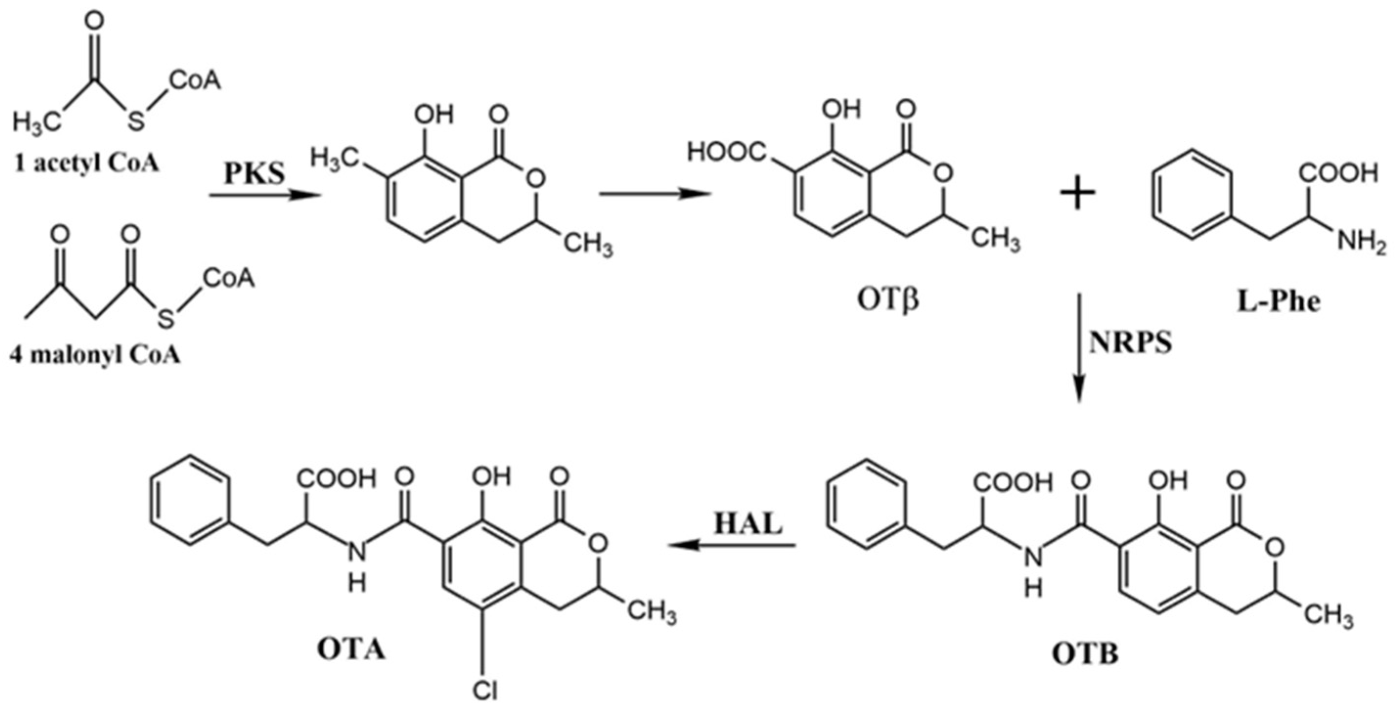
Based on OTA’s chemical structure, isotopic tracer technique, and studies on OTA degradation, researchers confirmed that the phenylalanine moiety in the OTA structure is derived from the shikimate pathway, while the dihydroisocoumarin portion originates from the polyketide pathway. The catalytic reactions in this pathway remain incompletely elucidated, with only hypothetical pathways proposed (see Figure 2). The initial substrates, acetyl-CoA and malonyl-CoA, underwent condensation catalyzed by polyketide synthase (PKS), forming a polyketide chain and producing a dihydroisocoumarin structure [45]. This dihydroisocoumarin intermediate was then conjugated with phenylalanine in a reaction catalyzed by nonribosomal peptide synthetase (NRPS). Finally, OTB was chlorinated by a halogenase (HAL) enzyme, yielding the final product, ochratoxin A (OTA) [46,47,48,49].
Generally, genes involved in fungal mycotoxin synthesis were clustered on the chromosome. Among these, pks and nrps played crucial roles in the secondary metabolites of fungi, serving as core genes within the cluster. In 2003, O’Callaghan et al. identified part of the pks sequence in A. ochraceus [51] and demonstrated that a pks-deficient mutant of A. ochraceus could not synthesize OTA. This study was the first to investigate genes associated with the OTA biosynthetic pathway. In 2005, Karolewiez et al. [52] discovered more complete pks and nrps gene sequences in P. nordicum, a known OTA producer. In 2006, building on Karolewiez’s work, Geisen et al. [53] identified genes within the OTA biosynthetic cluster in P. nordicum, which encode polyketide synthase, non-ribosomal peptide synthetase, alkaline serine protease.
In 2007, Pel et al. [54] sequenced the entire genome of A. niger CBS 513.88, publishing the complete sequence and predicting the biosynthetic pathway for OTA production in this strain. They proposed that the OTA biosynthetic gene cluster consists of eight genes: short-chain dehydrogenase (An15g07860), isopropanol dehydrogenase (An15g07870), hydroxylase (An15g07880), the bZIP transcription factor (An15g07890), the cytochrome P450 enzyme (An15g07900), polyketide synthase (An15g07910), nonribosomal peptide synthetase (An15g07920), and nitric oxide synthase (An15g07930). A schematic representation of the predicted OTA gene cluster is shown in Figure 3.
Gil-Serna et al. [55] pointed out that the basic leucine zipper (bZIP) transcription factor within the OTA biosynthetic gene cluster in A. steynii plays an important role in regulating OTA gene expression. In 2016, Ferrara et al. [56] demonstrated that the expression levels of p450 and bZIP genes were correlated with OTA production. These genes encoded a cytochrome P450 monooxygenase and a basic bZIP transcription factor, positioned between the nrps and hal genes within the proposed OTA biosynthetic gene cluster. A comparative analysis of OTA-producing gene clusters was conducted on five sequenced OTA-producing fungi (A. ochraceus fc-1, A. westerdijkiae CBS 112803, A. steynii IBT 23096, A. niger CBS 513.88, A. carbonarius ITEM 5010, and P. nordicum BFE487) [50], as shown in Figure 4. The OTA-related gene clusters in these five fungi displayed high homology, mainly consisting of five highly conserved biosynthetic genes encoding polyketide synthase (PKS), non-ribosomal peptide synthetase (NRPS), Cytochrome P450 Monooxygenase, halogenase (Hal), and the bZIP transcription factor. In A. ochraceus fc-1, these genes were knocked out and confirmed to be directly involved in OTA biosynthesis. However, variations in biosynthetic mechanisms were observed across species, and this result has not been validated in other strains. Li et al. [57] conducted a study within the theoretical framework of secondary metabolite regulation by the bZIP transcription factor. They showed that, in comparison to wild-type strains, bZIP knockout strains were nearly incapable of synthesizing OTA and its precursors (including OTα), indicating that the AnOTAbzip gene played a key positive regulatory role in OTA synthesis. The response of mutant strains under oxidative stress conditions may also suggest a dual role of the bZIP gene in secondary metabolism.

2.6. Regulatory Mechanisms of OT Biosynthesis

The synthesis of secondary metabolites in fungi was regulated by extensive metabolic regulation through various metabolic mechanisms. During secondary metabolite biosynthesis, gene clusters were primarily regulated by two categories of regulatory factors: global regulatory factors and pathway-specific regulators (i.e., cluster-specific regulators). Global transcription factors regulated secondary metabolite biosynthesis across a broad spectrum in fungi. A notable example was the light-responsive regulatory complex VeA/LaeA/VelB. Studies indicated that global transcription factors, such as LaeA, were responsive to environmental stimuli, including light, pH, carbon and nitrogen sources, temperature, and reactive oxygen species. The second category includes cluster-specific regulatory factors. According to current genomic data, approximately 60% of fungal secondary metabolite gene clusters contained such regulators [58]. These cluster-specific regulators were involved in regulating the biosynthetic levels of secondary metabolites within gene clusters, and numerous such regulators were identified.
A common type includes Zn(II)2Cys6 transcription factors. Examples included the gene cluster-specific transcription factor AflR involved in sterigmatocystin and aflatoxin biosynthesis [59,60]; the specific transcription factor LovE that regulates the lovastatin gene cluster in A. terreus [61], the AzaR responsible for the synthesis of azadiketone in A. niger [62], and MokH, required for monacolin K biosynthesis [63]. All of these transcription factors belong to the Zn(II)2Cys6 class. Additionally, the previously mentioned bZIP transcription factor was hypothesized to act as the cluster-specific regulator for the OTA biosynthetic gene cluster.

3. Toxicity of OTA

OTA was identified as a potent carcinogen with multiple toxicological effects, including teratogenicity, mutagenicity, hepatotoxicity, genotoxicity, immunotoxicity, and embryotoxicity [64]. These effects were observed in rodents and poultry, with the kidneys identified as the primary target organ [65].

3.1. Renal Toxicity

OTA exhibits toxicity in multiple organs, with the kidneys identified as its primary target [66]. Studies have shown that OTA exhibits significant nephrotoxicity in all monogastric mammals [67,68]. Although its toxic mechanism is complex and not fully understood, it is closely associated with impaired OTA transport and clearance in the body. For example, sulfamethoxazole can inhibit OTA metabolic clearance, leading to excessive accumulation within organs and exceeding their capacity to cope [68]. The nephrotoxicity of OTA may involve mechanisms such as the inhibition of protein synthesis, resulting in renal cell dysfunction, and the induction of oxidative stress, which exacerbates kidney tissue damage [69,70]. Although the precise mechanism by which OTA induces reactive oxygen species (ROS) generation remains unclear, it is generally accepted that ROS contribute to cell damage via lipid peroxidation and the suppression of antioxidant enzyme activity, thereby aggravating nephrotoxicity.
OTA was also implicated in Balkan endemic nephropathy, renal failure, and tumor development in humans. Additionally, OTA modulated transcription factors and induced DNA methylation, resulting in renal damage [71]. OTA further caused inflammation, glomerular and tubular injury, and even renal fibrosis by inhibiting protein synthesis, inducing DNA damage, cell cycle arrest, and apoptosis [72].
Bondy et al. [73] suggested that OTA was a potent carcinogen in poultry and rodents, leading to renal disease in livestock. Further research indicated that mitochondrial recombinant manganese superoxide dismutase could prevent OTA-induced hypertension and restore lipid peroxidation levels and tissue damage in rat kidneys [74]. Antioxidant molecules, including δ-tocotrienol, red orange extract, and lemon extract reduced or prevented OTA-induced nephrotoxicity by directly scavenging reactive oxygen species. This provided a basis for using antioxidants to counteract OTA toxicity [75,76].

3.2. Hepatotoxicity

Due to the decarboxylation of phenylalanine at OTA’s carboxyl group, the liver sustained damage second only to the kidneys. Qi et al. [77] analyzed the expression of microRNA, mRNA, and proteins in the livers of rats treated with OTA for 13 weeks using multi-omics approaches. Moderate and high doses of OTA exert different effects on the liver, inducing five distinct pathways. The primary bile acid biosynthesis and cytochrome P450 xenobiotic metabolism pathways are directly related to liver damage, while the remaining pathways (arginine and proline metabolism, cysteine and methionine metabolism, and PPAR signaling pathways) are associated with metabolic diseases. This study provided the first insights into early OTA-induced hepatotoxicity. In vivo liver injury resulting from OTA recirculation between the liver and intestine has also been widely reported. This effect is likely due to OTA’s inhibition of mitochondrial respiration, leading to oxidative stress, membrane peroxidation, calcium homeostasis disruption, and interference with oxidative phosphorylation, all contributing to liver damage [78,79].

3.3. Carcinogenic, Teratogenic, and Mutagenic Effects

Studies have shown that OTA exhibited genotoxic effects. Upon bioactivation, this toxin can form covalent DNA adducts, producing electrophilic metabolites that lead to mutations and malignant tumor formation [80]. Additionally, research indicated that OTA acted as an Nrf2 inhibitor, impairing cellular function by inducing oxidative stress, which led to DNA damage and mutations [81]. OTA can even cross the placental barrier, accumulating in the fetuses of various animals, including poultry, rabbits, hamsters, rats, and mice, causing fetal malformations. Bondy et al. [82] conducted a reproductive toxicity study in rats, showing that continuous feeding with OTA-contaminated feed induced post-implantation fetal toxicity. This treatment significantly reduced the number of offspring between implantation and the first postnatal day, with renal and reproductive toxicity observed in offspring.

3.4. Immunotoxicity

OTA was identified as an immunosuppressive mycotoxin that affected both humans and animals. It inhibited the proliferation of peripheral T and B lymphocytes, impaired macrophage phagocytic function, reduced levels of interleukin-2 and its receptors, decreased immunoglobulins G and M, and lowered natural killer cell activity, thereby increasing the risk of various infectious diseases [83]. Exposure to OTA led to oxidative stress in the bursa of Fabricius, lipid peroxidation, and various pathological lesions in the spleen and thymus of chickens [84]. OTA contamination in feed resulted in economic losses in the poultry industry [67]. Khan et al. [83] investigated the effects of OTA on the immune system of broiler chickens, demonstrating reductions in the weights of the thymus, spleen, and bursa of Fabricius, along with a decrease in white blood cell counts. Additionally, OTA reduced thymus size and serum globulin concentration, diminishing resistance to pathogenic microorganisms [85].

3.5. Gastrointestinal Toxicity

The gastrointestinal tract was linked to the liver and other tissues through enterohepatic circulation [86], and served as the primary defense barrier when livestock consume OTA-contaminated feed. Many mycotoxins acted as protein synthesis inhibitors, making intestinal cells and tissues primary targets. When intestinal epithelial cells (IECs) were fully exposed to OTA-contaminated feed, epithelial cells in the esophagus and gastrointestinal tract (GIT) may also be damaged [87]. This exposure led to intestinal dysbiosis, including increased permeability, immune suppression, and microbial imbalance, ultimately damaging the intestines and other organs [88,89,90].

3.6. Neurotoxicity

Since nerve cells are distributed throughout the body, OTA has a high likelihood of damaging these cells. Studies showed that OTA caused neuronal damage in the hippocampal region of the brain, leading to neurological disorders such as depression and memory impairment. Soleas et al. [91] reported that OTA could cause damage to the brain or lead to specific brain lesions. Therefore, OTA was highly toxic to nerve cells and had the potential to reach and damage neural tissues at any time.

4. Detoxification Technologies for OTA

Mycotoxins were potentially lethal or capable of causing severe illness within a relatively broad range of extremely low concentrations, typically measured in µg/kg or mg/kg [11]. Preventive controls implemented at all stages—pre-harvest, processing, and post-harvest—represented the primary approach to minimizing fungal growth and mycotoxin synthesis, thereby reducing human exposure to mycotoxins. During production, practical measures such as physical screening, mechanical removal, and air washing served as cost-effective strategies to eliminate “micro-contaminants” in food processing. However, as supplementary methods, detoxification technologies were also developed [92]. OTA was chemically stable, acid-resistant, heat-tolerant, and not easily degraded, meaning that conventional industrial processing cannot effectively remove OTA from feed and food raw materials, leaving it as a persistent component in final products. These detoxification techniques primarily degrade, modify, or adsorb mycotoxins to reduce or eliminate their toxicity. Most detoxification technologies remained in the research phase at the laboratory scale; currently, only adsorption and microbial methods have limited industrial-scale application in detoxifying fermented feed. In fact, most detoxification technologies remained in the research phase at a laboratory scale; currently, only adsorption and microbial technologies had limited industrial-scale application in detoxifying fermented feed. Developing cost-effective, efficient, adaptable, and stable detoxification technologies for industrial applications remained an urgent challenge for researchers.

4.1. OTA Conversion Pathway

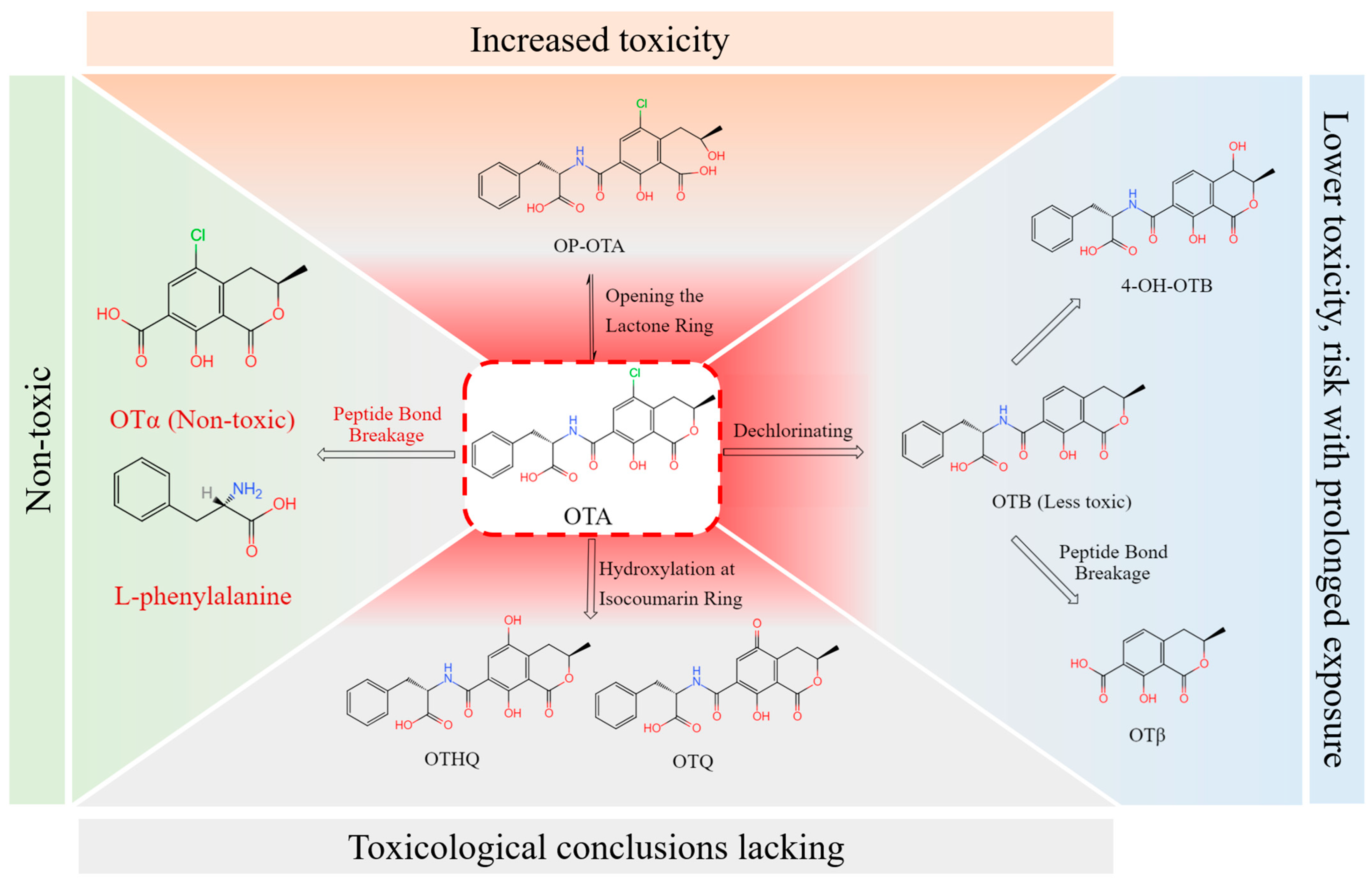
OTA was primarily degraded or transformed through four metabolic pathways [93], as shown in Figure 5: cleavage of the peptide bond to produce OTα and L-β-phenylalanine; dechlorination of the isocoumarin ring to produce OTB, which can further undergo peptide bond cleavage to form OTβ; hydroxylation of the isocoumarin ring to generate ochratoxin hydroquinone (OTHQ); and opening of the lactone ring to form open-lactone OTA (OP-OTA). In complex metabolic systems, OTA was also transformed into various forms, including 4-OH OTA, 4-OH-OTB, 10-OH OTA, hexose/pentose conjugates, and DNA adducts [6].
OTα lacked the hydrophilic L-β-phenylalanine structure, which significantly reduced lipid peroxidation damage when it formed complexes with other macromolecules. As a result, it exhibited much lower toxicity compared to the parent compound OTA. Studies [94,95] showed that OTα exhibited no cytotoxicity at a concentration of 50 μM in immortalized human kidney epithelial cells (IHKEs), a system highly sensitive to OTA with an IC50 (half-maximal inhibitory concentration) of 0.5 μM. Consequently, the conversion of OTA to OTα was considered one of the most effective detoxification pathways for OTA. Research by Ferenczi et al. [96] demonstrated that oral administration of OTA to male CD1 mice led to a significant increase in OTA concentration in the blood, transcriptional changes in OTA-dependent genes (annexinA2, clusterin, sulphotransferase, gadd45 and gadd153), and histopathological alterations in the renal cortex. These OTA-induced changes were not observed in the group fed with OTα.
OTB, a non-chlorinated analog structurally related to OTA, had a lower affinity for plasma proteins, allowing for faster metabolism compared to OTA and no specific retention in the kidneys; therefore, it was considered to be significantly less toxic [97]. However, other studies [6] indicated that this difference was typically evident only after a relatively short exposure time; as exposure duration increases, the toxicity difference diminished. Although OTB exhibited significantly lower nephrotoxicity than OTA in rodents, it showed only a slight reduction in cytotoxicity in vitro [98].
OTHQ was a minor metabolite of OTA that appeared in trace amounts in animals. A study conducted by Wu et al. [99] demonstrated that OTA underwent oxidative dechlorination to produce a hydroquinone–quinone redox pair, similar to other chlorinated phenols, which might play a role in OTA-mediated toxicity. However, limited data were available, making it difficult to draw meaningful toxicological conclusions [6].
OP-OTA was a highly toxic compound formed when the lactone ring of OTA opened under alkaline conditions after ingestion. The OP-OTA observed in rodents was found to be even more toxic than its parent compound, OTA [100]. In a study conducted by Li et al. [101], significant amounts of OP-OTA were detected in the bile and urine after OTA injection in rats. Their research also showed that OP-OTA can reconvert to OTA.

4.2. Physical Detoxification Technologies

Physical detoxification technologies included heat extrusion, adsorption, ultraviolet (UV) irradiation, gamma irradiation (γ-irradiation), and cold plasma. These technologies were widely used to remove OTA from food and feed. OTA was not completely degraded during baking [102]. OTA withstood high-pressure steam heating at 121 °C for 3 h, and, even at 250 °C, it was only partially degraded [65]. Irradiation served as a simple technology for removing mycotoxin contamination in aqueous solutions; 60Co-γ rays at a dose of 4 kGy degraded 90% of OTA in aqueous solutions. However, this effect depended on factors such as matrix composition, moisture content, and radiation dose. Irradiation degraded only a small portion of OTA in flour, grape juice, and wine [103]. Although irradiation and cold plasma treatment rapidly degraded OTA without significantly affecting laboratory product quality, they were limited by the requirement for specialized equipment, which made them difficult to popularize.
Adsorption was considered an economical, environmentally friendly, and easy-to-operate method for removing OTA contamination. Various natural and synthetic adsorbents, such as activated carbon [104], bentonite [105], β-cyclodextrin-polyurethane polymers [106], and chitosan [107], were reported to remove OTA from contaminated foods and agricultural products through physical adsorption. Clarification was a necessary step in winemaking, and positively charged and hydrophobic clarifying agents represented a low-cost option for removing OTA and other mycotoxins. Although traditional clarifying agents such as gelatin and chitosan were not highly effective at removing OTA at an initial concentration of 100 μg/L in wine, applications without new processing procedures still held potential [108].
Based on this, novel dual-functional composite materials that enhanced clarification and efficiently removed OTA were developed [23]. This dual-function design enabled the material to remove toxins while maintaining beverage clarity, balancing food safety with product quality. Xu et al. [109] demonstrated that magnetic multi-walled carbon nanotubes coated with polydopamine serve as adsorbents to extract OTA and five other mycotoxins from edible vegetable oil samples in solid-phase extraction. However, this technology was developed primarily for detection purposes. With technological advancements, modifications of specific functional groups on the material surface, pore size control, or selective film coatings increased affinity for toxins while reducing the adsorption of flavor substances and nutrients. However, vigilance was required: the comprehensive evaluation of the long-term stability, chemical inertness, and specific adsorption–desorption strategies of novel materials were essential to eliminate potential safety risks and provide more effective detoxification solutions.
Relatively safe materials with biocompatibility and biological inertness were widely used in the feed and food industries. Various commercially available and generally recognized as safe (GRAS) materials were directly applied to feed or simulated gastric and intestinal fluids for toxicity exposure and adsorbent intervention trials. In these trials, both reduced toxicity effects and potential impacts on nutrient absorption were observed [110,111]. The relatively low cost and mature preparation process made these adsorbents viable as feed additives with high economic efficiency for large-scale applications. However, suitable doses for safe use should be recommended.
Microbial adsorbents provided a mild approach for OTA purification, especially the use of fermentation strains with a strong safety record in the food and feed industries. OTA-adsorbing microorganisms included actinomycetes, lactic acid bacteria, filamentous fungi, and yeasts [112]. The most critical factor affecting microbial OTA adsorption capacity was cell wall composition. However, scholars debated specific cell wall components. For example, Taheur et al. [113] isolated strains from fermented cultures capable of adsorbing varying levels of AFB1, ZEA, and OTA, identifying galactomannan and β-glucan as the primary components responsible for adsorption. Caridi et al. [114], analyzing OTA in saline solutions used to wash wine lees, proposed that wine yeast exhibited adsorption activity via electrostatic and ionic interactions between mannoproteins on its cell wall and OTA. The removal of mycotoxins by microorganisms was influenced by multiple factors, such as cell weight, environmental conditions (pH, temperature, and duration), and the initial concentration of mycotoxins [115]. Although the binding between toxins and biomass was typically less stable than that with modified materials, posing a risk of re-release under washing conditions. Nonetheless, combining the economic value of these functional, cost-effective microorganisms with their OTA removal potential could positively impact consumer health [116,117].
Physical technologies effectively removed or degraded OTA in agricultural products. However, due to the nonspecific adsorption of OTA and nutrients, these technologies sometimes reduce nutritional value and palatability. Moreover, OTA was difficult to completely remove through adsorption; aside from the inherent safety risks of some non-biological adsorbents, certain adsorbents could also re-release OTA into the matrix. Nonetheless, adsorbent materials with potential for adsorbing, removing, or capturing multiple compounds remained an eco-friendly option. Development efforts focused on materials with enhanced multifunctionality, inertness, biocompatibility, low cost, and non-toxicity.

4.3. Chemical Detoxification Technologies

The chemical technology utilized the mechanism of altering the OTA structure through oxidants and similar agents for detoxification. Many chemical detoxifiers were investigated for their efficacy in mitigating mycotoxin contamination. Özcan et al. [118] reported a simple chemical technology to remove OTA from contaminated grapes. This method was based on the principle of amide bond hydrolysis under strongly alkaline conditions, rapidly degrading OTA into the OTα and L-β-phenylalanine. The addition of potassium carbonate enhanced OTA degradation more effectively, with potassium carbonate proving effective even at low concentrations. However, increased temperatures affected the degradation process. A study conducted by Yu et al. [119] indicated that acid treatment reduced OTA levels in grape pomace, with organic acids such as lactic acid, citric acid, and acetic acid being more effective than hydrochloric acid. However, the reduction efficiency of the same acid on OTA varies depending on the grape variety. A study conducted by Kogkaki et al. [120] showed that pine resin and natamycin significantly reduced OTA production by A. carbonarius. Pine resin completely inhibited fungal growth at low temperatures for up to 15 days, while natamycin required higher concentrations (800–1000 ng/mL) to effectively inhibit fungal growth. This demonstrated that preservatives could control fungal growth and toxin production, thereby reducing OTA contamination in grape materials and related products at the source.
Although chemical technologies effectively remove toxins, they could affect the nutritional value and flavor of food and pose safety risks due to chemical residues. Furthermore, due to overuse, many microorganisms developed resistance to these chemicals. Public concern arose over the potential health risks of chemical use. Stringent regulations on the use of chemicals in food and feed were implemented in many countries, with some even prohibiting chemical detoxification technologies in food processing.

4.4. Biological Detoxification Technologies

Biological detoxification technologies were categorized by mechanism into adsorption, antagonism, and degradation. Among detoxification technologies, microbial strains and enzymes for biological detoxification offered a range of advantages, including environmental friendliness, safety, and efficiency, making them a research focus in this field.

4.4.1. Microorganisms with Detoxification and/or Antagonistic Abilities

Yang et al. [121] optimized the conditions for OTA degradation by Yarrowia lipolytica by adjusting cell concentration, temperature, pH, and substrate concentration, identifying optimal degradation conditions for this species. The highest degradation rate was achieved at a cell concentration of 10^8 cells/mL. Wei et al. [122] demonstrated using HPLC-FLD and LC-MS that the intracellular proteins of Cryptococcus podzolicus Y3 effectively eliminate OTA across a temperature range of 4–28 °C. Pediococcus acidilactici NJB421 achieved a 48.53% degradation rate of OTA at an initial concentration of 2 μg/mL through adsorption and degradation within 48 h [123]. Significant protective effects against OTA-induced liver, kidney, and intestinal damage in mice were also observed under gastrointestinal conditions. Safety assessments also provided experimental evidence supporting the potential application of P. acidilactici NJB421 as a biocontrol agent in the food and feed industries. Some reports also suggested [124,125] that agricultural soil might be an important source of microorganisms capable of degrading mycotoxins. In fact, soil bacteria such as Bacillus sp. and Acinetobacter sp. were shown to transform a variety of aromatic compounds, playing an important role in the biodegradation of toxic molecules in contaminated soils [126,127]. Five soil samples from OTA-contaminated vineyards were screened for strains, and Acinetobacter calcoaceticus 396.1 and neg1 exhibited OTA degradation activity, converting 82% and 91% of OTA into OTα within six days, respectively [2].
Many microorganisms could degrade OTA, but some studies innovatively combined biodegradation with biocontrol, enhancing the practicality of microbial detoxification technologies. The Bacillus velezensis E2 strain showed excellent performance in degrading OTA and inhibiting the toxin-producing fungus A. westerdijkiae, achieving inhibition rates of 51.7% on a potato dextrose agar medium and 73.9% on pear fruit [128]. At an initial concentration of 2.5 μg/mL, the E2 strain removed 96.1% of OTA within 48 h. Its antifungal effect was likely due to competition with A. westerdijkiae for space and nutrients, especially through the action of diffusible lipopeptide metabolites.
In recent years, research has focused on screening highly effective microbial strains for degradation, with some studies further investigating the characteristics of degradation enzymes. The microbial technology utilized the adsorption or metabolic capacity of microorganisms to remove toxins and could be readily applied, making it commonly used for detoxifying fermented feed. It should be noted that the microorganisms used in this technology must be approved for use in fermented feed; nonetheless, the safety of metabolic detoxification remained uncertain and required validation through in vitro and in vivo experiments.

4.4.2. Enzymes for OTA Degradation

Although various enzymes in nature could degrade OTA, not all of them were suitable for OTA detoxification. Some enzymes degraded OTA, but their degradation products were not fully understood; others even produced more toxic compounds when acting on OTA. Currently, enzymatic cleavage of the amide bond connecting L-β-phenylalanine and OTα in OTA is widely considered the most reliable enzymatic detoxification technology. Several pure and crude enzyme preparations were reported to potentially hydrolyze OTA, but detailed information on these enzymes, including purification and characterization data, remained limited [129]. Carboxypeptidase A (CPA) (EC3.4.17.1) from bovine pancreas was the first reported protease capable of hydrolyzing OTA. Garcia et al. [130] evaluated the simultaneous degradation of OTA and ZEA by commercial peroxidase (POD) in model solutions and beer, optimizing the reaction parameters to enhance POD’s effectiveness. POD (0.6 U/mL) reached maximum degradation activities of 27.0% for OTA and 64.9% for ZEA after 6 h. In a beer matrix, it simultaneously degraded 4.8% of OTA and 10.9% of ZEA. Currently, commercially available hydrolytic enzymes capable of degrading OTA generally suffer from low efficiency or high costs, creating a demand for novel, high-efficiency OTA-DEs.
By comparing 23 commercial hydrolytic enzymes, Stander et al. [131] found that the crude lipase from A. niger exhibited high OTA degradation activity, with a specific activity of 2.32 U/mg. Abrunhosa et al. [132] isolated an enzyme extract from A. niger that could hydrolyze 99.8% of OTA in the substrate at pH 7.5. The inhibitory effects of EDTA and phenylmethylsulfonyl fluoride (PMSF) on the hydrolytic enzymes suggested that the enzymes involved in OTA hydrolysis were metalloenzymes. In subsequent research, Dobritzsch et al. [133] cloned and homologously expressed a putative amidase, OTase, encoding 480 amino acids from A. niger. The purified recombinant OTA-DEs exhibited approximately 600 times the efficiency of CPA at pH 7.5. Luo et al. [93] successfully heterologously expressed an ultra-efficient amide hydrolase, ADH3, identified from Acidovorax facilis. This enzyme can completely degrade 50 mg/L OTA within 90 s and demonstrated significant temperature adaptability, functioning effectively from as low as 0 °C to as high as 70 °C. ADH3 shares 26.57% amino acid sequence similarity with OTase. Experimental research conducted by Wei et al. [134] demonstrated that a fresh culture of Lysobacter sp. CW239 (2% inoculation) detoxified nearly 86.2% of OTA within 24 h, with an OTA residue of 4.04 mg/L. CP4 was successfully cloned and identified from CW239, though the detoxification activity of strain CW239 was significantly higher than that of the recombinant CP4. Other identified and successfully expressed OTA-DEs are listed in Table 2.
Biological detoxification offered the advantages of minimal side effects and environmental friendliness, making it a research focus in many countries. Samples from regions frequently subjected to pollutant stress were especially favored by researchers screening for degrading microbes. In polluted areas, native microbial communities gradually became enriched or developed degradation abilities. This evolution likely resulted from environmental stress or the adaptation to pollutants as carbon or energy sources. For example, several OTA-degrading bacteria of different species were isolated from vineyards. Genes responsible for OTA degradation were identified through correlation and differential gene expression analyses. The in vitro heterologous system was ultimately used to confirm the enzyme’s activity in degrading the compound [125,135,143].
Most currently identified OTA-DEs exhibited low efficiency, limiting their potential for commercial application. However, this information provided guidance for screening new enzymes, established a foundation for understanding enzyme structure–function relationships, and advanced the development of high-throughput screening and analysis technologies. The recently identified amide hydrolase PwADH from Pseudoxanthomonas wuyuanensis was considered one of the most effective OTA-DEs reported to date. It demonstrated catalytic efficiency 7.3 times greater than that of the previously leading enzyme, ADH3 [141]. Key detoxifying enzymes were identified through genetic engineering, then recombinantly expressed and purified, with promising applications in detoxifying agricultural products and animal feed. In the foreseeable future, accumulation of expertise in enzyme screening, structural analysis, and detection techniques may enable enzymatic detoxification to mitigate the threats posed by OTA and other mycotoxins to global food security and human health.

5. Conclusions and Future Perspectives

The complexity of food contamination persists as a global challenge in both public health and agriculture. This issue has underscored the need for stronger prevention and management measures. However, when contamination is unavoidable, developing cost-effective detoxification technologies becomes essential. This review provides an overview of the sources of OTA contamination and its biosynthesis. Physical, chemical, and biological detoxification methods are carefully reviewed, with a focus on microbial control applications. A broader perspective on OTA management is offered, laying the groundwork for removal techniques suitable for aquatic environments. Continuous research and refinement can advance OTA remediation technologies from theory to practice, enabling more effective protection of aquatic environments and ecosystems.
Future research and practice should prioritize the following key areas to achieve comprehensive OTA management:
(i) The main gaps in current studies include the distribution and migration patterns of OTA in water, as well as the efficient detection and quantification methods. Future research should prioritize addressing these issues. Key efforts should focus on clarifying the distribution of OTA in water and associated agricultural environments and investigating its migration pathways and environmental fate, particularly the potential risk of groundwater infiltration.
(ii) Although existing detoxification strategies have been extensively studied, mature techniques for removing OTA from aquatic environments remain lacking. The core challenges of current OTA purification technologies lie not only in technological development but also in validating their functional stability in aquatic applications. Larger-scale experiments and field tests need to be conducted under specific environmental conditions, balancing cost-effectiveness and sustainability.
(iii) Most research tended to focus on developing control measures for a single mycotoxin or a single fungal strain. Contamination was often complex and rarely caused by a single factor. OTA production was frequently accompanied by other types of mycotoxins, particularly citrinin (CIT). OTA and CIT were produced under similar conditions. This similarity led to the frequent coexistence of multiple toxins within the food chain. Consequently, the likelihood of consumer exposure to multiple mycotoxins through diet was heightened. However, these approaches face significant limitations in practical application. Control strategies targeting single contaminants or strains within the same species are often insufficient in complex field environments. Integrated control technologies suitable for multi-contaminant environments require further exploration. Such integrated strategies must account for the synergistic effects of different mycotoxins and address the challenges posed by the complex and variable conditions of environments.
(iiii) Globally, government agencies have implemented strict regulatory standards for OTA levels in food to protect public health. While these regulations have actively reduced OTA intake risks, they also impact international trade, especially in countries where OTA contamination is prevalent. For these countries, balancing regulatory compliance with economic feasibility is essential. Multidisciplinary collaborative research can explore emerging pollution issues from scientific principles to social management, enhancing the value and impact of the studies.

Author Contributions

Conceptualization, Y.Y. and K.L.; methodology, M.L.; software, M.L.; validation, Y.Y., M.L. and J.Z.; formal analysis, Y.Y.; investigation, Y.Y. and K.L.; resources, J.Z.; data curation, Y.Y. and J.L.; writing—original draft preparation, Y.Y.; writing—review and editing, F.F.; visualization, Y.Y.; supervision, F.F.; project administration, F.F.; funding acquisition, F.F. All authors have read and agreed to the published version of the manuscript.

Funding

This research was funded by the Key Research and Development Project of Shandong (No. 2020CXGC011404) and the Guangdong-Hong Kong Joint Laboratory for Water Security (No. 2020B1212030005).

Data Availability Statement

Dataset available upon request from the authors.

Conflicts of Interest

Author Junxiong Zhao was employed by the company CNPC Bohai Drilling Engineering Company Limited, Tianjin 300280, China. The remaining authors declare that the research was conducted in the absence of any commercial or financial relationships that could be construed as a potential conflict of interest.

References

  1. Pei, X.; Zhang, W.; Jiang, H.; Liu, D.; Liu, X.; Li, L.; Li, C.; Xiao, X.; Tang, S.; Li, D. Food-Origin Mycotoxin-Induced Neurotoxicity: Intend to Break the Rules of Neuroglia Cells. Oxidative Med. Cell. Longev. 2021, 2021, 9967334. [Google Scholar] [CrossRef] [PubMed]
  2. De Bellis, P.; Tristezza, M.; Haidukowski, M.; Fanelli, F.; Sisto, A.; Mule, G.; Grieco, F. Biodegradation of Ochratoxin A by Bacterial Strains Isolated from Vineyard Soils. Toxins 2015, 7, 5079–5093. [Google Scholar] [CrossRef] [PubMed]
  3. Peng, M.; Zhao, Z.; Liang, Z. Biodegradation of ochratoxin A and ochratoxin B by Brevundimonas naejangsanensis isolated from soil. Food Control 2022, 133, 108611. [Google Scholar] [CrossRef]
  4. Xiao, H.; Madhyastha, S.; Marquardt, R.R.; Li, S.; Vodela, J.K.; Frohlich, A.A.; Kemppainen, B.W. Toxicity of ochratoxin A, its opened lactone form and several of its analogs: Structure-activity relationships. Toxicol. Appl. Pharmacol. 1996, 137, 182–192. [Google Scholar] [CrossRef] [PubMed]
  5. Müller, G.; Burkert, B.; Möller, U.; Diller, R.; Rohrmann, B.; Rosner, H.; Köhler, H. Ochratoxin A and some of its derivatives modulate radical formation of porcine blood monocytes and granulocytes. Toxicology 2004, 199, 251–259. [Google Scholar] [CrossRef]
  6. Heussner, A.H.; Bingle, L.E.H. Comparative Ochratoxin Toxicity: A Review of the Available Data. Toxins 2015, 7, 4253–4282. [Google Scholar] [CrossRef] [PubMed]
  7. Yu, J.; Pedroso, I.R. Mycotoxins in Cereal-Based Products and Their Impacts on the Health of Humans, Livestock Animals and Pets. Toxins 2023, 15, 480. [Google Scholar] [CrossRef]
  8. Eskola, M.; Kos, G.; Elliott, C.T.; Hajšlová, J.; Mayar, S.; Krska, R. Worldwide contamination of food-crops with mycotoxins: Validity of the widely cited ‘FAO estimate’ of 25. Crit. Rev. Food Sci. Nutr. 2020, 60, 2773–2789. [Google Scholar] [CrossRef]
  9. Li, L.; Wu, S.; Yang, P.; Liu, Q.; Tang, S. Rapid detection and toxicity assessment of ochratoxin A by Photobacterium leiognathi in drinking water. Int. J. Food Sci. Technol. 2020, 55, 1359–1367. [Google Scholar] [CrossRef]
  10. Wang, S.-Y.; Herrera-Balandrano, D.D.; Shi, X.-C.; Chen, X.; Liu, F.-Q.; Laborda, P. Occurrence of aflatoxins in water and decontamination strategies: A review. Water Res. 2023, 232, 119703. [Google Scholar] [CrossRef] [PubMed]
  11. Juraschek, L.M.; Kappenberg, A.; Amelung, W. Mycotoxins in soil and environment. Sci. Total Environ. 2022, 814, 152425. [Google Scholar] [CrossRef] [PubMed]
  12. Markowiak, P.; Śliżewska, K.; Nowak, A.; Chlebicz, A.; Żbikowski, A.; Pawłowski, K.; Szeleszczuk, P. Probiotic microorganisms detoxify ochratoxin A in both a chicken liver cell line and chickens. J. Sci. Food Agric. 2019, 99, 4309–4318. [Google Scholar] [CrossRef]
  13. Gong, Q.; Meng, S.; Liu, D.; You, T. Direct Z-scheme NiTiO3/polyaniline heterojunction based photoelectrochemical aptasensor for the efficient detection of ochratoxin A in corn and soil. Sens. Actuators B Chem. 2024, 401, 134976. [Google Scholar] [CrossRef]
  14. Odey, M.O.; Gulack, A.O.; Imojara, B.; Benjamin, I. Surface tailoring of Graphene via silicon co-doping with Group 15 Elements for the Detection of Ochratoxin (OTX): An Insilco Investigation. Mater. Today Commun. 2024, 40, 109713. [Google Scholar] [CrossRef]
  15. Oliveira, B.R.; Mata, A.T.; Ferreira, J.P.; Barreto Crespo, M.T.; Pereira, V.J.; Bronze, M.R. Production of mycotoxins by filamentous fungi in untreated surface water. Environ. Sci. Pollut. Res. 2018, 25, 17519–17528. [Google Scholar] [CrossRef] [PubMed]
  16. Samuel, M.S.; Jeyaram, K.; Datta, S.; Chandrasekar, N.; Balaji, R.; Selvarajan, E. Detection, Contamination, Toxicity, and Prevention Methods of Ochratoxins: An Update Review. J. Agric. Food Chem. 2021, 69, 13974–13989. [Google Scholar] [CrossRef] [PubMed]
  17. Wang, G.; Li, E.; Gallo, A.; Perrone, G.; Varga, E.; Ma, J.; Yang, B.; Tai, B.; Xing, F. Impact of environmental factors on ochratoxin A: From natural occurrence to control strategy. Environ. Pollut. 2023, 317, 120767. [Google Scholar] [CrossRef] [PubMed]
  18. Koko, D.T.; Alfred, M.O.; Bolujoko, N.B.; Olorunnisola, D.; Otitoju, O.B.; Alabi, P.; Ogunlaja, O.O.; Okonofua, F.; Omonkhua, A.A.; Msagati, T.A.M.; et al. Prevalence and health risk evaluations of mycotoxins in drinking water sources in Nigeria. RSC Adv. 2024, 14, 34435–34447. [Google Scholar] [CrossRef] [PubMed]
  19. Bonerba, E.; Manfredi, A.; Dimuccio, M.M.; Lorusso, P.; Pandiscia, A.; Terio, V.; Di Pinto, A.; Panseri, S.; Ceci, E.; Bozzo, G. Ochratoxin A in Poultry Supply Chain: Overview of Feed Occurrence, Carry-Over, and Pathognomonic Lesions in Target Organs to Promote Food Safety. Toxins 2024, 16, 487. [Google Scholar] [CrossRef]
  20. González-Curbelo, M.Á.; Kabak, B. Occurrence of Mycotoxins in Dried Fruits Worldwide, with a Focus on Aflatoxins and Ochratoxin A: A Review. Toxins 2023, 15, 576. [Google Scholar] [CrossRef] [PubMed]
  21. Varga, J.; Kozakiewicz, Z. Ochratoxin A in grapes and grape-derived products. Trends Food Sci. Technol. 2006, 17, 72–81. [Google Scholar] [CrossRef]
  22. Ganesan, A.R.; Mohan, K.; Karthick Rajan, D.; Pillay, A.A.; Palanisami, T.; Sathishkumar, P.; Conterno, L. Distribution, toxicity, interactive effects, and detection of ochratoxin and deoxynivalenol in food: A review. Food Chem. 2022, 378, 131978. [Google Scholar] [CrossRef] [PubMed]
  23. Alford, R.; Mishael, Y.G. Bifunctional clay based sorbent for ‘Ochratoxin A’ removal and wine fining. Food Chem. 2023, 416, 135827. [Google Scholar] [CrossRef] [PubMed]
  24. Dahal, S.; Lee, H.J.; Gu, K.; Ryu, D. Heat Stability of Ochratoxin A in an Aqueous Buffered Model System. J. Food Prot. 2016, 79, 1748–1752. [Google Scholar] [CrossRef] [PubMed]
  25. Bazin, I.; Faucet-Marquis, V.; Monje, M.-C.; Khoury, M.; Marty, J.-L.; Leszkowicz, A. Impact of pH on the Stability and the Cross-Reactivity of Ochratoxin A and Citrinin. Toxins 2013, 5, 2324–2340. [Google Scholar] [CrossRef] [PubMed]
  26. Pagkali, V.; Petrou, P.S.; Salapatas, A.; Makarona, E.; Peters, J.; Haasnoot, W.; Jobst, G.; Economou, A.; Misiakos, K.; Raptis, I.; et al. Detection of ochratoxin A in beer samples with a label-free monolithically integrated optoelectronic biosensor. J. Hazard. Mater. 2017, 323, 75–83. [Google Scholar] [CrossRef] [PubMed]
  27. Chen, R.; Sun, Y.; Huo, B.; Yuan, S.; Sun, X.; Zhang, M.; Yin, N.; Fan, L.; Yao, W.; Wang, J.; et al. Highly sensitive detection of ochratoxin A based on bio-barcode immunoassay and catalytic hairpin assembly signal amplification. Talanta 2020, 208, 120405. [Google Scholar] [CrossRef] [PubMed]
  28. GB 2761-2017; National Food Safety Standard: Maximum Levels of Mycotoxins in Food. China Standards Press: Beijing, China, 2017.
  29. Chen, R.; de Sherbinin, A.; Ye, C.; Shi, G. China’s Soil Pollution: Farms on the Frontline. Science 2014, 344, 691. [Google Scholar] [CrossRef]
  30. Denizel, T.; Jarvis, B.; Rolfe, E.J. A field survey of pistachio (Pistacia vera) nut production and storage in turkey with particular reference to aflatoxin contamination. J. Sci. Food Agric. 1976, 27, 1021–1026. [Google Scholar] [CrossRef] [PubMed]
  31. Khalesi, M.; Khatib, N. The effects of different ecophysiological factors on ochratoxin A production. Environ. Toxicol. Pharmacol. 2011, 32, 113–121. [Google Scholar] [CrossRef]
  32. Amézqueta, S.; Schorr-Galindo, S.; Murillo-Arbizu, M.; González-Peñas, E.; López de Cerain, A.; Guiraud, J.P. OTA-producing fungi in foodstuffs: A review. Food Control 2012, 26, 259–268. [Google Scholar] [CrossRef]
  33. Malir, F.; Ostry, V.; Pfohl-Leszkowicz, A.; Malir, J.; Toman, J. Ochratoxin A: 50 Years of Research. Toxins 2016, 8, 191. [Google Scholar] [CrossRef] [PubMed]
  34. Duarte, S.C.; Pena, A.; Lino, C.M. A review on ochratoxin A occurrence and effects of processing of cereal and cereal derived food products. Food Microbiol. 2010, 27, 187–198. [Google Scholar] [CrossRef] [PubMed]
  35. Esteban, A.; Abarca, M.L.; Bragulat, M.R.; Cabañes, F.J. Effects of temperature and incubation time on production of ochratoxin A by black aspergilli. Res. Microbiol. 2004, 155, 861–866. [Google Scholar] [CrossRef]
  36. Baraldi, R.; Neri, L.; Costa, F.; Facini, O.; Rapparini, F. Ecophysiological and micromorphological characterization of green roof vegetation for urban mitigation. Urban Urban Green. 2018, 37, 24–32. [Google Scholar] [CrossRef]
  37. Medina, A.; Mateo, R.; López-Ocaña, L.; Valle-Algarra, F.M.; Jiménez, M. Study of Spanish grape mycobiota and ochratoxin a production by isolates of Aspergillus tubingensis and other members of Aspergillus section Nigri. Appl. Environ. Microbiol. 2005, 71, 4696–4702. [Google Scholar] [CrossRef]
  38. Javier Cabanes, F.; Rosa Bragulat, M.; Castella, G. Ochratoxin A Producing Species in the Genus Penicillium. Toxins 2010, 2, 1111–1120. [Google Scholar] [CrossRef] [PubMed]
  39. Limay-Rios, V.; Miller, J.D.; Schaafsma, A.W. Occurrence of Penicillium verrucosum, ochratoxin A, ochratoxin B and citrinin in on-farm stored winter wheat from the Canadian Great Lakes Region. PLoS ONE 2017, 12, e0181239. [Google Scholar] [CrossRef]
  40. Haggblom, P. Production of Ochratoxin-A in barley by Aspergillus Ochraceus and Penicillium Viridicatum—Effect of fungal growth, time, temperature, and inoculum size. Appl. Environ. Microbiol. 1982, 43, 1205–1207. [Google Scholar] [CrossRef] [PubMed]
  41. Delgado, J.; Rondan, J.J.; Nunez, F.; Rodriguez, A. Influence of an industrial dry-fermented sausage processing on ochratoxin A production by Penicillium nordicum. Int. J. Food Microbiol. 2021, 339, 109016. [Google Scholar] [CrossRef] [PubMed]
  42. Zhang, H.; Apaliya, M.T.; Mahunu, G.K.; Chen, L.; Li, W. Control of ochratoxin A-producing fungi in grape berry by microbial antagonists: A review. Trends Food Sci. Technol. 2016, 51, 88–97. [Google Scholar] [CrossRef]
  43. Steyn, P.S.; Holzapfel, C.W.; Ferreira, N.P. The biosynthesis of the ochratoxins, metabolites of Aspergillus ochraceus. Phytochemistry 1970, 9, 1977–1983. [Google Scholar] [CrossRef]
  44. Ruhland, M.; Engelhardt, G.; Wallnöter, P.R. Production of14C-ochratoxin A byPenicillium verrucosum sp. 1761 in liquid culture. Mycotoxin Res. 1996, 12, 7–13. [Google Scholar] [CrossRef]
  45. Gallo, A.; Bruno, K.S.; Solfrizzo, M.; Perrone, G.; Mulè, G.; Visconti, A.; Baker, S.E. New insight into the ochratoxin A biosynthetic pathway through deletion of a nonribosomal peptide synthetase gene in Aspergillus carbonarius. Appl. Environ. Microbiol. 2012, 78, 8208–8218. [Google Scholar] [CrossRef] [PubMed]
  46. Harris, J.P.; Mantle, P.G. Biosynthesis of ochratoxins by Aspergillus ochraceus. Phytochemistry 2001, 58, 709–716. [Google Scholar] [CrossRef]
  47. Wang, L.; Wang, Y.; Wang, Q.; Liu, F.; Selvaraj, J.N.; Liu, L.; Xing, F.; Zhao, Y.; Zhou, L.; Liu, Y. Functional Characterization of New Polyketide Synthase Genes Involved in Ochratoxin A Biosynthesis in Aspergillus ochraceus fc-1. Toxins 2015, 7, 2723–2738. [Google Scholar] [CrossRef] [PubMed]
  48. O’Callaghan, J.; Stapleton, P.C.; Dobson, A.D.W. Ochratoxin A biosynthetic genes in Aspergillus ochraceus are differentially regulated by pH and nutritional stimuli. Fungal Genet. Biol. 2006, 43, 213–221. [Google Scholar] [CrossRef] [PubMed]
  49. Huffman, J.; Gerber, R.; Du, L. Recent advancements in the biosynthetic mechanisms for polyketide-derived mycotoxins. Biopolymers 2010, 93, 764–776. [Google Scholar] [CrossRef]
  50. Wang, Y.; Wang, L.; Wu, F.; Liu, F.; Wang, Q.; Zhang, X.; Selvaraj, J.N.; Zhao, Y.; Xing, F.; Yin, W.B.; et al. A Consensus Ochratoxin A Biosynthetic Pathway: Insights from the Genome Sequence of Aspergillus ochraceus and a Comparative Genomic Analysis. Appl. Environ. Microbiol. 2018, 84, 19. [Google Scholar] [CrossRef] [PubMed]
  51. O’Callaghan, J.; Caddick, M.X.; Dobson, A.D.W. A polyketide synthase gene required for ochratoxin A biosynthesis in Aspergillus ochraceus. Microbiology 2003, 149, 3485–3491. [Google Scholar] [CrossRef]
  52. Keller, N.P.; Turner, G.; Bennett, J.W. Fungal secondary metabolism—From biochemistry to genomics. Nat. Rev. Microbiol. 2005, 3, 937–947. [Google Scholar] [CrossRef] [PubMed]
  53. Karolewiez, A.; Geisen, R. Cloning a part of the ochratoxin A biosynthetic gene cluster of Penicillium nordicum and characterization of the ochratoxin polyketide synthase gene. Syst. Appl. Microbiol. 2005, 28, 588–595. [Google Scholar] [CrossRef] [PubMed]
  54. Pel, H.J.; de Winde, J.H.; Archer, D.B.; Dyer, P.S.; Hofmann, G.; Schaap, P.J.; Turner, G.; de Vries, R.P.; Albang, R.; Albermann, K.; et al. Genome sequencing and analysis of the versatile cell factory Aspergillus niger CBS 513.88. Nat. Biotechnol. 2007, 25, 221–231. [Google Scholar] [CrossRef]
  55. Gil-Serna, J.; Vázquez, C.; González-Jaén, M.T.; Patiño, B. Clustered array of ochratoxin A biosynthetic genes in Aspergillus steynii and their expression patterns in permissive conditions. Int. J. Food Microbiol. 2015, 214, 102–108. [Google Scholar] [CrossRef]
  56. Ferrara, M.; Perrone, G.; Gambacorta, L.; Epifani, F.; Solfrizzo, M.; Gallo, A. Identification of a Halogenase Involved in the Biosynthesis of Ochratoxin A in Aspergillus carbonarius. Appl. Environ. Microbiol. 2016, 82, 5631–5641. [Google Scholar] [CrossRef]
  57. Zhang, J.; Li, L.; Yang, Y.; Zhao, C.; Hu, J.; Xue, X.; Gao, Q.; Wang, D.; Zhuang, Z.; Zhang, Y. Deletion and Overexpression of the AnOTAbzip Gene, a Positive Regulator of Ochratoxin A Biosynthesis in Aspergillus niger. J. Agric. Food Chem. 2022, 70, 2169–2178. [Google Scholar] [CrossRef] [PubMed]
  58. Bergmann, S.; Funk, A.N.; Scherlach, K.; Schroeckh, V.; Shelest, E.; Horn, U.; Hertweck, C.; Brakhage, A.A. Activation of a silent fungal polyketide biosynthesis pathway through regulatory cross talk with a cryptic nonribosomal peptide synthetase gene cluster. Appl. Environ. Microbiol. 2010, 76, 8143–8149. [Google Scholar] [CrossRef] [PubMed]
  59. Woloshuk, C.P.; Foutz, K.R.; Brewer, J.F.; Bhatnagar, D.; Cleveland, T.E.; Payne, G.A. Molecular characterization of aflR, a regulatory locus for aflatoxin biosynthesis. Appl. Environ. Microbiol. 1994, 60, 2408–2414. [Google Scholar] [CrossRef]
  60. Fernandes, M.; Keller, N.P.; Adams, T.H. Sequence-specific binding by Aspergillus nidulans AflR, a C6 zinc cluster protein regulating mycotoxin biosynthesis. Mol. Microbiol. 1998, 28, 1355–1365. [Google Scholar] [CrossRef]
  61. Huang, X.; Li, H.M. Cloning and bioinformatic analysis of lovastatin biosynthesis regulatory gene lovE. Chin. Med. J. 2009, 122, 1800–1805. [Google Scholar] [PubMed]
  62. Zabala, A.O.; Xu, W.; Chooi, Y.H.; Tang, Y. Characterization of a silent azaphilone gene cluster from Aspergillus niger ATCC 1015 reveals a hydroxylation-mediated pyran-ring formation. Chem. Biol. 2012, 19, 1049–1059. [Google Scholar] [CrossRef] [PubMed]
  63. Chen, Y.-P.; Yuan, G.-F.; Hsieh, S.-Y.; Lin, Y.-S.; Wang, W.-Y.; Liaw, L.-L.; Tseng, C.-P. Identification of the mokH Gene Encoding Transcription Factor for the Upregulation of Monacolin K Biosynthesis in Monascus pilosus. J. Agric. Food Chem. 2010, 58, 287–293. [Google Scholar] [CrossRef] [PubMed]
  64. Elhady, M.A.; Khalaf, A.A.A.; Ibrahim, M.A.; Hassanen, E.I.; Abdelrahman, R.E.; Noshy, P.A. Protective Effects of Bacillus subtilis Fermentation Extract Against Ochratoxin A-induced Nephrotoxicity and Immunotoxicity in Broiler Chickens. J. Vet. Res. 2022, 66, 167–177. [Google Scholar] [CrossRef] [PubMed]
  65. Kőszegi, T.; Poór, M. Ochratoxin A: Molecular Interactions, Mechanisms of Toxicity and Prevention at the Molecular Level. Toxins 2016, 8, 111. [Google Scholar] [CrossRef]
  66. Khoi, C.S.; Chen, J.H.; Lin, T.Y.; Chiang, C.K.; Hung, K.Y. Ochratoxin A-Induced Nephrotoxicity: Up-to-Date Evidence. Int. J. Mol. Sci. 2021, 22, 1237. [Google Scholar] [CrossRef]
  67. Denli, M.; Perez, J.F. Ochratoxins in feed, a risk for animal and human health: Control strategies. Toxins 2010, 2, 1065–1077. [Google Scholar] [CrossRef] [PubMed]
  68. Gao, J.; Liu, H.; Zhang, Z.; Liang, Z. Production, toxicity and biosynthesis of ochratoxin A: A review. Microbiol. China 2023, 50, 1265–1280. [Google Scholar] [CrossRef]
  69. Guadalupe, G.A.; Grandez-Yoplac, D.E.; Arellanos, E.; Doménech, E. Probabilistic Risk Assessment of Metals, Acrylamide and Ochratoxin A in Instant Coffee from Brazil, Colombia, Mexico and Peru. Foods 2024, 13, 726. [Google Scholar] [CrossRef] [PubMed]
  70. Song, C.; Qin, J. High-performance fabricated nano-adsorbents as emerging approach for removal of mycotoxins: A review. Int. J. Food Sci. Technol. 2022, 57, 5781–5789. [Google Scholar] [CrossRef]
  71. Özcan, Z.; Gül, G.; Yaman, I. Ochratoxin A activates opposing c-MET/PI3K/Akt and MAPK/ERK 1-2 pathways in human proximal tubule HK-2 cells. Arch. Toxicol. 2015, 89, 1313–1327. [Google Scholar] [CrossRef]
  72. Gong, L.; Zhu, H.; Li, T.; Ming, G.; Duan, X.; Wang, J.; Jiang, Y. Molecular signatures of cytotoxic effects in human embryonic kidney 293 cells treated with single and mixture of ochratoxin A and citrinin. Food Chem. Toxicol. 2019, 123, 374–384. [Google Scholar] [CrossRef] [PubMed]
  73. Bondy, G.S.; Caldwell, D.S.; Aziz, S.A.; Coady, L.C.; Armstrong, C.L.; Curran, I.H.; Koffman, R.L.; Kapal, K.; Lefebvre, D.E.; Mehta, R. Effects of Chronic Ochratoxin A Exposure on p53 Heterozygous and p53 Homozygous Mice. Toxicol. Pathol. 2015, 43, 715–729. [Google Scholar] [CrossRef] [PubMed]
  74. Vlachou, M.; Pexara, A.; Solomakos, N.; Govaris, A. Ochratoxin A in Slaughtered Pigs and Pork Products. Toxins 2022, 14, 67. [Google Scholar] [CrossRef] [PubMed]
  75. Damiano, S.; Navas, L.; Lombari, P.; Montagnaro, S.; Forte, I.M.; Giordano, A.; Florio, S.; Ciarcia, R. Effects of δ-tocotrienol on ochratoxin A-induced nephrotoxicity in rats. J. Cell. Physiol. 2018, 233, 8731–8739. [Google Scholar] [CrossRef] [PubMed]
  76. Damiano, S.; Iovane, V.; Squillacioti, C.; Mirabella, N.; Prisco, F.; Ariano, A.; Amenta, M.; Giordano, A.; Florio, S.; Ciarcia, R. Red orange and lemon extract prevents the renal toxicity induced by ochratoxin A in rats. J. Cell. Physiol. 2020, 235, 5386–5393. [Google Scholar] [CrossRef]
  77. Qi, X.; Yang, X.; Chen, S.; He, X.; Dweep, H.; Guo, M.; Cheng, W.-H.; Xu, W.; Luo, Y.; Gretz, N.; et al. Ochratoxin A induced early hepatotoxicity: New mechanistic insights from microRNA, mRNA and proteomic profiling studies. Sci. Rep. 2014, 4, 5163. [Google Scholar] [CrossRef]
  78. Li, X.; Ma, W.; Ma, Z.; Zhang, Q.; Li, H. Recent progress in determination of ochratoxin a in foods by chromatographic and mass spectrometry methods. Crit. Rev. Food Sci. Nutr. 2022, 62, 5444–5461. [Google Scholar] [CrossRef] [PubMed]
  79. Zheng, Q.W.; Ding, X.F.; Cao, H.J.; Ni, Q.Z.; Zhu, B.; Ma, N.; Zhang, F.K.; Wang, Y.K.; Xu, S.; Chen, T.W.; et al. Ochratoxin A Induces Steatosis via PPARγ-CD36 Axis. Toxins 2021, 13, 802. [Google Scholar] [CrossRef] [PubMed]
  80. Pfohl-Leszkowicz, A.; Manderville, R.A. An update on direct genotoxicity as a molecular mechanism of ochratoxin a carcinogenicity. Chem. Res. Toxicol. 2012, 25, 252–262. [Google Scholar] [CrossRef] [PubMed]
  81. Limonciel, A.; Jennings, P. A review of the evidence that ochratoxin A is an Nrf2 inhibitor: Implications for nephrotoxicity and renal carcinogenicity. Toxins 2014, 6, 371–379. [Google Scholar] [CrossRef] [PubMed]
  82. Bondy, G.S.; Curran, I.H.C.; Coady, L.C.; Armstrong, C.; Bourque, C.; Bugiel, S.; Caldwell, D.; Kwong, K.; Lefebvre, D.E.; Maurice, C.; et al. A one-generation reproductive toxicity study of the mycotoxin ochratoxin A in Fischer rats. Food Chem. Toxicol. 2021, 153, 112247. [Google Scholar] [CrossRef] [PubMed]
  83. Khan, S.A.; Venancio, E.J.; Ono, M.A.; Fernandes, E.V.; Hirooka, E.Y.; Shimizu, C.F.; Oba, A.; Flaiban, K.; Itano, E.N. Effects of Subcutaneous Ochratoxin-A Exposure on Immune System of Broiler Chicks. Toxins 2019, 11, 264. [Google Scholar] [CrossRef] [PubMed]
  84. Abdelrahman, R.E.; Khalaf, A.A.A.; Elhady, M.A.; Ibrahim, M.A.; Hassanen, E.I.; Noshy, P.A. Quercetin ameliorates ochratoxin A-Induced immunotoxicity in broiler chickens by modulation of PI3K/AKT pathway. Chem.-Biol. Interact. 2022, 351, 109720. [Google Scholar] [CrossRef]
  85. Choi, S.-Y.; Kim, T.H.; Hong, M.-W.; Park, T.S.; Lee, H.; Lee, S.-J. Transcriptomic alterations induced by aflatoxin B1 and ochratoxin A in LMH cell line. Poult. Sci. 2020, 99, 5265–5274. [Google Scholar] [CrossRef] [PubMed]
  86. Zhai, S.; Zhu, Y.; Feng, P.; Li, M.; Wang, W.; Yang, L.; Yang, Y. Ochratoxin A: Its impact on poultry gut health and microbiota, an overview. Poult. Sci. 2021, 100, 101037. [Google Scholar] [CrossRef] [PubMed]
  87. Assunção, R.; Martins, C.; Dupont, D.; Alvito, P. Patulin and ochratoxin A co-occurrence and their bioaccessibility in processed cereal-based foods: A contribution for Portuguese children risk assessment. Food Chem. Toxicol. 2016, 96, 205–214. [Google Scholar] [CrossRef]
  88. Ruan, D.; Wang, W.C.; Lin, C.X.; Fouad, A.M.; Chen, W.; Xia, W.G.; Wang, S.; Luo, X.; Zhang, W.H.; Yan, S.J.; et al. Effects of curcumin on performance, antioxidation, intestinal barrier and mitochondrial function in ducks fed corn contaminated with ochratoxin A. Anim. Int. J. Anim. Biosci. 2019, 13, 42–52. [Google Scholar] [CrossRef] [PubMed]
  89. Yao, H.; Wen, J.; Xiong, Y.; Liu, Y.; Lu, Y.; Cao, W. Microstructures, Mechanical Properties, and Corrosion Behavior of As-Cast Mg2.0Zn0.5Zr⁻xGd (wt %) Biodegradable Alloys. Materials 2018, 11, 1564. [Google Scholar] [CrossRef] [PubMed]
  90. Bazhenov, V.E.; Li, A.V.; Komissarov, A.A.; Koltygin, A.V.; Tavolzhanskii, S.A.; Bautin, V.A.; Voropaeva, O.O.; Mukhametshina, A.M.; Tokar, A.A. Microstructure and mechanical and corrosion properties of hot-extruded Mg–Zn–Ca–(Mn) biodegradable alloys. J. Magnes. Alloys 2021, 9, 1428–1442. [Google Scholar] [CrossRef]
  91. Soleas, G.J.; Yan, J.; Goldberg, D.M. Assay of Ochratoxin A in Wine and Beer by High-Pressure Liquid Chromatography Photodiode Array and Gas Chromatography Mass Selective Detection. J. Agric. Food Chem. 2001, 49, 2733–2740. [Google Scholar] [CrossRef]
  92. Varga, J.; Kocsubé, S.; Péteri, Z.; Vágvölgyi, C.; Tóth, B. Chemical, physical and biological approaches to prevent ochratoxin induced toxicoses in humans and animals. Toxins 2010, 2, 1718–1750. [Google Scholar] [CrossRef]
  93. Luo, H.; Wang, G.; Chen, N.; Fang, Z.; Xiao, Y.; Zhang, M.; Gerelt, K.; Qian, Y.; Lai, R.; Zhou, Y. A Superefficient Ochratoxin A Hydrolase with Promising Potential for Industrial Applications. Appl. Environ. Microbiol. 2022, 88, e0196421. [Google Scholar] [CrossRef]
  94. Bittner, A.; Cramer, B.; Harrer, H.; Humpf, H.U. Structure elucidation and in vitro cytotoxicity of ochratoxin α amide, a new degradation product of ochratoxin A. Mycotoxin Res. 2015, 31, 83–90. [Google Scholar] [CrossRef]
  95. Haq, M.; Gonzalez, N.; Mintz, K.; Jaja-Chimedza, A.; De Jesus, C.L.; Lydon, C.; Welch, A.; Berry, J.P. Teratogenicity of Ochratoxin A and the Degradation Product, Ochratoxin α, in the Zebrafish (Danio rerio) Embryo Model of Vertebrate Development. Toxins 2016, 8, 40. [Google Scholar] [CrossRef] [PubMed]
  96. Ferenczi, S.; Cserháti, M.; Krifaton, C.; Szoboszlay, S.; Kukolya, J.; Szőke, Z.; Kőszegi, B.; Albert, M.; Barna, T.; Mézes, M.; et al. A New Ochratoxin A Biodegradation Strategy Using Cupriavidus basilensis Őr16 Strain. PLoS ONE 2014, 9, e109817. [Google Scholar] [CrossRef] [PubMed]
  97. Breitholtz Emanuelsson, A.; Fuchs, R.; Hult, K.; Appelgren, L.E. Distribution of 14C-ochratoxin A and 14C-ochratoxin B in rats: A comparison based on whole-body autoradiography. IARC Sci. Publ. 1991, 115, 201–203. [Google Scholar]
  98. Mally, A.; Keim-Heusler, H.; Amberg, A.; Kurz, M.; Zepnik, H.; Mantle, P.; Völkel, W.; Hard, G.C.; Dekant, W. Biotransformation and nephrotoxicity of ochratoxin B in rats. Toxicol. Appl. Pharmacol. 2005, 206, 43–53. [Google Scholar] [CrossRef]
  99. Wu, Q.; Dohnal, V.; Huang, L.; Kuča, K.; Wang, X.; Chen, G.; Yuan, Z. Metabolic pathways of ochratoxin A. Curr. Drug Metab. 2011, 12, 1–10. [Google Scholar] [CrossRef]
  100. Ortiz-Villeda, B.; Lobos, O.; Aguilar-Zuniga, K.; Carrasco-Sánchez, V. Ochratoxins in Wines: A Review of Their Occurrence in the Last Decade, Toxicity, and Exposure Risk in Humans. Toxins 2021, 13, 478. [Google Scholar] [CrossRef] [PubMed]
  101. Li, S.; Marquardt, R.R.; Frohlich, A.A. Identification of ochratoxins and some of their metabolites in bile and urine of rats. Food Chem. Toxicol. 2000, 38, 141–152. [Google Scholar] [CrossRef] [PubMed]
  102. Vidal, A.; Sanchis, V.; Ramos, A.J.; Marín, S. Thermal stability and kinetics of degradation of deoxynivalenol, deoxynivalenol conjugates and ochratoxin A during baking of wheat bakery products. Food Chem. 2015, 178, 276–286. [Google Scholar] [CrossRef]
  103. Calado, T.; Fernández-Cruz, M.L.; Verde, S.C.; Venâncio, A.; Abrunhosa, L. Gamma irradiation effects on ochratoxin A: Degradation, cytotoxicity and application in food. Food Chem. 2018, 240, 463–471. [Google Scholar] [CrossRef] [PubMed]
  104. Espejo, F.J.; Armada, S. Effect of activated carbon on ochratoxin A reduction in “Pedro Ximenez” sweet wine made from off-vine dried grapes. Eur. Food Res. Technol. 2009, 229, 255–262. [Google Scholar] [CrossRef]
  105. Vila-Donat, P.; Marín, S.; Sanchis, V.; Ramos, A.J. New mycotoxin adsorbents based on tri-octahedral bentonites for animal feed. Anim. Feed Sci. Technol. 2019, 255, 114228. [Google Scholar] [CrossRef]
  106. Appell, M.; Jackson, M.A. Sorption of ochratoxin A from aqueous solutions using β-cyclodextrin-polyurethane polymer. Toxins 2012, 4, 98–109. [Google Scholar] [CrossRef] [PubMed]
  107. Abbasi Pirouz, A.; Abedi Karjiban, R.; Abu Bakar, F.; Selamat, J. A Novel Adsorbent Magnetic Graphene Oxide Modified with Chitosan for the Simultaneous Reduction of Mycotoxins. Toxins 2018, 10, 361. [Google Scholar] [CrossRef] [PubMed]
  108. Zhang, J.; Cai, R.; Yue, T.; Yuan, Y.; Gao, Z.; Wang, Z. Assessment of traditional clarifiers on the adsorption of ochratoxin A in Cabernet sauvignon red wine and their kinetics. Food Chem. 2022, 373, 131592. [Google Scholar] [CrossRef]
  109. Xu, H.; Sun, J.; Wang, H.; Zhang, Y.; Sun, X. Adsorption of aflatoxins and ochratoxins in edible vegetable oils with dopamine-coated magnetic multi-walled carbon nanotubes. Food Chem. 2021, 365, 130409. [Google Scholar] [CrossRef] [PubMed]
  110. Appell, M.; Wegener, E.C.; Sharma, B.K.; Eller, F.J.; Evans, K.O.; Compton, D.L. In Vitro Evaluation of the Adsorption Efficacy of Biochar Materials on Aflatoxin B1, Ochratoxin A, and Zearalenone. Animals 2023, 13, 3311. [Google Scholar] [CrossRef]
  111. Bhatti, S.A.; Khan, M.Z.; Saleemi, M.K.; Hassan, Z.U.; Khan, A. Ameliorative role of dietary activated carbon against ochratoxin-A induced oxidative damage, suppressed performance and toxicological effects. Toxin Rev. 2022, 41, 108–118. [Google Scholar] [CrossRef]
  112. Chen, W.; Li, C.; Zhang, B.; Zhou, Z.; Shen, Y.; Liao, X.; Yang, J.; Wang, Y.; Li, X.; Li, Y.; et al. Advances in Biodetoxification of Ochratoxin A-A Review of the Past Five Decades. Front. Microbiol. 2018, 9, 1386. [Google Scholar] [CrossRef]
  113. Taheur, F.B.; Fedhila, K.; Chaieb, K.; Kouidhi, B.; Bakhrouf, A.; Abrunhosa, L. Adsorption of aflatoxin B1, zearalenone and ochratoxin A by microorganisms isolated from Kefir grains. Int. J. Food Microbiol. 2017, 251, 1–7. [Google Scholar] [CrossRef]
  114. Caridi, A.; Sidari, R.; Pulvirenti, A.; Meca, G.; Ritieni, A. Ochratoxin A adsorption phenotype: An inheritable yeast trait. J. Gen. Appl. Microbiol. 2012, 58, 225–233. [Google Scholar] [CrossRef]
  115. Sheikh-Zeinoddin, M.; Khalesi, M. Biological detoxification of ochratoxin A in plants and plant products. Toxin Rev. 2019, 38, 187–199. [Google Scholar] [CrossRef]
  116. Zheng, X.; Xia, F.; Li, J.; Zheng, L.; Rao, S.; Gao, L.; Yang, Z. Reduction of ochratoxin A from contaminated food by Lactobacillus rhamnosus Bm01. Food Control 2023, 143, 109315. [Google Scholar] [CrossRef]
  117. Piotrowska, M. The Adsorption of Ochratoxin A by Lactobacillus Species. Toxins 2014, 6, 2826–2839. [Google Scholar] [CrossRef] [PubMed]
  118. Ozcan, S.; Gökmen, V. Alkali-based pre-treatment may prevent ochratoxin A in grapes. World Mycotoxin J. 2016, 9, 517–523. [Google Scholar] [CrossRef]
  119. Yu, J.; Smith, I.N.; Mikiashvili, N. Reducing Ochratoxin A Content in Grape Pomace by Different Methods. Toxins 2020, 12, 424. [Google Scholar] [CrossRef]
  120. Kogkaki, E.A.; Natskoulis, P.I.; Panagou, E.Z. Modeling the effect of natamycin, pine-resin and environmental factors on the growth and OTA production by Aspergillus carbonarius using response surface methodology. Food Res. Int. 2016, 79, 19–28. [Google Scholar] [CrossRef]
  121. Yang, Q.; Wang, J.; Zhang, H.; Li, C.; Zhang, X. Ochratoxin A is degraded by Yarrowia lipolytica and generates non-toxic degradation products. World Mycotoxin J. 2015, 9, 269–278. [Google Scholar] [CrossRef]
  122. Wei, M.; Dhanasekaran, S.; Legrand Ngolong Ngea, G.; Abiso Godana, E.; Zhang, X.; Yang, Q.; Zheng, X.; Zhang, H. Cryptococcus podzolicus Y3 degrades ochratoxin A by intracellular enzymes and simultaneously eliminates citrinin. Biol. Control 2022, 168, 104857. [Google Scholar] [CrossRef]
  123. Tang, J.; Yin, L.; Zhao, Z.; Ge, L.; Hou, L.; Liu, Y.; Chen, X.; Huang, K.; Gan, F. Isolation, identification and safety evaluation of OTA-detoxification strain Pediococcus acidilactici NJB421 and its effects on OTA-induced toxicity in mice. Food Chem. Toxicol. 2023, 172, 113604. [Google Scholar] [CrossRef] [PubMed]
  124. Wang, M.; Huang, S.; Chen, J.; Chen, S.; Long, M. Complete Genome Sequence of Zearalenone Degrading Bacteria Bacillus velezensis A2. Curr. Microbiol. 2021, 78, 347–350. [Google Scholar] [CrossRef] [PubMed]
  125. Yang, Y.; Zhong, W.; Liu, Z.; Xue, X.; Gao, Q.; Wang, D.; Zhang, Y.; Zhang, J. Isolation and identification of a Cytobacillus oceanisediminis strain with ochratoxin A detoxification ability. Food Control 2023, 151, 109797. [Google Scholar] [CrossRef]
  126. Cao, B.; Nagarajan, K.; Loh, K.-C. Biodegradation of aromatic compounds: Current status and opportunities for biomolecular approaches. Appl. Microbiol. Biotechnol. 2009, 85, 207–228. [Google Scholar] [CrossRef]
  127. Islam, R.; Zhou, T.; Christopher Young, J.; Goodwin, P.H.; Peter Pauls, K. Aerobic and anaerobic de-epoxydation of mycotoxin deoxynivalenol by bacteria originating from agricultural soil. World J. Microbiol. Biotechnol. 2012, 28, 7–13. [Google Scholar] [CrossRef] [PubMed]
  128. Zhang, Y.; Li, Z.; Lu, Y.; Zhang, J.; Sun, Y.; Zhou, J.; Tu, T.; Gong, W.; Sun, W.; Wang, Y. Characterization of Bacillus velezensis E2 with abilities to degrade ochratoxin A and biocontrol against Aspergillus westerdijkiae fc-1. Toxicon 2022, 216, 125–131. [Google Scholar] [CrossRef] [PubMed]
  129. Abrunhosa, L.; Paterson, R.R.M.; Venâncio, A. Biodegradation of Ochratoxin A for Food and Feed Decontamination. Toxins 2010, 2, 1078–1099. [Google Scholar] [CrossRef] [PubMed]
  130. de Oliveira Garcia, S.; Sibaja, K.V.M.; Nogueira, W.V.; Feltrin, A.C.P.; Pinheiro, D.F.A.; Cerqueira, M.B.R.; Badiale Furlong, E.; Garda-Buffon, J. Peroxidase as a simultaneous degradation agent of ochratoxin A and zearalenone applied to model solution and beer. Food Res. Int. 2020, 131, 109039. [Google Scholar] [CrossRef]
  131. Stander, M.A.; Bornscheuer, U.T.; Henke, E.; Steyn, P.S. Screening of Commercial Hydrolases for the Degradation of Ochratoxin A. J. Agric. Food Chem. 2000, 48, 5736–5739. [Google Scholar] [CrossRef]
  132. Abrunhosa, L.; Santos, L.; Venâncio, A. Degradation of Ochratoxin A by Proteases and by a Crude Enzyme of Aspergillus niger. Food Biotechnol. 2006, 20, 231–242. [Google Scholar] [CrossRef]
  133. Dobritzsch, D.; Wang, H.; Schneider, G.; Yu, S. Structural and functional characterization of ochratoxinase, a novel mycotoxin-Degrading enzyme. Biochem. J. 2014, 462. [Google Scholar] [CrossRef]
  134. Wei, W.; Qian, Y.; Wu, Y.; Chen, Y.; Peng, C.; Luo, M.; Xu, J.; Zhou, Y. Detoxification of ochratoxin A by Lysobacter sp. CW239 and characteristics of a novel degrading gene carboxypeptidase cp4. Environ. Pollut. 2020, 258, 113677. [Google Scholar] [CrossRef] [PubMed]
  135. Liuzzi, V.C.; Fanelli, F.; Tristezza, M.; Haidukowski, M.; Picardi, E.; Manzari, C.; Lionetti, C.; Grieco, F.; Logrieco, A.F.; Thon, M.R.; et al. Transcriptional Analysis of Acinetobacter sp. neg1 Capable of Degrading Ochratoxin A. Front. Microbiol. 2017, 7, 2162. [Google Scholar] [CrossRef] [PubMed]
  136. Xiong, K.; Liu, J.; Wang, X.; Sun, B.; Zhang, Y.; Zhao, Z.; Pei, P.; Li, X. Engineering a carboxypeptidase from Aspergillus niger M00988 by mutation to increase its ability in high Fischer ratio oligopeptide preparation. J. Biotechnol. 2021, 330, 1–8. [Google Scholar] [CrossRef] [PubMed]
  137. Chang, X.; Wu, Z.; Wu, S.; Dai, Y.; Sun, C. Degradation of ochratoxin A by Bacillus amyloliquefaciens ASAG1. Food Addit. Contam. Part A 2015, 32, 564–571. [Google Scholar] [CrossRef] [PubMed]
  138. Qing, H.; Huo, X.; Huang, S.; Zhao, L.; Zhang, J.; Ji, C.; Ma, Q. Bacillus subtilis ANSB168 Producing d-alanyl-d-alanine Carboxypeptidase Could Alleviate the Immune Injury and Inflammation Induced by Ochratoxin A. Int. J. Mol. Sci. 2021, 22, 12059. [Google Scholar] [CrossRef] [PubMed]
  139. Hu, H.-N.; Jia, X.-k.; Wang, Y.P.; Liang, Z. Removal of ochratoxin A by a carboxypeptidase and peptides present in liquid cultures of Bacillus subtilis CW14. World Mycotoxin J. 2018, 11, 559–570. [Google Scholar] [CrossRef]
  140. Zhang, L.; Zhang, X.; Chen, X.; Zhang, W.; Zhao, L.; Wang, Z.; Guo, Y. Biodegradation of ochratoxin A by Brevundimonas diminuta HAU429: Characterized performance, toxicity evaluation and functional enzymes. Food Res. Int. 2024, 187, 114409. [Google Scholar] [CrossRef] [PubMed]
  141. Hu, Y.; Dai, L.; Xu, Y.; Niu, D.; Yang, X.; Xie, Z.; Shen, P.; Li, X.; Li, H.; Zhang, L.; et al. Functional characterization and structural basis of an efficient ochratoxin A-degrading amidohydrolase. Int. J. Biol. Macromol. 2024, 278, 134831. [Google Scholar] [CrossRef]
  142. Jahan, I.; Tai, B.; Ma, J.; Hussain, S.; Du, H.; Guo, L.; Wang, G.; Adegoke, T.V.; Ma, L.; Xing, F. Identification of a Novel Bacillus velezensis IS-6 Nudix Hydrolase Nh-9 Involved in Ochratoxin A Detoxification by Transcriptomic Profiling and Functional Verification. J. Agric. Food Chem. 2023, 71, 10155–10168. [Google Scholar] [CrossRef] [PubMed]
  143. Yang, Y.; Zhong, W.; Wang, Y.; Yue, Z.; Zhang, C.; Sun, M.; Wang, Z.; Xue, X.; Gao, Q.; Wang, D.; et al. Isolation, identification, degradation mechanism and exploration of active enzymes in the ochratoxin A degrading strain Acinetobacter pittii AP19. J. Hazard. Mater. 2024, 465, 133351. [Google Scholar] [CrossRef] [PubMed]
  144. Peng, M.; Zhang, Z.; Xu, X.; Zhang, H.; Zhao, Z.; Liang, Z. Purification and characterization of the enzymes from Brevundimonas naejangsanensis that degrade ochratoxin A and B. Food Chem. 2023, 419, 135926. [Google Scholar] [CrossRef] [PubMed]
  145. Zhang, H.; Zhang, Y.; Yin, T.; Wang, J.; Zhang, X. Heterologous expression and characterization of a novel ochratoxin a degrading enzyme, N-acyl-L-amino acid amidohydrolase, from Alcaligenes faecalis. Toxins 2019, 11, 518. [Google Scholar] [CrossRef] [PubMed]
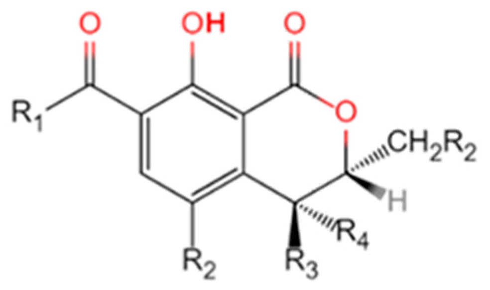
Figure 1. The molecular structure of naturally occurring OTs.
Water 16 03620 g001
Figure 2. Hypothetical OTA Biosynthetic Pathway [50].
Water 16 03620 g002
Figure 3. Proposed OTA biosynthetic gene cluster.
Water 16 03620 g003
Figure 4. OTA biosynthetic gene clusters in different fungi (redraw after [50]).
Water 16 03620 g004
Figure 5. The biodegradation mechanisms of OTA.
Water 16 03620 g005
Table 1. The molecular structure of naturally occurring OTs.
NameR1R2R3R4R5
OTαOHClHHH
OTβOHHHHH
OTAPhenylalanyl groupClHHH
OTBPhenylalanyl groupHHHH
OTCPhenylalanyl ethyl esterClHHH
4R-OH-OTAPhenylalanyl groupClHOHH
4S-OH-OTAPhenylalanyl groupClOHHH
4R-OH-OTBPhenylalanyl groupHHOHH
4S-OH-OTBPhenylalanyl groupHOHHH
10-OH-OTAPhenylalanyl groupClHHOH
Table 2. Comparison of characteristics of various OTA-DEs.
SourceGenBank IDMolecular Weight (kDa)Expression VectorOptimal pHOptimal
Temperature (°C)
Ref.
Aspergillus niger UVK143An14 g0208047A. niger AP4 strain/pGAPT5.6–6.066[133]
Stenotrophomonas acidaminiphila CW117H7691_1293545.6BL21/pGEX-4T-18.040–50[93]
Acinetobacter sp. neg1PJ15_154044.15BL21-CodonPlus(DE3)-
RIL/pET-28a
NRNR[135]
Aspergillus niger M00988KJ85492051.1BL21/pET-28aNRNR[136]
Bacillus amyloliquefaciens ASAG1KP16149348.6Rosetta/pEBNRNR[137]
Bacillus subtilis ANSB168DacA (Gene name)46Rosseta/pET-31b7.037[138]
Bacillus subtilis ANSB168DacB (Gene name)41Rosseta/pET-31b7.537[138]
Lysobacter sp. CW239H2514_0099543.32BL21/pET-32a7.037[134]
Bacillus subtilis CW14BCV50_1278548.62BL21/pET-28aNRNR[139]
Brevundimonas diminuta HAU429BT6 (Gene name)NRRosseta/pET-31b8.047–52[140]
Brevundimonas diminuta HAU429BT7 (Gene name)NRRosseta/pET-31b7.047[140]
Brevundimonas diminuta HAU429BT7 (Gene name)NRRosseta/pET-31b7.037[140]
Pseudoxanthomonas wuyuanensisWP_176520186356.43 (octamer)BL21/pET467.5–8.5 40–50[141]
Bacillus velezensis IS-6AAV34_RS1719014BL21/pET-28a7.0NR[142]
Acinetobacter pittii AP19BDGL_00190541BL21/pET-28aNRNR[143]
Brevundimonas naejangsanensis ML17gene009 (Gene name)79.7BL21/pET-28aNRNR[144]
Brevundimonas naejangsanensis ML17gene1826 (Gene name)58.4BL21/pET-28aNRNR[144]
Brevundimonas naejangsanensis ML17gene2253 (Gene name)67.7BL21/pET-28aNRNR[144]
Brevundimonas naejangsanensis ML17gene0484 (Gene name)45.8BL21/pET-28aNRNR[144]
Alcaligenes faecalis DSM 16305OSZ3702547.0BL21/pET-28a6.550[145]
Notes: NR = not reported.
Disclaimer/Publisher’s Note: The statements, opinions and data contained in all publications are solely those of the individual author(s) and contributor(s) and not of MDPI and/or the editor(s). MDPI and/or the editor(s) disclaim responsibility for any injury to people or property resulting from any ideas, methods, instructions or products referred to in the content.