Article

Biogeochemical Fe-Redox Cycling in Oligotrophic Deep-Sea Sediment

1
Beijing Research Institute of Chemical Engineering and Metallurgy, China National Nuclear Corporation, Beijing 101149, China
2
School of Historical Culture and Tourism, Fuyang Normal University, Fuyang 236037, China
3
State Key Laboratory of Biogeology and Environmental Geology, China University of Geosciences, Beijing 100083, China
4
UNSW Water Research Centre, School of Civil and Environmental Engineering, The University of New South Wales, Sydney, NSW 2052, Australia
*
Author to whom correspondence should be addressed.
Water 2024, 16(19), 2740; https://doi.org/10.3390/w16192740
Submission received: 16 August 2024 / Revised: 18 September 2024 / Accepted: 24 September 2024 / Published: 26 September 2024
(This article belongs to the Special Issue Soil and Groundwater Quality and Resources Assessment)

Abstract

:
Biogeochemical redox cycling of iron (Fe) essentially governs various geochemical processes in nature. However, the mechanistic underpinnings of Fe-redox cycling in deep-sea sediments remain poorly understood, due to the limited access to the deep-sea environment. Here, abyssal sediment collected from a depth of 5800 m in the Pacific Ocean was characterized for its elemental, mineralogical, and biological properties. The sedimentary environment was determined to be oligotrophic with limited nutrition, yet contained a considerable amount of trace elements. Fe-redox reactions in sediment progressed through an initial lag phase, followed by a fast Fe(II) reduction and an extended period of Fe(III) oxidation before achieving equilibrium after 58 days. The presence of an external H2 electron donor significantly increased the extent of Fe(III) bio-reduction by 7.73% relative to an amendment-free control under high pressure of 58 MPa. A similar enhancement of 11.20% was observed following lactate amendment under atmospheric pressure. Fe(II) bio-oxidation occurred after 16 days’ anaerobic culturing, coupled with nitrate reduction. During Fe bio-redox reactions, microbial community composition was significantly shaped by the presence/absence of an electron donor, while the hydrostatic pressure levels were the controlling factor. Shewanella spp. emerged as the primary Fe(III)-reducing microorganisms, and were stimulated by supplemented lactate. Marinobacter hydrocarbonoclasticus was the predominant Fe(II)-oxidizing microorganism across all conditions. Our findings illustrate continuous Fe-redox reactions occurring in the deep-sea environment, with coexisting Fe-redox microorganisms determining the oscillation of Fe valence states within the abyssal sediment.

1. Introduction

Iron (Fe) is the most abundant redox-active metal element in the Earth’s crust. The biogeochemical redox cycle of Fe plays a critical role in various environmental processes, including the cycling of essential elements, carbon storage, transformation of contaminants, and ocean productivity [1,2,3]. Fe-redox cycling in the epigenetic natural environment has been widely investigated over the last few decades. Iron cycles between its reduced (Fe2+) and oxidized (Fe3+) states, driven by both biotic and abiotic reactions. In marine and terrestrial ecosystems, microorganisms mediate these redox processes, influencing nutrient availability and the fate of toxic metals [4]. Biologically, Fe is physiologically essential for nearly all living organisms. Fe(II) supplies the requisite chemical energy for metal-oxidizing microorganisms, such as Acidithiobacillus or Leptospirillum, whereas Fe(III) functions as the electron acceptor for dissimilatory reduction in anaerobic environments [5,6].
Due to the difficulty in accessing deep-sea environments, little was known about Fe bio-redox reactions in deep-sea sediments until recently. Significant progress has been made in understanding these environments, including the ultradeep sea, due to the advancements in deep-sea exploration technology. However, much remains to be discovered [7,8,9]. Recent advances have demonstrated that Fe bio-redox cycling is active and prevalent in the anaerobic environments of deep sea, especially in oxygen minimum zones, coupled with organic carbon mineralization [10,11,12]. Despite the harsh environment of deep sea, characterized by the absence of light, extremely high hydrostatic pressure, low temperature, limited amount of organic matter, and low dissolved oxygen, a variety of microorganisms have been identified in abyssal sediments, dominated by bacteria and archaea [13,14], which significantly contribute to Fe-redox processes. Some species of Shewanella and Geobacter are known to transfer electrons from organics to Fe(III) through direct physical contact [15,16] or via nanowires [17,18]. Fe-oxidizing bacteria are also extensively distributed in deep-sea environments, such as Marinobacter hydrocarbonoclasticus, with dissolved oxygen or nitrate as electron acceptors [19]. In particular, nitrate concentration in the deep sea (16–43 µmol/kg) is generically higher than that at the epigenetic level (0–30 µmol/kg), making nitrate a dominant electron acceptor [20]. Genuine deep-sea sedimentary environments are generally complex and characterized by the coexistence of Fe(II), Fe(III), oxygen, and nitrate across diverse spatial and temporal scales [13]. Nitrate-reducing Fe(II)-oxidizing bacteria may have a symbiotic relationship with dissimilatory Fe(III)-reducing bacteria, facilitating Fe(III) oxide migration and Fe(II) regeneration [21]. However, the specific electron transfer mechanisms between Fe(III) and electron donors in abyssal sediment remain poorly understood. In particular, the long-term kinetics of Fe(III) reduction and Fe(II) oxidation in deep-sea sediments have not been thoroughly explored. Additionally, the impact of hydrostatic pressure on Fe-redox cycling and the corresponding microbial composition has not been sufficiently investigated.
The objective of this study was therefore to examine the biogeochemical cycling of Fe in the deep-sea environment. Abyssal sediments collected from a depth of 5800 m in the Pacific Ocean were characterized for elemental, mineralogical, and microbial compositions. The relationships between microorganisms, hydrostatic pressure. and electron donors/acceptors were statistically analyzed. The microbial functions related to Fe-redox properties were predicted based on FAPROTAX or BugBase, established databases. Our results provide novel insights into the Fe-redox reactions in nutrient-deficient abyssal environments, which are closely related to the global biogeochemical cycling of contaminants and essential elements.

2. Materials and Methods

2.1. Sample Collection

Sediment samples were collected from depths of 5800 m at 21.909° N, 130.141° E in the Pacific Ocean. A 30 cm-long undisturbed core was obtained using a box-type collector. To ensure sample freshness during repeated experiments, the sediments were divided into several equal parts and placed in airtight sterilized polyethylene containers. The samples were stored at 4 °C before use.

2.2. Bio-Redox Reactions of Fe in Abyssal Sediment

The artificial seawater medium was prepared as follows: 0.4 M NaCl, 27.6 mM Na2SO4, 7.65 mM NH4Cl, 24.5 mM MgCl2·6H2O, 10 mM CaCl2, 2.29 mM NaHCO3, 8.91 mM KCl, 50 μM KBr, 0.32 mM H3BO3, 0.15 mM SrCl2, 71.4 μM NaF, 17.91 μM FeSO4·7H2O, 7.72 μM ZnSO4·5H2O, 9.15 μM MnCl2·4H2O, 1.61 μM Na2MoO4·2H2O, 0.17 μM Co(NO3)2·6H2O, 0.32 μM CuSO4·5H2O, 26.86 μM Na2EDTA·2H2O, 40.93 nM D-biotin, 7.38 nM cobalamin, 296.50 nM thiamine, and 1 L ddH2O.
The properties of abyssal sediment and its microbial composition can change due to a lack of pressure-holding technology. Hence, Fe reduction experiments were conducted under atmospheric pressure (as a mechanistic comparison to high hydrostatic pressure) or 58 MPa in the presence or absence of an external electron donor (H2 or lactate), labeled as follows: H-Original (no external electron donor under high pressure), H-H2, H-Lactate, A-Original (no external electron donor under atmospheric pressure), A-H2, and A-Lactate. For the atmospheric experiments (A-), 100 mL of artificial seawater medium was added to a 200 mL glass container. To ensure anaerobic conditions, nitrogen or hydrogen was used to replace the air and the media were autoclaved at 121 °C for 20 min. After sterilization, a stock solution containing 0.5 g of abyssal sediments was added. Oxygen-free lactate was filtered into the medium through a sterilized filter (0.22 µm). Lactate (5 mM) or hydrogen (headspace) served as electron donors. For high-pressure experiments (H-), the systems were set up in custom-made high-strength polyethylene bags and placed in a high-pressure vessel. The cultures were maintained at 20 °C in the dark. Before use, all glassware was washed with neutral detergent and soaked in 1 M HNO3 overnight. All cultures were prepared in duplicate due to the excellent reproducibility observed in preliminary triplicate experiments. Fe-redox experiments were performed more than 3 times each, exhibiting excellent reproducibility. Total Fe(II) and total Fe were determined at days 0, 5, 16, 20, 39, and 58.
The preparation for the Fe oxidation experiment was similar to that of the reduction experiment, with the exception that the hydrostatic pressure was a uniform 0.1 Mpa and FeCl2 (1 mM) and NaNO3 (1 mM) were used as the electron donor and acceptor, respectively. After autoclaving, an FeCl2 stock solution was filtered through a sterilized filter (0.22 µm) into the medium. Abiotic controls were prepared to verify that Fe oxidation was driven by microbial activity. Total Fe(II) and total Fe were measured at days 0, 1, 3, 5, 7, 10, 17, 28, and 45. All cultures were prepared in duplicate.

2.3. Bioinformatic Analysis

2.3.1. DNA Extraction, 16S rRNA Gene Amplification, and High-Throughput Sequencing

DNAs were extracted from 0.25 g of sediment using the MoBio Power Soil DNA isolation kit (MoBio Laboratories, Carlsbad, CA, USA) according to the manufacture’s protocol. The V4 hypervariable region of the prokaryotic 16S rRNA genes for both bacteria and archaea was amplified using the primer set 515F (5′-CAGCMGCCGCGGTAA-3′) and 806R (5′-GGACTACHVGGGTWTCTAAT-3′) with unique barcode sequences at both 5′ ends. PCR amplification was carried out in a 50 μL reaction system containing 5 μL 10× PCR buffer, 1.5 μL dNTP mixture (10 mM for each), 1.5 μL forward and reverse primers (10 μM), 0.5 μL Taq DNA Enzyme (TaKaRa), 2 μL of template DNA (5–30 ng), 1 μL of BSA, and 37 μL ddH2O. The PCR program was as follows: 94 °C for 1 min, 30 cycles at 94 °C for 20 seconds, 57 °C for 25 seconds, and 68 °C for 45 seconds, and a final extension at 68 °C for 10 min. PCR products were purified by gel electrophoresis and quantified with a Qubit fluorimeter (Invitrogen, Carlsbad, CA). Equal molar amounts of DNA were pooled for library construction and sequencing on the Illumina Hiseq platform at Magigene Biotechnology Co., Ltd. (Guangzhou, China).

2.3.2. Data Processing and Statistical Analysis

The 16S rRNA gene sequences were analyzed using tools available on the BMKCloud platform (www.biocloud.net, accessed on 5 June 2024). The raw sequences were demultiplexed by barcode identification, with the maximum barcode error rate set at 1.5. Quality filtering of the raw data was performed using Trimmomatic (version 0.33) [22], with a sliding window size of 50 bp. Sequences were trimmed if the quality within the window fell below 20. Cutadapt (version 1.9.1) [23] was used to identify and remove primer sequences, with a maximum mismatch rate of 20% and a minimum coverage of 80%. USEARCH (version 10.0) [24] was employed to concatenate paired-end reads, and UCHIME (version 8.1) [25] was used to remove chimeras, resulting in high-quality sequences for further analysis. For USEARCH, the minimum overlap length was set to 10 bps, with a required minimum similarity of 90% and a maximum of 5 mismatched bases. High-quality reads were used to generate operational taxonomic units (OTUs) at a 97% similarity level using UPARSE [26]. Taxonomic assignment for representative sequences was performed based on the Silva database (release 138).
The rarefaction, Shannon, and rank abundance curves were analyzed using QIIME2 [27]. The data of 6 groups were processed through principal component analysis (PCA) using R, without further grouping. To create a heatmap, the 80 most abundant genera were extracted and visualized using R. The values for the heatmap were normalized with the R scale function to compute Z-scores across different samples of the same species. Redundancy analysis (RDA) or canonical correspondence analysis (CCA) was chosen based on the decision curve analysis (DCA) results of species sample abundance, depending on the length of the first axis of the DCA. CCA was selected if this length exceeded 4.0; otherwise, RDA was used. RDA or CCA was performed using the Vegan (version 2.3) package of R. The functional composition was predicted using FAPROTAX [28], based on the evidence from published and cultured microorganisms. FAPROTAX annotation can greatly enhance understanding of biogeochemical cycles in environmental samples. The microbial phenotypes of each group were predicted using BugBase [29], and the results were normalized based on the predicted 16S copy numbers. For each sample in the biological dataset, the relative abundance of traits was estimated across a range of coverage thresholds (0 to 1, in increments of 0.01). The coverage threshold was determined with the highest variance among all samples for each feature, and the final organism level trait prediction table was generated, which included the relative abundance of predicted traits for each sample. The predicted phenotypes were aerobic, anaerobic, facultative anaerobic, Gram-positive, Gram-negative, biofilm forming, mobile element containing, oxygen utilization, and oxidative stress tolerance.

2.3.3. Data Submission and Accession Numbers

The full-length 16S rRNA gene sequences were deposited in the NCBI database and can be accessed under project ID PRJNA1141258.

2.4. Analytical Methods

2.4.1. Measurements of Total Fe(II), Total Fe and Nitrate

At preselected time points during Fe-redox reactions, a 0.2 mL sediment suspension was withdrawn from the experimental bottle or bag using a sterile syringe. As described earlier [30], the sediment suspension was mixed with 0.48 mL of 3.6 N H2SO4 in a dark tube, then 40 μL of 48% HF and 80 μL of 10% (w/v) 1,10-phenanthroline were added. The tubes were then heated to 100 °C and boiled for 30 min. After cooling, 0.4 mL of 5% (w/v) boric acid was added to the mixture. Following this, 0.1 mL of the sample was combined with 1 mL of 1% (w/v) Na citrate to develop color for 20 min. A UV-vis spectrophotometer (Thermo Fisher, Waltham, MA, USA) was utilized to measure the absorbance at 510 nm. To determine the concentration of total Fe, including both Fe(III) and Fe(II), a 0.1 mL sample was added to a 1 mL mixture of Na citrate (1% w/v) and hydroxylammonium chloride (10% w/v). All Fe(III) was reduced to Fe(II), and then the concentration of Fe(II) was measured again. Additionally, at least 1 mL sediment suspension was filtrated through a 0.2 μm nylon filter, and the nitrate concentrations of the filtrate were measured using ion chromatography (Thermo Fisher ICS 600) with an electrical conductivity detector.

2.4.2. X-ray Diffraction Analysis

Abyssal sediment samples were freeze-dried and crushed to powder below 300 mesh using an agate mortar. X-ray diffraction (XRD) pattern was obtained following a previously described protocol [31]. Briefly, an X-ray powder diffractometer (Rigaku Smart Lab X, Tokyo, Japan) was used, equipped with a Cu Kα radiation source, a rotating-anode generator, and power of 9000 W (200 kV, 45 mA). The samples were scanned from 3° to 70° (2θ) in steps of 0.02° with 1 second per step. The mineralogical analysis was performed using MDI Jade software (Version 6.5).

2.4.3. Scanning Electron Microscopy Analysis

Scanning electron microscopy (SEM) was performed to examine mineral–microbe associations at the end of bio-reduction experiments. Samples were prepared following procedures described in our previous work [32]. Briefly, an aliquot of the sediment suspension was withdrawn from the experimental serum bottle, smeared onto a glass coverslip, and fixed using a mixture of 2.5% glutaraldehyde and 2% paraformaldehyde in phosphate-buffered saline (PBS). After fixation, gradient dehydration was performed with various proportions of ethanol and water. Afterwards, the samples were dried with a critical point dryer (CPD, Quorum K850) and Pt-coated with a Quorum SC7620 sputter coater. SEM (Zeiss Supra 55 SAPPHIRE) was used for morphological observation.

2.4.4. Major and Trace Element Analysis

Carbon (C), hydrogen (H), nitrogen (N), and sulfur (S) content were analyzed on decarbonated samples using an element analyzer (Vario Macro Cube, Elementar, Langenselbold, Germany). Other elements were determined at the Analytical Laboratory of Beijing Research Institute of Uranium Geology, China National Nuclear Corporation. For major elements (Si, Al, Fe, Mg, Ca, Na, K, Mn, Ti, P), samples were ground to 200 mesh and oven-dried overnight at 105 °C. Each sample of 2.0 g was weighed, ignited at 1100 °C in a platinum crucible, and reweighed to determine the loss on ignition (LOI). A 0.5 g (weight before ignition) ignited sample was mixed with 4.5 g Li2B4O7–LiBO2–LiF (4.5:1:0.4, wt%), fused in a platinum crucible at 1100 °C, and then pressed to create a flattened disk. The pressed discs were analyzed using an X-ray fluorescence spectrometer (XRF, PANalytical, Almelo, the Netherlands) at 25 kV with 144 mA emission current. Calibrations of accuracy and reproducibility were conducted using the GB/T 14506.14-2010 and GB/T 14506.28-2010 standards, with a standard deviation consistently below 5%. For trace element analysis, samples were crushed in an agate mortar and digested with HNO3 and HF to obtain the sample solution. Trace element concentrations were determined using ICP mass spectrometry (NEXION 300D, PerkinElmer, Shelton, CT, USA) coupled with an ESI SC-2 DX4 auto-sampler (Elemental Scientific, Omaha, NE, USA). Samples were diluted (1:20 v:v) with a diluent consisting of 0.05% Triton X-100 and 1% HNO3 in 18.2 MΩ·cm distilled deionized water. Calibrations for accuracy and reproducibility were conducted using the GB/T 14506.30-2010 standard, with a standard deviation consistently below 5%.

3. Results and Discussion

3.1. Characterization of the Abyssal Sediment

The sedimentary environment at 5800 m depth was carbon-deficient (0.22%, Table 1), significantly lower than the average carbon content (0.66%) in deep-sea sediments [33]. The C/N value in the sediment was slightly above 4, indicating the presence of chemoautotrophic microbial communities [34].
Substantial amounts of redox-active metals (e.g., Fe and Mn) were observed (Table 1), suggesting that the redox reactions may be prevalent at this depth. The high content of SiO2, Al2O3, and Fe2O3 indicated the argillaceous features of abyssal sediment, which was confirmed by XRD analysis (Figure 1), showing the dominant mineralogical composition of quartz, muscovite, albite, and lizardite.
A variety of trace elements were detected in the abyssal sediment (Table 1), which were closely associated with the co-present minerals. Clay minerals of muscovite can adsorb trace elements through ion exchange [35,36,37], while Fe–Mn (hydr)oxides also play a role in scavenging trace elements [38,39]. Active Fe-redox regions in deep-sea sediment can accumulate various trace elements, which may serve as active centers for coenzymes and catalyze methane production and nitrogen fixation [40,41,42].

3.2. Fluctuation of Fe-Redox State in the Abyssal Sediment

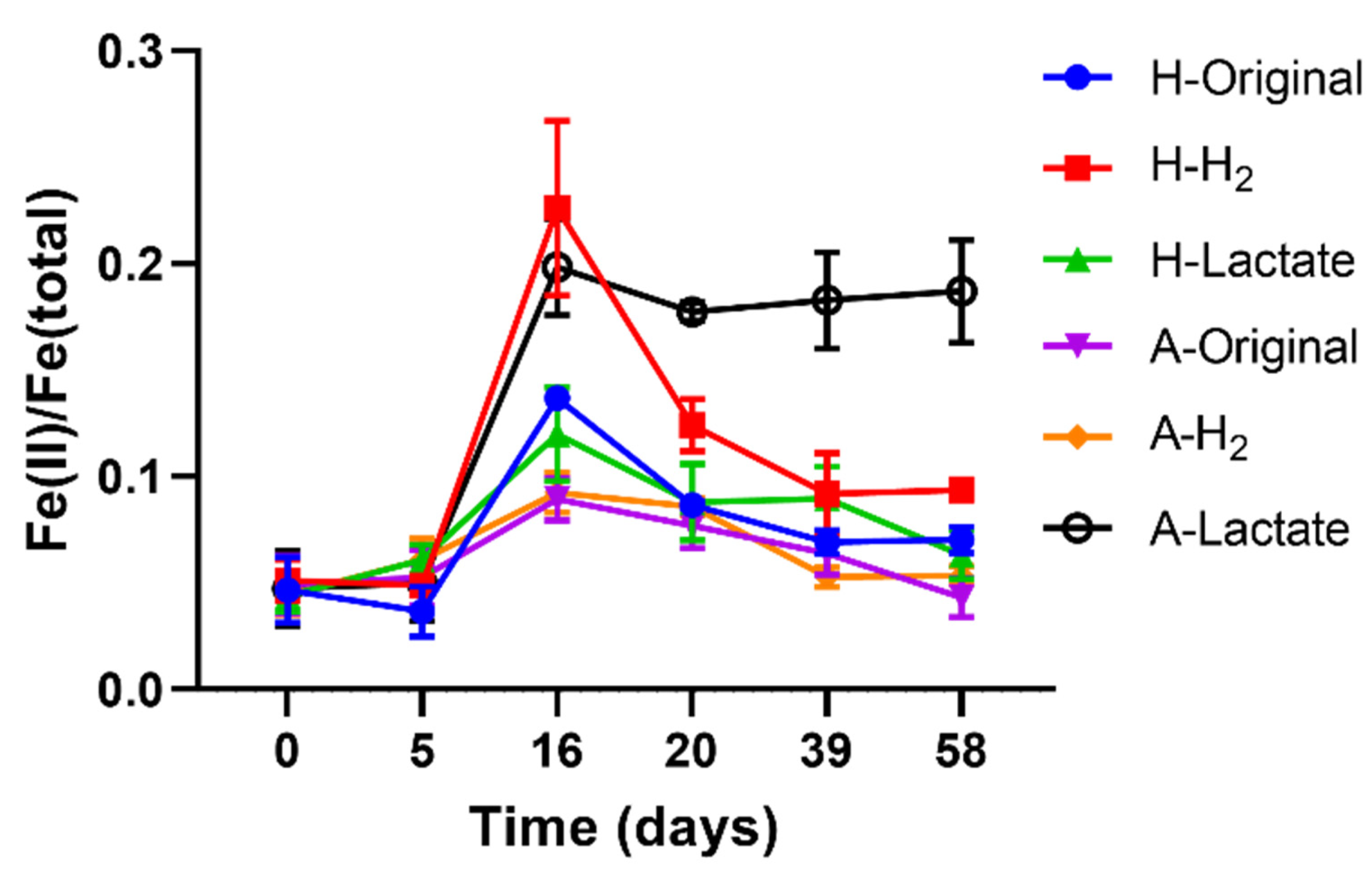
Due to the presence of dissolved oxygen, the majority of Fe in shallow abyssal sediment existed as Fe(III) (t = 0 d, Figure 2). Bio-induced oscillation of Fe-redox states in the abyssal sediment was observed over 58 days’ anaerobic culturing, regardless of the presence/absence of an external electron donor and environmental pressures (Figure 2).
During the initial 5 days, bio-redox activity was low and Fe(II) concentration remained unchanged. From day 5 to 16, Fe reduction increased sharply. Under high pressure of 58 MPa (H-), Fe(III) reduction in the H2-amended group (H-H2) reached 17.74%, which was 11.84% and 7.73% higher than that in the lactate-amended group (5.9%, H-Lactate) and amendment-free original group (10.01%, H-Original), respectively. Under atmospheric pressure (A-), Fe(III) reduction in the lactate group (A-Lactate) reached 14.86%, 11.69%, and 11.2% higher than that in the H2-amended group (3.17%, A-H2) and original group (3.66%, A-Original).
These findings indicate a shift in the Fe-redox equilibrium toward Fe reduction during the first 16 days, which was kinetically affected by the presence or absence of external electron donors and environmental pressures. Even in the absence of external electron donors, biogenic Fe(II) increased from 3.67% to 13.68% (high pressure) and from 5.27% to 8.93% (atmospheric pressure), suggesting a spontaneous Fe reduction using in situ electron donors. However, external electron donors further accelerated the reduction processes (Figure 2).
Environmental pressures also had an impact on the Fe redox. Under atmospheric pressure, lactate significantly promoted Fe(III) reduction from 8.93% (A-Original) to 19.84% (A-Lactate), but high pressure diminished this effect (Figure 2). Previous studies have similarly reported that the Fe reduction efficiency of Shewanella spp. was inhibited by high hydrostatic pressure due to the lack of piezotolerance [43]. Our observations suggest that high pressure universally inhibits microbial growth, thereby suppressing Fe(III) reduction. In contrast to lactate, H2 as an electron donor significantly enhanced Fe(III) reduction under high pressure (58 Mpa), increasing from 13.68% (H-Original) to 22.62% (H-H2), while showing negligible effects under atmospheric pressure. This enhancement likely resulted from piezotolerant or piezophilic Fe-reducing bacteria utilizing H2 as an electron donor [44]. This discovery aligns with the observed serpentinization in the 5800 m sample (Figure 1), wherein reactions between ultrabasic rock and water generate H2, which acts as a bioenergy source [45].
All groups reached Fe(II) concentration peaks at day 16 and started an extended period of gradual Fe oxidation before achieving a redox equilibrium after 58 days, except for A-Lactate, in which Fe(II) concentration stabilized between 17.75% and 18.71%, possibly due to a sufficient supply of effective electron donors (Figure 2). Despite measurable Fe-redox reactions, most sediment-bound Fe remained inactive (Figure 2).
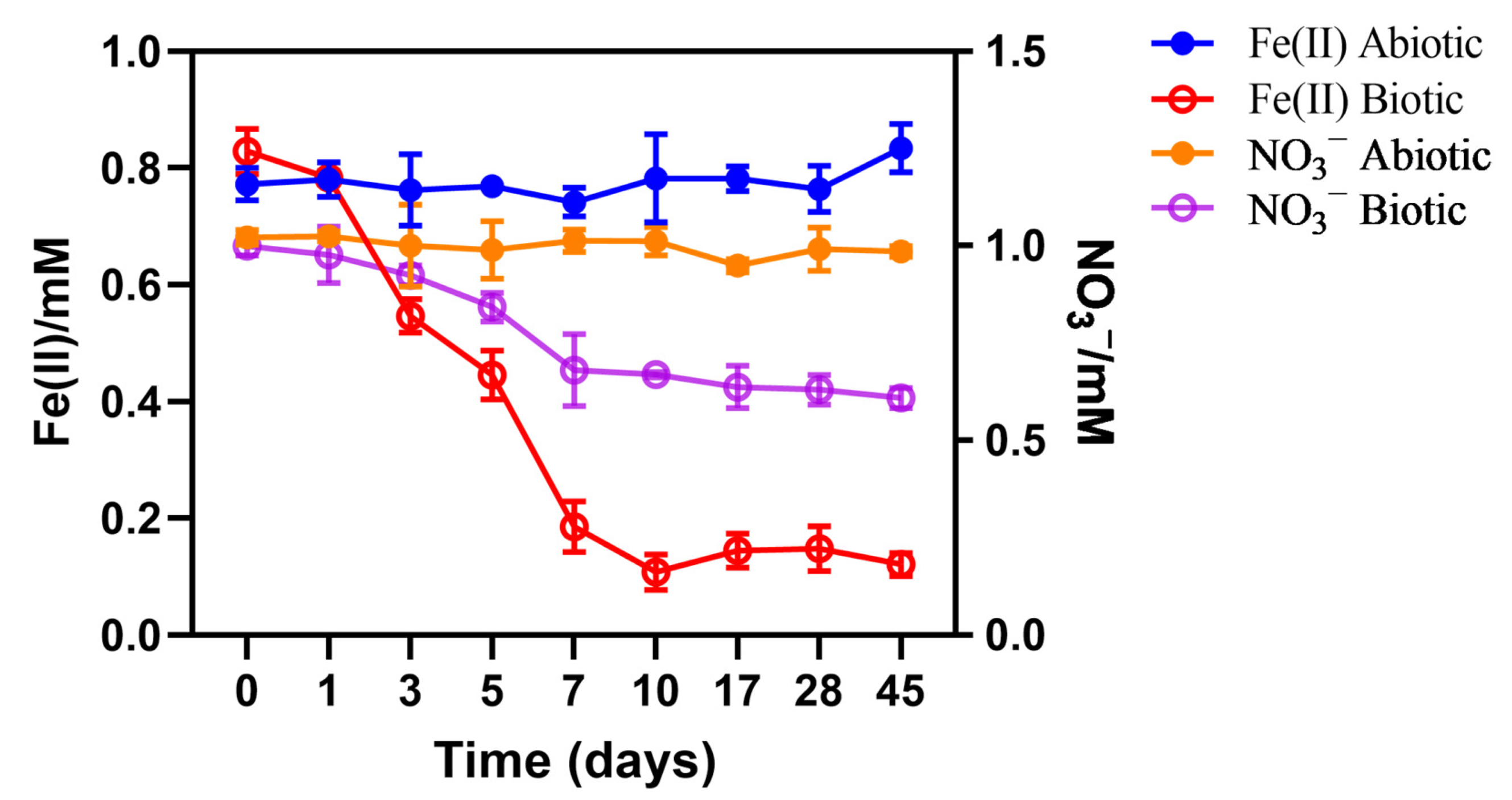
The validity of the Fe bio-oxidation process was confirmed through an independent Fe oxidation experiment supplemented with nitrate as the electron acceptor (Figure 3). Fe(II) decreased significantly from 0.829 mM to 0.108 mM within a 10-day period, coupled with a decrease in nitrate from 1.0 mM to 0.669 mM. The abiotic control maintained stable Fe(II) and nitrate concentrations, suggesting the absence of chemical oxidation of Fe(II) by nitrate under these conditions (Figure 3).
Oxidation of Fe(II) by nitrate or other oxidants is a frequently observed phenomenon in redox transition areas. The Fe(III) involved in dissimilatory reduction is typically generated through the microbial oxidation of Fe(II). This process is particularly prevalent in abyssal zones where organic carbon is limited, facilitating the growth of autotrophic organisms that rely on Fe(II) as an energy source and nitrate as an electron acceptor [46].

3.3. Pressure Controlling the Microbial Community Composition in Abyssal Sediment

A total of 525 operational taxonomic units (OTUs) was recognized within the six treated samples, with A-Original exhibiting the greatest OTU count (Figure 4a). Rarefaction and Shannon curves validated the sufficiency and dependability of the sampling and sequencing process (Figure S1). The introduction of external H2 or lactate did not influence the diversity of the anaerobic system, as the OTU counts remained comparable to the original groups. Elevated pressure significantly reduced the microbial diversity. For instance, samples subjected to 0.1 MPa displayed a microbial diversity of over 150 OTUs, in contrast to samples under 58 MPa, which exhibited fewer than 78 OTUs (Figure 4a and Figure S2). Despite the cumulative OTUs surpassing 500 across the samples, only 12 OTUs were shared (Figure 4b). This pattern was similarly evident at the genus level (Figure S3). These findings indicate that the addition of electron donors and the variations in pressure conditions significantly impacted the microbial compositions.
Principal component analysis (PCA, Figure 4c) and a clustering heatmap (Figure 4d) confirmed that environmental pressure primarily shaped the microbial community structures. PC1 explained 89% of the overall data variance, while PC2 accounted for 7.7%. The distinct separation of the three groups under atmospheric pressure and the three groups under 58 MPa along PC1 indicates a notable similarity within groups sharing the same pressure levels.
Consistent with previous observations [47,48], Proteobacteria, especially Gammaproteobacteria and Alphaproteobacteria, were the most prevalent bacteria in the abyssal sediment cultures (Figure 4e). Proteobacteria demonstrate extensive phylogenetic diversity within the Bacteria domain, enabling numerous species to adapt and thrive in distinct ecological niches within the deep-sea sediment environment [49]. These Proteobacteria found in deep-sea sediments are commonly characterized as psychrophilic and chemolithoautotrophic, displaying a preference for cold temperatures, low nutrient availability, and high pressure typical of abyssal sediment environments [50]. Proteobacteria have been discovered thriving in deep-sea hydrothermal fields, showcasing their remarkable diversity and ability to thrive in extreme environments [51]. Their diverse metabolic capabilities enable Proteobacteria to harness various energy sources, including the oxidation of iron and sulfur compounds abundant in deep-sea environments [52]. In comparison to other bacterial phyla, Proteobacteria have a higher representation in high-pressure environments, indicating that non-piezotolerant organisms are inhibited by high pressure, paving the way for piezotolerant Proteobacteria to flourish.
Bacteroidetes, a phylum with very low abundance across the treated sedimentary groups, revealed an interesting anomaly within the lactate group under 58 MPa (H-Lactate). In this unique case, Bacteroidetes accounted for a notable 5.72%. Further analysis unveiled that 98.6% of these Bacteroidetes were identified as Tenacibaculum, suggesting its piezophilic nature and preference for lactate as a carbon source. Notably, while Tenacibaculum’s metabolic activities indicate an affinity for lactate [53], its involvement in the oxidation or reduction of Fe remains unreported.
At the genus level, pressure was also the controlling factor for microbial community compositions. The microbial communities exhibited relatively similar characteristics across the three samples subjected to the same pressure conditions (Figure 4f), consistent with the findings of PCA and clustering analyses. Notably, Alcanivorax and Halomonas stood out as the predominant genera in the three high-pressure groups, with Altereythrobacter, Pseudoalteromonas, and Marinobacter also being prevalent. Conversely, under atmospheric conditions, Marinobacter and Pseudoalteromonas were the dominant genera, while the proportion of Alcanivorax was considerably lower.
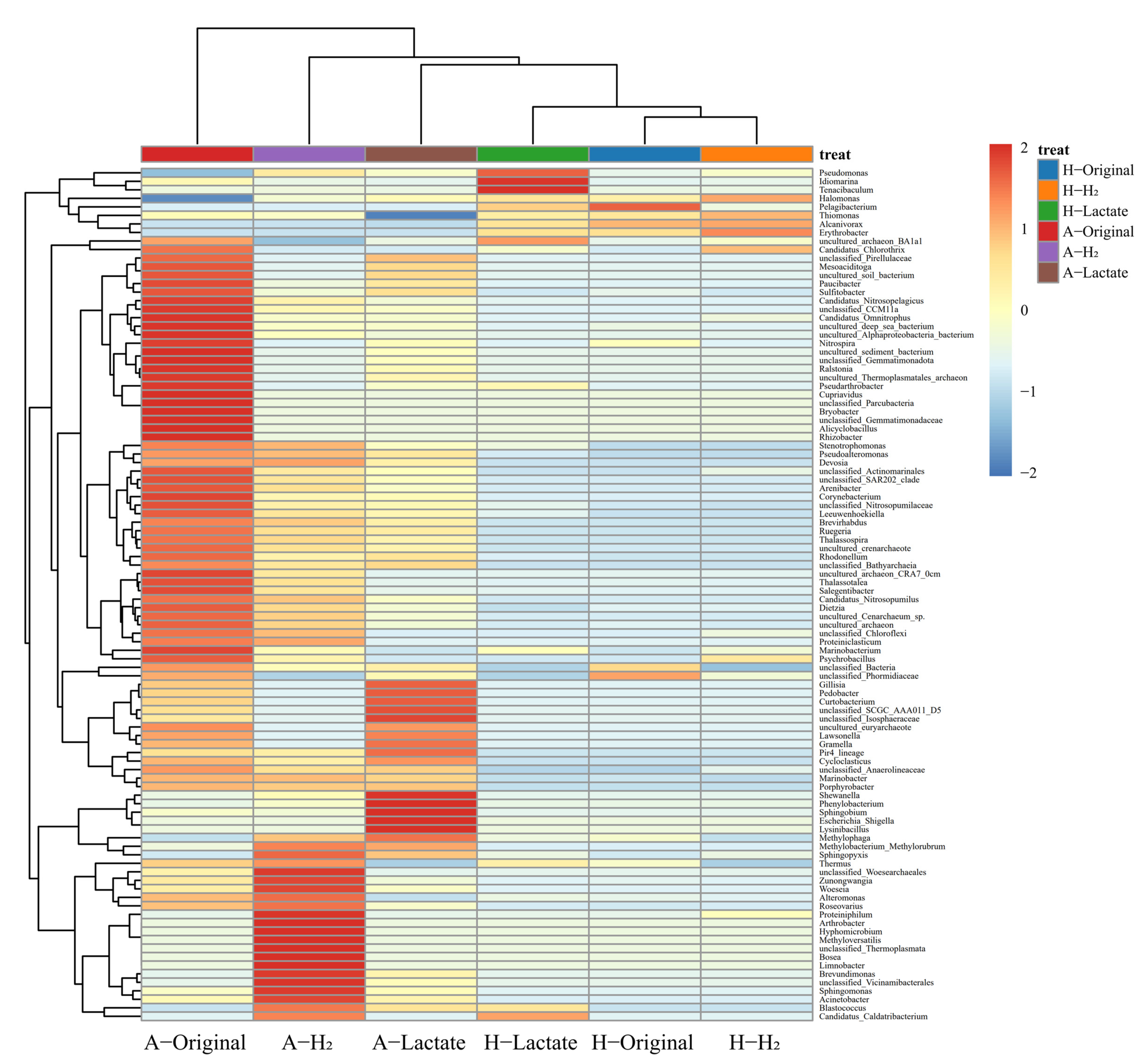
The heatmap depicts the distribution of the primary genera among the six treated abyssal sediment samples (Figure 5), revealing two distinct microbial compositions based on varying levels of pressure. Under high pressure, H2 or lactate had minimal impact on the microbial composition, except for Pseudomonas, Idiomarina, and Tenacibaculum, which displayed a significant positive relationship with lactate. Conversely, under atmospheric pressure, lactate and H2 selectively enriched the presence of specific genera that were not originally abundant within the group. Genera such as Shewanella, Phenylobacterium, Sphingobium, Escherichia_Shigella, and Lysinibacillus had a strong positive correlation with lactate, while Thermus, unclassified Woesearchaeales, Zunongwangia, Woeseia, Alteromonas, Roseovarius, Proteiniphilum, Arthrobacter, Hyphomicrobium, Methyloversatilis, unclassified Thermoplasmata, Bosea, Limnobacter, Brevundimonas, unclassified Vicinamibacterales, Sphingomonas, Acinetobacter, Blastococcus, and Candidatus Caldatribacterium had a strong positive correlation with H2. Additionally, Methylophaga, Methylobacterium_Methylorubrum, and Sphingopyxia showed a strong positive correlation with both H2 and lactate.
The genera positively correlated with lactate were predominantly heterotrophic, utilizing various organic compounds like short-chain organic acids, ketones, aldehydes, or esters as carbon source [54,55,56,57,58,59,60,61]. Notably, Lysinibacillus and Shewanella were identified as potential participants in Fe(III) reduction [62,63,64]. On the other hand, the genera associated with H2 were more diverse, including Thermus, Alteromonas, and Sphingomonas as potential Fe-reducing microorganisms [65,66,67], while Methyloversatilis was recognized as a potential Fe-oxidizing microorganism [68]. These microorganisms rely on simpler carbon and energy sources such as hydrogen and formate. Additionally, certain unidentified species of Thermoplasmata might function as potential Fe-oxidizing archaea or utilize hydrogen for energy production [69,70].
However, under atmospheric pressure, the majority of genera appeared to be more negatively correlated with H2 or lactate (Table S1). Among the negatively enriched genera, the proportions of uncultured and unclassified species were high, as was the proportion of archaea, including uncultured archaeon_CRA7_0cm, Candidatus Nitrosopumilus, uncultured Cenarchaeum spp., uncultured euryarchaeote, Candidatus Nitrosopelagicus, and uncultured Thermoplasmatales archaeon. This suggests that the preferential enrichment of H2 or lactate impaired symbiosis and microbial diversity in abyssal sediments, leading to a decrease in extremophilic archaea populations.

3.4. The Dynamics between Microbial Community and Fe-Redox Fluctuation

Except for Lysinibacillus, Thermus, Alteromonas, and Sphingomonas, which exhibited a positive correlation with the supplied H2 or lactate, Shewanella, Thalassospira, and Thermodesulfovibrio have been identified as potential iron-reducing bacteria (Table 2) [71,72]. Thalassospira demonstrated enhanced growth under atmospheric conditions compared to high-pressure environments, while the proportion of Thermodesulfovibrio remained consistently low across all cultivation systems (Table 2).
It is worth noting that the proportion of Shewanella, a genus capable of dissimilatory Fe reduction [73], varied significantly across the six treated abyssal sediments, influenced by both pressure levels and the availability of electron donors. The genus Shewanella encompasses more than 70 species, a few of which demonstrate a notable tolerance to high-pressure conditions [74]. Notably, in the present investigation, Shewanella was represented by three species: Shewanella putrefaciens (<0.02%), Shewanella oneidensis (<0.01%), and an unclassified Shewanella sp. (Table 2). The piezotolerance of Shewanella putrefaciens and Shewanella oneidensis is evident, while the unclassified Shewanella sp. was more prevalent under 0.1 MPa (Table 2). In general, most Shewanella spp. demonstrate a preference for atmospheric conditions, with even piezotolerant strains such as Shewanella piezotolerans WP3 showing optimal growth under pressures below 20 MPa [74]. Additionally, the abundance of Shewanella was significantly correlated with the supply of electron donors.
Under atmospheric pressure, the amendment of lactate (A-Lactate) significantly increased the abundance of Shewanella putrefaciens, Shewanella oneidensis, and the unclassified Shewanella sp. compared to amendment-free original samples (A-Original) (Table 2), with notably elevated Fe(II) concentrations (A-Lactate: 19.84%) compared to the other experimental cohorts (A-Original: 8.93%, A-H2: 9.25%, Figure 2). The supply of H2 (A-H2) also slightly promoted the growth of the unclassified Shewanella sp. compared to the original samples (A-Original) (Table 2). In contrast, under high pressure (58 MPa), Shewanella putrefaciens and Shewanella oneidensis exhibited enhanced growth in the presence of H2 (H-H2), aligning with the observations of water–rock interactions in ultrabasic rocks (serpentinization), as evidenced in the 5800 m sample (Figure 1). The abundance of Shewanella spp. correlated closely with Fe(II) levels (Table 2; Figure 2).
Hydrogen-utilizing Fe(III)-reducing microorganisms were expected to be prevalent in the H-H2 group, coinciding with the peak concentration of Fe(II) observed during incubation (Figure 2). Contrary to expectations, microbial community analysis did not support this hypothesis, possibly due to a gradual decrease in the abundance of Fe(III)-reducing microorganisms during incubation (Figure 4f), while sampling was conducted at the end of incubation.
The four dominant Fe-oxidizing genera, including Methyloversatilis, which was positively correlated with H2 amendment, were identified in the six treated abyssal sediment samples (Table 2). Thiomonas preferred high-pressure environments, whereas Fe-oxidizing archaea Sulfolobus were minimally present across all cultivation conditions. Certain species of Marinobacter have the capacity to oxidize Fe(II), such as Marinobacter hydrocarbonoclasticus, which utilizes Dps proteins for the oxidation of Fe(II) as a detoxification mechanism under anaerobic or oxygen-limited growth conditions [19]. The abundance of Marinobacter hydrocarbonoclasticus was observed to be relatively lower under high pressure (0.918–1.292%), but increased significantly when exposed to atmospheric conditions (14.953–19.154%), indicating a potential contribution to the enhanced Fe oxidation observed in later stages (Figure 2; Table 2).
Therefore, the presence of diverse microbial communities, especially Fe-redox microorganisms, contributed to the fluctuation observed in Fe-redox processes, which were further influenced by a range of environmental factors such as electron donor availability and ambient pressure levels.
The relationship between environmental factors (pressure, H2, lactate) and microbial communities was examined via redundancy analysis (RDA), revealing a distinct separation of the six treated abyssal sediment groups across four quadrants (Figure 6a). Notably, the two lactate groups (A-Lactate, H-Lactate) were distinctively situated in separate quadrants, whereas the original group and the H2 group at the same pressure level were clustered together in another quadrant. This distribution pattern suggests that environmental pressure levels played a pivotal role in shaping the microbial composition with respect to lactate and H2, as further confirmed by the elongation along the RDA1 axis. Notably, certain genera, such as Alanivorax, Erythrobacter, Pelagibacterium, and Halomonas, displayed positive correlations with pressure, while Dietzia, Thalassospia, Pseudoalteromonas, and Marinobacter exhibited negative associations, indicating varying degrees of resilience to the ambient pressure levels. This relationship was further confirmed by the correlation heatmap (Figure S4), illustrating the positive correlation of Halomonas, Pelagibacterium, Thiomonas, Alcanivorax, and Erythrobacter with pressure, and conversely the negative correlation of Shewanella and Marinobacter with pressure (Figure S4).
The functional diversity (Figure 6b) observed was closely correlated with the microbial diversity (Figure 4f), with atmospheric consortia exhibiting more comprehensive functions compared to the high-pressure microbial community. Under high-pressure conditions, the supplement of exogenous electron donors did not exert a notable influence on the metabolic traits of the microbial communities. Conversely, in atmospheric pressure settings, the amendment of lactate distinctly influenced the prevalence of specific metabolic pathways (Figure 6b and Figure S5). On one hand, lactate was previously observed to accelerate nitrate reduction [75], which explained the observed enhancement of nitrogen respiration, nitrate respiration, and nitrate reduction in A-Lactate compared to A-Original well (Figure 6b). On the other hand, high levels of lactate were reported to impede Fe(II) oxidation and nitrate reduction processes [76], which in turn supports the observation that Fe(II) concentrations in A-Lactate remained elevated compared to other experimental groups, where a decline of Fe(II) concentration was noted after day 16 (Figure 2).
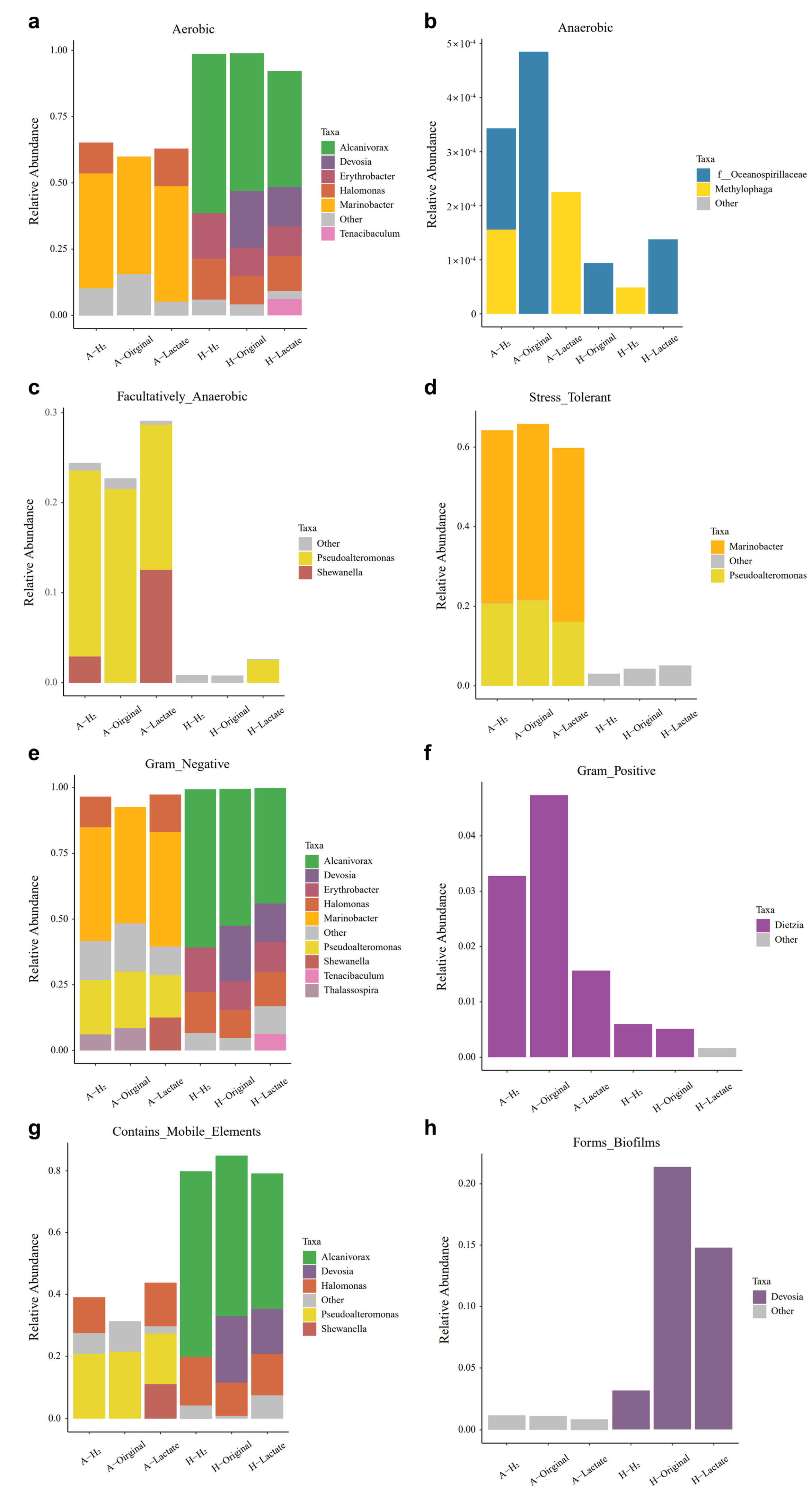
The analysis revealed a general prevalence of aerobic microorganisms across six treated abyssal sediment samples (Figure 7a). Facultatively anaerobic microorganisms were found to make up approximately 22.70–29.12% of the atmospheric groups (A-, Figure 7c). Anaerobic microorganisms exhibited a notably low relative abundance, accounting for less than 0.05%, predominantly observed within the atmospheric groups (A-, Figure 7b). These findings coincide with the established knowledge that contemporary oceanic and shallow abyssal sediments typically harbor oxygen [77], thus supporting the proliferation of aerobic microorganisms. The findings suggest that piezotolerant microorganisms tend to be aerobic, whereas those adapted to low-pressure settings are often anaerobic. Marinobacter and Pseudoalteromonas are recognized as genera capable of withstanding stress, making the three atmospheric groups stress-tolerant (Figure 7d). Notably, a significant proportion of microorganisms found in abyssal sediments were Gram-negative (Figure 7e). In contrast, Gram-positive microorganisms, predominantly represented by Dietzia, accounted for less than 5% of the total population and were primarily situated within the atmospheric groups (Figure 7f).
Ocean microorganisms often possess flagella, aiding their migration towards favorable environments [78]. Flagellated microorganisms were identified to make up 79.19–84.91% of high-pressure groups (H-), while only 31.30–43.75% was observed in atmospheric pressure groups (A-) (Figure 7g). Devosia, found in all three high-pressure groups, exhibited biofilm formation capability, a feature less common in microorganisms from atmospheric pressure groups (Figure 7h). Biofilms, composed of numerous microbial cells, play a vital role in facilitating symbiotic relationships among different microorganisms [79]. This symbiosis is essential for deep-sea microorganisms to thrive under extreme conditions and drive various biogeochemical processes.
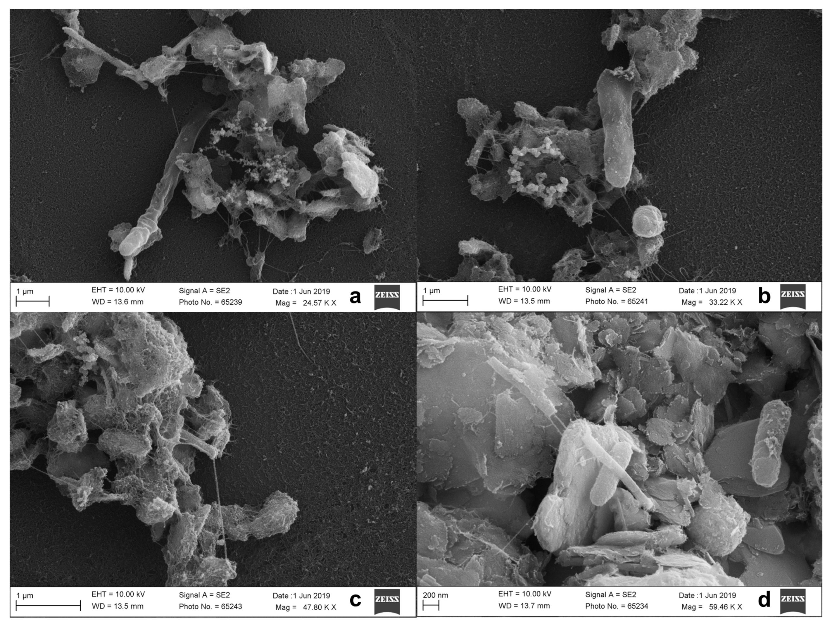
Abyssal sediments are composed of various Fe-redox microorganisms. Shewanella was identified as the predominant genus involved in dissimilatory Fe(III) reduction, constituting approximately 20.679% of the total biomass in the A-Lactate group. Scanning electron microscopy (SEM) images illustrated the direct interactions between microbial entities and phyllosilicate minerals within the abyssal sediments (Figure 8). Of particular interest were the microbial filaments that bridged cells and minerals, likely serving as microbial nanowires to facilitate extracellular electron transfer between the microbial community and iron-containing substances such as Fe(III) oxides. This intriguing phenomenon of microbial nanowires has been documented in the iron reduction mechanisms of both Geobacter spp. and Shewanella spp. [17,80].

4. Conclusions

The oligotrophic abyssal sediment experienced oscillations in Fe-redox states during anaerobic culturing, progressing through an initial lag phase, followed by a fast Fe reduction, and an extended period of gradual Fe oxidation before achieving equilibrium after 58 days. The supplement of external electron donors (H2 or lactate) significantly enhanced the extent of Fe(III) bio-reduction: (1) 7.73% by H2 under high pressure (58 MPa, H-H2) and (2) 11.20% by lactate under atmospheric pressure (A-Lactate). Fe(II) bio-oxidation occurred after 16 days’ anaerobic culturing, coupled with nitrate reduction. Bacteria were the dominant microorganisms in the abyssal sediment. Microbial community composition was heavily influenced by the presence or absence of an electron donor and the level of hydrostatic pressure, with pressure being the dominant factor. Fe(III) reducing and Fe(II) oxidizing were abundant in the abyssal sediment. Shewanella spp. were identified as the major Fe(III)-reducing microorganisms, particularly with lactate supplemented (A-Lactate). However, high pressure significantly lowered the proportion of Shewanella. Marinobacter hydrocarbonoclasticus was the dominant Fe(II)-oxidizing microorganism in all conditions.

Supplementary Materials

The following supporting information can be downloaded at: https://www.mdpi.com/article/10.3390/w16192740/s1, Figure S1: Rarefaction and Shannon curves; Figure S2: Rank abundance curves; Figure S3: Changes in the number of total and shared microbial communities at genera; Figure S4: Correlation heatmap; Figure S5: Comparison of metabolic functions; Table S1: Microbial community at genera negatively correlated to electron donor.

Author Contributions

Conceptualization, D.Z.; data curation, G.L.; formal analysis, X.L.; funding acquisition, Q.X.; investigation, D.Z., X.L. and Z.Z.; methodology, D.Z., G.L. and D.H.; resources, Q.X.; software, J.H.; supervision, Q.X.; validation, D.Z. and Q.X.; visualization, G.L., Y.L. and Y.S.; writing—original draft, D.Z.; writing—review and editing, Q.X. All authors have read and agreed to the published version of the manuscript.

Funding

This research was funded by grants from the National Natural Science Foundation (NSFC-42407333), Young Elite Scientist Sponsorship Program by Bast (BYESS2024071), and Young Talents Sponsorship Program by CNNC (2024-58).

Data Availability Statement

The original contributions presented in the study are included in the article/Supplementary Material, further inquiries can be directed to the corresponding author.

Conflicts of Interest

The authors declare no conflict of interest.

References

  1. Boyd, P.W.; Ellwood, M.J. The Biogeochemical Cycle of Iron in the Ocean. Nat. Geosci. 2010, 3, 675–682. [Google Scholar] [CrossRef]
  2. Lu, J.; Yu, P.; Zhang, J.; Guo, Z.; Li, Y.; Wang, S.; Hu, Z. Biotic/Abiotic Transformation Mechanisms of Phenanthrene in Iron-Rich Constructed Wetland under Redox Fluctuation. Water Res. 2024, 261, 122033. [Google Scholar] [CrossRef] [PubMed]
  3. Sheng, Y.; Baars, O.; Guo, D.; Whitham, J.; Srivastava, S.; Dong, H. Mineral-Bound Trace Metals as Cofactors for Anaerobic Biological Nitrogen Fixation. Environ. Sci. Technol. 2023, 57, 7206–7216. [Google Scholar] [CrossRef] [PubMed]
  4. Dong, H.; Zeng, Q.; Sheng, Y.; Chen, C.; Yu, G.; Kappler, A. Coupled Iron Cycling and Organic Matter Transformation across Redox Interfaces. Nat. Rev. Earth Environ. 2023, 4, 659–673. [Google Scholar] [CrossRef]
  5. He, H.; Zhang, C.-G.; Xia, J.-L.; Peng, A.-A.; Yang, Y.; Jiang, H.-C.; Zheng, L.; Ma, C.-Y.; Zhao, Y.-D.; Nie, Z.-Y.; et al. Investigation of Elemental Sulfur Speciation Transformation Mediated by Acidithiobacillus Ferrooxidans. Curr. Microbiol. 2009, 58, 300–307. [Google Scholar] [CrossRef]
  6. Sheng, Y.; Dong, H.; Kukkadapu, R.K.; Ni, S.; Zeng, Q.; Hu, J.; Coffin, E.; Zhao, S.; Sommer, A.J.; McCarrick, R.M. Lignin-Enhanced Reduction of Structural Fe (III) in Nontronite: Dual Roles of Lignin as Electron Shuttle and Donor. Geochim. Cosmochim. Acta 2021, 307, 1–21. [Google Scholar] [CrossRef]
  7. Danovaro, R.; Snelgrove, P.V.; Tyler, P. Challenging the Paradigms of Deep-Sea Ecology. Trends Ecol. Evol. 2014, 29, 465–475. [Google Scholar] [CrossRef]
  8. Leng, D.; Shao, S.; Xie, Y.; Wang, H.; Liu, G. A Brief Review of Recent Progress on Deep Sea Mining Vehicle. Ocean Eng. 2021, 228, 108565. [Google Scholar] [CrossRef]
  9. Zhou, R.; Bai, B.; Cai, G.; Chen, X. Thermo-Hydro-Mechanic-Chemical Coupling Model for Hydrate-Bearing Sediment within a Unified Granular Thermodynamic Theory. Comput. Geotech. 2024, 167, 106057. [Google Scholar] [CrossRef]
  10. Kim, J.; Dong, H.; Yang, K.; Park, H.; Elliott, W.C.; Spivack, A.; Koo, T.; Kim, G.; Morono, Y.; Henkel, S.; et al. Naturally Occurring, Microbially Induced Smectite-to-Illite Reaction. Geology 2019, 47, 535–539. [Google Scholar] [CrossRef]
  11. Sheng, Y.; Dong, H.; Coffin, E.; Myrold, D.; Kleber, M. The Important Role of Enzyme Adsorbing Capacity of Soil Minerals in Regulating β-Glucosidase Activity. Geophys. Res. Lett. 2022, 49, e2021GL097556. [Google Scholar] [CrossRef]
  12. Gao, X.; Han, Z.; Zhao, Y.; Zhou, G.; Lyu, X.; Qi, Z.; Liu, F.; Tucker, M.E.; Steiner, M.; Han, C. Interaction of Microorganisms with Carbonates from the Micro to the Macro Scales during Sedimentation: Insights into the Early Stage of Biodegradation. J. Environ. Manag. 2024, 356, 120714. [Google Scholar] [CrossRef] [PubMed]
  13. Roden, E.E. Microbial Iron-Redox Cycling in Subsurface Environments. Biochem. Soc. Trans. 2012, 40, 1249–1256. [Google Scholar] [CrossRef] [PubMed]
  14. Hu, L.; Wang, Z.; Wang, Z.; Wang, L.; Fang, J.; Liu, R. Community Composition and Functional Characterization of Microorganisms in Surface Sediment of the New Britain Trench. Curr. Microbiol. 2024, 81, 282. [Google Scholar] [CrossRef] [PubMed]
  15. Lovley, D.R. Dissimilatory metal reduction. Annu. Rev. Microbiol. 1993, 47, 263–290. [Google Scholar] [CrossRef]
  16. Dong, H.; Coffin, E.S.; Sheng, Y.; Duley, M.L.; Khalifa, Y.M. Microbial Reduction of Fe(III) in Nontronite: Role of Biochar as a Redox Mediator. Geochim. Cosmochim. Acta 2023, 345, 102–116. [Google Scholar] [CrossRef]
  17. Reguera, G.; McCarthy, K.D.; Mehta, T.; Nicoll, J.S.; Tuominen, M.T.; Lovley, D.R. Extracellular Electron Transfer via Microbial Nanowires. Nature 2005, 435, 1098–1101. [Google Scholar] [CrossRef]
  18. Wang, Z.; Hu, Y.; Dong, Y.; Shi, L.; Jiang, Y. Enhancing Electrical Outputs of the Fuel Cells with Geobacter Sulferreducens by Overexpressing Nanowire Proteins. Microb. Biotechnol. 2023, 16, 534–545. [Google Scholar] [CrossRef]
  19. Penas, D.; Pereira, A.S.; Tavares, P. Direct Evidence for Ferrous Ion Oxidation and Incorporation in the Absence of Oxidants by Dps from Marinobacter hydrocarbonoclasticus. Angew. Chem. 2019, 131, 1025–1030. [Google Scholar] [CrossRef]
  20. Nagata, T.; Tamburini, C.; Arístegui, J.; Baltar, F.; Bochdansky, A.B.; Fonda-Umani, S.; Fukuda, H.; Gogou, A.; Hansell, D.A.; Hansman, R.L. Emerging Concepts on Microbial Processes in the Bathypelagic Ocean–Ecology, Biogeochemistry, and Genomics. Deep Sea Res. Part II Top. Stud. Oceanogr. 2010, 57, 1519–1536. [Google Scholar] [CrossRef]
  21. Blöthe, M.; Roden, E.E. Composition and Activity of an Autotrophic Fe(II)-Oxidizing, Nitrate-Reducing Enrichment Culture. Appl. Environ. Microbiol. 2009, 75, 6937–6940. [Google Scholar] [CrossRef] [PubMed]
  22. Bolger, A.M.; Lohse, M.; Usadel, B. Trimmomatic: A Flexible Trimmer for Illumina Sequence Data. Bioinformatics 2014, 30, 2114–2120. [Google Scholar] [CrossRef] [PubMed]
  23. Martin, M. Cutadapt Removes Adapter Sequences from High-Throughput Sequencing Reads. EMBnet J. 2011, 17, 10–12. [Google Scholar] [CrossRef]
  24. Edgar, R.C. Search and Clustering Orders of Magnitude Faster than BLAST. Bioinformatics 2010, 26, 2460–2461. [Google Scholar] [CrossRef]
  25. Edgar, R.C.; Haas, B.J.; Clemente, J.C.; Quince, C.; Knight, R. UCHIME Improves Sensitivity and Speed of Chimera Detection. Bioinformatics 2011, 27, 2194–2200. [Google Scholar] [CrossRef]
  26. Edgar, R.C. UPARSE: Highly Accurate OTU Sequences from Microbial Amplicon Reads. Nat. Methods 2013, 10, 996–998. [Google Scholar] [CrossRef]
  27. Bolyen, E.; Rideout, J.R.; Dillon, M.R.; Bokulich, N.A.; Abnet, C.C.; Al-Ghalith, G.A.; Alexander, H.; Alm, E.J.; Arumugam, M.; Asnicar, F. Reproducible, Interactive, Scalable and Extensible Microbiome Data Science Using QIIME 2. Nat. Biotechnol. 2019, 37, 852–857. [Google Scholar] [CrossRef]
  28. Louca, S.; Parfrey, L.W.; Doebeli, M. Decoupling Function and Taxonomy in the Global Ocean Microbiome. Science 2016, 353, 1272–1277. [Google Scholar] [CrossRef]
  29. Ward, T.; Larson, J.; Meulemans, J.; Hillmann, B.; Lynch, J.; Sidiropoulos, D.; Spear, J.R.; Caporaso, G.; Blekhman, R.; Knight, R. BugBase Predicts Organism-Level Microbiome Phenotypes. BioRxiv 2017, 133462. [Google Scholar] [CrossRef]
  30. Xia, Q.; Zhang, L.; Dong, H.; Li, Z.; Zhang, Y.; Hu, J.; Chen, H.; Chen, Y. Bio-Weathering of a Uranium-Bearing Rhyolitic Rock from Xiangshan Uranium Deposit, Southeast China. Geochim. Cosmochim. Acta 2020, 279, 88–106. [Google Scholar] [CrossRef]
  31. Xia, Q.; Chen, J.; Dong, H. Effects of Organic Ligands on the Antibacterial Activity of Reduced Iron-Containing Clay Minerals: Higher Extracellular Hydroxyl Radical Production Yet Lower Bactericidal Activity. Environ. Sci. Technol. 2023, 57, 6888–6897. [Google Scholar] [CrossRef]
  32. Xia, Q.; Jin, Q.; Chen, Y.; Zhang, L.; Li, X.; He, S.; Guo, D.; Liu, J.; Dong, H. Combined Effects of Fe(III)-Bearing Nontronite and Organic Ligands on Biogenic U(IV) Oxidation. Environ. Sci. Technol. 2022, 56, 1983–1993. [Google Scholar] [CrossRef] [PubMed]
  33. Parameswaran, N.K.; González, E.; Burwicz-Galerne, E.; Braack, M.; Wallmann, K. NN-TOC v1: Global Prediction of Total Organic Carbon in Marine Sediments Using Deep Neural Networks. EGUsphere 2024. [Google Scholar] [CrossRef]
  34. Debbarma, R.; Singh, S.K.; Waikhom, G.; Biswas, P.; Meena, D.K.; Choudhary, B.K. Chapter 12—Biofloc Technology: A Strategic Way to Waste Recycling in Aquaculture. In Organic Farming, 2nd ed.; Sarathchandran, M.U., Thomas, S., Meena, D.K., Eds.; Woodhead Publishing Series in Food Science, Technology and Nutrition; Woodhead Publishing: Cambridge, UK, 2023; pp. 395–419. ISBN 978-0-323-99145-2. [Google Scholar] [CrossRef]
  35. Joshi, P.; Gorski, C.A. Anisotropic Morphological Changes in Goethite during Fe2+-Catalyzed Recrystallization. Environ. Sci. Technol. 2016, 50, 7315–7324. [Google Scholar] [CrossRef] [PubMed]
  36. Yang, M.; Liang, X.; Ma, L.; Huang, J.; He, H.; Zhu, J. Adsorption of REEs on Kaolinite and Halloysite: A Link to the REE Distribution on Clays in the Weathering Crust of Granite. Chem. Geol. 2019, 525, 210–217. [Google Scholar] [CrossRef]
  37. Zhang, L.; Dong, H.; Li, R.; Liu, D.; Bian, L.; Chen, Y.; Pan, Z.; Boyanov, M.I.; Kemner, K.M.; Wen, J.; et al. Effect of Siderophore DFOB on U(VI) Adsorption to Clay Mineral and Its Subsequent Reduction by an Iron-Reducing Bacterium. Environ. Sci. Technol. 2022, 56, 12702–12712. [Google Scholar] [CrossRef]
  38. Sheng, Y.; Hu, J.; Kukkadapu, R.; Guo, D.; Zeng, Q.; Dong, H. Inhibition of Extracellular Enzyme Activity by Reactive Oxygen Species upon Oxygenation of Reduced Iron-Bearing Minerals. Environ. Sci. Technol. 2023, 57, 3425–3433. [Google Scholar] [CrossRef]
  39. Cui, S.; Wang, R.; Chen, Q.; Pugliese, L.; Wu, S. Geobatteries in Environmental Biogeochemistry: Electron Transfer and Utilization. Environ. Sci. Ecotechnol. 2024, 22, 100446. [Google Scholar] [CrossRef]
  40. Dong, H.; Huang, L.; Zhao, L.; Zeng, Q.; Liu, X.; Sheng, Y.; Shi, L.; Wu, G.; Jiang, H.; Li, F. A Critical Review of Mineral–Microbe Interaction and Co-Evolution: Mechanisms and Applications. Natl. Sci. Rev. 2022, 9, nwac128. [Google Scholar] [CrossRef]
  41. Ji, X.; Zhou, C.; Chen, L.; Li, Y.; Hua, T.; Li, Y.; Wang, C.; Jin, S.; Ding, H.; Lu, A. Reduction, Mineralization, and Magnetic Removal of Chromium from Soil by Using a Natural Mineral Composite. Environ. Sci. Ecotechnol. 2022, 11, 100181. [Google Scholar] [CrossRef]
  42. Wang, H.; Bao, W.; Sarwar, M.T.; Tian, L.; Tang, A.; Yang, H. Mineral-Enhanced Manganese Ferrite with Multiple Enzyme-Mimicking Activities for Visual Detection of Disease Markers. Inorg. Chem. 2023, 62, 8418–8427. [Google Scholar] [CrossRef]
  43. Liu, D.; Wang, F.; Dong, H.; Wang, H.; Zhao, L.; Huang, L.; Wu, L. Biological Reduction of Structural Fe (III) in Smectites by a Marine Bacterium at 0.1 and 20 MPa. Chem. Geol. 2016, 438, 1–10. [Google Scholar] [CrossRef]
  44. Nixon, S.L.; Bonsall, E.; Cockell, C.S. Limitations of Microbial Iron Reduction under Extreme Conditions. FEMS Microbiol. Rev. 2022, 46, fuac033. [Google Scholar] [CrossRef] [PubMed]
  45. Klein, F.; Bach, W.; McCollom, T.M. Compositional Controls on Hydrogen Generation during Serpentinization of Ultramafic Rocks. Lithos 2013, 178, 55–69. [Google Scholar] [CrossRef]
  46. He, Y.; Zeng, X.; Xu, F.; Shao, Z. Diversity of Mixotrophic Neutrophilic Thiosulfate-and Iron-Oxidizing Bacteria from Deep-Sea Hydrothermal Vents. Microorganisms 2022, 11, 100. [Google Scholar] [CrossRef] [PubMed]
  47. Zhang, J.; Chen, M.; Huang, J.; Guo, X.; Zhang, Y.; Liu, D.; Wu, R.; He, H.; Wang, J. Diversity of the Microbial Community and Cultivable Protease-Producing Bacteria in the Sediments of the Bohai Sea, Yellow Sea and South China Sea. PLoS ONE 2019, 14, e0215328. [Google Scholar] [CrossRef]
  48. Hoshino, T.; Doi, H.; Uramoto, G.-I.; Wörmer, L.; Adhikari, R.R.; Xiao, N.; Morono, Y.; D’Hondt, S.; Hinrichs, K.-U.; Inagaki, F. Global Diversity of Microbial Communities in Marine Sediment. Proc. Natl. Acad. Sci. USA 2020, 117, 27587–27597. [Google Scholar] [CrossRef]
  49. Sharma, V.; Vashishtha, A.; Jos, A.L.M.; Khosla, A.; Basu, N.; Yadav, R.; Bhatt, A.; Gulani, A.; Singh, P.; Lakhera, S.; et al. Phylogenomics of the Phylum Proteobacteria: Resolving the Complex Relationships. Curr. Microbiol. 2022, 79, 224. [Google Scholar] [CrossRef]
  50. Cristóbal, H.A.; Benito, J.; Lovrich, G.A.; Abate, C.M. Phylogenentic and Enzymatic Characterization of Psychrophilic and Psychrotolerant Marine Bacteria Belong to γ-Proteobacteria Group Isolated from the Sub-Antarctic Beagle Channel, Argentina. Folia Microbiol. 2015, 60, 183–198. [Google Scholar] [CrossRef]
  51. Takai, K.; Inagaki, F.; Nakagawa, S.; Hirayama, H.; Nunoura, T.; Sako, Y.; Nealson, K.H.; Horikoshi, K. Isolation and Phylogenetic Diversity of Members of Previously Uncultivated ε-Proteobacteria in Deep-Sea Hydrothermal Fields. FEMS Microbiol. Lett. 2003, 218, 167–174. [Google Scholar]
  52. Edwards, K.J.; Rogers, D.R.; Wirsen, C.O.; McCollom, T.M. Isolation and Characterization of Novel Psychrophilic, Neutrophilic, Fe-Oxidizing, Chemolithoautotrophic α- and γ-Proteobacteria from the Deep Sea. Appl. Environ. Microbiol. 2003, 69, 2906–2913. [Google Scholar] [CrossRef] [PubMed]
  53. Oh, Y.-S.; Kahng, H.-Y.; Lee, D.-H.; Lee, S.B. Tenacibaculum Jejuense Sp. Nov., Isolated from Coastal Seawater. Int. J. Syst. Evol. Microbiol. 2012, 62, 414–419. [Google Scholar] [CrossRef] [PubMed]
  54. Urakami, T.; Komagata, K. Characterization of Species of Marine Methylotrophs of the Genus Methylophaga. Int. J. Syst. Bacteriol. 1987, 37, 402–406. [Google Scholar] [CrossRef]
  55. Nataro, J.P.; Bopp, C.A.; Fields, P.I.; Kaper, J.B.; Strockbine, N.A. Escherichia, Shigella, and Salmonella. In Manual of Clinical Microbiology; Versalovic, J., Carroll, K.C., Funke, G., Jorgensen, J.H., Landry, M.L., Warnock, D.W., Eds.; Wiley: Hoboken, NJ, USA, 2011; pp. 603–626. ISBN 978-1-68367-411-5. [Google Scholar]
  56. Roy, M.; Khara, P.; Basu, S.; Dutta, T.K. Catabolic Versatility of Sphingobium Sp. Strain PNB Capable of Degrading Structurally Diverse Aromatic Compounds. J. Bioremed. Biodeg. 2013, 4, 2. [Google Scholar] [CrossRef]
  57. Jo, J.H.; Choi, G.-M.; Lee, S.-Y.; Im, W.-T. Phenylobacterium Aquaticum Sp. Nov., Isolated from the Reservoir of a Water Purifier. Int. J. Syst. Evol. Microbiol. 2016, 66, 3519–3523. [Google Scholar] [CrossRef] [PubMed]
  58. Khan, I.U.; Hussain, F.; Habib, N.; Wadaan, M.A.M.; Ahmed, I.; Im, W.-T.; Hozzein, W.N.; Zhi, X.-Y.; Li, W.-J. Phenylobacterium Deserti Sp. Nov., Isolated from Desert Soil. Int. J. Syst. Evol. Microbiol. 2017, 67, 4722–4727. [Google Scholar] [CrossRef]
  59. Green, P.N.; Ardley, J.K. Review of the Genus Methylobacterium and Closely Related Organisms: A Proposal That Some Methylobacterium Species Be Reclassified into a New Genus, Methylorubrum Gen. Nov. Int. J. Syst. Evol. Microbiol. 2018, 68, 2727–2748. [Google Scholar] [CrossRef]
  60. Saratale, R.G.; Cho, S.K.; Saratale, G.D.; Ghodake, G.S.; Bharagava, R.N.; Kim, D.S.; Nair, S.; Shin, H.S. Efficient Bioconversion of Sugarcane Bagasse into Polyhydroxybutyrate (PHB) by Lysinibacillus Sp. and Its Characterization. Bioresour. Technol. 2021, 324, 124673. [Google Scholar] [CrossRef]
  61. Sharma, M.; Khurana, H.; Singh, D.N.; Negi, R.K. The Genus Sphingopyxis: Systematics, Ecology, and Bioremediation Potential-A Review. J. Environ. Manag. 2021, 280, 111744. [Google Scholar] [CrossRef]
  62. Gan, C.; Wu, R.; Luo, Y.; Song, J.; Luo, D.; Li, B.; Yang, Y.; Xu, M. Visualizing and Isolating Iron-Reducing Microorganisms at the Single-Cell Level. Appl. Environ. Microbiol. 2021, 87, e02192-20. [Google Scholar] [CrossRef]
  63. Zhu, Y.; He, X.; Xu, J.; Fu, Z.; Wu, S.; Ni, J.; Hu, B. Insight into Efficient Removal of Cr(VI) by Magnetite Immobilized with Lysinibacillus Sp. JLT12: Mechanism and Performance. Chemosphere 2021, 262, 127901. [Google Scholar] [CrossRef] [PubMed]
  64. Li, Y.; Liu, Y.; Guo, D.; Dong, H. Differential Degradation of Petroleum Hydrocarbons by Shewanella Putrefaciens under Aerobic and Anaerobic Conditions. Front. Microbiol. 2024, 15, 1389954. [Google Scholar] [CrossRef]
  65. Fredrickson, J.K.; Balkwill, D.L.; Romine, M.F.; Shi, T. Ecology, Physiology, and Phylogeny of Deep Subsurface Sphingomonas sp. J. Ind. Microbiol. Biotechnol. 1999, 23, 273–283. [Google Scholar] [CrossRef] [PubMed]
  66. Kieft, T.L.; Fredrickson, J.K.; Onstott, T.C.; Gorby, Y.A.; Kostandarithes, H.M.; Bailey, T.J.; Kennedy, D.W.; Li, S.W.; Plymale, A.E.; Spadoni, C.M.; et al. Dissimilatory Reduction of Fe(III) and Other Electron Acceptors by a Thermus Isolate. Appl. Environ. Microbiol. 1999, 65, 1214–1221. [Google Scholar] [CrossRef] [PubMed]
  67. Lovley, D.R.; Phillips, E.J.P.; Lonergan, D.J. Hydrogen and Formate Oxidation Coupled to Dissimilatory Reduction of Iron or Manganese by Alteromonas Putrefaciens. Appl. Environ. Microbiol. 1989, 55, 700–706. [Google Scholar] [CrossRef]
  68. Jakus, N.; Blackwell, N.; Straub, D.; Kappler, A.; Kleindienst, S. Presence of Fe(II) and Nitrate Shapes Aquifer-Originating Communities Leading to an Autotrophic Enrichment Dominated by an Fe(II)-Oxidizing Gallionellaceae sp. FEMS Microbiol. Ecol. 2021, 97, fiab145. [Google Scholar] [CrossRef]
  69. Golyshina, O.V.; Kublanov, I.V.; Tran, H.; Korzhenkov, A.A.; Lünsdorf, H.; Nechitaylo, T.Y.; Gavrilov, S.N.; Toshchakov, S.V.; Golyshin, P.N. Biology of Archaea from a Novel Family Cuniculiplasmataceae (Thermoplasmata) Ubiquitous in Hyperacidic Environments. Sci. Rep. 2016, 6, 39034. [Google Scholar] [CrossRef]
  70. Greening, C.; Cabotaje, P.R.; Alvarado, L.E.V.; Leung, P.M.; Land, H.; Rodrigues-Oliveira, T.; Ponce-Toledo, R.I.; Senger, M.; Klamke, M.A.; Milton, M.; et al. Minimal and Hybrid Hydrogenases Are Active from Archaea. Cell 2024, 187, 3357–3372.e19. [Google Scholar] [CrossRef]
  71. Wu, J.; Wang, P.; Zhang, D.; Chen, S.; Sun, Y.; Wu, J. Catalysis of Oxygen Reduction Reaction by an Iron-Reducing Bacterium Isolated from Marine Corrosion Product Layers. J. Electroanal. Chem. 2016, 774, 83–87. [Google Scholar] [CrossRef]
  72. Garber, A.I.; Nealson, K.H.; Okamoto, A.; McAllister, S.M.; Chan, C.S.; Barco, R.A.; Merino, N. FeGenie: A Comprehensive Tool for the Identification of Iron Genes and Iron Gene Neighborhoods in Genome and Metagenome Assemblies. Front. Microbiol. 2020, 11, 499513. [Google Scholar] [CrossRef]
  73. Ross, D.E.; Ruebush, S.S.; Brantley, S.L.; Hartshorne, R.S.; Clarke, T.A.; Richardson, D.J.; Tien, M. Characterization of Protein-Protein Interactions Involved in Iron Reduction by Shewanella Oneidensis MR-1. Appl. Environ. Microbiol. 2007, 73, 5797–5808. [Google Scholar] [CrossRef] [PubMed]
  74. Wang, F.; Wang, J.; Jian, H.; Zhang, B.; Li, S.; Wang, F.; Zeng, X.; Gao, L.; Bartlett, D.H.; Yu, J.; et al. Environmental Adaptation: Genomic Analysis of the Piezotolerant and Psychrotolerant Deep-Sea Iron Reducing Bacterium Shewanella Piezotolerans WP3. PLoS ONE 2008, 3, e1937. [Google Scholar] [CrossRef]
  75. Akunna, J.C.; Bizeau, C.; Moletta, R. Nitrate and Nitrite Reductions with Anaerobic Sludge Using Various Carbon Sources: Glucose, Glycerol, Acetic Acid, Lactic Acid and Methanol. Water Res. 1993, 27, 1303–1312. [Google Scholar] [CrossRef]
  76. Pang, Y.; Wang, J. Various Electron Donors for Biological Nitrate Removal: A Review. Sci. Total Environ. 2021, 794, 148699. [Google Scholar] [CrossRef]
  77. Kendall, B.; Anbar, A.D.; Kappler, A.; Konhauser, K.O. The Global Iron Cycle. In Fundamentals of Geobiology; Knoll, A.H., Canfield, D.E., Konhauser, K.O., Eds.; Wiley: Hoboken, NJ, USA, 2012; pp. 65–92. ISBN 978-1-118-28081-2. [Google Scholar]
  78. Eloe, E.A.; Lauro, F.M.; Vogel, R.F.; Bartlett, D.H. The Deep-Sea Bacterium Photobacterium Profundum SS9 Utilizes Separate Flagellar Systems for Swimming and Swarming under High-Pressure Conditions. Appl. Environ. Microbiol. 2008, 74, 6298–6305. [Google Scholar] [CrossRef]
  79. Flemming, H.-C.; Wuertz, S. Bacteria and Archaea on Earth and Their Abundance in Biofilms. Nat. Rev. Microbiol. 2019, 17, 247–260. [Google Scholar] [CrossRef]
  80. Gorby, Y.A.; Yanina, S.; McLean, J.S.; Rosso, K.M.; Moyles, D.; Dohnalkova, A.; Beveridge, T.J.; Chang, I.S.; Kim, B.H.; Kim, K.S.; et al. Electrically Conductive Bacterial Nanowires Produced by Shewanella oneidensis Strain MR-1 and Other Microorganisms. Proc. Natl. Acad. Sci. USA 2006, 103, 11358–11363. [Google Scholar] [CrossRef]
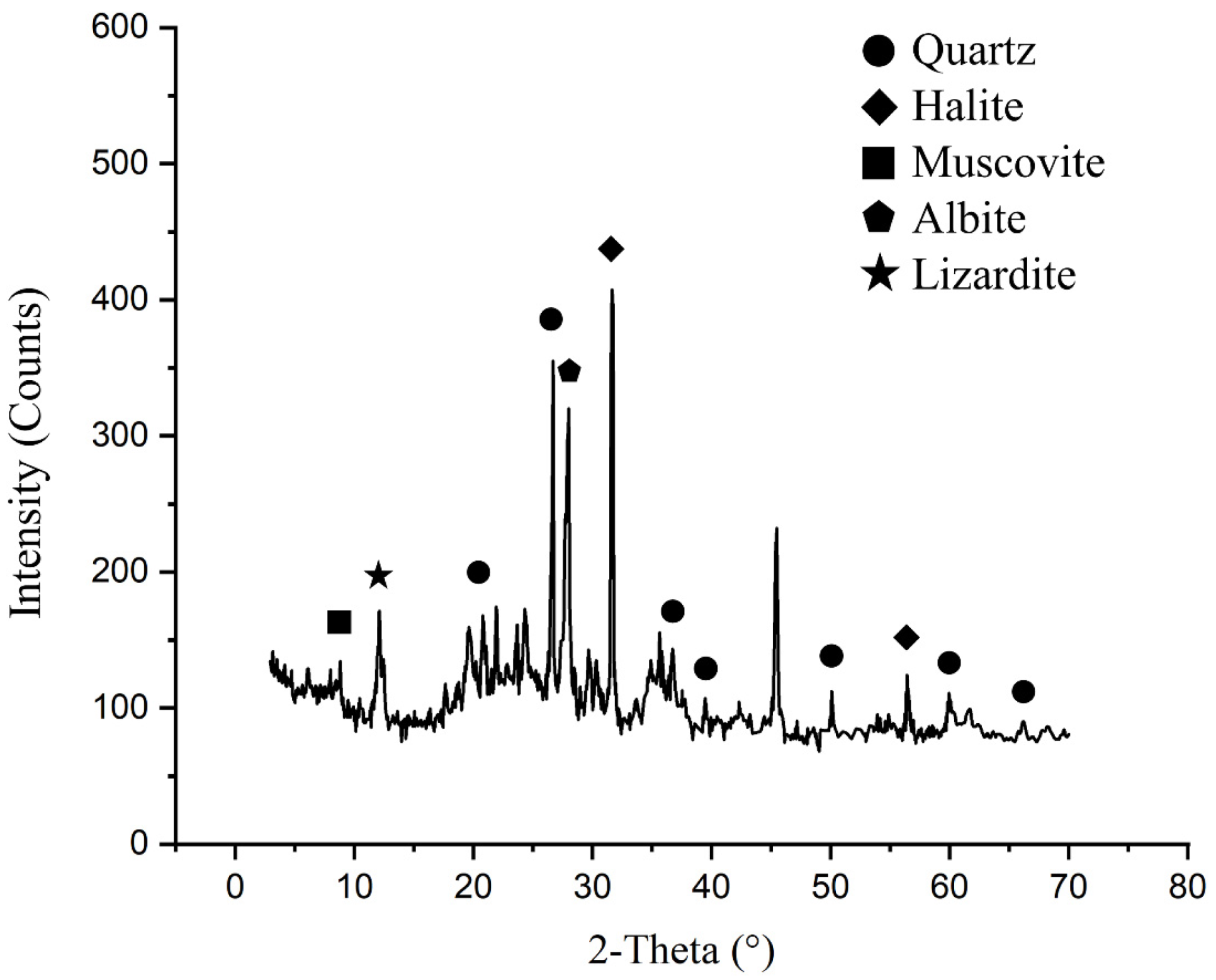
Figure 1. XRD pattern of the untreated abyssal sediment.
Water 16 02740 g001
Figure 2. Fe-redox curves of the abyssal sediment over 58 days’ anaerobic culturing under atmospheric pressure or 58 MPa in the presence or absence of an external electron donor (H2 or lactate).
Water 16 02740 g002
Figure 3. Nitrate-amended oxidation of the abyssal sediment over 45 days.
Water 16 02740 g003
Figure 4. (a) OTU number and (b) corresponding distribution; (c) principal component analysis; (d) sample clustering; relative abundance of (e) phylum and (f) genus in the 6 treated abyssal sediment cultures.
Water 16 02740 g004
Figure 5. Correlation heatmap indicating major microbial composition across the 6 treated abyssal sediment samples.
Water 16 02740 g005
Figure 6. (a) Redundancy analysis (RDA) among the 6 treated abyssal sediment samples and environmental factors (pressure, H2 and lactate) and (b) the community functional prediction based on the FAPROTAX database.
Water 16 02740 g006
Figure 7. Phenotypic predictions of the dominant phyla within the 6 treated abyssal sediment samples based on BugBase: (a) aerobic, (b) anaerobic, (c) facultative anaerobic, (d) stress-tolerant, (e) Gram-negative, (f) Gram-positive, (g) containing mobile elements, and (h) forming biofilms.
Water 16 02740 g007
Figure 8. Scanning electron microscopic images of mineral–microbe interactions in A-Lactate.
Water 16 02740 g008
Table 1. Measures of carbon (C), hydrogen (H), nitrogen (N), sulfur (S), 11 major elements, and 44 trace elements in the abyssal sediment.
C, H, N, S (%)C (%)H (%)N (%)S (%)C/NC/H
0.220.6820.0530.6394.1770.3238
Major elements (%)SiO2Al2O3Fe2O3MgOCaONa2O
49.1114.059.784.692.914.49
K2OMnOTiO2P2O5LOIFeO
2.071.20.7590.35610.280.36
Trace elements (μg/g)LiBeScVCrCoNiCuZnGaRb
45.21.5224.217190.672.116833114917.561.1
SrYMoCdInSbCsBaLaCePr
23177.311.10.3940.1181.885.52735496712.5
NdSmEuGdTbDyHoErTmYbLu
55.512.43.2810.22.39142.897.551.287.721.05
WReTlPbBiThUNbTaZrHf
3.440.0061.7935.50.6387.731.510.81.111293.75
Table 2. The proportions of Fe-reducing and Fe-oxidizing microorganisms (%).
Genus/SpeciesH-OriginalH-H2H-LactateA-OriginalA-H2A-Lactate
Fe-reducing microorganismsShewanella0.06370.05530.0230.9985.40720.679
S. putrefaciens00.01640000.00369
S. oneidensis00.006140.00628000.00738
Unclassified S. sp.0.06370.03280.01670.9985.40720.668
Thalassospira0.005540.006150.002095.2333.8642.438
Thermodesulfovibrio0.0013900000
Lysinibacillus00.0020500.0083300.0148
Thermus0.006920.002050.01260.006250.005570.00123
Alteromonas0.1660.1910.1490.2810.4430.0701
Sphingomonas0.001390.01230.008370.01670.04450.0184
Fe-oxidizing microorganismsMarinobacter4.462.3562.9934.45829.99528.034
Uncultured M. sp.0.3700.1540.2341.0130.6650.873
M. lacisalsi0.0320.0470.0151.2150.8460.216
M. hydrocarbonc-lasticus1.2920.9181.02916.61714.95419.154
Thiomonas0.02630.03070.02930.01250.01110.00861
Sulfolobus000000.00246
Methyloversatilis0000.002080.4200
Disclaimer/Publisher’s Note: The statements, opinions and data contained in all publications are solely those of the individual author(s) and contributor(s) and not of MDPI and/or the editor(s). MDPI and/or the editor(s) disclaim responsibility for any injury to people or property resulting from any ideas, methods, instructions or products referred to in the content.