Article

An Analysis of Microplastics Ingested by the Mediterranean Detritivore Holothuria tubulosa (Echinodermata: Holothuroidea) Sheds Light on Patterns of Contaminant Distribution in Different Marine Areas

1
Department of Science and Biological and Environmental Technologies-DiSTeBA, University of Salento, Via Prov.le Lecce-Monteroni, 73100 Lecce, Italy
2
National Biodiversity Future Center (NBFC), 90133 Palermo, Italy
3
Museo di Biologia Marina “Pietro Parenzan”, Via Vespucci 13/17, Porto Cesareo, 73010 Lecce, Italy
4
Department of Science, University Roma Tre, 00146 Rome, Italy
5
Interdepartmental Laboratory of Electronic Microscopy (LIME), University Roma Tre, 00146 Rome, Italy
*
Author to whom correspondence should be addressed.
Water 2023, 15(8), 1597; https://doi.org/10.3390/w15081597
Submission received: 20 March 2023 / Revised: 15 April 2023 / Accepted: 18 April 2023 / Published: 20 April 2023

Abstract

:
Microplastic pollution constitutes a serious environmental problem that requires more effective scientific research to describe its potential impacts on marine fauna. The interaction between microplastics and marine biota can have significant negative effects through the trophic chain, up to human health. To date, several steps forward have been made in our understanding of this phenomenon; however, large knowledge gaps still exist for several taxa and areas. In particular, the pattern of spatial and temporal distribution of microplastics in marine sediments and their interaction with benthic detritivore species still needs to be addressed. The Mediterranean Sea is one of the most impacted areas of the world, and its biota is deeply affected by microplastic pollution. To investigate the effects of the presence of microplastics in the sediments in this area, the echinoderm Holothuria tubulosa was chosen as a model species, and specimens were collected along the Salento peninsula in Apulia, Southern Italy. This peculiar geographic area extends between two ecoregions of the Mediterranean Sea, the Northern Ionian and the Southern Adriatic seas, characterized by peculiar and distinct currents and submarine topologies, resulting in a complex and dynamic ecosystem affected by seasonal fluctuations that make the Salento peninsula an interesting natural laboratory for predictions of future dispersion events on a wider scale. Microplastics were analyzed by investigating the gut contents of H. tubulosa individuals, and the SEM/EDX method was used to confirm the plastic material extracted. Results revealed microplastics in all the specimens analyzed and with a homogeneous pattern of distribution in time and some differences in space, suggesting that the presence of this anthropogenic material is constant throughout the year and its quantity is only slightly affected by the level of conservation and management strategies characterizing the sampling sites.

1. Introduction

Microplastics are particles smaller than 5 mm obtained from the chemical, physical, and mechanical degradation of plastics in contact with the environment [1]. The pollution caused by microplastics in the environment is a topic of increasing interest because as a result of their slow degradation and small size, these particles can be ingested by different animal species, including, ultimately, humans. The ingestion of microplastics may cause complications at a physiological level, with the possibility of transport to different organs and tissues of the organism and along the trophic network, giving rise to bioaccumulation processes [2]. The routes for plastics to reach the marine environment are numerous, as they can enter through inadequate plastic waste management, illegal dumping into the water, unintentional or accidental releases from vessels, and/or from litter left behind on beaches [3]. A significant proportion settles in the sediments [4]. Recently, several studies have shown that plastic litter affects different marine species [5,6,7,8] and that easily enters the marine trophic webs [9,10]. Exposure to microplastics has been associated with a number of negative health effects, including an increased immune response [11], decreased food consumption and weight loss [2,12], reduced growth rate [13], decreased fecundity [14], energy depletion [2], and negative impacts on subsequent generations [14]. Microplastics have also been shown to readily accumulate persistent organic pollutants, including pesticides, solvents, and pharmaceuticals, which can lead to additional health effects such as endocrine disruption and mobility impairments [15,16,17]. Microplastic pollution is thus a serious problem that acts on all living compartments and through different biological levels of the trophic chain in different ways. Such a problem is even more relevant if particular marine conditions occur, such as in the case of semi-closed basins with reduced seawater exchange.
Studying the pattern of distribution of microplastics in the Mediterranean Sea is particularly important due to the specific characteristic of this marine basin, which is connected to the Atlantic Ocean only through the Gibraltar strait and to the Red Sea by the Suez Canal. The Mediterranean Sea is one of the most impacted areas of the world due to the intense human activity and concentration of people along its coasts [18]. Indeed, improving the knowledge gap on this complex topic could provide important insights that would help predict and manage possible future scenarios, even at a larger geographic scale, and could aid in optimizing conservation strategies. Located in the Central Mediterranean basin and between two different ecoregions, the Northern Ionian and the Southern Adriatic seas, is the Salento peninsula (Apulia, Italy) (Figure 1), a strip of land characterized by peculiar and distinct currents and submarine topography. It is exposed to the Western Adriatic current and to the anticyclonic Northern Ionian Gyre, the intersection of which may lead to a high accumulation of microplastics in the Salento coastal waters [19]. This peninsula is considered a highly complex and dynamic ecosystem affected by seasonal fluctuations and is thus an interesting natural laboratory that may provide insight for wider geographical scales.
It seems that microplastics are more abundant in the western Mediterranean Sea than in the eastern basin, reaching minimum values in the Adriatic Sea [20]. However, recent studies have revealed the presence of microplastics in 45% of Adriatic Sea habitats [5] and in up to 95% of the benthic flatfish Solea solea (Linnaeus, 1758) [21], suggesting that benthic species can be strongly influenced by microplastics stored in sediments [22] and thus calling for studies focusing on these seabed-related communities.
To date, several methodologies have been proposed to detect and quantify microplastics in marine samples. However, all of them involve, as a first common step, the extraction of microplastic polymers from the environmental and/or biological matrices in question. At present, the most commonly used procedures to extract microplastics from biological tissues can be classified as acidic, alkaline, oxidative, and enzymatic methods [5]; however, new innovative methods (such as visual identification using polarized light or electron microscopy) are continuously being proposed and evaluated [23]. After microplastics are isolated from biological tissues, the most frequently used techniques for their confirmation and characterization are FTIR and Raman spectroscopy [24]. These methods allow the recording of spectra for the correct identification of debris and, to a certain extent, the quantitative description of its morphology [25]. Each spectrum of an unknown element is then compared with spectra of known standard polymers if they are present in a public database [26]. The Raman analysis allows for the identification of microplastics directly on filters without extensive visual pre-sorting; however, any remaining biotic material must be removed to avoid fluorescence quenching the signal [27]. Even if these two methodologies are considered very effective in determining microplastic materials, they suffer some important limitations such as the impossibility of obtaining detailed pictures of the surface morphology of the studied material, which could be very useful for confirming identification, for providing information on weathering degradation processes [28], and for a decrease in the identification power if a reference spectrum is lacking [29]. These drawbacks can be overcome using SEM-EDX analysis, which merges information from the external morphology with the microanalysis of the elementary composition [29]. Scanning electron microscopy (SEM) can provide rapid information on the morphology, aging, and origin of the samples examined as it provides high-resolution surface-state data and qualitative information on chemical composition. Furthermore, the use of electron microscopy combined with energy-dispersive X-ray spectroscopy provides detailed information on the elemental composition of microplastics, with additional information on the inorganic additives they can contain [28,29]. Thus, this methodology is as complementary and as effective as the Raman to quantify MPs.
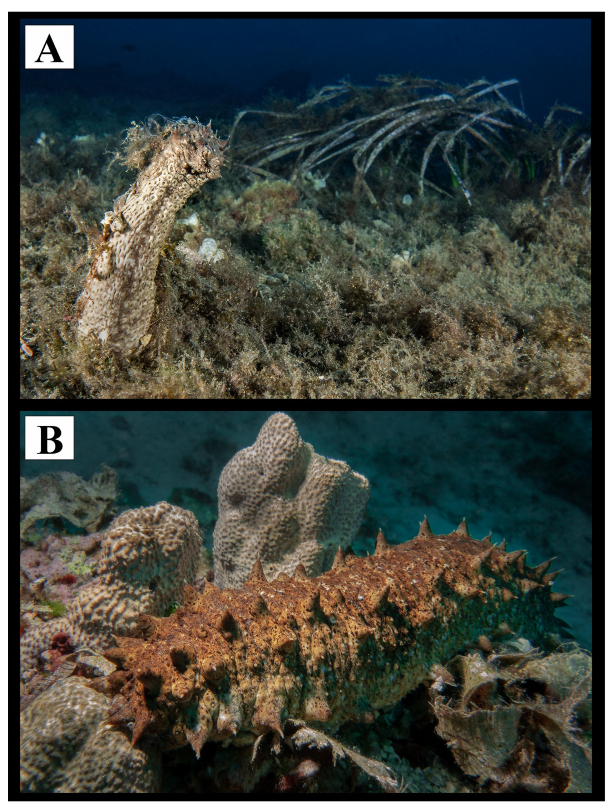
Several studies have been conducted on microplastics floating in the water column, transported through surface seawater [30], or deposited in sediments, with a rich literature demonstrating that the ingestion of microplastics by benthic organisms [31,32,33,34] can lead to the impairment of their digestive systems [35,36,37] and their ability to feed [38] and reproduce or cause the potential transfer of harmful toxic substances and ultimately death [39]. However, less attention has been given to the interactions between the microplastics present in the sediment and the benthic organisms that inhabit the seabed [22]. Among marine benthic species, the echinoderms are a group of invertebrates that are particularly diversified in terms of morphology and ecology. They are distributed worldwide, and they are widespread from the shoreline to the deep sea. These include the benthic holothurians, which demonstrate a detritivore trophic habitus and are also interesting because of their commercial purposes. Previous studies have been conducted on sea cucumbers and their interactions with microplastics present in the sediments [22,40], confirming the presence of a plastic percentage in the gut content of the animals analyzed and the positive correlation existing between it and the amount of plastic present in the sediment, confirming this group as a good descriptor of environmental status. Detritivore holothurian species may thus be good sentinels of microplastic pollution in a specific zone due to their low mobility and trophic behavior [41]. It is clear that the specimens collected for the present study are representative of the potential pollution by microplastics in the study area. Species belonging to the lower trophic levels, which are indiscriminate feeders, are not able to differentiate between plastics and prey, ingesting high levels of these pollutants [22]. Holothurians, as nonselective feeders, ingest large quantities of sediment into their mouths to extract nutrients from biofilms, organic debris, and microorganisms [42]. The transit through their digestive tract could allow for the absorption of plastic leachates and adhered contaminants, representing a possible risk for potential transfer to higher trophic levels [22,42,43]. The translocation of the ingested plastic could produce significant damage to these animals, affecting their reproductive success and reducing population abundance in marine ecosystems [22]. However, studies linking the presence of microplastics in holothurian species with their potential physiological or biochemical effects are still scarce and were developed mainly in controlled laboratory conditions that generally do not reflect environmental settings and expose the organisms to concentrations of pollutants higher than the concentrations normally found in the animals’ habitats [44,45]. In these latter studies, the spatial distribution of microplastics in the natural environment is completely lacking—this is significant because understanding the pathways of distribution of environmental microplastics eaten by organisms is the baseline step for planning effective conservation actions. Here, Holothuria tubulosa Gmelin, 1791 (Figure 2) was selected as a benthic detritivore species which is particularly interesting because it represents an initial step from the detrital network towards the trophic network in shallow marine ecosystems [22] and also because it is among the most common and widely distributed species in the Mediterranean Sea [46], feeding continuously and indiscriminately upon the sediment [47] and taking up microplastics through both respiration and feeding [48]. Moreover, in many countries, H. tubulosa is a species of great commercial interest; in fact, in Apulia, it is irregularly fished and trafficked outside the Italian jurisdiction.
The main aims of the present study are to (i) investigate the possible presence of microplastics in the gut content of H. tubulosa individuals collected along the Salento peninsula in different seasons to elucidate potential changes in space and time, and (ii) to extract, quantify, and classify the possible microplastics found, exploring the possibility of a spatial pattern depending on the collecting zone.

2. Materials and Methods

2.1. Sampling Protocol

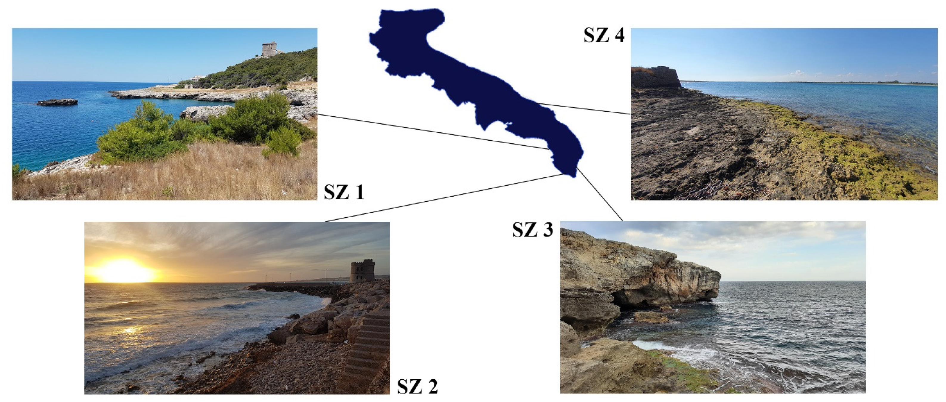
Two sampling campaigns were carried out in the spring and autumn of 2020, respectively, along the Salento peninsula. In particular, four different sites located in the Ionian Sea (Stations 1 and 2) and in the Adriatic Sea (Stations 3 and 4) were sampled (Figure 3). The sampling stations are characterized by different degrees of anthropogenic impacts, from areas that are influenced by high tourism to a Marine Protected Area. At least four adult specimens of H. tubulosa were collected per station and in the two seasons through scuba diving at a depth of 10 m and without using gloves to avoid possible contaminations. Each individual was wrapped in aluminum foil directly underwater and placed in an aluminum thermos bottle with a wide opening. Once collected, the samples were catalogued with a tag indicating the station and sampling date and were finally stored at −20 °C at the Department of Biological and Environmental Sciences and Technologies of the University of Salento (DiSTeBA) for later anatomical dissection and subsequent laboratory analysis.

2.2. Anatomical Dissection

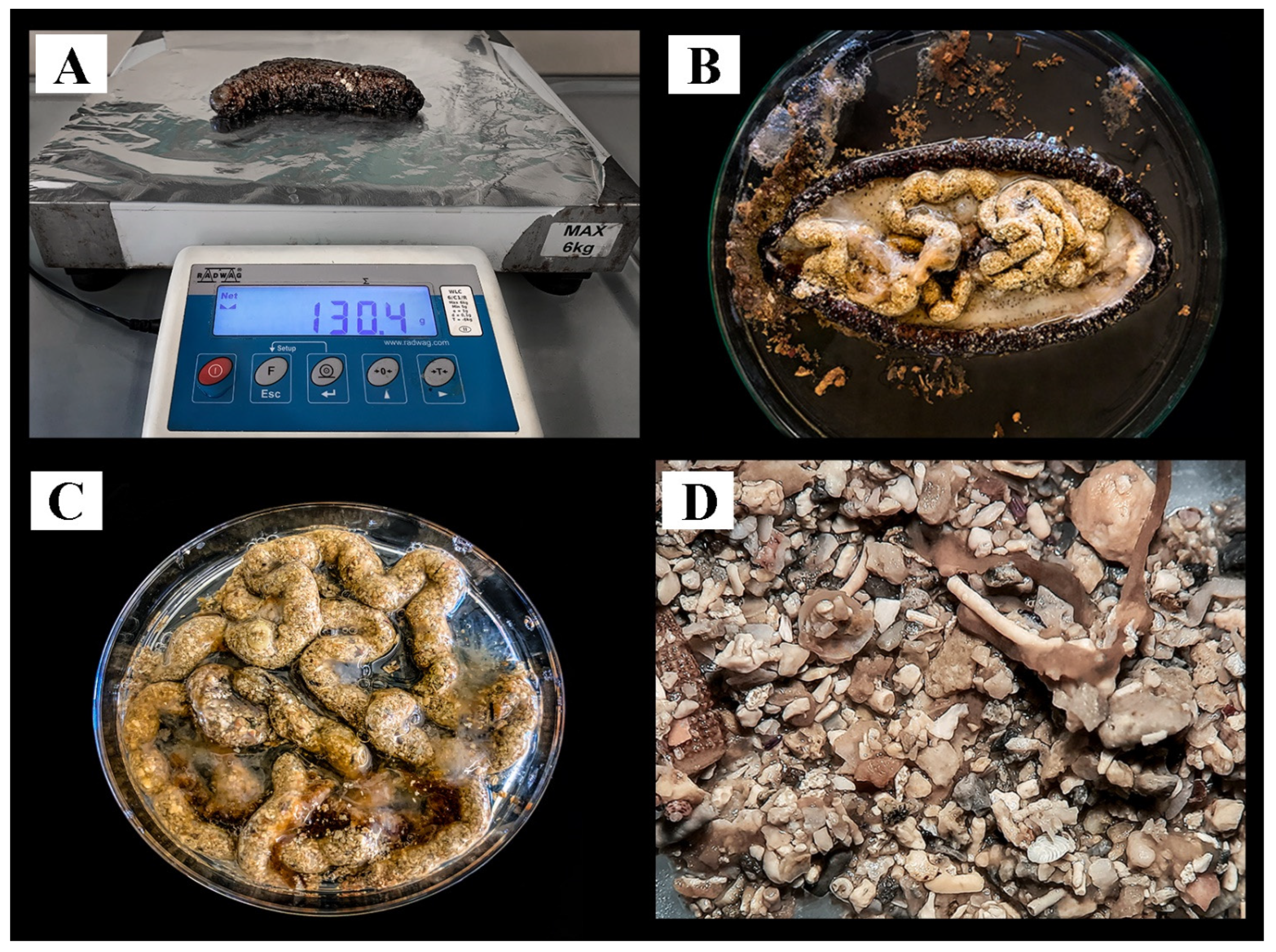
The collected samples were photographed, thawed at 37 °C, and then measured for both dimensions and weight. The dissection of the individuals was carried out by making a longitudinal incision on the dorsal side to keep the digestive system intact. Each digestive tract, isolated from each specimen, was placed in a biological glass tube with ethanol EtOH 96% until further analysis. To avoid possible contamination [23,49,50], all glassware was washed with filtered deionized water and rinsed with ethanol. Cotton lab coats, nitrile gloves, and face masks were worn throughout the experiment. The work surface was thoroughly cleaned with 70% ethanol and analyzed under a stereomicroscope to remove any contamination.

2.3. Microplastic Extraction and Analysis

From the total individuals collected and analyzed, six of the most representative digestive systems (three per each collecting season) from each station were selected for the following microplastic analysis. The selection was made extracting the digestive systems from individuals that were as similar as possible in terms of size and the quantity of material in the swallowed content to avoid variables that could alter the results. Each digestive tract was analyzed using a Leica MZ12 stereomicroscope. All fragments and plastic fibres were carefully hand-collected using laboratory forceps, transferred to separate sterile glass plates, and coded for subsequent confirmation by SEM/EDX analysis. To optimize the microplastic isolation process and to avoid possible misidentifications, an enzymatic digestion method was used. Five hundred microliters of distilled water and 20 µL of proteinase K were added to each sample and incubated overnight at 56 °C to optimize the digestion. Successively, microplastic samples were removed from the digestive solution, rinsed in clean distilled and filtered water, and finally counted and classified according to structure (fibres or fragments) and color.
To confirm the validity of the stereomicroscope observations and the identity of the microplastic material, a scanning electron microscopy–energy-dispersive X-ray analysis (SEM/EDX) was used. The selected particles were placed on aluminum stubs using double-sided carbon tape and subsequently analyzed using a Gemini 300 SEM (Carl Zeiss AG, Jena, Germany) equipped with an XFlash 6-60 EDS system (Bruker Nano GmbH, Berlin, Germany) at the LIME (Electron Microscopy Interdepartmental Laboratory, Rome, Italy) of the University of Roma Tre. Plastic microparticles were identified by analyzing both their external features and elemental composition. Finally, an SEM/EDX analysis was also carried out on fragments of dubious nature in order to provide EDX spectra useful for future comparisons.

2.4. Statistical Analysis

For statistical comparison, we first considered two periods: spring and autumn. The four sites were also considered for comparisons in the initial analysis. The dependent variables were fibers, fragments, and total microplastics (the sum of the previous two variables). A two-way ANOVAs test was used to test the potential differences, but the limited number of samples (three per season and location) made this approach not viable. The preliminary ANOVA results provided no significant differences between the locations and seasons. We thus considered only the four locations to carry out a one-way ANOVA with Scheffer’s post hoc. The data were not distributed normally and had different variances but met both criteria for a parametric analysis after logarithmic transformation (Brown–Forsythe test and Levene test, p = 0.05; Shapiro–Wilk test, p = 0.1), thus permitting the use of an ANOVA [51]. The program used was the STATISTICA 7.1 software package. Seasonal average values of the fibers, fragments, and the total microplastics, taken at each individual station, were represented with their respective standard deviations in three separated histograms with clustered columns. Particle coloration data were also represented in a grouped column histogram.

3. Results

During the two sampling campaigns that occurred in the spring and autumn of 2020, 24 scuba dives were carried out at the four sampling stations along the Salento peninsula (Figure 3). A total number of 47 H. tubulosa specimens was collected, with 25 individuals obtained during the spring sampling and 22 in the autumn campaign. All the collected specimens were weighted, measured (length and width), and their digestive systems were extracted (Table S1, Figure 4). Within this group, the digestive systems of six representatives per each station (three per each sampling seasons) were selected for the following microplastic extraction and analysis.

3.1. Statistical Analysis

The microplastic extraction analysis revealed a total of 741 microparticles. Of these microparticles, 406 were fibers and 335 were fragments. The list of all the microplastic samples obtained from each analyzed digestive system is reported in Table 1.
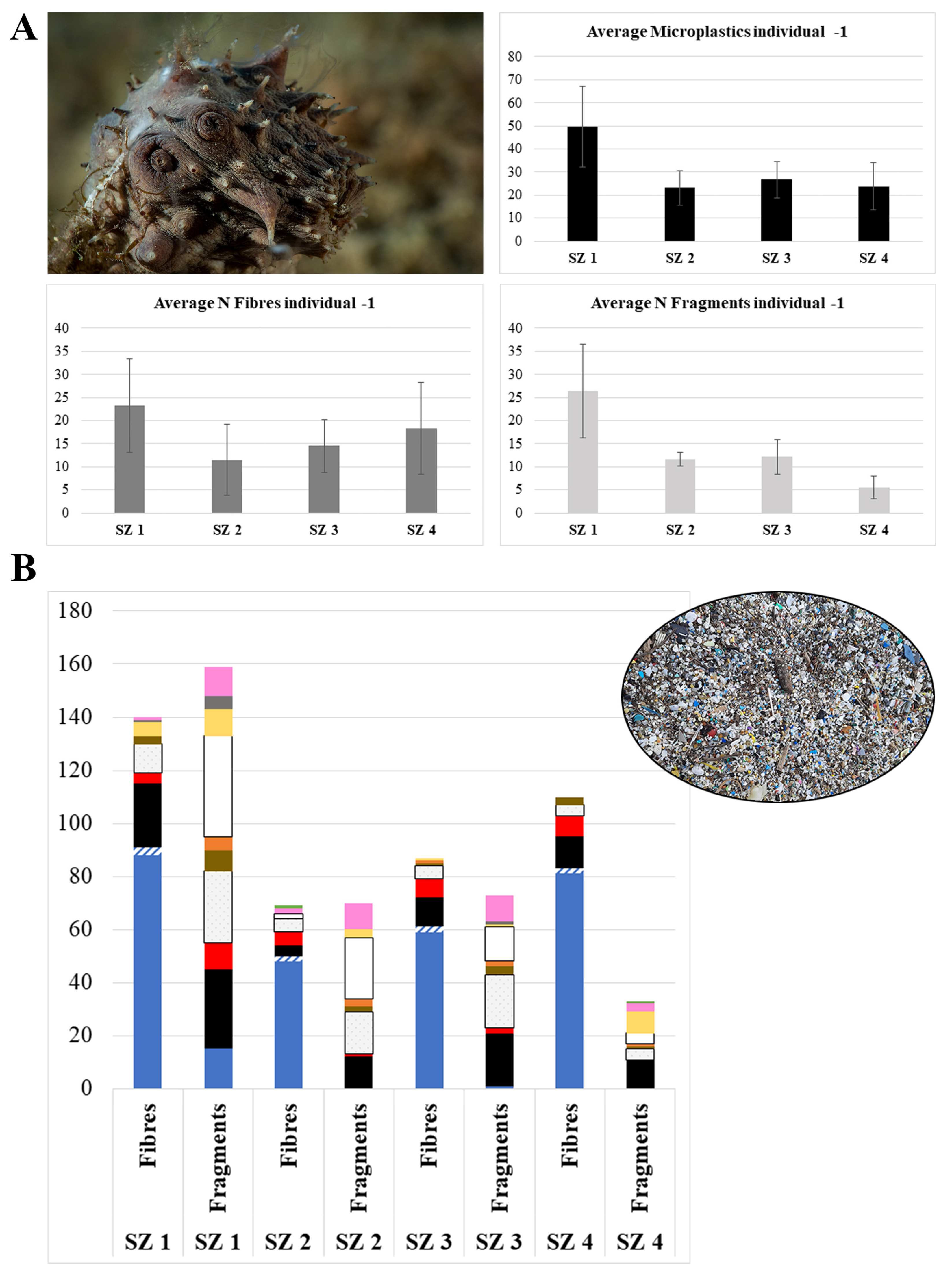
The total number of microplastics per individual in the different zones showed significant differences (one-way ANOVA, F3,20 = 0.729; p < 0.002); Chiapparo (SZ 1) was different from all the other areas, with the highest differences between the “Torre Guaceto” M.P.A. (SZ 4) (mean of 24 microplastics) and Chiapparo (mean of 50 microplastics). When fibers were considered, no significant differences between zones were found (one-way ANOVA, F3,20 = 42.12; p = 0.1289). There were, however, significant differences for the fragments (one-way ANOVA, F3,20 = 15.13; p < 0.0001); in this case, Chiapparo was also different from the other three locations, with the maximum amount of fragments found in holothurians from Chiapparo (mean of 27) and the minimum from “Torre Guaceto” M.P.A. (mean of six) (Figure 5).

3.2. SEM/EDX Microanalyses

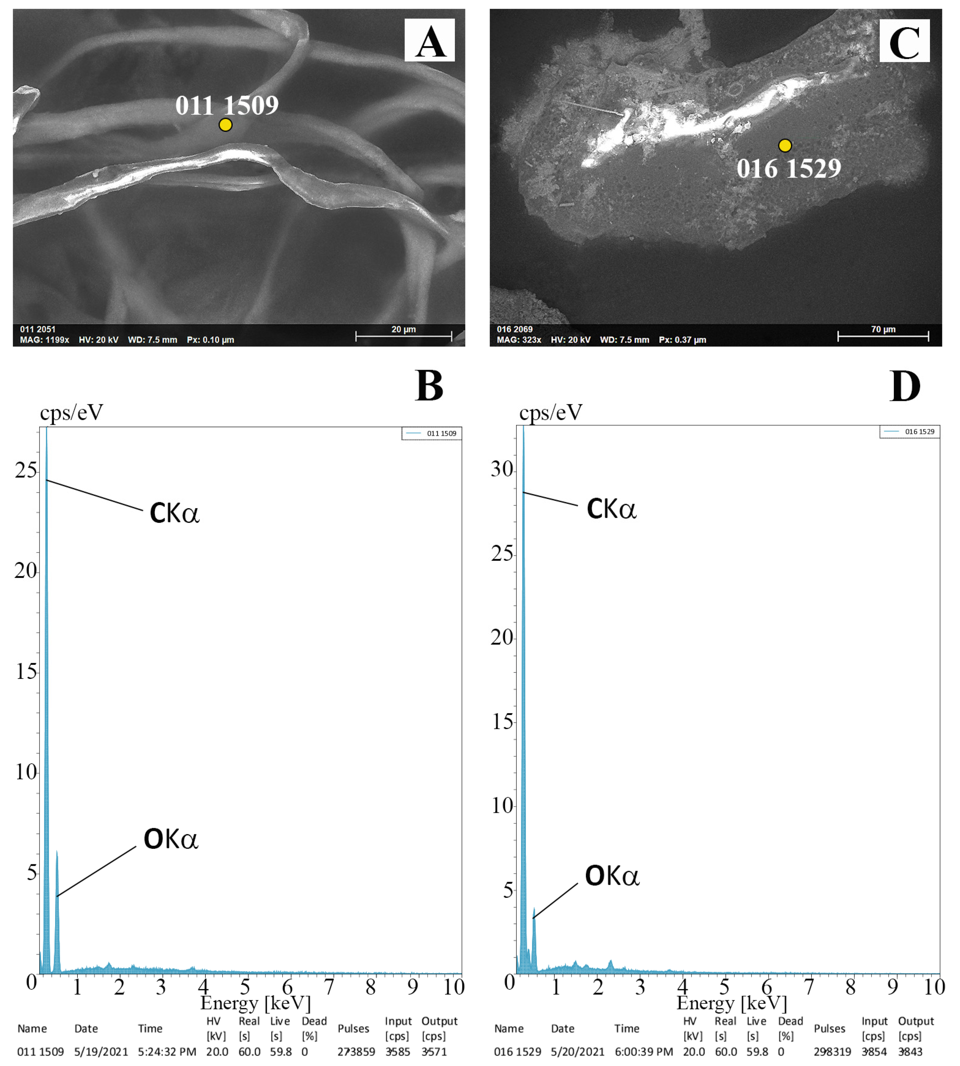
Thirty-four high resolution SEM images and EDX spectra were obtained from plastic fibers and fragments and from other biotic and abiotic materials that were similar in their external shape to plastic materials. The spectra obtained from the present study were compared with some reported by published studies [29], and the results matched with those from the morphological identifications. The resulting SEM images and the relative EDX spectra of a typical plastic fiber and fragment are shown in Figure 6.
In addition to the canonical spectra, spectra belonging to another important class of plastic material, fiberglass, were found. Fiberglass is characterized by Carbon (C), Oxygen (O), and Silicon (Si) [29,52,53] (https://www.nrc.gov/docs/ML0530/ML053040493.pdf (accessed on 30 March 2023), and it results from the addition of glass fibers that are commonly added to reinforce plastic structures [54,55].
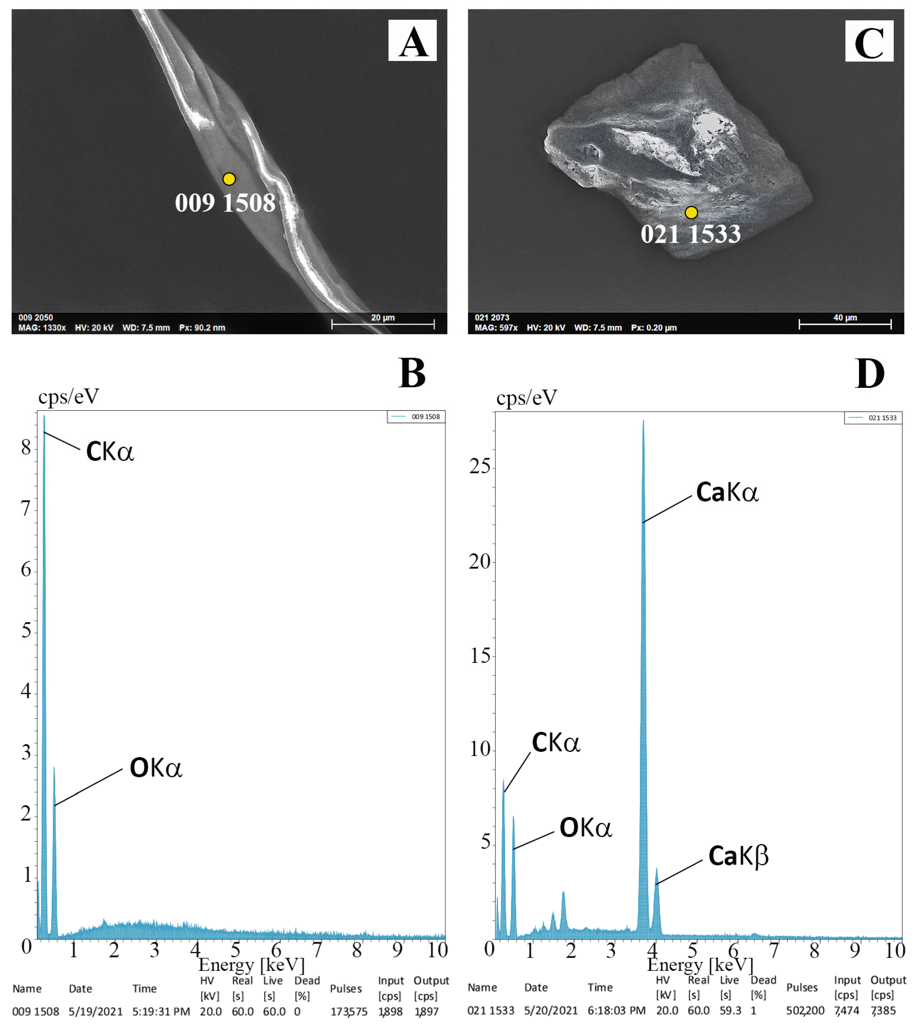
Finally, samples that were deemed to be doubtful when observed with the optical microscope were checked with SEM/EDX method and confirmed to be non-plastic. In fact, among these dubious samples were cotton fibers, characterized by a typical twisted morphology [29], and also the presence of peaks resembling those reported for cellulose (Figure 7A,B) [29,56,57,58], gastropod shells, and tubes of polychaetes (Figure 7C,D), all containing a high concentration of calcium carbonate (CaCO3), which is coherent with spectra from the literature [29,59,60,61,62,63].

4. Discussion

Results from the present study revealed microplastic fibers and fragments in all the Holothuria tubulosa individuals investigated and a pattern of MP distribution that was equivalent in the four studied sites except for the “Chiapparo” station (SZ 1) (Figure 3), which proved to have the highest number of microplastics with respect to the other areas investigated. In fact, microplastics were widely distributed along the Salento peninsula regardless of the level of anthropogenic impact acting on the particular investigated stations. This corroborates the evidence that microplastics are widespread all over the peninsula. This finding is a bit disturbing, considering that highly disturbed areas may have more microplastics, but sites that are considered of low anthropogenic impact or under special conservation protocols (such as the “Torre Guaceto” Marine Protected Area, SZ 4) also have a non-negligible concentration of these pollutants. Such a presence, even in MPAs, represents a potential threat for marine species [64,65]. The presence of microplastics in the digestive systems of all Holothuria specimens investigated herein suggests that microplastics could enter the trophic chain, beginning their long journey through primary consumers to the upper-level predators. For this reason, the present study could become a baseline for future studies on the possible presence of microplastics in other compartments of the body of holothurians, further improving our knowledge on the ways microplastics spread.
The results reported here are worrying and stimulate new questions regarding guidelines to be adopted for the management of natural marine environments in terms of plastic pollution, the need to find new effective directions to somehow reduce the amount of plastic entering the marine system, and the need for more effective and rapid conservation strategies in the case of such pollutants. The statistical analysis revealed that the only single significant difference was observed at the “Chiapparo” SZ1 (Ionian Sea) site, where the microplastics were even more abundant than in the other sites. This unexpected result is interesting and requires further studies to be better understood, although we can hypothesize that the great influence of tourism in that area causes an increase in the human impact on the local environment. It has been shown that in areas heavily frequented by humans, especially tourists, the presence of microplastics is significantly different [66]. In coastal areas, multiple anthropogenic factors can affect the accumulation and dispersion of microplastics [67,68]. As we already know, different anthropogenic activities (e.g., coastal tourism, recreational boating, agriculture, ports, industrial activities, fishing, and aquaculture), proximity to large cities [68,69,70,71,72], and natural factors can significantly contribute to the amount of marine litter and to the variability of its concentration and distribution in the environments. The fragmentation and degradation of macro-, meso-, and microplastics drive the production of the smaller plastics that are found in different areas. Although the presence of environmental microplastics and their flow through the food chain are beginning to be understood, grey areas remain to be investigated. Plastic fragments break down into smaller pieces and degrade further when exposed to UV radiation, oxygen, temperature, and physical stress [73]. A combination of the properties of the polymer and sunlight and temperature influence the disintegration of macroplastic debris [74]. Ultraviolet radiation causes the oxidation of the polymer matrix, which leads to the cleavage of bonds. This process is most effective on beaches due to the high UV light, physical abrasion, and turbulence [75,76,77,78]. A range of variability in the transport and fragmentation of plastics may be caused by seasonal changes in river outflows, currents, mechanisms of degradation and fragmentation, changes in litter size, shape, buoyancy, and movement to and from other compartments [70,79,80,81,82], but it also can be associated with events of different time durations, such as tidal conditions, short-term wind and rain events, and seasonal extremes [81].
The microplastics analyzed in the present study showed characteristic abrasion grooves, which are considered indicative signs of the degradation process due to external environmental agents [29,83,84]. This determination was made possible by the method of MP determination that was adopted here. In fact, contrary to what is seen in other methods of MP identification (such as FTIR and Raman spectroscopy), SEM/EDX analysis allowed for the identification of an environmental microplastic even if it was degraded or mixed with other pollutants. Moreover, the addition of the enzymatic step during the phase of microplastic extraction was revealed to be powerful in removing non-plastic materials and avoiding misidentifications due to the enzyme’s ability to digest biological tissues without damaging the plastic. This improvement in protocol was also confirmed by the absence of Nitrogen (N) in all the EDX spectra analyzed. Bearing in mind that the applied methods of extraction and identification of microplastics can influence the density, size, morphology, and polymer composition, ultimately negatively affecting the results [85], the power of the present method is even more consistent when considering that environmental microplastics and not virgin material were the main focus of the present study [29].
Considering that the area of study, the Salento peninsula, is characterized by an extreme heterogeneous environment that includes two different seas (Adriatic and Ionian seas), each of which are subject to completely different natural and anthropogenic conditions, it may be hypothesized that the presence of such a large quantity of microplastics could be explained as the consequence of a chain of multiple causes affecting the study area, as reported by other authors for similar contexts [86,87]. Previous studies that were focused on the composition, quantity, and spatial distribution of microplastics in Italian coastal and offshore areas showed that microplastics were widespread and without seasonal differences in concentration [86], agreeing with the results presented here. The results herein reported provide an additional demonstration of the high level of microplastic pollution characterizing marine environments and in the study area in particular. In fact, the Salento peninsula is a very resilient area due to the different dominant marine currents, which can move plastic materials away, and/or due to the presence of several underwater canyons that could act as deep plastic accumulation sites. A future study of the possible sources of microplastics in the environment should be the next step, with the goal of reducing the continuous release of plastic into the sea. In this regard, previous studies have reported household textile recycling and wastewater treatment plants as the most common pathway for the release of fibers into the environment [88,89], considerations that should stimulate further in-depth analyses.
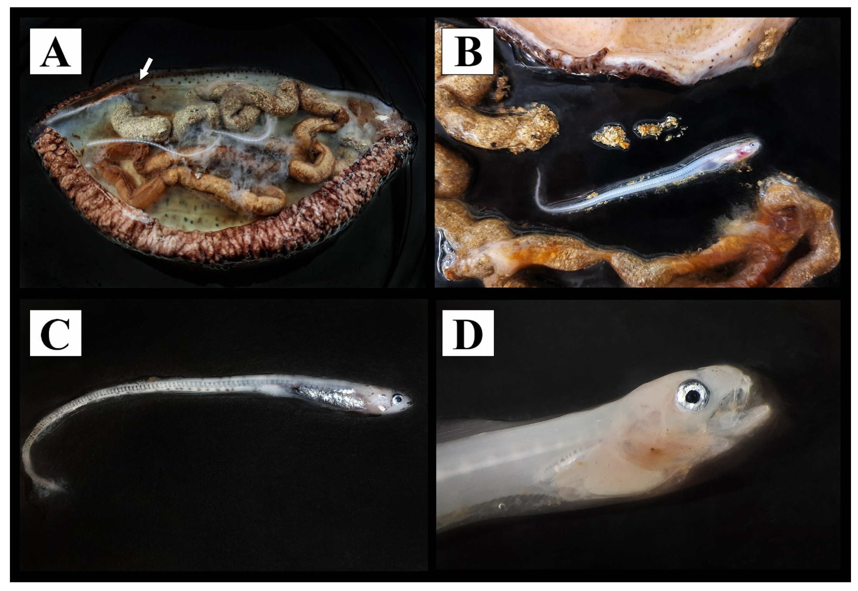
The complexity of natural environments and communities and the interconnections existing between organisms at different biological scales are the main challenges for delineating the best conservation practices. The most effective practice of such conservation studies is difficult and deserves to be investigated in each specific area. In this regard, it is noteworthy to report that inside all the holothurian specimens collected in autumn from “Grotta Verde” in Station 3 (SZ 3) (Figure 3), we found individuals of the commensal fish Carapus acus (Brünnich, 1768) (Figure 8). This finding confirms the delicate and profound association which is known to exist between two very distant taxa—the commensalism between the H. tubulosa and the fish Carapus acus (Brünnich, 1768).
Results from the present study add data on the ecology of this inconspicuous pearlfish and its Apulian population that should be considered in future conservation strategies and in planning actions aimed to protect H. tubulosa living in that region of Italy. Currently a large gap of knowledge still exists on the possible negative effects of microplastics ingested by holothurians on C. acus.

5. Conclusions

Quantifying plastic pollution and understanding its negative effects on the marine environment are two of the main goals of research groups all over the world. All the information gathered thus far demonstrates the long-term impacts of microplastics on marine fauna, the echinoderm studied herein being no exception. Most past studies did not include an analysis of the distribution of MPs in study organisms from different areas, the present study being an example of how to quantitatively approach the problem at a regional level. Studying the distribution of microplastics in a particular geographic area is complex and requires a considerable effort in terms of field sampling, laboratorial investigations, analysis, and interpretation of the evidence obtained. Herein, we reported results from an analysis of the spatial distribution of microplastics ingested by a detritivore benthic species, Holothuria tubulosa, by extracting plastic fibers and debris from gut contents and with the help of the SEM/EDX method of identification. Interestingly, all the specimens had microplastics, and the statistical analysis showed no major differences among sampled areas. This reinforces the hypothesis that microplastics are widespread and are found in every part of our planet. The sediments of the Apulian region are contaminated by these small debris, which are present even in protected areas such as the “Torre Guaceto” MPA, highlighting the importance of a new perspective on management and conservation related to the presence of microplastics.

Supplementary Materials

The following supporting information can be downloaded at: https://www.mdpi.com/article/10.3390/w15081597/s1. Table S1: Stations, sampling localities, seasons, and measurements of length, width, and weights of all the specimens analyzed in the present study.

Author Contributions

Conceptualization, G.F., A.M. and S.R.; methodology, G.F., A.M. and S.R.; software, A.M. and S.R.; validation, G.F., A.M. and S.R.; formal analysis, M.M., A.D.G., A.M. and S.R.; investigation, M.S. and G.F.; resources, M.S. and G.F.; data curation, A.M., M.S., M.M., A.D.G., and S.R.; writing—original draft preparation, A.M. and G.F.; writing—review and editing, A.M., G.F., M.S., M.M., A.D.G. and S.R.; supervision, S.R. All authors have read and agreed to the published version of the manuscript.

Funding

This research received no external funding.

Data Availability Statement

Not Applicable.

Acknowledgments

The authors are deeply grateful to the Director of the Consortium of Management of the Marine Protected Area “Torre Guaceto” for the permission to collect samples and for the precious collaboration during the sampling activities. The authors wish to thank Richard C. Brusca (Arizona, USA) for agreeing to review the English language and for his useful suggestions that improved the quality of the manuscript. G.F. and S.R. wish to thank the “MoSSHa-Monitoring of Species Sites and Habitats” project, funded by the Apulian Region, within the POR Puglia 2014/2020 funds for monitoring actions of “Natura 2000” species, sites and habitats. G.F. wishes to thank the Italian Ministry of Education, University and Research (MIUR, PON 2014–2020, Grant AIM 1848751-2, Linea 2) for support. S.R. and A.M. thank the project “i-Plastics: Dispersion and impacts of micro- and nano-plastics in the tropical and temperate oceans: from regional land-ocean interface to open ocean”, acknowledging the funding from the JPI Oceans International Consortium. Finally, the authors would like to thank the three reviewers that helped to improve the quality of the final paper.

Conflicts of Interest

The authors declare no conflict of interest.

References

  1. Thompson, R.C.; Swan, S.H.; Moore, C.J.; Vom Saal, F.S. Our plastic age. Philos. Trans. R. Soc. B Biol. Sci. 2009, 364, 1973–1976. [Google Scholar] [CrossRef]
  2. Wright, S.L.; Rowe, D.; Thompson, R.C.; Galloway, T.S. Microplastic ingestion decreases energy reserves in marine worms. Curr. Biol. 2013, 23, R1031–R1033. [Google Scholar] [CrossRef]
  3. Crawford, C.B.; Quinn, B. Microplastic Pollutants; Elsevier: Amsterdam, The Netherlands, 2016. [Google Scholar]
  4. Simon, L.; Grelaud, M.; i Orellana, J.G.; Ziveri, P. Microplastic particles in the Ebro Delta, Spain: Occurrence, composition and sources. In MICRO 2018. Fate and Impact of Microplastics: Knowledge, Actions and Solutions; MSFS-RBLZ: Lanzarote, Spain, 2018; p. 136. [Google Scholar]
  5. Dehaut, A.; Cassone, A.-L.; Frère, L.; Hermabessiere, L.; Himber, C.; Rinnert, E.; Rivière, G.; Lambert, C.; Soudant, P.; Huvet, A. Microplastics in seafood: Benchmark protocol for their extraction and characterization. Environ. Pollut. 2016, 215, 223–233. [Google Scholar] [CrossRef]
  6. Avio, C.G.; Gorbi, S.; Regoli, F. Experimental development of a new protocol for extraction and characterization of microplastics in fish tissues: First observations in commercial species from Adriatic Sea. Mar. Environ. Res. 2015, 111, 18–26. [Google Scholar] [CrossRef] [PubMed]
  7. Collard, F.; Gilbert, B.; Compère, P.; Eppe, G.; Das, K.; Jauniaux, T.; Parmentier, E. Microplastics in livers of European anchovies (Engraulis encrasicolus, L.). Environ. Pollut. 2017, 229, 1000–1005. [Google Scholar] [CrossRef] [PubMed]
  8. Fossi, M.C.; Marsili, L.; Baini, M.; Giannetti, M.; Coppola, D.; Guerranti, C.; Caliani, I.; Minutoli, R.; Lauriano, G.; Finoia, M.G. Fin whales and microplastics: The Mediterranean Sea and the Sea of Cortez scenarios. Environ. Pollut. 2016, 209, 68–78. [Google Scholar] [CrossRef] [PubMed]
  9. do Sul, J.A.I.; Costa, M.F. The present and future of microplastic pollution in the marine environment. Environ. Pollut. 2014, 185, 352–364. [Google Scholar] [CrossRef]
  10. Setälä, O.; Fleming-Lehtinen, V.; Lehtiniemi, M. Ingestion and transfer of microplastics in the planktonic food web. Environ. Pollut. 2014, 185, 77–83. [Google Scholar] [CrossRef]
  11. Von Moos, N.; Burkhardt-Holm, P.; Köhler, A. Uptake and effects of microplastics on cells and tissue of the blue mussel Mytilus edulis L. after an experimental exposure. Environ. Sci. Technol. 2012, 46, 11327–11335. [Google Scholar] [CrossRef]
  12. Besseling, E.; Wegner, A.; Foekema, E.M.; Van Den Heuvel-Greve, M.J.; Koelmans, A.A. Effects of microplastic on fitness and PCB bioaccumulation by the lugworm Arenicola marina (L.). Environ. Sci. Technol. 2013, 47, 593–600. [Google Scholar] [CrossRef]
  13. Huerta Lwanga, E.; Gertsen, H.; Gooren, H.; Peters, P.; Salánki, T.; Van Der Ploeg, M.; Besseling, E.; Koelmans, A.A.; Geissen, V. Microplastics in the terrestrial ecosystem: Implications for Lumbricus terrestris (Oligochaeta, Lumbricidae). Environ. Sci. Technol. 2016, 50, 2685–2691. [Google Scholar] [CrossRef] [PubMed]
  14. Sussarellu, R.; Suquet, M.; Thomas, Y.; Lambert, C.; Fabioux, C.; Pernet, M.E.J.; Le Goïc, N.; Quillien, V.; Mingant, C.; Epelboin, Y. Oyster reproduction is affected by exposure to polystyrene microplastics. Proc. Natl. Acad. Sci. USA 2016, 113, 2430–2435. [Google Scholar] [CrossRef] [PubMed]
  15. Wardrop, P.; Shimeta, J.; Nugegoda, D.; Morrison, P.D.; Miranda, A.; Tang, M.; Clarke, B.O. Chemical pollutants sorbed to ingested microbeads from personal care products accumulate in fish. Environ. Sci. Technol. 2016, 50, 4037–4044. [Google Scholar] [CrossRef] [PubMed]
  16. Bakir, A.; Rowland, S.J.; Thompson, R.C. Enhanced desorption of persistent organic pollutants from microplastics under simulated physiological conditions. Environ. Poll. 2014, 185, 16–23. [Google Scholar] [CrossRef] [PubMed]
  17. Rochman, C.M. The complex mixture, fate and toxicity of chemicals associated with plastic debris in the marine environment. Mar. Anthropog. Litter 2015, 117–140. [Google Scholar] [CrossRef]
  18. Balzan, M.V.H.; Hausson, A.E.R.; Aroua, N.; Baldy, V.; Bou Dagher, M.; Branquinho, C.; Dutay, J.-C.; El Bour, M.; Médail, F.; Mojtahid, M.; et al. Ecosystems. In Climate and Environmental Change in the Mediterranean Basin—Current Situation and Risks for the Future; First Medi-terranean Assessment Report; Cramer, W., Guiot, J., Marini, K., Eds.; Union for the Mediterranean, Plan Bleu, UNEP/MAP: Marseille, France, 2020; 151p. [Google Scholar]
  19. Kalimeris, A.; Kassis, D. Sea surface circulation variability in the Ionian-Adriatic Seas. Prog. Oceanogr. 2020, 189, 102454. [Google Scholar] [CrossRef]
  20. Suaria, G.; Avio, C.G.; Mineo, A.; Lattin, G.L.; Magaldi, M.G.; Belmonte, G.; Moore, C.J.; Regoli, F.; Aliani, S. The Mediterranean Plastic Soup: Synthetic polymers in Mediterranean surface waters. Sci. Rep. 2016, 6, 37551. [Google Scholar] [CrossRef]
  21. Pellini, G.; Gomiero, A.; Fortibuoni, T.; Ferrà, C.; Grati, F.; Tassetti, A.N.; Polidori, P.; Fabi, G.; Scarcella, G. Characterization of microplastic litter in the gastrointestinal tract of Solea solea from the Adriatic Sea. Environ. Pollut. 2018, 234, 943–952. [Google Scholar] [CrossRef]
  22. Renzi, M.; Blašković, A.; Bernardi, G.; Russo, G.F. Plastic litter transfer from sediments towards marine trophic webs: A case study on holothurians. Mar. Pollut. Bull. 2018, 135, 376–385. [Google Scholar] [CrossRef]
  23. Lusher, A.L.; Mchugh, M.; Thompson, R.C. Occurrence of microplastics in the gastrointestinal tract of pelagic and demersal fish from the English Channel. Mar. Pollut. Bull. 2013, 67, 94–99. [Google Scholar] [CrossRef]
  24. Käppler, A.; Fischer, D.; Oberbeckmann, S.; Schernewski, G.; Labrenz, M.; Eichhorn, K.-J.; Voit, B. Analysis of environmental microplastics by vibrational microspectroscopy: FTIR, Raman or both? Anal. Bioanal. Chem. 2016, 408, 8377–8391. [Google Scholar] [CrossRef] [PubMed]
  25. Mecozzi, M.; Pietroletti, M.; Monakhova, Y.B. FTIR spectroscopy supported by statistical techniques for the structural characterization of plastic debris in the marine environment: Application to monitoring studies. Mar. Pollut. Bull. 2016, 106, 155–161. [Google Scholar] [CrossRef] [PubMed]
  26. Rocha-Santos, T.; Duarte, A.C. A critical overview of the analytical approaches to the occurrence, the fate and the behavior of microplastics in the environment. Trends Analyt. Chem. 2015, 65, 47–53. [Google Scholar] [CrossRef]
  27. Käppler, A.; Windrich, F.; Löder, M.G.; Malanin, M.; Fischer, D.; Labrenz, M.; Eichhorn, K.-J.; Voit, B. Identification of microplastics by FTIR and Raman microscopy: A novel silicon filter substrate opens the important spectral range below 1300 cm− 1 for FTIR transmission measurements. Anal. Bioanal. Chem. 2015, 407, 6791–6801. [Google Scholar] [CrossRef]
  28. Gniadek, M.; Dąbrowska, A. The marine nano-and microplastics characterisation by SEM-EDX: The potential of the method in comparison with various physical and chemical approaches. Mar. Pollut. Bull. 2019, 148, 210–216. [Google Scholar] [CrossRef]
  29. Furfaro, G.; D’Elia, M.; Mariano, S.; Trainito, E.; Solca, M.; Piraino, S.; Belmonte, G. SEM/EDX analysis of stomach contents of a sea slug snacking on a polluted seafloor reveal microplastics as a component of its diet. Sci. Rep. 2022, 12, 10244. [Google Scholar] [CrossRef]
  30. Dai, Z.; Zhang, H.; Zhou, Q.; Tian, Y.; Chen, T.; Tu, C.; Fu, C.; Luo, Y. Occurrence of microplastics in the water column and sediment in an inland sea affected by intensive anthropogenic activities. Environ. Pollut. 2018, 242, 1557–1565. [Google Scholar] [CrossRef]
  31. Denuncio, P.; Bastida, R.; Dassis, M.; Giardino, G.; Gerpe, M.; Rodríguez, D. Plastic ingestion in Franciscana dolphins, Pontoporia blainvillei (Gervais and d’Orbigny, 1844), from Argentina. Mar. Pollut. Bull. 2011, 62, 1836–1841. [Google Scholar] [CrossRef]
  32. Laist, D.W. Impacts of marine debris: Entanglement of marine life in marine debris including a comprehensive list of species with entanglement and ingestion records. In Marine Debris: Sources, Impacts, and Solutions; Springer: Berlin/Heidelberg, Germany, 1997; pp. 99–139. [Google Scholar]
  33. Lazar, B.; Gračan, R. Ingestion of marine debris by loggerhead sea turtles, Caretta caretta, in the Adriatic Sea. Mar. Pollut. Bull. 2011, 62, 43–47. [Google Scholar] [CrossRef]
  34. Van Franeker, J.A.; Blaize, C.; Danielsen, J.; Fairclough, K.; Gollan, J.; Guse, N.; Hansen, P.-L.; Heubeck, M.; Jensen, J.-K.; Le Guillou, G. Monitoring plastic ingestion by the northern fulmar Fulmarus glacialis in the North Sea. Environ. Pollut. 2011, 159, 2609–2615. [Google Scholar] [CrossRef]
  35. Sheavly, S.; Register, K. Marine debris & plastics: Environmental concerns, sources, impacts and solutions. J. Polym. Environ. 2007, 15, 301–305. [Google Scholar]
  36. Ryan, P.G.; Moore, C.J.; Van Franeker, J.A.; Moloney, C.L. Monitoring the abundance of plastic debris in the marine environment. Philos. Trans. R. Soc. Lond. B Biol. Sci. 2009, 364, 1999–2012. [Google Scholar] [CrossRef] [PubMed]
  37. Butterworth, A.; Clegg, I.; Bass, C. Marine Debris: A Global Picture of the Impact on Animal Welfare and of Animal-Focused Solutions; WSPA International: London, UK, 2012. [Google Scholar]
  38. Derraik, J.G. The pollution of the marine environment by plastic debris: A review. Mar. Pollut. Bull. 2002, 44, 842–852. [Google Scholar] [CrossRef] [PubMed]
  39. Gregory, M.R. Environmental implications of plastic debris in marine settings—Entanglement, ingestion, smothering, hangers-on, hitch-hiking and alien invasions. Philos. Trans. R. Soc. Lond. B Biol. Sci. 2009, 364, 2013–2025. [Google Scholar] [CrossRef] [PubMed]
  40. Sayogo, B.; Patria, M.; Takarina, N. The density of microplastic in sea cucumber (Holothuria sp.) and sediment at Tidung Besar and Bira Besar island, Jakarta. J. Phys. Conf. Ser. 2020, 1524, 012064. [Google Scholar] [CrossRef]
  41. Navarro, P.; García-Sanz, S.; Barrio, J.; Tuya, F. Feeding and movement patterns of the sea cucumber Holothuria sanctori. Mar. Biol. 2013, 160, 2957–2966. [Google Scholar] [CrossRef]
  42. Graham, E.R.; Thompson, J.T. Deposit-and suspension-feeding sea cucumbers (Echinodermata) ingest plastic fragments. J. Exp. Mar. Biol. Ecol. 2009, 368, 22–29. [Google Scholar] [CrossRef]
  43. Mohsen, M.; Wang, Q.; Zhang, L.; Sun, L.; Lin, C.; Yang, H. Microplastic ingestion by the farmed sea cucumber Apostichopus japonicus in China. Environ. Pollut. 2019, 245, 1071–1078. [Google Scholar] [CrossRef]
  44. Liu, J.; Xu, D.; Chen, Y.; Zhao, C.; Liu, L.; Gu, Y.; Ren, Y.; Xia, B. Adverse effects of dietary virgin (nano) microplastics on growth performance, immune response, and resistance to ammonia stress and pathogen challenge in juvenile sea cucumber Apostichopus japonicus (Selenka). J. Hazard. Mater. 2022, 423, 127038. [Google Scholar] [CrossRef]
  45. Rabeh, I.; Telahigue, K.; Bejaoui, S.; Hajji, T.; Chouba, L.; EL Cafsi, M.h.; Soudani, N. Effects of mercury graded doses on redox status, metallothionein levels and genotoxicity in the intestine of sea cucumber Holothuria forskali. Chem. Ecol. 2019, 35, 204–218. [Google Scholar] [CrossRef]
  46. Tortonese, E.; Vadon, C. Oursins et Holothuries. In Fiches FAO D’identification des Espèces Pour les Besoins de la Pêche—Mediterranee et Mer Noire; Fischer, W., Bouchon, M.L., Scneider, M., Eds.; FAO Publication: Rome, Italy, 1987; pp. 734–760. [Google Scholar]
  47. Massin, C.J.; Jangoux, M. Observations écologiques sur Holothuria tubulosa, H. poli et H. forskali (Echinodermata-Holothuroidea) et comportement alimentaire de H. tubulosa. Cah. Biol. Mar. 1976, 17, 45–59. [Google Scholar]
  48. Iwalaye, O.A.; Moodley, G.K.; Robertson-Andersson, D.V. The possible routes of microplastics uptake in sea cucumber Holothuria cinerascens (Brandt, 1835). Environ. Pollut. 2020, 264, 114644. [Google Scholar] [CrossRef] [PubMed]
  49. Claessens, M.; Van Cauwenberghe, L.; Vandegehuchte, M.B.; Janssen, C.R. New techniques for the detection of microplastics in sediments and field collected organisms. Mar. Pollut. Bull. 2013, 70, 227–233. [Google Scholar] [CrossRef] [PubMed]
  50. Goldstein, M.C.; Goodwin, D.S. Gooseneck barnacles (Lepas spp.) ingest microplastic debris in the North Pacific Subtropical Gyre. PeerJ 2013, 1, e184. [Google Scholar] [CrossRef]
  51. Zar, J.H. Biostatistical Analysis, 3rd ed.; Prentice Hall International Editions: Englewood Cliffs, NJ, USA, 1996. [Google Scholar]
  52. Etcheverry, M.; Ferreira, M.L.; Capiati, N.J.; Pegoretti, A.; Barbosa, S.E. Strengthening of polypropylene–glass fiber interface by direct metallocenic polymerization of propylene onto the fibers. Compos. A Appl. Sci. Manuf. 2008, 39, 1915–1923. [Google Scholar] [CrossRef]
  53. Ivanič, A.; Kravanja, G.; Kidess, W.; Rudolf, R.; Lubej, S. The influences of moisture on the mechanical, morphological and thermogravimetric properties of mineral wool made from basalt glass fibers. Materials 2020, 13, 2392. [Google Scholar] [CrossRef] [PubMed]
  54. Alsayed, S.; Al-Salloum, Y.; Almusallam, T. Performance of glass fiber reinforced plastic bars as a reinforcing material for concrete structures. Compos. B Eng. 2000, 31, 555–567. [Google Scholar] [CrossRef]
  55. Kavad, B.; Pandey, A.; Tadavi, M.; Jakharia, H. A review paper on effects of drilling on glass fiber reinforced plastic. Proc. Technol. 2014, 14, 457–464. [Google Scholar] [CrossRef]
  56. Barathi, M.; Kumar, A.S.K.; Rajesh, N. Efficacy of novel Al–Zr impregnated cellulose adsorbent prepared using microwave irradiation for the facile defluoridation of water. J. Environ. Chem. Eng. 2013, 1, 1325–1335. [Google Scholar] [CrossRef]
  57. Ibrahim, N.A.; Eid, B.M.; Abd El-Aziz, E.; Abou Elmaaty, T.M.; Ramadan, S.M. Multifunctional cellulose-containing fabrics using modified finishing formulations. RSC Adv. 2017, 7, 33219–33230. [Google Scholar] [CrossRef]
  58. Bahsis, L.; El Ayouchia, H.B.; Anane, H.; Benhamou, K.; Kaddami, H.; Julve, M.; Stiriba, S.-E. Cellulose-copper as bio-supported recyclable catalyst for the clickable azide-alkyne [3+ 2] cycloaddition reaction in water. Int. J. Biol. Macromol. 2018, 119, 849–856. [Google Scholar] [CrossRef]
  59. Taylor, P.D.; Vinn, O.; Kudryavtsev, A.; Schopf, J.W. Raman spectroscopic study of the mineral composition of cirratulid tubes (Annelida, Polychaeta). J. Struct. Biol. 2010, 171, 402–405. [Google Scholar] [CrossRef] [PubMed]
  60. Lakshmanna, B.; Jayaraju, N.; Prasad, T.L.; Sreenivasulu, G.; Nagalakshmi, K.; Kumar, M.P.; Madakka, M. Data on Molluscan Shells in parts of Nellore Coast, southeast coast of India. Data Brief 2018, 16, 705–712. [Google Scholar] [CrossRef] [PubMed]
  61. Naji, A.; Nuri, M.; Vethaak, A.D. Microplastics contamination in molluscs from the northern part of the Persian Gulf. Environ. Pollut. 2018, 235, 113–120. [Google Scholar] [CrossRef] [PubMed]
  62. Ding, J.; Li, J.; Sun, C.; Jiang, F.; Ju, P.; Qu, L.; Zheng, Y.; He, C. Detection of microplastics in local marine organisms using a multi-technology system. Anal. Methods 2019, 11, 78–87. [Google Scholar] [CrossRef]
  63. Nguyen, V.Q.; Van, H.T.; Sy, H.L.; Nguyen, T.M.L.; Nguyen, D.K. Application of Mussell-derived biosorbent to remove NH 4+ from aqueous solution: Equilibrium and Kinetics. SN Appl. Sci. 2021, 3, 496. [Google Scholar] [CrossRef]
  64. Zhang, K.; Su, J.; Xiong, X.; Wu, X.; Wu, C.; Liu, J. Microplastic pollution of lakeshore sediments from remote lakes in Tibet plateau, China. Environ. Pollut. 2016, 219, 450–455. [Google Scholar] [CrossRef]
  65. Yu, X.; Ladewig, S.; Bao, S.; Toline, C.A.; Whitmire, S.; Chow, A.T. Occurrence and distribution of microplastics at selected coastal sites along the southeastern United States. Sci. Total Environ. 2018, 613, 298–305. [Google Scholar] [CrossRef]
  66. Simon-Sánchez, L.; Grelaud, M.; Franci, M.; Ziveri, P. Are research methods shaping our understanding of microplastic pollution? A literature review on the seawater and sediment bodies of the Mediterranean Sea. Environ. Pollut. 2022, 292, 118275. [Google Scholar] [CrossRef]
  67. Suaria, G.; Aliani, S. Floating debris in the Mediterranean Sea. Mar. Pollut. Bull. 2014, 86, 494–504. [Google Scholar] [CrossRef]
  68. Thiel, M.; Hinojosa, I.; Miranda, L.; Pantoja, J.; Rivadeneira, M.; Vásquez, N. Anthropogenic marine debris in the coastal environment: A multi-year comparison between coastal waters and local shores. Mar. Pollut. Bull. 2013, 71, 307–316. [Google Scholar] [CrossRef]
  69. Christina Araújo, M.; Costa, M. An analysis of the riverine contribution to the solid wastes contamination of an isolated beach at the Brazilian Northeast. Manag. Environ. Qual. 2007, 18, 6–12. [Google Scholar] [CrossRef]
  70. Jambeck, J.R.; Geyer, R.; Wilcox, C.; Siegler, T.R.; Perryman, M.; Andrady, A.; Narayan, R.; Law, K.L. Plastic waste inputs from land into the ocean. Science 2015, 347, 768–771. [Google Scholar] [CrossRef] [PubMed]
  71. Rech, S.; Macaya-Caquilpán, V.; Pantoja, J.; Rivadeneira, M.; Madariaga, D.J.; Thiel, M. Rivers as a source of marine litter–a study from the SE Pacific. Mar. Pollut. Bull. 2014, 82, 66–75. [Google Scholar] [CrossRef]
  72. Hanke, G.; Galgani, F.; Werner, S.; Oosterbaan, L.; Nilsson, P.; Fleet, D.; Kinsey, S.; Thompson, R.; Van Franeker, J.A.; Vlachogianni, T.; et al. Guidance on Monitoring of Marine Litter in European Seas; Publications Office of the European Union: Luxembourg, 2013. [Google Scholar]
  73. Gewert, B.; Plassmann, M.M.; MacLeod, M. Pathways for degradation of plastic polymers floating in the marine environment. Environ. Sci. 2015, 17, 1513–1521. [Google Scholar] [CrossRef]
  74. Auta, H.S.; Emenike, C.; Fauziah, S.H. Distribution and importance of microplastics in the marine environment: A review of the sources, fate, effects, and potential solutions. Environ. Inter. 2017, 102, 165–176. [Google Scholar] [CrossRef] [PubMed]
  75. Andrady, A. Microplastics in the marine environment. Mar. Poll. Bull. 2011, 62, 1596–1605. [Google Scholar] [CrossRef]
  76. Cole, M.; Lindeque, P.K.; Fileman, E.; Clark, J.; Lewis, C.; Halsband, C.; Galloway, T.S. Microplastics alter the properties and sinking rates of zooplankton faecal pellets. Environ. Sci. Technol. 2016, 50, 3239–3246. [Google Scholar] [CrossRef] [PubMed]
  77. Wagner, M.; Scherer, C.; Alvarez-Muñoz, D.; Brennholt, N.; Bourrain, X.; Buchinger, S.; Fries, E.; Grosbois, C.; Klasmeier, J.; Marti, T. Microplastics in freshwater ecosystems: What we know and what we need to know. Environ. Sci. Eur. 2014, 26, 12. [Google Scholar] [CrossRef]
  78. Cai, L.; Wang, J.; Peng, J.; Wu, Z.; Tan, X. Observation of the degradation of three types of plastic pellets exposed to UV irradiation in three different environments. Sci. Total Environ. 2018, 628, 740–747. [Google Scholar] [CrossRef]
  79. Atwood, E.C.; Falcieri, F.M.; Piehl, S.; Bochow, M.; Matthies, M.; Franke, J.; Carniel, S.; Sclavo, M.; Laforsch, C.; Siegert, F. Coastal accumulation of microplastic particles emitted from the Po River, Northern Italy: Comparing remote sensing and hydrodynamic modelling with in situ sample collections. Mar. Pollut. Bull. 2019, 138, 561–574. [Google Scholar] [CrossRef]
  80. Cózar, A.; Sanz-Martín, M.; Martí, E.; González-Gordillo, J.I.; Ubeda, B.; Gálvez, J.Á.; Irigoien, X.; Duarte, C.M. Plastic Accumulation in the Mediterranean Sea. PLoS ONE 2015, 10, e0121762. [Google Scholar] [CrossRef] [PubMed]
  81. GESAMP. Guidelines for the Monitoring and Assessment of Plastic Litter in the Ocean; GESAMP Reports and Studies, No. 99; Kershaw, P.J., Turra, A., Galgani, F., Eds.; IMO/FAO/UNESCO-IOC/UNIDO/WMO/IAEA/UN/UNEP/UNDP/ISA Joint Group of Experts on the Scientific Aspects of Marine Environmental Protection: London, UK, 2019; p. 130. Available online: http://www.gesamp.org/publications/guidelines-for-the-monitoring-and-assessment-of-plastic-litter-in-the-ocean (accessed on 19 October 2022).
  82. Mansui, J.; Darmon, G.; Ballerini, T.; Van Canneyt, O.; Ourmieres, Y.; Miaud, C. Predicting marine litter accumulation patterns in the Mediterranean basin: Spatio-temporal variability and comparison with empirical data. Prog. Oceanogr. 2020, 182, 102268. [Google Scholar] [CrossRef]
  83. Zbyszewski, M.; Corcoran, P.L.; Hockin, A. Comparison of the distribution and degradation of plastic debris along shorelines of the Great Lakes, North America. J. Great Lakes Res. 2014, 40, 288–299. [Google Scholar] [CrossRef]
  84. Wang, Z.-M.; Wagner, J.; Ghosal, S.; Bedi, G.; Wall, S. SEM/EDS and optical microscopy analyses of microplastics in ocean trawl and fish guts. Sci. Total Environ. 2017, 603, 616–626. [Google Scholar] [CrossRef]
  85. Lusher, A.; Welden, N.; Sobral, P.; Cole, M. Sampling, isolating and identifying microplastics ingested by fish and invertebrates. In Analysis of Nanoplastics and Microplastics in Food; CRC Press: Boca Raton, FL, USA, 2020; pp. 119–148. [Google Scholar]
  86. Sbrana, A.; Valente, T.; Bianchi, J.; Franceschini, S.; Piermarini, R.; Saccomandi, F.; de Lucia, A.G.; Camedda, A.; Matiddi, M.; Silvestri, C. From inshore to offshore: Distribution of microplastics in three Italian seawaters. Environ. Sci. Pollut. Res. 2023, 30, 21277–21287. [Google Scholar] [CrossRef]
  87. Rios-Fuster, B.; Alomar, C.; González, G.P.; Martínez, R.M.G.; Rojas, D.L.S.; Hernando, P.F.; Deudero, S. Assessing microplastic ingestion and occurrence of bisphenols and phthalates in bivalves, fish and holothurians from a Mediterranean marine protected area. Environ. Res. 2022, 214, 114034. [Google Scholar] [CrossRef]
  88. Browne, M.A.; Crump, P.; Niven, S.J.; Teuten, E.; Tonkin, A.; Galloway, T.; Thompson, R. Accumulation of microplastic on shorelines woldwide: Sources and sinks. Environ. Sci. Technol. 2011, 45, 9175–9179. [Google Scholar] [CrossRef] [PubMed]
  89. Gavigan, J.; Kefela, T.; Macadam-Somer, I.; Suh, S.; Geyer, R. Synthetic microfiber emissions to land rival those to waterbodies and are growing. PLoS ONE 2020, 15, e0237839. [Google Scholar] [CrossRef]
Figure 1. Mediterranean Sea: the Apulia (in the black box) and the Salento peninsula (highlighted with the striped pattern).
Water 15 01597 g001
Figure 2. Underwater photographs of Holothuria tubulosa (A) An adult showing the typical reproductive and gamete release behavior. (B) Close-up of H. tubulosa individual. Pictures credits: Michele Solca.
Water 15 01597 g002
Figure 3. Sampling stations along the Salento peninsula. SZ 1—“Chiapparo”, Santa Caterina, Lecce (40°08′28.2″ N 17°58′46.1″ E); SZ 2—“Torre Vado”, Morciano di Leuca, Lecce (39°49′46.3″ N 18°16′38.6″ E); SZ 3—“Grotta Verde”, Marina di Andrano, Lecce (39°57′48.1″ N 18°24′18.2″ E); SZ 4—“Torre Guaceto” M.P.A., Brindisi (40°42′48.2″ N 17°48′30.5″ E).
Water 15 01597 g003
Figure 4. Anatomical dissections. (A) H. tubulosa individual on the scale for quantifying weight. (B) Longitudinal incision of a sea cucumber. (C) The entire digestive system extracted from an individual. (D) Detail of the debris found in the gut contents.
Water 15 01597 g004
Figure 5. (A) Comparison of the sampling sites’ potential variation in microplastics in Holothuria tubulosa. Bars represent mean ± standard deviation. (B) Analyses of the colors of all the extracted microplastics grouped according to stations. SZ 1—“Chiapparo”, Santa Caterina, Nardò; SZ 2—“Torre Vado”, Morciano di Leuca; SZ 3—“Grotta Verde”, Tricase; SZ 4—“Torre Guaceto” M.P.A., Brindisi. The colors of the different rectangles reflect the real colors of the microplastics analyzed.
Water 15 01597 g005
Figure 6. SEM/EDX example of images and microanalysis from canonical microplastic. (A,C) SEM images and (B,D) EDX spectra from (A,B) a typical microplastic fibers and (C,D) typical microplastic debris extracted from the gut content of H. tubulosa from Salento peninsula.
Water 15 01597 g006
Figure 7. SEM/EDX example of images and microanalyses from non-plastic materials. (A,C) SEM images and (B,D) EDX spectra from (A,B) a natural cotton fiber and (C,D) possible part of a mollusk shell made mainly of Calcium Carbonate.
Water 15 01597 g007
Figure 8. Ecological association between H. tubulosa and the commensal fish Carapus acus. (A) The larva of C. acus is visible in the body cavity of the sea cucumber, with the small head of the fish and its eye highlighted by a white arrow. (B) C. acus after dissection of the H. tubulosa specimen. (C) An adult of C. acus extracted from a H. tubulosa collected in “Grotta verde”, SZ 3. (D) Detail of the cephalic portion of the commensal fish.
Water 15 01597 g008
Table 1. Stations and mean ± standard deviation of all the microplastic relative data with respective standard deviation. SZ 1—“Chiapparo”, Santa Caterina, Nardò; SZ 2—“Torre Vado”, Morciano di Leuca, SZ 3—“Grotta Verde”, Tricase; SZ 4—“Torre Guaceto” M.P.A., Brindisi.
StationAverage Fibers/Indiv.Average Fragments/Indiv.Average MPs/Indiv.
SZ 123 ± 1027 ± 1050 ± 17
SZ 212 ± 812 ± 223 ± 8
SZ 315 ± 612 ± 427 ± 8
SZ 418 ± 106 ± 324 ± 10
Disclaimer/Publisher’s Note: The statements, opinions and data contained in all publications are solely those of the individual author(s) and contributor(s) and not of MDPI and/or the editor(s). MDPI and/or the editor(s) disclaim responsibility for any injury to people or property resulting from any ideas, methods, instructions or products referred to in the content.