Article

Quantitative Morphometric Analysis of Morphologically Similar Species of Fragilaria (Fragilariaceae, Bacillariophyta) Allows Detection of Non-Indigenous Taxa: A Case Study from Lake Ladoga (North of European Russia)

1
St. Petersburg Federal Research Center, Institute of Limnology of the Russian Academy of Sciences, 196105 St. Petersburg, Russia
2
Faculty of Water Sciences, University of Public Service, H-6500 Baja, Hungary
3
National Laboratory for Water Science and Water Safety, University of Public Service, H-6500 Baja, Hungary
4
Faculty of Biology, Lomonosov Moscow State University, 119991 Moscow, Russia
5
Department of Hydrobiology, University of Debrecen, H-4032 Debrecen, Hungary
*
Author to whom correspondence should be addressed.
Water 2023, 15(22), 3994; https://doi.org/10.3390/w15223994
Submission received: 2 October 2023 / Revised: 13 November 2023 / Accepted: 14 November 2023 / Published: 17 November 2023
(This article belongs to the Special Issue Aquatic Ecology and Biological Invasions)

Abstract

:
In Lake Ladoga (northwestern Russia), we found a diatom, putatively Fragilaria sublanceolata-baikali, an endemic species from Lake Baikal (southeastern Siberia, Russia). To determine whether this population matches a previously recognized species from Lake Baikal and assess how it differs from other similar Fragilaria taxa, we studied the valve morphology of three morphologically similar Fragilaria populations (the putative F. sublanceolata-baikali, F. pectinalis and F. perminuta) sampled in Lake Ladoga, along with a population of F. sublanceolata-baikali sampled in Lake Baikal. We used light and scanning electron microscopy with a combination of traditional and geometric morphometric methods. To analyze covariation between the valve shape and size (i.e., allometry), we examined differences in the ontogenetic–allometric trajectories at both the interspecific and intraspecific levels. In addition, the effect of size correction of the valve shape on species differentiation was tested. Traditional morphometrics revealed that F. sublanceolata-baikali is distinguished from F. pectinalis and F. perminuta by valve length, while F. pectinalis and F. perminuta are distinguished by striae density. All three species of Fragilaria showed separate and parallel allometric trajectories. In contrast, the two populations of F. sublanceolata-baikali were on a common allometric trajectory, indicating the conspecificity between these populations. Prior to allometric correction, geometric morphometrics was not able fully discriminate between the three Fragilaria species. After allometric correction, the three Fragilaria species were clearly separated in a size-corrected morphospace, whereas the two populations of F. sublanceolata-baikali formed a tightly overlapping group. Thus, we conclude that geometric morphometrics can reliably distinguish between these morphologically similar species of Fragilaria, but only after accounting for allometric shape variation. Our study confirmed morphological similarity between the two geographically distant populations of F. sublanceolata-baikali, which indicates that this taxon can be considered as invasive in Lake Ladoga.

1. Introduction

Invasions are one of the most important aspects of modern ecology. A biological invasion occurs when a species spreads beyond its native area of distribution and becomes established and persists in a new geographic area [1]. Diatoms possess many attributes of successful invasive species, including world-wide distribution, broad range of environmental tolerances, short generation times, rapid growth, high dispersal ability and human commensalism [2]. Many studies document introductions of non-indigenous diatom species and their implications for aquatic ecosystems [3,4,5,6,7,8]. While most introduced diatoms have little or no effect on native communities [3,4,5], others such as Stephanodiscus binderanus (Kütz.) Krieg. and Didymosphenia geminata (Lyngb.) M. Schmidt have dramatic impacts [6,8]. In most of the cases, non-indigenous diatoms were introduced and well established long before they were recognized by routine monitoring studies [7]. One of the reasons for the low detectability of invasive diatoms during their colonization of natural ecosystems is taxonomic uncertainty [7], especially in groups of morphologically similar species with overlapping ranges of morphometric characteristics. Therefore, it is necessary to have clear morphometric criteria to distinguish species in taxonomically difficult groups, to recognize non-native diatom species.
Lake Ladoga, located in northwestern Russia, is the largest freshwater lake in Europe. During an ongoing inventory of the diatom flora of Lake Ladoga, we found a putative Lake Baikal endemic, Fragilaria sublanceolata-baikali [9], which belongs to the Fragilaria capucina/vaucheriae complex. According to Krammer and Lange-Bertalot [10], diatom species from the Fragilaria capucina/vaucheriae complex are among the most common, numerous and well-illustrated diatom taxa in the scientific literature. However, in practice, the species diversity of this complex remains insufficiently studied due to a general tendency in taxonomic identification to rely on the morphological characteristics of several species with a cosmopolitan distribution. Recently, several species belonging to the Fragilaria capucina/vaucheriae complex were described and characterized in detail after re-examination of type materials via scanning electron microscopy (SEM). Among them is the diatom endemic of Lake Baikal, F. sublanceolata-baikali (Flower et D. M. Williams) Novais et al., which was initially described as two new forms of F. capucina: F. capucina f. lanceolata-baikali Flower et D. M. Williams and F. capucina f. sublanceolata-baikali Flower et D. M. Williams [11]. A further SEM study of the type material revealed that both forms differed substantially from F. capucina based on the absence of spatulate linking spines and one rimoportula per valve [12]. In addition, since detailed morphological analysis showed a continuum in valve shape and dimension, both forms were merged into one and raised to species level as F. sublanceolata-baikali [13]. Another example is F. pectinalis (O. F. Müll.) Lyngb., which was considered for a long time to be a synonym of F. capucina Desm. and remained forgotten until it was re-described on the basis of type material by Tuji and Williams [14,15]. Also, due to difficulties with clear diagnostic features, this species was in the past often identified as F. vaucheriae (Kütz.) J. B. Petersen [16]. Recent examination of frustule morphology under SEM showed that F. pectinalis possess some features (such as an absence of spines on the valve surface and uninterrupted striae at the valve margin) that reliably differentiate it from F. vaucheriae [16]. Finally, F. perminuta (Grunow) Lange-Bertalot was initially viewed by Krammer and Lange-Bertalot [10] as F. capucina var. perminuta. A series of morphological studies showed that F. perminuta can be distinguished from F. capucina based on ultrastructural characteristics such as a lack of any spines and only one rimoportula per valve [15,17,18].
Despite considerable progress in the taxonomy of the genus Fragilaria, the separation of species belonging to different Fragilaria complexes remains challenging. In this regard, a combination of traditional and geometric morphometrics has proved to be successful for the taxonomic differentiation of closely related diatom species [12,17,19,20,21,22]. Traditional morphometrics is characterized by collecting linear measurements and applying multivariate statistics to these data [23]. The most commonly used multivariate statistical techniques are principal component analysis, canonical variate analysis and discriminant functions [21,22]. Geometric morphometrics, as an alternative way to analyze shape, provide more details about the geometry of morphological structure than can be obtained using linear measurements [23]. Landmark-based geometric morphometric approaches begin with identifying biologically meaningful landmarks and recording their coordinates [24]. Because the raw landmark coordinates used to characterize shape vary depending on their position, orientation and scale, the direct use of them is inappropriate for shape analysis [25]. By eliminating non-shape variation, the Procrustes superimposition makes the landmark coordinates invariant to position, orientation and isometric size [26]. Superimposed landmark coordinates are usable as shape variables and can be subjected to multivariate analyses of covariance structure and group differences; therefore, they have wide applications in species discrimination and systematics.
Within geometric morphometric approaches, allometry refers to the variation in various morphological traits that vary as a function of size [27,28]. Pennate diatoms undergo a severe modification of valve shape during vegetative reproduction, accompanied by a reduction in size [29]. Despite significant changes in valve outline during diatom morphogenesis, an overall specific valve shape for each species is retained throughout the size-diminution series [30]. Therefore, ontogenetic–allometric trajectories visualizing the variation of valve shape with size can be used for the morphological characterization of diatom species [20,31,32]. The main idea of applying allometric regression lines for species recognition is that the shape of different specimens is easier to compare when the specimens all have the same size [32]. In traditional morphometrics, much effort has been spent to develop methods for size adjustment, so that size-related shape variation can be excluded and species correctly discriminated [33]. An important application of allometry in geometric morphometrics is size correction by means of removing the common allometric component of shape variation used to facilitate discrimination within multiple groups such as sexes and species [34,35,36]. Although variation in size and shape is of central significance for diatom valve morphogenesis, little attention has been paid to size-corrected geometric morphometrics in the studies of diatom taxonomy.
In Lake Ladoga, the species of the Fragilaria capucina/vaucheriae complex, such as F. capucina, F. vaucheriae and F. gracilis Østrup, usually occupy subdominant positions in the benthic diatom assemblages [20,37]. A recent study showed that three other morphologically close species from the Fragilaria capucina/vaucheriae complex—F. sublanceolata-baikali, F. pectinalis and F. perminuta—are present in Lake Ladoga [9]. To determine whether the Ladoga population of F. sublanceolata-baikali matches a previously recognized species from Lake Baikal and assess how it differs from other similar Fragilaria taxa, we made detailed investigations of valve morphology for three sympatric populations of Fragilaria (F. sublanceolata-baikali, F. pectinalis and F. perminuta) sampled in Lake Ladoga (northwestern Russia) and one geographically distant population of F. sublanceolata-baikali sampled in Lake Baikal (southeastern Siberia, Russia). A combination of traditional and geometric morphometric methods with light microscopy (LM) and scanning electron microscopy (SEM) was applied to (1) investigate whether differences in valve size and shape occur among sympatric populations of the three different Fragilaria species (the interspecific level) and between the two geographically distant populations of F. sublanceolata-baikali (the intraspecific level), (2) analyze the relationships between valve shape and size (i.e., allometry) and compare ontogenetic–allometric trajectories at both the interspecific and intraspecific levels, and (3) test the applicability of size correction (by means of a pooled within-group allometric regression) in species differentiation.

2. Materials and Methods

2.1. Samples

Field samples containing three populations of the Fragilaria capucina/vaucheriae complex (F. cf. sublanceolata-baikali, F. pectinalis and F. perminuta) were collected in Lake Ladoga (northwestern Russia). Epiphyton samples were taken by brushing reed (Phragmites australis (Cav.) Trin. ex Steud.) stems in the Shchuchiy bay (61°05.113′ N, 30°05.398′ E) in June 2014. In addition, F. sublanceolata-baikali was also collected in Lake Baikal (southeastern Siberia, Russia). Epilithon samples were taken by scraping the upper surface of stones near the settlements Babushkin (51°43.292′ N, 105°51.583′ E) and Tankhoi (51°32.908′ N, 105°04.280′ E) in September 2021. The epiphyton and epilithon samples collected in the field were placed in plastic bottles and fixed in 4% formaldehyde solution. To clean diatom valves for the preparation of permanent slides, the field samples were treated by cold burning with sulphuric acid (H2SO4) and potassium dichromate (K2Cr2O7) [38]. In the laboratory, 1 g of K2Cr2O7 was added to 100 mL of warm concentrated H2SO4 and heated until the dissolution of K2Cr2O7. Then, 1 mL of this mixture was added to the diatom material for 10 min. The cleaned diatom material was then washed three times in distilled water via centrifugation at 2000 rpm for 10 min and decantation of the water layer. All field samples and permanent slides were stored in the diatom collection of the Laboratory of Hydrobiology, Institute of Limnology of the Russian Academy of Sciences (RAS).

2.2. Scanning Electron and Light Microscopy

Scanning electron microscope observations were performed using a Zeiss EVO MA 10 (Carl Zeiss AG, Jena, Germany) (Centre for Ecological Research, Institute of Aquatic Ecology (Budapest, Hungary)), JSM-6380LA (JEOL Ltd., Tokyo, Japan) and ThermoScientific Quattro S (Thermo Fisher Scientific, Waltham, MA, USA) (Lomonosov Moscow State University (Moscow, Russia)) microscopes. For SEM analysis on the Zeiss EVO MA 10, samples were filtered through an Isopore polycarbonate membrane filter (Merck KGaA, Darmstadt, Germany) of 3 μm pore size and 13 mm diameter. The filters were fixed onto aluminium stubs (15 mm diameter) using double-sided carbon discs and coated with gold using a rotary-pumped spatter coater Quorum Q150R S (Carl Zeiss AG, Germany). For SEM analysis on JSM-6380LA and ThermoScientific Quattro S, coverslips with dried material were mounted on the surface of aluminium stubs with nail-polish and coated with Au-Pd in an Eiko IB3 ion coater (Eiko Engineering Co., Hitachinaka, Japan). Scanning electron micrographs of diatom valves were taken and then saved as TIFF images for examination of ultrastructural characteristics. For LM analysis, diatoms were mounted on permanent slides with a Naphrax mountant (Naphrax Ltd., Bedford, UK) and observed with a light microscope (Zeiss AxioImager D1 (Carl Zeiss AG, Germany)) equipped with differential interference contrast (DIC) optics at a magnification of 1000×. LM images were obtained using a Zeiss AxioCam MRm digital camera (Carl Zeiss AG, Germany) (Institute of Limnology RAS (St. Petersburg, Russia)).

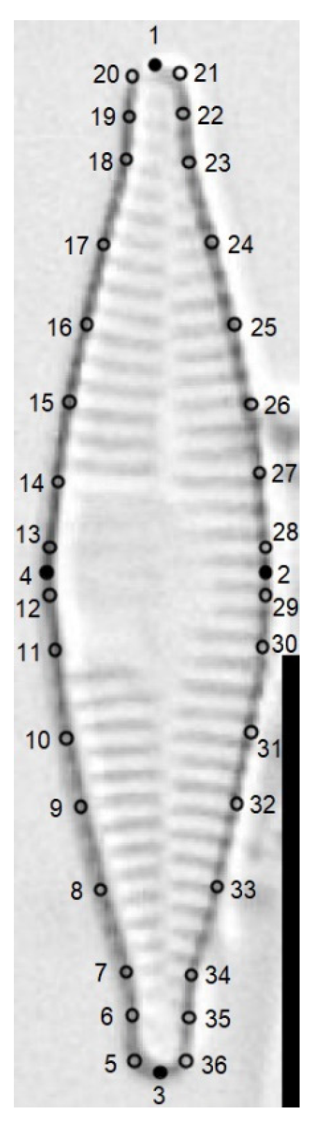
2.3. Morphological Analysis

The morphological examination of diatom valves was performed by means of two methods: traditional morphometric analysis of valve variables and geometric morphometric analysis of valve shape, both performed following [22]. Morphological terminology follows [39,40,41].
During observation of the permanent slides, the lengths of the smallest and longest valves of the taxon of interest were recorded. We collected 60 images for each of the two populations of F. sublanceolata-baikali, 30 images for F. pectinalis and 30 images for F. perminuta, giving a total of 180 LM images of individual valves. The following “traditional” morphometric characteristics were recorded for each valve: length, width, length-to-width ratio and striae density.
For geometric morphometric analysis, we selected valves from previously obtained LM images in the following way: First, the length range of each taxon was divided into five equal-length intervals. Then, four valves were randomly selected in each length interval for the two populations of F. sublanceolata-baikali, and two valves in each interval for F. pectinalis and F. perminuta. As a result, the selected LM images consisted of 20 images for each of the two populations of F. sublanceolata-baikali, 10 images for F. pectinalis and 10 images for F. perminuta, giving a total of 60 LM images.
Geometric morphometrics was employed to obtain shape variables, which preserve the geometry of morphological structure [24]; in contrast, traditional morphometrics uses quantitative characteristics (measurements and their ratios) as variables in statistical analysis. We used semilandmark-based geometric morphometrics as a more effective approach than landmark-based methods for capturing information about shape curvature [42]. Unlike landmarks representing discrete homologous points of shape, semilandmarks quantitatively slide along the outline curve until they match the shape in the best possible way [42].
The LM images were used to record the Cartesian X and Y coordinates of landmarks by means of the tpsDig2 software ver. 2.32 [43]. Of these 36 landmarks, 4 were fixed and the remaining landmarks were semilandmarks that were allowed to slide along the outline curve [42]. The four fixed landmarks were positioned at the intersections of the valve outline with the apical and transapical axes, and four sets of eight semilandmarks were placed between the four fixed landmarks (Figure 1). The average shape (called the consensus configuration) was estimated and then the coordinates of all specimens were aligned (translated, rotated and scaled) to this average shape by using the generalized Procrustes superimposition procedure [26] in the tpsRelw software ver. 1.75 [43].

2.4. Multivariate Statistical Analysis

For traditional morphometrics, canonical variate analysis (CVA), also known as discriminant analysis for multiple groups, was performed to test the separation between Fragilaria populations on the base of morphometric characteristics in the PAST software ver. 4.13 [44]. CVA provided an ordination that maximized the separation between the group means relative to the variation within groups. All morphometric variables were log-transformed.
For geometric morphometrics, Procrustes-superimposed coordinates were subjected to principal component analysis (PCA, i.e., relative warp analysis), which describes the positions of specimens relative to the consensus configuration and characterizes the directions of shape deformation in the morphospace [24]. To describe the variation in shape of the four Fragilaria populations, we plotted PC1 vs. PC2 and compared the positions of the clouds of points (i.e., specimens) belonging to different populations. PCA was performed using tpsRelw [43]. Non-parametric testing of the multivariate analysis of variance (NPMANOVA) was used to test for differences between populations in terms of their valve shape represented by the first two PC scores. NPMANOVA is a test of significant difference between two or more groups, based on any distance measure [45]. NPMANOVA based on the Euclidean distance was performed in PAST [44].

2.5. Allometric Variability and Size Correction

Size was measured for each specimen as the centroid size of the landmark configuration. The centroid size was computed as the square root of the summed squared deviations of the coordinates from their centroid [46]. To analyze the allometric ontogenetic trajectories, we first regressed PC1 against the log centroid size for each Fragilaria population. We then performed an analysis of covariance (ANCOVA) to test for differences in the slopes of the allometric trajectories between populations, using the centroid size as covariate. If the slope differences were not significant, the populations had the same allometric trajectory or were characterized by parallel allometric trajectories [36]. To verify this, a further test for differences in the intercepts of allometric trajectories was conducted using ANCOVA. ANCOVA and regression analyses were performed in PAST [44].
As analysis of the allometric trajectories revealed substantial size-related shifts along PC1 in the shape of all populations, an allometric correction was necessary to compare the positions of the four different Fragilaria populations in a size-standardized morphospace. Size-correction was performed in the MorphoJ software ver. 1.07a [47] by means of a pooled within-group allometric regression using the centroid size as covariate. The allometrically corrected PCA using the covariance matrix of the residuals from the allometric regression permits more meaningful comparisons of the shapes of specimens of different sizes [28]. To test for significant differences between populations in terms of their shapes as represented by the first two PC scores, NPMANOVA with pairwise comparisons was performed using PAST [44].

3. Results

3.1. Microscopical Analysis

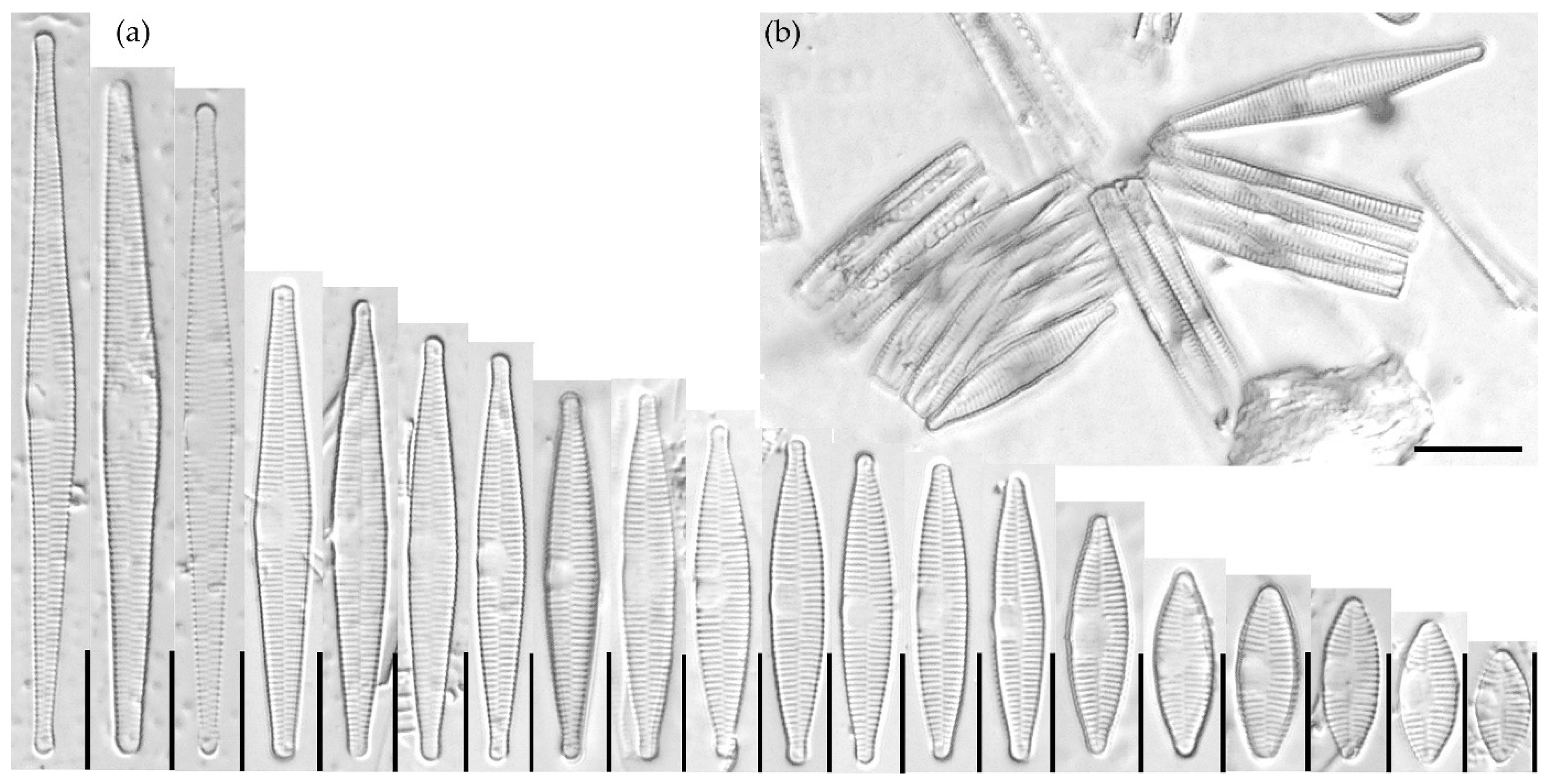
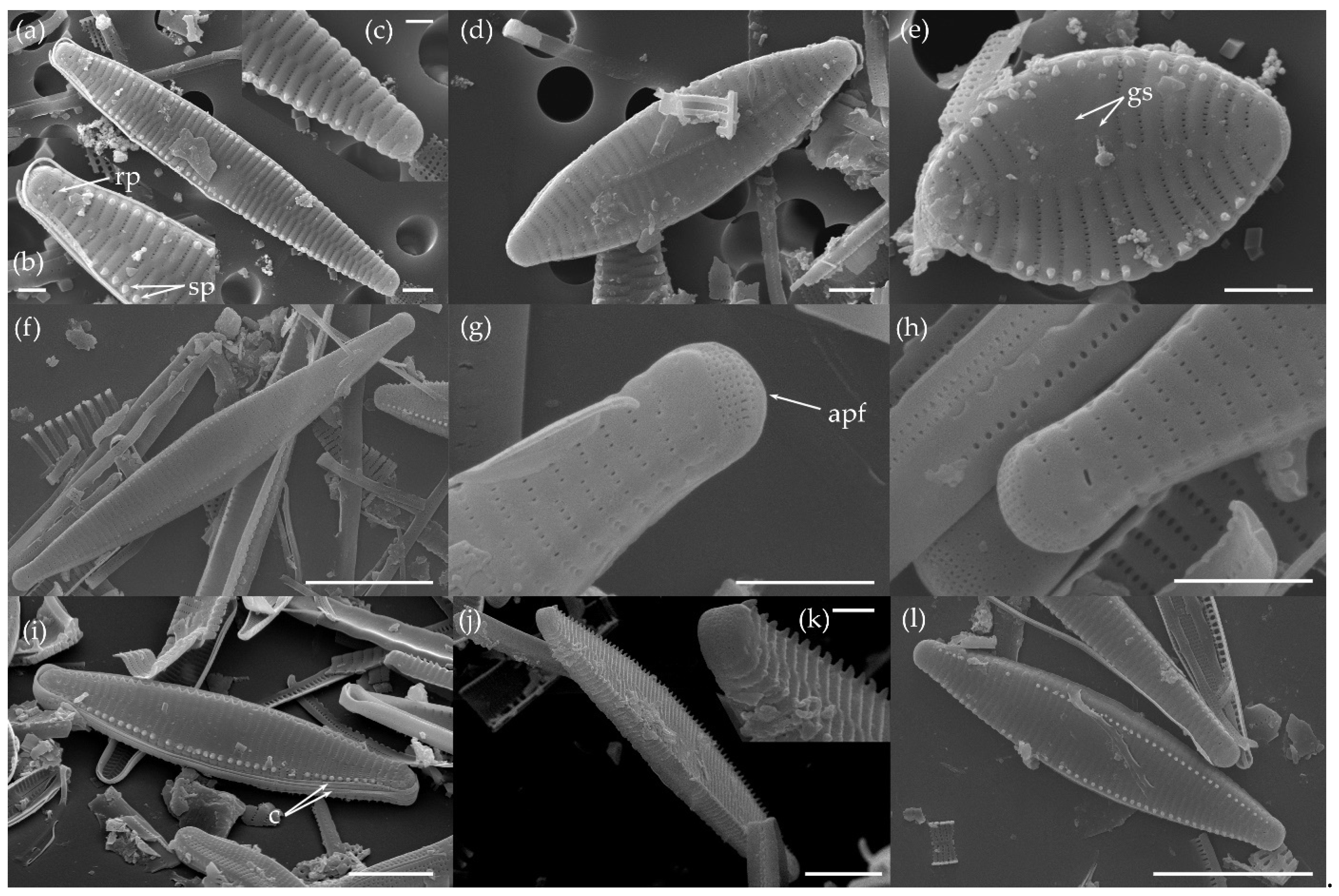
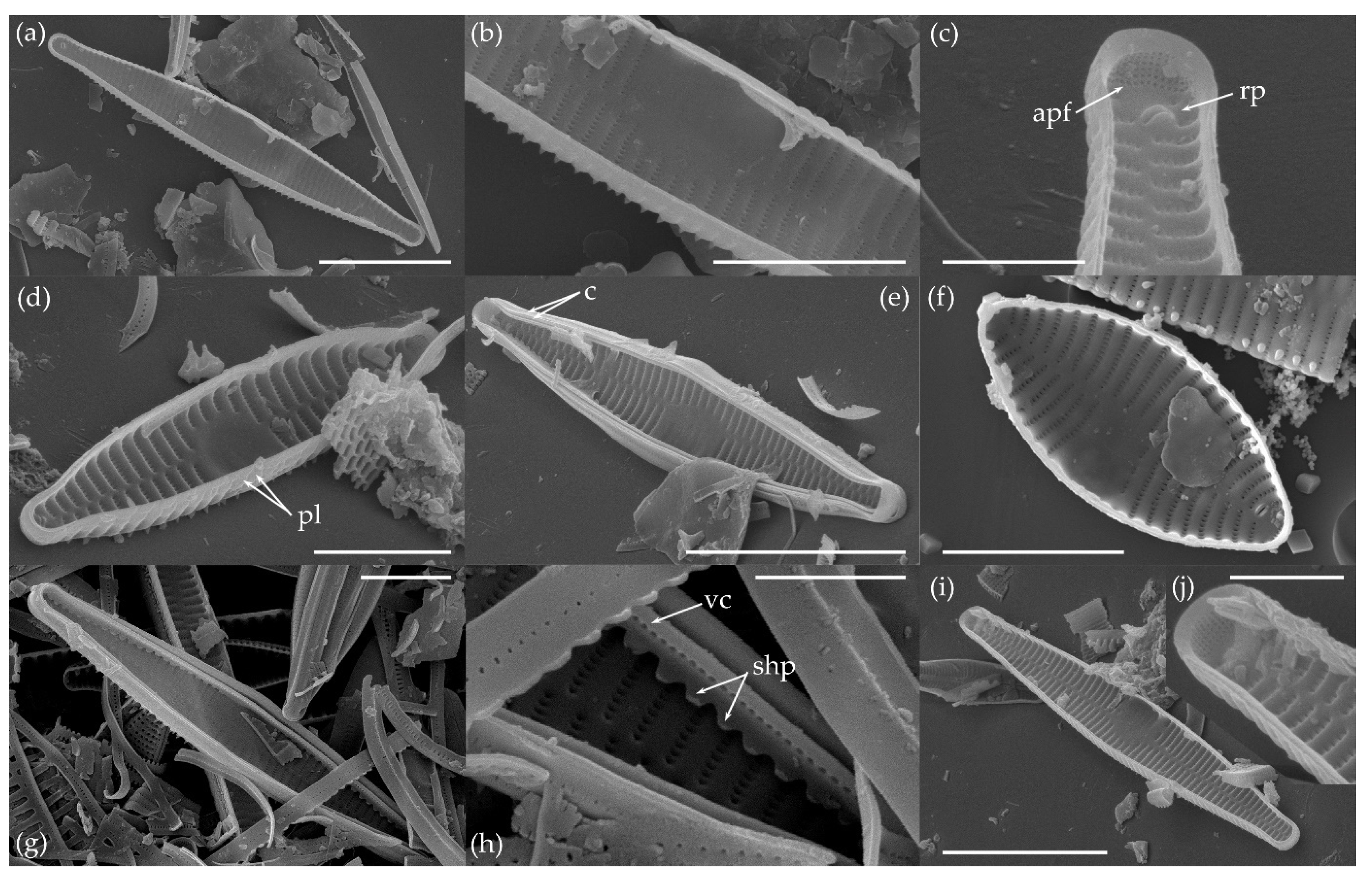
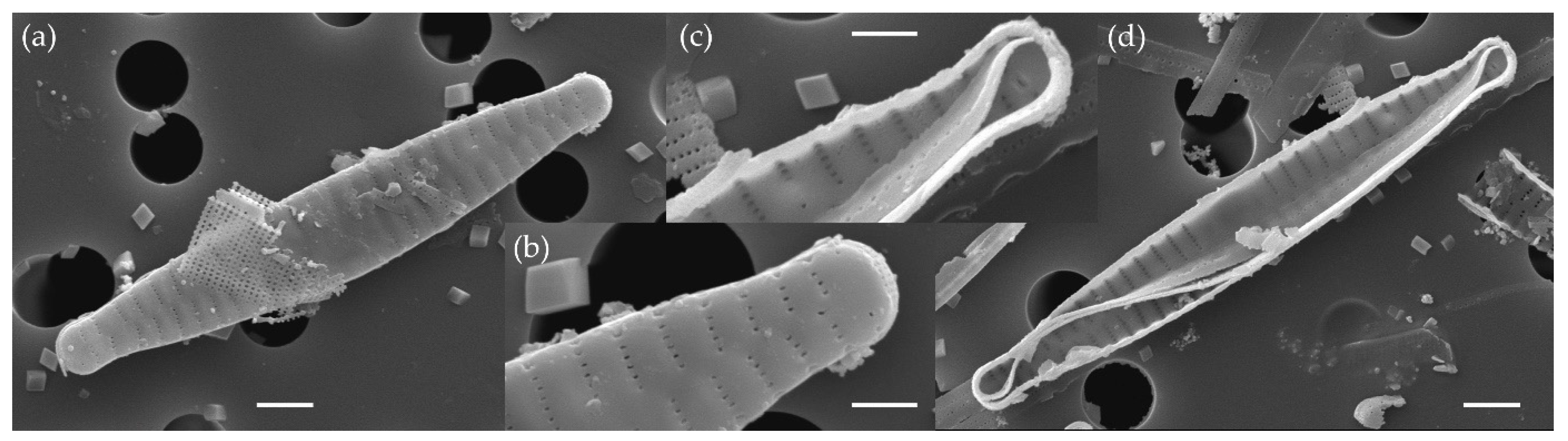
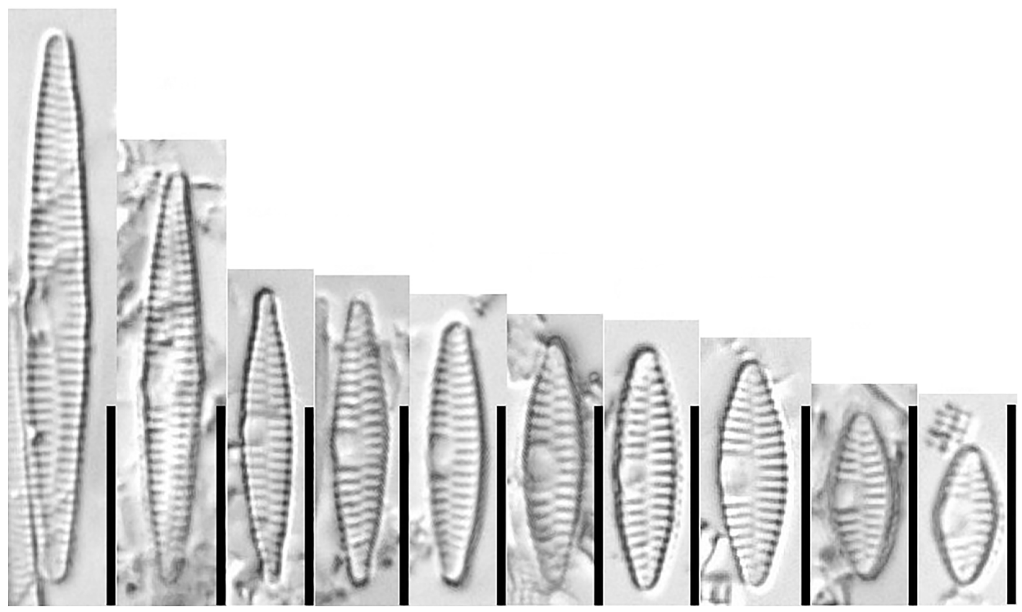
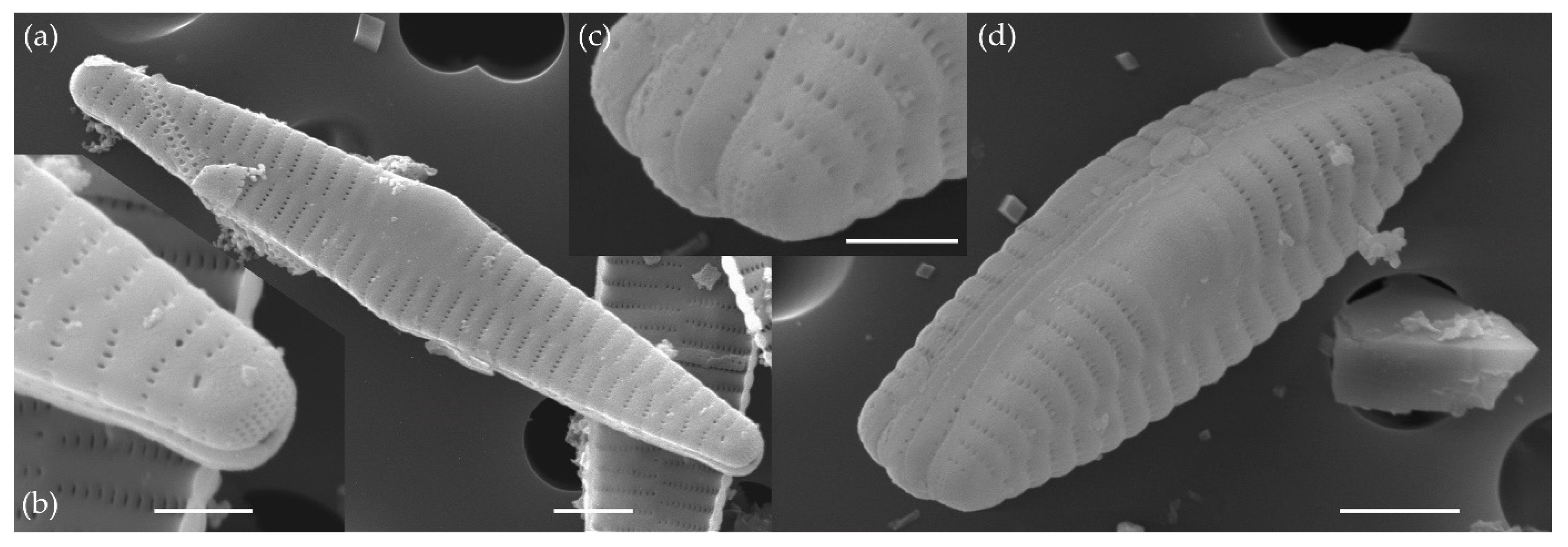
3.1.1. Fragilaria sublanceolata-baikali (Flower et D. M. Williams) Novais, C. Delgado et S. Blanco (Figure 2, Figure 3, Figure 4, Figure 5 and Figure 6)

Basionym: Fragilaria capucina f. sublanceolata-baikali Flower et D. M. Williams.
Figure 2. LM photographs of Fragilaria sublanceolata-baikali sampled in Lake Ladoga: (a) size diminution series and (b) several cells joined together. Scale bars: 10 μm.
Water 15 03994 g002
Figure 3. LM photographs showing size diminution series of Fragilaria sublanceolata-baikali sampled in Lake Baikal. Scale bars: 10 μm.
Water 15 03994 g003
Figure 4. SEM micrographs of Fragilaria sublanceolata-baikali from Lake Ladoga; external valve view. Abbreviations: apf—apical pore field, c—copula, gs—ghost striae, rp—rimoportula, sp—conical spines. Scale bars: (a,d,e,g,h): 2 μm; (b,c,k): 1 μm; (f,l): 10 μm; (i,j): 5 μm.
Water 15 03994 g004
Figure 5. SEM micrographs of Fragilaria sublanceolata-baikali from Lake Ladoga; internal valve view. Abbreviations: apf—apical pore field, c—copula, p—plaques, rp—rimoportula, shp—sawtooth-shaped projections, vc—valvocopula. Scale bars: (a,e,i): 10 μm; (b,d,f,g): 5 μm; (f,c,h,j): 2 μm.
Water 15 03994 g005
Figure 6. SEM micrographs of Fragilaria sublanceolata-baikali from Lake Baikal. (aj), (l, upper valve): external valve view; (k), (l, lower valve)–(n): internal valve view. Abbreviations: apf—apical pore field, c—copula, p—plaques, rp—rimoportula, shp—sawtooth-shaped projections, sp—conical spines, v—velum, vc—valvocopula. Scale bars: (a,d,e,g,l): 10 μm; (b,c,f,j,k): 2 μm; (h,i,m,n): 5 μm.
Water 15 03994 g006
Synonym: Fragilaria capucina f. lanceolata-baikali Flower et D. M. Williams.
Description: Frustules rectangular in girdle view; solitary or 2–4 cells together. Valves narrowly lanceolate in larger specimens to elliptic-lanceolate in smaller specimens with protracted, subcapitate (in larger specimens) to rostrate apices (in smaller specimens). Valve length: 8.0–61.2 μm; width: 3.5–5.2 μm. Axial area narrow, linear, not widening towards the central area. An asymmetrical central area formed by unilateral hyaline fascia, clearly swollen on one side; opposite side with several shortened or almost not shortened striae; ghost striae only rarely observed. Striae alternating, not interrupted in the valve face/mantle junction, uniseriate, parallel to very weakly radiate throughout, becoming slightly more radiate near the apices; 16–20 per 10 μm. Striae compose of small, rounded to apically elongated areolae; 7 per 1 μm; areolae occluded by individual hymenes (areolae occlusions only rarely observed, probably due to severe sample preparation). Valves with marginal thick, conical spines (spinules) located on the virgae; spinules occasionally absent (independent from valve length); linking spines absent. Apical pore fields present on both poles; pore field composed of up to 6 regular rows of small pores (porelli). Plaques rarely observed on abvalvar margin of mantle. One rimoportula present per valve; rimoportula transapically elongated, located at apex. Cingulum composed of several copulae. Girdle bands with 1 row of small unoccluded perforations, located at mid-line. Valvocopula open, closely attached to mantle interior, with sawtooth-shaped projections attached to the valve.
Distribution: Fragilaria sublanceolata-baikali is a freshwater species, registered in Lake Baikal [48]. This species was recorded for the first time for Lake Ladoga.
Remarks: More taxonomic information about F. sublanceolata-baikali can be found in Flower et al. [11], Novais et al. [12] and Van de Vijver et al. [13].
Morphological study of the species previously identified by Rusanov et al. [9] as Fragilaria cf. sublanceolata-baikali in Lake Ladoga (Figure 2, Figure 4 and Figure 5) showed that this taxon is considered conspecific with F. sublanceolata-baikali (Figure 3 and Figure 6) and corresponds to the description provided by Van de Vijver et al. [13]. Both populations of F. sublanceolata-baikali from Lake Ladoga and Lake Baikal overlap considerably in their main morphological features (Table 1; Figure 2, Figure 3, Figure 4, Figure 5 and Figure 6). The specimens of the F. sublanceolata-baikali population from Baikal have slightly shorter and narrower valves compared to the Ladoga population. The analysis of the F. sublanceolata-baikali valve shape in the cell-size reduction series showed that in both populations the valves are strictly narrowed lanceolate in large specimens, while valves of smaller specimens are more elliptic-lanceolate with gradually tapering margins (Figure 2 and Figure 3). Valve apices vary from subcapitate (in larger specimens) to rostrate (in smaller specimens) (Figure 2 and Figure 3). There were no differences between Ladoga and Baikal specimens according to other morphological characteristics, such as the structure of axial and central areas, arrangement of the apical pore field, structure and location of spinules, number and location of rimoportulae and the structure of girdle bands (Figure 4, Figure 5 and Figure 6; Table 1).

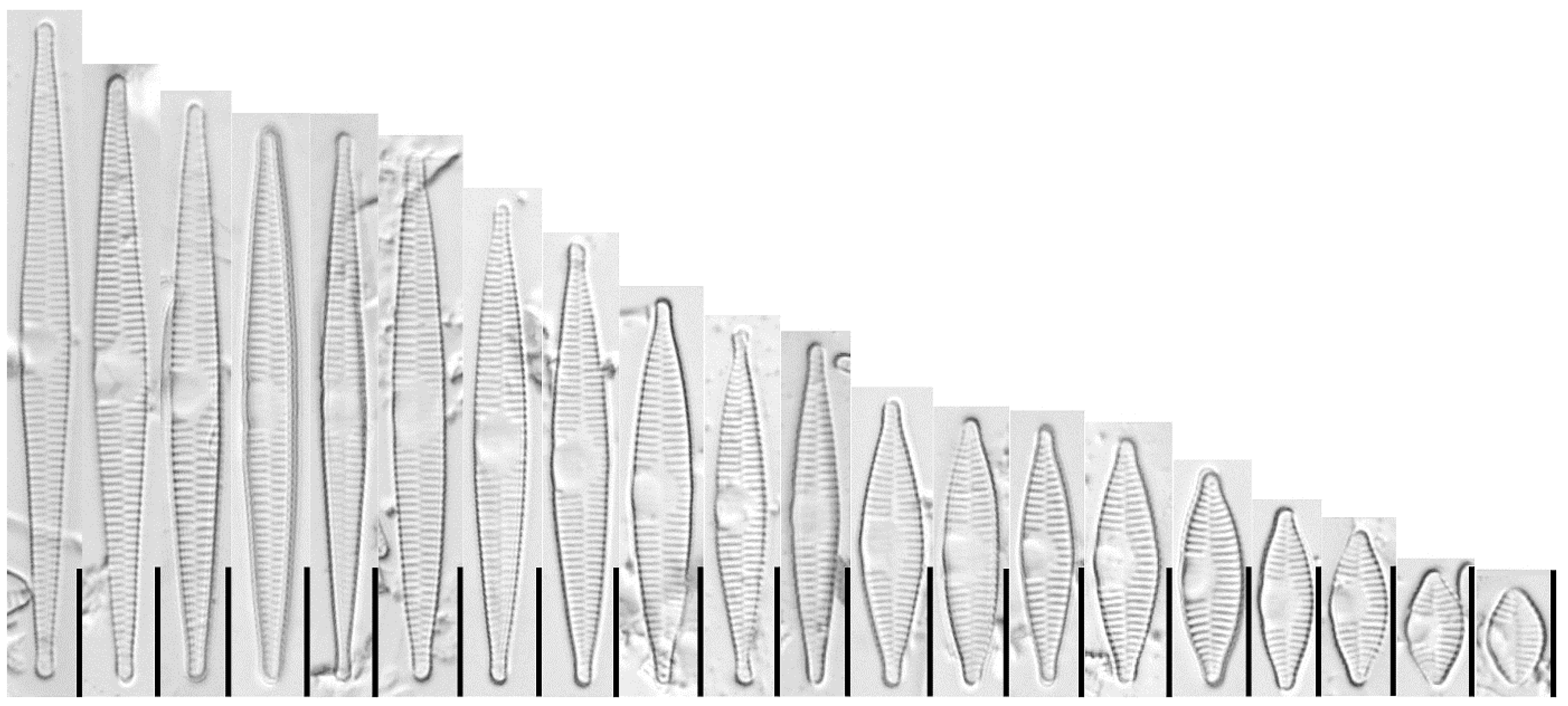
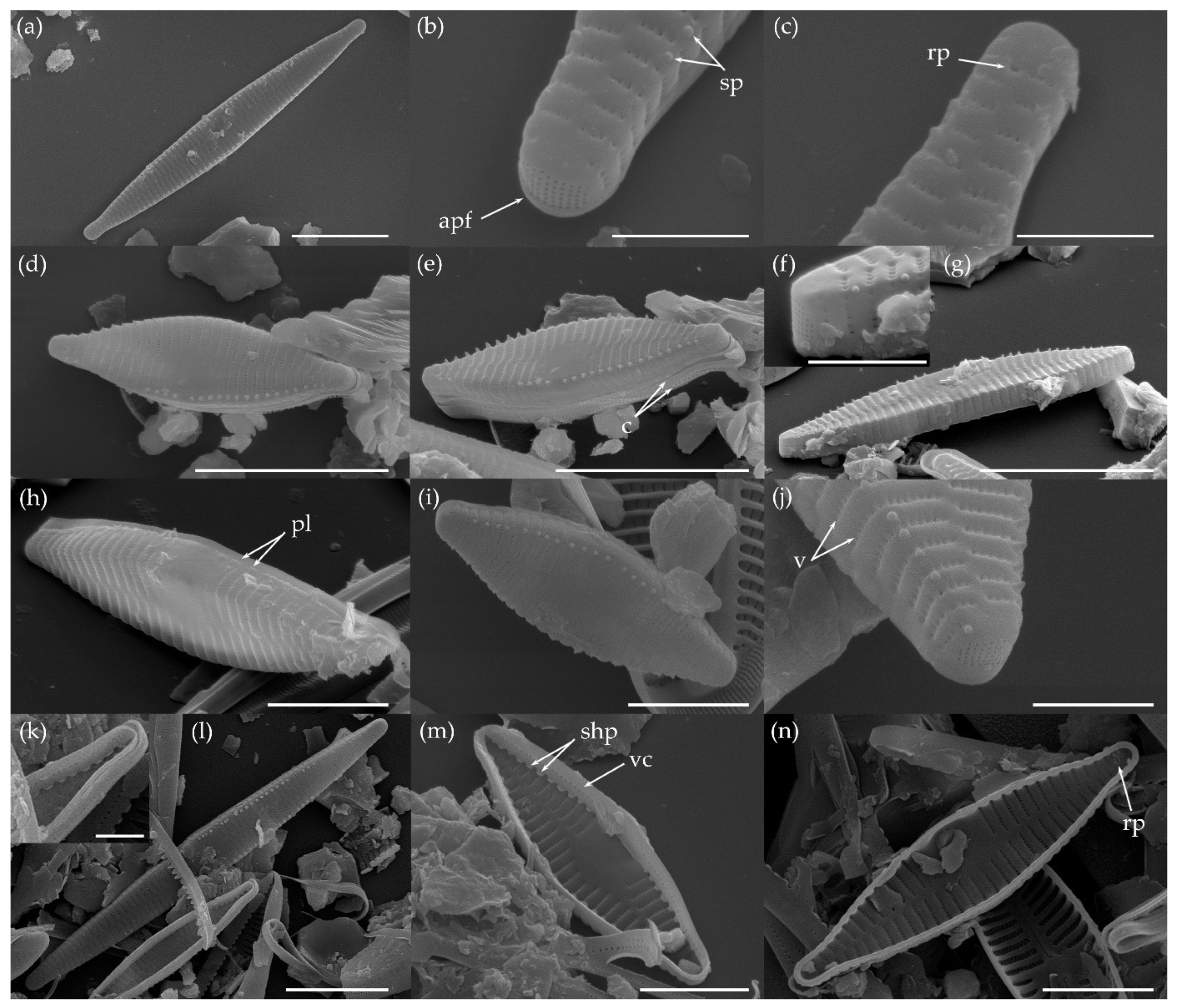
3.1.2. Fragilaria pectinalis (O. F. Müll.) Lyngb. (Figure 7 and Figure 8)

Basionym: Conferva pectinalis O. F. Müll.
Description: Valves linear lanceolate in larger specimens to lanceolate in smaller specimens with protracted, subcapitate (in larger specimens) to approximately rostrate apices (in smaller specimens). Valve length: 11.3–25.8 μm; width: 3.2–4.2 μm. Axial area narrow, linear, not widening towards the central area. An asymmetrical central area formed by unilateral hyaline fascia; opposite side with several shortened striae. Striae alternating, not interrupted in the valve face/mantle junction, uniseriate, parallel to very slightly radiate near the apices; 14–17 per 10 μm. Striae composed of small, rounded to apically elongated areolae; 5–6 per 1 μm. Linking spines and spinules absent. Apical pore fields present on both poles; pore field composed of several regular rows of porelli. One rimoportula present per valve, located at apex. Girdle bands not observed.
Distribution: Fragilaria pectinalis is a widespread freshwater species, registered in different countries of Europe, North America and Southwest Asia (for more details see [48]). The species was recorded for the first time for Lake Ladoga.
Remarks: More taxonomic information about Fragilaria pectinalis can be found in Tuji, Williams [14,15], Wetzel, Ector [16] and Van de Vijver et al. [49].
Figure 7. LM photographs showing size diminution series of Fragilaria pectinalis. Scale bars: 10 μm.
Water 15 03994 g007
Figure 8. SEM photographs of Fragilaria pectinalis: (a,b) external valve view and (c,d) internal valve view. Scale bars: (a,d): 2 μm; (b,c): 1 μm.
Water 15 03994 g008

3.1.3. Fragilaria perminuta (Grunow) Lange-Bertalot (Figure 9 and Figure 10)

Basionym: Synedra perminuta Grunow.
Description: Valves narrowly rhombic-lanceolate in larger specimens to broadly lanceolate in smaller specimens with slightly protracted, approximately rostrate (in larger specimens) to not protracted, cuneately rounded apices (in smaller specimens). Valve length: 6.9–28.1 μm; width: 2.8–3.6 μm. Axial area narrow, linear, not widening towards the central area. An asymmetrical central area formed by unilateral hyaline fascia, often swollen on one side; opposite side featuring several shortened striae. Striae alternating, not interrupted in the valve face/mantle junction, uniseriate, parallel to very slightly radiate near the apices; 18–20 per 10 μm. Striae composed of small, rounded to apically elongated areolae; 7 per 1 μm. Linking spines and spinules absent. Apical pore fields present on both poles, pore field composed of several (at least 4) regular rows of porelli. One rimoportula present per valve, located at apex. Girdle bands not observed.
Distribution: Fragilaria perminuta is a widespread freshwater species, registered in different countries of Europe, North America, the Middle East and Asia (for more details see [48]). The species was recorded for the first time for Lake Ladoga.
Remarks: More taxonomic information about Fragilaria perminuta can be found in Tuji, Williams [15], Delgado et al. [17], Wetzel, Ector [16] and Van de Vijver, Ector [18].
Figure 9. LM photographs showing size diminution series of Fragilaria perminuta. Scale bars: 10 μm.
Water 15 03994 g009
Figure 10. SEM photographs of Fragilaria perminuta; external valve view. Scale bars: (a,d): 2 μm and (b,c): 1 μm.
Water 15 03994 g010

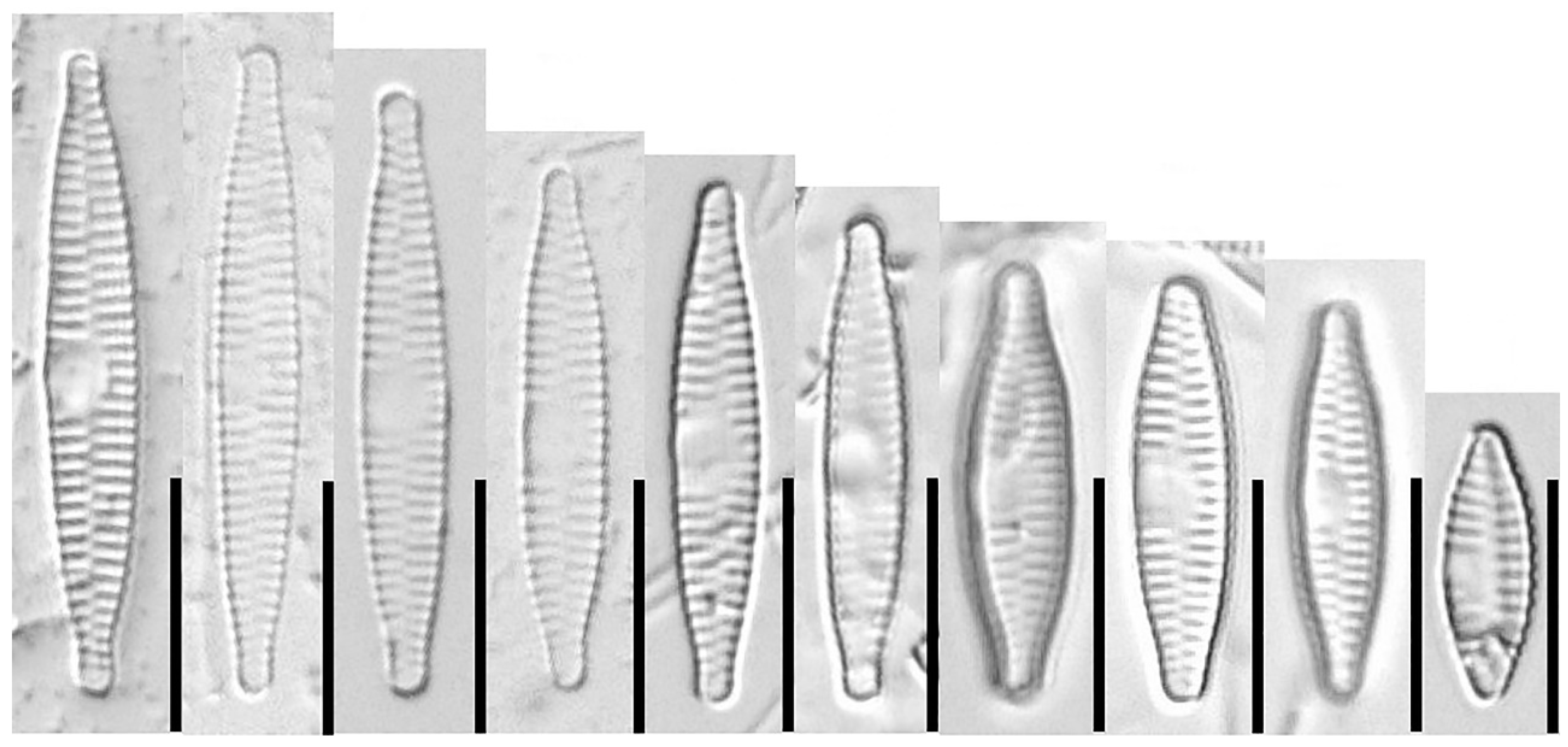
3.2. Traditional Morphometric Analysis

The traditional morphometric characteristics (length, width, length-to-width ratio and striae density) overlapped among the four populations of Fragilaria (Table 1). In the CVA using “population” as the grouping variable, the greater part of total variance (99.9%) was explained along the first two canonical variates (CVs): CV1 and CV2 explained 72.5% and 27.4% of the total variance, respectively, while CV3 explained only 0.1% of the total variance. The scatterplot of CV1 and CV2 showed aggregation of the individual specimens into three groups (Figure 11). The two populations of F. sublanceolata-baikali were grouped together and showed little separation. At the same time, F. pectinalis and F. perminuta were separated from each other and from the two populations of F. sublanceolata-baikali. CV1 segregated the two populations of F. sublanceolata-baikali from F. pectinalis and F. perminuta, while CV2 explained the difference between F. pectinalis and F. perminuta.
Numeric canonical loadings indicated the strength and direction of the relationships between the CVs and morphometric characteristics. Valve length was the morphometric characteristic with the highest loading on CV1, while striae density had the highest loading on CV2 (Table 2). Thus, along CV1, F. sublanceolata-baikali was separated from F. pectinalis and F. perminuta by the higher valve length; meanwhile, along CV2, F. perminuta was distinguished from F. pectinalis by the higher striae density.

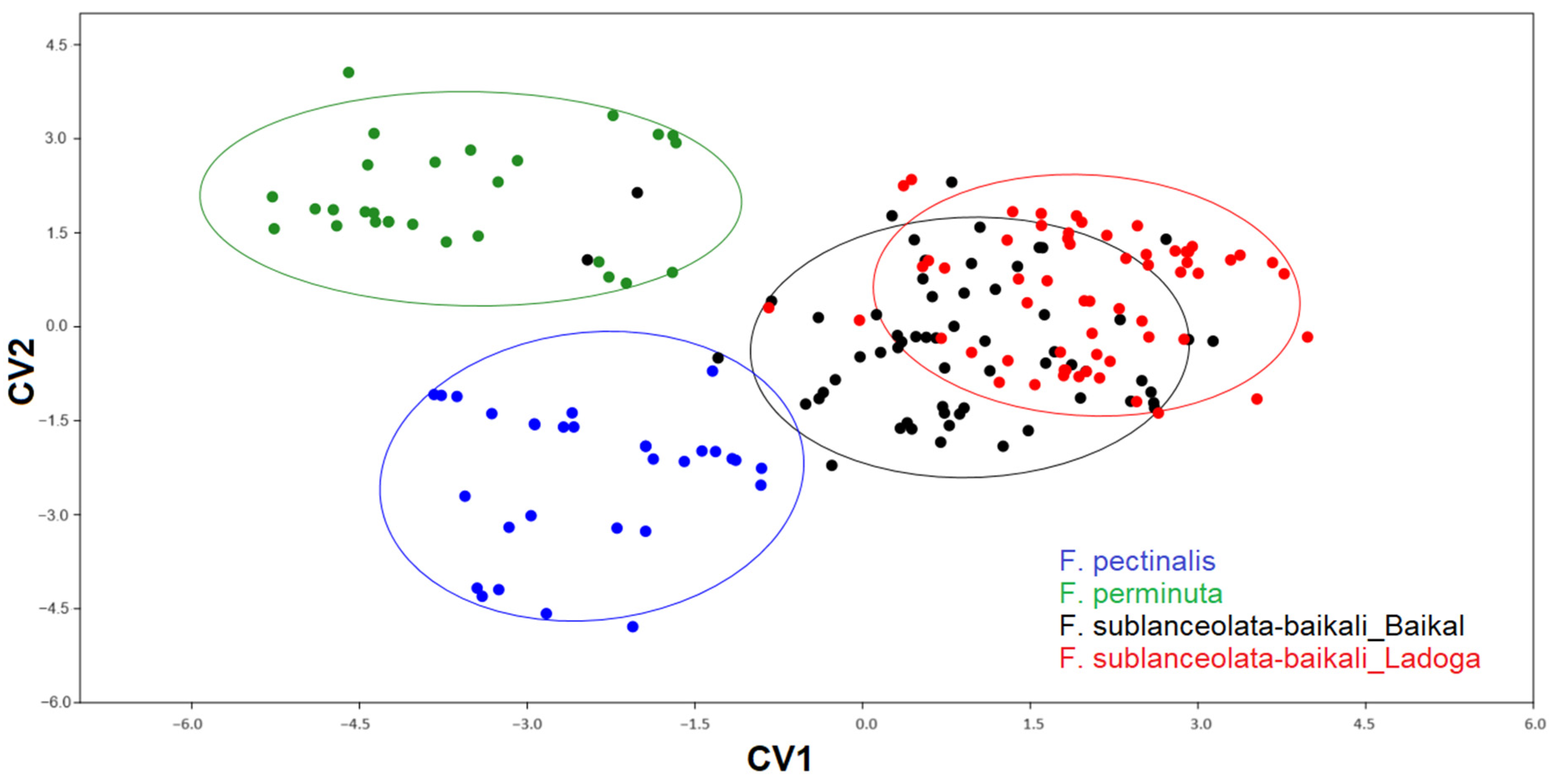
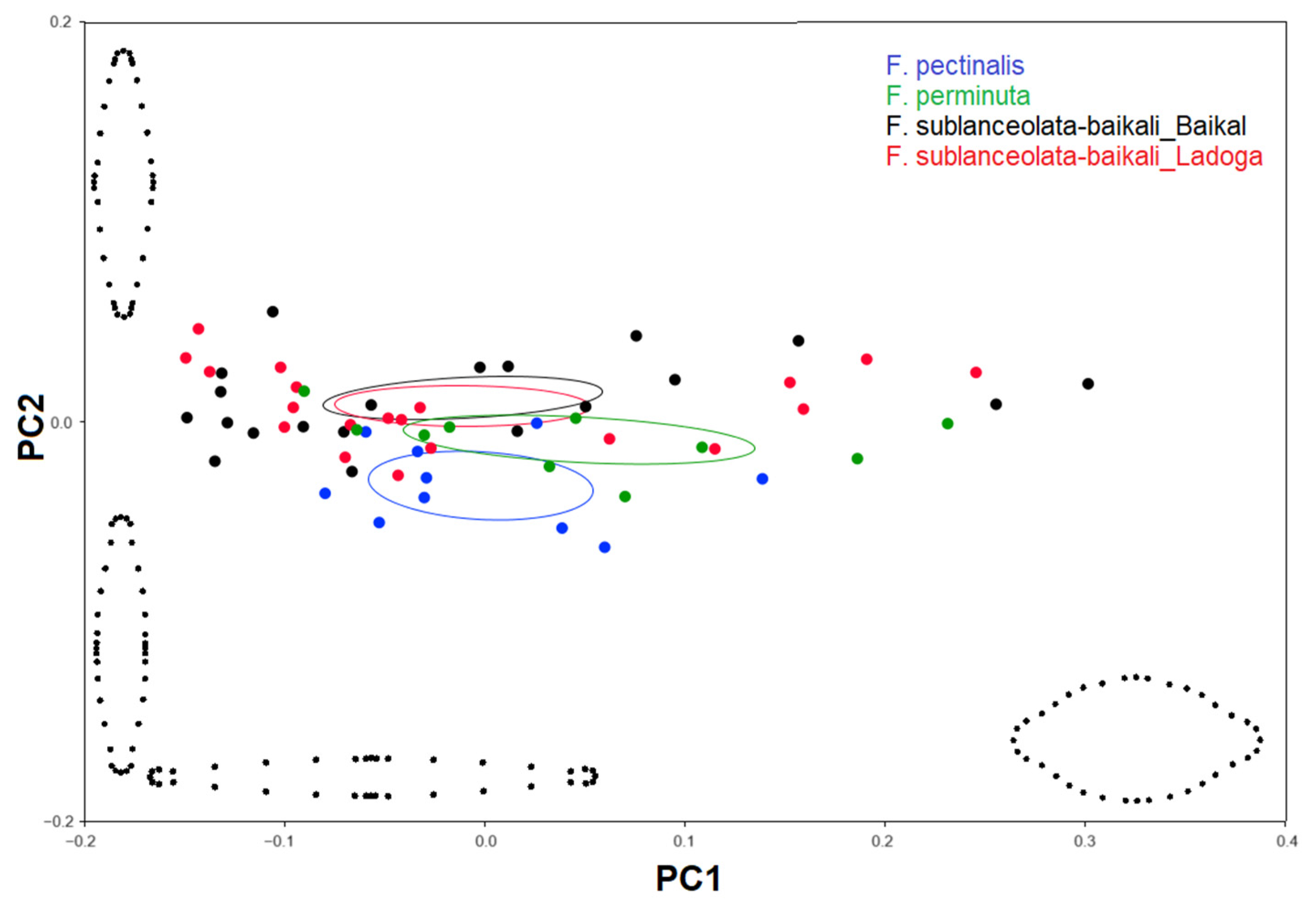
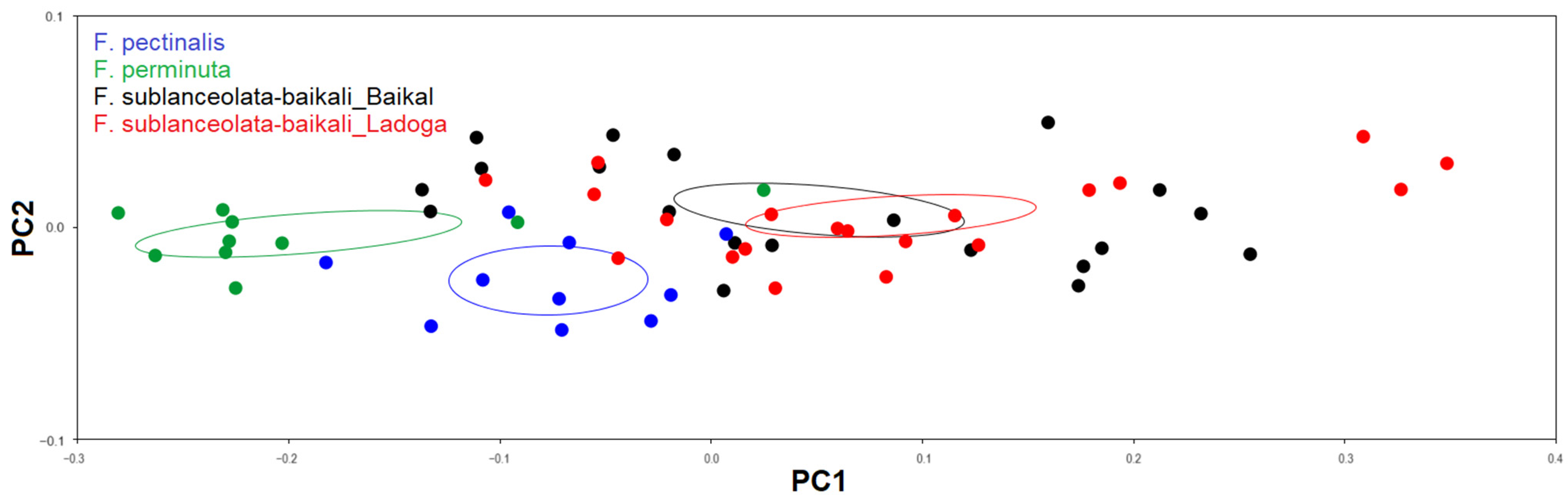
3.3. Geometric Morphometric Analysis

3.3.1. Testing Shape Variation in Populations

The PCA of Procrustes coordinates showed that the first three PCs accounted for 90.6%, 4.1% and 0.9% of the shape variability across populations, respectively (95.6% of the total variance). Because inclusion of PCs beyond the second did not contribute substantially to increase the explained shape variability in these populations, only the first two PCs were included in further analysis.
PC1 represented a gradient in the valve shape from narrowly lanceolate to broadly lanceolate from the left to the right of the plot (Figure 12). PC2 displayed a gradual variation in the shape of the valve ends from subcapitate to slightly protracted cuneate from the bottom to the top of the plot (Figure 12). The scatterplot of PC1 and PC2 showed considerable overlap between populations (Figure 12), which made their separation statistically insignificant (NPMANOVA; F = 0.994, p = 0.405).

3.3.2. Testing the Effect of Size on Shape and Size Correction

Correlation analysis showed that only PC1 correlated substantially with log centroid size (Pearson correlation coefficient r = 0.907, p < 0.0001), while PC2 and PC3 did not (r = −0.232, p = 0.075 and r = −0.097, p = 0.459, respectively). These values indicate that the allometric shape change is confined to a single axis with the largest variance in PC1.
Linear regression confirmed a strong allometric effect on shape variation, as captured by PC1 for all four populations of Fragilaria (Table 3, Figure 13). ANCOVA showed that the slopes of regression lines did not differ statistically (F = 0.407, p = 0.749) (Table 4). To determine which populations had the same allometric trajectory or parallel allometric trajectories, we performed ANCOVA with pairwise comparisons to test for differences in the intercepts of allometric trajectories. ANCOVA revealed that there was an overall statistical difference in the intercepts between different populations (F = 33.63, p < 0.0001). Pairwise comparison showed that the two populations of F. sublanceolata-baikali differed significantly (p < 0.0001) from F. pectinalis and F. perminuta (Table 4). Also, there were significant differences between (p < 0.01) F. pectinalis and F. perminuta (Table 4). Taken together, these results clearly indicate parallel allometric trajectories between F. sublanceolata-baikali, F. pectinalis and F. perminuta. In contrast, the difference in the intercepts of allometric trajectories was not significant (p = 0.110) between the two populations of F. sublanceolata-baikali (Table 4), suggesting that these populations were on a common allometric trajectory.
As a result of the strong allometric effect in the geometric data, effective discrimination between populations required the removal of size-dependent aspects of shape variation by means of a pooled within-group allometric regression. PCA of the residuals of the allometric regression showed that the first two PCs accounted for 95.4% of the total variance (92.6% and 2.8% for PC1 and PC2, respectively). A scatterplot of the allometrically corrected axes PC1 and PC2 revealed three reasonably well-defined groups of specimens belonging to F. sublanceolata-baikali (both populations together), F. pectinalis and F. perminuta (Figure 14). The clear separation of F. sublanceolata-baikali, F. pectinalis and F. perminuta was confirmed by NPMANOVA (F = 15.62, p < 0.0001; Table 5). Conversely, the two populations of F. sublanceolata-baikali formed tightly overlapping groups, indicating no significant differences in their shape (p = 0.420; Table 5).

4. Discussion

4.1. Microscopic Examination

Our results showed that morphological features visible both in LM and SEM allowed us to distinguish the three species of the Fragilaria capucina/vaucheriae complex: F. sublanceolata-baikali, F. pectinalis and F. perminuta.
Several SEM characteristics such as uniseriate striae, round or slightly elongated areolae composition, a well-developed central area, apical pore fields of ocellulimbus type and the presence of one rimoportula per valve confirm the placement of the three studied taxa to the genus Fragilaria [50]. Another ultrastructural feature shared by these taxa with the genus Fragilaria is open girdle bands [51]. All three taxa discussed in this paper have an asymmetrical central area formed by unilateral hyaline fascia. Despite similarity between these taxa with respect to the hyaline area, there are some subtle differences between them. The central area in F. sublanceolata-baikali and F. perminuta is clearly unilaterally swollen, which is usually not observed in F. pectinalis valves. The presence/absence of spines is not currently a decisive characteristic in the definition of the genus Fragilaria, but it remains a distinguishing characteristic allowing the characterization of species within the genus, especially if the position of spines on the valve surface is considered [52]. In this respect, F. pectinalis and F. perminuta have no spines, whereas F. sublanceolata-baikali has thick conical spines located on the virgae (interstriae).
Other SEM and LM characteristics distinguishing F. sublanceolata-baikali from another morphologically similar species are summarized in Table A1 (Appendix A).

4.2. Traditional and Geometric Morphometrics

The canonical variate analysis (CVA) using traditional morphometric data further corroborated that the three species can be separated on the basis of morphological characteristics. On the other hand, the morphological analysis supplemented by CVA showed the homogeneity of morphological characteristics between the Ladoga and Baikal populations of F. sublanceolata-baikali, suggesting conspecificity between the two populations.
The three Fragilaria species can also be differentiated by distinct morphological characteristics discernable in LM. CVA results showed that valve length contributes most to the separation of F. sublanceolata-baikali from F. pectinalis and F. perminuta along CV1. The valve length of F. sublanceolata-baikali is much higher (maximum valve length = 61 μm) in comparison to those of F. pectinalis (26 μm) and F. perminuta (28 μm). It should be noted, however, that in our study the maximum values of valve length are lower than those recorded for F. pectinalis (36 μm) and F. perminuta (46 μm) in other studies [16,18,49]. Thus, the valve length is not a strong enough characteristic to unequivocally distinguish F. perminuta from F. sublanceolata-baikali over their entire valve length ranges. According to CVA results, striae density is the main character contributing to the distinction between F. pectinalis and F. perminuta. F. perminuta has a higher striae density (18–20 per 10 μm) than F. pectinalis (14–17 per 10 μm), lacking any overlap between both taxa. F. sublanceolata-baikali also has denser striation (16–20 per 10 μm) than F. pectinalis, and thus can be easily differentiated from F. pectinalis by this characteristic. The valve width (having high canonical loadings for CV1) is also important to separate wider F. sublanceolata-baikali (3.5–5.2 μm) from narrower F. perminuta (2.8–3.6 μm) and F. pectinalis (3.2–4.2 μm), despite some overlap between them in this character.
The semilandmark-based geometric morphometric shape analysis provides a quantitative means to evaluate taxonomic identification within groups of closely related species. In our study, PCA of Procrustes-superimposed coordinates showed that there was an extensive overlap in valve outline between the four studied populations of Fragilaria. As a result, the three Fragilaria species were not distinguished by the location of their specimens in PCA morphospace, which was not subjected to prior size correction. Thus, the next step of our study was to test for allometric effects in the shape variation of studied populations, to justify the expediency of applying the size-correction technique [28].

4.3. Allometric Effect and Size Correction

The strong allometric effect on shape variation was corroborated by statistically significant relationships between PC1 and valve size (expressed as log centroid size) for each studied population of Fragilaria. Testing for the differences in allometric patterns between populations (i.e., slope and intercept of allometric trajectories) showed that populations belonging to different species have separate and parallel ontogenetic trajectories. This implies that each species has a specific pattern of shape variation throughout the valve ontogenetic process. In contrast, the two geographically distant populations of F. sublanceolata-baikali were on a common allometric trajectory, indicating the conspecificity between these populations.
For size correction in taxonomic studies, an estimation of the common allometric component for several groups (species) is required [34,35,36]. A statistical method that can achieve such an estimate is pooled within-group regression [28]. After Procrustes superimposition of landmark coordinates, the shape data may still contain a component of size-related shape variation because of common allometries. Such allometric shape variation can confound the results of taxonomic studies [35]. Using the residuals from the pooled within-group regression of shape on size, the common allometric component of shape variation can be identified and removed. Removing size-related shape variability leaves size-unrelated shape variability, which is free of the confounding effects of common allometry and substantially improves differentiation among species [35]. A key assumption in pooled within-group regression is that all species have parallel ontogenetic trajectories [28]. Only in this case, a single estimate of the allometric pattern can be calculated for all species.
In the present study, the three Fragilaria species were clearly separated in the size-corrected morphospace, whereas the two populations of F. sublanceolata-baikali formed tightly overlapping groups. Although the valve shape of all three studied species varies with size reduction from narrowly lanceolate to elliptic-lanceolate, species-specific characteristics separated Fragilaria species in the morphospace after removal of the allometric variation. F. sublanceolata-baikali has the most convex valves with the widest range of variation in the size-adjusted morphospace. F. perminuta has a more rhomboid outline, whereas F. pectinalis has a more linear shape. Subtle differences in the shape of valve ends separate F. sublanceolata-baikali and F. pectinalis from F. perminuta. The first two species have subcapitate to rostrate apices, while the latter has only rostrate to cuneate apices observed in smaller valves. All these shape characteristics are usually discernable under LM, and therefore useful for species identification and separation.

4.4. Geographical Distribution and Ecology of F. sublanceolata-baikali

The results of traditional morphometric and size-corrected geometric morphometric analyses of the two populations of Fragilaria sublanceolata-baikali from Lake Ladoga and Lake Baikal made it possible to confirm that both populations represent the same species. Thus, our results indicate that F. sublanceolata-baikali, which has until now been known as a Baikal endemic [11,12], is growing and reproducing quite successfully in Lake Ladoga. In this regard, a few questions arise. First, can F. sublanceolata-baikali be considered as a true Baikal endemic species or a species with a wider geographical distribution? Unfortunately, it is still difficult to unequivocally answer this question due to insufficient knowledge of the distribution of freshwater diatoms in Russia. This lack of knowledge partially stems from a very limited use of electron microscopy in diatom research in Russia. Therefore, we cannot say positively that the distribution of F. sublanceolata-baikali is restricted to Lake Baikal. On the other hand, dozens of endemic algal taxa are known in Lake Baikal, including diatoms [53,54,55,56,57,58]. Thus, the second question: how could the Baikal endemic appear in Lake Ladoga? It can be assumed that F. sublanceolata-baikali was likely brought into the lake along with other invaders from Lake Baikal, which were specifically introduced into Lake Ladoga in the 20th century. There have been well-known attempts to acclimatize Baikal fish species in Lake Ladoga, including the Baikal omul Coregonus autumnalis migratorius (Georgi, 1775) in the 1950s and the Baikal sturgeon Acipenser baerii baicalensis (Nikolsky, 1896) in the 1960s [59]. There is also an example of successful invasion and active distribution in Lake Ladoga of the Baikal amphipod Gmelinoides fasciatus (Stebbing, 1899), which appeared in the lake in the late 1980s [60]. G. fasciatus invaded Lake Ladoga as a consequence of its intentional introduction aimed at enhancing fish production in some Karelian Isthmus lakes close to Lake Ladoga’s western coast in the early 1970s [61]. Thus, we believe that the most probable time of appearance of F. sublanceolata-baikali in Lake Ladoga is from the early 1970s to the late 1980s. It is difficult to say exactly when F. sublanceolata-baikali invaded Lake Ladoga, since in previous studies SEM was used mainly for planktonic diatoms [62,63]. However, it is obvious that the ecological environments of Lake Ladoga turned out to be favorable for the further successful population growth and distribution of F. sublanceolata-baikali.
In Lake Ladoga, the distribution of Fragilaria sublanceolata-baikali is restricted to a few sites on the western and northern coasts featuring oligo-mesotrophic conditions. In particular, the Shchuchiy bay (the western coast), where periphyton samples containing F. sublanceolata-baikali were collected, is characterized by low levels of total phosphorus (median value = 27 μg P l−1), total nitrogen (0.510 mg N l−1) and conductivity (107 μS cm−1) and an alkaline pH (7.8). The accompanying diatom flora for F. sublanceolata-baikali (with 20% relative abundance) asides from F. pectinalis (7%) and F. perminuta (1%) are F. gracilis (13%), Encyonema minutum (Hilse) D. G. Mann (13%), Achnanthidium minutissimum (Kütz.) Czarn. (8%), Diatoma moniliformis (Kütz.) D. M. Williams (4%) and Tabellaria flocculosa (Roth) Kütz. (2%). F. pectinalis and F. perminuta have a much wider distribution across the lake coastline, including Volkhov Bay on the southern coast, the most eutrophic and polluted embayment of Lake Ladoga.

5. Conclusions

In the present study, we applied traditional and geometric morphometrics to characterize the morphology of three species within the Fragilaria capucina/vaucheriae complex: F. sublanceolata-baikali, F. pectinalis and F. perminuta. Traditional morphometrics reliably differentiated between all studied species and showed high similarity between the two geographically distant populations of F. sublanceolata-baikali. Geometric morphometrics yielded similar results, but only after accounting for allometric shape variation. To our knowledge, this is the first study to examine the applicability of size correction (by means of applying a pooled within-group allometric regression) in diatom taxonomy. Our results provide evidence that the estimation of allometric shape variation may be a very useful tool for distinguishing between morphologically close diatom species. Both traditional and geometric morphometrics confirmed a morphological similarity between the two populations of F. sublanceolata-baikalii, which indicates that this taxon can be considered invasive in Lake Ladoga.

Author Contributions

Conceptualization, A.G.R.; methodology, A.G.R. and M.A.G.; statistical analysis, A.G.R.; investigation, A.G.R., M.A.G., M.D., A.A.G. and M.Y.K.; resources, É.Á., M.A.G. and M.Y.K.; writing—original draft preparation, A.G.R. and M.A.G.; writing—review and editing, M.D., É.Á., K.T.K., I.G. and I.S.; visualization, A.G.R., M.D., M.A.G. and A.A.G.; supervision, É.Á. All authors have read and agreed to the published version of the manuscript.

Funding

Project no. TKP2021-NKTA-32 has been implemented with the support provided by the Ministry of Innovation and Technology of Hungary from the National Research, Development and Innovation Fund, financed under the TKP2021-NKTA funding scheme. The research presented in the article was carried out within the framework of the Széchenyi Plan Plus program with the support of the RRF 2.3.1 21 2022 00008 project.

Data Availability Statement

Data are contained within the article.

Acknowledgments

The authors are thankful to the anonymous reviewers for their comments. This research was carried out as a part of the state assignment of the Institute of Limnology RAS (No. 0154-2019-0001 for A.G.R.), the scientific project of Lomonosov Moscow State University (No. 121032300081-7 for M.A.G. and A.A.G.; No. 121032300124-1 for M.Y.K.). SEM studies were carried out at the Centre for Ecological Research, Institute of Aquatic Ecology (Budapest, Hungary) and the Shared Research Facility “Electron microscopy in life sciences” at Moscow State University (Unique Equipment “Three-dimensional electron microscopy and spectroscopy”) (Moscow, Russia).

Conflicts of Interest

The authors declare no conflict of interest.

Appendix A

A literature search revealed that Fragilaria sublanceolata-baikali is morphologically similar to some Fragilaria species such as F. battarbeeana Van de Vijver et al., F. malouana Van de Vijver et Jarlman and F. sandellii Van de Vijver et Jarlman, which were identified from different freshwater bodies of Europe [64,65] (Table A1). Unfortunately, the majority of the morphological characteristics of all these species overlap (e.g., valve dimensions; shape of axial and central areas; stria pattern and density; areola shape; location of spines; absence of linking spines; number, location and ultrastructure of rimoportulae; etc.); therefore, a combination of characters must be used to distinguish these taxa from each other. F. sublanceolata-baikali is distinguished from F. battarbeeana by its larger valve length with a more lanceolate valve shape and apical pore field ultrastructure. F. sublanceolata-baikali differs from F. malouana in the shape of the valve and apices, shape of the marginal spines and apical pore field ultrastructure. F. sublanceolata-baikali can be distinguished from F. sandellii by the valve shape and dimensions, shape of apices, shape of marginal spines and apical pore field ultrastructure. Both F. malouana and F. sandellii differ from F. sublanceolata-baikali and F. battarbeeana by the presence of clearly visible plaques at the mantle edge. F. battarbeeana, F. malouana and F. sandellii have never been observed to form long ribbon-shaped colonies: they occur as solitary cells or as two cells joined together (the latter two species). F. sublanceolata-baikali generally occurs as solitary cells but can also be found as two, three and even four cells joined together (Figure 2b); the species has never been observed to form long ribbon-shaped colonies [11,12,13]. Unfortunately, the cingulum structure of these Fragilaria species has been poorly studied and described only for F. sandellii [65], so we cannot compare taxa based on this characteristic. A more detailed study of these species will make it possible to find other morphological differences.
Table A1. Comparison of Fragilaria sublanceolata-baikali with morphologically similar taxa.
FeaturesF. sublanceolata-baikaliF. battarbeeanaF. malouanaF. sandellii
CellsSolitary or 2–4 cells togetherSolitarySolitary or two cells togetherSolitary or maximum two cells together
Length, μm8.0–61.212–3825–6011–24
Width, μm3.5–5.23.5–53–44.5–6
Valve outlineNarrowly lanceolate in larger specimens to elliptic-lanceolate in smaller specimensLanceolate, with weakly to clearly convex margins (in smaller specimens)Linear to very narrowly linear–lanceolate, with almost-parallel to weakly convex marginElliptic-lanceolate to lanceolate in longer valves with distinctly convex margins
ApicesProtracted, subcapitate (in larger specimens) to rostrate apices (in smaller specimens)Protracted, rostrate, subcapitate to even capitateSlightly protracted, rostrate to acutely rounded Slightly protracted, subrostrate to acutely rounded
Sternum (axial area)Narrow, linear, not widening towards the central areaVery narrow, up to 1/8 of total valve width, linear, occasionally weakly widening only at central areaVery narrow, up to 1/8 of total valve width, linear, only at central area weakly wideningVery narrow, maximum 1/10 of the total valve width, linear, gradually widening towards central area
Central areaAsymmetrical, formed by unilateral hyaline fascia, clearly swollen on one side with shortened or almost not-shortened striaeClearly asymmetrical, unilaterally expanded, forming a distinct hyaline zone on one side with almost not-shortened striae on the otherClearly asymmetrical, forming a distinct unilaterally hyaline zone with almost not-shortened striae on the other sideLarge unilateral hyaline zone with weakly shortened striae on the other side
Ghost striaeOnly rarely observedOnly rarely observedOnly rarely observedOccasionally visible
Striae per 10 μm16–2018–1917–1918–19
Stria patternNot interrupted in the valve face/mantle junction, uniseriate, parallel to very weakly radiate throughout, becoming slightly more radiate near the apicesNot interrupted in the valve face/mantle junction, uniseriate, parallel to very weakly radiate, only becoming more radiate near apicesNot interrupted in the valve face/mantle junction, uniseriate, parallel to weakly radiate throughout valveNot interrupted in the valve face/mantle junction, uniseriate, weakly radiate throughout valve
Areolae per 1 μm7ndndnd
Areola shapeRounded to apically elongatedRoundedRoundedRounded
SpinesThick, conical spines located on the virgae; spines occasionally absent (independent from valve length); linking spines absentThick, conical, located on the virgae (occasionally two spines per virga)Small, thick, acute, located on the virgae *Small, narrow, acute, located on the virgae **
Apical pore fieldComposed of up to 6 regular rows of poresLarge, composed of up to 7 parallel rows of small poresLarge, composed of up to 7 parallel rows of small poresRelatively large, well-defined, composed of 5–8 parallel rows of small pores
Rimo-portula per valve1111
Mantle plaquesOnly rarely observedndLarge, visible at mantle edgeClearly visible at mantle edge
GirdleComposed of several copulae; girdle bands with one row of small unoccluded perforations, located at mid-line; valvocopula open, closely attached to mantle interior, with sawtooth-shaped projections attached to the valvendndComposed of several open perforated copulae
Refe-rencesThis study[65][65][64,65]
Notes: * According to [65] (pp. 142–143; Figures 193–197), spines are located on the virgae, but according to [65] (pp. 136; Table 2), the spines are located on the striae. ** According to [65] (Figures 141 and 142), the spines are located on the virgae, but according to [65] (p. 136; Table 2), the spines are located on the striae. nd—no data.

References

  1. Williamson, M. Biological Invasions; Chapman and Hall: London, UK, 1996; p. 244. [Google Scholar]
  2. Ricciardi, A.; Rasmussen, J.B. Predicting the identity and impact of future biological invaders: A priority for aquatic resource management. Can. J. Fish. Aquat. Sci. 1998, 55, 1759–1765. [Google Scholar] [CrossRef]
  3. Buczkó, K.; Trábert, Z.; Stenger-Kovács, C.; Tapolczai, K.; Bíró, T.; Duleba, M.; Földi, A.; Korponai, J.; Vadkerti, E.; Végvári, Z.; et al. Rapid expansion of an aquatic invasive species (AIS) in Central-European surface waters; a case study of Achnanthidium delmontii. Ecol. Indic. 2022, 135, 108547. [Google Scholar] [CrossRef]
  4. Duleba, M.; Ector, L.; Horváth, Z.; Kiss, K.T.; Molnár, L.F.; Pohner, Z.; Szilágyi, Z.; Tóth, B.; Vad, C.F.; Várbíró, G.; et al. Biogeography and phylogenetic position of a warm-stenotherm centric diatom, Skeletonema potamos (C.I. Weber) Hasle and its long-term dynamics in the River Danube. Protist 2014, 165, 715–729. [Google Scholar] [CrossRef]
  5. Rimet, F.; Couté, A.; Piuz, A.; Berthon, V.; Druart, J.-C. Achnanthidium druartii sp. nov. (Achnanthales, Bacillariophyta), a new species invading European rivers. Vie Milieu Environ. 2010, 60, 185–195. [Google Scholar]
  6. Blanco, S.; Ector, L. Distribution, ecology and nuisance effects of the freshwater invasive diatom Didymosphenia geminata (Lyngbye) M. Schmidt: A literature review. Nova Hedwig. 2009, 88, 347–422. [Google Scholar] [CrossRef]
  7. Spaulding, S.A.; Kilroy, C.; Edlund, M.B. Diatoms as non-native species. In The Diatoms: Applications for the Environmental and Earth Sciences, 2nd ed.; Smol, J.P., Stoermer, E.F., Eds.; Cambridge University Press: Cambridge, UK, 2010; pp. 560–569. [Google Scholar]
  8. Hohn, M.H. Qualitative and quantitative analyses of plankton diatoms, Bass Island area, Lake Erie, 1938–1965. Bull. Ohio Biol. Surv. 1969, 3, 1–211. [Google Scholar]
  9. Rusanov, A.G.; Duleba, M.; Kiss, K.T.; Ács, É. Morphological characteristics of selected species of Fragilaria capucina/vaucheriae species complex in Lake Ladoga. In Proceedings of the XVII International Scientific Conference “Diatom Algae: Morphology, Biology, Systematics, Floristics, Ecology, Paleogeography, Biostratigraphy”, Kolorgrad, Minsk, Belarus, 23–28 August 2021. (In Russian with English Summary). [Google Scholar]
  10. Krammer, K.; Lange-Bertalot, H. Bacillariophyceae. 2/3. Teil: Centrales, Fragilariaceae, Eunotiaceae. Süsswasserflora von Mitteleuropa, 2nd ed.; Spektrum Akademischer Verlag: Heidelberg, Germany, 2000; p. 599. [Google Scholar]
  11. Flower, R.J.; Pomazkina, G.V.; Rodionova, E.; Williams, D.M. Local and meso-scale diversity patterns of benthic diatoms in Lake Baikal. In Proceedings of the 17th International Diatom Symposium, Ottawa, ON, Canada, 25–31 August 2002. [Google Scholar]
  12. Novais, M.H.; Almeida, S.F.P.; Blanco, S.; Delgado, C. Morphology and ecology of Fragilaria misarelensis sp. nov. (Bacillariophyta), a new diatom species from southwest of Europe. Phycologia 2019, 58, 128–144. [Google Scholar] [CrossRef]
  13. Van de Vijver, B.; Hürimann, J.; Williams, D.M.; Wetzel, C.E.; Ector, L. Type analysis of Fragilaria capucina f. lanceolata-baikali and Fragilaria capucina f. sublanceolata-baikali (Bacillariophyta, Fragilariaceae). Not. Algarum 2021, 181, 1–5. [Google Scholar]
  14. Tuji, A.; Williams, D.M. Typification of Conferva pectinalis O. F. Müll. (Bacillariophyceae) and the identity of the type of an alleged synonym, Fragilaria capucina Desm. Taxon 2006, 55, 193–199. [Google Scholar] [CrossRef]
  15. Tuji, A.; Williams, D.M. Examination of types in the Fragilaria pectinalis-capitellata species complex. In Proceedings of the 19th International Diatom Symposium, Irkutsk, Russia, 28 August–3 September 2006. [Google Scholar]
  16. Wetzel, C.E.; Ector, L. Taxonomy and ecology of Fragilaria microvaucheriae sp. nov. and comparison with the type materials of F. uliginosa and F. vaucheriae. Cryptogam. Algol. 2015, 36, 271–289. [Google Scholar] [CrossRef]
  17. Delgado, C.; Novais, M.H.; Blanco, S.; Almeida, S.F.P. Examination and comparison of Fragilaria candidagilae sp. nov. with type material of Fragilaria recapitellata, F. capucina, F. perminuta, F. intermedia and F. neointermedia (Fragilariales, Bacillariophyceae). Phytotaxa 2015, 231, 1–18. [Google Scholar] [CrossRef]
  18. Van de Vijver, B.; Ector, L. Analysis of the type material of Synedra perminuta (Bacillariophyceae) with the description of two new Fragilaria species from Sweden. Phytotaxa 2020, 468, 89–100. [Google Scholar] [CrossRef]
  19. Delgado, C.; Novais, M.H.; Blanco, S.; Almeida, S.F.P. Fragilaria rinoi sp. nov. (Fragilariales, Fragilariophyceae) from periphytic river samples in Central Portugal. Eur. J. Taxon. 2016, 248, 1–16. [Google Scholar] [CrossRef]
  20. Rusanov, A.G.; Ector, L.; Morales, E.A.; Kiss, K.T.; Ács, É. Morphometric analyses of Staurosira inflata comb. nov. (Bacillariophyceae) and the morphologically related Staurosira tabellaria from north-western Russia. Eur. J. Phycol. 2018, 53, 336–349. [Google Scholar] [CrossRef]
  21. Beszteri, B.; Ács, É.; Medlin, L. Conventional and geometric morphometric studies of valve ultrastructural variation in two closely related Cyclotella species (Bacillariophyta). Eur. J. Phycol. 2005, 40, 89–103. [Google Scholar] [CrossRef]
  22. Potapova, M.; Hamilton, P.B. Morphological and ecological variation within the Achnanthidium minutissimum (Bacillariophyceae) species complex. J. Phycol. 2007, 43, 561–575. [Google Scholar] [CrossRef]
  23. Rohlf, F.J.; Marcus, L.F. A revolution in morphometrics. Trends Ecol. Evol. 1993, 8, 129–132. [Google Scholar] [CrossRef]
  24. Bookstein, F.L. Morphometric Tools for Landmark Data: Geometry and Biology; Cambridge University Press: Cambridge, UK, 1991; p. 435. [Google Scholar]
  25. Adams, D.C.; Rohlf, F.J.; Slice, D.E. Geometric morphometrics: Ten years of progress following the ‘revolution’. Ital. J. Zool. 2004, 71, 5–16. [Google Scholar] [CrossRef]
  26. Rohlf, F.J.; Slice, D. Extensions of the Procrustes method for the optimal superimposition of landmarks. Syst. Zool. 1990, 39, 40–59. [Google Scholar] [CrossRef]
  27. Klingenberg, C.P. Heterochrony and allometry: The analysis of evolutionary change in ontogeny. Biol. Rev. 1998, 73, 79–123. [Google Scholar] [CrossRef]
  28. Klingenberg, C.P. Size, shape, and form: Concepts of allometry in geometric morphometrics. Dev. Genes Evol. 2016, 226, 113–137. [Google Scholar] [CrossRef] [PubMed]
  29. Geitler, L. Der Formwechsel der pennaten Diatomeen (Kieselalgen). Arch. Protistenk. 1932, 78, 1–226. [Google Scholar]
  30. Mann, D.G. The origins of shape and form in diatoms: The interplay between morphogenetic studies and systematics. In Shape and Form in Plants and Fungi; Ingra, D.S., Hudson, A.J., Eds.; Academic Press: London, UK, 1994; pp. 17–38. [Google Scholar]
  31. English, J.D.; Potapova, M.G. Ontogenetic and interspecific valve shape variation in the Pinnatae group of the genus Surirella and the description of S. lacrimula sp. nov. Diatom Res. 2012, 27, 9–27. [Google Scholar] [CrossRef]
  32. Edgar, R.K.; Saleh, A.I.; Edgar, S.M. A morphometric diagnosis using continuous characters of Pinnunavis edkuensis, sp. nov. (Bacillariophyta: Bacillariophyceae), a brackish-marine species from Egypt. Phytotaxa 2015, 212, 1–56. [Google Scholar] [CrossRef]
  33. Jungers, W.L.; Falsetti, A.B.; Wall, C.E. Shape, relative size, and size-adjustments in morphometrics. Am. J. Phys. Anthropol. 1995, 38, 137–161. [Google Scholar] [CrossRef]
  34. Mitteroecker, P.; Gunz, P.; Bernhard, M.; Schaefer, K.; Bookstein, F.L. Comparison of cranial ontogenetic trajectories among great apes and humans. J. Hum. Evol. 2004, 46, 679–697. [Google Scholar] [CrossRef]
  35. Sidlauskas, B.L.; Mol, J.H.; Vari, R.P. Dealing with allometry in linear and geometric morphometrics: A taxonomic case study in the Leporinus cylindriformis group (Characiformes: Anostomidae) with description of a new species from Suriname. Zool. J. Linn. Soc. 2011, 162, 103–130. [Google Scholar] [CrossRef]
  36. Viscosi, V.; Antonecchia, G.; Lepais, O.; Fortini, P.; Gerber, S.; Loy, A. Leaf shape and size differentiation in white oaks: Assessment of allometric relationships among three sympatric species and their hybrids. Int. J. Plant Sci. 2012, 173, 875–884. [Google Scholar] [CrossRef]
  37. Davydova, N.N. The composition and conditions of formation diatom complexes on the surface layer of bottom deposits of Ladoga Lake. In Plant Resources of Lake Ladoga; Nauka: Leningrad, Russia, 1968; pp. 131–174. (In Russian) [Google Scholar]
  38. Balonov, I.M. Preparation of diatoms and golden algae for electron microscopy. In Study Methods of Biogeocenoses of Inland Waterbodies; Mordukhai-Boltovskoi, F.D., Ed.; Nauka: Moscow, Russia, 1975; pp. 87–90. (In Russian) [Google Scholar]
  39. Round, F.E.; Crawford, R.M.; Mann, D.G. The Diatoms: Biology and Morphology of the Genera; Cambridge University Press: Cambridge, UK, 1990; p. 747. [Google Scholar]
  40. Gogorev, R.M.; Chudaev, D.A.; Stepanova, V.A.; Kulikovskiy, M.S. Russian and English terminological glossary on morphology of diatoms. Nov. Sist. Nizs. Rast. 2018, 52, 265–309. [Google Scholar] [CrossRef]
  41. Gololobova, M.A.; Gogorev, R.M.; Lyakh, A.M.; Dorofeyuk, N.I. The main valve shapes of diatoms: Terminology. I. Valve shapes symmetrical to the apical axis and valve shapes with radial symmetry. Nov. Sist. Nizs. Rast. 2022, 56, 29–54. [Google Scholar] [CrossRef]
  42. Bookstein, F.L. Landmark methods for forms without landmarks: Morphometrics of group differences in outline shape. Med. Image Anal. 1997, 1, 225–243. [Google Scholar] [CrossRef] [PubMed]
  43. Rohlf, F.J. The tps series of software. Hystrix 2015, 26, 9–12. [Google Scholar] [CrossRef]
  44. Hammer, Ø.; Harper, D.A.T.; Ryan, P.D. PAST: Paleontological statistics software package for education and data analysis. Palaeontol. Electron. 2001, 4, 9. [Google Scholar]
  45. Anderson, M.J. A new method for non-parametric multivariate analysis of variance. Austral Ecol. 2001, 26, 32–46. [Google Scholar] [CrossRef]
  46. Mitteroecker, P.; Gunz, P. Advances in geometric morphometrics. Evol. Biol. 2009, 36, 235–247. [Google Scholar] [CrossRef]
  47. Klingenberg, C.P. MorphoJ: An integrated software package for geometric morphometrics. Mol. Ecol. Resour. 2011, 11, 353–357. [Google Scholar] [CrossRef]
  48. AlgaeBase. Available online: https://www.algaebase.org (accessed on 19 September 2023).
  49. Van de Vijver, B.; Mertens, A.; Ector, L. Analysis of the type material of Synedra deformis W. Sm. and Synedra vaucheriae var. deformis Grunow (Fragilariaceae, Bacillariophyta). Cryptogam. Algologie 2020, 41, 137–149. [Google Scholar] [CrossRef]
  50. Morales, E.A. Morphological studies in selected fragilarioid diatoms (Bacillariophyceae) from Connecticut waters (U.S.A.). Proc. Acad. Nat. Sci. Philadelphia 2001, 151, 105–120. [Google Scholar] [CrossRef]
  51. Williams, D.M.; Round, F.E. Revision of the genus Fragilaria. Diatom Res. 1987, 2, 267–288. [Google Scholar] [CrossRef]
  52. Williams, D.M. Spines and homologues in ‘araphid’ diatoms. Plant Ecol. Evol. 2019, 152, 150–162. [Google Scholar] [CrossRef]
  53. Popovskaya, G.I.; Likhoshway, Y.V.; Genkal, S.I.; Firsova, A.D. The role of endemic diatom algae in the phytoplankton of Lake Baikal. Hydrobiologia 2006, 568, 87–94. [Google Scholar] [CrossRef]
  54. Popovskaya, G.I.; Genkal, S.I.; Likhoshway, Y.V. Diatoms of the Plankton of Lake Baikal: Atlas and Key; Nauka: Novosibirsk, Russia, 2016; p. 180. (In Russian) [Google Scholar]
  55. Kulikovskiy, M.S.; Lange-Bertalot, H.; Metzeltin, D.; Witkowski, A. Lake Baikal: Hotspot of endemic diatoms I. Iconogr. Diatomol. 2012, 23, 1–861. [Google Scholar]
  56. Kulikovskiy, M.S.; Lange-Bertalot, H.; Kuznetsova, I.V. Lake Baikal: Hotspot of endemic diatoms II. Iconogr. Diatomol. 2015, 26, 1–657. [Google Scholar]
  57. Bukhtiyarova, L.N.; Pomazkina, G.V. Bacillariophyta of Lake Baikal: Baikalia, Slavia, Navigeia, Placogeia, Grachevia, Goldfishia, Nadiya, Cymbelgeia; M.G. Kholodny Institute of Botany: Lviv, Ukraine, 2013; p. 184. (In Russian) [Google Scholar]
  58. Pomazkina, G.V.; Rodionova, E.V.; Sherbakova, T.A. Benthic Diatom Agae of the Family Naviculaceae of Lake Baikal: Atlas and Key; Nauka: Novosibirsk, Russia, 2018; p. 313. (In Russian) [Google Scholar]
  59. Kudersky, L.A. Fish acclimatization in water bodies of Russia: State and course of development. Probl. Fish. 2001, 2, 6–85. (In Russian) [Google Scholar]
  60. Kurashov, E.A.; Barbashova, M.A.; Barkov, D.V.; Rusanov, A.G.; Lavrova, M.S. Invasive amphipods as a factor of transformation of Lake Ladoga ecosystems. Russ. J. Biol. Invasions 2012, 3, 202–212. [Google Scholar] [CrossRef]
  61. Panov, V.E. Establishment of the Baikalian endemic amphipod Gmelinoides fasciatus Stebb. in Lake Ladoga. Hydrobiologia 1996, 322, 187–192. [Google Scholar] [CrossRef]
  62. Genkal, S.I.; Trifonova, I.S. Centric diatoms (Centrophyceae, Bacillariophyta) in plankton of Lake Ladoga and it tributaries. Int. J. Algae 2003, 5, 56–67. [Google Scholar] [CrossRef]
  63. Genkal, S.I.; Trifonova, I.S. Diatom Algae of the Plankton of Lake Ladoga and Water-Bodies of Its Basin; Rybinsk Printing House: Rybinsk, Russia, 2009; p. 72. (In Russian) [Google Scholar]
  64. Van de Vijver, B.; Jarlman, A.; De Haan, M.; Ector, L. New and interesting diatom species (Bacillariophyceae) from Swedish rivers. Nova Hedwig. 2012, 141, 237–253. [Google Scholar]
  65. Van de Vijver, B.; Williams, D.M.; Kelly, M.; Jarlman, A.; Wetzel, C.E.; Ector, L. Analysis of some species resembling Fragilaria capucina (Fragilariaceae, Bacillariophyta). Fottea 2021, 21, 128–151. [Google Scholar] [CrossRef]
Figure 1. LM image of Fragilaria sublanceolata-baikali showing the position of the 4 landmarks (filled circles No. 1–4) and the 32 semilandmarks (unfilled circles No. 5–36) on the valve outline used to perform the morphometric analysis. Scale bar: 10 μm.
Water 15 03994 g001
Figure 11. Plot of the canonical variate analysis (CVA) based on the traditional morphometric dataset. Canonical variate scores calculated by grouping the specimens according to different Fragilaria populations (red circles = F. sublanceolata-baikali sampled in Lake Ladoga; black circles = F. sublanceolata-baikali sampled in Lake Baikal; blue circles = F. pectinalis; green circles = F. perminuta). Variation around the means is illustrated with 90% confidence ellipses.
Water 15 03994 g011
Figure 12. Plot of the principal component analysis (PCA) for different Fragilaria populations (red circles = F. sublanceolata-baikali sampled in Lake Ladoga; black circles = F. sublanceolata-baikali sampled in Lake Baikal; blue circles = F. pectinalis; green circles = F. perminuta). Variation around the means is illustrated with 90% confidence ellipses. Illustrations inside the plot represent approximations of the extremes of the first two principal components (PC1–2).
Water 15 03994 g012
Figure 13. Plot of the first principal component (PC1) scores versus log centroid size for different Fragilaria populations. Regression lines represent ontogenetic–allometric trends of F. sublanceolata-baikali from Lake Ladoga (red), F. sublanceolata-baikali from Lake Baikal (black), F. pectinalis (blue) and F. perminuta (green).
Water 15 03994 g013
Figure 14. Plot of the allometrically-corrected PCA results for different Fragilaria populations (red circles = F. sublanceolata-baikali sampled in Lake Ladoga; black circles = F. sublanceolata-baikali sampled in Lake Baikal; blue circles = F. pectinalis; green circles = F. perminuta). Variation around the mean is illustrated with 90% confidence ellipses.
Water 15 03994 g014
Table 1. Main morphological characteristics of the studied Fragilaria populations.
CharactersFragilaria
sublanceolata-baikali
Fragilaria
pectinalis
Fragilaria
perminuta
LadogaBaikalBaikal *LadogaLadoga
(n = 60)(n = 60) (n = 30)(n = 30)
Valve length, μm9.6–61.28.0–51.310–4511.3–25.86.9–28.1
Valve width, μm3.8–5.23.5–5.24.0–5.53.2–4.22.8–3.6
Length-to-width ratio2.0–14.61.7–12.8nd3.0–7.82.2–8.3
Valve outlineNarrowly lanceolate (larger specimens) to elliptic-lanceolate (smaller specimens)Linear lanceolate (larger specimens) to lanceolate (smaller specimens)Narrowly rhombic-lanceolate (larger specimens) to broadly lanceolate (smaller specimens)
ApicesProtracted, subcapitate (larger specimens) to rostrate (smaller specimens)Protracted, subcapitate (larger specimens) to approximately rostrate (smaller specimens)Slightly protracted, approximately rostrate (larger specimens) to not protracted, cuneately rounded (smaller specimens)
Axial areaNarrow, linear, not widening towards the central area
Central areaUnilateral hyaline fascia, clearly swollen on one side; opposite side with several shortened or almost not shortened striaeUnilateral hyaline fascia; opposite side with several shortened striaeUnilateral hyaline fascia, often swollen on one side; opposite side with several shortened striae
Striae in 10 μm17–2016–2017–2014–1718–20
Stria patternAlternating, not interrupted in the valve face/mantle junction, uniseriate, parallel to very weakly radiate throughout, becoming slightly more radiate near the apicesAlternating, not interrupted in the valve face/mantle junction, uniseriate, parallel to very weakly radiate near the apices
Areolae in 1 μm77nd5–67
Areola shapeSmall, rounded to apically elongated
SpinesMarginal spines (spinules) located on the virgae usually present, conical, never in the shape of linking spines; spinules occasionally absent (independent from valve length)AbsentAbsent
Apical pore fieldPresent on both apices, composed of
up to 6 rows of porelli
Present on both apices; composed of several regular rows of porelli
RimoportulaPresent at 1 apex per valve
Notes: * According to Van de Vijver et al. [13]. nd—no data.
Table 2. Canonical loadings for the first two canonical variates (CV1 and CV2) with substantial influences indicated in bold.
Morphological Characteristics CV1CV2
Valve length115.8−19.0
Valve width−85.96.1
Length-to-width ratio−102.318.6
Striae density14.646.2
Table 3. Least squares regression statistics (± SE) for the relationships between the first principal component (PC1) scores and log centroid size for studied Fragilaria populations.
Fragilaria PopulationsSlopeInterceptR2p
F. sublanceolata-baikali from Lake Ladoga−0.530 ± 0.0310.757 ± 0.0450.942<0.0001
F. sublanceolata-baikali from Lake Baikal−0.551 ± 0.0250.771 ± 0.0350.965<0.0001
F. pectinalis−0.586 ± 0.0580.730 ± 0.0740.928<0.0001
F. perminuta−0.585 ± 0.0640.707 ± 0.0730.912<0.0001
Table 4. Results (p-values *) of ANCOVA with pairwise comparisons for allometric trajectories of studied Fragilaria populations (1 = F. sublanceolata-baikali from Lake Ladoga; 2 = F. sublanceolata-baikali from Lake Baikal; 3 = F. pectinalis; 4 = F. perminuta).
Fragilaria Populations1234
1 0.110<0.0001<0.0001
20.605 <0.0001<0.0001
30.5250.649 <0.01
40.4240.5780.994
Notes: * p-values (sequential Bonferroni correction) of tests for differences in the regression slopes are shown below the diagonal, while those for differences in the intercepts are shown above the diagonal.
Table 5. Results * of NPMANOVA with pairwise comparisons for the allometrically-corrected PC1 and PC2 scores of studied Fragilaria populations (1 = F. sublanceolata-baikali from Lake Ladoga; 2 = F. sublanceolata-baikali from Lake Baikal; 3 = F. pectinalis; 4 = F. perminuta).
Fragilaria Populations1234
1 0.420<0.01<0.0001
20.66 <0.001<0.0001
38.9414.04 <0.01
427.6436.0911.89
Notes: * p-values (sequential Bonferroni correction) are shown above the diagonal, while F-values are shown below the diagonal.
Disclaimer/Publisher’s Note: The statements, opinions and data contained in all publications are solely those of the individual author(s) and contributor(s) and not of MDPI and/or the editor(s). MDPI and/or the editor(s) disclaim responsibility for any injury to people or property resulting from any ideas, methods, instructions or products referred to in the content.

