Article

Development and Validation of a Method for the Simultaneous Quantification of 21 Microbial Volatile Organic Compounds in Ambient and Exhaled Air by Thermal Desorption and Gas Chromatography–Mass Spectrometry

1
Department of Environmental and Occupational Health (DSEST), Université de Montréal, Montréal, QC H3N 1X9, Canada
2
Centre de Recherche en Santé Publique (CReSP), Montréal, QC H3N 1X9, Canada
3
Institut de Recherche Robert-Sauvé en Santé et en Sécurité du Travail (IRSST), Montréal, QC H3A 3C2, Canada
*
Author to whom correspondence should be addressed.
Atmosphere 2022, 13(9), 1432; https://doi.org/10.3390/atmos13091432
Submission received: 28 July 2022 / Revised: 11 August 2022 / Accepted: 23 August 2022 / Published: 5 September 2022
(This article belongs to the Section Air Quality and Health)

Abstract

:
Microbial volatile organic compounds (mVOCs) are metabolites developed by indoor molds responsible for several health effects. Their detection may be an alternative approach for fungal exposure assessment, given that the classical methods have limitations. The goal of this study was to develop and validate an analytical method to quantify 21 mVOCs in ambient and exhaled air using active sampling on sorbent tubes followed by thermal desorption and gas chromatography–mass spectrometry analysis. Tenax/Carbograph sorbent was selected for its extraction/desorption efficiency. Reliable linearity was obtained over the concentration range of mVOCs with low limits of detection (≥1.76 ng/m3) and quantification (≥5.32 ng/m3). Furthermore, accuracy and precision in the percentage recoveries ranged between 80–118% with coefficients of variations lower than 4.35% for all mVOCs. Feasibility tests with ambient air of different places (toxicology laboratory, office, and mold contaminated bedroom) showed that variations between settings were observable and that the highest mVOCs concentrations in the bedroom. Consequently, concentrations of 17 mVOCs were higher in the volunteer’s exhalate after exposure in the bedroom than in the laboratory. In conclusion, this method allows the detection of mVOCs in a new matrix, i.e., exhaled air and targeting the contaminated environment and, therefore, intervening for the protection of human health.

1. Introduction

In today’s increasingly urbanized societies, people tend to spend 80–90% of their time indoors (e.g., home, school, office, industry, etc.) [1]. Therefore, there is a growing interest in investigating and monitoring the air quality of indoor environments. Exposure to indoor air components have direct impacts on human health [2,3,4,5,6,7], and among the multiple pollutants that may be present, molds must be considered with caution due to their health effects. Even though in some cases, there is no known quantitative correlation between exposure to molds and health effects, in other conditions the causal link is well established [8,9,10,11]. Depending on the mold species and/or their produced metabolites, on the dose of exposure and on the individual susceptibility, health effects can range from irritant and infectious to immunological, toxic, and cancerogenic effects [12]. Thus, mold exposure can cause inflammations, allergies, asthma, and rhinitis [9,10] and in some cases it can lead to hypersensitivity pneumonitis, acute toxic effects [12], and neurological dysfunctions [13,14].
Given the fact that molds can develop on a multitude of substrates of various origins [12] when humidity and temperature conditions are favorable, the microbial air quality, and thus occupational health and safety, can be of concern in the majority of sectors (industrial and non-industrial). Indeed, workers in some professional sectors known for their potential mold contamination (e.g., composting centers, agricultural sectors, dairy industries) are often directly exposed and are, therefore, at the highest risk. However, in other sectors where the fungal contamination is more subtle (e.g., mold contaminated buildings or offices due to excessive humidity and/or ventilation system issues), workers may also be at risk. Thus, mold exposure assessments are crucial to prevent adverse effects on exposed workers.
The classical methods used for assessing exposure to indoor molds rely on the environmental evaluation of molds in the air, surface sampling, visual or microscopic examinations, cultures in specific media, and the use of quantitative polymerase chain reaction (q-PCR) [10,15,16,17]. However, these methods have several limitations. Due to the variability of mold levels in the environment, a large number of samples may be needed, which can result in time-consuming and expensive analyses. In addition, the environmental air sampling can be inefficient when the molds are hidden and difficult to reach, leading to an underestimated exposure. In some cases, destructive actions, such as wall removal, may be necessary to find the origin of the contamination. Therefore, to overcome all these drawbacks, an alternative approach for the evaluation of exposure to indoor molds is needed.
Mold growth, favored by the presence of nutrients, humidity, and appropriate temperature, can cause the development of primary and secondary metabolites [18,19,20], such as mycotoxins and microbial volatile organic compounds (mVOCs) [21]. These mVOCs, known for their musty and earthy odor, are small molecules capable of diffusing easily in air and water [22] and include a wide range of alcohols, ketones, aldehyde, esters, carboxylic acids, terpenes, and sulfur and nitrogen compounds [21,22,23,24]. These mVOCs have been used as markers of molds in different applications, such as water quality, indoor air contamination, and food safety [25]. The food-processing industry was the first to develop an interest in using mVOCs to diagnose biocontamination in the 1970s. Then, microbial (fungal) species have been identified and separated using mVOC analyses and profiles [21]. The emission of mVOCs can vary depending on mold species, growth support, ambient temperature, and other environmental factors [26]. To assess the relationship between humidity in indoor environments and mold contamination, many studies relied on the measurement of the concentration of mVOCs. Wilkins and Larsen (1995) [27] found that humidity in occupational settings was related to the measurement of several mVOCs, such as 2-methyl-1-propanol, dimethyldisulfide, dimethyltrisulfide, dimethyltetrasulfide, and 2-ethyl-1-hexanol. Additionally, Elke et al., (1999) [28] reported that some mVOCs, such as 3-methylbutan-1-ol, 2-hexanone, 2-heptanone, and 3-octanol exhibited major indices of molds development. In the 1990s, mVOCs were measured in indoor air, mainly in Germany [28], indicating the presence of hidden mold development. The species-specific mVOCs profile [29] and the ability of mVOCs to pass through porous materials make these compounds promising indicators of the presence of microbial growth [30]. In workplaces, some mVOCs concentrations have reached high levels (in the order of µg/m3) [31,32,33]. The sampling of mVOCs from indoor air could be an efficient method to exploit [29,34,35,36,37]. However, as it may have some limitations, such as those of the environmental evaluation of cultivable molds in indoor air, the method can be complemented by measuring mVOCs in biological matrices (e.g., urine, blood, exhaled air) of exposed workers. Given that these mVOCs can easily penetrate the human body by inhalation and can potentially accumulate in the workers’ biological matrices, they can, therefore, be considered as potential biomarkers of occupational exposure to molds.
A list of 21 mVOCs (ethyl acetate, butan-2-one, pentan-2-one, pentan-3-ol, pentan-2-ol, pent-1-en-3-ol, cyclopentanone, 3-methylbutan-1-ol, octan-3-one, cyclohexanone, pent-2-en-1-ol, 2-ethyl-1,6-dioxaspiro [4.4] nonane, nonan-2-one, octan-2-ol, oct-1-en-3-ol, decanal, pentyl hexanoate, undecan-6-one, octan-1-ol, non-3-en-1-ol, and 5-ethyloxolan-2-one) was selected in a recent literature review [38] to serve as potential biomarkers of exposure to molds. These mVOCs were selected for (i) their association with the most familiar indoor molds known for their health effects, such as Acremonium spp., Fusarium spp., Alternaria spp., Aspergillus spp., Cladosporium spp., Penicillium spp., Stachybotrys spp. [17,21,39,40], and (ii) their physicochemical properties (i.e., pH and boiling point) and estimated toxicokinetic parameters (i.e., blood: air partition coefficients and volume of distribution). Estimation of the latter was based on the mVOCs physicochemical properties Log P and Henry’s law constant, and the tissue composition of the organs of the human body [41]. These two toxicokinetic parameters are fundamental because they provide a good indication of the capacity of mVOCs to be absorbed into the blood and to accumulate in the body after inhalation.
Recently an analytical method using solid-phase microextraction coupled to gas chromatography–mass spectrometry for the quantification of the aforementioned 21 mVOCs in human blood and urine was developed [42]. In addition, a recent study developed a method for the quantification of seven mVOCs, including two of our target mVOCs (octan-3-one and oct-1-en-3-ol) in human serum. However, no applications were done on humans [43]. Despite the different developed methods for the analysis of VOCs in biological matrices (urine and blood) [42,43,44] and in the air [45,46,47,48,49,50], there is no method developed for the simultaneous quantification of these 21 mVOCs in ambient and exhaled air. In fact, the use of biological matrices, such as blood and urine, can be beneficial and essential for detection and quantification of some compounds so that the latter can serve as potential biomarkers. However, these approaches can have drawbacks, such as the need for a specialist (e.g., blood sampling), the high cost, and the accessibility, which make them less well suited for some applications. Therefore, to surmount such limitations, it is necessary to concentrate on another important matrix, which is the exhaled breath. This matrix is characterized by simplicity, low cost, and non-invasive collection in different contexts with no need of specialists and clinical settings. In addition, a healthy person can exhale approximately 8 L of breath every minute [51], which allows the collection of large samples just by increasing sampling time. Thus, this matrix can be an important and endless source of information, adapted for continuous sampling to evaluate variations in time [51].
For air VOCs sampling, different methods were developed using sorbent-based and canister-based methods [45,52,53]. Canister sampling involves pumping the air into a stainless steel canister, then a portion of this air is analyzed by focusing the VOCs using a cryotrap or sorbent bed before GC analysis, while active or passive sampling of VOCs using sorbent tubes consists of pulling the air through a glass or metal tube containing suitable sorbent such as polymers (e.g., Tenax TA, Chromosorb 102), activated charcoal (e.g., Anasorb R 747), graphitised carbon blacks (e.g., Carbograph, Carbopack B), or a combination of different sorbents depending on the types and properties of VOCs (e.g., polarity, volatility) and the sampling conditions (e.g., humidity) [21,47]. After the adsorption of the VOCs in the sorbent tubes, their transfer to the gas chromatography for analysis is accomplished by thermal desorption [45]. Sorbent-based methods followed by thermal desorption and gas chromatography–mass spectrometry (GC/MS) analysis are the most used methods for VOCs in indoor, occupational, and ambient air [45,46,49,54,55]. Based on a recent review, the sampling method using thermal adsorbent tubes is characterized by high extraction capability, robustness, and high performance for VOCs extraction [56]. The analytical method based on thermal desorption tubes coupled to gas chromatography–mass spectrometry, is generally preferred due to its high sensitivity, since the sample is not diluted [46,51,57], it has high recovery for reactive and polar analytes, it has compatibility with non-polar and semi-volatile organic compounds, and it has low cost [46,47,56]. In addition, it allows the detection of directly desorbed compounds into the GC-MS/MS [58], since sample preparation is not needed [29]. Sorbent tubes are small, reusable, and easy to condition, carry, transport, and stock [46,47,50,59]. Based on all of these advantages, the sorbent-based method was adopted in this study.
The objective of this study was to develop a sorbent-based analytical method using thermal desorption coupled to gas chromatography–triple quadrupole mass spectrometry (GC-MS/MS) to simultaneously quantify the 21 selected mVOCs in ambient and exhaled air. A thermal desorption system, allowing the desorption, the focusing of the compounds onto cold trap and the injection into the GC was used. The method was validated in terms of selectivity, linearity, limits of detection (LOD), limits of quantification (LOQ), accuracy, and precision.

2. Materials and Methods

2.1. Chemicals and Materials

The 21 selected compounds were purchased as commercial neat liquid standards with a minimum purity of 95%: butan-2-one, cyclopentanone, pentan-2-one, nonan-2-one, cyclohexanone, 5-ethyloxolan-2-one, undecan-6-one, pent-1-en-3-ol, pent-2-en-1-ol, pentan-2-ol, pentan-3-ol, 3-methylbutan-1-ol, oct-1-en-3-ol, octan-1-ol, octan-2-ol, non-3-en-1-ol, ethyl acetate, pentyl hexanoate, and decanal (Sigma-Aldrich, Saint Louis, MO, USA), 2-ethyl-1,6-dioxaspiro [4.4] nonane (Biosynth International, Inc., Naperville, IL, USA) and octan-3-one (TCI, Portland, OR, USA). Isotopically labeled versions of four of the analytes were used as internal standards: nonan-2-one-1,1,1,3,3-d5, pentan-2-one-1,1,1,3,3-d5, octan-1-ol-d17 and d4-3-methylbutan-1-ol (CDN Isotopes Inc., Pointe-Claire, QC, Canada), and had a minimum purity of 99%. Analytical grade methanol (Sigma-Aldrich, Saint Louis, MO, USA) was used for the preparation of the standard and calibration solutions.
Glass material, including 2 mL glass vials with PTFE/silicone septa (Agilent Technologies, Mississauga, ON, Canada) and crimp cap 20 mL glass vials with magnetic bimetal cap (LabSphere Inc., Brossard, QC, Canada), was conditioned at 200 °C for 1 h before use for the preparation of standard and calibration solutions.
Different pre-packed stainless steel sorbent tubes: Tenax TA (35/60 mesh), Tenax TA (60/80 mesh), and Tenax TA/Carbograph 1 TD (Markes International, Llantrisant, UK) capped with ¼-inch brass storage caps were tested. Prior to the first use, the sorbent tubes were conditioned using the multi-tube conditioner TC-20 (Markes International, Llantrisant, UK) at 330 °C for 2 h (Tenax TA) and at 320 °C for 2 h followed by 4 h at 330 °C (Tenax TA/Carbograph 1 TD) with an ultra-pure nitrogen flow rate of 80 mL/min, as suggested by the supplier. Before each use, conditioning at 250 °C for 1 h with a flow rate of 80 mL/min was carried out. After conditioning, tubes were capped with ¼-inch brass storage caps fitted with ¼-inch combined PTFE ferrules and stored in a clean environment.
SKC Tedlar bags (3 L) and an air sampling SKC AirLite pump (SKC Ltd., Dorset, UK) were used for method development and validation.

2.2. Standard Solutions and Sorbent Tubes

A primary stock solution (S1) containing a mix of the 21 analytes was prepared by adding 20 µL of each compound into 20 mL of methanol in a clear glass vial to obtain an individual mVOC concentration of 800–1000 mg/L. S1 is then diluted 20-fold in methanol to obtain a secondary stock solution (S2) with an individual mVOC concentration of 40–50 mg/L (Figure 1). The S2 solution is used for the optimization of the breakthrough volume and thermal desorption conditions. Similar to the mVOCs stock solutions, a primary internal standard solution (ISTD1) was prepared by injecting 20 µL of each of the four ISTDs into 20 mL of methanol (final concentration of 800–1000 mg/L each), followed by a 20-fold dilution in methanol to obtain a secondary internal standard solution (ISTD2) with a final concentration of 40–50 mg/L for each ISTD. All these stock solutions were stocked at −20 °C away from light. The ISTD2 was diluted in methanol before use to obtain a third internal standard solution (ISTD3) with a concentration of approximately 400 µg/L (Figure 1). An aliquot of 1 µL of ISTD3 was injected manually using a 10 µL GC syringe into the sorbent tubes for each level of the internal standard calibration.
For the optimization of the thermal desorption conditions, 1 µL of S2 was loaded into the sampling end of a sorbent tube connected to the inlet of a Calibration Solution Loading Rig (CSLR) (Markes International, Llantrisant, UK), using a 10 µL GC syringe. The CSLR’s unheated injector port was supplied with a carrier gas flow (100 mL/min of ultra-pure nitrogen N2) adjusted with a valve. To ensure that (i) the mVOCs were completely evaporated and adsorbed on the sorbents and (ii) the solvent was purged, needle injection through the inlet septum of the CSLR unit was kept for about 20–30 s, then N2 flow was passed into the tube for 2 more minutes, before desorption of analytes and injection into the GC for analysis.

2.3. Instrumentation, Desorption, and GC-MS/MS Analysis

The desorption and the analysis of the targeted mVOCs were carried out using an automated thermal desorption system (TD100-xr, Markes International, Llantrisant, UK) coupled to a triple quadrupole GC-MS/MS system (Agilent 7890A series GC; Agilent 7000 triple quadrupole mass spectrometer; Agilent Technologies, Palo Alto, Santa Clara, CA, USA) (Figure 1). The sorbent tubes were initially pre-purged for 3 min with pure helium (He) at a flow rate of 20 mL/min to get rid of trapped water and air before desorption. The thermal desorption was then performed in two steps. During the primary desorption, the sorbent tubes were heated to 250 °C for 3 min with He flow rate of 20 mL/min to desorb the mVOCs from the tubes and to focus them into the general-purpose graphitized carbon focusing trap without the application of a flow split. After the primary desorption, the trap was purged for 1 min at 40 °C with a flow rate of 20 mL/min. During the secondary desorption, the focus trap was rapidly heated to 320 °C with a heating rate of 100 °C/s and temperature was maintained for 3 min. The analytes were then injected to the GC column with a split of 20 mL/min.
The chromatographic separation of the 21 mVOCs and the optimization of GC conditions were carried out in a previous study [42] and the data are summarized in Table 1. In this work, we resorted to the dMRM (dynamic multiple reaction monitoring) mode that does not require time segments and allows the improvement of the data quality and triple quadrupole quantification in complex analysis, as compared to the time-segmentation method (MRM). Qualitative identification of the 21 selected mVOCs was based on the retention times (RT) of pure reference compounds and on the comparison of the measured mass spectra with those belonging to the compounds from the National Institute of Standards and Technology (NIST) library. The physicochemical properties, the retention times, and the MRM transitions of the 21 mVOCs are summarized in Table S1. Data acquisition was processed using MassHunter software (Agilent Technologies).

2.4. Breakthrough Volume

To determine the mVOCs breakthrough, two sorbent tubes were connected in series with a Swagelok union with PTFE fittings, to the inlet of the CSLR system. The exit end of the front tube was connected to the sampling end of the backup tube. An aliquot of 1 µL of S2 standard solution was spiked into the sampling end of the front tube. Different flow rates and sampling volumes were tested: (i) 50 mL/min, 2 L; (ii) 50 mL/min, 3 L; (iii) 100 mL/min, 2 L; (iv) 100 mL/min, 3 L; (v) 100 mL/min, 6 L; (vi) 100 mL/min, 10 L; (vii) 150 mL/min, 3 L; (viii) 150 mL/min, 6 L; (ix) 150 mL/min, 10 L. A more concentrated solution (S1) was also tested for breakthrough effect with different flow rates and volumes: (i) 50 mL/min, 3 L; (ii) 100 mL/min, 3 L; (iii) 150 mL/min, 3 L and (iv) 150 mL/min, 10 L. The sorbent tubes were analyzed individually by TD-GC-MS/MS and the breakthrough value (BV) was calculated for each compound as the percentage of the mVOC response in the back tube related to the sum of the responses in both tubes. A BV (%) < 5% is recommended for all the compounds to prevent the breakthrough of single mVOCs at the selected sampling volume [53].

2.5. Method Validation

Standard solutions for calibration were prepared in 2 mL glass vials by dilution from S2 in methanol to obtain mVOC standard concentrations ranging from approximately 12 µg/L to 50 000 µg/L. Standard tubes were then prepared by spiking 1 µL of one standard solution (approximately 0.012 to 50 ng introduced onto the standard thermal tubes) and 1 µL of ISTD3 (0.37 ng per tube) and passing a flow of 150 mL/min of N2 into each tube for 20 min via the CSLR, resulting in 8 calibration levels. At least five levels were retained for the calibration curve of each mVOC. Two sorbent tubes, one spiked with 1 µL of methanol and the second one spiked with 1 µL of ISTD3, served as blank tubes. These calibration standard solutions and tubes were freshly prepared before moving them to the tray of the thermal desorption system for desorption and analysis.
The method was validated for ambient air samples in terms of selectivity, linearity of the calibration curves, limits of detection and quantification (LOD, LOQ), accuracy and precision as described in our previous study [42] (Figure 1). The precision in terms of repeatability and intermediate precision was assessed by the coefficient of variation (CV%) at low and high concentrations. For repeatability assessment, 3 replicas were analyzed on the same day. For intermediate precision (inter-day precision) the same procedure was repeated on 2 different days of 2 different weeks. The coefficient of variation was obtained by dividing the percentage mean concentration of the replicas by the standard deviation of the replicas. The accuracy in terms of percentage recovery (Rec%) was assessed by analyzing 4 different concentrations (approximately 0.097, 0.38, 6 and 24 ng per tube), prepared independently from the calibration standard solutions. The percentage recovery (Rec%) was calculated as the ratio between the theoretical value and the calculated value from the calibration curve.
To assess the applicability of this developed method for the exhaled air, four different solutions at low and high concentrations (approximately 0.097, 0.38, 6 and 24 ng per tube) were prepared in the linearity range of the mVOCs. Four Tedlar bags were filled with 3 L exhaled air. To ensure that exactly 3 L of air are available for analysis, a preliminary step allowing the collection of more than 3 L of exhaled air in a first Tedlar bag is conducted. Afterwards, exactly 3 L are transferred into a second Tedlar bag using an air pump and flow meter (150 mL/min for 20 min). Each of the four final bags was then spiked with 1 µL of one of prepared solution, respectively, followed by 1 µL of ISTD3. The content of each bag was then transferred to a sorbent tube using the air sampling pump with a flow rate of 150 mL/min for 20 min (3 L). After the analysis of the four sorbent tubes by TD-GC-MS/MS, the recovery was calculated as the percentage ratio between the calculated value from the calibration curves and the theoretical value, to confirm that the developed method is applicable for both ambient and exhaled air.

2.6. Method Application

A preliminary method application was carried out in ambient air as follows: a volume of 3 L of ambient air was sampled in duplicate by loading two sorbent tubes using a sampling pump at a flow rate of 150 mL/min for 20 min in three places in different days of the month of February 2022: a toxicology laboratory (TL) at a university in the region of Montreal (Canada), an office at the university (OU), and a room in an apartment (RA) that has visible molds contamination on walls. After sampling, the sorbent tubes were directly spiked with 1 µL of ISTD3 via the CSLR unit. In addition, three different volunteers (V) from different workplaces (V1: working in a pharmacology and physiology laboratory, V2: working in an office in the university and V3: working in a toxicology laboratory, as well as living in mold-contaminated apartment) were asked to fill a 3 L Tedlar bag by blowing their exhaled air in duplicate (the same procedure, assuring an exact 3 L volume as described in the previous paragraph, was performed). Exceptionally, V3 was asked to fill two 3 L Tedlar bags in the morning after a night spent (8 h) in a mold contaminated bedroom and two other 3 L Tedlar bags after his work shift (8 h) in the laboratory. All the Tedlar bags were then spiked with 1 µL of ISTD3 and the content of each bag was loaded into a sorbent tube using the sampling pump at a flow rate of 150 mL/min for 20 min. All the sorbent tubes were then analyzed by the developed TD-GC-MS/MS method. The mVOCs responses (mean of the duplicate) were used to calculate their concentrations from the constructed calibration curves and the spatial profile of the mVOCs was elaborated for each test.

3. Results and Discussion

3.1. Sorbent Selection

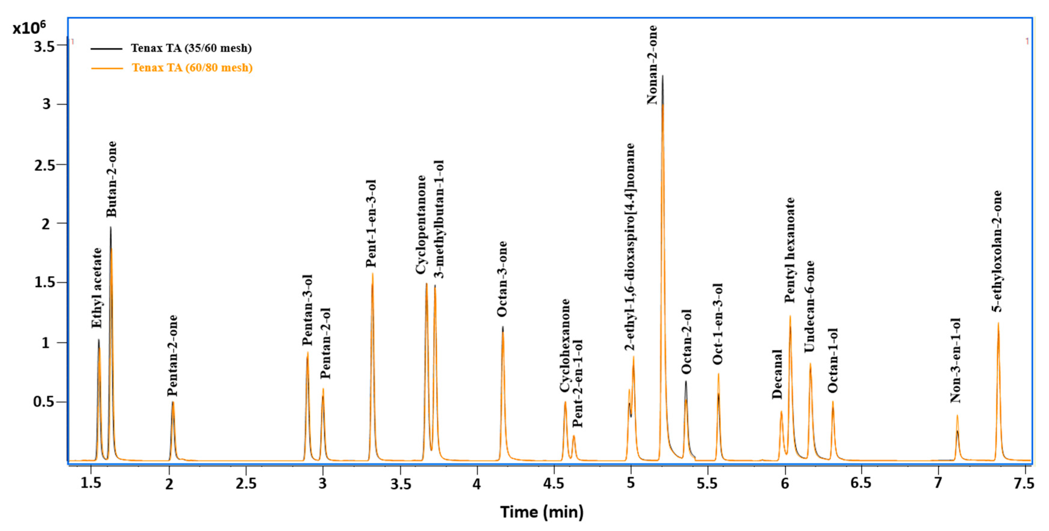
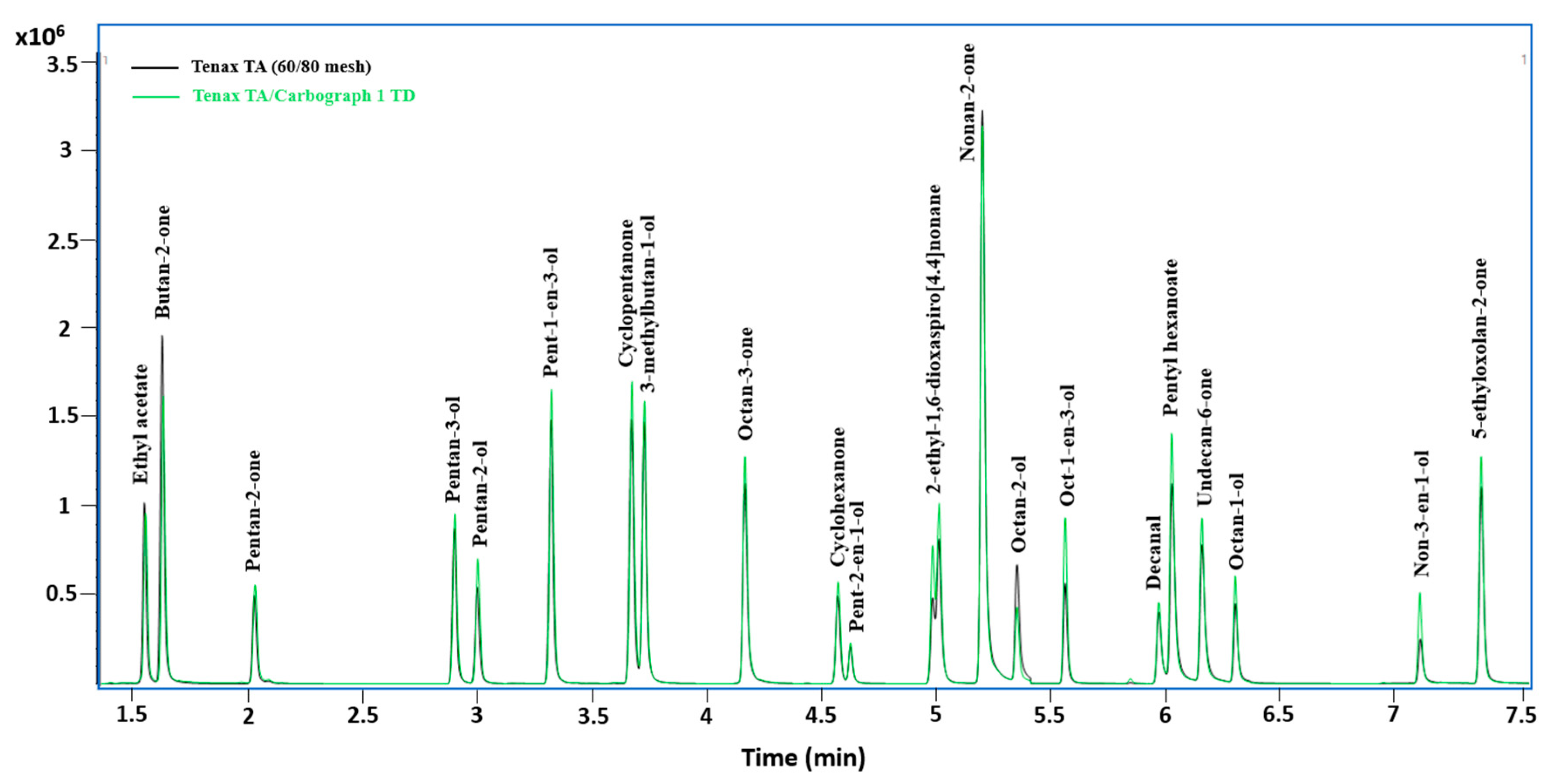
In this study, three sorbent materials were tested, including Tenax TA with two different mesh sizes (Tenax TA 35/60 mesh and Tenax TA 60/80 mesh) and a combination of two different sorbents: Tenax TA/Carbograph 1 TD. Figure 2 shows two superposed chromatograms for the 21 mVOCs following their adsorption into two sorbent tubes of the same type (Tenax TA) but containing different mesh sizes: 35/60 mesh and 60/80 mesh. By comparing the chromatograms, no significant difference in terms of peak area and shape was detected (Figure 2). Similarly, it was found in other studies that particle size has no influence on the adsorption and desorption of VOCs from a sorbent tube packed with Tenax TA of different mesh sizes [50,60]. The mesh size, ranging from 30 to 80, is not crucial for choosing the sorbent type because the retention volume of the compound will be the same [61]. On the other hand, Figure 3 shows two superposed chromatograms for the 21 mVOCs following their adsorption into two different types of sorbent tubes: Tenax TA (60/80 mesh) and Tenax TA/Carbograph 1 TD. By comparing the chromatograms, slightly better results in terms of peak area were found for the multibed sorbents tube (Tenax TA/Carbograph 1 TD) for the majority of the mVOCs (Figure 3). The selection of the sorbent material depends on different criteria, such as the sorbent strength, the hydrophobicity, and the artefacts [62]. Selected sorbents should be highly efficient for analytes adsorption and weak enough for analytes desorption [62]. Tenax TA is the most used polymer sorbent for the quantification of nonpolar to slightly polar compounds between n-hexane (C6) and n-hexadecane (C16) and less volatile than benzene (BP = 80 °C) [50,59] but is considered weak for polar compounds. Therefore, in order to improve the quantification of selected mVOCs, more volatile than n-hexane (ethyl acetate, butan-2-one, pentan-2-one, pentan-3-ol, pentan-2-ol, pent-1-en-3-ol, cyclopentanone and 3-methylbutan-1-ol), Carbograph 1 TD (graphitized carbon blacks GCB), considered as medium sorbent, was packed with Tenax TA. Both sorbents are very hydrophobic. There was no need to test stronger and highly sorptive materials, such as carbon molecular sieves (CMS), known to be associated with humidity problems [62,63]. The presence of water can cause analytical problems, such as column damage, retention times changes and reduction in the adsorption capability of the stationary phase [64]. In addition, these sorbents are generally used for very volatile organic compounds (VVOCs) defined as the substances, which elutes before n-hexane or with carbon atoms < C6 [50,65,66]. In addition, it is important to choose a sorbent with minimum levels of artefacts, such as Tenax TA (0.1–1 ng), whereas other polymers, such as Chromosorb, can reach high artefacts levels (5–10 ng) [61]. For all these reasons, sorbent tubes packed with Tenax TA/Carbograph 1 TD were selected for the rest of the study.

3.2. Breakthrough Volume

The selected sorbent tubes packed with Tenax TA/Carbograph 1 TD were used for the breakthrough volume tests. The breakthrough values (BV) calculated for different volumes and flow rates and for each mVOCs using S1 and S2 are shown in Tables S2 and S3, respectively. S1 and S2 were tested to show the effect of mVOCs concentrations on the breakthrough volume. The results for S1 showed saturation for some mVOCs based on their peak shapes (pent-1-en-3-ol, cyclopentanone, 3-methylbutan-1-ol, nonan-2-one, oct-1-en-3-ol, undecane-6-one and 5-ethyloxolan-2-one), whereas ethyl acetate and butan-2-one showed very low peak areas, compared to other mVOCs (Figures S1 and S2). BVs were slightly higher than 5% for ethyl acetate and butan-2-one at 50, 100, and 150 mL/min for 3 L volume, whereas higher BVs were observed for ethyl acetate and butan-2-one at 150 mL/min for 10 L volume (7.44% for ethyl acetate and 26.13% for butan-2-one). However, all the other mVOCs showed no significant breakthrough (BV < 1%) (Table S2). At high concentrations (S1), the saturation of some mVOCs with higher boiling temperature can cause the breakthrough of the compounds with the lowest boiling temperature, such as ethyl acetate and butan-2-one.
The results for S2 showed that no significant breakthrough (<1%) was observed for all the mVOC for the tested volumes (2 L, 3 L, 6 L, and 10 L) and flow rates (50 mL/min, 100 mL/min, and 150 mL/min) with a maximum BV of 0.86% for ethyl acetate (2 L at 50 mL/min) (Table S3). Therefore, to choose the corresponding volume and flow rate, the extraction efficiency in terms of peak areas and the time saving criteria were added. By fixing each flow rate and increasing the volume, the peak areas slightly decreased for the majority of the mVOCs showing slightly better results for 3 L (Figures S3–S5). On the other hand, by fixing the volume of 3 L and varying the flow rate, slightly better results were shown for 50 mL/min. Given that there was no big difference in peak areas between 50 mL/min and 150 mL/min (Figure S6), 150 mL/min was selected to save time for the calibration. A volume of 3 L at a flow rate of 150 mL/min were selected for this study.

3.3. Method Validation

After the optimization of TD-GC-MS/MS conditions, the developed method was validated in ambient air for the 21 mVOCs, then applied to exhaled air to verify its applicability. Eight different concentration levels were analyzed in triplicates and at least five levels were selected for the calibration curve of each mVOC (Figure S7). All the parameters used for method validation are presented in Table 2. The concentrations in ng/m3 were calculated by determining the quantity injected (ng) in tubes for each mVOC, following the injection of 1 µL of mVOCs mix, divided by 3 L volume. The linearity of the calibration curves, within the selected concentration ranges, was acceptable with a coefficient of determination R2 ≥ 0.999 for the 21 mVOCs (Table 2). Indeed, ISTDs used for linearity correction were crucial for a good quantification. The LODs ranged between 1.76 ng/m3 (pentan-2-ol) and 108.37 ng/m3 (decanal) and the LOQs ranged between 5.32 ng/m3 (pentan-2-ol) and 328.39 ng/m3 (decanal). In this study, the LOD of butan-2-one (75.30 ng/m3) was circa six-fold lower than the value (434 ng/m3) obtained by Peng and Batterman (2000) [46]. In the study of Ribes et al. (2007) [47], two different LODs values were determined for two quantification ions with high and low abundances in the VOC mass spectra. Thus, their LODs for octan-1-ol (7 and 14 ng in tube) were 200- to 400-fold higher than the one obtained in this study (10.53 ng/m3 equivalent to 0.031 ng in tube). In addition, the LOD obtained here for ethyl acetate (26 ng/m3 equivalent to 0.078 ng in tube) was lower than the value for one quantification ion (0.5 ng in tube) but slightly higher than the value for the second quantification ion (0.02 ng in tube) obtained by Ribes et al. (2007) [47]. These values showed the capacity of this method to quantify the selected mVOCs at low concentrations in ambient air. Furthermore, a satisfactory precision (CV% < 5%) was obtained for this developed method by evaluating the coefficient of variation (CV%). For repeatability, the CV% varied from 0.07% to 4.35% at the lowest level of the calibration curve and from 0.13% to 0.71% at the highest level of the calibration curve. For intermediate precision, the CV% varied from 0.11% to 2.04% at the lowest level of the calibration and from 0.22% to 0.84% at the highest level of the calibration curve. A good method accuracy was demonstrated by the percentage recovery (R%) ranging from 80% to 113% at low concentration level and from 94% to 118% at high concentration level for all mVOCs. These values were within the acceptable range of 80–120% [67].
To validate this developed method and verify its applicability in exhaled air, the recovery assessed as the percentage ratio between the calculated value from the calibration curves and the theoretical value was obtained by the analysis of four sorbent tubes loaded with exhaled air from four 3 L Tedlar bags spiked with the mVOCs (two bags with high mVOCs concentration levels and two bags with low mVOCs concentration levels as mentioned in Section 2.5). Depending on the linearity range of each mVOC, the recovery was calculated for one high concentration level and one low concentration level, as shown in Table 3. The recoveries obtained for all the mVOCs varied from 84% to 118% at low concentration level and from 83% to 119% at high concentration level, within the acceptable range of 80–120%, thus indicating the applicability of this method to exhaled air.

3.4. Method Application

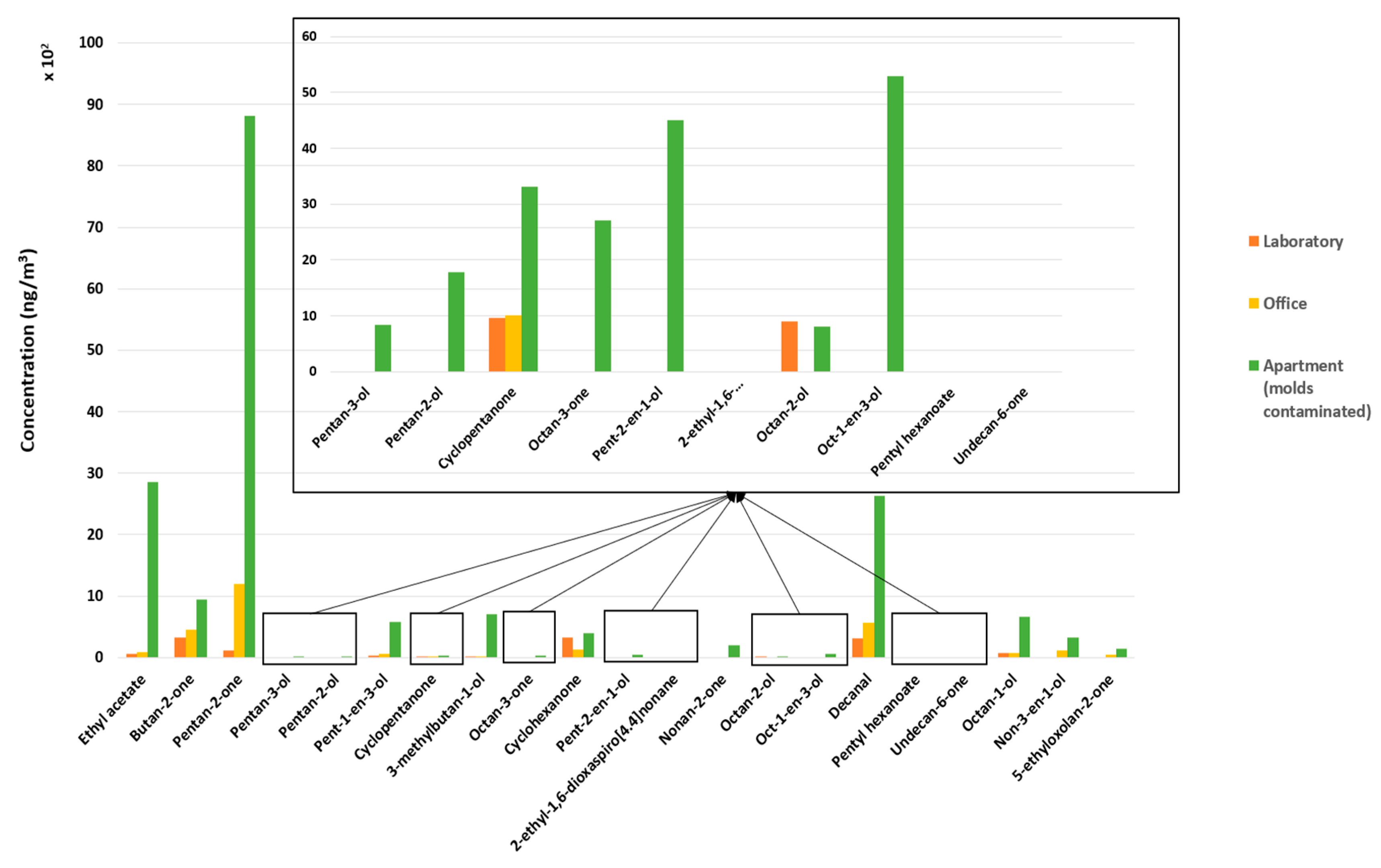
Samples of ambient air collected in duplicate from three different indoor places (TL, OU and RA) were analyzed. The profiles and concentrations of mVOCs in each place are presented in Figure 4 and Table S4. The results showed that, with the exception of octan-2-ol, RA presented the highest concentrations for 17 mVOCs (1.5 to 75-fold higher), in comparison with OU and TL (Table S4), which could be indicative of the relation between the detection of these mVOCs and the presence of molds. The three other mVOCs were not quantified (<LOQ) in all the places (2-ethyl-1,6-dioxaspiro [4.4] nonane, pentyl hexanoate and undecane-6-one). Among the most concentrated mVOCs detected in the RA, pent-1-en-3-ol, nonan-2-one, non-3-en-1-ol, and 5-ethyloxolan-2-one are the compounds known to possibly be specific to molds with no other known non-microbial sources, whereas ethyl acetate, butan-2-one, pentan-2-one, 3-methylbutan-1-ol, cyclohexanone, octan-1-ol, and decanal can have other non-microbial sources [38]. However, most of the latter were demonstrated to be produced by specific mold species. Sunesson et al. (1995) [68], proved the production of ethyl acetate by Penicillium commune, butan-2-one by Penicillium commune, Paecilomyces variotii, and Phialophora fastigiate and 3-methylbutan-1-ol by Aspergillus versicolor, Penicillium commune, Paecilomyces variotii, and Phialophora fastigiate. In the study of Kamiński et al. (1972) [69], 3-methylbutan-1-ol and octan-1-ol were produced by Aspergillus flavus, whereas pentan-2-one and decanal are known to be produced by Coniophora puteana as shown in the study of Korpi et al. (1999) [70].
In OU, half of the mVOCs were quantified but with much lower concentrations than those in the RA, which can possibly indicate the presence of some invisible molds, probably with lower concentrations or in a different stage of growth. Their presence in indoor offices can be due to an inadequate ventilation system or to an excess of water/humidity. The other mVOCs (pentan-3-ol, pentan-2-ol, octan-3-one, pent-2-en-1-ol, 2-ethyl-1,6-dioxaspiro [4.4] nonane, nonan-2-one, octan-2-ol, oct-1-en-3-ol, pentyl hexanoate, and undecane-6-one) were not quantified (i.e., <LOQ) in the OU. However, only nine mVOCs were quantified in TL with the lowest concentrations, compared to OU and RA, except for cyclohexanone and octan-2-ol showing slightly higher concentrations than those detected in the OU. This may be due to a good ventilation system in the laboratory, whereas the use of chemicals may be the reason of higher concentrations for some mVOCs.
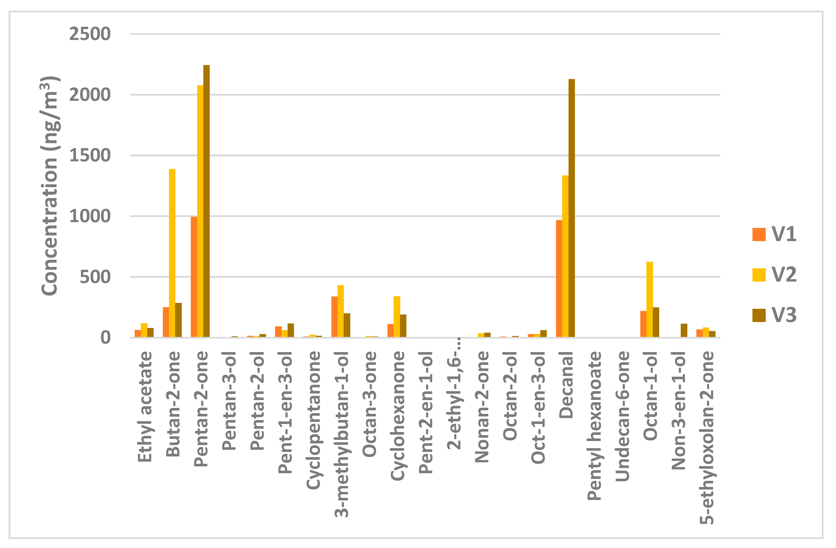
The samples of exhaled air collected in duplicate in Tedlar bags from three volunteers (V1, V2, and V3) then loaded into sorbent tubes, were analyzed and the results of comparison of the mVOCs profile and concentrations between volunteers are presented in Figure 5 and Table S5. The concentrations of more than half of the detected mVOCs (pentan-2-one, pentan-3-ol, pentan-2-ol, pent-1-en-3-ol, nonan-2-one, octan-2-ol, oct-1-en-3-ol, decanal, and non-3-en-1-ol) were higher in the exhaled air of V3 who spent the night sleeping in RA, compared to the other volunteers. This result may be explained by (i) differences in the concentrations measured in the air for these mVOCs and (ii) a higher presence of molds responsible for the production of these mVOCs [29,38]. However, the concentrations of eight other mVOCs (ethyl acetate, butan-2-one, cyclopentanone, 3-methylbutan-1-ol, octan-3-one, cyclohexanone, octan-1-ol and 5-ethyloxolan-2-one) were higher in the exhaled air of the V2 working in OU in comparison with the other volunteers. This result may be due to a different concentration profile of the mVOCs between RA and OU that may also be combined with a different toxicokinetic behavior of these groups of compounds. Among the 21 mVOCs, 4 (ethyl acetate, butan-2-one, 3-methylbutan-1-ol and cyclohexanone) are regulated by Québec/Canada (by “Règlement sur la santé et la sécurité du travail (RSST)”) of Québec with weighted average exposure values (WAEV) much higher than the concentrations detected in the exhaled air of the volunteers. The highest concentrations of these four mVOCs were detected in the exhaled air of V2 (Figure 5 and Table S5). The WAEV were 1440 mg/m3 for ethyl acetate, compared to 117 ng/m3 (V2), 150 mg/m3 for butan-2-one compared to 1388 ng/m3 (V2), 361 mg/m3 for 3-methylbutan-1-ol compared to 432 ng/m3 (V2), and 100 mg/m3 for cyclohexanone compared to 339 ng/m3 (V2) [71].
A final test was carried out by analyzing the mVOCs profile and concentrations in the exhaled air of V3 collected in the morning after spending a night (8 h) in RA and after their 8 h work shift in TL. Figure 6 shows the ratio between the concentrations of the mVOCs in the morning and after the work shift in the laboratory. The ratios varied between 1.04 for octan-1-ol and 11.20 for 5-ethyloxolan-2-one. For some mVOCs (ethyl acetate, pentan-3-ol, nonan-2-one, octan-2-ol, non-3-en-1-ol, and 5-ethyloxolan-2-one), where the concentration after the work shift were lower than the LOQ; half of the LOD value was used to calculate the concentration ratio. Since pent-2-en-1-ol, 2-ethyl-1,6-dioxaspiro [4.4] nonane, pentyl hexanoate, and undecan-6-one were not quantified (<LOQ) in the exhaled air of V3 in both locations, their ratios were not calculated. These results support the main hypothesis in this study, which is that there is a relation between the detection and quantification of mVOCs in ambient air and/or human biological matrices (e.g., exhaled air, urine, and blood) and the presence or the exposure to molds in indoor environments. This also supports the potential use of these mVOCs as potential biomarkers for mold exposure.
However, since the aim of the present study was to demonstrate the good quantification of these mVOCs in ambient and exhaled air using Tedlar bags and sorbent tubes with the thermal desorption system, these preliminary field measurements were performed to show the difference of mVOCs profile and concentrations between different volunteers (V1, V2, V3) and for the same volunteer (V3) at different places, including a contaminated apartment (Figure S8), without taking into account the background of each volunteer.

4. Conclusions

The new method developed in this study allows the detection and quantification in ambient and exhaled air of 21 mVOCs, previously selected as potential biomarkers of mold exposure [38,42]. This method uses Tenax TA/Carbograph 1 TD sorbent tubes and thermal desorption system coupled to gas chromatography–mass spectrometry (TD-GC-MS/MS). The validated method showed a reliable linearity for the calibration curves, a good selectivity for the selected compounds, and satisfactory accuracy (80% < R% <120%) and precision (CV < 4.35%). These results prove the efficiency of this method to identify and quantify mVOCs at low levels in ambient and exhaled air (1.76 ng/m3 < LOD < 108.37 ng/m3). Furthermore, a preliminary application of this method in ambient air of different places showed the higher mVOCs concentrations in the visible-mold-contaminated apartment bedroom in comparison with the other places, which had no evident trace of molds. In addition, more than half of the detected mVOCs were at the highest concentration in the exhaled air of a volunteer who spent a night in the mold-contaminated bedroom. These results are in line with the hypothesis that the presence of indoor molds contamination may be assessed by the detection and quantification of mVOCs in ambient and exhaled air. In addition, it allows us to explore the difference between the contaminated and non-contaminated environment and to better target contaminated environments to interfere with people’s health protection.

Supplementary Materials

The following supporting information can be downloaded at: https://www.mdpi.com/article/10.3390/atmos13091432/s1. Figure S1: A room in an apartment that has visible mold contamination on walls. The apartment is on the ground floor and the room (approximately: 60 m3) has two windows with poor ventilation system and excessive humidity. Figure S2: Chromatogram presenting the peak area of the 21 mVOCs in S1 adsorbed on Tenax TA/Carbograph 1 TD for 10 L at a flow rate of 150 mL/min. Figure S3: Chromatogram presenting the peak area of 8 mVOCs in S1 (presenting saturation) adsorbed on Tenax TA/Carbograph 1 TD for 10 L at a flow rate of 150 mL/min. Figure S4: Chromatogram presenting the peak area of the 21 mVOCs adsorbed on Tenax TA/Carbograph 1 TD for 2 L and 3 L air volumes at a flow rate of 50 mL/min. Figure S5: Chromatogram presenting the peak area of the 21 mVOCs adsorbed on Tenax TA/Carbograph 1 TD for 2 L, 3 L, 6 L and 10 L air volumes at a flow rate of 100 mL/min. Figure S6: Chromatogram presenting the peak area of the 21 mVOCs adsorbed on Tenax TA/Carbograph 1 TD for 3 L, 6 L and 10 L air volumes at a flow rate of 150 mL/min. Figure S7: Chromatogram presenting the peak area of the 21 mVOCs adsorbed on Tenax TA/Carbograph 1 TD for 3 L air volume at a flow rate of 50 mL/min and 150 mL/min. Figure S8: Calibration curves (relative response mVOC/ISTD vs. mVOC concentrations (ng/L)) for the 21 mVOCs in ambient air, including equation and R2. Table S1: Properties (molecular weight MW and boiling temperature BT), transitions and internal standard (ISTD) used for quantification for each mVOC. Table S2: BVs for each mVOC in S1 (% mVOC response in the back tube related to the sum of the responses in both tubes) for different volumes and flow rate. Table S3: BVs for each mVOC in S2 (% mVOC response in the back tube related to the sum of the responses in both tubes) for different volumes and flow rates. Table S4: Concentrations of the mVOCs found in three different places: toxicology laboratory (TL), an office at the university (OU) and a room in an apartment (RA) with molds contamination. Table S5: The mVOCs concentrations in the exhaled air of three different volunteers: V1 (in a pharmacology and physiology laboratory), V2 (in an office in the university), and V3 (in a room in a mold contaminated apartment).

Author Contributions

Methodology: S.T., B.E.A., S.H., G.M.; Validation: S.T.; Investigation: S.T.; Conceptualization: B.E.A., S.H., G.M.; Funding acquisition: M.B., S.H., G.M.; Writing—original draft: S.T.; Writing—review and editing: B.E.A., M.B., S.H., G.M. All authors have read and agreed to the published version of the manuscript.

Funding

This research was funded by the Institut de recherche Robert-Sauvé en santé et en sécurité du travail (IRSST), grant number 2018-0021.

Institutional Review Board Statement

Ethical review and approval were waived by the Clinical Research Ethics Committee (CERC) of the Université de Montréal for this study. The reason for this waiver is that the proposed activity is not currently part of a formal research context as defined in the Canadian Tri-Council Policy Statement, the objective being to develop a chemical analysis method to measure contaminants in air, which will serve as a basis for subsequent studies in workers whose participation will be necessary for the validation of the use of these microbial volatile organic compounds (MVOCs) as biomarkers of exposure to molds in the workplace. Furthermore, at this initial stage, no human participants are involved.

Informed Consent Statement

Consent was obtained from all 3 volunteers involved in the study.

Data Availability Statement

Not applicable.

Conflicts of Interest

The authors declare that they have no known competing financial interests or personal relationships that could have appeared to influence the work reported in this paper.

References

  1. Pietrogrande, M.C.; Casari, L.; Demaria, G.; Russo, M. Indoor Air Quality in Domestic Environments during Periods Close to Italian COVID-19 Lockdown. Int. J. Environ. Res. Public Health 2021, 18, 4060. [Google Scholar] [CrossRef] [PubMed]
  2. Shaddick, G.; Salter, J.M.; Peuch, V.H.; Ruggeri, G.; Thomas, M.L.; Mudu, P.; Tarasova, O.; Baklanov, A.; Gumy, S. Global Air Quality: An Inter-Disciplinary Approach to Exposure Assessment for Burden of Disease Analyses. Atmosphere 2021, 12, 48. [Google Scholar] [CrossRef]
  3. Settimo, G.; Manigrasso, M.; Avino, P. Indoor Air Quality: A Focus on the European Legislation and State-of-the-Art Research in Italy. Atmosphere 2020, 11, 370. [Google Scholar] [CrossRef]
  4. Leung, D.Y.C. Outdoor-indoor air pollution in urban environment: Challenges and opportunity. Front. Environ. Sci. 2015, 2, 1–7. [Google Scholar] [CrossRef]
  5. Li, Z.; Wen, Q.; Zhang, R. Sources, health effects and control strategies of indoor fine particulate matter (PM2.5): A review. Sci. Total Environ. 2017, 586, 610–622. [Google Scholar] [CrossRef]
  6. Health Effects Institute. State of Global Air 2020; Special Report; Health Effects Institute: Boston, MA, USA, 2020.
  7. Zar, H.J.; Ferkol, T.W. The global burden of respiratory disease-Impact on child health. Pediatr. Pulmonol. 2014, 49, 430–434. [Google Scholar] [CrossRef]
  8. Tischer, C.G.; Heinrich, J. Exposure assessment of residential mould, fungi and microbial components in relation to children’s health: Achievements and challenges. Int. J. Hyg. Environ. Health 2013, 216, 109–114. [Google Scholar] [CrossRef]
  9. Hurraß, J.; Heinzow, B.; Aurbach, U.; Bergmann, K.-C.; Bufe, A.; Buzina, W.; Cornely, O.A.; Engelhart, S.; Fischer, G.; Gabrio, T.; et al. Medical diagnostics for indoor mold exposure. Int. J. Hyg. Environ. Health 2017, 220, 305–328. [Google Scholar] [CrossRef]
  10. Borchers, A.T.; Chang, C.; Eric Gershwin, M. Mold and Human Health: A Reality Check. Clin. Rev. Allergy Immunol. 2017, 52, 305–322. [Google Scholar] [CrossRef]
  11. Reboux, G.; Bellanger, A.-P.; Roussel, S.; Grenouillet, F.; Millon, L. Moulds in dwellings: Health risks and involved species. Rev. Des Mal. Respir. 2010, 27, 169–179. [Google Scholar] [CrossRef]
  12. D’Halewyn, M.A.; Leclerc, J.M.; King, N.; Bélanger, M.; Legris, M.; Frenette, Y. Les risques à la santé associés à la présence de moisissures en milieu intérieur: Rapport scientifique. Inst. Natl. De St. Publique Québec 2002, 15–27. Available online: https://www.inspq.qc.ca/pdf/publications/126_RisquesMoisissuresMilieuInterieur.pdf (accessed on 22 August 2022).
  13. Hyvönen, S.; Lohi, J.; Tuuminen, T. Moist and Mold Exposure is Associated with High Prevalence of Neurological Symptoms and MCS in a Finnish Hospital Workers Cohort. Saf. Health Work 2020, 11, 173–177. [Google Scholar] [CrossRef]
  14. Harding, C.F.; Pytte, C.L.; Page, K.G.; Ryberg, K.J.; Normand, E.; Remigio, G.J.; DeStefano, R.A.; Morris, D.B.; Voronina, J.; Lopez, A.; et al. Mold inhalation causes innate immune activation, neural, cognitive and emotional dysfunction. Brain Behav. Immun. 2020, 87, 218–228. [Google Scholar] [CrossRef]
  15. Mensah-Attipoe, J.; Täubel, M. Analysis Approaches for Fungi in Indoor Environmental Assessments. In Exposure to Microbiological Agents in Indoor and Occupational Environments; Viegas, C., Viegas, S., Gomes, A., Täubel, M., Sabino, R., Eds.; Springer International Publishing: Cham, Switzerland, 2017; pp. 109–127. ISBN 978-3-319-61688-9. [Google Scholar]
  16. Shorter, C.; Täubel, M.; Pierse, N.; Douwes, J.; Howden-Chapman, P.; Hyvärinen, A.; Crane, J. Objective assessment of domestic mold contamination using quantitative PCR. J. Allergy Clin. Immunol. 2016, 137, 622–624. [Google Scholar] [CrossRef]
  17. Oneț, A.; Ilieș, D.C.; Ilieș, A.; Herman, V.; Burta, L.; Marcu, F.; Buhas, R.; Baias, Ș.; Oneț, C.; Ilieș, M.; et al. Indoor Air Quality Assessement and Its Perception. Case Study Historic Wooden Church, Romania. Rom. Biotechnol Lett. 2020, 25, 1547–1553. [Google Scholar] [CrossRef]
  18. Alborch, L.; Bragulat, M.R.; Abarca, M.L.; Cabañes, F.J. Temperature and incubation time effects on growth and ochratoxin A production by Aspergillus sclerotioniger and Aspergillus lacticoffeatus on culture media. Lett. Appl. Microbiol. 2011, 52, 208–212. [Google Scholar] [CrossRef]
  19. Mateo, J.; Mateo, R.; Jiménez, M. Accumulation of type A trichothecenes in maize, wheat and rice by Fusarium sporotrichioides isolates under diverse culture conditions. Int. J. Food Microbiol. 2002, 72, 115–123. [Google Scholar] [CrossRef]
  20. Cahagnier, B.; Melcion, D.; Richard-Molard, D. Growth of Fusarium moniliforme and its biosynthesis of fumonisin B1 on maize grain as a function of different water activities. Lett. Appl. Microbiol. 1995, 20, 247–251. [Google Scholar] [CrossRef]
  21. Korpi, A.; Järnberg, J.; Pasanen, A.-L. Microbial Volatile Organic Compounds. Crit. Rev. Toxicol. 2009, 39, 139–193. [Google Scholar] [CrossRef]
  22. Weisskopf, L.; Schulz, S.; Garbeva, P. Microbial volatile organic compounds in intra-kingdom and inter-kingdom interactions. Nat. Rev. Genet. 2021, 19, 391–404. [Google Scholar] [CrossRef]
  23. Brodhagen, M.; Tsitsigiannis, D.I.; Hornung, E.; Goebel, C.; Feussner, I.; Keller, N.P. Reciprocal oxylipin-mediated cross-talk in the Aspergillus-seed pathosystem. Mol. Microbiol. 2007, 67, 378–391. [Google Scholar] [CrossRef]
  24. Splivallo, R.; Valdez, N.; Kirchhoff, N.; Ona, M.C.; Schmidt, J.-P.; Feussner, I.; Karlovsky, P. Intraspecific genotypic variability determines concentrations of key truffle volatiles. New Phytol. 2012, 194, 823–835. [Google Scholar] [CrossRef]
  25. Wu, S.; Hayati, S.K.; Kim, E.; de la Mata, A.P.; Harynuk, J.J.; Wang, C.; Zhao, R. Henry’s Law Constants and Indoor Partitioning of Microbial Volatile Organic Compounds. Environ. Sci. Technol. 2022, 56, 7143–7152. [Google Scholar] [CrossRef]
  26. Hung, R.; Lee, S.; Bennett, J.W. Fungal volatile organic compounds and their role in ecosystems. Appl. Microbiol. Biotechnol. 2015, 99, 3395–3405. [Google Scholar] [CrossRef]
  27. Wilkins, K.; Larsen, K. Variation of volatile organic compound patterns of mold species from damp buildings. Chemosphere 1995, 31, 3225–3236. [Google Scholar] [CrossRef]
  28. Elke, K.; Begerow, J.; Oppermann, H.; Kråmer, U.; Jermann, E.; Dunemann, L. Determination of selected microbial volatile organic compounds by diffusive sampling and dual-column capillary GC-FID-a new feasible approach for the detection of an exposure to indoor mould fungi? J. Environ. Monit. 1999, 1, 445–452. [Google Scholar] [CrossRef] [PubMed]
  29. Garcia-Alcega, S.; Nasir, Z.A.; Ferguson, R.; Whitby, C.; Dumbrell, A.J.; Colbeck, I.; Gomes, D.; Tyrrel, S.; Coulon, F. Fingerprinting outdoor air environment using microbial volatile organic compounds (MVOCs)—A review. TrAC Trends Anal. Chem. 2017, 86, 75–83. [Google Scholar] [CrossRef]
  30. Kuske, M.; Romain, A.-C.; Nicolas, J. Microbial volatile organic compounds as indicators of fungi. Can an electronic nose detect fungi in indoor environments? Build. Environ. 2005, 40, 824–831. [Google Scholar] [CrossRef]
  31. Garcia-Alcega, S.; Nasar, Z.A.; Ferguson, R.; Noël, C.; Cravo-Laureau, C.; Whitby, C.; Dumbrell, A.J.; Colbeck, I.; Tyrrel, S.; Coulon, F. Can chemical and molecular biomarkers help discriminate between industrial, rural and urban environments? Sci. Total Environ. 2018, 631–632, 1059–1069. [Google Scholar] [CrossRef]
  32. Kim, J.L.; Elfman, L.; Mi, Y.; Wieslander, G.; Smedje, G.; Norbäck, D. Indoor molds, bacteria, microbial volatile organic compounds and plasticizers in schools? associations with asthma and respiratory symptoms in pupils. Indoor Air 2006, 17, 153–163. [Google Scholar] [CrossRef]
  33. Ryan, T.J.; Beaucham, C. Dominant microbial volatile organic compounds in 23 US homes. Chemosphere 2013, 90, 977–985. [Google Scholar] [CrossRef] [PubMed]
  34. Jakubowski, M. Biological Monitoring versus Air Monitoring Strategies in Assessing Environmental–Occupational Exposure. J. Environ. Monit. 2012, 14, 348–352. [Google Scholar] [CrossRef] [PubMed]
  35. Matysik, S.; Herbarth, O.; Mueller, A. Determination of microbial volatile organic compounds (MVOCs) by passive sampling onto charcoal sorbents. Chemosphere 2009, 76, 114–119. [Google Scholar] [CrossRef]
  36. Schuchardt, S.; Kruse, H. Quantitative volatile metabolite profiling of common indoor fungi: Relevancy for indoor air analysis. J. Basic Microbiol. 2009, 49, 350–362. [Google Scholar] [CrossRef]
  37. Claeson, A.-S.; Sandström, M.; Sunesson, A.-L. Volatile organic compounds (VOCs) emitted from materials collected from buildings affected by microorganisms. J. Environ. Monit. 2007, 9, 240–245. [Google Scholar] [CrossRef]
  38. El Aroussi, B.; Marchand, G.; Aubin, S.; Bouchard, M.; Haddad, S. Utilisation des Composés Organiques Volatils Microbiens Comme Biomarqueurs de L’exposition Aux Moisissures en Milieu de Travail—Étude de Faisabilité; Rapports Scientifiques: (R-1037); Institut de Recherche Robert-Sauvé en Santé et en Sécurité de Travail (IRSST): Montréal, QC, Canada, 2018. [Google Scholar]
  39. Schuchardt, S.; Strube, A. Microbial volatile organic compounds in moldy interiors: A long-term climate chamber study. J. Basic Microbiol. 2012, 53, 532–538. [Google Scholar] [CrossRef]
  40. Piecková, E.; Jesenská, Z. Microscopic Fungi in Dwellings and Their Health Implications in Humans. Ann. Agric. Environ. Med. AAEM 1999, 6, 1–11. [Google Scholar] [PubMed]
  41. Peyret, T.; Poulin, P.; Krishnan, K. A unified algorithm for predicting partition coefficients for PBPK modeling of drugs and environmental chemicals. Toxicol. Appl. Pharmacol. 2010, 249, 197–207. [Google Scholar] [CrossRef]
  42. Tabbal, S.; El Aroussi, B.; Bouchard, M.; Marchand, G.; Haddad, S. A new headspace solid-phase microextraction coupled with gas chromatography-tandem mass spectrometry method for the simultaneous quantification of 21 microbial volatile organic compounds in urine and blood. Chemosphere 2022, 296, 133901. [Google Scholar] [CrossRef]
  43. Wazeerud-Din, I.J.; Silva, L.K.; Smith, M.M.; Newman, C.A.; Blount, B.C.; De Jesús, V.R. Quantification of seven microbial volatile organic compounds in human serum by solid-phase microextraction gas chromatography-tandem mass spectrometry. Chemosphere 2021, 266, 128970. [Google Scholar] [CrossRef]
  44. Drabińska, N.; Flynn, C.; Ratcliffe, N.; Belluomo, I.; Myridakis, A.; Gould, O.; Fois, M.; Smart, A.; Devine, T.; Costello, B.D.L. A literature survey of all volatiles from healthy human breath and bodily fluids: The human volatilome. J. Breath Res. 2021, 15, 034001. [Google Scholar] [CrossRef] [PubMed]
  45. Pankow, J.F.; Luo, W.; Isabelle, L.M.; Bender, D.A.; Baker, R.J. Determination of a Wide Range of Volatile Organic Compounds in Ambient Air Using Multisorbent Adsorption/Thermal Desorption and Gas Chromatography/Mass Spectrometry. Anal. Chem. 1998, 70, 5213–5221. [Google Scholar] [CrossRef]
  46. Peng, C.-Y.; Batterman, S. Performance evaluation of a sorbent tube sampling method using short path thermal desorption for volatile organic compounds. J. Environ. Monit. 2000, 2, 313–324. [Google Scholar] [CrossRef] [PubMed]
  47. Ribes, A.; Carrera, G.; Gallego, E.; Roca, X.; Berenguer, M.J.; Guardino, X. Development and validation of a method for air-quality and nuisance odors monitoring of volatile organic compounds using multi-sorbent adsorption and gas chromatography/mass spectrometry thermal desorption system. J. Chromatogr. A 2007, 1140, 44–55. [Google Scholar] [CrossRef]
  48. Zhang, S.; Cai, L.; Koziel, J.A.; Hoff, S.J.; Schmidt, D.R.; Clanton, C.J.; Jacobson, L.D.; Parker, D.B.; Heber, A.J. Field air sampling and simultaneous chemical and sensory analysis of livestock odorants with sorbent tubes and GC–MS/olfactometry. Sensors Actuators B Chem. 2010, 146, 427–432. [Google Scholar] [CrossRef]
  49. Rodríguez-Navas, C.; Forteza, R.; Cerdà, V. Use of Thermal Desorption–Gas Chromatography–Mass Spectrometry (TD–GC–MS) on Identification of Odorant Emission Focus by Volatile Organic Compounds Characterisation. Chemosphere 2012, 89, 1426–1436. [Google Scholar] [CrossRef]
  50. Schieweck, A.; Gunschera, J.; Varol, D.; Salthammer, T. Analytical procedure for the determination of very volatile organic compounds (C3–C6) in indoor air. Anal. Bioanal. Chem. 2018, 410, 3171–3183. [Google Scholar] [CrossRef]
  51. Haworth, J.J.; Pitcher, C.K.; Ferrandino, G.; Hobson, A.R.; Pappan, K.L.; Lawson, J.L.D. Breathing new life into clinical testing and diagnostics: Perspectives on volatile biomarkers from breath. Crit. Rev. Clin. Lab. Sci. 2022, 59, 353–372. [Google Scholar] [CrossRef]
  52. U.S.EPA. Compendium of Methods for the Determination of Toxic Organic Compounds in Ambient Air, Method TO-14; Center for Environmental Research Information, Office of Research and Development: Cincinnati, OH, USA, 1999.
  53. U.S.EPA. Compendium of Methods for the Determination of Toxic Organic Compounds in Ambient Air, Method TO-17; Center for Environmental Research Information, Office of Research and Development: Cincinnati, OH, USA, 1999.
  54. Tang, Y.; Fellin, P.; Otson, R. Evaluation of a Transportable Gas Chromatograph for Monitoring of Indoor Airborne Volatile Organic Compounds with a Gas Sampling Valve or a Concentrator. Indoor Built Environ. 1995, 4, 27–36. [Google Scholar] [CrossRef]
  55. Ras, M.R.; Borrull, F.; Marcé, R.M. Sampling and preconcentration techniques for determination of volatile organic compounds in air samples. TrAC Trends Anal. Chem. 2009, 28, 347–361. [Google Scholar] [CrossRef]
  56. Belizário, J.E.; Faintuch, J.; Malpartida, M.G. Breath Biopsy and Discovery of Exclusive Volatile Organic Compounds for Diagnosis of Infectious Diseases. Front. Cell. Infect. Microbiol. 2021, 10, 564194. [Google Scholar] [CrossRef]
  57. Bortoli, M.; Knoppel, H.; Pecchio, E.; Schauenburg, H.; Vissers, H. Comparison of Tenax and Carbotrap for VOC Sampling in Indoor Air. Indoor Air 1992, 2, 216–224. [Google Scholar] [CrossRef]
  58. Kozicki, M.; Wiejak, A.; Piasecki, M.; Abram, A. Identification of MVOCs Produced by Coniophora puteana and Poria placenta Growing on WPC Boards by Using Subtraction Mass Spectra. Int. J. Environ. Res. Public Health 2019, 16, E2499. [Google Scholar] [CrossRef] [PubMed]
  59. Woolfenden, E. Monitoring VOCs in Air Using Sorbent Tubes Followed by Thermal Desorption-Capillary GC Analysis: Summary of Data and Practical Guidelines. J. Air Waste Manag. Assoc. 1997, 47, 20–36. [Google Scholar] [CrossRef]
  60. Patil, S.; Lonkar, S. Thermal Desorption—Gas Chromatography for the Determination of Benzene, Aniline, Nitrobenzene and Chlorobenzene in Workplace Air. J. Chromatogr. A 1992, 600, 344–351. [Google Scholar] [CrossRef]
  61. Woolfenden, E. Sorbent-based sampling methods for volatile and semi-volatile organic compounds in air. Part 2. Sorbent selection and other aspects of optimizing air monitoring methods. J. Chromatogr. A 2010, 1217, 2685–2694. [Google Scholar] [CrossRef]
  62. Khan, M.F.; Sahani, M.; Nadzir, M.S.M.; Yik, L.C.; Hoque, H.M.S.; Hamid, H.H.A.; Wahab, M.I.A.; Munna, F.T.; Amin, N.; Misran, H.; et al. Volatile Organic Compound Analysis by Sorbent Tube-Thermal Desorption-Gas Chromatography: A Review. Int. J. Eng. Technol. 2018, 7, 165–175. [Google Scholar] [CrossRef]
  63. Maceira, A.; Vallecillos, L.; Borrull, F.; Marcé, R.M. New approach to resolve the humidity problem in VOC determination in outdoor air samples using solid adsorbent tubes followed by TD-GC–MS. Sci. Total Environ. 2017, 599-600, 1718–1727. [Google Scholar] [CrossRef]
  64. Wu, C.-H.; Feng, C.-T.; Lo, Y.-S.; Lin, T.-Y.; Lo, J.-G. Determination of volatile organic compounds in workplace air by multisorbent adsorption/thermal desorption-GC/MS. Chemosphere 2004, 56, 71–80. [Google Scholar] [CrossRef]
  65. SIST EN. 16516:2018+A1:2020—Construction Products: Assessment of Release of Dangerous Substances—Determination of Emissions into Indoor Air. Available online: https://standards.iteh.ai/catalog/standards/sist/fe1939d9-7a49-4f83-b4f2-38e7c4758d1c/sist-en-16516-2018a1-2020 (accessed on 10 August 2022).
  66. Wang, B.; Zhao, Y.; Lan, Z.; Yao, Y.; Wang, L.; Sun, H. Sampling methods of emerging organic contaminants in indoor air. Trends Environ. Anal. Chem. 2016, 12, 13–22. [Google Scholar] [CrossRef]
  67. Food and Drug Administration (FDA); Office of Regulatory Affairs (ORA). Methods, method verification and validation. In Laboratory Manual Volume II, ORA-LAB.5.4.5; FDA: Silver Spring, MD, USA, 2020. Available online: https://www.fda.gov/media/73920/download (accessed on 7 June 2021).
  68. Sunesson, A.; Vaes, W.; Nilsson, C.; Blomquist, G.; Andersson, B.; Carlson, R. Identification of volatile metabolites from five fungal species cultivated on two media. Appl. Environ. Microbiol. 1995, 61, 2911–2918. [Google Scholar] [CrossRef] [PubMed] [Green Version]
  69. Kaminśki, E.; Libbey, L.M.; Stawicki, S.; Wasowicz, E. Identification of the Predominant Volatile Compounds Produced by Aspergillus flavus. Appl. Microbiol. 1972, 24, 721–726. [Google Scholar] [CrossRef] [PubMed]
  70. Korpia, A.; Pasanen, A.-L.; Viitanenb, H. Volatile metabolites of Serpula lacrymans, Coniophora puteana, Poria placenta, Stachybotrys chartarum and Chaetomium globosum. Build. Environ. 1998, 34, 205–211. [Google Scholar] [CrossRef]
  71. Règlement sur la Santé et la Sécurité du Travail (RSST) du Québec. Loi Sur la Santé et la Sécurité du Travail. 2022. chapitre S-2.1, a. 223. Available online: https://www.legisquebec.gouv.qc.ca/fr/pdf/rc/S-2.1,%20R.%2013.pdf (accessed on 8 August 2022).
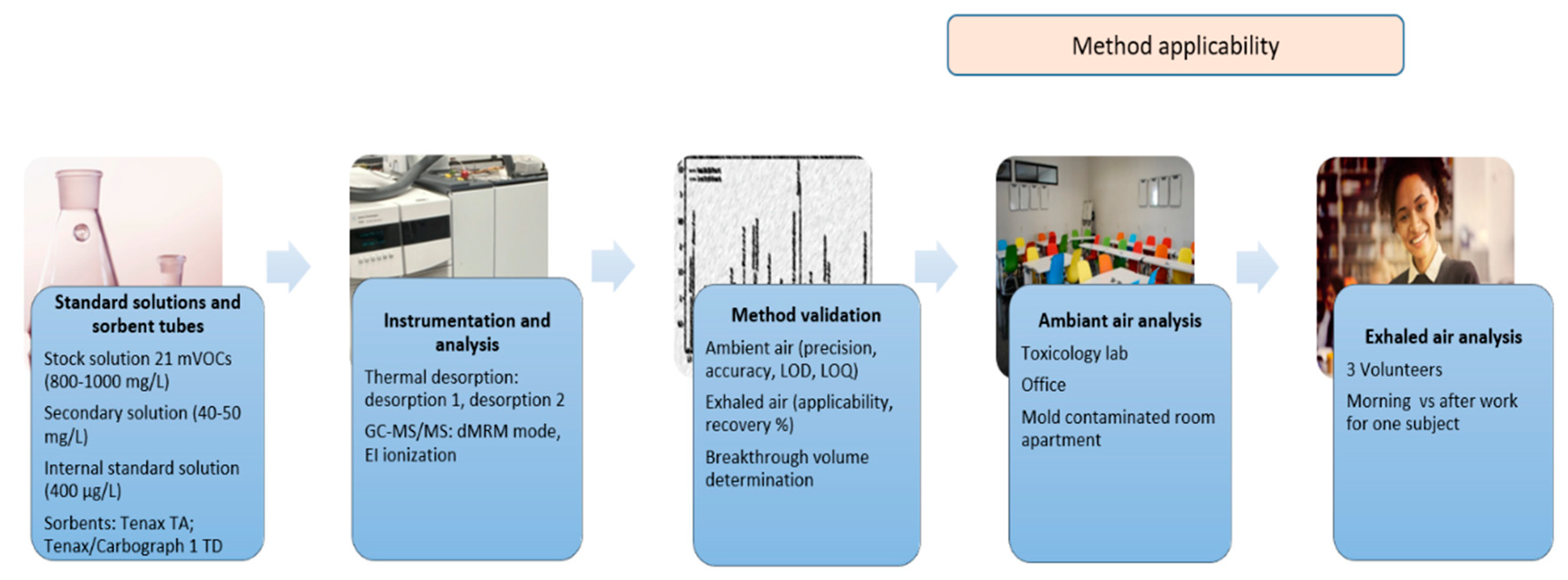
Figure 1. A summary of the methodology steps.
Atmosphere 13 01432 g001
Figure 2. dMRM chromatogram presenting the peak area of the 21 mVOCs adsorbed on Tenax TA (35/60 mesh) and on Tenax (60/80 mesh).
Atmosphere 13 01432 g002
Figure 3. dMRM chromatogram presenting the peak area of the 21 mVOCs adsorbed on Tenax TA (60/80 mesh) and on Tenax TA/Carbograph 1 TD.
Atmosphere 13 01432 g003
Figure 4. Comparison of the mVOCs profile between three different places (Toxicology laboratory, office at university, and mold contaminated apartment room).
Atmosphere 13 01432 g004
Figure 5. The mVOCs in the exhaled air of the three volunteers: V1 (working in the laboratory), V2 (working in the office) and V3 (exhaled air sampled after a night spent in a mold contaminated apartment room).
Atmosphere 13 01432 g005
Figure 6. Concentration ratio of the mVOCs in exhaled air of volunteer V3 in the morning (after a night spent in a mold contaminated bedroom) vs. after a work shift in the laboratory.
Atmosphere 13 01432 g006
Table 1. Analytical parameters for GC-MS/MS.
ParameterRetained Conditions
GC
ColumnDB-wax (30 m × 0.32 mm, 0.5 µm)
Carrier gasUltra-high purity He (99.999%)
Carrier gas flow1.8 mL/min
Oven programInitial temperature: 65 °C
3 °C/min to 70 °C
20 °C/min to 220 °C
Total run time: 9.17 min
MS/MS
ModedMRM (dynamic multiple reaction monitoring)
Ionization sourceEI, 70 eV
Transfer line temperature245 °C
MS source temperature230 °C
Emission current35 µA
Quad temperatureQ1: 150 °C
Q2: 150 °C
Table 2. Method validation parameters for the 21 mVOCs in the air: range of linearity, coefficient of determination (R2), limits of detection and quantification (LOD and LOQ), accuracy presented as percentage recovery R% at low (Min) and high (Max) concentration.
CompoundsRange of Linearity (ng/m3)R2LOD (ng/m3)LOQ (ng/m3)R% (Min)R% (Max)Repeatability CV% (Min)Repeatability CV% (Max)Intermediate Precision CV% (Min)Intermediate Precision CV% (Max)
Ethyl acetate30–80700.9992678.78901020.690.560.520.59
Butan-2-one120–14,2500.99975.30228.1996950.220.540.210.47
Pentan-2-one 120–13,8900.99972.04218.3196960.140.250.110.37
Pentan-3-ol4–18600.9992.096.34871070.160.271.140.34
Pentan-2-ol4–17900.9991.765.32801070.160.230.490.44
Pent-1-en-3-ol8–20100.9996.5819.931101040.230.190.550.48
Cyclopentanone4–20900.9992.166.54941070.180.140.580.34
3-methylbutan-1-ol4–17500.9993.049.22901080.070.520.860.42
Octan-3-one4–17600.9993.3410.121071050.250.480.560.84
Cyclohexanone30–81600.99911.5234.90921050.270.290.710.55
Pent-2-en-1-ol30–69900.99925.1176.081041040.600.382.040.48
2-ethyl-1,6-dioxaspiro [4.4] nonane30–78800.99926.4380.08841100.320.320.850.76
Nonan-2-one30–69500.99922.6568.65921100.430.200.400.28
Octan-2-ol 7–17700.9992.407.26801181.160.130.250.23
Oct-1-en-3-ol7–17700.9994.9815.111131130.710.230.610.48
Decanal 120–13,8000.999108.37328.39801024.350.160.610.22
Pentyl hexanoate120–14,4900.99955.71168.83931060.210.210.370.35
Undecan-6-one30–69700.99925.0775.97951050.160.620.410.47
Octan-1-ol 30–69600.99910.5331.90921080.210.270.390.54
Non-3-en-1-ol 120–13,8800.99970.95214.9982940.600.711.380.58
5-ethyloxolan-2-one40–88200.9999.5929.06871100.270.220.290.25
Table 3. The recoveries (R%) calculated at low (Min) and high (Max) concentration levels for the 21 mVOCs in exhaled air.
CompoundsR% (Min)R% (Max)
Ethyl acetate118118
Butan-2-one103119
Pentan-2-one 107105
Pentan-3-ol9793
Pentan-2-ol10293
Pent-1-en-3-ol10383
Cyclopentanone108117
3-methylbutan-1-ol8496
Octan-3-one104117
Cyclohexanone107118
Pent-2-en-1-ol87100
2-ethyl-1,6-dioxaspiro [4.4] nonane106109
Nonan-2-one111115
Octan-2-ol 102102
Oct-1-en-3-ol10099
Decanal 8593
Pentyl hexanoate94102
Undecan-6-one105109
Octan-1-ol 96107
Non-3-en-1-ol 90110
5-ethyloxolan-2-one101117
Publisher’s Note: MDPI stays neutral with regard to jurisdictional claims in published maps and institutional affiliations.