1. Introduction
Arthrogryposis, also known as Arthrogryposis Multiplex Congenita (AMC), encompasses a diverse group of conditions characterized by multiple congenital joint contractures and limited joint mobility present at birth. These conditions are typically classified into isolated and multiple contractures, with the latter affecting two or more body regions.
The term “arthrogryposis” is a clinical descriptor rather than a specific diagnosis and is observed in over 300 distinct syndromes [
1]. One of the most recognizable forms is amyoplasia, which presents characteristic limb positioning, including internally rotated shoulders, extended elbows, flexed wrists, and equinovarus feet. Despite significant musculoskeletal involvement, most individuals have normal intelligence [
2].
Distal arthrogryposes (DAs) are a subset of autosomal dominant syndromes primarily affecting the distal limbs. Unlike amyoplasia, DAs typically spare the proximal joints and lack underlying neurological or primary muscle disease [
3]. They are classified based on phenotypic features, ranging from DA1, characterized by camptodactyly and clubfoot, to more complex forms such as DA2A (Freeman–Sheldon syndrome) and DA2B (Sheldon–Hall syndrome), which also present with craniofacial anomalies [
4]. Additional variants include DA3 through DA10, each with unique features, such as scoliosis (DA4), hearing loss (DA6), and ophthalmoplegia (DA5) [
3,
4,
5].
Molecular studies have revealed that pathogenic variants in genes encoding fast-twitch muscle fiber proteins, including
TNNI2,
TNNT3,
TPM2,
MYH3, and
MYH8, are implicated in DA pathogenesis [
6].
MYH3, encoding the embryonic myosin heavy chain, plays a central role.
MYH3 is expressed during early development and remains active postnatally in skeletal and cardiac muscle tissues [
7]. Monoallelic pathogenic variants in
MYH3 are known to cause both DA2A and DA2B, with overlapping clinical presentations involving joint contractures [
8]. Moreover,
MYH3 variants have been linked to contractures, pterygia, and spondylocarpotarsal fusion syndrome 1A (CPSFS1A), a condition marked by vertebral and carpal/tarsal fusions, and to contractures, pterygia, and spondylocarpotarsal fusion syndrome 1B (CPSFS1B), with both autosomal dominance and autosomal recessive inheritance [
8]. These variants may disrupt TGF-β signaling, which is essential for proper muscle development and maintenance, further illustrating the complex molecular basis of these disorders [
8].
Given the heterogeneity of AMC, accurate diagnosis requires a multidisciplinary approach combining clinical, radiological, and genetic assessments. Treatment strategies must be individualized, often involving coordinated orthopedic, rehabilitative, and surgical care to optimize patient outcomes.
Here, we report on a pair of siblings carrying three compound heterozygous MYH3 variants (c.[749G>A;787G>T];[4130_4138del]) and showing the different expression of distal contractures. Based on their genotype–phenotype correlation, this report’s major aim is widening the knowledge of the phenotypic and molecular heterogeneity of MYH3-related disorders.
2. Case Reports
Two siblings, a six-year-old girl and a thirteen-month-old boy, born to non-consanguineous healthy parents of Moroccan origin, were referred to our clinic due to congenital musculoskeletal abnormalities. Family history was non-contributory, with no similar features or developmental disorders among relatives. Fetal movements were reported to be normal. Both were born at term after an uneventful pregnancy and normal delivery, with birth weight, length, and head circumference within the normal centiles for gestational age. At clinical examination, distal arthrogryposes, as well as mild deviation in the spinal column in the female patient and bilateral camptodactyly in the male patient, were reported without further description. No other documentation was available prior to our evaluation.
On physical examination, both presented with a short neck and congenital contractures affecting the hands.
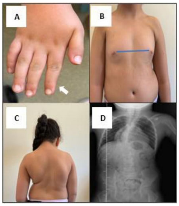
The older sibling had a length at the fifth percentile and a weight between the 25th and 50th percentiles, and additional findings included bilateral single transverse palmar creases, fifth-finger camptodactyly and contracture of the distal phalanges, a triggering left thumb, cubitus valgus, and a widened internipple distance. A severe right-convex thoracolumbar scoliosis was present without skeletal malformations or vertebral fusions on radiographic study (
Figure 1).
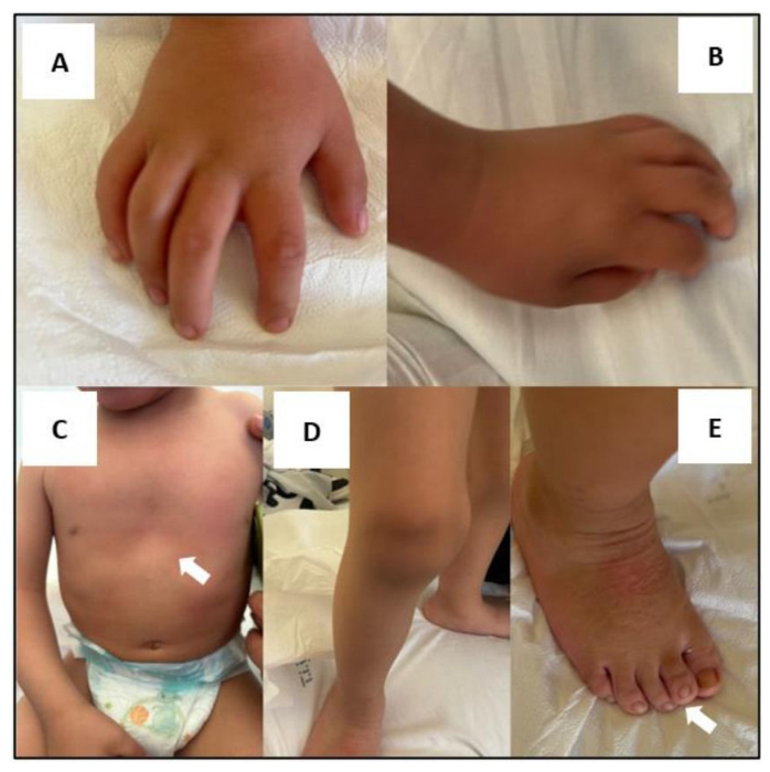
The male infant had a length and weight at the 50th percentile and exhibited low-set ears, bilateral ulnar deviation of the hands and first thumbs, bilateral polycamptodactyly, pectus excavatum, genu varum and bilateral knee flexion contractures, and overlapping of the second toe onto the first (
Figure 2). Audiological examination and auditory brainstem response (ABR) evaluation revealed moderate-to-severe bilateral sensorineural hearing loss.
Neuropsychiatric evaluation showed IQ in the normal range; neurological examination and electromyography, aCGH, and molecular analysis for skeletal dysplasia were normal in both patients. Given the clinical suspicion of AMC, molecular analysis was performed using a next-generation sequencing (NGS) panel for skeletal dysplasias and arthrogryposis.
3. Molecular Analysis
Genomic DNA was extracted from peripheral blood, followed by next-generation sequencing (NGS). Library preparation was performed using Clinical Exome Solution (Clinical Exome_Exp_v2, CNVs available) by Twist (South San Francisco, CA, USA), a target enrichment workflow that targets 6700 known disease-associated genes, according to the manufacturer’s instructions. Library quality and quantity were assessed using D1000 and High-Sensitivity D1000 Reagent Kits on a TapeStation 4150 system (Agilent Technologies, Santa Clara, CA, USA). Normalized libraries were sequenced on an Illumina NextSeq platform using a NextSeq 500/550 Mid Output Kit v2.5 (300 cycles) (Illumina, San Diego, CA, USA). Base calling and quality scoring were performed using the manufacturer’s software Real-Time Analysis v.1.18.54, followed by the generation of FASTQ sequence files. Reads were mapped to the GRCh38 reference genome (mean target coverage: 125.9821, target regions 100X: 70.34% for male sibling; mean target coverage: 141.08, target regions 100X: 81.41% for female sibling). A virtual panel for skeletal dysplasias and arthrogryposis (
Supplementary Table S1) was applied for the secondary bioinformatic analysis. Variant annotation, filtering, and interpretation were performed using Expert Variant Interpreter—eVai (enGenome, Pavia, Italy) according to the American College of Medical Genetics and Genomics (ACMG) guidelines and ACGS best practice guidelines for variant classification in rare diseases [
9,
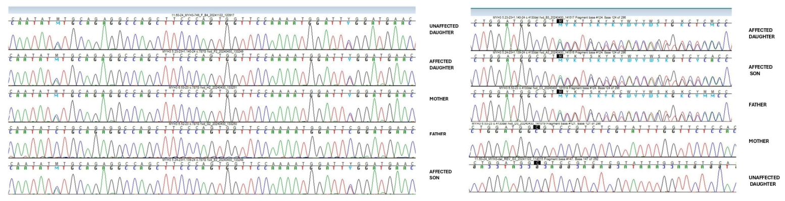
10]. Variants identified in probands and in their relatives were confirmed by Sanger sequencing using the primers in
Supplementary Table S2. Sequencing reactions were performed using the BigDye™ Terminator v.3.1 Cycle Sequencing Kit (Applied Biosystems, Life Technologies, Waltham, MA, USA), followed by capillary electrophoresis on a SeqStudio Genetic Analyzer (Applied Biosystems). Sequence analysis was carried out with Sequencher v5.4.6 (Gene Codes Corporation, Ann Arbor, MI, USA).
Genetic testing was performed in accordance with the Declaration of Helsinki, after obtaining written informed consent from the patients’ parents, who also authorized data publication.
4. Results
Genetic testing identified three heterozygous variants in the
MYH3 gene in both patients: two maternal VUS variants in
cis (c.749G>A and c.787G>T) and a paternal, likely pathogenic variant in
trans (c.4130_4138del) (
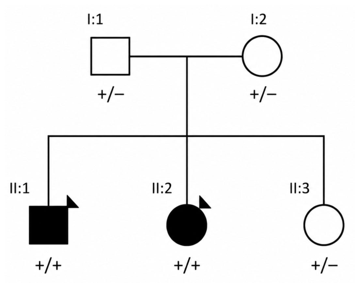
Figure 3).
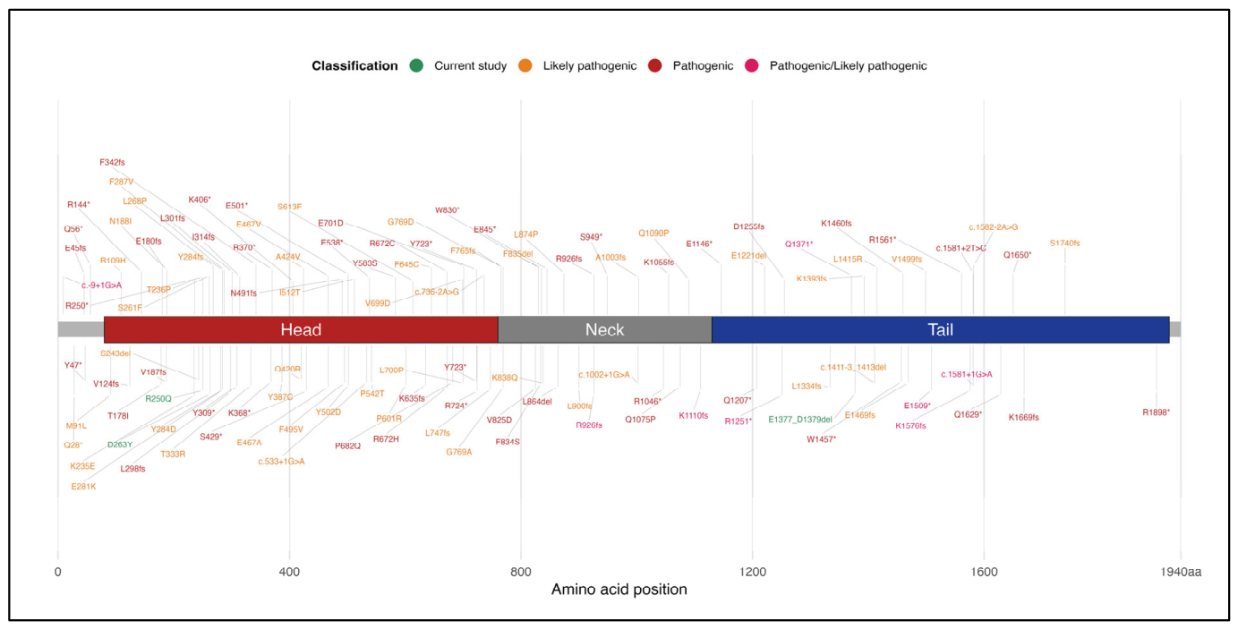
The c.749G>A, p.(Arg250Gln) variant is a missense variant occurring in a well-established mutational hotspot of the gene, with a low population frequency in gnomAD; in silico prediction tools indicate that this variant is likely to have a deleterious effect on protein structure/function (PP2, PM2, PP3, PP1). The c.787G>T, p.(Asp263Tyr) variant is a missense alteration involving a mutational hotspot, also with a very low population frequency and predicted to have a damaging effect by in silico analysis (PP2, PM2, PP3, PP1). The c.4130_4138del p.(Glu1377_Asp1379del) variant is a novel variant, an in-frame deletion causing the loss of three amino acids in a non-repetitive region, located in an evolutionarily conserved domain, absent from public databases (PM2, PM4, PP1_Mod) (
Figure 4). All three variants co-segregated with disease. According to the ACMG and ACGS criteria, the c.749G>A and c.787G>T variants can be classified as VUS, while the c.4130_4138del variant can be classified as likely pathogenic (
Supplementary Table S3; Supplementary Figure S1).
No other candidate variants in arthrogryposis genes were detected.
Subsequent genetic testing of an asymptomatic sister revealed the same two maternal variants, confirming her carrier status (
Figure 5).
5. Discussion
The present report highlights the clinical and molecular heterogeneity of MYH3-related arthrogryposis. The phenotypic overlap observed in both siblings, including congenital hand contractures and axial skeletal anomalies, aligns with the spectrum described for DAs. However, despite carrying the same compound heterozygous variants in MYH3, the two siblings exhibited distinct clinical manifestations, showing interindividual variability. The female patient presented with pronounced scoliosis and minor craniofacial involvement, while the male sibling showed more extensive musculoskeletal abnormalities and moderate-to-severe hearing loss.
In
Table 1, we compare the two affected siblings with the defining features of DA2A, DA2B, DA4, DA6, and recessive
MYH3-related CPSFS phenotypes.
However, it cannot be excluded that this different expressivity may be related to other factors, such as sex-related modifiers, additional genetic modifiers, and environmental factors.
The identification of the above-mentioned three
MYH3 variants in compound heterozygosity raises important questions regarding their individual and combined pathogenic potential. Given their segregation with disease in the two affected probands, alongside the heterozygous state retrieved in the asymptomatic parents and sister, a probable
MYH3 autosomal recessive inheritance can be hypothesized, as recently reported in the literature [
11,
12]. The absence of clinical manifestations in heterozygous carriers makes a fully penetrant dominant mechanism less likely, although functional studies are required to establish the precise molecular effects of the identified variants in order to elucidate whether the two variants in
cis act synergistically and how the variant in
trans may contribute to the overall phenotype. In this context, some authors have proposed the term
MYH3-associated skeletal fusion syndrome (MASF) to capture this expanded clinical variability. Furthermore, these studies indicate that, given the considerable phenotypic variability observed both between and within families, drawing a clear and consistent boundary between dominant and recessive presentations may be challenging and not always straightforward from a clinical or biological standpoint [
8].
From a diagnostic perspective, this report underscores the limitations of phenotype-driven classification in arthrogryposis syndromes. Although
MYH3 is a well-recognized cause of several distal arthrogryposes syndromes [
8], the clinical manifestations observed in our patients differ from the classical presentations most commonly associated with
MYH3-related disorders. In particular, the severe scoliosis observed in the female sibling and the sensorineural hearing loss identified in the male sibling resemble features that have been reported in association with DA4 and DA6, respectively. Nevertheless, these observations are based on a single family and further descriptions are necessary.
The overlapping and atypical features observed in the present cases highlight the need for comprehensive genomic approaches, particularly in patients who do not fully meet established diagnostic criteria. Early molecular diagnosis may facilitate appropriate surveillance, genetic counseling, and the anticipatory management of associated complications, including progressive spinal deformities and sensory deficits.
Moreover, this report emphasizes the importance of recognizing the wide phenotypic spectrum associated with MYH3 variants. Notably, the observed manifestations do not fully correspond to any known subtype of distal arthrogryposis, supporting the concept that MYH3-associated conditions may represent a continuum rather than distinct clinical entities.
6. Conclusions
We report a family with arthrogryposis and variable clinical manifestations in two affected siblings carrying compound heterozygous MYH3 variants, including two variants in cis (c.749G>A and c.787G>T) and one variant in trans (c.4130_4138del). The segregation pattern and clinical findings provide suggestive evidence of a possible genotype–phenotype association and further support the expanding phenotypic spectrum of MYH3-related disorders, which may be influenced also by both inheritance patterns and gene dosage effects. However, the interpretation of the identified variants remains limited by the absence of functional validation, and a causal relationship with the observed phenotype cannot be established with certainty. Additional families, detailed segregation analyses, and functional studies are needed to clarify the pathogenic role of these variants and their contribution to phenotypic variability.
Author Contributions
Concept and design of study, A.D. and D.C.; investigations and interpretation of molecular data, E.F., A.B. and R.B.; validation, D.V. and D.C.; clinical data curation, A.D., C.M. and D.C.; writing—original draft preparation, A.D., D.V. and E.F.; review and editing, D.V., E.F. and D.C.; supervision, D.C. All authors have read and agreed to the published version of the manuscript.
Funding
This research received no external funding.
Institutional Review Board Statement
Ethics committee approval was not required for this retrospective single-patient case report according to local regulations. Written informed consent for the publication of clinical data and images was obtained from the patients in accordance with the Declaration of Helsinki. All personal data were anonymized to protect patient privacy. The study was reviewed by the Regional Ethics Committee of Calabria, Italy, which issued an acknowledgment (Protocol No. 147/2026).
Informed Consent Statement
Informed consent was obtained from all subjects involved in the study.
Data Availability Statement
The clinical and molecular data supporting the findings are included within the article. Additional anonymized information may be made available by the corresponding authors upon reasonable request, subject to ethical and privacy restrictions.
Acknowledgments
The authors would like to acknowledge Giada Cuconato (Biogenet; Department of Molecular Medicine, University of Pavia, Italy) for her assistance in the final revision of the molecular data.
Conflicts of Interest
Dr. Elena Falcone, Dr. Alessia Bauleo, and Dr. Rossella Brando are employees of BIOGENET, Medical and Forensic Genetics Laboratory. The molecular analyses reported in this study were performed at this institution. The authors declare that no commercial or financial relationships existed that could be construed as a potential conflict of interest.
Abbreviations
The following abbreviations are used in this manuscript:
| DAs | Distal arthrogryposes |
| aCGH | Chromosomal microarray analysis |
| ABR | Auditory brainstem response |
| VUS | Variants of uncertain significance |
References
- Hall, J.G. Arthrogryposis multiplex congenita: Etiology, genetics, classification, diagnostic approach, and general aspects. J. Pediatr. Orthop. B 1997, 6, 159–165. [Google Scholar] [CrossRef] [PubMed]
- Hall, J.G.; Reed, S.D.; Greene, G. The distal arthrogryposes: Delineation of new entities—Review and nosologic discussion. Am. J. Med. Genet. 1982, 11, 185–239. [Google Scholar] [CrossRef] [PubMed]
- Bamshad, M.; Van Heest, A.E.; Pleasure, D. Arthrogryposis: A review and update. J. Bone Jt. Surg. Am. 2009, 91, 40–46. [Google Scholar] [CrossRef]
- Stevenson, D.A.; Carey, J.C.; Palumbos, J.; Rutherford, A.; Dolcourt, J.; Bamshad, M.J. Clinical characteristics and natural history of Freeman–Sheldon syndrome. Pediatrics 2006, 117, 754–762. [Google Scholar] [CrossRef] [PubMed]
- Desai, D.; Stiene, D.; Song, T.; Sadayappan, S. Distal arthrogryposis and lethal congenital contracture syndrome—An overview. Front. Physiol. 2020, 11, 689. [Google Scholar] [CrossRef] [PubMed]
- Tolonen, J.P.; Schnekenberg, R.P.; McGowan, S.; Sims, D.; McEntagart, M.; Elmslie, F.; Shears, D.; Stewart, H.; Tofaris, G.K.; Dabir, T.; et al. Detailed analysis of ITPR1 missense variants guides diagnostics and therapeutic design. Mov. Disord. 2024, 39, 141–151. [Google Scholar] [PubMed]
- Cameron-Christie, S.R.; Wells, C.F.; Simon, M.; Wessels, M.; Tang, C.Z.N.; Wei, W.; Takei, R.; Aarts-Tesselaar, C.; Sandaradura, S.; Sillence, D.O.; et al. Recessive Spondylocarpotarsal Synostosis Syndrome Due to Compound Heterozygosity for Variants in MYH3. Am. J. Hum. Genet. 2018, 102, 1115–1125. [Google Scholar] [CrossRef] [PubMed]
- Zhao, S.; Zhang, Y.; Hallgrimsdottir, S.; Zuo, Y.; Li, X.; Batkovskyte, D.; Liu, S.; Lindelöf, H.; Wang, S.; Hammarsjö, A.; et al. Expanding the mutation and phenotype spectrum of MYH3-associated skeletal disorders. npj Genom. Med. 2022, 7, 11. [Google Scholar] [CrossRef] [PubMed]
- Richards, S.; Aziz, N.; Bale, S.; Bick, D.; Das, S.; Gastier-Foster, J.; Grody, W.W.; Hegde, M.; Lyon, E.; Spector, E.; et al. ACMG Laboratory Quality Assurance Committee. Standards and guidelines for the interpretation of sequence variants: A joint consensus recommendation of the American College of Medical Genetics and Genomics and the Association for Molecular Pathology. Genet. Med. 2015, 17, 405–424. [Google Scholar] [CrossRef] [PubMed]
- Durkie, M.; Cassidy, E.-J.; Berry, I.; Owens, M.; Turnbull, C.; Scott, R.H.; Taylor, R.W.; Deans, Z.C.; Ellard, S.; Baple, E.L.; et al. ACGS Best Practice Guidelines for Variant Classification in Rare Disease; Association for Clinical Genomic Science (ACGS): London, UK, 2024. [Google Scholar]
- Zgheib, O.; Rio-Frio, T.; Guipponi, M.; Nouspikel, T.; Fokstuen, S.; Abramowicz, M.; Khau Van Kien, P. Biallelic MYH3 Variants Cause Distal Arthrogryposis in Compound Heterozygosity and a Subclinical Phenotype in Simple Heterozygosity. Codominance or Recessive Inheritance? Clin. Genet. 2026, 109, 1059–1063. [Google Scholar] [CrossRef] [PubMed]
- Morali, B.; Miranda, V.; Raelson, J.; Grimard, G.; Glavas, P.; Audibert, F.; Dumont, N.A.; Barone, J.; Bamshad, M.; Lemyre, E.; et al. Bi-allelic variants in MYH3 cause recessively-inherited arthrogryposis. Clin. Genet. 2024, 106, 483–487. [Google Scholar] [CrossRef] [PubMed]
Figure 1.
Female patient at 6 years: (A) fifth-finger camptodactylty and distal phalange contracture, (B) widened internipple distance, and (C,D) right-convex thoracolumbar scoliosis.
Figure 1.
Female patient at 6 years: (A) fifth-finger camptodactylty and distal phalange contracture, (B) widened internipple distance, and (C,D) right-convex thoracolumbar scoliosis.
Figure 2.
Male patient at 13 months: (A,B) bilateral polycamptodactyly, (C) pectus excavatum, (D) genu varum and bilateral knee flexion contractures, and (E) overlapping second toe.
Figure 2.
Male patient at 13 months: (A,B) bilateral polycamptodactyly, (C) pectus excavatum, (D) genu varum and bilateral knee flexion contractures, and (E) overlapping second toe.
Figure 3.
Family pedigrees. Black squares and circles represent affected individuals with distal contractures; +/+ and +/− symbols, respectively, represent compound heterozygosity (II:1 and II:2 c.[749G>A;787G>T];[4130_4138del]) and heterozygosity (I:1 c.[4130_4138del];[=], I:2 and II:3 c.[749G>A;787G>T];[=]). The arrowhead in the pedigree indicates the probands.
Figure 3.
Family pedigrees. Black squares and circles represent affected individuals with distal contractures; +/+ and +/− symbols, respectively, represent compound heterozygosity (II:1 and II:2 c.[749G>A;787G>T];[4130_4138del]) and heterozygosity (I:1 c.[4130_4138del];[=], I:2 and II:3 c.[749G>A;787G>T];[=]). The arrowhead in the pedigree indicates the probands.
Figure 4.
Variant distribution across MYH3 protein domains: Likely pathogenic (orange), pathogenic (brownish), and pathogenic/likely pathogenic (purple) variants identified in the literature (ClinVar database) and the three variants in the current study, D263Y, R250Q, and E1377_D1379del (green). *: stop codon.
Figure 4.
Variant distribution across MYH3 protein domains: Likely pathogenic (orange), pathogenic (brownish), and pathogenic/likely pathogenic (purple) variants identified in the literature (ClinVar database) and the three variants in the current study, D263Y, R250Q, and E1377_D1379del (green). *: stop codon.
Figure 5.
Sanger sequencing analysis of the three variants in the family. On the left, the two missense variants in cis (c.749G>A and c.787G>T); on the right, the in-frame deletion (c.4130_4138del).
Figure 5.
Sanger sequencing analysis of the three variants in the family. On the left, the two missense variants in cis (c.749G>A and c.787G>T); on the right, the in-frame deletion (c.4130_4138del).
Table 1.
Comparison of the clinical features observed in the two affected siblings with the principal distal arthrogryposis subtypes and recessive MYH3-related congenital pterygium, scoliosis, and vertebral fusion syndrome (CPSFS). Abbreviations: CPSFS, congenital pterygium, scoliosis, and vertebral fusion syndrome; AD, autosomal dominant; AR, autosomal recessive; O.R., occasionally reported.
Table 1.
Comparison of the clinical features observed in the two affected siblings with the principal distal arthrogryposis subtypes and recessive MYH3-related congenital pterygium, scoliosis, and vertebral fusion syndrome (CPSFS). Abbreviations: CPSFS, congenital pterygium, scoliosis, and vertebral fusion syndrome; AD, autosomal dominant; AR, autosomal recessive; O.R., occasionally reported.
| Clinical Feature | Sibling 1 (Female) | Sibling 2 (Male) | DA2A | DA2B | DA3 | DA4 | DA5 | DA6 | DA7 | DA8 | DA9 | DA10 | Recessive MYH3-
Related CPSFS |
|---|
| Inheritance | Hypothesized
AR | Hypothesized
AR | AD | AD | AD | AD | AD | AD | AD | AD | AD | AD | AR |
| Distal limb contractures | + | + | + | + | + | + | + | + | + | + | + | + | + |
| Proximal joint involvement | - | Knee flexion contractures | Usually absent | Usually absent | Usually absent | Usually absent | Usually absent | Usually absent | Variable | Variable | Variable | Variable | May be present |
| Camptodactyly | + | + | Typical | Typical | Typical | Common | Common | Common | Common | Common | Common | Common | Frequent |
| Clubfoot | - | - | Frequent | Frequent | Frequent | Frequent | Frequent | Frequent | Variable | Variable | Variable | Variable | Variable |
| Craniofacial abnormalities | Not identified | Low-set
ears only | Distinctive facies | Mild
involvement | Cleft palate may occur | Usually absent | Distinctive facies | Usually absent | Usually absent | Usually absent | Usually absent | Usually absent | Mild or absent |
| Cleft palate | - | - | O.R. | O.R. | Typical feature | Not typical | Not typical | Not typical | Not typical | Not typical | Not typical | Not typical | Rare |
| Short neck | + | + | Not typical | Not typical | Not typical | Variable | Variable | Variable | Variable | Variable | Variable | Variable | Reported |
| Scoliosis | Severe
Thoracolumbar
scoliosis | Not observed | Uncommon | O.R. | O.R. | Defining feature | O.R. | O.R. | O.R. | Common | Variable | Variable | Frequent |
| Sensorineural hearing loss | - | Moderate-to-severe | Not typical | Not typical | Not typical | Not typical | Not typical | Defining feature | Not typical | Not typical | Not typical | Not typical | Reported in some patients |
| Ophthalmoplegia | - | - | - | - | - | - | Defining feature | - | - | - | - | - | O.R. |
| Ptosis | - | - | - | - | - | - | Common | - | - | - | - | - | Variable |
| Pectus abnormalities | - | Pectus excavatum | O.R. | O.R. | Variable | Variable | Variable | Variable | Variable | Variable | Variable | Variable | Reported |
| Pterygia | - | - | - | - | - | - | - | - | - | Not
typical | Typical | Not
typical | May be
present |
| Vertebral fusion | - | - | - | - | - | - | - | - | - | Typical | Variable | Variable | Typical |
| Carpal/tarsal fusion | Not detected | Not detected | - | - | - | - | - | - | - | Typical | Variable | Variable | Typical |
| Neurological abnormalities | - | - | - | - | - | - | - | - | - | - | - | - | Typically absent |
| Overall phenotypic concordance with present family | - | - | Limited | Limited | Partial | Partial overlap due to scoliosis | Limited | Partial overlap due to hearing
loss | Limited | Partial | Partial | Partial | Greatest overall concordance |
| Disclaimer/Publisher’s Note: The statements, opinions and data contained in all publications are solely those of the individual author(s) and contributor(s) and not of MDPI and/or the editor(s). MDPI and/or the editor(s) disclaim responsibility for any injury to people or property resulting from any ideas, methods, instructions or products referred to in the content. |




 ,
,