Article

Phylo-Epigenetic Conservation and CpG Erosion in OCT4, SOX2, and hTERT Intragenic CpG Islands: A Waddingtonian Perspective on Mammalian Developmental Evolution

by
Simeon Santourlidis
Epigenetics Laboratory for Human Health and Longevity, Institute of Transplantation Diagnostics and Cell Therapeutics, Medical Faculty, Heinrich-Heine University Duesseldorf, Moorenstr. 5, 40225 Duesseldorf, Germany
Genes 2025, 16(9), 1102; https://doi.org/10.3390/genes16091102
Submission received: 25 July 2025 / Revised: 27 August 2025 / Accepted: 14 September 2025 / Published: 18 September 2025
(This article belongs to the Section Animal Genetics and Genomics)

Abstract

Background/Objectives: Developmental biologist Conrad Waddington proposed that evolution is shaped not only by genetic mutations and natural selection but also by environmentally responsive developmental mechanisms. Building on this premise, the epigenetic regulation of three master genes central to mammalian embryogenesis—OCT4, SOX2, and hTERT—focusing on their intragenic CpG islands (iCpGIs), which are crucial for transcriptional control and chromatin state modulation, were investigated. Methods: By performing a phylo-epigenetic comparison across 12 primate species, strong conservation of CpG-rich regions, punctuated by lineage-specific CpG transitions, particularly CpG→TpG and CpG→CpA was identified. Results: These mutational patterns align with methylation-dependent deamination mechanisms and highlight iCpGIs as evolutionarily constrained, epigenetically plastic elements. Notably, CpG variation alone recapitulated known primate phylogenies, suggesting that methylation-sensitive sites within iCpGIs encode both developmental and evolutionary information. Conclusions: It is proposed that such sites are prone to Environmentally Determined Epimutations (EDEMs)—methylation-driven, nutrition-sensitive changes that persist across generations and modulate gene regulatory capacity. This integrative framework advances Waddington’s concept of canalization by providing a molecular mechanism through which environmental factors can reshape developmental trajectories and contribute to evolutionary innovation.

1. Introduction

Conrad Hal Waddington envisioned evolution as a long-term process governing life. Across countless generations, the successful interaction of organisms with their environment has yielded increasingly robust life forms [1]. These organisms possess the intrinsic capacity to respond to fluctuating environmental conditions and, over successive generations, become progressively better adapted. Notably, certain acquired characteristics manifest already during embryonic development [1]. New individuals inherit the capacity not only to survive and reproduce, but also to pass on both ancestral and newly acquired traits to their offspring.
In Waddington’s view, a complete understanding of this adaptive progression requires expanding the Neo-Darwinian framework, which combines natural selection with random mutation, by incorporating a crucial epigenetic component [1]. This addition provides a missing link: a functional bridge between environmental pressures and the organism’s adaptive developmental responses.
Waddington’s work gave rise to two foundational concepts. First, he proposed that environmental stressors can induce phenotypic changes that eventually become genetically assimilated and heritable [1,2]. A central question that arises is how does such genetic assimilation manifest in the DNA itself?
Second, he introduced the concept of canalization, which describes the robustness of developmental processes [1,2]. It is evident that immediately following conception, the zygote already harbors all the essential information required to initiate an evolutionarily canalized developmental program. This enables the formation of a complex, highly adapted organism—be it an ape or a human—well adapted to anticipated environmental conditions.
This raises several fundamental questions: In what form is the information that canalizes development encoded within the zygote? How is it propagated across generations? And how do environmental stimuli influence its transgenerational plasticity?
Significant progress in epigenetics now allows us to assert with confidence that epigenetic regulation is as fundamentally important as classical genetics—a principle that holds true across biological disciplines, from cancer biology to embryology and evolutionary science. Integrating this expanding body of knowledge into evolutionary theory could deepen our understanding of processes that unfold on evolutionary time scales, which are otherwise only accessible through fragmentary, present-day observations.
It is well established that genome-wide, cell type-specific DNA methylation patterns demarcate active and repressed genomic regions and are central to establishing correct baseline gene expression profiles.
DNA methylation, which occurs predominantly at CpG dinucleotides in mammalian genomes, has been extensively studied over the past four decades and is widely recognized as a repressive epigenetic mark [3]. Over 60% of human genes possess a CpG-rich 5′ region, characterized by at least 50% of GC content [4], known as a CpG island (CGI), typically spanning 0.4–2 kb and surrounding the transcription start site of developmental, tissue-specific, and housekeeping genes [5]. Notably, the promoter CGIs of housekeeping genes remain consistently unmethylated throughout development and across all differentiated tissues, consistent with their ubiquitous expression [5]. This hypomethylated state serves to protect these CpG-rich regions from mutational erosion [6].
Spontaneous hydrolytic deamination of cytosine leads to uracil, which is efficiently detected and repaired [6]. In contrast, 5-methylcytosine deamination occurs 2- to 3-fold more frequently and generates TpG mismatches, which are less efficiently corrected [7]. Consequently, the transition rate from methylated CpG to TpG is 10–50 times higher than for other dinucleotide transitions [8,9]. Due to the palindromic nature of CpGs, this leads to CpG-to-TpG/CpA erosion over evolutionary time.
Studies analyzing more than 30 silenced and 100 non-silenced genes have shown that methylation of a relatively small core CGI region overlapping the transcription start site correlates consistently with gene silencing [10]. Depending on their precise methylation state, CpG sites within a CGI can have varying effects on gene expression, reflecting a complex and still not fully resolved regulatory logic [10].
Importantly, methylation extending into exonic regions does not necessarily block transcription initiation or elongation [10,11]. On the contrary, evidence indicates that methylation within gene bodies can modulate gene expression by enhancing transcription and affecting alternative splicing [12,13]. For instance, the CpG architecture at exon–intron junctions influences DNA methylation’s role in co-transcriptional splicing [13]. Another study found that exonic DNA methylation interacts with nucleosome occupancy and the histone mark H3K36me3 to regulate splicing outcomes [14].
Intragenic CpG islands (iCpGIs) have emerged as key regulatory elements, with their influence dependent on CpG architecture [13]. CpG density of CGIs is an important regulatory feature that contributes to the formation of histone signatures associated with transcription [15]. During development, unmethylated iCpGIs can adopt bivalent chromatin states, marked by both H3K27me3 (repressive) and H3K4me3 (activating) histone marks [16,17]. This poised configuration allows developmental genes to remain transcriptionally silent yet ready for rapid activation. As embryonic stem (ES) cells differentiate, these bivalent domains resolve into either active or repressive chromatin, depending on lineage commitment [17].
Moreover, intragenic CpG islands positioning within genes is not arbitrary and is instead selected for; iCpGIs can influence pre-mRNA processing, affecting mRNA isoform length and enhancer function, depending on their methylation status [16]. They contribute to the expansion of transcriptomic diversity. ICpGIs are frequently subject to DNA methylation changes during development, implying that their epigenetic regulation is crucial for tissue specific programming [16].
DNA methylation is thus a central determinant of mammalian embryogenesis. It is essential for embryonic viability [18], and its absence severely impairs differentiation [19]. Despite this, we are only beginning to uncover the full extent of its developmental impact [18].
Key pluripotency regulators such as OCT4, SOX2, and hTERT are under epigenetic control [20,21,22,23,24,25,26]. Their CpG islands spanning promoters and exons are unmethylated in early development and become subsequently methylated upon differentiation [27]. Furthermore, SOX2 has been shown to be marked by bivalent histone modifications, and hTERT is subject to regulation via histone modifications and DNA methylation [25].
Following fertilization, global epigenetic reprogramming resets the methylation landscape, and essential pluripotency genes such as Oct4 and Sox2 become transcriptionally activated [28]. Together, these genes initiate a regulatory network involving hundreds to thousands of developmental genes. The precise expression balance between Oct4, Sox2, and their targets is critical for maintaining pluripotency, enabling the generation of all three embryonic germ layers [28]. Even minor deviations in expression levels of Oct4 or Sox2 can tip the balance toward differentiation, with long-term effects on embryonic development [28].
As differentiation begins, pluripotency-related transcriptional circuits are silenced, in part via de novo DNA methylation [19]. In this context, it has been observed that OCT4 fails to bind its target sites in embryonic stem cells (ESCs) when methylation occurs within 100 bp on each side of the target sequence [11].
Additionally, rapidly dividing ESCs must address telomere maintenance, as DNA polymerases cannot fully replicate chromosome ends [29]. Robust telomerase activity—regulated via transcriptional, epigenetic, and splicing mechanisms—ensures telomere length, self-renewal, and protection against premature differentiation [29,30,31,32,33,34].
In humans, telomerase has been detected in germ-line tissues, blastocysts, and 16–20-week-old fetal tissue, but not in most normal somatic tissues [35]. Telomerase expression can impact the expression of a variety of genes required for extended self-renewal and lifespan [36]. Undifferentiated stem cells are responsible for telomerase activity in the human fetus, and downregulation of telomerase activity occurs during differentiation and gestational development [35]. It has been demonstrated that an alternative splicing event, centered around hTERT exon 2, triggers hTERT mRNA decay in differentiating cells, whereas in pluripotent cells, inclusion of exon 2 promotes telomerase accumulation [34]. This tissue-specific and developmental regulation of telomerase in the human fetus suggests an important role for this ribonucleoprotein in human fetal tissue differentiation and development [35]. Thus, the regulation mechanisms finely tune the downregulation of telomerase right at the beginning of and during the various differentiation paths to be pursued, constituting a crucial adjusting screw for the developmental fate of the embryo.
Taken together, the activity of these three hierarchically positioned master genes and their target networks must be precisely tuned. Small perturbations in the epigenetic regulation of OCT4, SOX2, or hTERT can have profound consequences on the outcome of embryogenesis—ultimately affecting the development of a viable, well-adapted organism.
All three genes harbor intragenic CpG islands extending from upstream regions near the transcription start site into exon and intron (except SOX2) sequences: OCT4 iCpGI spans exon 1 and intron 1; SOX2 iCpGI includes the entire coding sequence of the single exon; and hTERT iCpGI covers exon 1, intron 1, exon 2, and part of intron 2.
Given the central importance of DNA methylation in early developmental regulation, it is hypothesized here that evolutionary changes in the CpG profiles, the basis of differential methylation of these crucial epigenetic regulatory regions, may represent a key feature of mammalian evolution.
To explore this, a phylo-epigenetic comparison of CpG versus non-CpG dinucleotide variation in the intragenic CpG islands (iCpGIs) of OCT4, SOX2, and hTERT across 12 primate species has been performed. These included hominins (Homo sapiens, H. neanderthalensis, and Denisovan human), great apes (Pan troglodytes, Pan paniscus, Gorilla gorilla, Pongo abelii), and Old World monkeys (Macaca mulatta, Macaca fascicularis, Papio anubis, Chlorocebus sabaeus, and Rhinopithecus roxellana).
Among extant species, the common chimpanzee and bonobo are the closest relatives to humans, having diverged ~1.7 million years ago [37]. The Neanderthal and Denisovan genomes were included as calibration points. Neanderthals and modern humans are estimated to have diverged at least 430,000 years ago [38,39], while Denisovans represent a distinct lineage that separated from modern humans approximately 600,000–800,000 years ago based on nuclear and mitochondrial DNA analyses [40,41,42].

2. Material and Methods

To investigate the evolutionary conservation and divergence of epigenetically relevant CpG-rich regions, three canonical pluripotency-associated genes—OCT4 (POU5F1), SOX2, and hTERT—were selected based on their well-established regulation via DNA methylation. The reference human mRNA and genomic sequences of these genes were retrieved from the NCBI nucleotide database (https://www.ncbi.nlm.nih.gov/nuccore/?term=, accessed on 20 February 2025) under the following accession numbers: OCT4—NM_002701, BX088580; SOX2—NM_003106, AC117415; hTERT—NM_198253, NW_001838923.
The intragenic CpG islands (iCpGIs) within the 5′ regions of these genes were identified and extracted for twelve primate species using the UCSC Genome Browser (http://genome.ucsc.edu, accessed on 5 March 2025) [43]. These species included P. troglodytes (panTro6), P. paniscus (panPan3), G. gorilla (gorGor6), P. abelii (ponAbe3), R. roxellana (rhiRox1), P. anubis (papAnu4), M. fascicularis (macFas5), C. sabaeus (chlSab2), M. mulatta (rheMac10), archaic human relatives, H. neanderthalensis (Altai Neanderthal), and Denisova hominin, with sequences obtained from the Max Planck Institute’s JBrowse genome browser (https://bioinf.eva.mpg.de/jbrowse/, v1.12.1; accessed on 30 March 2025) [39].
To exclude potentially confounding repetitive elements (e.g., LINE-1, Alu), CpG-rich regions were screened using RepeatMasker (https://www.repeatmasker.org; accessed on 8 April 2025). The concatenated iCpGI sequences of OCT4, SOX2, and hTERT (in that order) were aligned using the multiple sequence alignment tool MAFFT (v7.511) with default settings and the nucleotide scoring matrix 1PAM/κ = 2, optimized for closely related sequences. The FFT-NS-i algorithm was employed to refine the alignment (https://mafft.cbrc.jp/alignment/server/; accessed on 15 April 2025) [44,45].
Phylogenetic relationships were inferred using the UPGMA clustering method based on the molecular clock assumption (https://mafft.cbrc.jp/alignment/server/phylogeny.html; accessed on 30 April 2025) [46]. To explore patterns of CpG conservation and divergence, all orthologous CpG dinucleotide positions were annotated and systematically converted: conserved CpGs across all species were replaced with “A”, while same-position dinucleotides lacking universal CpG conservation (i.e., variable CpG presence) were replaced with “T”.
Visual inspection and comparison of phylogenetic trees were performed using Archaeopteryx.js (http://phylo.io; accessed on 30 April 2025) [47]. Pairwise sequence identity was assessed using Clustal 2.1 to generate a percent identity matrix (https://www.ebi.ac.uk/; accessed on 30 April 2025).

3. Results

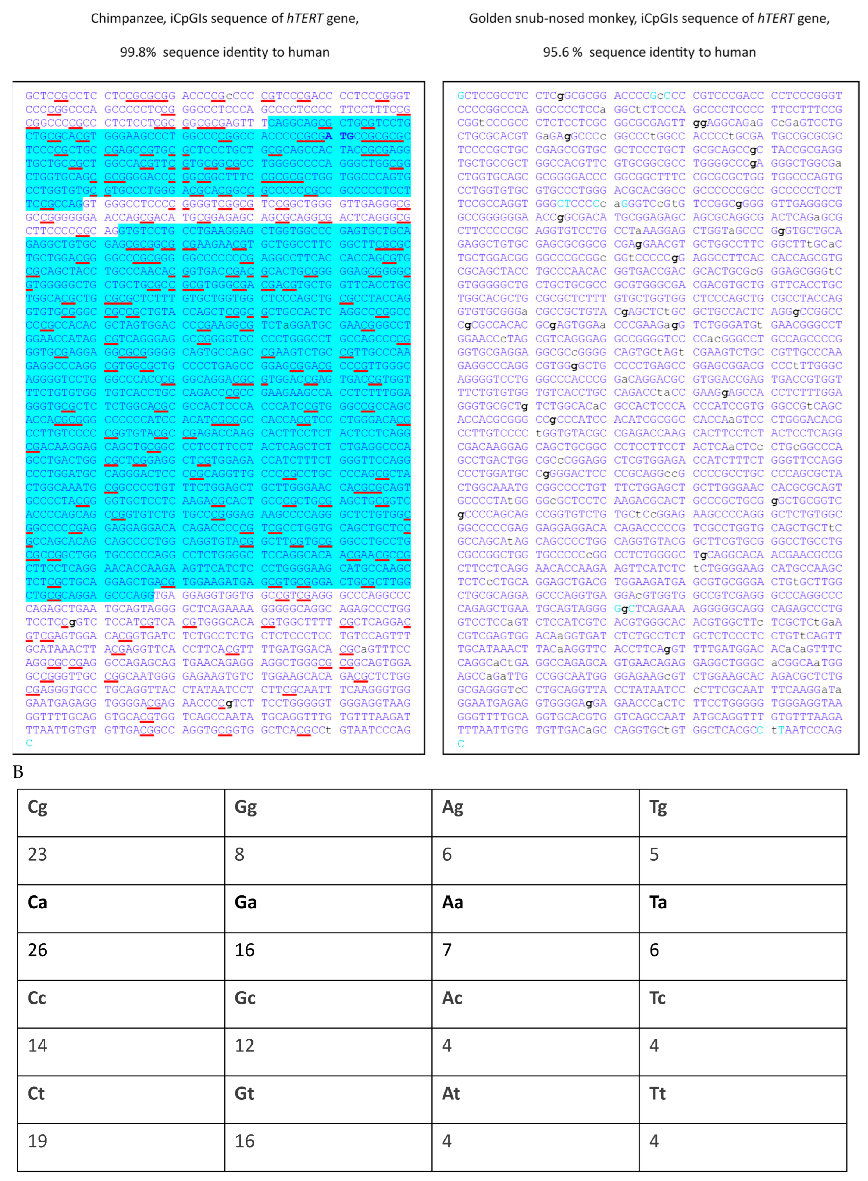
Initial sequence alignments were performed using the BLAT tool available via the UCSC Genome Browser, focusing on CpG-rich regions (iCpGIs) of the human genes OCT4, SOX2, and hTERT. These human sequences were first compared to their orthologs in the closely related chimpanzee (P. troglodytes) and subsequently to the more distantly related golden snub-nosed monkey (R. roxellana) (Figure 1A). All three iCpGIs exhibited dense clusters of CpG dinucleotides, spanning the first exon and extending into intron 1 in OCT4, and in the case of hTERT, further including exon 2 and part of intron 2.
These initial alignments revealed high sequence identity between human and chimpanzee: 99.7% for OCT4, 99.9% for SOX2, and 99.8% for hTERT (Figure 1A). Sequence identity between human and the golden snub-nosed monkey was moderately lower: 96.8% (OCT4), 98.3% (SOX2), and 95.6% (hTERT). Notably, the Colobinae subfamily, to which the golden snub-nosed monkey belongs, diverged from the Cercopithecinae (e.g., macaques, baboons, green monkeys) approximately 16.2 million years ago, as supported by fossil-calibrated molecular data [48]. In comparison, the divergence between orangutans and African apes occurred more recently, approximately 14 million years ago [48].
Across these alignments, nucleotide substitutions were frequently observed at guanine (G) positions. This trend is particularly evident in the hTERT iCpGIs of R. roxellana, where divergent G positions are marked in bold in the sequence alignment (Figure 1A). Further inspection of all interspecies alignments shown in Figure 1 revealed that these altered guanines were distributed across 23 CpG, 8 GpG, 6 ApG, and 5 TpG dinucleotides in the chimpanzee and golden snub-nosed monkey sequences. Additionally, adenine changes in 26 CpA dinucleotides were found (Figure 1B). All other changed dinucleotides appeared less frequently than the CpG and CpA dinucleotides (Figure 1B). Of note, none of the 18 CpG sites within the exonic regions of the OCT4 and SOX2 genes exhibiting interspecies variation between human, chimpanzee, and golden snub-nosed monkey resulted in an amino acid change.
These observations motivated the inclusion of additional primate species, which we used to perform a comprehensive alignment of the iCpGIs from OCT4, SOX2, and hTERT, resulting in a dataset comprising the introduced 12 primates. The aligned sequences ranged from 4928 to 5000 nucleotides, with an average length of 4977 nucleotides.
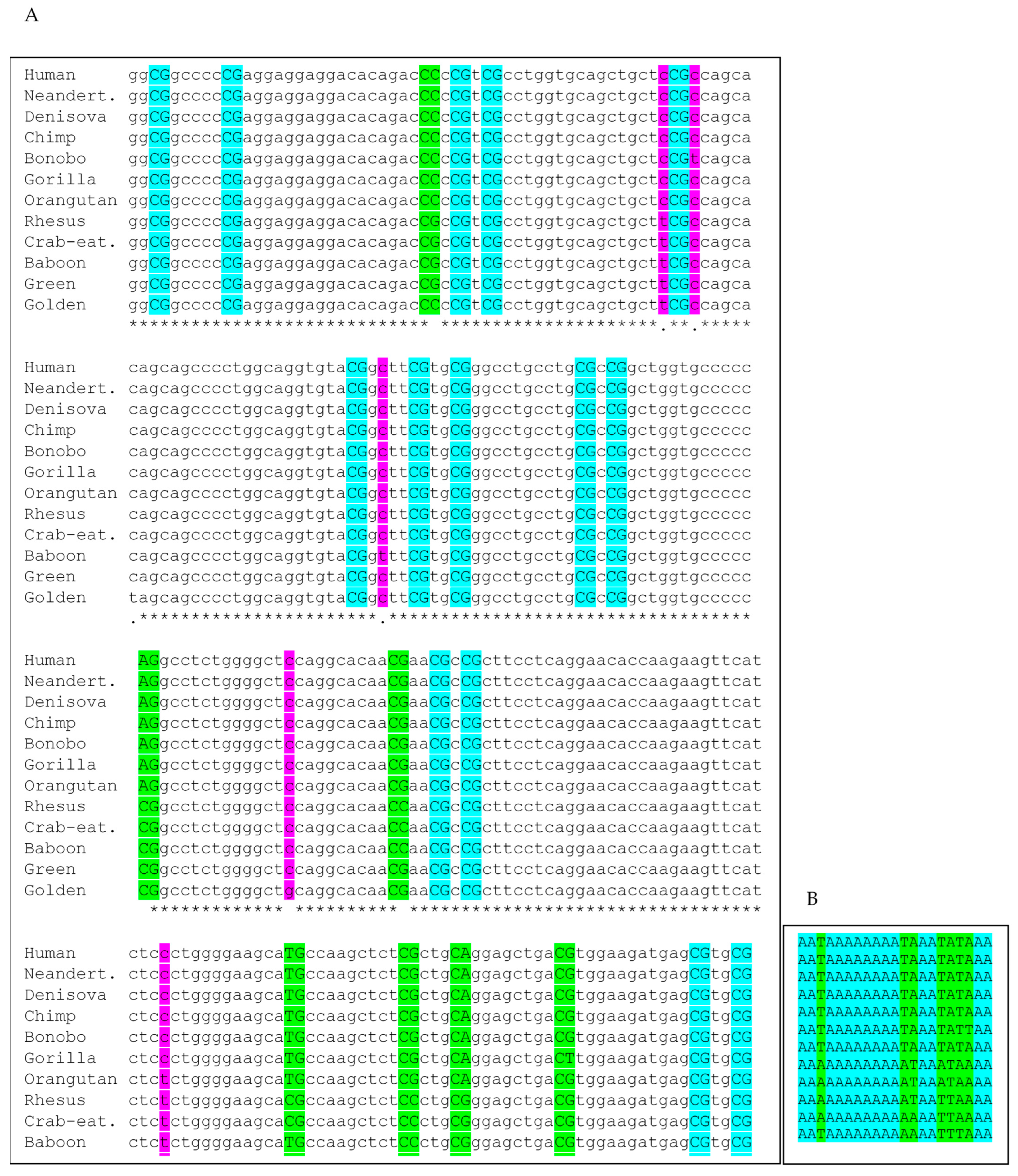
Within this full alignment, inspecting orthologous single-nucleotide positions, 132 single-nucleotide positions were changed outside of CpG dinucleotides in at least one of the primate sequences, corresponding to 2.7% of all nucleotide positions. Assuming these changes affect dinucleotide positions, 5.3% of all possible orthologous dinucleotides were altered. Of the 2489 total dinucleotide positions analyzed, 426 were orthologous CpG dinucleotide positions. Among these, 143 (34%) were variable in at least one species, translating to about 5.9% of all 2489 dinucleotides, while 283 (66%) were fully conserved. It should be noted that of the 143 orthologous CpG positions with variability between the selected species, 47 (33%) have either a CpG in all representatives of the hominid group or in the Old World monkey group. These differences at these orthologous CpG positions therefore distinguish the two major groups. There are 34 single-nucleotide variants (SNVs) at non-CpG positions that do the same. This translate to 25% of all orthologous SNV positions of all variant non-CpG positions.
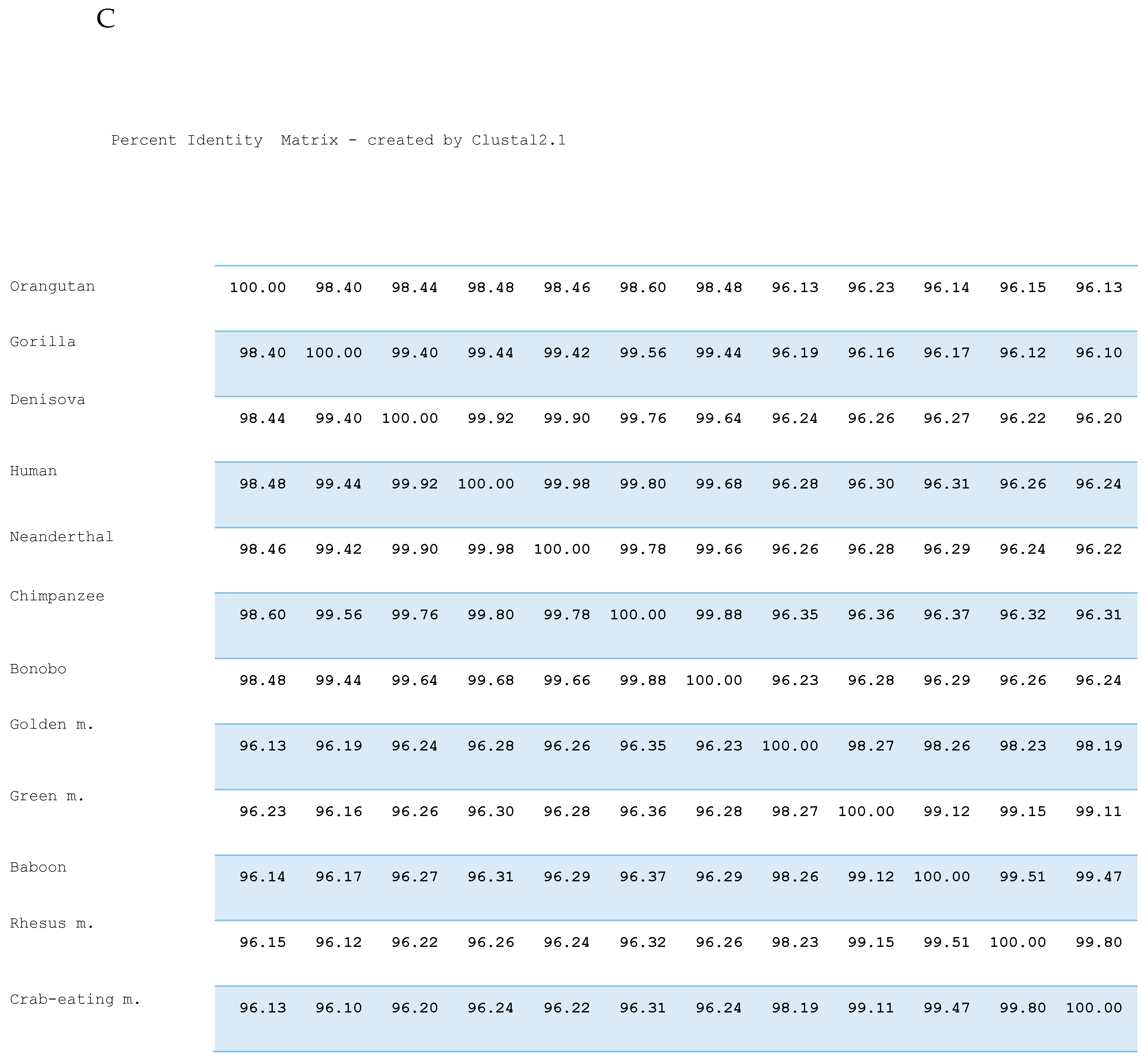
A representative 240-nucleotide alignment excerpt (Figure 2A) highlights conserved and altered CpG and non-CpG dinucleotide positions across species. Percent identity matrix analyses showed over 99% sequence identity among all hominid species except P. abelii (98%). Among the Old World monkeys, intra-group sequence identity was similarly high (≥99%), with R. roxellana again showing slightly reduced identity (98%). Inter-clade identity (Old World monkeys vs. hominids) averaged 96%, underscoring the high degree of conservation in these regulatory sequences.
These results contrast with prior studies of CpG islands in highly conserved housekeeping genes, where 4.8% of all orthologous dinucleotides were altered at non-CpG sites and 3.2% at orthologous CpG positions across human and great ape lineages [49]. In the present study, when focusing exclusively on the same hominins and great apes, 1.6% of non-CpG and 1.8% of CpG dinucleotides were variable, corresponding to 12% of all orthologous CpG sites—suggesting an even greater evolutionary constraint on iCpGIs associated with pluripotency genes.
Detailed analysis of CpG changes of the full alignment revealed specific substitution patterns: CG→CA (n = 45), CG→TG (n = 42), CG→CC (n = 23), CG→CT (n = 16), CG→GG (n = 15), and CG→AG (n = 20), with rarer transitions/transversions such as CG→AC, GC, GA, TA, and TT (n ≤ 3). CpA (on the sense strand) corresponds to TpG on the antisense strand, both of which are typical outcomes of methylated CpG deamination events. Of note, T→G belongs to the medium frequency substitutions in the eukaryotic genomes [50], suggesting that the slightly increased CG→CC rate could have resulted from the CG→TG transition and the subsequent TG→GG transversion in the complementary strand.
To visualize evolutionary dynamics specifically at CpG sites, a subalignment was generated by recoding conserved CpG dinucleotides as “A” and altered CpG positions as “T.” A representative excerpt is shown in Figure 2B. This transformation highlights the elevated substitution rate at CpG dinucleotides and the predominance of CG→CA and CG→TG transitions within these developmental regulatory regions.
To further explore evolutionary relationships, two UPGMA phylogenetic trees were constructed: one based on all nucleotide changes (including CpG sites, insertions, and deletions) and another based solely on CpG dinucleotides and changes at orthologous positions harboring CpGs in at least one species (Figure 3A and Figure 3B, respectively). The latter, referred to as a “phylo-epigenetic tree”, more accurately recapitulated the evolutionary relationships and divergence times previously inferred from genome-wide analyses and fossil records [49].
Calibration of both trees was anchored to the divergence of P. troglodytes and P. paniscus, estimated at 1.7 million years ago [37]. The phylo-epigenetic tree reliably reconstructed established divergence events, including the ~14 Ma separation of orangutans from African apes, the ~16.2 Ma split between Colobinae and Cercopithecinae, and the ~6 Ma divergence between humans and chimpanzees, consistent with both fossil and molecular evidence [37,51,52,53,54,55].
Collectively, these findings demonstrate that CpG-rich regions of OCT4, SOX2, and hTERT exhibit strong evolutionary conservation across primates, with a subset of orthologous CpG sites undergoing lineage-specific transitions consistent with known epigenetic mutational mechanisms. These patterns underscore the relevance of iCpGIs as evolutionarily constrained elements involved in developmental gene regulation and suggest potential utility in phylo-epigenetic reconstructions.

4. Discussion

Fundamental questions concerning the nature and direction of embryonic and evolutionary development have long captured scientific interest. Pioneering work by Conrad Waddington introduced the concepts of developmental canalization and the genetic assimilation of acquired traits into heritable forms—cornerstones of epigenetic thought [1,2]. The present study aimed to extend Waddington’s framework by integrating over seven decades of subsequent advances in molecular biology and epigenetics, particularly as they relate to early embryonic development.
This work represents an attempt to further pursue the central postulates Waddington articulated, namely that the critical information guiding development must be encoded in the zygote and that this information must be modifiable to accommodate environmental input—both at the transgenerational and evolutionary scale.
To explore this hypothesis, interspecies sequence variation has been examined across a panel of primates, focusing on highly conserved intragenic CpG islands (iCpGIs) within key pluripotency-associated genes. The analysis revealed that the most frequently observed genetic changes involved CpG dinucleotides, with a notable predominance of CpG-to-TpG and CpA transitions. Moreover 33% of the variable CpG positions were uniformly altered either in the hominid or the Old World clade, hence forming a clear distinguishing mark (Figure 3).
While this finding aligns with known molecular mechanisms—specifically, the elevated deamination rate of 5-methylcytosine at CpG sites—it holds further significance in light of recent observations. CpG-to-TpG mutations have been shown to frequently generate de novo binding sites for transcription factors such as OCT4 with higher functional efficiency than other mutational events, thereby increasing regulatory plasticity and potentially conferring an evolutionary advantage [56]. Similar perspectives underscore that these transitions contribute to genomic flexibility and can be associated with phenotypic innovations relevant to speciation [57].
What is more surprising, however, is the specific localization of these frequently occurring CpG changes within the iCpGIs of master developmental regulators. It is well established that CpG density and methylation status within such regions play critical roles in modulating gene expression. Importantly, OCT4, SOX2, and hTERT are situated at the apex of the regulatory hierarchy that orchestrates pluripotency and lineage specification. Even minor perturbations in the expression or epigenetic regulation of these genes are known to propagate through downstream networks, altering the tightly coordinated spatiotemporal gene expression patterns required for proper morphogenesis and differentiation [58].
Environmental factors—most prominently, nutrition—are known to shape the DNA methylation landscape on a genome-wide scale [59]. Approximately 28 million CpG dinucleotides and their methylation pattern in the human genome must be faithfully maintained during somatic cell division to preserve phenotypic identity [60]. S-adenosylmethionine (SAM), derived from dietary components such as folate, methionine, and riboflavin, serves as the principal methyl donor in one-carbon metabolism and DNA methylation processes [59]. Epigenetically metastable genomic regions are particularly sensitive to fluctuations in SAM availability and nutritional input during early embryogenesis, and several studies have confirmed that maternal diet can influence heritable epigenetic phenotypes in offspring [61,62].
Importantly, it has been demonstrated that CpG islands, although typically protected from methylation, may acquire aberrant methylation patterns following transient local sequence alterations. Such changes can escape erasure and be transmitted across generations [63]. Mutations that impact epigenetic stability and expression variability have thus been proposed to offer selective advantages in fluctuating environments [63,64].
It has been further hypothesized that during the critical epigenetic reprogramming window of the zygote and early embryo, incomplete demethylation events may allow residual methylation marks to persist at specific CpG sites—particularly within iCpGIs of developmental genes. These sites may act as epigenetically metastable loci whose modulation could influence gene dosage and, ultimately, phenotype [65].
Given that complete demethylation in primordial germ cells has been experimentally confirmed [64], it is proposed here that isolated CpG methylation signatures—particularly those within iCpGIs of pluripotency genes—may occasionally escape this process. Their persistence, although infrequent, may underlie subtle heritable changes that only become evident across evolutionary timescales. Moreover, the original methylation signal may be masked by subsequent deamination-induced transitions (e.g., CpG to TpG), obscuring their historical epigenetic origin.
Remarkably, phylogenetic trees constructed solely on CpG variation within these conserved gene regions (Figure 3B) recapitulate species relationships consistent with phylogenies derived from whole-genome and mitochondrial DNA analyses, as well as fossil records. This finding highlights the evolutionary informativeness of CpG variation and supports the notion that epigenetically mediated sequence changes contribute to lineage divergence.
It is therefore proposed that CpG transitions within regulatory regions of developmental genes represent a distinct class of mutations: Environmentally Determined Epimutations (EDEMs). These do not conform to the classical concept of random mutations central to the modern synthesis but rather arise via methylation-dependent processes modulated by environmental factors such as diet. Over evolutionary time, these CpG alterations may exert profound effects on embryonic development by reshaping the regulatory capacity of master genes.
This leads to the important inference that artificial alteration of key CpGs—e.g., by CRISPR/Cas-based CpG editing—could significantly impact embryonic development. Suggested first test candidates are those CpGs distinguishing hominids and New World monkeys.
Such CpGs may constitute pivotal epigenetic control points, and their modification may yield unpredictable and potentially detrimental consequences. Provocatively speaking, they may contribute to fundamental differences between humans and apes.
Taken together, these findings close the conceptual loop connecting environmental inputs, epigenetic regulation, developmental processes, and evolutionary trajectories. They provide a molecular framework that supports Waddington’s postulate of acquired character assimilation and offer a mechanistic explanation for how developmental pathways may canalize and branch in response to persistent environmental pressures—most notably, food availability.
It is proposed here that in an evolutionary timescale, the epigenetic regulation of the pluripotency network is influenced by the availability of food. This bridge between a crucial environmental factor for life and the epigenetic regulation of developmental genes is a flexible driver of evolution that enables adaptable rates of development. It is a possible answer to many inconsistencies in evolutionary biology that are related to the fact that various examples contradict gradualism [66], with the observation of unexpected rates of development.

5. Conclusions

This study provides first-time evidence that specific CpG sites within intragenic CpG islands of master developmental regulators serve as epigenetically metastable loci. They may act as molecular fulcrums that modulate the tempo of evolution in response to external environmental cues—most notably, nutritional input. The identification of these sites as hotspots for methylation-sensitive sequence transitions, coupled with their localization in key pluripotency genes, points to their dual role as both mediators and recorders of environmentally driven epigenetic change. These findings support the concept of Environmentally Determined Epimutations (EDEMs) as a distinct category of mutation whose origin is rooted in dynamic methylation processes rather than stochastic genomic alterations. Nutrition-sensitive methylation patterns at specific CpGs may shape evolutionary trajectories through modulation of gene regulation and expression plasticity. This work hints to an understanding of how epigenetic mechanisms may integrate environmental information into the genetic fabric of developmental programs. It thus offers a mechanistic bridge between fluctuating ecological contexts and variable evolutionary rates—providing a compelling alternative to purely gradualist models of biological change.

Funding

This research received no external funding.

Institutional Review Board Statement

Not applicable.

Informed Consent Statement

Not applicable.

Data Availability Statement

All genomic sequences analyzed in this study are publicly available through the UCSC Genome Browser and Ensembl Genome Browser. Alignment data and annotated regions are available from the author upon request.

Acknowledgments

I would like to thank my grandson Simeon Stefanos Santourlidis.

Conflicts of Interest

The author declares no conflicts of interest.

References

  1. Waddington, C.H. The Strategy of the Genes, 1st ed.; Routledge: London, UK, 1957. [Google Scholar]
  2. Waddington, C.H. Canalization of development and genetic assimilation of acquired characters. Nature 1959, 183, 1654–1655. [Google Scholar] [CrossRef] [PubMed]
  3. Bird, A.P.; Wolffe, A.P. Methylation-induced repression—Belts, braces, and chromatin. Cell 1999, 99, 451–454. [Google Scholar] [CrossRef]
  4. Zampieri, M.; Ciccarone, F.; Calabrese, R.; Franceschi, C.; Bürkle, A.; Caiafa, P. Reconfiguration of DNA methylation in aging. Mech. Ageing Dev. 2015, 151, 60–70. [Google Scholar] [CrossRef]
  5. Vavouri, T.; Peinado, M.A. CpG Islands; Springer: New York, NY, USA, 2018; Volume 1766, ISBN 978-1-4939-7767-3. [Google Scholar]
  6. Schulz, W.A. Molecular Biology of Human Cancers, 2nd ed.; Springer: Berlin/Heidelberg, Germany, 2023; Chapter 8.2. [Google Scholar]
  7. Shen, J.C.; Rideout, W.M., 3rd; Jones, P.A. The rate of hydrolytic deamination of 5-methylcytosine in double-stranded DNA. Nucleic Acids Res. 1994, 22, 972–976. [Google Scholar] [CrossRef]
  8. Sved, J.; Bird, A. The expected equilibrium of the CpG dinucleotide in vertebrate genomes under a mutation model. Proc. Natl. Acad. Sci. USA 1990, 87, 4692–4696. [Google Scholar] [CrossRef]
  9. Fryxell, K.J.; Moon, W.J. CpG mutation rates in the human genome are highly dependent on local GC content. Mol. Biol. Evol. 2005, 22, 650–658. [Google Scholar] [CrossRef] [PubMed]
  10. Ushijima, T. Detection and interpretation of altered methylation patterns in cancer cells. Nat. Rev. Cancer 2005, 5, 223–231. [Google Scholar] [CrossRef]
  11. Jones, P.A. Functions of DNA methylation: Islands, start sites, gene bodies and beyond. Nat. Rev. Genet. 2012, 13, 484–492. [Google Scholar] [CrossRef]
  12. Jones, P.A. The DNA methylation paradox. Trends Genet. 1999, 15, 34–37. [Google Scholar] [CrossRef]
  13. Gelfman, S.; Cohen, N.; Yearim, A.; Ast, G. DNA-methylation effect on cotranscriptional splicing is dependent on GC architecture of the exon-intron structure. Genome Res. 2013, 23, 789–799. [Google Scholar] [CrossRef]
  14. Choi, J.K. Contrasting chromatin organization of CpG islands and exons in the human genome. Genome Biol. 2010, 11, R70. [Google Scholar] [CrossRef]
  15. Angeloni, A.; Bogdanovic, O. Sequence determinants, function, and evolution of CpG islands. Biochem. Soc. Trans. 2021, 49, 1109–1119. [Google Scholar] [CrossRef] [PubMed]
  16. Cain, J.A.; Montibus, B.; Oakey, R.J. Intragenic CpG Islands and Their Impact on Gene Regulation. Front. Cell Dev. Biol. 2022, 10, 832348. [Google Scholar] [CrossRef] [PubMed]
  17. Bernstein, B.E.; Mikkelsen, T.S.; Xie, X.; Kamal, M.; Huebert, D.J.; Cuff, J.; Fry, B.; Meissner, A.; Wernig, M.; Plath, K.; et al. A bivalent chromatin structure marks key developmental genes in embryonic stem cells. Cell 2006, 125, 315–326. [Google Scholar] [CrossRef]
  18. Klose, R.J.; Bird, A.P. Genomic DNA methylation: The mark and its mediators. Trends Biochem. Sci. 2006, 31, 89–97. [Google Scholar] [CrossRef]
  19. Smith, Z.D.; Meissner, A. DNA methylation: Roles in mammalian development. Nat. Rev. Genet. 2013, 14, 204–220. [Google Scholar] [CrossRef] [PubMed]
  20. Deb-Rinker, P.; Ly, D.; Jezierski, A.; Sikorska, M.; Walker, P.R. Sequential DNA methylation of the Nanog and Oct-4 upstream regions in human NT2 cells during neuronal differentiation. J. Biol. Chem. 2005, 280, 6257–6260. [Google Scholar] [CrossRef]
  21. Li, J.Y.; Pu, M.T.; Hirasawa, R.; Li, B.-Z.; Huang, Y.-N.; Zeng, R.; Jing, N.-H.; Chen, T.; Li, E.; Sasaki, H.; et al. Synergistic function of DNA methyltransferases Dnmt3a and Dnmt3b in the methylation of Oct4 and Nanog. Mol. Cell. Biol. 2007, 27, 8748–8759. [Google Scholar] [CrossRef]
  22. Rowland, T.J.; Bonham, A.J.; Cech, T.R. Allele-specific proximal promoter hypomethylation of the telomerase reverse transcriptase gene (TERT) associates with TERT expression in multiple cancers. Mol. Oncol. 2020, 14, 2358–2374. [Google Scholar] [CrossRef]
  23. Ott, P.; Araúzo-Bravo, M.J.; Hoffmann, M.J.; Poyet, C.; Bendhack, M.L.; Santourlidis, S.; Erichsen, L. Differential DNA Methylation of THOR and hTAPAS in the Regulation of hTERT and the Diagnosis of Cancer. Cancers 2022, 14, 4384. [Google Scholar] [CrossRef]
  24. Ghanjati, F.; Santourlidis, S. Effect on Multipotency and Phenotypic Transition of Unrestricted Somatic Stem Cells from Human Umbilical Cord Blood after Treatment with Epigenetic Agents. Stem Cells Int. 2016, 2016, 7643218. [Google Scholar] [CrossRef]
  25. Santourlidis, S.; Wernet, P.; Ghanjati, F.; Graffmann, N.; Springer, J.; Kriegs, C.; Zhao, X.; Brands, J.; Araúzo-Bravo, M.J.; Neves, R.; et al. Unrestricted somatic stem cells (USSC) from human umbilical cord blood display uncommitted epigenetic signatures of the major stem cell pluripotency genes. Stem Cell Res. 2011, 6, 60–69. [Google Scholar] [CrossRef]
  26. Athanasiadou, R.; de Sousa, D.; Myant, K.; Merusi, C.; Stancheva, I.; Bird, A.; Feil, R. Targeting of de novo DNA methylation throughout the Oct-4 gene regulatory region in differentiating embryonic stem cells. PLoS ONE 2010, 5, e9937. [Google Scholar] [CrossRef]
  27. Auclair, G.; Guibert, S.; Bender, A.; Weber, M. Ontogeny of CpG island methylation and specificity of DNMT3 methyltransferases during embryonic development in the mouse. Genome Biol. 2014, 15, 545. [Google Scholar] [CrossRef] [PubMed]
  28. Rizzino, A.; Wuebben, E.L. Sox2/Oct4: A delicately balanced partnership in pluripotent stem cells and embryogenesis. Biochim. Biophys. Acta 2016, 1859, 780–791. [Google Scholar] [CrossRef]
  29. Batista, L.F. Telomere biology in stem cells and reprogramming. Prog. Mol. Biol. Transl. Sci. 2014, 125, 67–88. [Google Scholar]
  30. Cong, Y.S.; Wright, W.E.; Shay, J.W. Human telomerase and its regulation. Microbiol. Mol. Biol. Rev. 2002, 66, 407–425. [Google Scholar] [CrossRef]
  31. Santourlidis, S.; Araúzo-Bravo, M.J.; Brodell, R.T.; Hassan, M.; Bendhack, M.L. hTERT Epigenetics Provides New Perspectives for Diagnosis and Evidence-Based Guidance of Chemotherapy in Cancer. Int. J. Mol. Sci. 2024, 25, 7331. [Google Scholar] [CrossRef] [PubMed]
  32. Serakinci, N.; Mega Tiber, P.; Orun, O. Chromatin modifications of hTERT gene in hTERT-immortalized human mesenchymal stem cells upon exposure to radiation. Eur. J. Med. Genet. 2018, 61, 288–293. [Google Scholar] [CrossRef] [PubMed]
  33. Yang, R.; Han, Y.; Guan, X.; Hong, Y.; Meng, J.; Ding, S.; Long, Q.; Yi, W. Regulation and clinical potential of telomerase reverse transcriptase (TERT/hTERT) in breast cancer. Cell Commun. Signal. 2023, 21, 218. [Google Scholar] [CrossRef]
  34. Penev, A.; Bazley, A.; Shen, M.; Boeke, J.D.; Savage, S.A.; Sfeir, A. Alternative splicing is a developmental switch for hTERT expression. Mol. Cell 2021, 81, 2349–2360.e6. [Google Scholar] [CrossRef]
  35. Ulaner, G.A.; Giudice, L.C. Developmental regulation of telomerase activity in human fetal tissues during gestation. Mol. Hum. Reprod. 1997, 3, 769–773. [Google Scholar] [CrossRef]
  36. Armstrong, L.; Saretzki, G.; Peters, H.; Wappler, I.; Evans, J.; Hole, N.; von Zglinicki, T.; Lako, M. Overexpression of telomerase confers growth advantage, stress resistance, and enhanced differentiation of ESCs toward the hematopoietic lineage. Stem Cells 2005, 23, 516–529. [Google Scholar] [CrossRef] [PubMed]
  37. Mao, Y.; Catacchio, C.R.; Hillier, L.W.; Porubsky, D.; Li, R.; Sulovari, A.; Fernandes, J.D.; Montinaro, F.; Gordon, D.S.; Storer, J.M.; et al. A high-quality bonobo genome refines the analysis of hominid evolution. Nature 2021, 594, 77–81. [Google Scholar] [CrossRef] [PubMed]
  38. Arsuaga, J.L.; Martínez, I.; Arnold, L.J.; Aranburu, A.; Gracia-Téllez, A.; Sharp, W.D.; Quam, R.M.; Falguères, C.; Pantoja-Pérez, A.; Bischoff, J.; et al. Neandertal roots: Cranial and chronological evidence from Sima de los Huesos. Science 2014, 344, 1358–1363. [Google Scholar] [CrossRef] [PubMed]
  39. Prüfer, K.; Racimo, F.; Patterson, N.; Jay, F.; Sankararaman, S.; Sawyer, S.; Heinze, A.; Renaud, G.; Sudmant, P.H.; De Filippo, C.; et al. The complete genome sequence of a Neanderthal from the Altai Mountains. Nature 2014, 505, 43–49. [Google Scholar] [CrossRef]
  40. Reich, D.; Green, R.E.; Kircher, M.; Krause, J.; Patterson, N.; Durand, E.Y.; Viola, B.; Briggs, A.W.; Stenzel, U.; Johnson, P.L.F.; et al. Genetic history of an archaic hominin group from Denisova Cave in Siberia. Nature 2010, 468, 1053–1060. [Google Scholar] [CrossRef]
  41. Rogers, A.R.; Harris, N.S.; Achenbach, A.A. Neanderthal-Denisovan ancestors interbred with a distantly related hominin. Sci. Adv. 2020, 6, eaay5483. [Google Scholar] [CrossRef]
  42. Krause, J.; Fu, Q.; Good, J.M.; Viola, B.; Shunkov, M.V.; Derevianko, A.P.; Pääbo, S. The complete mitochondrial DNA genome of an unknown hominin from southern Siberia. Nature 2010, 464, 894–897. [Google Scholar] [CrossRef]
  43. Kent, W.J. BLAT—The BLAST-like alignment tool. Genome Res. 2002, 12, 656–664. [Google Scholar]
  44. Katoh, K.; Misakwa, K.; Kei-ichi, K.; Miyata, T. MAFFT: A novel method for rapid multiple sequence alignment based on fast Fourier transform. Nucleic Acids Res. 2002, 30, 3059–3066. [Google Scholar] [CrossRef]
  45. Katoh, K.; Rozewicki, J.; Yamada, K.D. MAFFT online service: Multiple sequence alignment, interactive sequence choice and visualization. Brief. Bioinform. 2019, 20, 1160–1166. [Google Scholar] [CrossRef]
  46. Michener, C.D.; Sokal, R.R. A quantitative approach to a problem in classification. Evolution 1957, 11, 130–162. [Google Scholar] [CrossRef]
  47. Robinson, O.; Dylus, D.; Dessimoz, C. Phylo.io: Interactive Viewing and Comparison of Large Phylogenetic Trees on the Web. Mol. Biol. Evol. 2016, 33, 2163–2166. [Google Scholar] [CrossRef]
  48. Raaum, R.L.; Sterner, K.N.; Noviello, C.M.; Stewart, C.-B.; Disotell, T.R. Catarrhine primate divergence dates estimated from complete mitochondrial genomes. J. Hum. Evol. 2005, 48, 237–257. [Google Scholar] [CrossRef]
  49. Santourlidis, S. Phylo-Epigenetics in Phylogeny Analyses and Evolution. Genes 2024, 15, 1198. [Google Scholar] [CrossRef]
  50. Polak, P.; Querfurth, R.; Arndt, P.F. The evolution of transcription-associated biases of mutations across vertebrates. BMC Evol. Biol. 2010, 18, 187. [Google Scholar] [CrossRef] [PubMed]
  51. Heath, T.A.; Hedtke, S.M.; Hillis, D.M. Taxon Sampling and the Accuracy of Phylogenetic Analyses. J. Syst. Evol. 2008, 46, 239–257. [Google Scholar]
  52. Steiper, M.E.; Young, N.M. Molecular-clock analyses estimate the Cercopithecoides—Hominoide split at 32 million years ago. Proc. Natl. Acad. Sci. USA 2006, 103, 8870–8875. [Google Scholar]
  53. Guschanski, K.; Krause, J.; Sawyer, S.; Valente, L.M.; Bailey, S.; Finstermeier, K.; Sabin, R.; Gilissen, E.; Sonet, G.; Nagy, Z.T.; et al. Next-generation museomics disentangles one of the largest primate radiations. Syst. Biol. 2013, 62, 539–554. [Google Scholar] [CrossRef]
  54. Scally, A.; Dutheil, J.Y.; Hillier, L.W.; Jordan, G.E.; Goodhead, I.; Herrero, J.; Hobolth, A.; Lappalainen, T.; Mailund, T.; Marques-Bonet, T.; et al. Insights into hominid evolution from the gorilla genome sequence. Nature 2012, 483, 169–175. [Google Scholar] [CrossRef]
  55. Stone, A.C.; Battistuzzi, F.U.; Kubatko, L.S.; Perry, G.H.; Trudeau, E.; Lin, H.; Kumar, S. More reliable estimates of divergence times in Pan using complete mtDNA sequences and accounting for population structure. Philos. Trans. R. Soc. Lond. B Biol. Sci. 2010, 365, 3277–3288. [Google Scholar] [CrossRef]
  56. Zemojtel, T.; Kiełbasa, S.M.; Arndt, P.F.; Behrens, S.; Bourque, G.; Vingron, M. CpG deamination creates transcription factor-binding sites with high efficiency. Genome Biol. Evol. 2011, 3, 1304–1311. [Google Scholar] [CrossRef]
  57. Pértille, F.; Da Silva, V.H.; Johansson, A.M.; Lindström, T.; Wright, D.; Coutinho, L.L.; Jensen, P.; Guerrero-Bosagna, C. Mutation dynamics of CpG dinucleotides during a recent event of vertebrate diversification. Epigenetics 2019, 14, 685–707. [Google Scholar] [CrossRef] [PubMed]
  58. Silva, J.; Smith, A. Capturing pluripotency. Cell 2008, 132, 532–536. [Google Scholar] [CrossRef]
  59. Erichsen, L.; Thimm, C.; Santourlidis, S. Methyl Group Metabolism in Differentiation, Aging, and Cancer. Int. J. Mol. Sci. 2022, 23, 8378. [Google Scholar] [CrossRef]
  60. Lister, R.; Pelizzola, M.; Dowen, R.H.; Hawkins, R.D.; Hon, G.; Tonti-Filippini, J.; Nery, J.R.; Lee, L.; Ye, Z.; Ngo, Q.-M.; et al. Human DNA methylomes at base resolution show widespread epigenomic differences. Nature 2009, 462, 315–322. [Google Scholar] [CrossRef]
  61. Wolff, G.L.; Kodell, R.L.; Moore, S.R.; Cooney, C.A. Maternal epigenetics and methyl supplements affect agouti gene expression in Avy/a mice. FASEB J. 1998, 12, 949–957. [Google Scholar] [CrossRef] [PubMed]
  62. Waterland, R.A.; Jirtle, R.L. Transposable elements: Targets for early nutritional effects on epigenetic gene regulation. Mol. Cell. Biol. 2003, 23, 5293–5300. [Google Scholar] [CrossRef]
  63. Takahashi, Y.; Valencia, M.M.; Yu, Y.; Ouchi, Y.; Takahashi, K.; Shokhirev, M.N.; Lande, K.; Williams, A.E.; Fresia, C.; Kurita, M.; et al. Transgenerational inheritance of acquired epigenetic signatures at CpG islands in mice. Cell 2023, 186, 715–731. [Google Scholar] [CrossRef] [PubMed]
  64. Horsthemke, B.; Bird, A. Loss of CpG island immunity to DNA methylation induced by mutation. Epigenet. Chromatin 2023, 16, 17. [Google Scholar] [CrossRef] [PubMed]
  65. Branciamore, S.; Rodin, A.S.; Riggs, A.D.; Rodin, S.N. Enhanced evolution by stochastically variable modification of epigenetic marks in the early embryo. Proc. Natl. Acad. Sci. USA 2014, 111, 6353–6358. [Google Scholar] [CrossRef] [PubMed]
  66. Darwin, C. On the Origin of Species by Means of Natural Selection, or the Preservation of Favoured Races in the Struggle for Life; John Murray: London, UK, 1859. [Google Scholar]
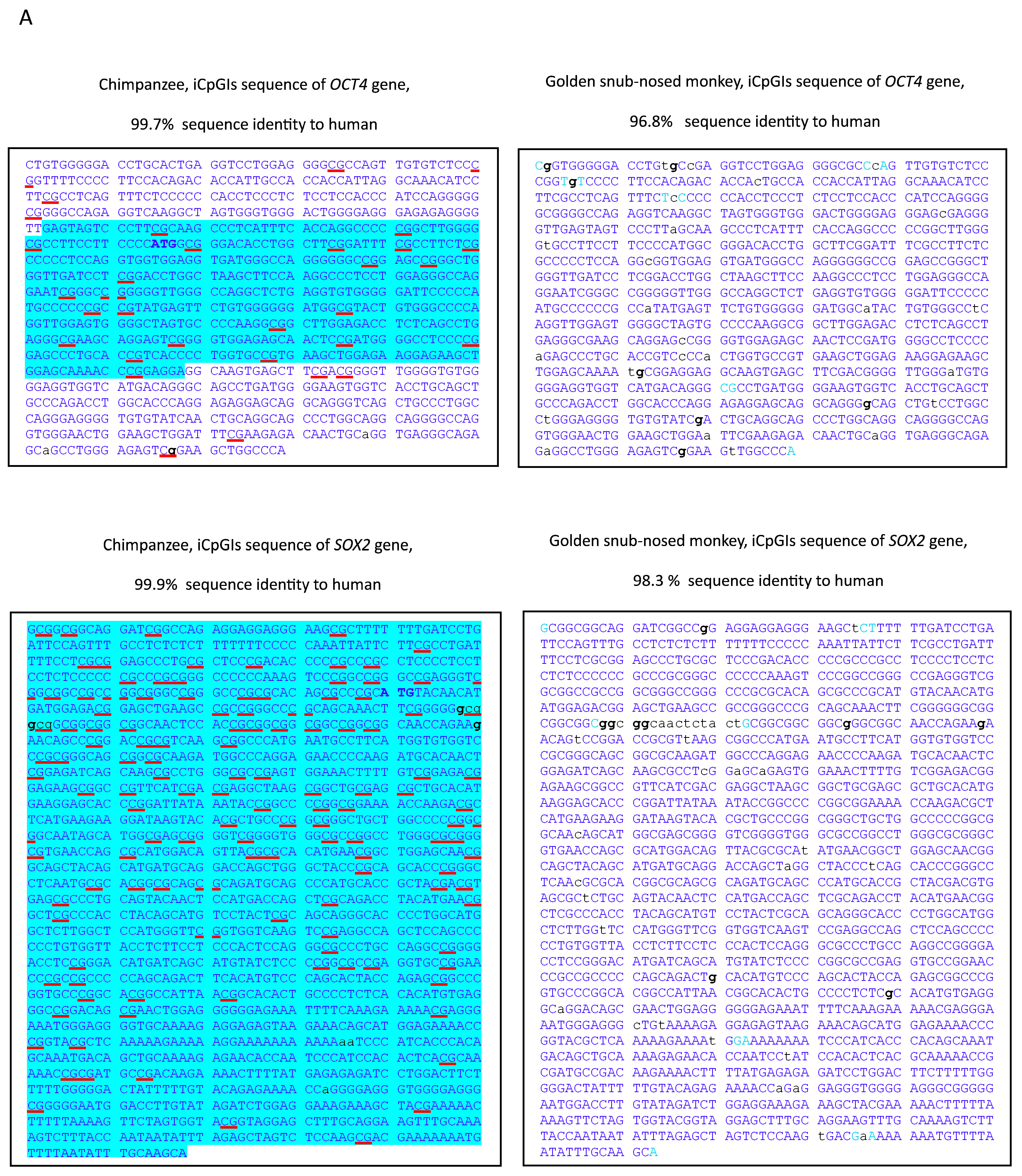
Figure 1. Comparative analysis of iCpGIs in OCT4, SOX2, and hTERT between human, chimpanzee, and golden snub-nosed monkey. (A) Representative excerpts from the UCSC Genome Browser BLAT alignment of the 5′ iCpGIs of OCT4, SOX2, and hTERT are shown for humans, chimpanzee, and golden snub-nosed monkey. Conserved nucleotides are denoted by uppercase blue letters; divergent bases appear in lowercase. Notably frequent X→G substitutions are highlighted in bold. CpG dinucleotides are underlined in red. The translation start codon is shown in bold blue uppercase. Exonic sequences are indicated by a light blue background. (B) The frequency of single-nucleotide substitutions involving guanine (“g”) versus non-“g” residues is quantified for both CpG and non-CpG contexts. The analysis includes all aligned iCpGI regions from OCT4, SOX2, and hTERT in chimpanzee and golden snub-nosed monkey. Gaps of ≥2 nucleotides were excluded from the analysis.
Genes 16 01102 g001aGenes 16 01102 g001b
Figure 2. Conservation and divergence of iCpGIs across twelve primate species. (A) A segment (last part exon 2 hTERT) of a multiple alignment (~4977 nt average length) representing the concatenated iCpGI sequences of OCT4, SOX2, and hTERT from twelve primates. Purple highlights denote species-specific single-nucleotide variants (SNVs) in non-CpG contexts (5.3% of all aligned dinucleotide positions). Green marks variable CpG sites—positions where a CpG occurs in at least one species and is absent or mutated in others (5.9% of all positions, 34% of CpG sites). Universally conserved CpG sites are marked with light blue. Asterisk (*) indicate fully conserved nucleotides; single substitutions are marked with a dot. (B) Corresponding “A/T” transformation of the alignment: all CpGs are replaced with “A”, and all orthologous non-CpG dinucleotides at positions containing a CpG in at least one species are replaced by “T”. Continuous CpG blocks are shown with a light blue background, with interrupted (non-uniform) CpG positions in green. (C) A clustal 2.1-derived percent identity matrix depicting pairwise similarity across the twelve full-length sequences (~4977 nt each).
Genes 16 01102 g002aGenes 16 01102 g002b
Figure 3. Phylogenetic and “phylo-epigenetic” relationships among 12 primate species based on OCT4, SOX2, and hTERT iCpGIs. (A) Standard phylogram based on all single-nucleotide polymorphisms (SNPs), including CpG and non-CpG substitutions, insertions, and deletions, reconstructed from aligned iCpGI sequences. (B) “Phylo-epigenetic” tree based exclusively on patterns of CpG conservation and divergence: conserved CpGs and all species-specific CpG substitutions within CpG islands of OCT4, SOX2, and hTERT were used to reconstruct alternative hierarchy reflecting epigenetic divergence. Numbers at branch nodes indicate divergence events, with values representing millions of years.
Genes 16 01102 g003
Disclaimer/Publisher’s Note: The statements, opinions and data contained in all publications are solely those of the individual author(s) and contributor(s) and not of MDPI and/or the editor(s). MDPI and/or the editor(s) disclaim responsibility for any injury to people or property resulting from any ideas, methods, instructions or products referred to in the content.