Article

Gene-by-Environment Interactions Involving Maternal Exposures with Orofacial Cleft Risk in Filipinos

by
Zeynep Erdogan-Yildirim
1,*,†,
Jenna C. Carlson
2,3,
Nandita Mukhopadhyay
1,
Elizabeth J. Leslie-Clarkson
4,
Carmencita D. Padilla
5,6,7,
Jeffrey C. Murray
8,
Terri H. Beaty
9,
Seth M. Weinberg
1,2,
Mary L. Marazita
1,2,10 and
John R. Shaffer
1,2,*
1
Center for Craniofacial and Dental Genetics, Department of Oral and Craniofacial Sciences, School of Dental Medicine, University of Pittsburgh, Pittsburgh, PA 15261, USA
2
Department of Human Genetics, School of Public Health, University of Pittsburgh, Pittsburgh, PA 15261, USA
3
Department of Biostatistics and Health Data Science, School of Public Health, University of Pittsburgh, Pittsburgh, PA 15261, USA
4
Department of Human Genetics, Emory University School of Medicine, Atlanta, GA 30322, USA
5
Department of Pediatrics, College of Medicine, University of the Philippines Manila, Ermita, Manila 1000, Metro Manila, Philippines
6
Institute of Human Genetics, National Institutes of Health, University of the Philippines Manila, Ermita, Manila 1000, Metro Manila, Philippines
7
Newborn Screening Reference Center, National Institutes of Health, University of the Philippines Manila, Ermita, Manila 1000, Metro Manila, Philippines
8
Department of Pediatrics, University of Iowa School of Medicine, Iowa City, IA 52242, USA
9
Department of Epidemiology, Bloomberg School of Public Health, John Hopkins University, Baltimore, MD 21205, USA
10
Departments of Psychiatry and Clinical and Translational Sciences, School of Medicine, University of Pittsburgh, Pittsburgh, PA 15261, USA
*
Authors to whom correspondence should be addressed.
Lead contact.
Genes 2025, 16(8), 876; https://doi.org/10.3390/genes16080876
Submission received: 12 June 2025 / Revised: 11 July 2025 / Accepted: 16 July 2025 / Published: 25 July 2025
(This article belongs to the Section Genes & Environments)

Abstract

Background/Objectives: Maternal exposures are known to influence the risk of isolated cleft lip with or without cleft palate (CL/P)—a common and highly heritable birth defect with a multifactorial etiology. Methods: To identify new risk loci, we conducted a genome-wide gene–environment interaction (GEI) analysis of CL/P with maternal smoking and vitamin use in Filipinos (Ncases = 540, Ncontrols = 260). Since GEI analyses are typically low in power and the results can be difficult to interpret, we applied multiple testing frameworks to evaluate potential GEI effects: a one degree-of-freedom (1df) GxE test, the 3df joint test, and the two-step EDGE approach. Results: While no genome-wide significant interactions were detected, we identified 11 suggestive GEIs with smoking and 24 with vitamin use. Several implicated loci contain biologically plausible genes. Notable interactions with smoking include loci near FEZF1, TWIST2, and NET1. While FEZF1 is involved in early neuronal development, TWIST2 and NET1 regulate epithelial–mesenchymal transition, which is required for proper lip and palate fusion. Interactions with vitamins encompass CECR2—a chromatin remodeling protein required for neural tube closure—and FURIN, a critical protease during early embryogenesis that activates various growth factors and extracellular matrix proteins. The activity of both proteins is influenced by folic acid. Conclusions: Our findings highlight the critical role of maternal exposures in identifying genes associated with structural birth defects such as CL/P and provide new paths to explore for CL/P genetics.

1. Introduction

Orofacial clefts (OFCs) are common birth defects that result mainly from failure in the processes required for the complete fusion of the structures involving the lip and/or the palate. On average, 1 in 700 live births are affected by OFCs worldwide with significant variability in incidence rates across populations [1,2]. In a majority of OFC cases, the condition presents as cleft lip with and without cleft palate (CL/P), with 70% manifesting as an isolated, non-syndromic feature [3]. The rate of isolated CL/P is high in the Philippines, with 1 in 500 newborns affected [1]. Siblings of affected Filipinos have an 11.5-fold increased CL/P risk compared to the general population, indicating a strong familial component [1,2]. A significant disparity in CL/P risk is evident across socioeconomic status (SES), with incidence rate dropping to approximately 1 in 1000 among Filipinos living under higher SES, suggesting poor maternal nutrition could be a contributing factor [1,4,5].
Epidemiological studies in individuals from the Philippines have identified links between increased CL/P risk and inadequate levels of vitamin B6 (<20 nmol/L) as well as low plasma zinc levels in mothers [4,5,6]. Although maternal smoking is another risk factor for clefts, studies involving Filipino mothers did not show statistically significant associations between smoking and the risk of cleft [7], which may be due to Southeast Asian women having the lowest prevalence of smoking (0.9%) compared to other continental groups (1.4% through 17.5%) [8]. However, household smoking, as a possible indicator of passive smoke exposure, increased the odds for OFCs in offsprings [7,9]. These findings align with previous reports showing evidence for increased risk for OFC with exposure to maternal smoking, with environmental tobacco smoke exposure, and with a lack of multivitamin (with/without folic acid) supplementation during pregnancy [10]. These facts underscore the significance of both the genetic predisposition and the influence of environmental factors in CL/P etiology in this population [1].
Genetic studies of isolated CL/P in diverse populations, including both candidate and genome-wide approaches, have been used to identify genetic risk loci [11,12]. Although numerous CL/P risk loci have been identified, they collectively do not fully account for the estimated genetic variance. Interactions between genetic and environmental factors during development may explain some of the missing heritability; hence, several studies have explored gene–environment interactions (GEIs) in cleft risk in different populations and detected significant interactions with maternal smoking (NOS3 [13], GRID2 [14], and ELAVL2 [14]), environmental tobacco smoke (RUNX2 [15]), and maternal vitamin use (NOS3 [13], CACNG3 [16], and ESRRG [17]). Several suggestive interactions have been reported with maternal smoking (MUSK [18] and PRL [16]) and maternal vitamin intake (RETREG1 [18], FLJ0838 [17], COBL [17], CASP9 [18], and ANTXR1 [18]). These findings highlight the significance of GEI studies, especially for complex disorders such as CL/P, for unveiling underlying mechanisms that cannot be detected by examining the genetic main effects alone.
Relatively little is known about genetic factors underlying CL/P or their interactions with environmental exposures in Filipinos, despite being a population with a high CL/P prevalence and the documented prevalence differences by SES that are evidence of environmental effects. Hence, the goal of our study was to discover new genes involved in orofacial cleft risk through a genome-wide interrogation of GEIs in individuals from the Philippines. In particular, we focus on maternal smoking and vitamin use during the periconceptional period.

2. Materials and Methods

2.1. Study Population Description

The discovery sample for this GEI analysis was derived from a multiethnic sample of 4114 participants genotyped as part of the POFC2 study. The participants of the genetic and phenotyping studies led by the Center for Craniofacial and Dental Genetics (University of Pittsburgh) were recruited at regional recruitment sites in 10 countries across five different continental regions and included affected probands and their family members as well as controls with no personal or family history of craniofacial defects. Mothers of both cases and controls provided retrospective information on maternal exposures during pregnancy for each offspring, including smoking, alcohol consumption, and the use of any vitamin supplement during each trimester of their pregnancies through self-reported (yes/no) questionnaires.
From a total of 2155 genotyped Filipinos in the study, we selected CL/P cases and unaffected unrelated controls who had complete data on maternal smoking and vitamin intake during the periconceptional period that spans from 3 months prior to conception to the end of first trimester. An individual was considered exposed if the mother confirmed personal smoking or vitamin intake (1) within the three months prior to pregnancy and/or (2) within the first trimester. Individuals with a cleft palate only (CPO), CL/P cases with a family history of CPO or a diagnosis of a syndrome, and controls reporting family history of any type of craniofacial defect were excluded from analysis. We also removed individuals with unknown maternal exposure status across both time periods. The final sample comprised 540 cases and 260 controls (sample flowchart in Figure S1A). Details on the exposure count are provided in Table 1.

2.2. Genotyping and Imputation

The genotyping of the Filipino cohort used in this analysis was performed as part of the Pittsburgh Orofacial Cleft 2 (POFC2) study that included 4114 participants recruited worldwide across South Asia, Africa, Latin America, USA, East Asia, and Europe (Figure S1A). The genomic DNA was extracted from saliva samples collected with Oragene kits (DNA Genotek Inc., Ottawa, ON, Canada) and genotyped at the Center for Inherited Disease Research (CIDR) at John Hopkins University using the Illumina Global Diversity Array-8 v1.0 (GDA, Illumina Inc., San Diego, CA, USA) covering approximately 1.9 million markers. After extensive quality control and quality assurance steps [19] (removing poorly performing samples and those with sex discrepancies and variants with missing call rates (≥2%), discordant calls on duplicate probes, ≥2 Mendelian errors, or deviation from Hardy–Weinberg equilibrium (p < 3.45 × 10⁻³)). The imputation of unobserved variants was conducted via the TOPMed Imputation Server with the TOPMed reference panel (version r2) using minimac4 (v1.6.0) [20,21]. Monomorphic variants and variants with low imputation quality (R2 < 0.8) were removed prior to analysis. The imputed dosages were converted to a binary dosage file format via the R package BinaryDosage (v1.0.0) [22].
The genotype data are available via the database of Genotypes and Phenotypes (dbGaP) via the accession number phs002815.v2.p1; the phenotypic and pregnancy history data are available through FaceBase (facebase.org; accession number: FB00001368, doi: 10.25550/56-ES6P, and accession number: FB00001369, doi: 10.25550/5A-FJBJ).

2.3. Statistical Analyses

2.3.1. Gene-by-Environment Interaction Analyses

For discovering G × E interactions (GEIs) with maternal smoking and vitamin intake implicated in CL/P risk, we performed a genome-wide scan on common variants with a minor allele frequency (MAF) ≥ 0.10, leveraging the analytical approaches implemented in the R package GxEScanR (v2.0.2) [23,24]. We employed logistic regression on the imputed dosages adjusting for reported sex and the first five principal components of ancestry (Figure S2). For tests where GxEScanR failed to produce results due to reaching the maximum number of iterations, we computed test statistics using the same model and covariates using glm().
There are many existing statistical approaches to detect GEI and interpreting the results from statistical models for GEI can be challenging. Thus, we employed three complementary strategies as implemented in GxEScanR for detecting and interpreting GEI in this study (Figure 1). The first approach used the standard 1 degree-of-freedom (1df) G × E test from a model with both gene and environment main effects and their interaction. While the 1df G × E test is the easiest to interpret, it has the lowest power. The second approach was a unified 3df joint test of the main genetic effect (DG) from a model without an interaction, the same G × E effect as the first approach, and the gene–environment (GE) association. Since the 3df joint test combines these components, it does not distinguish which one is driving the association. Hence, we conclude the presence of a GEI effect if the 1df G × E test and/or if the GE association test in cases but not in controls has a p < 0.05. The 3df test is powerful to detect signals by any combination of the three components; however, when the true effect is driven only by one component, the power to detect is reduced. Lastly, our third approach, called the two-step EDGE approach, uses a 2df joint test of DG and GE to screen variants, followed by a test for the same G × E effect as the first approach. This two-step EDGE approach ultimately tests the same effect as the first approach, but with more statistical power, as the filtering step reduces the total number of tests in step 2. However, SNPs with weak marginal genetic or environmental effects may lead to missed true interactions [23,24,25].
We used separate significance thresholds for each approach. For the first approach, our significance threshold was p1df < 5 × 10⁻⁸. In the second approach (3df test), findings with p3df < 5 × 10⁻⁸/3 (≈2 × 10⁻⁸) were considered significant and those with p3df < 1 × 10⁻⁵ were considered suggestive; however, interaction was concluded only when either pG×E or pcase-only was < 0.05. For our third approach (two-step EDGE), we used a filtering threshold of p2df_EDGE < 0.05 for step 1, followed by a significance threshold of pG×E ≈ 8 × 10⁻⁷ for step 2, which was adjusted for the effective number of independent loci (0.05/61681 and 0.05/61173 for vitamins and smoking, respectively) determined via simpleM [23,26]. Step 2 associations with a pG×E < 5 × 10⁻⁴ were considered suggestive.

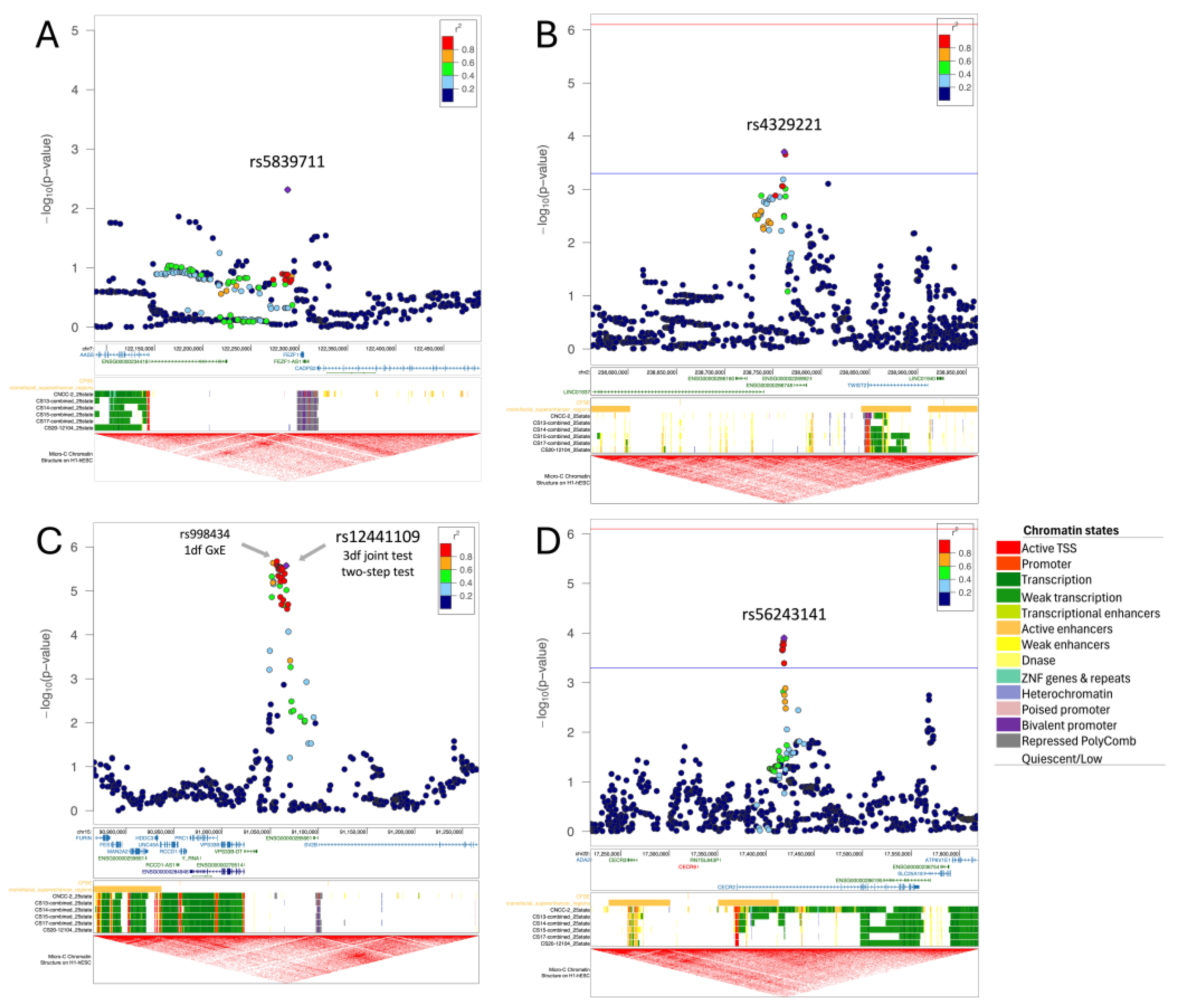
2.3.2. Regional Plots

To investigate the genomic region of interest, we used LocusZoom [27] to visualize the association signals within the context of nearby genes and regulatory elements. We incorporated data on strong craniofacial-specific enhancers [28] and the topologically associated domain information from the H1-hESC Micro-C track available through the UCSC Genome Browser into regional plots (accessed on 10 September 2024) [29]. To accurately reflect the genetic architecture of the study sample, we calculated the linkage disequilibrium in individuals from the Philippines in our discovery sample.

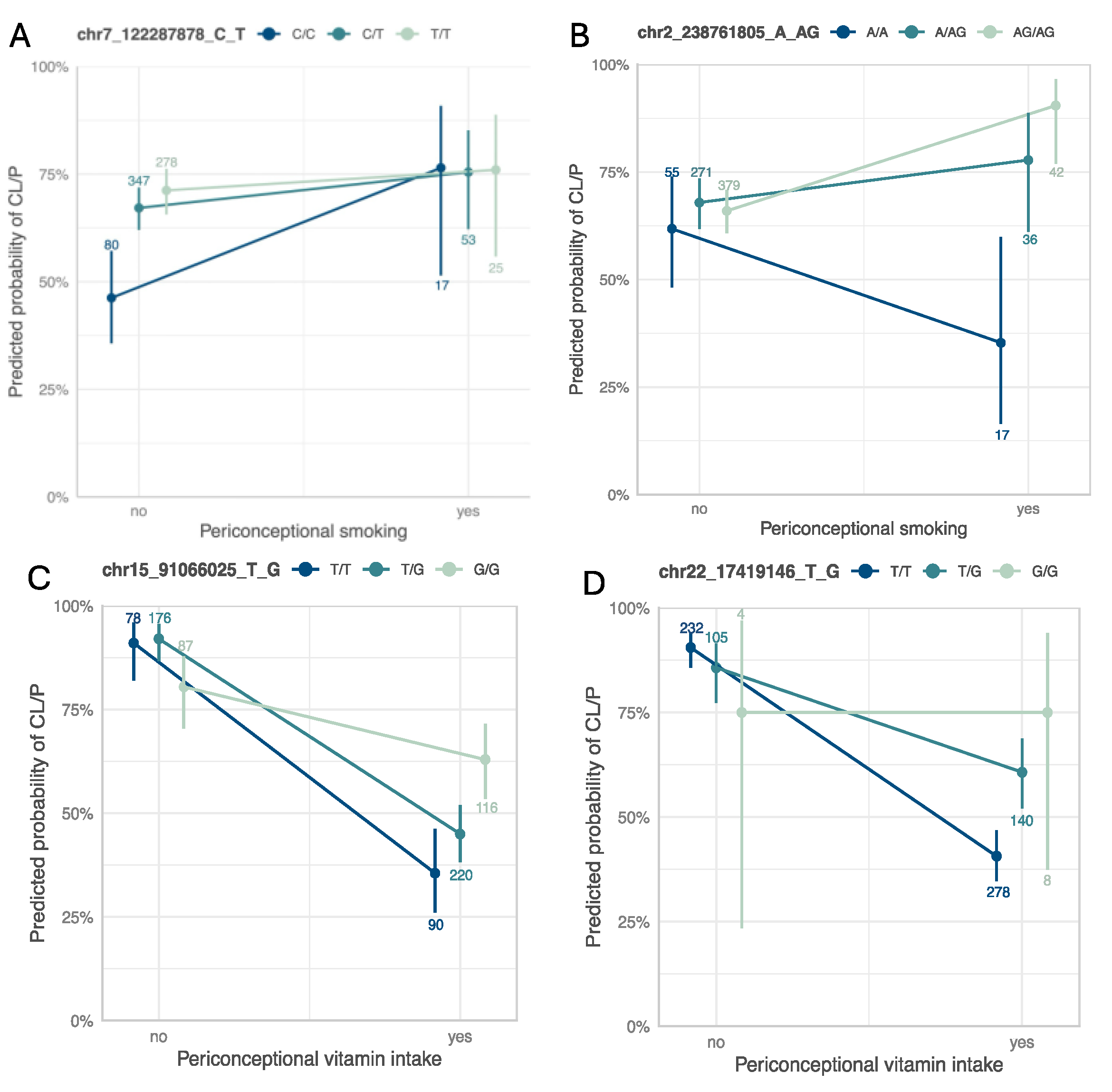
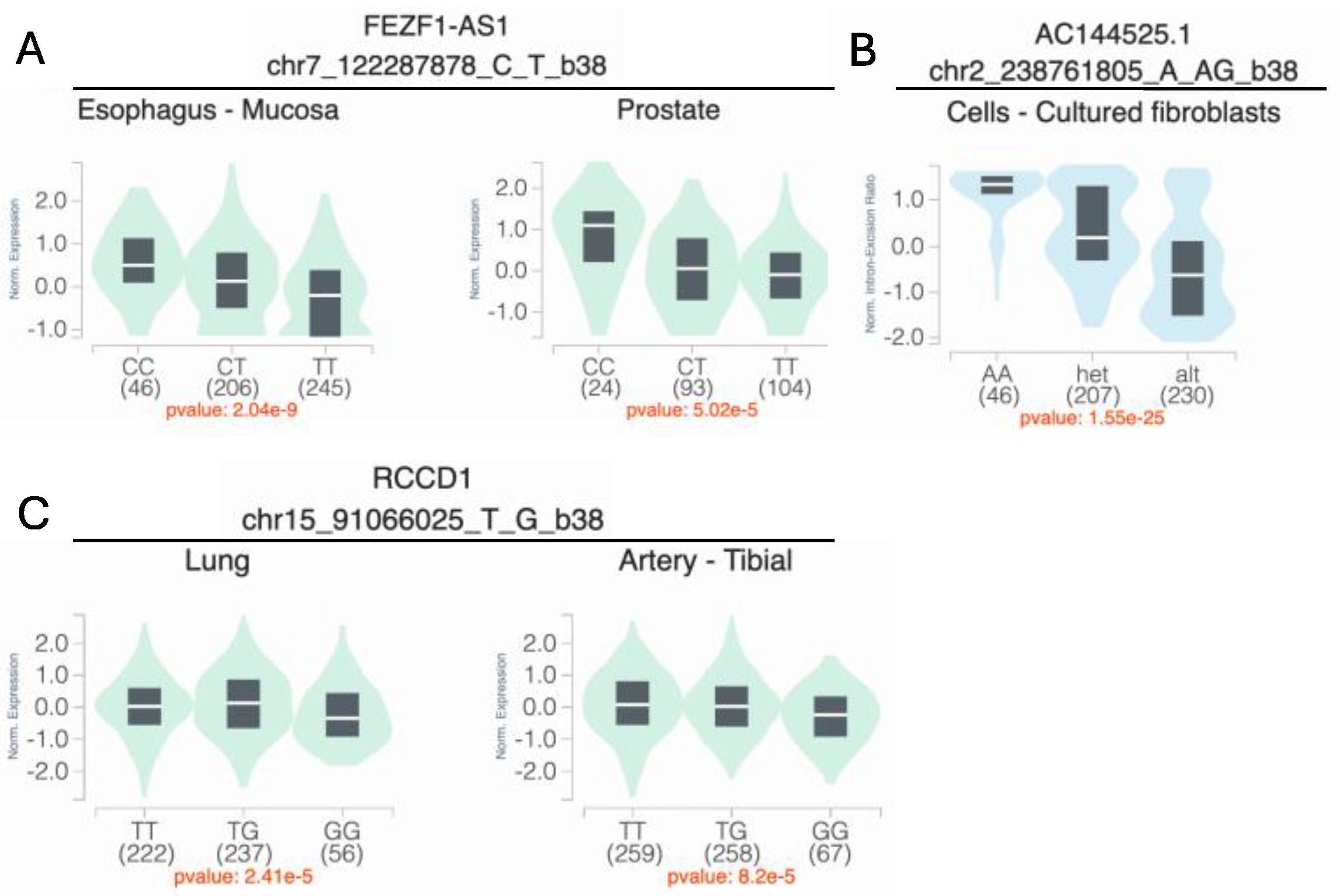
2.3.3. Interaction Plots and eQTL/sQTL Plots

To evaluate and interpret the detected GEIs, we used interaction plots and visualized the predicted probabilities stratified by environment and genotype. The predicted probabilities were estimated using a logistic regression model, incorporating the genotype, maternal exposure status, and their interaction term.
To determine whether the lead variant had a regulatory effect on the gene expression and its potential relevance, we looked up in the publicly available Genotype-Tissue Expression (GTEx Release v8, accessed on 14 September 2024 [30]) database if the lead variant was associated with gene expression (eQTL) or alternative splicing (sQTL) and retrieved their single tissue violin plots from the interactive data portal.

2.3.4. Sensitivity Analysis

Because maternal smoking and vitamin use are moderately correlated (Cramér’s V coefficient = 0.183, p = 3.81 × 10⁻⁷), to disentangle the effects of smoking, we reran the GEI analyses for vitamin use by excluding all individuals exposed to periconceptional smoking (n = 99), leaving 676 individuals consisting of 439 CL/P cases (vitamin use: 44.19%) and 237 controls (vitamin use: 86.08%).

2.3.5. Replication

To validate the GEI findings from the discovery cohort, we performed replication analysis with variants passing the suggestive threshold for the 3df test and/or the two-step approach. To reduce multiple testing burden at the replication stage, we ran replication analysis for only those that demonstrated a GEI association from variants identified by the 3df test, either (1) via 1df G × E test with pG × E < 0.05 or (2) via GE association in cases only with pcase_only < 0.05 with an insignificant GE association in controls.
We used a case-only replication approach using three independent sets of individuals with CL/P who were recruited from the Philippines (Table 1): n = 137 from the POFC1 Study conducted by the University of Pittsburgh (Replication 1) [31], n = 88 from the GENEVA Study (Replication 2) [32], and n = 213 from the Gabriella Miller Kids First (GMKF) initiative (Replication 3). Replication 2 and Replication 3 samples were originally enrolled in cleft studies conducted by the University of Iowa. Each of the three replication studies had differing family-based recruitment designs, so we extracted independent CL/P cases only for this replication analysis (removing overlapping and dependent individuals) so that the same statistical method could be used across each replication set (Figure S1B). Each study measured periconceptional exposure to maternal smoking and maternal vitamin use in the same way as the POFC2 cohort. In each replication sample separately, we measured the GEI by evaluating the GE association in cases using GxEScanR. We considered an interaction to be replicated if the p-value was < 0.05 in any replication sample with a consistent direction of effect between the discovery sample and the replication sample.
The genotyping for the POFC1 study (dbGaP accession number: phs000774.v2.p1) and the GENEVA study (dbGaP accession number: phs000094.v1.p1) were performed using Illumina HumanCore + Exome Array and Illumina Human 610 Quadv1_B array, respectively. More details about the genotyping for the POFC1 and the GENEVA studies has been described previously [31,32]. We re-imputed both cohorts using the TOPMED r2 after removing low-frequency variants (MAF < 0.01) and phasing the haplotypes via SHAPEIT. The whole genome sequences from GMKF (dbGaP accession number: phs002595.v1.p1) were generated by the Broad Institute using Illumina HiSeqX with a target read depth of ~30 × coverage. The data were aligned to the GRCh38/hg38 reference genome and processed by the Kids First Data Research Center via Cavatica using a custom pipeline based on Genome Analysis Toolkit (GATK) best practices [33].
The GATK genotyping workflow incorporated base quality score recalibration (BQSR), single-sample variant calling for SNVs and indels using HaplotypeCaller, joint variant calling across multiple samples, and final refinement with the variant quality score recalibration (VQSR) and filtering of called variants. Kids First DRC pipelines are publicly accessible as open-source tools on GitHub (link for alignment workflow and joint genotyping workflow in Web Resources).

2.3.6. Examination of Known GEI Loci Implicated in CL/P Risk in This GEI Analysis

We explored our GEI analysis results for known statistically significant GEI loci with maternal smoking (rs4389540 in GRID2 [14], rs1799983 in NOS3 [13], and rs2257210 at ELAVL2 [14]) and vitamin intake (rs1339221 in ESRRG [17], rs1799983 in NOS3 [13], and rs9930171 in CACNG3 [16]) reported in association with CL/P risk. For each locus, we selected the lead SNV with the lowest p-value and considered it replicated if the pG×E and/or pcase_only in our analysis was <0.05.

3. Results

In this study, we carried out a comprehensive analysis to detect GEIs with maternal smoking and vitamin use implicated in isolated CL/P risk in people recruited from the Philippines. Maternal smoking showed a trend with an increased risk of CL/P (OR = 1.58 [95%-CI = 0.95–2.73]), although the association was not statistically significant (Fisher’s exact p = 0.0796), and maternal vitamin intake was associated with reduced cleft risk (OR = 0.11 [95%-CI = 0.08–0.17]; Fisher’s exact p < 2.2 × 10⁻¹⁶). The descriptive characteristics of the study sample are shown in Table 1.

3.1. Genome-Wide Interaction Analysis for Maternal Smoking

The statistical testing for GEI interaction with periconceptional exposure to smoking did not yield any genome-wide significant results using all three analytical approaches; however, several suggestive associations were detected (Table S1). Our first statistical approach, the 1df G × E test, showed suggestive association with one variant (rs10150710-C, p = 5.90 × 10⁻⁶, Table S2) mapping to the second intron of BCL11B that encodes a transcription factor with key functions in the maturation of T-cells, and neurological and craniofacial development [34]. The second approach, the 3df test, detected 13 independent suggestive loci (p < 1 × 10⁻⁵) (Table S3 and Figure S3A), of which five independent loci (at PTPRD, TTBK1, SLFN12L, TNS1, and OTOR) showed moderate G × E interaction with a pG×E < 0.05 (p-values ranging from 7.46 × 10⁻⁴ to 3.62 × 10⁻²) and one locus, rs4329221 at FEZF1, indicated interaction via the case-only analysis (pcase_only = 4.87 × 10⁻³, Table 2 and Figure 2A).
Table 2. Main GEI findings with exposures to maternal smoking and maternal vitamin intake. The alternate allele frequency (alt AF) is calculated within our sample.
ExposureMethod Variant InfoDiscoveryReplication 1Replication 2Replication 3
SNV (hg38)rsIDNearest GeneTypeAlt
AF
3df Test2df EDGEGxEDGGE AllGE CasesGE ControlsCase Only Case Only Case Only
PPβPPPβPPβPβPβP
Smoking3df test7-122287878-C-Trs4329221FEZF1intergenic0.639.87 × 10−66.18 × 10⁻⁶−0.581.63 × 10−12.87 × 10−51.09 × 10−2−0.574.87 × 10−35.71 × 10⁻¹−0.06 a9.10 × 10−1−1.39 a2.63 × 10−1−0.873.44 × 10−3
SmokingTwo-step test2-238761805-A-AGrs5839711TWIST2intergenic0.727.93 × 10−52.08 × 10−21.561.98 × 10−46.62 × 10−16.00 × 10−3−0.077.53 × 10−11.31 × 10⁻⁵ a0.158.22 × 10−150.57 a3.50 × 10−1 0.617.15 × 10−2
Vitamin intake3df test and two-step test15-91066025-T-Grs12441109SV2Bintergenic0.532.65 × 10−63.66 × 10−71.452.68 × 10−62.02 × 10−12.55 × 10−20.651.02 × 10−59.69 × 10⁻³−0.254.51 × 10−10.049.34 × 10−1 0.039.02 × 10−1
Vitamin intakeTwo-step test22-17419146-T-Grs56243141CECR2intronic0.172.58 × 10−42.52 × 10−21.561.26 × 10−42.28 × 10−21.39 × 10−10.654.98 × 10−42.60 × 10⁻³−0.1237.90 × 10−10.712.21 × 10−1−0.363.11 × 10−1
Vitamin intake3df test and two-step test5-57049741-A-Grs179464MIER3intergenic0.518.33 × 10−61.02 × 10⁻³−1.204.04 × 10−42.54 × 10−45.34 × 10−1−0.182.31 × 10−14.43 × 10⁻⁵−0.501.83 × 10−10.108.42 × 10−1−0.137.02 × 10−1
a p-values are obtained from retesting using a generalized linear model (glm) as GxEScanR did not report results due to maximum iterations exceeded.
Using the two-step EDGE approach, we found another five suggestively associated risk loci (PAMR1, SPAG16, TWIST2, NET1, and ZNF722, Table S4 and Figure S3B). Two of these loci, TWIST2 and NET1, are implicated in the regulation of epithelial–mesenchymal transition (EMT), a critical process in lip and palate fusion during embryonic development. The lead signal at TWIST2 (rs5839711-AG, p G × E = 1.98 × 10⁻⁴) is an intergenic variant (Figure 2B). This variant does not show evidence of a genetic main effect overall or when restricting to just the unexposed CL/P cases; however, among those with exposure to maternal smoking, the CL/P risk is higher in individuals carrying one or two copies of the inserted G allele compared to homozygotes for the A allele (Figure 3B). Per GTEx (v8) [30] project data, this variant regulates the alternative splicing of the nearby lincRNA AC144525.1 in transformed fibroblast cells (p = 1.55 × 10⁻²⁵), where each copy of the alternate allele is associated with a decreasing intron–excision ratio (Figure 4B). Additionally, this lincRNA is located within the enhancer region GH02J238784 according to the GeneHancer [35] that also encompasses the craniofacial-specific enhancers [28], as shown in Figure 2B, and exhibits regulatory effects on the surrounding genes including TWIST2, LINC01937, and ASB1.

3.2. Genome-Wide Interaction Analysis for Maternal Vitamin Use

For the vitamin GEI analysis, while no variant achieved statistical significance using any of the three approaches, we identified four independent suggestive loci with a moderate G×E effect via the 3df test (at NLGN1, SV2B, CSMD1, and MIER3; Figure S4A) and 22 suggestive loci via the two-step approach, of which rs12441109-G and rs179464-G were detected via both methods (Table 2, Table S1, Figure S4B).
Using the 1df G×E test, the most significant association was at 15q26.1 in the intergenic region between VPS33B and SV2B, with rs998434-T (pGxE = 2.13 × 10⁻⁶) as the top signal (Figure 2C, Table S5). The same locus was identified by both the 3df test and the two-step approach (Table 2, Tables S6 and S7), through association with a variant in high LD (rs12441109-G, p3df = 2.65 × 10⁻⁶ and pGxE = 2.68 × 10⁻⁶, LD with rs998434 r2 = 0.95). While we did not observe a genetic main effect, rs12441109-G exhibited suggestive interaction with vitamin exposure. The protective effect of maternal vitamin use on CL/P risk differed by genotype with an estimated probability of CL/P of ~58% for GG individuals with maternal vitamin use compared to ~33% for TT (Figure 3C). For rs12441109-G, we observed a lower p-value after performing a sensitivity analysis by excluding smokers (p3df = 9.05 × 10⁻⁷ and pGxE = 9.34 × 10⁻⁷, Table S8). Furthermore, rs12441109-G acts as an eQTL on RCCD1, where the double copy of the G allele reduces the mean RCCD1 expression in the tibial artery (p = 8.2 × 10⁻⁵) and the lung tissue (p = 2.41 × 10⁻⁵) per GTEx (Figure 4C).
Among the 22 independent loci detected via the two-stage approach (Figure S4B), one of the noteworthy findings was the locus at CECR2 with the lead variant rs56243141-G (pGxE = 1.26 × 10⁻⁴, sensitivity analysis pGxE = 1.34 × 10⁻³) mapping to its first intron (Table 2 and Table S7). When stratified by vitamin use, unexposed individuals with the TT genotype had a higher predicted probability of CL/P, although not statistically significant, whereas exposed individuals with the TT genotype had a significantly lower predicted probability of CL/P compared to the genotypes with the alternate allele G (Figure 3D). The p-value of CL/P for individuals homozygous for the minor allele (G) remained the same across both strata, however, with wide confidence intervals as there were few homozygotes.

3.3. Replication Analysis

To validate our findings, we conducted a replication analysis across three independent samples consisting of individuals from the Philippines (Replication 1, Replication 2, and Replication 3). From the 39 suggestive findings, only rs4329221-T at the intergenic region near FEZF1 was replicated (Table 2 and Table S1). This variant demonstrated a GEI effect with maternal smoking and was detected via the 3df test (effect estimate: −0.57, pcase_only = 4.87 × 10⁻³). The interaction between rs4329221-T and smoking was replicated in the Replication 3 sample with an effect estimate of −0.87 and pcase_only = 3.44 × 10⁻³. Although this variant was not replicated in Replication 1 and Replication 2 samples, it demonstrated the same direction of effect. While the CL/P risk in smokers was almost identical across all genotype strata, in non-smokers the two copies of the reference allele (CC) were associated with lower odds of CL/P compared to T carriers (Figure 3A). This variant also serves as an eQTL and leads to a reduced expression of FEZF1-AS1 in the esophageal mucosa and the prostate (p = 2.04 × 10⁻⁹ and p = 5.02 × 10⁻⁵, respectively) in carriers of the T allele (Figure 4A).

3.4. Known GEI Loci with Maternal Smoking and Vitamin Intake in CL/P Risk

From six of the previously known significant GEIs, we replicated the interaction between NOS3 and maternal vitamin intake (Table 3). The candidate study by Shaw et al. (2005) investigating the interaction between loci at NOS3 and maternal smoking and/or vitamin intake detected an increased CL/P risk with rs1799983-T in offsprings who were exposed to maternal smoking without maternal vitamin supplementation during the periconceptional period (OR = 4.4, 95%-CI [1.8, 10.7]) [13]. In our study, offspring presenting with same exposure and genotypes were all affected. When stratified by vitamin supplementation in the complete sample or in the subset excluding smokers, the predicted probability of CL/P was the highest in non-exposed offspring carrying the T allele and lowest in exposed individuals (1.00, 95%-CI [0, 1] vs. 0.30, 95%-CI [0.10, 0.65]). In contrast, although offspring homozygous for the G allele demonstrated an advantage when their mothers did not take vitamins, there was no statistically significant difference in predicted probability among offspring whose mothers used vitamins (G/G 0.49 [0.43, 0.55] and T/G 0.47 [0.39, 0.55]).
Table 3. Lookup of previously known GEI loci with maternal smoking and vitamin intake implicated in CL/P risk in this GEI study.
ExposureVariant InfoDiscovery Sample
SNV (hg38)rsIDNearest GeneTypeStudy
MAF
Reference
AF
GxEDGGE Cases
ORPORPORP
Smoking4-92455080-G-Ars4389540GRID2Intronic0.14Beaty et al. (2013) [14]0.005------
Smoking7-150999023-T-Grs1799983NOS3Exonic-missense0.26Shaw et al. (2005) [13]0.170.766.16 × 10−10.999.33 × 10−10.917.19 × 10−1
Smoking9-24527359-G-Ars2257210ELAVL2Intergenic0.31Beaty et al. (2013) [14]0.211.118.23 × 10−10.978.24 × 10−11.312.30 × 10−1
Vitamin intake1-216999264-T-Crs1339221ESRRGIntronic0.40Haaland et al. (2018) [17]0.320.546.01 × 10−21.038.33 × 10−10.832.03 × 10−1
Vitamin intake7-150999023-T-Grs1799983NOS3Exonic-missense0.26Shaw et al. (2005) [13]0.173.936.42 × 10−30.853.60 × 10−11.048.59 × 10−1
Vitamin intake16-24342036-A-Grs9930171CACNG3Intronic0.35Carlson et al. (2022) [16]0.410.856.04 × 10−10.935.92 × 10−10.967.60 × 10−1

4. Discussion

The goal of this study was to identify genetic risk variants that interact with well-established environmental risk factors, specifically periconceptional exposure to smoking and vitamin use, contributing to isolated CL/P in individuals from the Philippines. To achieve this, we performed a comprehensive analysis of GEIs using a genome-wide approach by applying three complementary methods: the 1df G × E test, the unified 3df test and the two-step EDGE approach. While neither method identified genome-wide significant associations in our sample, our findings revealed 11 independent loci suggestively associated with maternal smoking and 24 independent loci suggestively associated with maternal vitamin use during periconceptional period. Among these suggestive loci, the GEI effect at the locus including FEZF1 was replicated in the three independent samples analyzed (Table 2). We also noted several of the remaining GEI findings with potential biological relevance to cleft risk: TWIST2, NET1, TNS1, and BCL11B for maternal smoking and SV2B, CECR2, and MIER3 for maternal vitamin use. These findings have not been reported in previous GEI analyses of CL/P [14,16,17,18,36,37,38,39].
Our main finding was that rs4329221-T at FEZF1 influenced CL/P risk via its interaction with smoking. FEZF1 encodes a zinc finger protein that serves as a transcriptional repressor [40]. FEZF1 is an important factor for neuronal differentiation and the migration of olfactory sensory neurons [40]. The disruption in the maturation process of olfactory neurons, which is intertwined with the migration of gonadotropin releasing hormone (GnRH) neurons, is associated with hypogonadotropic hypogonadism 22 with anosmia (also known as Kallmann syndrome) or without anosmia (HH22 [MIM: 616030]), both of which has been linked to missense mutations within FEZF1 [40,41,42]. The lead variant rs4329221-T is an eQTL for the nearby FEZF1-AS1 (Figure 4) [30]. Its expression is reduced in carriers of the alternate allele (T) [30]. Furthermore, FEZF1-AS1, which promotes cell proliferation and inhibits apoptosis, is found to be overexpressed in placental tissues collected from preeclampsia patients and in various tumors, predicting a poor outcome [43,44]. Moreover, the lead variant is 30 kb proximal to the variants associated with smoking initiation (rs1443753-C [45] and rs10953957-A [45]) and ever vs. never smokers (rs1443753-C [45], rs10252114-C [46], and rs10953957-A [47]).
Among the GEI loci identified in association with smoking, TWIST2 is another potential candidate with a biological role in CL/P risk. TWIST2 encodes a basic helix-loop-helix type transcription factor that modulates the chromatin binding activity and has a bifunctional role both as a transcriptional repressor or activator [48]. TWIST2 governs the mesenchymal cell fate and epithelial–mesenchymal transition (EMT) critical for normal embryonic morphogenesis and cancer progression [49]. It is expressed in the mesodermal tissues (including craniofacial mesenchyme, osteoblasts, myocytes, and adipocytes) during embryogenesis and prevents them from reaching their terminal differentiation [48,50]. Mutations that disrupt these processes are found to cause facial patterning defects as seen in ablepharon macrostomia and Barber-Say syndrome [49,51]. Additionally, TWIST2 overexpression in breast cancers leads to the downregulation of CDH1 by repressing its promoter [52]. A loss of CDH1/E-cadherin in neural crest cells impairs their migration, leading to craniofacial malformations including CL/P in animal models that recapitulated the observations in CL/P patients with rare CDH1 mutations [53,54,55,56]. Furthermore, experiments with continuous exposure to smoke extract over 21 and 40 weeks increased the expression of TWIST2 and reduced the expression of CDH1 in mammary epithelial cells (non-tumorigenic MCF10A and tumorigenic MCF-7), promoting the migratory ability of the cells [57]. Given its involvement in key developmental processes, TWIST2 is a potential candidate for further investigation to determine its role with smoking in CL/P.
Analysis with another key exposure, the maternal vitamin intake during periconception, revealed several genes that stand out for their potential relevance to CL/P, such as SV2B. Multiple variants with a GEI effect span the intergenic region between SV2B and VPS33B (Figure 2C), which is distally located to a large region of craniofacial enhancers that are linked by the same topologically associated domain (TAD). The lead variant rs12441109-G exhibits a regulatory role on the expression of the neighboring gene RCCD1 (Figure 4C), which is involved in chromatin organization and is recognized as a novel oncogene in lung cancer [30,58]. RCCD1 promotes the migration of tumor cells and TGF-β-induced EMT [58]. Within the same TAD, FURIN is another potential candidate gene underlying the observed GEI effect. FURIN encodes a critical serine protease that facilitates the conversion of a wide range of protein precursors, including growth factors, their receptors, and extracellular matrix proteins, into active forms and plays a vital role in early embryonic development. This is highlighted by the evidence that mouse embryos lacking Furin do not survive beyond 10–11 days after birth and present with notable defects in ventral closure and axial rotation which are under the control of BMP subfamily, to which the FURIN substrates TGFβ and related proteins, including BMP4, and Nodal precursors belong. Moreover, FURIN activity is shown to be inhibited by folic acid [59]. In addition to its mapping onto craniofacial-specific enhancers, another piece of evidence supporting its role for CL/P risk is that mutations in GDF11, which prevents its cleavage by FURIN, lead to CL/P [60,61].
A second promising candidate gene with vitamin use is CECR2, which is responsible for chromatin remodeling and the proper development of the neural tube and craniofacial structures. It is also reported in association with cat eye syndrome (CES [MIM: 115470]), which is caused by an inverted duplication involving the region at CECR2 [62]. A loss of cecr2 in mice was shown to cause neural tube defects [63]. The supplementation of folic acid in women during the periconceptional period is a well-established preventative measure against neural tube defects. Similarly, maternal folic acid use also reduces the occurrence as well as the recurrence risk of OFCs [64]. Studies have demonstrated that folate increased CECR2 expression, while folate deficiency and the resulting increase in homocysteine levels, especially its metabolite homocysteine thiolactone, led to a decreased expression of CECR2 [65]. Mutations within CECR2 have been reported to curtail its expression [63,65]. While there is evidence for interaction between CECR2 and folic acid, the mechanistic link to CL/P risk is unknown.
Furthermore, we reviewed the literature for known GEI loci with maternal smoking and vitamin intake and limited the lookup in our analysis to only the prior significant findings. We validated the interaction between the NOS3 variant rs1799983-G and maternal vitamin intake in our discovery sample. Although we confirmed one of the GEI findings in our sample, replicating GEI findings remains challenging due to heterogeneity across study samples, populations, and how exposure status is assessed.
Strengths
One of the major strengths of our study is the use of complementary GEI detection methods (1df G×E test, the 3df test, and the two-step EDGE approach) that allowed us a robust assessment of potential interactions that might not have been captured by a single method. Furthermore, this study uniquely contributes to the genetic research by focusing on the Filipino population, which is among the populations with highest CL/P risk and has a high level of exposure to adverse environmental factors, as reported [1]. Moreover, the examination of the interaction with vitamin intake (with/without folic acid) specifically within this population provides insights into the interaction as there was no fortification of food with folic acid at the time of recruitment [66].
Limitations
A major limitation of our study lies in the sample size, as GEI analyses generally require larger sample sizes than studies that examine the main genetic effects. Given the relatively small sample size of our sample, the study may have been underpowered to identify significant interactions, particularly for exposures with low prevalence such as smoking in Filipino women. Another limitation of our study may lie in the potential recall and reporting bias associated with exposure data, as these were collected retrospectively. There was also substantial heterogeneity in how exposure data are defined and collected and rates of exposures (especially vitamin use) across different study samples.

5. Conclusions

In conclusion, we conducted a comprehensive genome-wide gene-by-environment analysis with maternal smoking and vitamin intake influencing CL/P risk in individuals from the Philippines and identified several GEI loci that had not been reported before. These include notable interactions, although suggestive, with smoking near FEZF1 and TWIST2, and with vitamin intake within CECR2 and near FURIN, all of which demonstrate biological plausibility and warrant further investigation—both to statistically validate them in larger samples and to functionally characterize them in craniofacial animal models.

Supplementary Materials

The following supporting information can be downloaded at https://www.mdpi.com/article/10.3390/genes16080876/s1: Supplemental Figures (Figure S1–S4) and Supplemental Tables (Table S1–S8).

Author Contributions

Conceptualization, Z.E.-Y., J.C.C. and J.R.S.; Methodology, Z.E.-Y. and J.C.C., Formal Analysis, Z.E.-Y.; Visualization, Z.E.-Y.; Writing–Original Draft, Z.E.-Y.; Writing–Review and Editing, Z.E.-Y., J.R.S., S.M.W., J.C.C., M.L.M., E.J.L.-C. and N.M.; Resources, E.J.L.-C., C.D.P., J.C.M., T.H.B., S.M.W., M.L.M. and J.R.S.; Supervision, S.M.W., M.L.M. and J.R.S.; Funding Acquisition, E.J.L.-C., C.D.P., J.C.M., T.H.B., S.M.W., M.L.M. and J.R.S. All authors have read and agreed to the published version of the manuscript.

Funding

This research was funded by the National Institute of Health (NIH) through R01-DE032122 (PI: Shaffer JR), X01-HG011437 (PI: Shaffer JR and Marazita ML), R01-DE016148 (PI: Marazita ML and Weinberg SM), X01-HG0007845 (PI: Marazita ML), R37-DE008559 (PI: Murray JC), U01-DE018993 (PI: Beaty TH), and X01-HD100701-01(PI: Leslie-Clarkson EJ, Marazita ML, and Murray, JC). Research reported in this publication was supported by the National Institute of Dental & Craniofacial Research of the National Institutes of Health under Award Number T90-DE030853 (PI: Sfeir C). The content is solely the responsibility of the authors and does not necessarily represent the official views of the National Institutes of Health. This research was also supported in part by the University of Pittsburgh Center for Research Computing (CRC), RRID: SCR_022735, through the resources provided. Specifically, this work used the HTC cluster, which is supported by NIH award number S10OD028483.

Institutional Review Board Statement

This study was conducted in accordance with the Declaration of Helsinki, and approved by the Institutional Review Board. The study protocol was approved by Institutional Review Boards locally (University of the Philippines, Manila, FWA00003505) and at the University of Pittsburgh (FWA00006790). Written informed consent was obtained from all individuals prior to study participation.

Informed Consent Statement

Informed consent was obtained from all subjects involved in the study.

Data Availability Statement

All phenotype and genotype data used in this study are accessible via FaceBase and dbGaP. POFC2 Study: The demographic and phenotypic data including pregnancy history and medical history can be found on FaceBase (Record ID: 56-ES6P, accession#: FB00001368, doi: 10.25550/56-ES6P). Genotype data are available through dbGaP (accession#: phs002815.v2.p1). POFC1 Study: The demographic and phenotypic data are accessible through FaceBase (Record ID: 5A-FJBJ, accession#: FB00001369, doi: 10.25550/5A-FJBJ). Genotype data are available through dbGaP (accession #: phs000774.v2.p1). Trios from the Iowa Filipino Study genotyped via GENEVA Study: The demographic and phenotypic data are accessible through FaceBase (Record ID: 1-50DE, Accession #: FB00001040, doi: 10.25550/1-50DE). Trios from the Iowa Filipino Study whole genome sequenced via GMKF: The results analyzed and published here are based in part upon data generated by Gabriella Miller Kids First (GMKF) Pediatric Research Program projects phs002595.v1.p1, and were accessed from the Kids First Data Resource Portal (https://kidsfirstdrc.org/) and/or dbGaP. (www.ncbi.nlm.nih.gov/gap).

Acknowledgments

The authors thank the study participants for contributing to this study. The authors are also grateful for the support from the National Institute of Health (NIH).

Conflicts of Interest

The authors declare no conflicts of interest.

Abbreviations

The following abbreviations are used in this manuscript:
CL/PCleft lip with or without cleft palate
dfDegree-of-freedom
DGDisease–gene association, referring to genetic main effect
eQTLExpression quantitative trait locus
GEEnvironment–gene association
GEIGene–environment interaction
OFCOrofacial cleft
POFCPittsburgh Orofacial Cohort
sQTLSplicing quantitative trait locus

References

  1. Murray, J.C.; Daack-Hirsch, S.; Buetow, K.H.; Munger, R.; Espina, L.; Paglinawan, N.; Villanueva, E.; Rary, J.; Magee, K.; Magee, W. Clinical and epidemiologic studies of cleft Lip and palate in the Philippines. Cleft Palate Craniofacial J. 1997, 34, 7–10. [Google Scholar] [CrossRef]
  2. David-Padilla, C.; Paz EMCC, la.; Lucero, F.; Villafuerte, C.; Cardenas, J.; Villanueva, E. Profile of Oral Cleft Cases Reported in the Philippine Oral Cleft Registry from May 2003 to December 2006. Acta Med. Philipp. 2008, 42, 27–33. [Google Scholar]
  3. Jugessur, A.; Murray, J.C. Orofacial clefting: Recent insights into a complex trait. Curr. Opin. Genet. Dev. 2005, 15, 270–278. [Google Scholar] [CrossRef] [PubMed]
  4. Tamura, T.; Munger, R.G.; Corcoran, C.; Bacayao, J.Y.; Nepomuceno, B.; Solon, F. Plasma zinc concentrations of mothers and the risk of nonsyndromic oral clefts in their children: A case-control study in the Philippines. Birth Defects Res. Part A Clin. Mol. Teratol. 2005, 73, 612–616. [Google Scholar] [CrossRef] [PubMed]
  5. Tamura, T.; Munger, R.G.; Nepomuceno, B.; Corcoran, C.; Cembrano, J.; Solon, F. Maternal plasma pyridoxal-5′-phosphate concentrations and risk of isolated oral clefts in the Philippines. Birth Defects Res. Part A Clin. Mol. Teratol. 2007, 79, 276–280. [Google Scholar] [CrossRef] [PubMed]
  6. Munger, R.G.; Sauberlich, H.E.; Corcoran, C.; Nepomuceno, B.; Daack-Hirsch, S.; Solon, F.S. Maternal vitamin B-6 and folate status and risk of oral cleft birth defects in the Philippines. Birth Defects Res. Part A Clin. Mol. Teratol. 2004, 70, 464–471. [Google Scholar] [CrossRef] [PubMed]
  7. Figueiredo, J.C.; Ly, S.; Magee, K.S.; Ihenacho, U.; Baurley, J.W.; Sanchez-Lara, P.A.; Brindopke, F.; Nguyen, T.; Nguyen, V.; Tangco, M.I.; et al. Parental risk factors for oral clefts among Central Africans, Southeast Asians, and Central Americans. Birth Defects Res. Part A Clin. Mol. Teratol. 2015, 103, 863–879. [Google Scholar] [CrossRef] [PubMed]
  8. Jafari, A.; Rajabi, A.; Gholian-Aval, M.; Peyman, N.; Mahdizadeh, M.; Tehrani, H. National, regional, and global prevalence of cigarette smoking among women/females in the general population: A systematic review and meta-analysis. Environ. Health Prev. Med. 2021, 26, 5. [Google Scholar] [CrossRef] [PubMed]
  9. Auslander, A.; McKean-Cowdin, R.; Brindopke, F.; Sylvester, B.; DiBona, M.; Magee, K.; Kapoor, R.; Conti, D.V.; Rakotoarison, S.; Magee, W. The role of smoke from cooking indoors over an open flame and parental smoking on the risk of cleft lip and palate: A case-control study in 7 low-resource countries. J. Glob. Health 2020, 10, 020410. [Google Scholar] [CrossRef] [PubMed]
  10. Marazita, M.L. Gene × environment associations in orofacial clefting. Curr. Top. Dev. Biol. 2023, 152, 169–192. [Google Scholar] [CrossRef] [PubMed]
  11. Khan, M.I.; Cs, P.; Srinath, N.M. Genetic Factors in Nonsyndromic Orofacial Clefts. Glob. Med. Genet. 2020, 7, 101–108. [Google Scholar] [CrossRef] [PubMed]
  12. Thieme, F.; Ludwig, K.U. The role of noncoding genetic variation in isolated orofacial clefts. J. Dent. Res. 2017, 96, 1238–1247. [Google Scholar] [CrossRef] [PubMed]
  13. Shaw, G.M.; Iovannisci, D.M.; Yang, W.; Finnell, R.H.; Carmichael, S.L.; Cheng, S.; Lammer, E.J. Endothelial nitric oxide synthase (NOS3) genetic variants, maternal smoking, vitamin use, and risk of human orofacial clefts. Am. J. Epidemiol. 2005, 162, 1207–1214. [Google Scholar] [CrossRef] [PubMed]
  14. Beaty, T.H.; Taub, M.A.; Scott, A.F.; Murray, J.C.; Marazita, M.L.; Schwender, H.; Parker, M.; Hetmanski, J.B.; Balakrishnan, P.; Mansilla, M.A.; et al. Confirming genes influencing risk to cleft lip with/without cleft palate in a case-parent trio study. Hum. Genet. 2013, 132, 771–781. [Google Scholar] [CrossRef] [PubMed]
  15. Wu, T.; Fallin, M.D.; Shi, M.; Ruczinski, I.; Liang, K.Y.; Hetmanski, J.B.; Wang, H.; Ingersoll, R.G.; Huang, S.; Ye, X.; et al. Evidence of gene-environment interaction for the RUNX2 gene and environmental tobacco smoke in controlling the risk of cleft lip with/without cleft palate. Birth Defects Res. Part A Clin. Mol. Teratol. 2012, 94, 76–83. [Google Scholar] [CrossRef] [PubMed]
  16. Carlson, J.C.; Shaffer, J.R.; Deleyiannis, F.; Hecht, J.T.; Wehby, G.L.; Christensen, K.; Feingold, E.; Weinberg, S.M.; Marazita, M.L.; Leslie, E.J. Genome-wide interaction study implicates VGLL2 and alcohol exposure and PRL and smoking in orofacial cleft risk. Front. Cell Dev. Biol. 2022, 10, 621261. [Google Scholar] [CrossRef] [PubMed]
  17. Haaland, Ø.A.; Lie, R.T.; Romanowska, J.; Gjerdevik, M.; Gjessing, H.K.; Jugessur, A. A genome-wide search for gene-environment effects in isolated cleft lip with or without cleft palate triads points to an interaction between maternal periconceptional vitamin use and variants in ESRRG. Front. Genet. 2018, 9, 60. [Google Scholar] [CrossRef] [PubMed]
  18. Zhang, W.; Venkataraghavan, S.; Hetmanski, J.B.; Leslie, E.J.; Marazita, M.L.; Feingold, E.; Weinberg, S.M.; Ruczinski, I.; Taub, M.A.; Scott, A.F.; et al. Detecting gene-environment interaction for maternal exposures using case-parent trios ascertained through a case with non-syndromic orofacial cleft. Front. Cell Dev. Biol. 2021, 9, 621018. [Google Scholar] [CrossRef] [PubMed]
  19. Laurie, C.C.; Doheny, K.F.; Mirel, D.B.; Pugh, E.W.; Bierut, L.J.; Bhangale, T.; Boehm, F.; Caporaso, N.E.; Cornelis, M.C.; Edenberg, H.J.; et al. Quality control and quality assurance in genotypic data for genome-wide association studies. Genet. Epidemiol. 2010, 34, 591–602. [Google Scholar] [CrossRef] [PubMed]
  20. Das, S.; Forer, L.; Schönherr, S.; Sidore, C.; Locke, A.E.; Kwong, A.; Vrieze, S.I.; Chew, E.Y.; Levy, S.; McGue, M.; et al. Next-generation genotype imputation service and methods. Nat. Genet. 2016, 48, 1284–1287. [Google Scholar] [CrossRef] [PubMed]
  21. Fuchsberger, C.; Abecasis, G.R.; Hinds, D.A. minimac2: Faster genotype imputation. Bioinformatics 2015, 31, 782–784. [Google Scholar] [CrossRef] [PubMed]
  22. Morrison, J. BinaryDosage: A Package to Create, Merge, and Read Binary Genotype Files. Version 1.0.0. Available online: https://cran.rstudio.com/web/packages/BinaryDosage2020 (accessed on 10 July 2023).
  23. Morrison, J. GxEScanR: Run GWAS/GWEIS Scans Using Binary Dosage Files [R Package GxEScanR Version 2.0.2]. 2020. Available online: https://cran.r-project.org/web/packages/GxEScanR (accessed on 10 July 2023).
  24. Gauderman, W.J.; Zhang, P.; Morrison, J.L.; Lewinger, J.P. Finding novel genes by testing G × E interactions in a genome-wide association study. Genet. Epidemiol. 2013, 37, 603–613. [Google Scholar] [CrossRef] [PubMed]
  25. Gauderman, W.J.; Kim, A.; Conti, D.V.; Morrison, J.; Thomas, D.C.; Vora, H.; Lewinger, J.P. A unified model for the analysis of gene-environment interaction. Am. J. Epidemiol. 2019, 188, 760–767. [Google Scholar] [CrossRef] [PubMed]
  26. Gao, X.; Starmer, J.; Martin, E.R. A multiple testing correction method for genetic association studies using correlated single nucleotide polymorphisms. Genet. Epidemiol. 2008, 32, 361–369. [Google Scholar] [CrossRef] [PubMed]
  27. Pruim, R.J.; Welch, R.P.; Sanna, S.; Teslovich, T.M.; Chines, P.S.; Gliedt, T.P.; Boehnke, M.; Abecasis, G.R.; Willer, C.J. LocusZoom: Regional visualization of genome-wide association scan results. Bioinformatics 2010, 26, 2336–2337. [Google Scholar] [CrossRef] [PubMed]
  28. Wilderman, A.; van Oudenhove, J.; Kron, J.; Noonan, J.P.; Cotney, J. High-resolution epigenomic atlas of human embryonic craniofacial development. Cell Rep. 2018, 23, 1581–1597. [Google Scholar] [CrossRef] [PubMed]
  29. Krietenstein, N.; Abraham, S.; Venev, S.V.; Abdennur, N.; Gibcus, J.; Hsieh, T.H.S.; Parsi, K.M.; Yang, L.; Maehr, R.; Mirny, L.A.; et al. Ultrastructural details of mammalian chromosome architecture. Mol. Cell 2020, 78, 554–565.e7. [Google Scholar] [CrossRef] [PubMed]
  30. The GTEx Consortium. The GTEx Consortium atlas of genetic regulatory effects across human tissues. Science 2020, 369, 1318–1330. [Google Scholar] [CrossRef] [PubMed]
  31. Leslie, E.J.; Carlson, J.C.; Shaffer, J.R.; Feingold, E.; Wehby, G.; Laurie, C.A.; Jain, D.; Doheny, K.F.; McHenry, T.; Resick, J.; et al. A multi-ethnic genome-wide association study identifies novel loci for non-syndromic cleft lip with or without cleft palate on 2p24.2, 17q23 and 19q13. Hum. Mol. Genet. 2016, 25, 2862–2872. [Google Scholar] [CrossRef] [PubMed]
  32. Beaty, T.H.; Murray, J.C.; Marazita, M.L.; Munger, R.G.; Ruczinski, I.; Hetmanski, J.B.; Liang, K.Y.; Wu, T.; Murray, T.; Fallin, M.D.; et al. A genome-wide association study of cleft lip with and without cleft palate identifies risk variants near MAFB and ABCA4. Nat. Genet. 2010, 42, 525–529. [Google Scholar] [CrossRef] [PubMed]
  33. van der Auwera, G.A.; Carneiro, M.O.; Hartl, C.; Poplin, R.; del Angel, G.; Levy-Moonshine, A.; Jordan, T.; Shakir, K.; Roazen, D.; Thibault, J.; et al. From FastQ data to high confidence variant calls: The Genome Analysis Toolkit best practices pipeline. Curr. Protoc. Bioinform. 2013, 43, 11.10.1–11.10.33. [Google Scholar] [CrossRef] [PubMed]
  34. García-Aznar, J.M.; Alvarez, S.A.; del Castillo, T.B. Pivotal role of BCL11B in the immune, hematopoietic and nervous systems: A review of the BCL11B-associated phenotypes from the genetic perspective. Genes. Immun. 2024, 25, 232–241. [Google Scholar] [CrossRef] [PubMed]
  35. Fishilevich, S.; Nudel, R.; Rappaport, N.; Hadar, R.; Plaschkes, I.; Stein, T.I.; Rosen, N.; Kohn, A.; Twik, M.; Safran, M.; et al. GeneHancer: Genome-wide integration of enhancers and target genes in GeneCards. Database 2017, 1, bax028. [Google Scholar] [CrossRef] [PubMed]
  36. Haaland, Ø.A.; Romanowska, J.; Gjerdevik, M.; Lie, R.T.; Gjessing, H.K.; Jugessur, A. A genome-wide scan of cleft lip triads identifies parent-of-origin interaction effects between ANK3 and maternal smoking, and between ARHGEF10 and alcohol consumption. F1000Research 2019, 8, 960. [Google Scholar] [CrossRef] [PubMed]
  37. Beaty, T.H.; Ruczinski, I.; Murray, J.C.; Marazita, M.L.; Munger, R.G.; Hetmanski, J.B.; Murray, T.; Redett, R.J.; Fallin, M.D.; Liang, K.Y.; et al. Evidence for gene-environment interaction in a genome wide study of nonsyndromic cleft palate. Genet. Epidemiol. 2011, 35, 469–478. [Google Scholar] [CrossRef] [PubMed]
  38. Shi, M.; Christensen, K.; Weinberg, C.R.; Romitti, P.; Bathum, L.; Lozada, A.; Morris, R.W.; Lovett, M.; Murray, J.C. Orofacial cleft risk is increased with maternal smoking and specific detoxification-gene variants. Am. J. Hum. Genet. 2007, 80, 76–90. [Google Scholar] [CrossRef] [PubMed]
  39. Wu, T.; Schwender, H.; Ruczinski, I.; Murray, J.C.; Marazita, M.L.; Munger, R.G.; Hetmanski, J.B.; Parker, M.M.; Wang, P.; Murray, T.; et al. Evidence of gene−environment interaction for two genes on chromosome 4 and environmental tobacco smoke in controlling the risk of nonsyndromic cleft palate. PLoS ONE 2014, 9, e88088. [Google Scholar] [CrossRef] [PubMed]
  40. Shimizu, T.; Nakazawa, M.; Kani, S.; Bae, Y.K.; Shimizu, T.; Kageyama, R.; Hibi, M. Zinc finger genes Fezf1 and Fezf2 control neuronal differentiation by repressing Hes5 expression in the forebrain. Development 2010, 137, 1875–1885. [Google Scholar] [CrossRef] [PubMed]
  41. Kotan, L.D.; Hutchins, B.I.; Ozkan, Y.; Demirel, F.; Stoner, H.; Cheng, P.J.; Esen, I.; Gurbuz, F.; Bicakci, Y.K.; Mengen, E.; et al. Mutations in FEZF1 cause Kallmann syndrome. Am. J. Hum. Genet. 2014, 95, 326–331. [Google Scholar] [CrossRef] [PubMed]
  42. Cho, H.J.; Shan, Y.; Whittington, N.C.; Wray, S. Nasal placode development, GnRH neuronal migration and Kallmann Syndrome. Front. Cell Dev. Biol. 2019, 7, 121. [Google Scholar] [CrossRef] [PubMed]
  43. Shi, C.; Sun, L.; Song, Y. FEZF1-AS1: A novel vital oncogenic lncRNA in multiple human malignancies. Biosci. Rep. 2019, 39, BSR20191202. [Google Scholar] [CrossRef] [PubMed]
  44. Zhao, X.; Su, F.; Guo, Q.; Tao, X.; Wang, H.; Wang, H.; Li, Q.; Zhang, W. Preeclampsia-associated lncRNA FEZF1-AS1 regulates cell proliferation and apoptosis in placental trophoblast cells through the ELAVL1/NOC2L axis. Cell Div. 2023, 18, 17. [Google Scholar] [CrossRef] [PubMed]
  45. Saunders, G.R.B.; Wang, X.; Chen, F.; Jang, S.K.; Liu, M.; Wang, C.; Gao, S.; Jiang, Y.; Khunsriraksakul, C.; Otto, J.M.; et al. Genetic diversity fuels gene discovery for tobacco and alcohol use. Nature 2022, 612, 720–724. [Google Scholar] [CrossRef] [PubMed]
  46. Linnér, R.K.; Biroli, P.; Kong, E.; Meddens, S.F.W.; Wedow, R.; Fontana, M.A.; Lebreton, M.; Tino, S.P.; Abdellaoui, A.; Hammerschlag, A.R.; et al. Genome-wide association analyses of risk tolerance and risky behaviors in over 1 million individuals identify hundreds of loci and shared genetic influences. Nat. Genet. 2019, 51, 245–257. [Google Scholar] [CrossRef] [PubMed]
  47. Liu, M.; Jiang, Y.; Wedow, R.; Li, Y.; Brazel, D.M.; Chen, F.; Datta, G.; Davila-Velderrain, J.; McGuire, D.; Tian, C.; et al. Association studies of up to 1.2 million individuals yield new insights into the genetic etiology of tobacco and alcohol use. Nat. Genet. 2019, 51, 237–244. [Google Scholar] [CrossRef] [PubMed]
  48. Kim, J.Y.; Park, M.; Ohn, J.; Seong, R.H.; Chung, J.H.; Kim, K.H.; Jo, S.J.; Kwon, O. Twist2-driven chromatin remodeling governs the postnatal maturation of dermal fibroblasts. Cell Rep. 2022, 39, 110821. [Google Scholar] [CrossRef] [PubMed]
  49. Marchegiani, S.; Davis, T.; Tessadori, F.; van Haaften, G.; Brancati, F.; Hoischen, A.; Huang, H.; Valkanas, E.; Pusey, B.; Schanze, D.; et al. Recurrent Mutations in the Basic Domain of TWIST2 Cause Ablepharon Macrostomia and Barber-Say Syndromes. Am. J. Hum. Genet. 2015, 97, 99–110. [Google Scholar] [CrossRef] [PubMed]
  50. Merindol, N.; Riquet, A.; Szablewski, V.; Eliaou, J.F.; Puisieux, A.; Bonnefoy, N. The emerging role of Twist proteins in hematopoietic cells and hematological malignancies. Blood Cancer J. 2014, 4, e206. [Google Scholar] [CrossRef] [PubMed]
  51. de Maria, B.; Mazzanti, L.; Roche, N.; Hennekam, R.C. Barber-Say syndrome and Ablepharon-Macrostomia syndrome: An overview. Am. J. Med. Genet. Part A 2016, 170, 1989–2001. [Google Scholar] [CrossRef] [PubMed]
  52. Fang, X.; Cai, Y.; Liu, J.; Wang, Z.; Wu, Q.; Zhang, Z.; Yang, C.J.; Yuan, L.; Ouyang, G. Twist2 contributes to breast cancer progression by promoting an epithelial–mesenchymal transition and cancer stem-like cell self-renewal. Oncogene 2011, 30, 4707–4720. [Google Scholar] [CrossRef] [PubMed]
  53. Alvizi, L.; Nani, D.; Brito, L.A.; Kobayashi, G.S.; Passos-Bueno, M.R.; Mayor, R. Neural crest E-cadherin loss drives cleft lip/palate by epigenetic modulation via pro-inflammatory gene–environment interaction. Nat. Commun. 2023, 14, 2868. [Google Scholar] [CrossRef] [PubMed]
  54. Cox, L.L.; Cox, T.C.; Uribe, L.M.M.; Zhu, Y.; Richter, C.T.; Nidey, N.; Standley, J.M.; Deng, M.; Blue, E.; Chong, J.X.; et al. Mutations in the epithelial cadherin-p120-catenin complex cause mendelian non-syndromic cleft lip with or without cleft palate. Am. J. Hum. Genet. 2018, 102, 1143–1157. [Google Scholar] [CrossRef] [PubMed]
  55. Green, B.L.; Fasaye, G.A.; Samaranayake, S.G.; Duemler, A.; Gamble, L.A.; Davis, J.L. Frequent cleft lip and palate in families with pathogenic germline CDH1 variants. Front. Genet. 2022, 13, 1012025. [Google Scholar] [CrossRef] [PubMed]
  56. Bureau, A.; Parker, M.M.; Ruczinski, I.; Taub, M.A.; Marazita, M.L.; Murray, J.C.; Mangold, E.; Noethen, M.M.; Ludwig, K.U.; Hetmanski, J.B.; et al. Whole Exome sequencing of distant relatives in multiplex families implicates rare variants in candidate genes for oral clefts. Genetics 2014, 197, 1039–1044. [Google Scholar] [CrossRef] [PubMed]
  57. Di Cello, F.; Flowers, V.L.; Li, H.; Vecchio-Pagán, B.; Gordon, B.; Harbom, K.; Shin, J.; Beaty, R.; Wang, W.; Brayton, C.; et al. Cigarette smoke induces epithelial to mesenchymal transition and increases the metastatic ability of breast cancer cells. Mol. Cancer 2013, 12, 90. [Google Scholar] [CrossRef] [PubMed]
  58. Wu, J.; He, Z.; Yang, X.M.; Li, K.L.; Wang, D.L.; Sun, F.L. RCCD1 depletion attenuates TGF-β-induced EMT and cell migration by stabilizing cytoskeletal microtubules in NSCLC cells. Cancer Lett. 2017, 400, 18–29. [Google Scholar] [CrossRef] [PubMed]
  59. Sheybani, Z.; Dokoohaki, M.H.; Negahdaripour, M.; Dehdashti, M.; Zolghadr, H.; Moghadami, M.; Masoompour, S.M.; Zolghadr, A.R. The interactions of folate with the enzyme furin: A computational study. RSC Adv. 2021, 11, 23815–23824. [Google Scholar] [CrossRef] [PubMed]
  60. Cox, T.C.; Lidral, A.C.; McCoy, J.C.; Liu, H.; Cox, L.L.; Zhu, Y.; Anderson, R.D.; Uribe, L.M.M.; Anand, D.; Deng, M.; et al. Mutations in GDF11 and the extracellular antagonist, Follistatin, as a likely cause of Mendelian forms of orofacial clefting in humans. Hum. Mutat. 2019, 40, 1813–1825. [Google Scholar] [CrossRef] [PubMed]
  61. Ravenscroft, T.A.; Phillips, J.B.; Fieg, E.; Bajikar, S.S.; Peirce, J.; Wegner, J.; Luna, A.A.; Fox, E.J.; Yan, Y.L.; Rosenfeld, J.A.; et al. Heterozygous loss-of-function variants significantly expand the phenotypes associated with loss of GDF11. Genet. Med. 2021, 23, 1889–1900. [Google Scholar] [CrossRef] [PubMed]
  62. Dicipulo, R.; Norton, K.A.; Fairbridge, N.A.; Kibalnyk, Y.; Fox, S.C.; Hornberger, L.K.; McDermid, H.E. Cecr2 mutant mice as a model for human cat eye syndrome. Sci. Rep. 2021, 11, 3111. [Google Scholar] [CrossRef] [PubMed]
  63. Fairbridge, N.A.; Dawe, C.E.; Niri, F.H.; Kooistra, M.K.; King-Jones, K.; McDermid, H.E. Cecr2 mutations causing exencephaly trigger misregulation of mesenchymal/ectodermal transcription factors. Birth Defects Res. Part A Clin. Mol. Teratol. 2010, 88, 619–625. [Google Scholar] [CrossRef] [PubMed]
  64. Liu, C.; Wang, D.; Jin, L.; Zhang, J.; Meng, W.; Jin, L.; Shang, X. The relationship between maternal periconceptional micronutrient supplementation and non-syndromic cleft lip/palate in offspring. Birth Defects Res. 2023, 115, 545–554. [Google Scholar] [CrossRef] [PubMed]
  65. Bai, B.; Jiang, Q.; Liu, L.; Liu, C.; Zhang, Q. Double whammy: The genetic variants in CECR2 and high Hcy on the development of neural tube defects. Front. Genet. 2023, 14, 1189847. [Google Scholar] [CrossRef] [PubMed]
  66. Bernardo, A.K.M.; Gordoncillo, N.P.; Atienza, L.M.; Talavera, M.T.M.; Recuenco, M.C. Prevalence and factors associated with folate deficiency among Filipino women of child-bearing age. Malays. J. Nutr. 2020, 26, 229–243. [Google Scholar] [CrossRef]
Figure 1. The three GEI analysis approaches used in this study. In the models, Y represents the phenotype, βG×E the multiplicative interaction effect, βG the genotypic effect, βE the environmental effect, and C other covariates.
Genes 16 00876 g001
Figure 2. Regional plots for the main findings with maternal smoking (top) and maternal vitamin intake (bottom). The regional plots depict the genomic location of the identified variants (lead variant as purple diamond) in respect to nearby genes, the craniofacial-specific enhancers (CFSE) and the topologically associated domains from UCSC genome browser track. For (A) rs5839711 at FEZF1, which was identified via the 3df test, we display the –log10 of the p-values from case-only analysis. For (B) rs4329221-T, (C) rs12441109-G, and (D) rs56243141, we report the –log10 p-values from the 1df G×E test. For (B,D), which were identified via two-step testing, we provide the significance (red) and suggestive (blue) threshold for the second step. The regional plot for the locus at SV2B (C) depicts, in addition to the main finding rs12441109-G via the 3df test and two-step test, the lead variant rs998434-T from the 1df G × E test. The colors of the variants indicate their correlation (r2) with the lead variant. The chromatin states of CFSE are color-coded at each developmental time point (Carnegie Stage [CS] 13 through CS20) as shown on the right.
Genes 16 00876 g002
Figure 3. Interaction plots for the main findings with maternal smoking (top) and maternal vitamin use (bottom). Interaction plots between (A) rs4329221-T at FEZF1, and (B) rs5839711-AG at TWIST2 and maternal smoking, and between (C) rs12441109-G at SV2B, and (D) rs56243141-G at CECR2 and maternal vitamin use. Each panel is annotated with the chromosomal position in hg38 followed by the reference and the alternate allele. The color scheme ranges from dark (homozygous for the reference allele) to light (homozygous for the alternate allele). The count of individuals within each stratum and genotype is annotated.
Genes 16 00876 g003
Figure 4. Variant-specific expression (eQTL) and splicing quantitative trait loci (sQTL) for the main findings with maternal smoking (top) and maternal vitamin use (bottom). (A) rs4329221-T is associated with a reduced mean expression of FEZF1-AS1 in both the esophageal mucosa (β = −0.36, p = 2.0 × 10⁻⁹) and the prostate tissue (β = −0.38, p = 5.0 × 10⁻⁵). (B) rs5839711-AG significantly decreases the intron–excision ratio of the lincRNA AC144525.1, which acts as an enhancer on the nearby TWIST2, ASB1, and LINC01937 in the cultured fibroblast cells (β = −0.78, p = 1.5 × 10⁻²⁵). (C) rs12441109-G at SV2B reduces the mean expression of RCCD1 in lung cells (β = −0.12, p = 2.4 × 10⁻⁵) and the tibial artery (β = −0.11, p = 8.2 × 10⁻⁵). eQTL and sQTL data were retrieved from the GTEx (v8) browser.
Genes 16 00876 g004
Table 1. Descriptive characteristic of study participants in discovery and replication cohorts.
DiscoveryReplication 1Replication 2Replication 3
  CL/P casesControlsCL/P casesCL/P casesCL/P cases
 N54026013788213
Maternal vitamin useYes2042221082036
No303382963170
Unknown330057
Maternal smokingYes722310433
No46823712484180
Unknown00300
Assigned sex at birthMales3201238559139
Females220137522974
Disclaimer/Publisher’s Note: The statements, opinions and data contained in all publications are solely those of the individual author(s) and contributor(s) and not of MDPI and/or the editor(s). MDPI and/or the editor(s) disclaim responsibility for any injury to people or property resulting from any ideas, methods, instructions or products referred to in the content.

