Case Report

Phase Determination and Demonstration of Parental Mosaicism of Intragenic PRKN Deletions Initially Identified by Chromosomal Microarray Analysis

1
Division of Laboratory Genetics, Department of Laboratory Medicine and Pathology, Mayo Clinic, Rochester, MN 55905, USA
2
Division of Cytogenetics, Center for Advanced Molecular Diagnostics, Department of Pathology, Harvard Medical School and Brigham and Women’s Hospital, Boston, MA 02115, USA
*
Authors to whom correspondence should be addressed.
Genes 2025, 16(6), 630; https://doi.org/10.3390/genes16060630
Submission received: 8 May 2025 / Revised: 16 May 2025 / Accepted: 23 May 2025 / Published: 24 May 2025
(This article belongs to the Special Issue Clinical Cytogenetics: Current Advances and Future Perspectives)

Abstract

:
Background: Autosomal recessive juvenile Parkinson disease (ARJP) is an early-onset neurodegenerative disorder characterized by Parkinsonian motor symptoms with slow progression and preserved cognition. Biallelic pathogenic variants within the PRKN gene are associated with ARJP. Among PRKN pathogenic variants, deletions are a frequent occurrence and may be identified through chromosomal microarray testing. Methods: Here we present a case with two intragenic PRKN deletions initially identified as a secondary finding using chromosomal microarray. One deletion was paternally inherited and the second initially appeared to be de novo. In addition to microarray which initially identified the two deletions, long-range GAP-PCR and Sanger sequencing were used to further characterize the de novo deletion and phase of the deletions. Results: Molecular characterization of the apparently de novo deletion demonstrated low-level maternal mosaicism of this deletion, thus proving that these deletions are in trans in the proband, yielding a diagnosis of autosomal recessive juvenile Parkinson disease. Conclusions: This case highlights the utility of a diagnostic approach combining microarray, long-range PCR, and Sanger sequencing to establish the phase and confirm biallelic PRKN deletions in a patient with ARJP. Furthermore, these findings highlight the importance of investigating the possibility of parental mosaicism to determine the phase of autosomal recessive variants and establish accurate recurrence risks.

1. Introduction

Parkinson disease is the second-most common neurodegenerative disorder and is caused by a combination of aging, environmental factors, and genetics [1]. Both autosomal dominant and autosomal recessive mendelian forms of Parkinson disease exist, along with genetic risk loci that contribute to the formation of sporadic disease [2]. Autosomal recessive juvenile Parkinson disease (ARJP) is a form of Parkinson disease that has the classical findings of Parkinson disease, including resting tremor, muscle rigidity, and bradykinesia, but with a mean age of onset of 31 years. Progression of the disease is typically slow. Features commonly associated with progression include freezing of gait, postural deformity, and motor fluctuations; however, cognitive impairment is typically not seen [3].
ARJP has been associated with several genes; however, biallelic mutations within the PRKN gene (also known as PARK2) are the most common [4,5]. The PRKN gene encodes the parkin protein, which is an E3 ubiquitin ligase that targets proteins for proteasomal degradation and is thought to be involved in the maintenance of mitochondria [6,7,8]. Loss-of-function variants within PRKN cause disease, with both single-nucleotide variants, including missense, nonsense, and frameshift mutations, and structural variants, including deletions and duplications, being classified as pathogenic [9,10]. The loss-of-function variants result in defective or absent parkin protein, which leads to the disruption of the ubiquitin–proteasome system and the accumulation of proteins typically targeted for degradation [11].
Heterozygous PRKN single-nucleotide variants and structural variants may carry an increased risk of disease. Carriers of one PRKN mutation are more frequent among patients with Parkinson disease compared to controls [12], although they may only represent a genetic risk loci associated with the formation of sporadic disease [13]. However, the role of heterozygous carrier status in disease remains contested [14].
Structural variants are frequently found within the gene, with over 200 unique structural variants noted in population data (gnomAD SV v2.1) [15]. Greater than 0.5% of individuals within the UK Biobank cohort have copy number variants within the PRKN gene [14]. Several features of the PRKN gene contribute to the frequency of structural variants within the gene, including its localization within a common fragile site, FRA6E [16], its large size (~1.4 Mb, seventeenth-largest gene in the human genome), and the presence of intragenic repetitive elements [17].

2. Materials and Methods

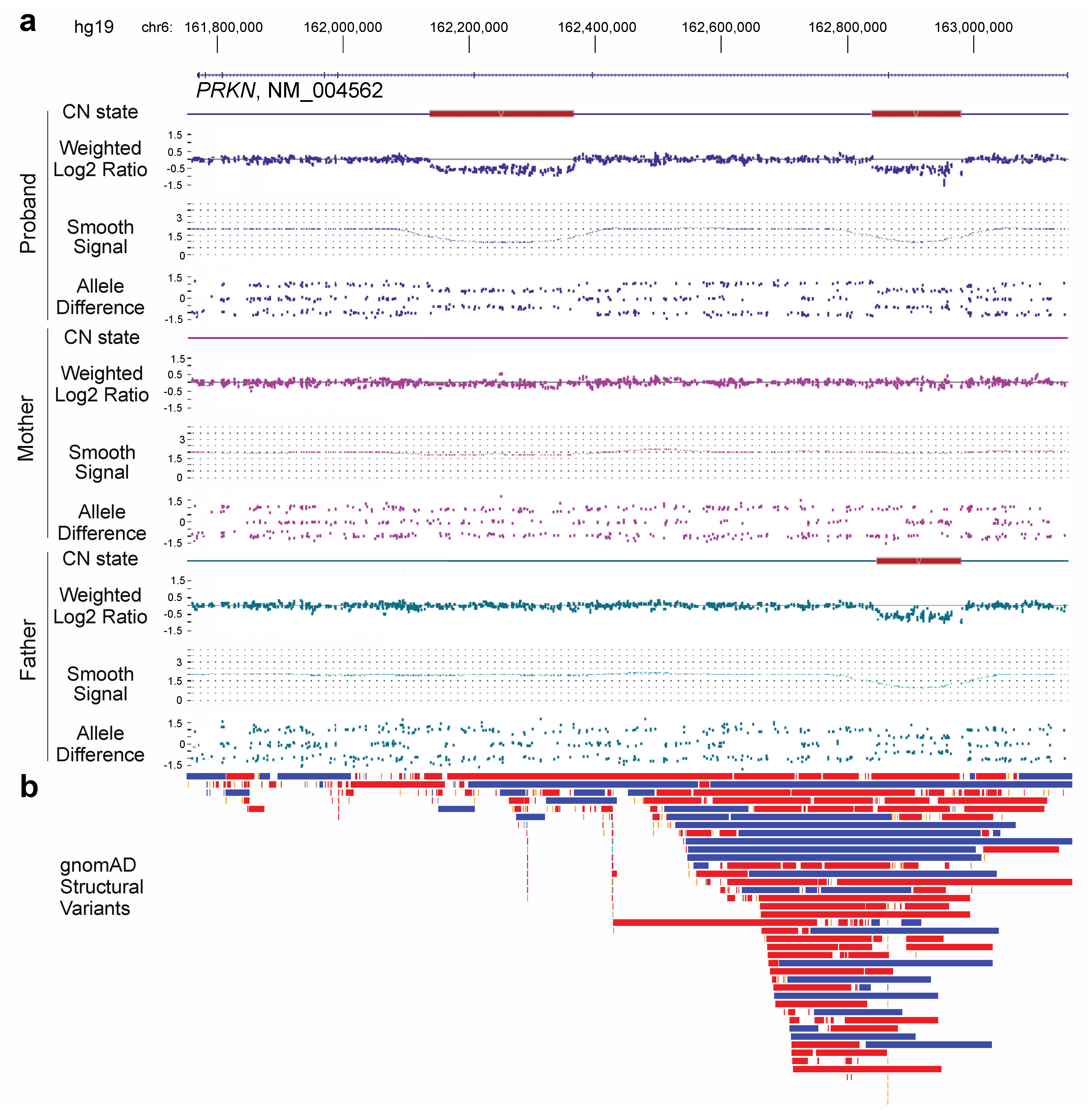
2.1. Chromosomal Microarray

Chromosomal microarray (CMA) analysis was performed using the Applied Biosystems (Affymetrix, Santa Clara, CA, USA) CytoScan HD platform, which has 1.9 million copy number probes and 750,000 single-nucleotide polymorphism (SNP) probes. Peripheral blood specimens from the proband, mother, and father were processed according to the manufacturer’s instructions. Data were analyzed using Chromosome Analysis Suite (ChAS) software version 3.3 (Thermo Fisher, Waltham, MA, USA) and reported using the NCBI human genome build 37.1 (hg19). The genotypes on chromosome 6 were extracted from each specimen using the ChAS software. The phase of the deletion was determined by comparing the proband’s genotypes within and surrounding deleted interval in the PRKN gene with the genotypes from the parents. Out of the 79 SNPs in the interval, 14 were informative for phase determination. The data presented reflect clinical testing performed within our diagnostic laboratory.

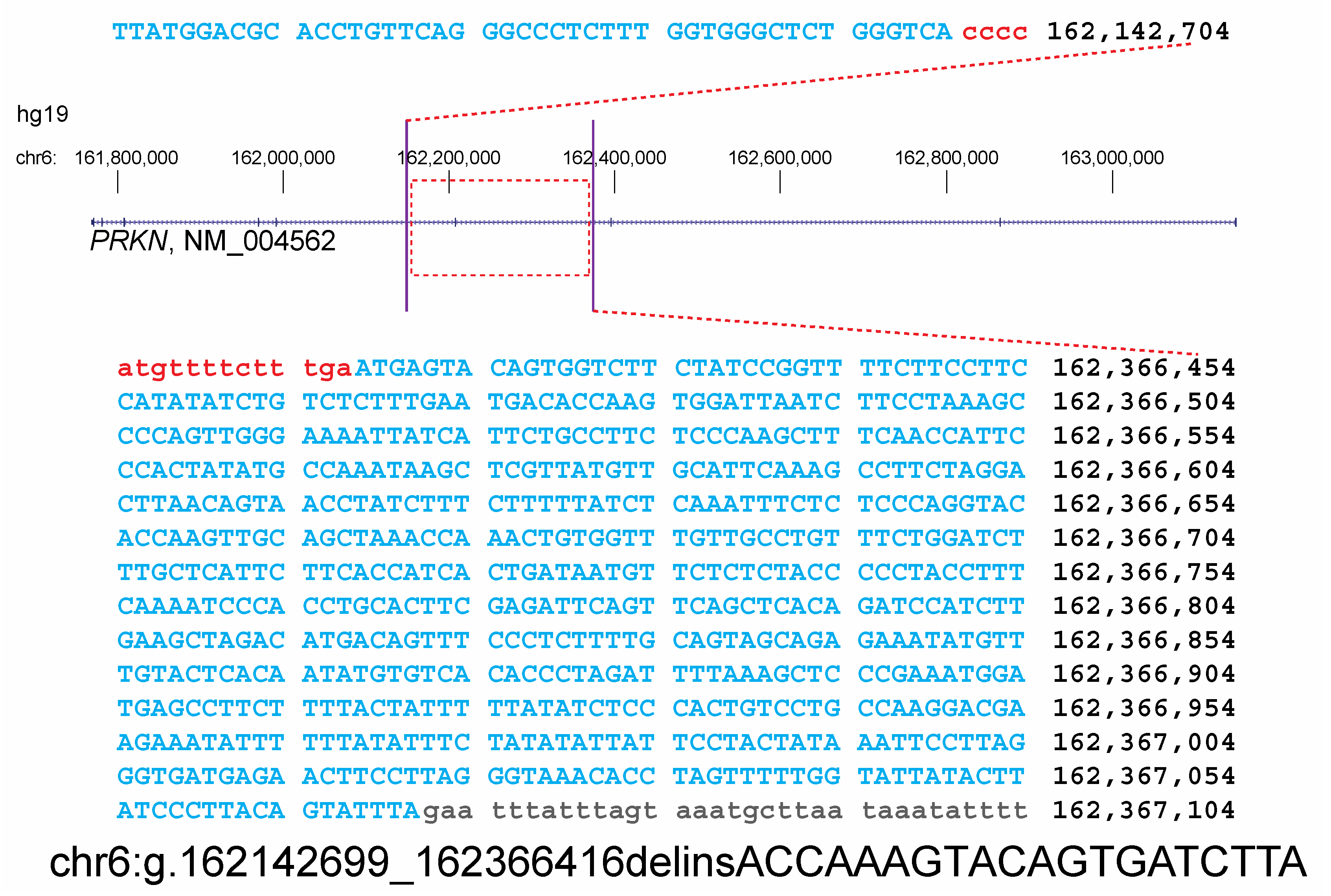
2.2. Long-Range PCR and Sanger Sequencing

GAP PCR and Sanger sequencing were performed on DNA extracted from the proband, maternal sample, and an unrelated control sample. PCR primers were designed to flank the edges of the deletion and anneal outside the area where the proband had informative SNPs for paternal inheritance. Primer sequences were TCCCATCACACCAGAAAACA and CTTGGGAGAAGGCAGAATGA. Long-range PCR was performed using the TaKaRa LATaq Hot Start Version according to the manufacturer’s instructions. The long-range PCR product was amplified with the following ~10 h thermocycling program:
  • Hold: 94 °C for 1 min
  • Denature: 95 °C for 15 min
  • Anneal: 65 °C for 30 min
  • Extend: 68 °C for 15 min
  • Steps 2–4, 35 cycles
  • Final extension: 72 °C for 10 min
The long-range PCR product was size-confirmed with a gel and purified using the AMPure XP purification kit. The purified product was Sanger-sequenced using the BigDye Terminator v1.1 Cycle Sequencing kit with UPS universal primer sequences GGGTTCCCTAAGGGTTGGA and GTGCCAGCAAGATCCAATCTAGA. Fastq files from Sanger sequencing were aligned to hg19 using BLAT to clarify the breakpoints in the proband and mother’s samples.

3. Results

Chromosomal microarray (CMA) testing was ordered for a peripheral blood specimen from a six-day old female with a family history of a 1.5 Mb 17q12 duplication. This recurrent duplication exhibits incomplete penetrance with 90% of cases being inherited. Reported phenotypes of the duplication include variable intellectual disability, speech and motor delay, hypotonia, and seizures [18]. The familial 1.5 Mb duplication was observed on chromosome 17 and included 41 genes. In addition to this duplication, two non-overlapping intragenic deletions were found within the PRKN gene on 6q26 (Figure 1a). These deletions were approximately 140 Kb and 227 Kb in size and encompassed exons 2 and 7 (NM_004562.2), respectively, based on CMA. Both deletions were predicted to be out-of-frame and resulted in loss of PRKN gene function; however, it was unclear if these deletions were in cis or in trans.
Parental CMA studies were performed to assist in phase determination for the proband’s PRKN deletions. The exon 2 deletion was clearly paternally inherited; however, the exon 7 deletion appeared to be de novo. Informative single-nucleotide polymorphisms (SNPs) from the CMA genotype data within and surrounding the exon 7 deletion supported paternal inheritance of the intact copy, suggesting that the deletion arose from the maternal homolog (Table 1). Interestingly, closer inspection of the maternal copy number probes from CMA demonstrated possible low-level mosaicism for the deletion below the limit of detection for the Cytoscan CMA platform.
Primers were designed flanking the exon 7 deletion to perform long-range GAP PCR in the proband, mother, and a control. In the absence of the deletion, the product should be too large to amplify given the thermocycling conditions. No product was identified in the control sample; however, a PCR product was produced in both the proband and mother. Sanger sequencing of this long-range PCR product confirmed that both the proband and mother carry the deletion. The size of the deletion was refined as 223.7 kb, with a breakpoint nomenclature of chr6(GRCh37):g.162142699_162366416delinsACCAAAGTACAGTGATCTTA (Figure 2). These data confirmed that the proband’s deletions are in trans. Thus, the proband has a molecular diagnosis of ARJP.

4. Conclusions

We provided a diagnosis of ARJP in a newborn child based on a secondary finding of two intragenic deletions within the PRKN gene identified through CMA. It is unclear when or how the disease will manifest in this patient, as age of onset, progression, and clinical symptoms can vary greatly [19,20,21,22]. Modifier genes may also contribute to the disease state [23]. The confirmation of carrier status for each parent changes the reproductive risk for this couple.
Due to the structure of the PRKN gene, deletions are thought to be recurrent, independent events while point mutations may be attributed to founder effects [24]. Accordingly, the deletions identified in the proband are localized to regions where structural variation is present in a control population, particularly the deletion containing exon 2 of PRKN; however, these deletions have not been reported before (Figure 1b). Heterozygous copy number variation within PRKN is a relatively common finding in patients that have received CMA testing in our laboratory, and the reporting of heterozygous copy number changes in this gene should be carefully considered.
We used a combination of methods to confirm that the proband in this case had biallelic deletions in the PRKN gene by identifying low-level maternal mosaicism. Methods such as CMA, multiplex ligation-dependent probe amplification (MLPA) [25], digital-droplet PCR (ddPCR) [26], and optical genome mapping (OGM) [27] have all been used to identify deletions and duplications in PRKN. Because of the large size of the PRKN gene, GAP-PCR was used for phase determination of the proband in this study due to the suspected maternal mosaicism. However, other methods may be utilized when the mode of inheritance is not clear or is de novo. Custom FISH probes were used to determine the phase of a compound heterozygote with de novo and inherited deletions [28]. RT-PCR has also been used for phase determination in a cohort study [29], which is simpler to design if the correct specimen is available. Trio whole genome sequencing is another option for phase determination as the underlying informative genotype data should resolve the parent haplotype of origin, as was the case in the study presented here at a smaller scale. In addition, long-read sequencing has been used to resolve a complex inversion involving the PRKN gene and could be used for phase determination [30]. As methods in cytogenomics evolve, there may be improved ways to determine phase of structural rearrangements, particularly in large genes such as PRKN. For now, a comprehensive approach using confirmatory molecular methods is the best method to ensure proper phasing.
When multiple variants are detected in genes associated with autosomal recessive disorders, it is essential to determine the phase to differentiate between a diagnosis and carrier status and to determine recurrence risks. Frequently, parental studies are sufficient to make this determination if neither variant is de novo. However, this case demonstrates the importance of additional follow-up studies when apparent de novo variants are detected. Low-level parental mosaicism should be considered and tested for using alternative methods, especially in the case of autosomal recessive disorders, and has significant implications for recurrence risk.

Author Contributions

L.A.C., C.R., N.L.H. and E.C.T. analyzed the data. L.A.C., F.H., J.H.N. and M.W. generated the experimental data. L.A.C. and E.C.T. wrote the manuscript. All authors have read and agreed to the published version of the manuscript.

Funding

This research received no external funding.

Institutional Review Board Statement

IRB approval was not necessary for this case report.

Informed Consent Statement

Informed consent was not required for this study as no patient-identifying information has been included.

Data Availability Statement

The original contributions presented in the study are included in the article; further inquiries can be directed to the corresponding author.

Conflicts of Interest

The authors declare no conflicts of interest.

References

  1. Willis, A.W.; Roberts, E.; Beck, J.C.; Fiske, B.; Ross, W.; Savica, R.; Eeden, S.K.V.D.; Tanner, C.M.; Marras, C.; Alcalay, R.; et al. Incidence of Parkinson disease in North America. npj Parkinsons Disease 2022, 8, 170. [Google Scholar] [CrossRef] [PubMed]
  2. Funayama, M.; Nishioka, K.; Li, Y.; Hattori, N. Molecular genetics of Parkinson’s disease: Contributions and global trends. J. Hum. Genet. 2023, 68, 125–130. [Google Scholar] [CrossRef]
  3. Doherty, K.M.; Silveira-Moriyama, L.; Parkkinen, L.; Healy, D.G.; Farrell, M.; Mencacci, N.E.; Ahmed, Z.; Brett, F.M.; Hardy, J.; Quinn, N.; et al. Parkin disease: A clinicopathologic entity? JAMA Neurol. 2013, 70, 571–579. [Google Scholar] [CrossRef] [PubMed]
  4. Kitada, T.; Asakawa, S.; Hattori, N.; Matsumine, H.; Yamamura, Y.; Minoshima, S.; Yokochi, M.; Mizuno, Y.; Shimizu, N. Mutations in the parkin gene cause autosomal recessive juvenile parkinsonism. Nature 1998, 392, 605–608. [Google Scholar] [CrossRef]
  5. Corti, O.; Lesage, S.; Brice, A. What genetics tells us about the causes and mechanisms of Parkinson’s disease. Physiol. Rev. 2011, 91, 1161–1218. [Google Scholar] [CrossRef] [PubMed]
  6. Imai, Y.; Soda, M.; Takahashi, R. Parkin suppresses unfolded protein stress-induced cell death through its E3 ubiquitin-protein ligase activity. J. Biol. Chem. 2000, 275, 35661–35664. [Google Scholar] [CrossRef] [PubMed]
  7. Shimura, H.; Hattori, N.; Kubo, S.-I.; Mizuno, Y.; Asakawa, S.; Minoshima, S.; Shimizu, N.; Iwai, K.; Chiba, T.; Tanaka, K.; et al. Familial Parkinson disease gene product, parkin, is a ubiquitin-protein ligase. Nat. Genet. 2000, 25, 302–305. [Google Scholar] [CrossRef]
  8. Zhang, Y.; Gao, J.; Chung, K.K.; Huang, H.; Dawson, V.L.; Dawson, T.M. Parkin functions as an E2-dependent ubiquitin- protein ligase and promotes the degradation of the synaptic vesicle-associated protein, CDCrel-1. Proc. Natl. Acad. Sci. USA 2000, 97, 13354–13359. [Google Scholar] [CrossRef]
  9. Kasten, M.; Hartmann, C.; Hampf, J.; Schaake, S.; Westenberger, A.; Vollstedt, E.; Balck, A.; Domingo, A.; Vulinovic, F.; Dulovic, M.; et al. Genotype-Phenotype Relations for the Parkinson’s Disease Genes Parkin, PINK1, DJ1: MDSGene Systematic Review. Mov. Disord. 2018, 33, 730–741. [Google Scholar] [CrossRef]
  10. Abbas, N.; Lücking, C.B.; Ricard, S.; Dürr, A.; Bonifati, V.; De Michele, G.; Bouley, S.; Vaughan, J.R.; Gasser, T.; Marconi, R.; et al. A wide variety of mutations in the parkin gene are responsible for autosomal recessive parkinsonism in Europe. French Parkinson’s Disease Genetics Study Group and the European Consortium on Genetic Susceptibility in Parkinson’s Disease. Hum. Mol. Genet. 1999, 8, 567–574. [Google Scholar] [CrossRef]
  11. Clausen, L.; Okarmus, J.; Voutsinos, V.; Meyer, M.; Lindorff-Larsen, K.; Hartmann-Petersen, R. PRKN-linked familial Parkinson’s disease: Cellular and molecular mechanisms of disease-linked variants. Cell. Mol. Life Sci. 2024, 81, 223. [Google Scholar] [CrossRef] [PubMed]
  12. Lubbe, S.J.; I Bustos, B.; Hu, J.; Krainc, D.; Joseph, T.; Hehir, J.; Tan, M.; Zhang, W.; Escott-Price, V.; Williams, N.M.; et al. Assessing the relationship between monoallelic PRKN mutations and Parkinson’s risk. Hum. Mol. Genet. 2021, 30, 78–86. [Google Scholar] [CrossRef] [PubMed]
  13. Yoshino, H.; Li, Y.; Nishioka, K.; Daida, K.; Hayashida, A.; Ishiguro, Y.; Yamada, D.; Izawa, N.; Nishi, K.; Nishikawa, N.; et al. Genotype-phenotype correlation of Parkinson’s disease with PRKN variants. Neurobiol. Aging 2022, 114, 117–128. [Google Scholar] [CrossRef] [PubMed]
  14. Zhu, W.; Huang, X.; Yoon, E.; Bandres-Ciga, S.; Blauwendraat, C.; Billingsley, K.J.; Cade, J.H.; Wu, B.P.; Williams, V.H.; Schindler, A.B.; et al. Heterozygous PRKN mutations are common but do not increase the risk of Parkinson’s disease. Brain 2022, 145, 2077–2091. [Google Scholar] [CrossRef] [PubMed]
  15. Collins, R.L.; Brand, H.; Karczewski, K.J.; Zhao, X.; Alföldi, J.; Francioli, L.C.; Khera, A.V.; Lowther, C.; Gauthier, L.D.; Wang, H.; et al. A structural variation reference for medical and population genetics. Nature 2020, 581, 444–451. [Google Scholar] [CrossRef]
  16. Denison, S.R.; Callahan, G.; Becker, N.A.; Phillips, L.A.; Smith, D.I. Characterization of FRA6E and its potential role in autosomal recessive juvenile parkinsonism and ovarian cancer. Genes Chromosomes Cancer 2003, 38, 40–52. [Google Scholar] [CrossRef]
  17. Hedrich, K.; Eskelson, C.; Wilmot, B.; Marder, K.; Harris, J.; Garrels, J.; Meija-Santana, H.; Vieregge, P.; Jacobs, H.; Bressman, S.B.; et al. Distribution, type, and origin of Parkin mutations: Review and case studiesz. Mov. Disord. 2004, 19, 1146–1157. [Google Scholar] [CrossRef]
  18. Mefford, H.; Mitchell, E.; Hodge, J. 17q12 Recurrent Duplication. In GeneReviews(®); Pagon, R.A., Adam, M.P., Ardinger, H.H., Wallace, S.E., Amemiya, A., Bean, L.J., Bird, T.D., Ledbetter, N., Mefford, H.C., Smith, R.J., et al., Eds.; University of Washington: Seattle, WA, USA, 1993. [Google Scholar]
  19. Chien, H.F.; Rohé, C.F.; Costa, M.D.L.; Breedveld, G.J.; Oostra, B.A.; Barbosa, E.R.; Bonifati, V. Early-onset Parkinson’s disease caused by a novel parkin mutation in a genetic isolate from north-eastern Brazil. Neurogenetics 2006, 7, 13–19. [Google Scholar] [CrossRef]
  20. Isaacs, D.; Claassen, D.; Bowman, A.B.; Hedera, P. Phenotypic Discordance in Siblings with Identical Compound Heterozygous PARK2 Mutations. Brain Sci. 2017, 7, 71. [Google Scholar] [CrossRef]
  21. Klein, C.; Pramstaller, P.P.; Kis, B.; Page, C.C.; Kann, M.; Leung, J.; Woodward, H.; Castellan, C.C.; Scherer, M.; Vieregge, P.; et al. Parkin deletions in a family with adult-onset, tremor-dominant parkinsonism: Expanding the phenotype. Ann. Neurol. 2000, 48, 65–71. [Google Scholar] [CrossRef]
  22. Puschmann, A. Monogenic Parkinson’s disease and parkinsonism: Clinical phenotypes and frequencies of known mutations. Park. Relat. Disord. 2013, 19, 407–415. [Google Scholar] [CrossRef]
  23. Cukier, H.N.; Kim, H.; Griswold, A.J.; Codreanu, S.G.; Prince, L.M.; Sherrod, S.D.; McLean, J.A.; Dykxhoorn, D.M.; Ess, K.C.; Hedera, P.; et al. Genomic, transcriptomic, and metabolomic profiles of hiPSC-derived dopamine neurons from clinically discordant brothers with identical PRKN deletions. npj Park. Dis. 2022, 8, 84. [Google Scholar] [CrossRef]
  24. Periquet, M.; Lücking, C.B.; Vaughan, J.R.; Bonifati, V.; Dürr, A.; De Michele, G.; Horstink, M.W.; Farrer, M.; Illarioshkin, S.N.; Pollak, P.; et al. Origin of the mutations in the parkin gene in Europe: Exon rearrangements are independent recurrent events, whereas point mutations may result from Founder effects. Am. J. Hum. Genet. 2001, 68, 617–626. [Google Scholar] [CrossRef] [PubMed]
  25. Mutez, E.; Swiderski, M.; Devos, D.; Moreau, C.; Baille, G.; Degardin, A.; Ryckewaert, G.; Carriere, N.; Kreisler, A.; Simonin, C.; et al. Indication for molecular testing by multiplex ligation-dependent probe amplification in parkinsonism. Eur. J. Neurol. 2023, 30, 1667–1675. [Google Scholar] [CrossRef]
  26. Bravo, P.; Darvish, H.; Tafakhori, A.; Azcona, L.J.; Johari, A.H.; Jamali, F.; Paisán-Ruiz, C. Molecular characterization of PRKN structural variations identified through whole-genome sequencing. Mol. Genet. Genom. Med. 2018, 6, 1243–1248. [Google Scholar] [CrossRef] [PubMed]
  27. Trinh, J.; Schaake, S.; Gabbert, C.; Lüth, T.; Cowley, S.A.; Fienemann, A.; Ullrich, K.K.; Klein, C.; Seibler, P. Optical genome mapping of structural variants in Parkinson’s disease-related induced pluripotent stem cells. BMC Genom. 2024, 25, 980. [Google Scholar] [CrossRef]
  28. Williams, E.S.; Barrett, M.J.; Dhamija, R.; Toran, L.; Chambers, C.; Mahadevan, M.S.; Golden, W.L. Phase determination using chromosomal microarray and fluorescence in situ hybridization in a patient with early onset Parkinson disease and two deletions in PRKN. Mol. Genet. Genom. Med. 2018, 6, 457–462. [Google Scholar] [CrossRef] [PubMed]
  29. Kim, S.Y.; Seong, M.W.; Jeon, B.S.; Kim, S.Y.; Ko, H.S.; Kim, J.Y.; Park, S.S. Phase analysis identifies compound heterozygous deletions of the PARK2 gene in patients with early-onset Parkinson disease. Clin. Genet. 2012, 82, 77–82. [Google Scholar] [CrossRef]
  30. Daida, K.; Funayama, M.; Billingsley, K.J.; Malik, L.; Miano-Burkhardt, A.; Leonard, H.L.; Makarious, M.B.; Iwaki, H.; Ding, J.; Gibbs, J.R.; et al. Long-Read Sequencing Resolves a Complex Structural Variant in PRKN Parkinson’s Disease. Mov. Disord. 2023, 38, 2249–2257. [Google Scholar] [CrossRef]
Figure 1. PRKN deletions. (a) Chromosomal microarray data for the proband, mother, and father at the PRKN gene. Two deletions are found in the proband. The exon 2 deletion is paternally inherited, while the exon 7 deletion appears to be de novo. (b) Structural variants (deletions in red, duplications in blue, and inversions in orange) found in a normal population cohort (gnomAD SV v2.1).
Genes 16 00630 g001
Figure 2. Breakpoint detection of the maternally inherited deletion. Sanger sequencing of the long-range PCR product demonstrates a 223.7 kb deletion with breakpoints at chr6: 162,142,699 and 162,366,416, as well as a 20 bp insertion, with the following nomenclature: chr6(GRCh37):g.162142699_162366416delinsACCAAAGTACAGTGATCTTA. Bases in blue align to the genome and bases in red are absent in the Sanger sequencing product.
Genes 16 00630 g002
Table 1. Informative SNPs. Informative single nucleotide variants within the deleted interval containing exon 7 show paternal inheritance (bolded alleles are shared between father and proband).
SNPPositionMotherProbandFather
rs6907465162250726TTAAAT
rs9347543162252602AAGGGA
rs9458393162255535GGTTTG
rs6926642162270981TTCCCT
rs9365329162271943TTGGGT
rs9347547162280309GGAAAG
rs7750426162281372TTGGGT
rs2186803162281492AACCCA
rs2155486162281533TTAAAT
rs6935164162282924CCTTTC
rs9689946162283949GGTTTG
rs9355958162322332TTCCCC
Disclaimer/Publisher’s Note: The statements, opinions and data contained in all publications are solely those of the individual author(s) and contributor(s) and not of MDPI and/or the editor(s). MDPI and/or the editor(s) disclaim responsibility for any injury to people or property resulting from any ideas, methods, instructions or products referred to in the content.

