Case Report

The Arg99Gln Substitution in HNRNPC Is Associated with a Distinctive Clinical Phenotype Characterized by Facial Dysmorphism and Ocular and Cochlear Anomalies

1
Molecular Genetics and Functional Genomics, Bambino Gesù Children’s Hospital, IRCCS, 00143 Rome, Italy
2
Medical and Molecular Genetics, AORN A. Cardarelli, 80131 Naples, Italy
3
Center for Rare Diseases and Birth Defects, Department of Woman and Child Health and Public Health, Fondazione Policlinico Universitario A. Gemelli, IRCCS, 00168 Rome, Italy
4
Department of Biochemical Sciences “Alessandro Rossi Fanelli”, Sapienza University, 00185 Rome, Italy
5
Facoltà di Medicina e Chirurgia, Università Cattolica del Sacro Cuore, 00168 Rome, Italy
6
Department of Oncology and Molecular Medicine, Istituto Superiore di Sanità, 00161 Rome, Italy
7
Department of Biomedicine and Prevention, University “Tor Vergata”, 00173 Rome, Italy
8
Department of Computer, Control and Management Engineering, Sapienza University, 00185 Rome, Italy
9
National Centre of Services and Research for Prevention of Blindness and Rehabilitation of Low Vision Patients, IAPB Italia Onlus, 00185 Rome, Italy
10
Pediatric Neuropsychiatric Unit, Department of Woman and Child Health and Public Health, Fondazione Policlinico Universitario A. Gemelli, IRCCS, 00168 Rome, Italy
*
Authors to whom correspondence should be addressed.
Genes 2025, 16(2), 176; https://doi.org/10.3390/genes16020176
Submission received: 30 December 2024 / Revised: 23 January 2025 / Accepted: 27 January 2025 / Published: 1 February 2025
(This article belongs to the Special Issue Feature Papers in Human Genomics and Genetic Diseases 2024)

Abstract

:
Background/Objectives: Heterozygous variants in the heterogeneous nuclear ribonucleoprotein C gene (HNRNPC) have recently been reported to cause intellectual developmental disorder-74 (MRD74), a neurodevelopmental disorder with no recurrent diagnostic handles. Affected individuals show variable, non-specific, and subtle dysmorphic features. The degree of developmental delay (DD)/intellectual disability (ID) is also wide, ranging from mild to severe. The mutational spectrum is relatively broad with exon deletions and splice site and frameshift variants distributed along the entire length of the gene leading to HNRNPC loss of function. Only two missense changes located within the RNA-binding motif (RBM) and adjacent linker region of the more abundant isoform (Arg64Trp and Arg99Gln) have been described. Notably, the Arg99Gln amino acid substitution was reported in a subject presenting with a more complex and unique clinical phenotype characterized by distinctive facial features, DD/ID, cochlear aplasia, and bilateral colobomatous microphthalmia, suggesting the possible occurrence of phenotypic heterogeneity. Results: Here, we report the second individual carrying the Arg99Gln change in HNRNPC and having clinical features with a significant overlap with the peculiar phenotype of the previously described subject, supporting the occurrence of a genotype–phenotype correlation. Conclusions: Due to the concomitant occurrence of ocular and cochlear involvement as recognizable diagnostic handles, we propose that the HNRNPCArg99Gln-related phenotype should be considered as a potential differential diagnosis in subjects with ID and major signs of CHARGE syndrome not fulfilling the minimum criteria for a clinical diagnosis.

1. Introduction

Heterogeneous nuclear ribonucleoproteins (HNRNPs) are a large family of ubiquitously expressed RNA-binding proteins involved in gene expression and nucleic acid metabolism, including pre-mRNA processing, mRNA stabilization during export from the nucleus to the cytoplasm, and regulation of the translational process [1,2]. To date, genes encoding members of this family have been proven to contribute to various neurodegenerative conditions, including amyotrophic lateral sclerosis (HNRNPA1), oculopharyngeal muscular dystrophy (HNRPA2B1), spinal muscular atrophy (HNRNPG), and fronto-temporal lobe dementia (HNRPA2B1) [1]. Disease-causing variants encompassing eight genes encoding members of this family have consistently been associated with syndromic neurodevelopmental disorders (NDDs) (HNRNPH1, MIM 601035; HNRNPH2, MIM 300610; HNRNPK, MIM 600712; HNRNPR, MIM 607201; HNRNPU, MIM 602869; HNRNPQ, MIM 616686; HNRNPG, MIM 300199; HNRNPC, MIM 164020) [3,4,5,6,7,8,9,10,11,12]. Among them, heterozygous HNRNPC loss-of-function (LoF) variants have recently been identified to cause a syndromic NDD (i.e., intellectual developmental disorder, autosomal dominant 74, MRD74; MIM 620688), mainly characterized by global developmental delay (DD), intellectual disability (ID), behavioral abnormalities, and variable facial dysmorphism [11].
HNRNPC codes for the heterogeneous nuclear ribonucleoprotein C (HNRNPC), a key protein involved in pre-mRNA splicing by promoting alternative exon usage [13], and in the export of transcripts longer than 700 nucleotides from the nucleus to the cytoplasm [14]. HNRNPC is characterized by five functional domains: a conserved N-terminal single-stranded RNA-binding motif (RBM) previously known as the RNA-binding domain (RBD) (residues 1 to 93), a linker region conferring RNA-binding specificity for the poly r(U)5 consensus sequence (residues 94 to 104), a positively charged zipper-like motif (bZLM) (residues 140 to 179), a leucine-zipper-like oligomerization domain (CLZ) (residues 180 to 207), and a C-terminal domain (residues 208 to 290) with a still poorly characterized function [11,15,16]. HNRNPC’s target accessibility is regulated by the mRNA post-transcriptional modification N(6)-methyl-adenosine (m6A), a mechanism known as “m6A-switch” [17], regulating the alternative splicing, export, maturation, and expression of several target mRNAs. Notably, HNRNPC haploinsufficiency has been shown to affect these processes, leading to the nuclear accumulation of ID-associated mRNAs [11,17].
The phenotypic spectrum of MRD74 is typically non-specific. Affected individuals show variable and subtle dysmorphic features, with brachycephaly, deep-set eyes, either hypo- or hypertelorism, a prominent nose, a smooth philtrum, and a thin upper lip occurring most commonly [11]. DD/ID and behavioral anomalies are invariably present, with the former characterized by wide variability, ranging from mild to severe. A relatively broad spectrum of pathogenic variants leading to HNRNPC LoF have been reported, the majority representing nonsense, frameshift, and intragenic in-frame deletions. In addition, two functionally uncharacterized missense changes (Arg64Trp and Arg99Gln) located within the RBM and adjacent linker region of the more abundant HNRNPC isoform (isoform b, NP_004491.2) have also been described [11]. Notably, among the previously reported individuals, the subject carrying the Arg99Gln amino acid substitution presented with a more complex and atypical clinical phenotype, characterized by distinctive facial features, DD/ID, cochlear aplasia, and bilateral colobomatous microphthalmia, suggesting the possible occurrence of phenotypic heterogeneity [11]. Of note, cochlear hypoplasia and colobomatous microphthalmia represent two major signs of CHARGE syndrome according to the diagnostic criteria available for this disorder [18,19].
Here, we report a second individual carrying the same missense mutation in HNRNPC and showing an overlapping phenotype with the previously described subject, indicating the occurrence of a genotype–phenotype correlation. Although the clinical phenotype of the two subjects does not fulfill the minimum criteria for a clinical diagnosis of CHARGE syndrome [19], due to the co-presence of two major diagnostic features, we propose that HNRNPCArg99Gln-related phenotype should be considered as a potential differential diagnosis in subjects with ID and major signs of CHARGE syndrome.

2. Materials and Methods

The subject was enrolled in a research program aiming to understand the molecular causes of unclassified pediatric disorders at the Ospedale Pediatrico Bambino Gesù, Rome, Italy. The study was approved by the local Institutional Ethical Committee (ref. RF-2021-12374963, 15 March 2023). Clinical data, pictures, and blood samples were collected, used, and stored after signed informed consent from the participating subject and her parents was secured, in accordance with the ethical standards outlined in the Declaration of Helsinki. Permission to publish the clinical pictures was obtained.

2.1. DNA Methylation Profiling Analysis

Peripheral blood (PB) DNA was extracted using standard techniques. Bisulfite conversion was performed, and samples were analyzed using the Infinium Methylation EPIC BeadChip v.1/v.2 (Illumina San Diego, CA, USA), according to the manufacturer’s protocol. IDAT files containing methylated and unmethylated signal intensities were imported for analysis into R v.4.4.0 by means of the minfi package, correcting for background intensities. DNA methylation (DNAm) analysis was performed using a previously described pipeline [20,21] and the currently available DNAm signature for CHARGE syndrome [22,23], and grouped by means of hierarchical clustering (HC) and multidimensional scaling (MDS), considering the pairwise Euclidean distances between samples. The training model of the support vector machine (SVM) machine learning (ML)-based classifier was carried out as previously described [24].

2.2. WGS Analysis

Trio-based WGS data were obtained using a 2×150 bp paired-end read protocol on a NovaSeq 6000 platform (Illumina, San Diego, CA, USA), reaching 30x median coverage. Base calling and data analysis were performed using Bcl2FASTQ (Illumina). Paired-end reads were mapped to the GRCh38 reference sequence; variant calling and joint genotyping were run using Sentieon v.2023-08 (https://www.sentieon.com). SNP and indel hard filtering were applied using Genome Analysis Toolkit, version 3.8.0 (Broad Institute). High-quality variants were first filtered by frequency ≤ 5% in the in-house WGS population-matched database (>350 WGS). The remaining coding sequence variants were annotated and filtered using a custom pipeline, as previously described [25,26]. Detected variants in non-coding regions were annotated and prioritized using Genomiser v.2309 [27]. Structural variants were detected using DELLY v.1.1.6 [28] and prioritized using AnnotSV v.3.4. [29]. The identified HNRNPC variant was validated by bidirectional Sanger sequencing.

2.3. Structural Analysis

The non-covalent intramolecular interactions involving Arg99 in the wild-type (WT) HNRNPC structure (PDB ID: 2MXY, and PDB ID: 2MZ1) [30] and the predicted structural consequences of the Arg99Gln amino acid change were inspected using UCSF Chimera software v.1.17.3 (https://www.cgl.ucsf.edu/chimera, accessed on 9 December 2024) [31].

3. Results

3.1. Clinical Findings

The proband was a single-born from an uneventful pregnancy of non-consanguineous parents without a family history of genetic or neurological diseases. The parents reported three previous spontaneous miscarriages during the first trimester. A cesarean section was performed because of the lack of progress in labor. Her auxological parameters at birth were as follows: weight, 3730 g (+1.37 SD); length, 52 cm (+1.61 SD); and occipitofrontal circumference (OFC), 36 cm (+1.87 SD). Her Apgar score was 8 and 9, at the first and fifth minute, respectively. She was fed with valid suction. At birth, a bilateral periauricolar fistula was noticed and auditory evoked potentials were performed, revealing hearing loss. At 1 year, brain and ear MRI was performed, which evidenced agenesis of the left cochlea, while a sketch of a cochlea was present on the right; therefore, acoustic prosthesization was prescribed. A CT scan was later performed, documenting the complex malformation of the inner ears (Figure 1). The ophthalmological evaluation showed iris, chorioretinal, and optic nerve coloboma with microphthalmia and subluxation of the crystalline lens on the right eye, and chorioretinal coloboma on the left eye. At the cardiological evaluation, a patent ductus arteriosus was documented. She underwent endocrinological evaluation because of growth retardation. Growth hormone (GH) stimulation tests ruled out GH deficiency. She presented with menarche at the age of 15 with irregular menses.
She presented with DD. At one year, she started rehabilitation. She achieved sitting at 18 months and independent walking at 30 months. A neuropsychological assessment documented moderate intellectual impairment (IQ 42). She currently attends high school with a support and communication assistant and use of alternative augmentative communication.
At the last evaluation (18 years), she presented with global growth delay: weight, 40.9 kg (−2.26 SD); height, 142.3 cm (−3.18 SD); and OFC, 50 cm (−4.05 SD). She presented with a broad and slightly receding forehead, a high hairline, thick eyebrows, narrow and wave-shaped palpebral fissures, bilateral microphthalmia with iris coloboma (right eye), a sharp nose, a thin upper lip, slightly posteriorly rotated ears with a fistula at the root of the helix bilaterally, a large triangular dimple, fleshy lobes, a prominent malar region, and a dimple chin (Figure 1). She also showed a short and broad distal phalanx of the first finger of the hands bilaterally.
Due to the presence of two major features of CHARGE syndrome, comparative genomic hybridization analysis and mutation scanning of the entire coding sequence of CHD7 (MIM 608892) was performed, both resulting as negative.

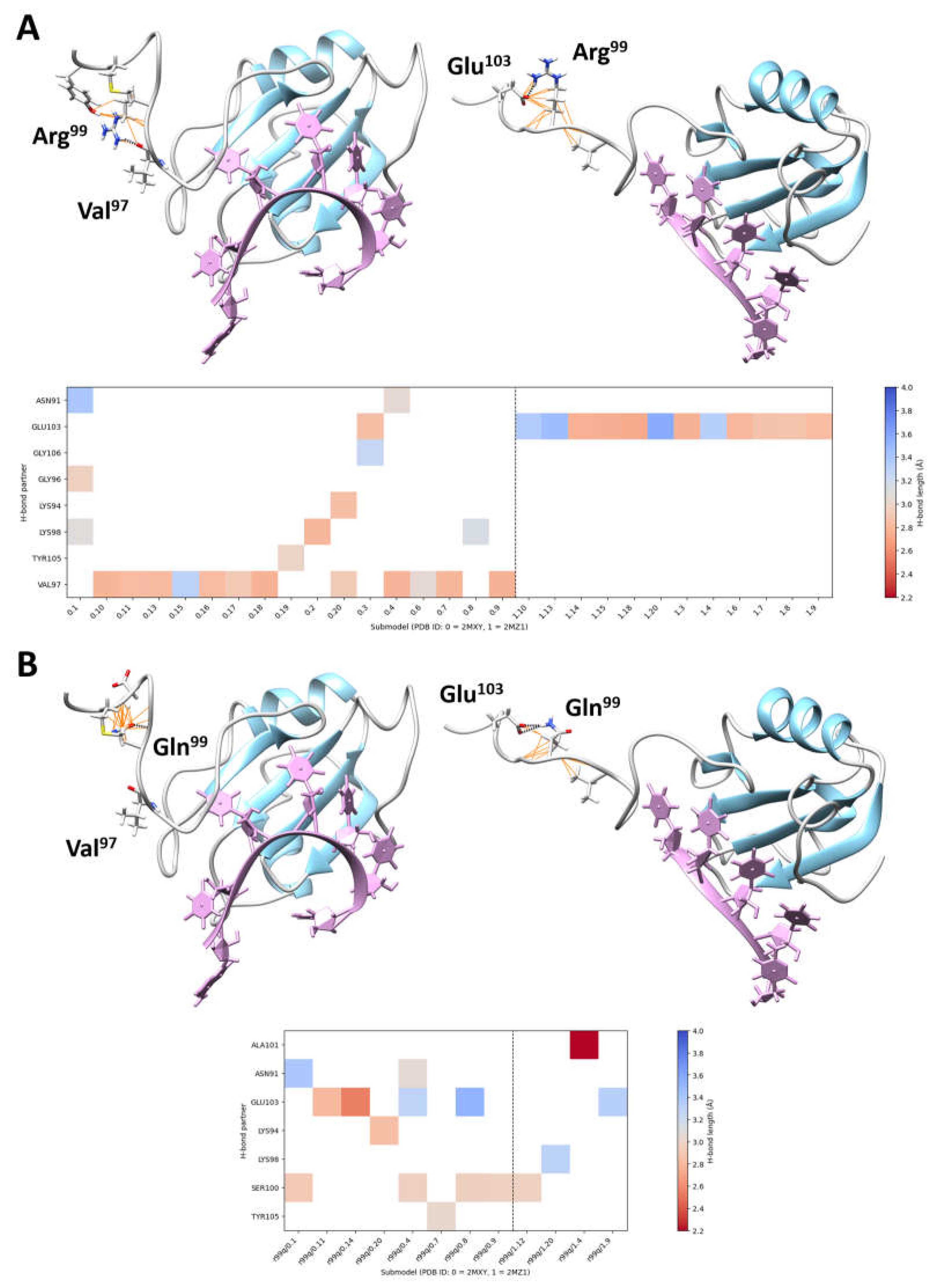
3.2. Molecular Findings and Structural Analysis

To definitively exclude the suspicion of an atypical presentation of CHARGE syndrome, DNAm profiling was performed. A set of 148 differentially methylated probes defining the previously reported episignature for CHARGE syndrome [22,23] was used to test and classify the DNAm profile of the subject, which was compared with two in-house cohorts composed of three patients with molecularly confirmed CHARGE syndrome (ID1: NM_017780.4:c.2839C>T, p.Arg947Ter; ID2: NM_017780.4:c.7252C>T, p.Arg2418Ter; ID3: and NM_017780.4:c.1972G>T, p.Glu658Ter) (“CHD7 training set”) and 400 controls (290 healthy subjects and 110 individuals affected with different rare disorders). Unsupervised and supervised analyses consistently grouped the tested sample together with controls (Supplemental Figure S1), in line with the previous negative genetic findings.
A subsequent WGS analysis identified a de novo single-nucleotide variant (NM_031314.3:c.296G>A; NP_112604.2:p.Arg99Gln) in the HNRNPC gene. Variant validation and segregation were attained by Sanger sequencing (Supplemental Figure S2). A likely pathogenic impact of the amino acid substitution was predicted by the AlphaMissense (score = 0.94) and CADD (PHRED score = 29) tools. The c.296G>A change was not present in gnomAD v.4.1.0 but had been reported in the original cohort of MRD74 patients reported by Niggl et al. [11]. The WGS data analysis confirmed the absence of intragenic or structural variants involving CHD7 and excluded the occurrence of other clinically relevant variants in genes previously reported to be implicated in neurodevelopmental disorders with features overlapping with CHARGE syndrome.
The structural consequences of the Arg99Gln substitution in HNRNPC folding and function were assessed using the available 3D nuclear magnetic resonance-spectroscopy models (PDB ID: 2MXY, and PDB ID: 2MZ1) [30]. In the generated structure, Arg99 forms two key intramolecular interactions with Val97 (H-bond, 2.92 Å ± 0.21 SD) and Glu103 (salt bridge, 2.94 Å ± 0.27 SD). These bonds contribute to the stabilization and proper folding of the linker region. The replacement of Arg99 by Gln was predicted to abolish both the intramolecular H-bond with Val97 and electrostatic component of the salt bridge with Glu103 (Figure 2).

3.3. Assessment of the Clinical Profile Associated with Arg99Gln

We clinically compared the present subject and the previously described individual carrying the same missense change (individual 11) [11]. Their characteristics are summarized in Table 1. Both the individuals shared bilateral cochlear aplasia/hypoplasia and colobomatous microphthalmia as major signs. They also presented with overlapping facial features, including a broad and receding forehead with a high upper hairline, mild synophrys, wave-shaped lids, anteverted nares, a long philtrum, and a thin upper lip with a prominent malar region. Notably, these features were only sporadically reported in the other subjects with HNRNPC variants as individual traits [11], indicating that this specific amino acid substitution leads to a unique clinical presentation.

4. Discussion

We report the second subject carrying the pathogenic c.296G>A missense variant (Arg99Gln) in HNRNPC showing a unique clinical presentation characterized by DD/ID, distinctive facial features, cochlear aplasia, and bilateral colobomatous microphthalmia. The clinical phenotype of the subject fit that of the previously described individual and only partially overlaps with the clinical spectrum of MRD74 [11]. This finding suggests the occurrence of phenotypic heterogeneity involving pathogenic variants in HNRNPC.
Haploinsufficiency has been shown to be the pathogenic mechanism underlying the majority of HNRNPC variants, affecting proper transcript processing of ID-associated genes. However, the pathological effect exerted by a small number of missense changes remains to be elucidated [11]. Notably, the two individuals carrying the Arg64Trp and the Val108Ile amino acid substitutions, the latter affecting the less abundant major isoform (NP_112604.2) only (the variant is an intronic change, c.317+5G>A, of the processed transcript encoding the short but more abundant HNRNPC isoform; NP_004491.2), presented with a non-specific phenotype with slight dysmorphic features, and peculiar behavior anomalies substantially overlapping with those found in other MRD74 individuals with HNRNPC LoF variants. Specifically, these individuals presented with feeding problems (observed in 8/10 individuals with LoF HNRNPC variants) and a typical happy demeanor associated with other behavioral anomalies (observed in 7/10 individuals with LoF HNRNPC variants). These two clinical signs have not been observed in the two individuals with the Arg99Gln amino acid substitution. On the other hand, excluding the two affected subjects with the Arg99Gln amino acid change, none of the subjects with LP/P variants in HNRNPC have been reported to show cochlear aplasia and coloboma. This specific genotype–phenotype correlation suggests the occurrence of a specific consequence of the Arg99Gln amino acid substitution on HNRNPC function.
While the RNA-binding activity of HNRNPC is conferred by the conserved RBM domain, the RNA-binding specificity towards the poly-r(U)5 consensus sequence and ability to recognize different transcripts is granted by the linker domain encompassing residues from K94 to M104 [15,16]. Consistently, two HNRNPC truncated mutants, Δ95–290 and Δ105–290, maintained their RNA-binding activity, but lost their high affinity for targets with the poly-r(U)5 tract, indiscriminately binding to transcripts with different poly-r(U) consensus sequences (i.e., poly r(U)8, poly r(U)13) [15]. Within the linker region, Arg99, Lys98, and Glu103 are highly intolerant to any amino acid substitutions, according to the AlphaMissense Pathogenicity Heatmap. Consistently, no substitution affecting Arg99 has been reported in public databases (gnomAD v.4.1.0 and All of Us). Moreover, only a few benign missense changes have been reported to affect residues within the entire linker domain, indicating that local structural rearrangements of this region may dramatically affect the target recognition process. Structural inspection of the HNRNPC RBM-linker region complexed with the poly-r(U)5 stretch showed that the substitution of the positively charged Arg99 with Gln, a polar residue, disrupts the salt bridge with the conserved Glu103 and abolishes the H-bond with Val97. Although functional characterization is not yet available, the structural data indicate that Arg99 is a key residue for maintaining the linker domain’s folding and consequently granting specificity in binding to poly-r(U)5-containing transcripts.
Cochlear hypo/aplasia and colobomatous microphthalmia are common findings in CHARGE syndrome [32,33,34], which is caused by heterozygous mutations in the chromodomain helicase DNA-binding protein 7 (CHD7) gene encoding a chromatin remodeler [35]. Indeed, though the clinical phenotype of the proband did not fulfill the diagnostic criteria of CHARGE syndrome, targeted sequencing and DNAm profiling were performed to exclude this diagnosis. A clinical diagnosis of CHARGE syndrome requires four major criteria or three major criteria and three minor criteria according to Blake et al. [19], with cochlear hypo/aplasia and colobomatous microphthalmia representing major criteria [18,19]. In individuals fulfilling the diagnostic criteria for CHARGE syndrome, CHD7 sequencing and targeted analyses directed to identify structural rearrangements involving the gene are expected to confirm the diagnosis in all cases. Notably, pathogenic variants in various genes associated with other well-recognized syndromic conditions have been reported in individuals with at least two major signs of CHARGE syndrome, suggesting the occurrence of genetic heterogeneity involving those genes with pathogenic variants specifically associated with phenotypes that should be considered as a differential diagnosis in individuals with an atypical/incomplete CHARGE phenotype [36]. Among them, Kabuki syndrome is a syndromic DD/ID disorder characterized by a distinctive facial gestalt, caused by LoF pathogenic variants in the KMT2D and KDM6A genes [37]. There is evidence that specific pathogenic variants affecting exons 38 and 39 of KMT2D result in a unique phenotype characterized by choanal atresia, hypoplastic nipples, branchial apparatus abnormalities, neck pits, lacrimal duct anomalies, hearing loss, external ear malformations, and thyroid abnormalities, resulting in a condition partially overlapping with CHARGE syndrome, and not resembling Kabuki syndrome [38,39,40]. Similarly, while heterozygous variants in RERE cause a neurodevelopmental disorder with or without anomalies of the brain, eye, or heart (NEDBEH, MIM 616975) [41,42], one single recurrent in-frame duplication has been reported in three independent individuals sharing a phenotype characterized by a variable combination of choanal atresia, iris coloboma, abnormal external ears, progressive sensorineural hearing loss with cochlear dysplasia, and ID [36,42]. This finding prompted the authors to propose the inclusion of RERE in the genes to be tested in individuals who fulfill the diagnostic criteria for CHARGE syndrome but do not carry pathogenic variants in CHD7 [42]. The specificity of this genotype–phenotype correlation further highlights the possibility of phenotypic heterogeneity associated with unique gene variants, as observed for the presently identified Arg99Gln substitution in HNRNPC. In line with these considerations, a single individual with a clinical diagnosis of CHARGE syndrome, showing a normal chromosomal microarray and negative for intragenic CHD7 mutations, was identified to carry a missense variant in EP300, a gene implicated in Rubinstein–Taybi syndrome 2 (MIM 613684) and Menke–Hennekam syndrome (MIM 618333) [36,43,44].
In conclusion, we provide evidence of a recognizable phenotype associated with the c.296G>A (p.Arg99Gln) change in HNRNPC that clinically diverges from MRD74. This phenotype is characterized by DD/ID, distinctive facial features, cochlear aplasia, and bilateral colobomatous microphthalmia. Due to the co-presence of ocular and cochlear involvement, two main features of CHARGE syndrome, we propose that the HNRNPCArg99Gln-related phenotype should be considered as a potential differential diagnosis in subjects with ID and major signs of CHARGE syndrome not fulfilling the minimum criteria for a clinical diagnosis of CHARGE syndrome.

Supplementary Materials

The following supporting information can be downloaded at: https://www.mdpi.com/article/10.3390/genes16020176/s1, Figure S1: DNA methylation profiling analyses; Figure S2: Sanger sequencing chromatograms of the HNRNPC coding region encompassing the c.296G>A variant in the proband.

Author Contributions

L.C., A.B., M.N., V.C., C.M., and A.C. performed the genomic analyses and data validation. R.O., C.L., D.R., and G.Z. contributed to patient assessment and clinical data collection. M.P. and F.C.R. analyzed the clinical data and revised the literature. M.C. carried out the structural analyses. C.C., M.F., and A.C. performed the DNA methylation profiling analysis. G.Z. and M.T. coordinated the study. L.C., M.P., and M.T. wrote the manuscript. All authors critically revised the manuscript. All authors have read and agreed to the published version of the manuscript.

Funding

This work was supported, in part, by grants from the Italian Ministry of Health (Current Research Funds to A.C. and M.T., and RF-2021-12374963 to M.T.).

Institutional Review Board Statement

The study was approved by the Bambino Gesù Children’s Hospital ethics committee (ref. RF-2021-12374963, 15 March 2023). Clinical data, pictures, and DNA samples were collected, used, and stored after signed informed consent from the participating family was secured. Written informed consent was obtained for publication of individual pictures.

Informed Consent Statement

Informed consent was obtained from all subjects involved in the study.

Data Availability Statement

The sequencing data that support the findings of this work are available on request from the corresponding author. The data are not publicly available due to privacy/ethical restrictions.

Acknowledgments

The authors wish to thank the family participating in this study.

Conflicts of Interest

The authors do not have conflicts of interest to declare.

References

  1. Geuens, T.; Bouhy, D.; Timmerman, V. The hnRNP family: Insights into their role in health and disease. Hum. Genet. 2016, 135, 851–867. [Google Scholar] [CrossRef] [PubMed]
  2. Gillentine, M.A.; Wang, T.; Hoekzema, K.; Rosenfeld, J.; Liu, P.; Guo, H.; Kim, C.N.; De Vries, B.B.A.; Vissers, L.E.L.M.; Nordenskjold, M.; et al. Rare deleterious mutations of HNRNP genes result in shared neurodevelopmental disorders. Genome Med. 2021, 13, 63–89. [Google Scholar] [CrossRef]
  3. Shashi, V.; Xie, P.; Schoch, K.; Goldstein, D.B.; Howard, T.D.; Berry, M.N.; Schwartz, C.E.; Cronin, K.; Sliwa, S.; Allen, A.; et al. The RBMX gene as a candidate for the Shashi X-linked intellectual disability syndrome. Clin. Genet. 2015, 88, 386–390. [Google Scholar] [CrossRef] [PubMed]
  4. Bain, J.M.; Cho, M.T.; Telegrafi, A.; Wilson, A.; Brooks, S.; Botti, C.; Gowans, G.; Autullo, L.A.; Krishnamurthy, V.; Willing, M.C.; et al. Variants in HNRNPH2 on the X Chromosome Are Associated with a Neurodevelopmental Disorder in Females. Am. J. Hum. Genet. 2016, 99, 728–734. [Google Scholar] [CrossRef]
  5. Lange, L.; Pagnamenta, A.T.; Lise, S.; Clasper, S.; Stewart, H.; Akha, E.S.; Quaghebeur, G.; Knight, S.J.L.; Keays, D.A.; Taylor, J.C.; et al. A de novo frameshift in HNRNPK causing a Kabuki-like syndrome with nodular heterotopia. Clin. Genet. 2016, 90, 258–262. [Google Scholar] [CrossRef]
  6. Bramswig, N.C.; Ludecke, H.J.; Hamdan, F.F.; Altmuller, J.; Beleggia, F.; Elcioglu, N.H.; Freyer, C.; Gerkes, E.H.; Demirkol, Y.K.; Knupp, K.G.; et al. Heterozygous HNRNPU variants cause early onset epilepsy and severe intellectual disability. Hum. Genet. 2017, 136, 821–834. [Google Scholar] [CrossRef]
  7. Yates, T.M.; Vasudevan, P.C.; Chandler, K.E.; Donnelly, D.E.; Stark, Z.; Sadedin, S.; Willoughby, J.; Broad Center for Mendelian Genomics; DDD Study; Balasubramanian, M. De novo mutations in HNRNPU result in a neurodevelopmental syndrome. Am. J. Med. Genet. 2017, 173, 3003–3012. [Google Scholar] [CrossRef]
  8. Duijkers, F.A.; McDonald, A.; Janssens, G.E.; Lezzerini, M.; Jongejan, A.; van Koningsbruggen, S.; Leeuwenburgh-Pronk, W.G.; Wlodarski, M.W.; Moutton, S.; Tran-Mau-Them, F.; et al. HNRNPR Variants that Impair Homeobox Gene Expression Drive Developmental Disorders in Humans. Am. J. Hum. Genet. 2019, 104, 1040–1059. [Google Scholar] [CrossRef] [PubMed]
  9. Reichert, S.C.; Li, R.; A Turner, S.; van Jaarsveld, R.H.; Massink, M.P.G.; van den Boogaard, M.J.H.; del Toro, M.; Rodrıguez-Palmero, A.; Fourcade, S.; Schluter, A.; et al. HNRNPH1-related syndromic intellectual disability: Seven additional cases suggestive of a distinct syndromic neurodevelopmental syndrome. Clin. Genet. 2020, 98, 91–98. [Google Scholar] [CrossRef]
  10. Semino, F.; Schroter, J.; Willemsen, M.H.; Bast, T.; Biskup, S.; Beck-Woedl, S.; Brennenstuhl, H.; Schaaf, C.P.; Kolker, S.; Hoffmann, G.F.; et al. Further evidence for de novo variants in SYNCRIP as the cause of a neurodevelopmental disorder. Hum. Mutat. 2021, 42, 1094–1100. [Google Scholar] [CrossRef]
  11. Niggl, E.; Bouman, A.; Briere, L.C.; Hoogenboezem, R.M.; Wallaard, I.; Park, J.; Admard, J.; Wilke, M.; Harris-Mostert, E.D.R.O.; Elgersma, M.; et al. HNRNPC haploinsufficiency affects alternative splicing of intellectual disability-associated genes and causes a neurodevelopmental disorder. Am. J. Hum. Genet. 2023, 110, 1414–1435. [Google Scholar] [CrossRef] [PubMed]
  12. Johansson, J.; Lidéus, S.; Frykhol, C.; Gunnarsson, C.; Mihalic, F.; Gudmundsson, S.; Ekvall, S.; Molin, A.M.; Pham, M.; Vihinen, M.; et al. Gustavson syndrome is caused by an in-frame deletion in RBMX associated with potentially disturbed SH3 domain interactions. Eur. J. Hum. Genet. 2024, 32, 333–341. [Google Scholar] [CrossRef] [PubMed]
  13. Herzner, A.M.; Khan, Z.; van Nostrand, E.L.; Chan, S.; Cuellar, T.; Chen, R.; Pechuan-Jorge, X.; Komuves, L.; Solon, M.; Modrusan, Z.; et al. ADAR and hnRNPC deficiency synergize in activating endogenous dsRNA-induced type I IFN responses. J. Exp. Med. 2021, 218, e20201833. [Google Scholar] [CrossRef] [PubMed]
  14. McCloskey, A.; Taniguchi, I.; Shinmyozu, K.; Ohno, M. hnRNP C tetramer measures RNA length to classify RNA polymerase II transcripts for export. Science 2012, 335, 1643–1646. [Google Scholar] [CrossRef] [PubMed]
  15. Görlach, M.; Burd, C.G.; Dreyfuss, G. The determinants of RNA-binding specificity of the heterogeneous nuclear ribonucleoprotein C proteins. J. Biol. Chem. 1994, 269, 23074–23078. [Google Scholar] [CrossRef] [PubMed]
  16. Koloteva-Levine, N.; Amichay, M.; Elroy-Stein, O. Interaction of hnRNP-C1/C2 proteins with RNA: Analysis using the yeast three-hybrid system. FEBS Lett. 2002, 523, 73–78. [Google Scholar] [CrossRef]
  17. Liu, N.; Dai, Q.; Zheng, G.; He, C.; Parisien, M.; Pan, T. N(6)-methyladenosine-dependent RNA structural switches regulate RNA-protein interactions. Nature 2015, 518, 560–564. [Google Scholar] [CrossRef]
  18. Verloes, A. Updated diagnostic criteria for CHARGE syndrome: A proposal. Am. J. Med. Genet. A 2005, 133, 306–308. [Google Scholar] [CrossRef] [PubMed]
  19. Blake, K.D.; Prasad, C. CHARGE syndrome. Orphanet. J. Rare Dis. 2006, 1, 34. [Google Scholar] [CrossRef] [PubMed]
  20. Ciolfi, A.; Foroutan, A.; Capuano, A.; Pedace, L.; Travaglini, L.; Pizzi, S.; Andreani, M.; Miele, E.; Invernizzi, F.; Reale, C.; et al. Childhood-onset dystonia-causing KMT2B variants result in a distinctive genomic hypermethylation profile. Clin. Epigenet. 2021, 13, 157. [Google Scholar] [CrossRef] [PubMed]
  21. Niceta, M.; Ciolfi, A.; Ferilli, M.; Pedace, L.; Cappelletti, C.; Nardini, C.; Hildonen, M.; Chiriatti, L.; Miele, E.; Dentici, M.L.; et al. DNA methylation profiling in Kabuki syndrome: Reclassification of germline KMT2D VUS and sensitivity in validating postzygotic mosaicism. Eur. J. Hum. Genet. 2024, 32, 819–826. [Google Scholar] [CrossRef] [PubMed]
  22. Butcher, D.T.; Cytrynbaum, C.; Turinsky, A.L.; Siu, M.T.; Inbar-Feigenberg, M.; Mendoza-Londono, R.; Chitayat, D.; Walker, S.; Machado, J.; Caluseriu, O.; et al. CHARGE and Kabuki syndromes: Gene-specific DNA Methylation signatures identify epigenetic mechanisms linking these clinically overlapping conditions. Am. J. Hum. Genet. 2017, 100, 773–788. [Google Scholar] [CrossRef] [PubMed]
  23. Aref-Eshghi, E.; Kerkhof, J.; Pedro, V.P.; Groupe DI France; Barat-Houari, M.; Ruiz-Pallares, N.; Andrau, J.C.; Lacombe, D.; Van-Gils, J.; Fergelot, P.; et al. Evaluation of DNA methylation episignatures for diagnosis and phenotype correlations in 42 Mendelian neurodevelopmental disorders. Am. J. Hum. Genet. 2020, 106, 356–370. [Google Scholar] [CrossRef]
  24. Ferilli, M.; Ciolfi, A.; Pedace, L.; Niceta, M.; Radio, F.C.; Pizzi, S.; Miele, E.; Cappelletti, C.; Mancini, C.; Galluccio, T.; et al. Genome-Wide DNA Methylation Profiling Solves Uncertainty in Classifying NSD1 Variants. Genes 2022, 13, 2163. [Google Scholar] [CrossRef]
  25. Motta, M.; Fasano, G.; Gredy, S.; Brinkmann, J.; Bonnard, A.A.; Simsek-Kiper, P.O.; Gulec, E.Y.; Essaddam, L.; Utine, G.E.; Prandi, I.G.; et al. SPRED2 loss-of-function causes a recessive Noonan syndrome-like phenotype. Am. J. Hum. Genet. 2021, 108, 2112–2129. [Google Scholar] [CrossRef] [PubMed]
  26. Radio, F.C.; Pang, K.; Ciolfi, A.; Levy, M.A.; Hernández-García, A.; Pedace, L.; Pantaleoni, F.; Liu, Z.; de Boer, E.; Jackson, A.; et al. SPEN haploinsufficiency causes a neurodevelopmental disorder overlapping proximal 1p36 deletion syndrome with an episignature of X chromosomes in females. Am. J. Hum. Genet. 2021, 108, 502–516. [Google Scholar] [CrossRef] [PubMed]
  27. Smedley, D.; Schubach, M.; Jacobsen, J.O.B.; Köhler, S.; Zemojtel, T.; Spielmann, M.; Jäger, M.; Hochheiser, H.; Washington, N.L.; McMurry, J.A.; et al. A Whole-Genome Analysis Framework for Effective Identification of Pathogenic Regulatory Variants in Mendelian Disease. Am. J. Hum. Genet. 2016, 99, 595–606. [Google Scholar] [CrossRef]
  28. Rausch, T.; Zichner, T.; Schlattl, A.; Stütz, A.M.; Benes, V.; Korbel, J.O. DELLY: Structural variant discovery by integrated paired-end and split-read analysis. Bioinformatics 2012, 28, 333–339. [Google Scholar] [CrossRef]
  29. Geoffroy, V.; Herenger, Y.; Kress, A.; Stoetzel, C.; Piton, A.; Dollfus, H.; Muller, J. AnnotSV: An integrated tool for Structural Variations annotation. Bioinformatics 2018, 34, 3572–3574. [Google Scholar] [CrossRef]
  30. Cieniková, Z.; Damberger, F.F.; Hall, J.; Allain, F.H.; Maris, C. Structural and mechanistic insights into poly(uridine) tract recognition by the hnRNP C RNA recognition motif. J. Am. Chem. Soc. 2014, 136, 14536–14544. [Google Scholar] [CrossRef] [PubMed]
  31. Pettersen, E.F.; Goddard, T.D.; Huang, C.C.; Couch, G.S.; Greenblatt, D.M.; Meng, E.C.; Ferrin, T.E. UCSF Chimera—A visualization system for exploratory research and analysis. J. Comput. Chem. 2004, 25, 1605–1612. [Google Scholar] [CrossRef] [PubMed]
  32. Lalani, S.R.; Safiullah, A.M.; Fernbach, S.D.; Harutyunyan, K.G.; Thaller, C.; Peterson, L.E.; McPherson, J.D.; Gibbs, R.A.; White, L.D.; Hefner, M.; et al. Spectrum of CHD7 mutations in 110 individuals with CHARGE syndrome and genotype-phenotype correlation. Am. J. Hum. Genet. 2006, 78, 303–314. [Google Scholar] [CrossRef] [PubMed]
  33. Lewis, M.A.; Juliano, A.; Robson, C.; Clement, E.; Nash, R.; Rajput, K.; D’Arco, F. The spectrum of cochlear malformations in CHARGE syndrome and insights into the role of the CHD7 gene during embryogenesis of the inner ear. Neuroradiology 2023, 65, 819–834. [Google Scholar] [CrossRef] [PubMed]
  34. Szleper, A.; Lachowska, M.; Wojciechowski, T.; Pronicka-Iwanicka, K. Detailed analysis of inner ear malformations in CHARGE syndrome patients—Correlation with audiological results and proposal for computed tomography scans evaluation methodology. Braz. J. Otorhinolaryngol. 2024, 90, 101383. [Google Scholar] [CrossRef] [PubMed]
  35. Janssen, N.; Bergman, J.E.; Swertz, M.A.; Tranebjaerg, L.; Lodahl, M.; Schoots, J.; Hofstra, R.M.; van Ravenswaaij-Arts, C.M.; Hoefsloot, L.H. Mutation update on the CHD7 gene involved in CHARGE syndrome. Hum. Mutat. 2012, 33, 1149–1160. [Google Scholar] [CrossRef] [PubMed]
  36. Moccia, A.; Srivastava, A.; Skidmore, J.M.; Bernat, J.A.; Wheeler, M.; Chong, J.X.; Nickerson, D.; Bamshad, M.; Hefner, M.A.; Martin, D.M.; et al. Genetic analysis of CHARGE syndrome identifies overlapping molecular biology. Genet. Med. 2018, 20, 1022–1029. [Google Scholar] [CrossRef] [PubMed]
  37. Bögershausen, N.; Gatinois, V.; Riehmer, V.; Kayserili, H.; Becker, J.; Thoenes, M.; Simsek-Kiper, P.Ö.; Barat-Houari, M.; Elcioglu, N.H.; Wieczorek, D.; et al. Mutation Update for Kabuki Syndrome Genes KMT2D and KDM6A and Further Delineation of X-Linked Kabuki Syndrome Subtype 2. Hum. Mutat. 2016, 37, 847–864. [Google Scholar] [CrossRef]
  38. Baldridge, D.; Spillmann, R.C.; Wegner, D.J.; Wambach, J.A.; White, F.V.; Sisco, K.; Toler, T.L.; Dickson, P.I.; Cole, F.S.; Shashi, V.; et al. Phenotypic expansion of KMT2D-related disorder: Beyond Kabuki syndrome. Am. J. Med. Genet. A 2020, 182, 1053–1065. [Google Scholar] [CrossRef] [PubMed]
  39. Cuvertino, S.; Hartill, V.; Colyer, A.; Garner, T.; Nair, N.; Al-Gazali, L.; Canham, N.; Faundes, V.; Flinter, F.; Hertecant, J.; et al. A restricted spectrum of missense KMT2D variants cause a multiple malformations disorder distinct from Kabuki syndrome. Genet. Med. 2020, 22, 867–877. [Google Scholar] [CrossRef]
  40. Stadelmaier, R.T.; Kenna, M.A.; Barrett, D.; Mullen, T.E.; Bodamer, O.; Agrawal, P.B.; Robson, C.D.; Wojcik, M.H. Neuroimaging in Kabuki syndrome and another KMT2D-related disorder. Am. J. Med. Genet. A 2021, 185, 3770–3783. [Google Scholar] [CrossRef] [PubMed]
  41. Fregeau, B.; Kim, B.J.; Hernández-García, A.; Jordan, V.K.; Cho, M.T.; Schnur, R.E.; Monaghan, K.G.; Juusola, J.; Rosenfeld, J.A.; Bhoj, E.; et al. De Novo Mutations of RERE Cause a Genetic Syndrome with Features that Overlap Those Associated with Proximal 1p36 Deletions. Am. J. Hum. Genet. 2016, 98, 963–970. [Google Scholar] [CrossRef] [PubMed]
  42. Jordan, V.K.; Fregeau, B.; Ge, X.; Giordano, J.; Wapner, R.J.; Balci, T.B.; Carter, M.T.; Bernat, J.A.; Moccia, A.N.; Srivastava, A. Genotype-phenotype correlations in individuals with pathogenic RERE variants. Hum. Mutat. 2018, 39, 666–675. [Google Scholar] [CrossRef] [PubMed]
  43. Roelfsema, J.H.; White, S.J.; Ariyürek, Y.; Bartholdi, D.; Niedrist, D.; Papadia, F.; Bacino, C.A.; den Dunnen, J.T.; van Ommen, G.J.; Breuning, M.H.; et al. Genetic heterogeneity in Rubinstein-Taybi syndrome: Mutations in both the CBP and EP300 genes cause disease. Am. J. Hum. Genet. 2005, 76, 572–580. [Google Scholar] [CrossRef] [PubMed]
  44. Menke, L.A.; DDD Study; Gardeitchik, T.; Hammond, P.; Heimdal, K.R.; Houge, G.; Hufnagel, S.B.; Ji, J.; Johansson, S.; Kant, S.G. Further delineation of an entity caused by CREBBP and EP300 mutations but not resembling Rubinstein-Taybi syndrome. Am. J. Med. Genet. A 2018, 176, 862–876. [Google Scholar] [CrossRef]
Figure 1. Clinical features of the subject carrying the missense HNRNPC pathogenic variant predicting the Arg99Gln amino acid substitution. Facial features (top panels). Note a broad and slightly receding forehead, high hairline, thick eyebrows, narrow and wave-shaped lids, bilateral microphthalmia, sharp nose, long philtrum, thin upper lip, prominent malar region, dimple chin, slightly posteriorly rotated ears, large triangular dimple, and fleshy lobes. CT scan of mastoids and petrous bone (bottom panel). Note aplasia of the left internal auditory canal (IAC), marked hypoplasia of the right IAC, and severe malformation of the vestibule, semicircular canals, and cochleae, bilaterally.
Genes 16 00176 g001
Figure 2. Intramolecular interactions involving Arg99 and perturbations introduced by the pathogenic Arg99Gln substitution. (A) The 3D structures of the single-stranded RNA-binding motif (RBM) of HNRNPC complexed with RNA consensus sequences (PDB ID 2MXY, submodel 18, 5′-AUUUUUC-3′, left; PDB ID 2MZ1, submodel 9, 5′-UUUUC-3′, right) are shown, with secondary structures and loops colored in sky blue and grey, respectively (top panel). The RNA consensus sequences are plum-colored. H-bonds (black dashed lines) and van der Waals bonds (orange lines) involving Arg99 are shown. The two most frequent H-bonds observed in the NMR spectrometry models (i.e., involving Val97 and Glu103) are also indicated (left and right, respectively). The plot (bottom panel) shows all the intramolecular H-bonds involving Arg99, colored by H-bond length. (B) By considering the same 3D structures, the substitution of the arginine residue by a non-charged polar residue, glutamine, abolishes binding to Val97 (left) and weakens binding to Glu103 (right) (top panel). The plot (bottom panel) shows all the intramolecular H-bonds involving Gln99, colored by H-bond length.
Genes 16 00176 g002
Table 1. Clinical features of individuals carrying the c.296G>A substitution in HNRNPC.
Present StudyIndividual 11 [11]
SexFemaleFemale
Age at evaluation 14 years4 years 5 months
HNRNPC variant c.296G>A, p.Arg99Gln (de novo)c.296G>A, p.Arg99Gln (de novo)
ACMG variant classificationPS2, PM2 (likely pathogenic)PS2, PM2 (likely pathogenic)
Molecular analysisWGS (trio)WGS (trio)
Other clinically relevant variantsNoneNone
Phenotypic information
Pregnancy and birthNormalNormal, urinary tract infection
Gestational age (weeks)4040 + 4
Weight at birth (g)37303540
Length at birth (cm)51
OFC at birth (cm)3633
Apgar score8–9
Growth parameters 18 years6 years, 8 months
Height (cm)142.3 cm (−3.0 SD); 122.9 cm (0.7 SD)
Weight (kg)40.9 kg (−1.8 SD); 29.5 kg (1.7 SD)
OFC (cm)50 cm (−3.0 SD)4y5m: 49.3 cm (−1.7 SD)
First words No words3 years
Walking 2.5 years2 years
Language developmentNo wordsFew single words and signs
Gross motor skillsMild delayMild delay
Cognitive deficitModerate (IQ 42)Mild
SeizuresNoNo
Hypo-/hypertoniaNoNo
Movement disorderNoNo
MRI anomaliesAplasia of left cochlea and hypoplasia of the right cochleaAbsent cochlea (right), hypoplasia of the left cochlea, absent cochlear nerves
Behavioral anomaliesStress-related hetero-aggressiveness, stereotypiesNone
Sleeping problemsNoNo
Facial dysmorphismBroad and slightly receding forehead, high upper hairline, thick eyebrows, mild synophrys, wave-shaped lids, mild eversion of lower lateral eyelids, narrow and upslanted palpebral fissures, mildly deep-set eyes, deep and long philtrum, sharp nose, anteverted nares, thin upper lip, prominent malar region, preauricular fistulaBroad and slightly receding forehead, high upper hairline, mildly thick eyebrows, mild synophrys, wave-shaped lids, everted lower lateral eyelids, mildly upslanted palpebral fissures, deep-set eyes, smooth and long philtrum, anteverted nares, thin upper lip, prominent malar region, notched lower lateral incisor
Eyes/visionBilateral colobomatous microphthalmiaBilateral colobomatous microphthalmia
HearingSensorineural hearing lossSensorineural hearing loss
Cardiac problemsPatent ductus arteriosus
Renal problemsNoneNone
Endocrinological problemsDelayed menarche, growth delay, pituitary gland hypoplasiaPremature thelarche
Additional featuresShort and broad distal phalanx of the first finger of the hands, hirsutismProminent finger pads, hirsute legs, single gray scalp hair
Disclaimer/Publisher’s Note: The statements, opinions and data contained in all publications are solely those of the individual author(s) and contributor(s) and not of MDPI and/or the editor(s). MDPI and/or the editor(s) disclaim responsibility for any injury to people or property resulting from any ideas, methods, instructions or products referred to in the content.

