Article

FOXM1 Maintains Homeostasis and Self-Renewal in Wharton’s Jelly Mesenchymal Stem Cells

1
Faculty of Chinese Medicine and the State Key Laboratory of Mechanism and Quality of Chinese Medicine, Macau University of Science and Technology, Macau 999078, China
2
The State Key Laboratory of Mechanism and Quality of Chinese Medicine, Macau University of Science and Technology, Macau 999078, China
*
Author to whom correspondence should be addressed.
Genes 2025, 16(12), 1517; https://doi.org/10.3390/genes16121517
Submission received: 20 November 2025 / Revised: 7 December 2025 / Accepted: 16 December 2025 / Published: 18 December 2025
(This article belongs to the Special Issue Genetics and Epigenetics of Cellular Differentiation)

Abstract

Background: The transcription factor FOXM1 is a master regulator of the cell cycle and is implicated in various cell fate decisions. However, its functional role and regulatory network in human Wharton’s jelly mesenchymal stem cells (WJ-MSCs) remain poorly defined. This study aimed to elucidate the comprehensive function of FOXM1 in maintaining WJ-MSC stemness, proliferation, and survival, and to delineate the underlying molecular mechanisms. Methods: We used RNA Interference to knock down FOXM1 in WJ-MSCs. The phenotypic impacts were assessed through CCK-8, colony formation, migration, and flow cytometry assays. We analyzed transcriptomic changes using RNA-seq and verified the results through qRT-PCR and Western blotting. Results: Knockdown of FOXM1 significantly reduced the expression of core pluripotency factors (OCT4, SOX2, and NANOG), impairing stem cell identity and abolishing colony formation and migration capacities. Furthermore, FOXM1 deficiency induced G0/G1 phase cell cycle arrest, downregulated CCND1, and triggered apoptosis through a mechanism involving p53 accumulation, an increased BAX/BCL-2 ratio, and Caspase-3 activation. RNA-seq analysis further corroborated the systematic downregulation of cell cycle pathways and upregulation of apoptotic pathways upon FOXM1 deficiency. Conclusions: Our findings establish FOXM1 as a critical regulatory node that integrates stem cell identity with proliferative and survival signals to maintain WJ-MSC homeostasis. This study redefines FOXM1’s role in stem cell biology and provides a theoretical foundation for enhancing the therapeutic efficacy of WJ-MSCs by modulating this key factor.

1. Introduction

Increasing evidence suggests that an imbalance in the pluripotency of stem cells not only promotes spontaneous differentiation but also compromises their therapeutic efficacy [1]. Therefore, effectively maintaining the undifferentiated state and therapeutic potential of mesenchymal stem cells (MSCs) during in vitro expansion is a critical challenge for their clinical translation [2]. Wharton’s jelly MSCs (WJ-MSCs) represent a highly promising cell source for overcoming this challenge. They possess the classic MSC features of unlimited self-renewal capacity, multidirectional differentiation potential, and immunoregulatory abilities [3]. WJ-MSCs also serve as an important source of primitive cells that originate from the ectoderm of umbilical cord tissue and are classified as medical waste discarded after delivery [4]. They are easily accessible and raise no ethical concerns, and no tumorigenicity has been observed. Importantly, WJ-MSCs can consistently and stably express pluripotency core markers such as OCT4, SOX2, and NANOG at high levels [5]. Even after multiple passages, WJ-MSCs can maintain their stemness and are less prone to aging [6]. These properties make them exceptionally suitable for clinical therapies and large-scale production [7].
The therapeutic potential of MSCs primarily depends on their ability to maintain an undifferentiated state and multidirectional differentiation capacity, demonstrating great promise in the fields of regenerative medicine and tissue engineering [8,9]. WJ-MSCs, in particular, have diverse clinical applications. For example, they can be induced to differentiate into neural-like cells for the treatment of neurological disorders [10] or into hepatocyte-like cells as a potential cell source for liver transplantation [11]. Additionally, their ability to differentiate into osteoblasts shows promise in the intervention of osteoporosis [12], and various other applications [13,14,15]. This ability to maintain an undifferentiated state during rapid proliferation suggests the presence of a complex molecular regulatory network within the cells.
Recent research has uncovered a direct molecular link between cell cycle regulation mechanisms and the maintenance of stem cell pluripotency [16]. For instance, c-Myc, a core cell cycle regulator, not only promotes the transition from G1 to S phase but also plays a critical role in maintaining the stability of the self-renewal and pluripotency gene networks [17]. This mechanism elucidates a key characteristic of stem cells: the ability to sustain an undifferentiated state while actively proliferating [18]. Consequently, we speculate that proliferative factors and differentiation inhibitory factors may form a synergistic regulatory network in stem cells. Based on this, we hypothesize that a center regulatory network can concurrently manage both “proliferative vigor” and “cell identity maintenance,” hence functioning as a “stemness guardian”. Therefore, exploring the molecular basis for maintaining the identity of WJ-MSCs becomes particularly important.
FOXM1, as a member of the FOX transcription factor family, plays a central role in regulating cell proliferation [19]. It governs the transitions between G1/S and G2/M phases, orchestrates DNA replication processes, and is pivotal in mitotic progression [20,21]. Indeed, multiple studies have confirmed that FOXM1 is a primary driver of cell proliferation across various tissues [22,23]. It achieves this function by regulating the expression of cell cycle-related genes, such as Cyclin B1 and PLK1 [24]. It is noteworthy that FOXM1 is highly expressed in various stem cells and is a key element in maintaining cellular proliferation homeostasis [25]. However, the expression characteristics, biological functions, and specific regulatory mechanisms of FOXM1 in more primitive WJ-MSCs remain to be elucidated.
Increasing evidence suggests that the function of FOXM1 is significantly context-dependent. Its role can vary based on the tumor microenvironment and other external factors. In certain cases, such as in lung cancer [26], breast cancer [27], and colorectal cancer [28], abnormal high expression of FOXM1 promotes oncogenic processes such as cell proliferation, invasion, and metastasis. This overexpression is associated with poor clinical outcomes for patients, highlighting FOXM1’s role as a potential therapeutic target in cancer treatment [29]. However, in other cases, such as promoting skin wound healing in diabetic foot ulcers by regulating neutrophil extracellular traps, FOXM1 exhibits a protective role in tissue regeneration. This indicates that FOXM1 may have different functions in various biological contexts, contributing to both tumor progression and tissue repair and regeneration [30]. During embryonic development, FOXM1 is also essential. Mice with a knockout of the Foxm1 gene experience postnatal death due to lung developmental defects [31]. At the cellular level, downregulation of FOXM1 leads to cell cycle arrest, decreased proliferation capacity, and increased genomic instability [32].
The therapeutic potential of WJ-MSCs is critically dependent on their proliferative capacity during in vitro expansion. However, the role of FOXM1 in integrating cell cycle and apoptotic pathways to maintain WJ-MSC homeostasis has remained unclear. This study aimed to systematically investigate FOXM1’s functions and test the hypothesis that it acts as a “central regulator” that coordinates proliferation and apoptosis. Our work is the first to define this core regulatory function of FOXM1, thereby providing a theoretical foundation and potential intervention targets for optimizing in vitro expansion and enhancing the efficacy of WJ-MSC-based therapies.

2. Materials and Methods

2.1. Cell Source and Culture

Human Wharton’s jelly mesenchymal stem cells (WJ-MSCs) obtained from ATCC (Manassas, VA, USA; cat. PCS-500-010™, neonate donor). WJ-MSCs were cultured in OriCell® Human Umbilical Cord Mesenchymal Stem Cell Complete Medium (HUXUC-90011, Cyagen Biosciences, Suzhou, China) supplemented with 12% OriCell® Fetal Bovine Serum and Culture Supplement (Cyagen Biosciences, Suzhou, China). WJ-MSCs were seeded into 6-well plates (Corning, Glendale, AZ, USA) at a density of 1 × 10⁵ cells per well. All cultures were incubated in a humidified atmosphere at 37 °C with 5% CO2 (Thermo Fisher Scientific, Waltham, MA, USA). To ensure optimal nutrient supply, the culture medium was refreshed every 24 h. Upon reaching 80–90% confluence, typically within 48 to 72 h, the cells were subcultured to sustain their logarithmic growth and viability. All experiments in this study were carried out using cells between the 3rd and 6th passages.

2.2. RNA Interference (siRNA)

WJ-MSCs were plated into 6-well culture plates at a density of 2 × 10⁵ cells per well. Two experimental groups were established: one group received FOXM1-siRNA (cat. sc-43769; Santa Cruz Biotechnology, Dallas, TX, USA) targeting all major isoforms (pan-isoform siRNA), while the other group received control-siRNA (cat.sc-37007; Santa Cruz Biotechnology). To perform transfection, siRNA-LipoRNAi™ complexes (Invitrogen, Dallas, TX, USA) were prepared in serum-free Opti-MEM™ medium (Gibco, Dallas, TX, USA). These complexes were then added to the wells containing the pre-plated cells, following the manufacturer’s instructions. After the transfection process, the cells were cultured in high-serum medium, specifically DMEM supplemented with 12% FBS (Gibco). The cells were incubated under standard conditions of 37 °C and 5% CO2 for a duration of 48 h.

2.3. Real-Time Quantitative PCR (RT-qPCR)

Total RNA from the cells was extracted using TRIzol Reagent (Thermo Fisher Scientific) according to the manufacturer’s instructions. The cDNA was synthesized from the extracted RNA using the NovoScript® III Reverse Transcriptase kit (Invitrogen). For quantitative real-time PCR analysis, Fast SYBR Green Master Mix (Bio-Rad Laboratories, Hercules, CA, USA) was utilized. The PCR procedure began with an initial denaturation step at 95 °C for 10 min. This was followed by 40 cycles, each consisting of denaturation at 95 °C for 15 s, annealing at 60 °C for 1 min, and extension at 72 °C for 1 min. To determine mRNA levels, the relative quantification was performed using the 2−ΔΔCt method, with GAPDH as the internal control gene. Details of the primers used for RT-qPCR were provided in Table S1.

2.4. Western Blotting

For the Western blotting experiment, cells were first lysed on ice using RIPA lysis buffer (P0013C, Beyotime, Shanghai, China). The total protein concentration was then measured using a BCA assay kit (P0010S, Beyotime, Shanghai, China). Next, total proteins were separated using 10% SDS-PAGE (Bio-Rad) and transferred to a PVDF membrane (Millipore, Burlington, MA, USA) through wet transfer. After transferring, the membrane was blocked with 5% non-fat milk for 1 h to reduce non-specific binding. The membrane was then incubated with the primary antibody (listed in Table S2, including catalog numbers, clone identifiers and dilutions) overnight at 4 °C, followed by the corresponding HRP-conjugated secondary antibodies (in Table S2) for 2 h at room temperature. Finally, the membrane was washed with TBST, and the signal was developed using ECL reagent (Pierce, Thermo™ Fisher, Dallas, TX, USA). The imaging system (Bio-Rad) was used to capture the signals. The list of all primary and secondary antibodies used is provided in Table S2.

2.5. RNA Sequencing

Prior to RNA extraction, WJ-MSCs underwent FOXM1 knockdown treatment. Total RNA was extracted when the cultures reached approximately 80% confluence and displayed healthy morphology. Sequencing libraries were constructed from qualified RNA using the VAHTS Universal V10 RNA-seq Library Prep Kit for Illumina (premixed, Vazyme, cat. no. NR616-02, San Diego, CA, USA). The prepared libraries were sequenced on the Illumina NovaSeq X Plus platform, generating 150 bp paired-end reads. For data analysis, the raw sequencing data underwent quality control using Fastp. High-quality readings were then aligned to the human reference genome for differential expression analysis.

2.6. Colony Formation

A colony formation assay was performed to assess the clonogenic potential of WJ-MSCs following FOXM1 knockdown. Cells were transfected with either a negative control siRNA or FOXM1 siRNA. After transfection, the cells were harvested and plated into 6-well culture plates (Corning, Glendale, AZ, USA) at a density of 1 × 10³ cells per well. The cultures were maintained with routine medium replacement every 3 to 4 days. Upon completion of a 14-day incubation period, the resulting cell colonies were subjected to staining. Briefly, the culture medium was aspirated, and the cells were fixed with formaldehyde. The fixed colonies were then stained with 0.2% crystal violet (1 mL per well) for 30 min at ambient temperature. Subsequently, unbound dye was thoroughly removed by gently washing the plates three times with phosphate-buffered saline (PBS). Finally, the plates were air-dried in a ventilated space, and the stained colonies were documented by photography for morphological analysis.

2.7. Wound Healing Assay

WJ-MSCs from the control and experimental groups were seeded in 6-well plates and cultured for 24 h until the cell density exceeded 80%. A scratch was then made on the cell layer using a 200 µL pipette tip. The plates were then washed with PBS to remove any floating cells, and the culture medium was replaced with serum-free medium. The cells were observed under an Olympus microscope (Olympus, Tokyo, Japan) to capture images of the scratched area. Duplicate wells from each control and experimental group were analyzed. The scratch distance was quantified and compared to the difference from the initial scratch distance. The percentage represents the degree of scratch closure, i.e., the migration rate. Statistical analysis was performed to assess the significance of the differences between the two groups.

2.8. CCK-8 Cell Proliferation Assay

WJ-MSCs were seeded in 96-well plates (Corning, USA) at a density of 5 × 10³ cells/well. Cells were transfected with FOXM1 siRNA and control siRNA at 24, 48, and 72 h. Before assessing cell proliferation at each transfection time point, the following steps were performed: the culture medium was removed, and 100 μL of complete DMEM (containing 10 μL of CCK-8 reagent, Beyotime Biotechnology) was added to each well. The plates were then incubated at 37 °C in a 5% CO2 incubator for 1 h. After incubation, the optical density (OD) at 450 nm was measured using an automated microplate reader (Hitachi, Tokyo, Japan). Data were recorded at 24, 48, and 72 h post-transfection.

2.9. Cell Cycle and Apoptotic Analysis

For cell cycle analysis, WJ-MSCs were seeded in culture dishes and transfected with either negative control or FOXM1-siRNA for 48 h. After transfection, the cells were collected and resuspended in pre-chilled PBS buffer. The cells were then mixed with 70% ethanol that had been cooled to 4 °C and fixed at this temperature for 24 h. Staining was performed using the Cell Cycle Staining Kit (100-107, GOONIE, Guangzhou, China), following the manufacturer’s instructions closely. After a 30 min incubation at 37 °C in the dark, the samples were analyzed using a FACScan flow cytometer (Becton Dickinson, Franklin Lakes, NJ, USA) to determine cell cycle distribution.
For apoptosis detection, the cells were similarly transfected with negative control or FOXM1-siRNA for 48 h. According to the manufacturer’s guidelines, the cells were collected and stained using the Annexin V-FITC/PI Apoptosis Kit (100-101, GOONIE, Guangzhou, China). Approximately 1 × 10⁵ cells per sample were resuspended in 100 μL of binding buffer. The cells were then stained with 5 μL of Annexin V-FITC and 5 μL of Propidium Iodide (PI) for 15 min at room temperature in the dark. After a 30 min incubation, the samples were analyzed for apoptosis using the FACScan flow cytometer (Becton Dickinson). Fluorescence compensation was performed using single-stained controls (cells stained with Annexin V-FITC only or PI only) to correct for spectral overlap. All flow cytometry results were analyzed with FlowJo software v10 (Becton Dickinson, Franklin Lakes, NJ, USA).

2.10. Statistical Analysis

Statistical analysis of the data was carried out using GraphPad Prism 9.5, with significance determined through Student’s t-test. The experiments were conducted in triplicate to ensure reliability, and results were expressed as mean ± standard deviation from at least three independent experiments. A probability value of less than 0.05 was considered statistically significant, with the following thresholds applied: * p < 0.05, ** p < 0.01, *** p < 0.001.

3. Results

3.1. FOXM1 Depletion Compromises Clonogenicity and Survival of WJ-MSCs

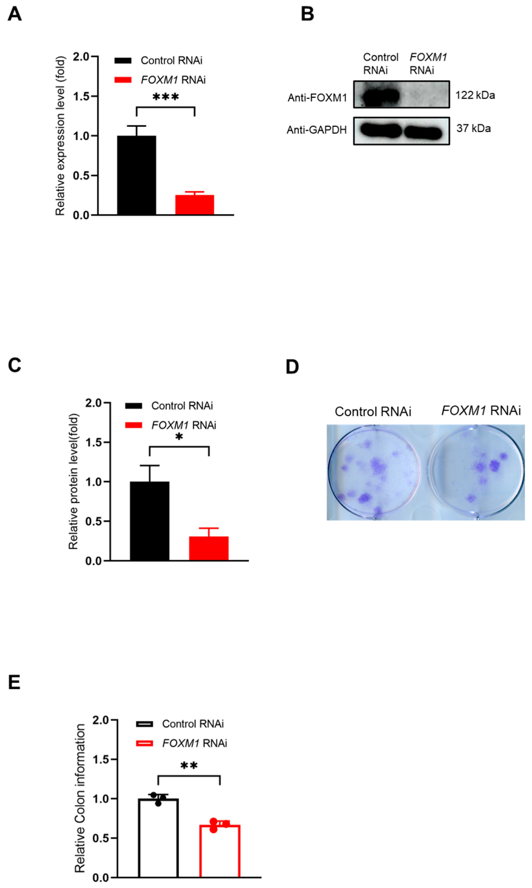
To investigate the role of FOXM1 in WJ-MSCs, we first achieved efficient knockdown by transfecting cells with FOXM1-targeting siRNA, using a non-targeting siRNA as a negative control. The success of the knockdown was confirmed at both the transcriptional and protein levels. RT-qPCR analysis showed that FOXM1 mRNA expression was reduced to less than 20% of the level in the control group (Figure 1A). Consistent with this, Western blot analysis demonstrated a significant decrease in FOXM1 protein expression (Figure 1B,C).
Having established the model, we assessed the functional impact of FOXM1 loss on proliferative capacity using a colony formation assay. The control group formed a high number of colonies, whereas the FOXM1-knockdown group exhibited a stark reduction in both the number and coverage area of colonies (Figure 1D,E, ** p < 0.01). These results demonstrate that FOXM1 is essential for the clonogenic proliferation and survival of WJ-MSCs.

3.2. Global Gene Expression Changes upon FOXM1 Knockdown

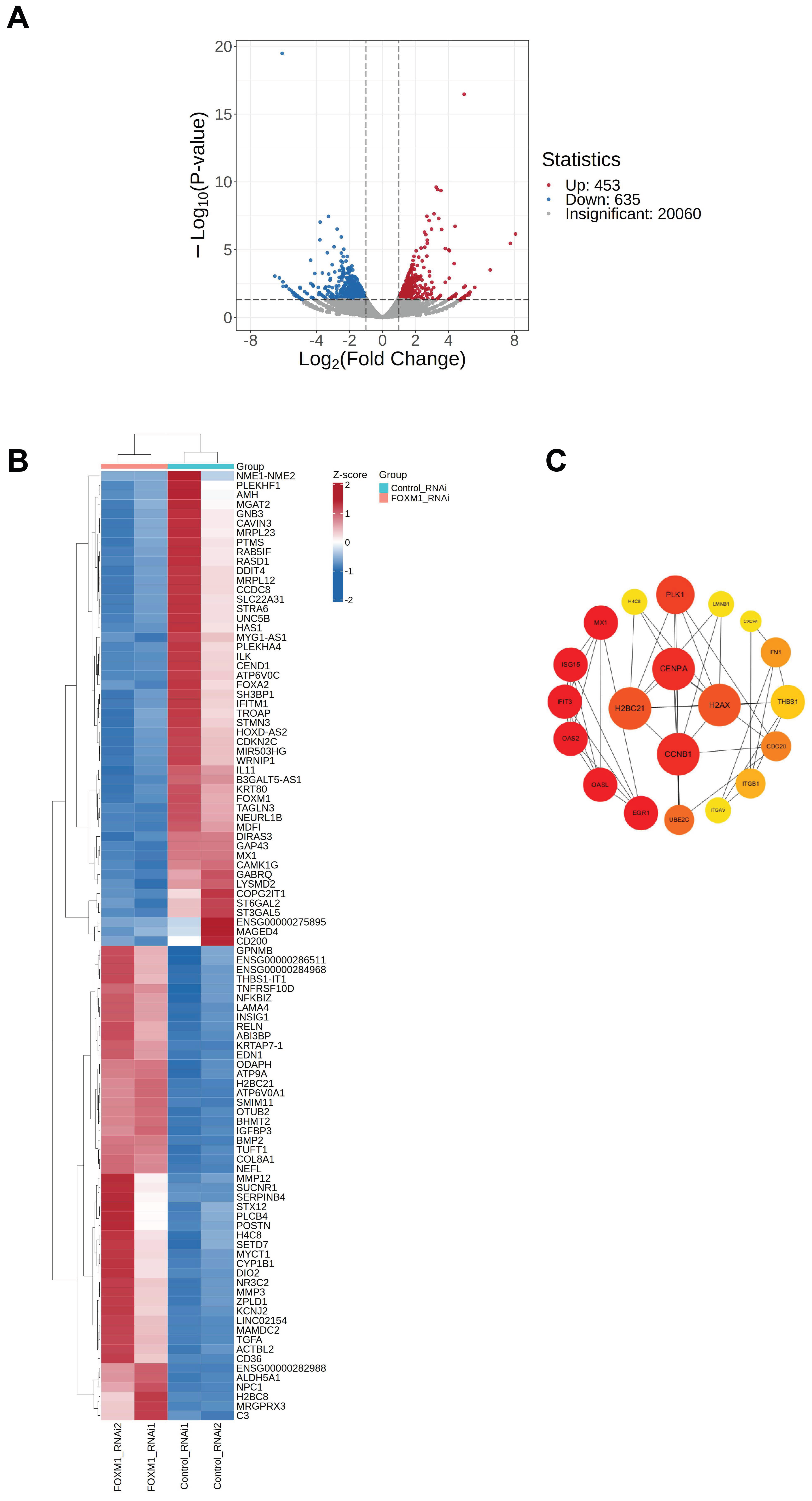
To elucidate the role of the transcription factor FOXM1 in WJ-MSC pluripotency, we performed RNA-seq analysis following FOXM1 knockdown. Using a threshold of |log2FoldChange| ≥ 1 and a p-value < 0.05, we identified 453 significantly upregulated and 635 downregulated genes (Figure 2A). Unsupervised clustering of the top 100 most significantly altered genes (50 up, 50 down) revealed a clear distinction between FOXM1-depleted and control cells (Figure 2B). Gene ontology analysis indicated that these differentially expressed genes were primarily enriched in processes including cell cycle regulation, signal transduction, and immune responses.
To identify key network nodes, we constructed a protein–protein interaction network using Cytoscape v3.10.1. The cytoHubba plugin highlighted the top 20 hub genes, which are strongly associated with the cell cycle, DNA damage repair, and immune signaling (Figure 2C). Notably, the cell cycle gene CCNB1 was among the highest-ranked hub genes, underscoring a potential central role for FOXM1 in regulating cell proliferation in WJ-MSCs.

3.3. FOXM1 Knockdown Leads to Identity Disruption in WJ-MSCs

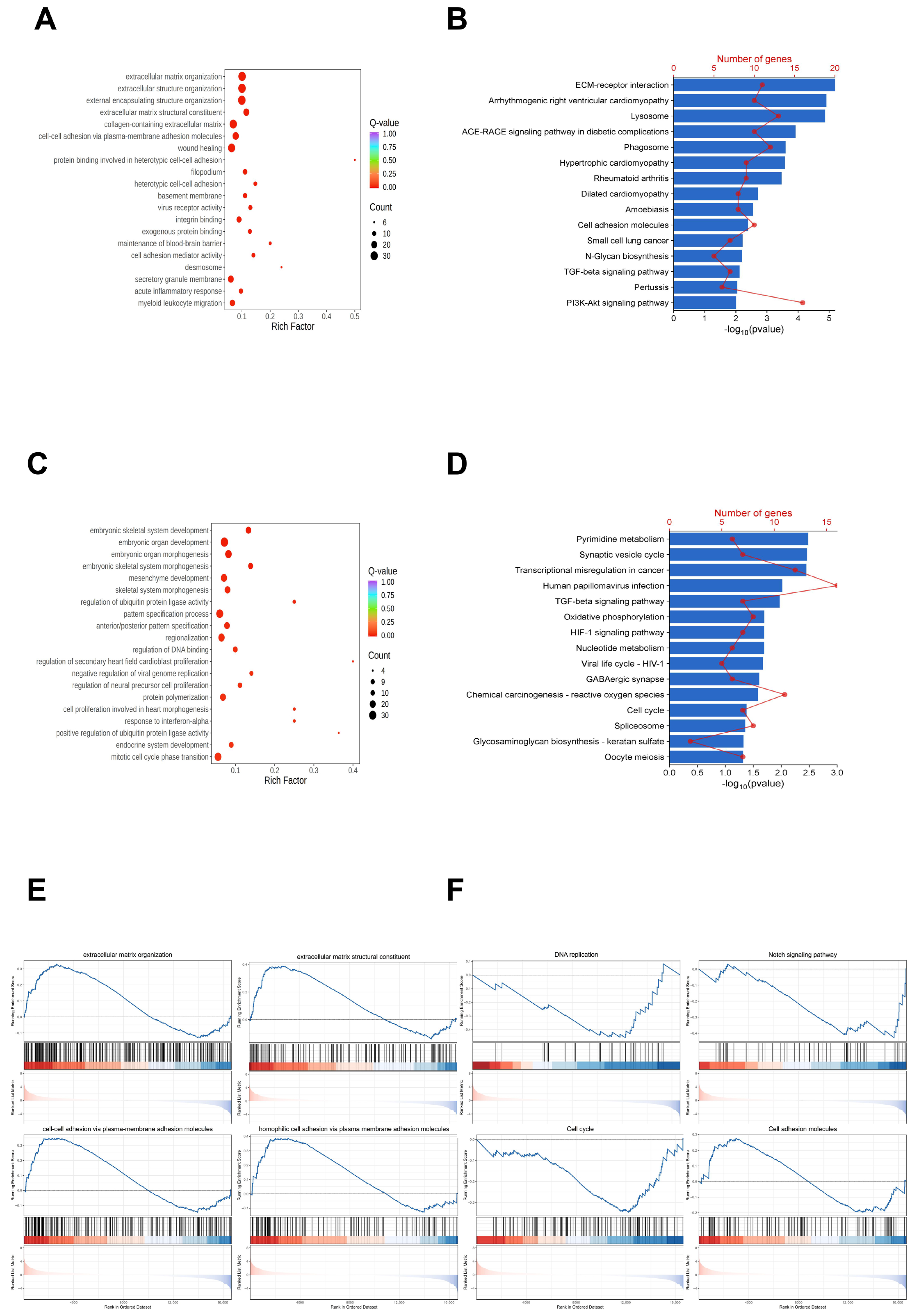
To further explore how FOXM1 knockdown affects biological functions of WJ-MSCs, we conducted Gene Ontology (GO) and KEGG analyses on both upregulated and downregulated genes. The GO analysis revealed that upregulated genes were significantly enriched in key biological processes such as extracellular matrix organization, cell adhesion, and wound healing. In contrast, the downregulated genes were primarily associated with biological processes related to embryonic development, cell proliferation and differentiation, and protein degradation and regulation (Figure 3A,C).
To gain more insights into the signaling pathways in FOXM1-knockdown WJ-MSCs, we performed KEGG analysis on the upregulated and downregulated genes. We selected the top 15 pathways with a p-value < 0.05 to indicate significant enrichment. The results showed that upregulated genes were mainly linked to pathways involving extracellular matrix remodeling, lysosomal function alterations, and mechanisms related to diabetes complications. Conversely, pathways associated with downregulated genes were primarily related to cellular metabolism, signal transduction, cell cycle regulation, and rheumatoid arthritis (Figure 3B,D). Additionally, Gene Set Enrichment Analysis (GSEA) indicated that the enrichment results were closely related to intercellular adhesion, extracellular matrix interactions, as well as signaling pathways involved in the cell cycle and apoptosis (Figure 3E,F).
To visually confirm the coordinated changes within these highlighted pathways and to provide a global view of gene expression, we generated expression heatmaps for all genes comprising the Cell Cycle (Figure S1A) and Apoptosis pathways (Figure S1B). We identified these two pathways as the most significantly enriched and phenotypically relevant pathways. The heatmaps clearly demonstrated a consistent downregulation trend across most genes in the cell cycle pathway and a concurrent upregulation trend in the apoptosis pathway. Together, these results demonstrated that FOXM1 may play multifaceted roles in WJ-MSCs.

3.4. The Important Regulatory Role of FOXM1 in WJ-MSCs

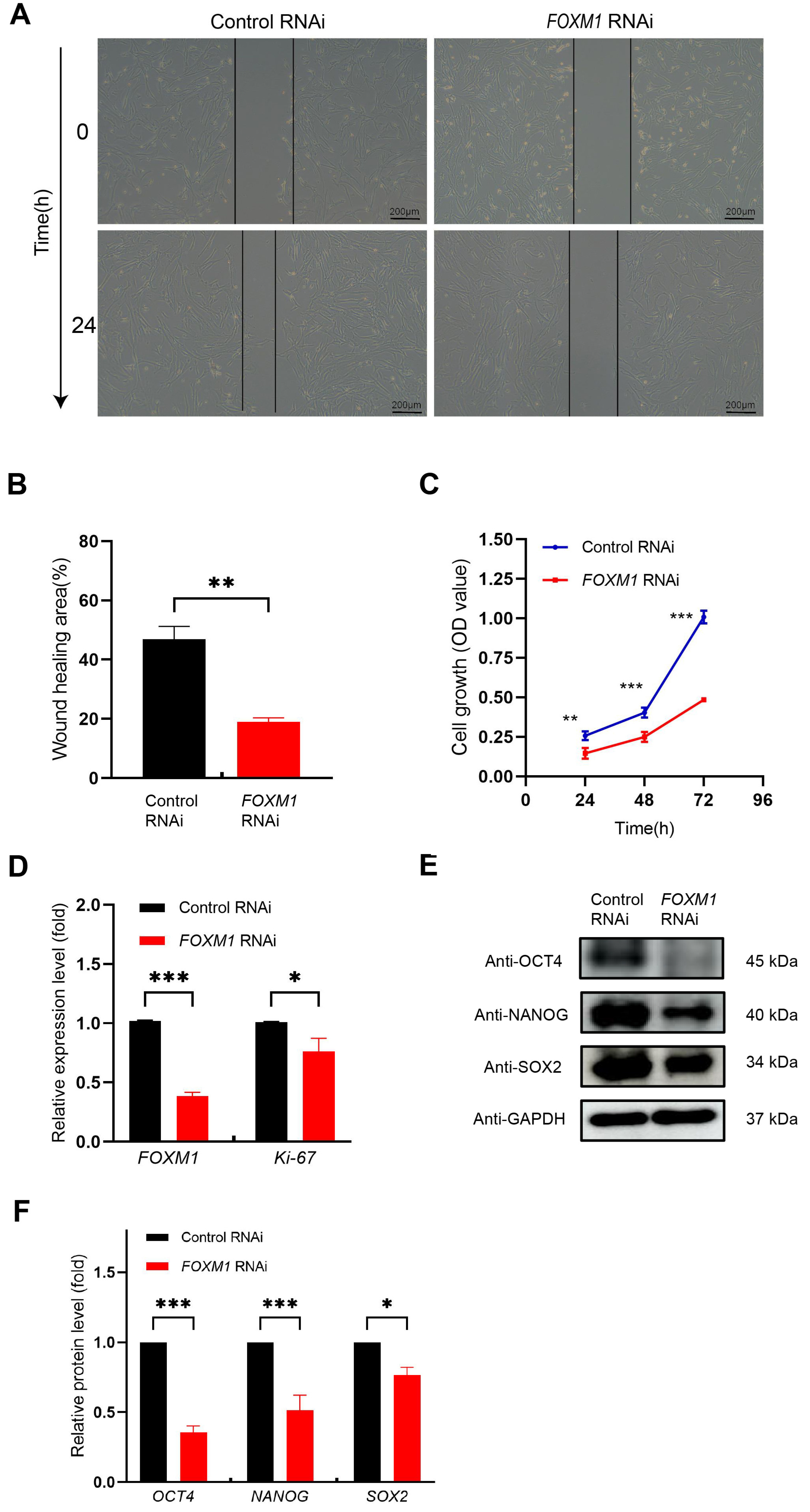
We next investigated the impact of FOXM1 knockdown on the migration, proliferation, and pluripotency of WJ-MSCs. The migratory capacity of FOXM1-knockdown cells was significantly impaired, as demonstrated by a wound healing assay showing a markedly reduced migration rate after 24 h compared to the control group (Figure 4A,B).
Consistent with this functional deficit, FOXM1 knockdown also severely compromised proliferative capacity. CCK-8 assays revealed a significant reduction in cell proliferation after 48 h (Figure 4C, *** p < 0.001). This was further supported by a concomitant decrease in the mRNA expression of both FOXM1 itself and the proliferation marker MKI67 (which encodes Ki-67) (Figure 4D, ** p < 0.001, * p < 0.05).
Furthermore, FOXM1 depletion critically impaired stem cell pluripotency. Western blot analysis showed a substantial reduction in the core pluripotency factors OCT4, NANOG, and SOX2 (Figure 4E). Densitometric quantification confirmed these decreases were statistically significant (Figure 4F). Collectively, these findings suggest that FOXM1 serves as an essential factor for maintaining the migratory, proliferative, and pluripotent properties of WJ-MSCs.

3.5. FOXM1 Knockdown Inhibits WJ-MSCs Proliferation by Regulating the Cell Cycle Process

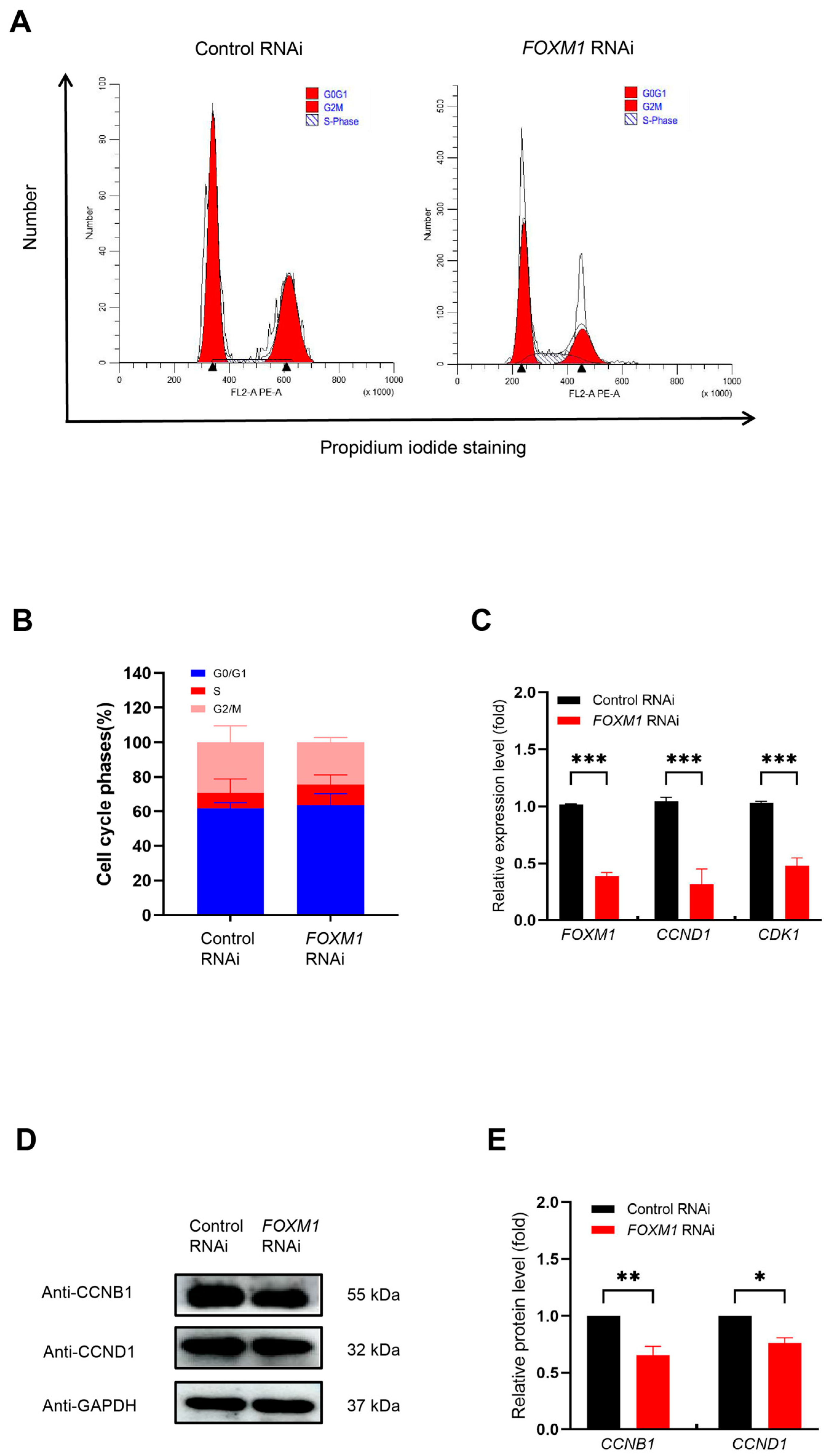
We further investigated the effects of FOXM1 knockdown on cell cycle progression in WJ-MSCs. Initially, we examined cell cycle distribution using flow cytometry. We found that FOXM1 RNAi led to an increase in the number of cells in the G0/G1 phase, along with a decrease in the number of cells in the S and G2/M phases (Figure 5A,B). These results suggest that FOXM1 has an important regulatory role in the cell cycle.
To investigate the molecular mechanisms by which FOXM1 impacts the cell cycle, we utilized RT-qPCR to measure the expression levels of key regulatory genes (CCND1 and CDK1). Our results demonstrated that FOXM1 RNAi significantly reduced CCND1 and CDK1 mRNA expression to less than 50% (Figure 5C, *** p < 0.001). These findings, aligning with the RNA-seq data, reinforced that FOXM1 may influence cell cycle progression through the regulation of these genes. Additionally, we validated the protein expression levels of the key cell cycle proteins CCNB1 and CCND1 via Western blot. Our results showed that FOXM1 RNAi significantly diminished CCNB1 and CCND1 protein levels (Figure 5D,E, ** p < 0.01). These results are consistent with the changes in gene expression and further emphasize the critical role of FOXM1 in cell cycle regulation.
In summary, our findings establish that FOXM1 sustains WJ-MSC proliferation by directly promoting cell cycle progression; its knockdown dysregulates cell cycle genes, inducing a G0/G1 arrest and halting proliferation.

3.6. FOXM1 Knockdown Promotes Apoptosis in WJ-MSCs

To validate the RNA-seq findings and further investigate FOXM1’s regulatory role, we confirmed the expression of key differentially expressed genes using RT-qPCR. The RNA-seq data had initially indicated that FOXM1 knockdown significantly altered the transcriptome, with GO and KEGG analyses linking these changes to critical processes such as cell proliferation, apoptosis, inflammatory response, and stem cell maintenance (Figure 3).
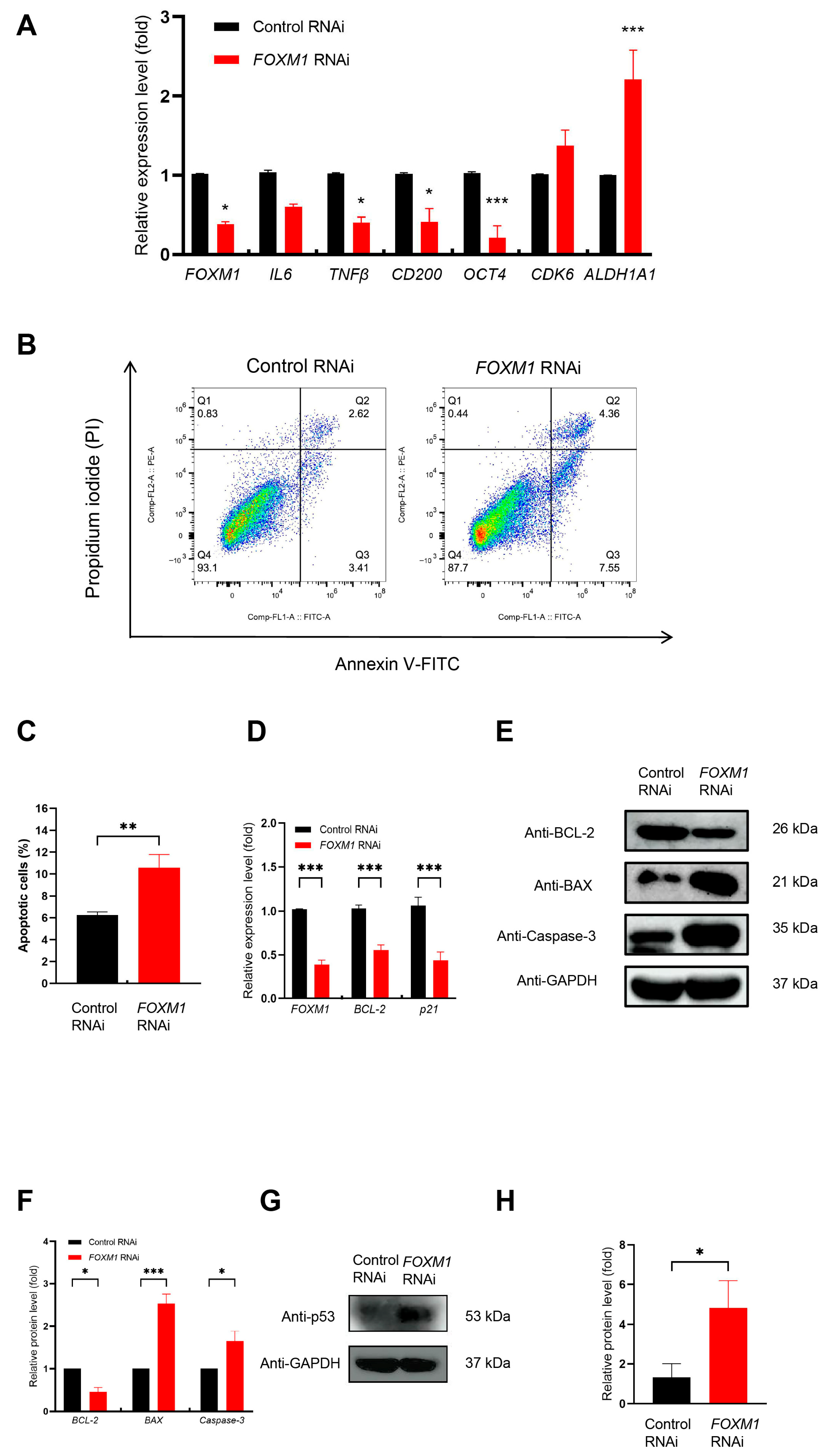
To validate the reliability of the RNA-seq results, we selected representative genes from the significantly enriched key pathways for RT-qPCR analysis. These genes included IL6, TNFβ, CD200, OCT4, CDK6, and ALDH1A1 (Figure 6A). We observed a significant downregulation of genes critical for inflammation, immune modulation (IL6, TNFβ, CD200), and stem cell identity (OCT4, ALDH1A1), alongside dysregulation of the cell cycle gene CDK6. The pronounced decrease in CD200 provides a clear validation of the sequencing data. Collectively, these results verify that FOXM1 knockdown disrupts core cellular functions, ultimately leading to the loss of stem cell characteristics in WJ-MSCs.
To determine whether the observed G0/G1 phase cell cycle arrest and inhibited proliferation (Figure 5) upon FOXM1 knockdown were coupled with an apoptotic response, we investigated its effects on apoptosis and associated mechanisms. The apoptosis pathway was prominently enriched in our KEGG analysis and displayed a global upregulation pattern (Figure S1B). Flow cytometry analysis revealed a significantly higher apoptosis rate in FOXM1-deficient cells compared to controls, with a notable increase of 5.88% (Figure 6B,C). This was supported by a significant reduction in the expression levels of BCL-2 and p21 (Figure 6D). Critically, the analysis of protein levels provided additional evidence for pathway activation. These results together indicate that the reduction in FOXM1 may promote cell death. Western blot analysis confirmed these findings, demonstrating a decrease in the anti-apoptotic protein BCL-2 and a concomitant increase in the pro-apoptotic proteins BAX and Caspase-3 (Figure 6E). Quantitative analysis of the protein levels confirmed these changes were statistically significant (Figure 6F).
To clarify the upstream mechanism by which FOXM1 deficiency triggers this apoptotic cascade, we studied the change in p53, a main regulatory factor of the intrinsic apoptosis pathway. Compared with the control group, FOXM1 knockdown significantly led to an upregulation of p53 protein expression (Figure 6G,H). This finding indicated that p53 accumulation may be associated with FOXM1 inhibition in WJ-MSCs. Collectively, these results demonstrated that FOXM1 may be a critical regulator of apoptosis in WJ-MSCs.

4. Discussion

Previous research has shown that FOXM1 plays a significant role in the proliferation [33], differentiation, and survival of various cell types [34]. However, its specific functions and molecular mechanisms in WJ-MSCs remain unclear. In this study, our data suggests that FOXM1 acts as an important transcription factor for maintaining the homeostasis of WJ-MSCs, with its knockdown impacting multiple functions of these cells. We systematically explored its potential roles in proliferation, migration, cell cycle progression, apoptosis, and stem cell pluripotency in vitro.
FOXM1 appears to support the colony-forming ability, proliferation, and migration of WJ-MSCs. Our data indicate that FOXM1 knockdown significantly impairs the colony-forming potential of WJ-MSCs, leading to reduced cell proliferation and migration. Consistent with the downregulation of CCND1 and G1 phase cell cycle arrest, CCK-8 assays demonstrated that FOXM1 knockdown significantly diminished WJ-MSC proliferation. These findings reinforce the notion that FOXM1 may be indispensable for the proliferation of WJ-MSCs. Numerous studies have shown that FOXM1 promotes cell proliferation [35] and migration [36], and support the notion that FOXM1 contributes to the proliferative capacity of WJ-MSCs.
Additionally, FOXM1 impairs the stem cell characteristics and cell cycle regulation of WJ-MSCs. OCT4, NANOG, and SOX2 are well-known core transcription factors that sustain the self-renewal and pluripotency of human pluripotent stem cells [37,38]. Our results showed that FOXM1 RNAi decreased the expression of pluripotency-related genes, including OCT4, SOX2, and NANOG. This is consistent with a previous study which has identified FOXM1 as a key target upstream of the pluripotency regulatory network, which directly binds to the OCT4 promoter to maintain the expression of pluripotency markers [39]. However, since OCT4, SOX2, and NANOG are expressed at much lower levels in WJ-MSCs, whether this direct regulatory mechanism operates in WJ-MSCs still remains to be experimentally tested. Additionally, FOXM1 acts as a transcriptional activator in cell cycle regulation, playing a significant role in the G1/S and G2/M transitions [40,41]. We hypothesize that the cell cycle arrest and proliferation defects caused by FOXM1 knockdown are a synergistic process driven by both direct and indirect mechanisms. On the direct level, FOXM1 knockdown may lead to the inactivation of its downstream targets, such as CCNB1 and CDK1, which directly impede cell cycle progression. On the indirect level, FOXM1 knockdown results in decreased expression of OCT4, whose deficiency has been shown to induce cell cycle arrest and impair proliferation [42]. Thus, the concurrent disruption of these key cellular processes upon FOXM1 knockdown demonstrates its central role in sustaining WJ-MSC homeostasis. Future studies employing promoter-binding assays and other mechanistic experiments are necessary to definitively establish if FOXM1 directly regulates those genes in WJ-MSCs.
Moreover, we also found that FOXM1 knockdown relieved the inhibition of pro-apoptotic factors such as BAX, while the subsequent upregulation of Caspase-3 marked the initiation of the apoptotic execution phase. Critically, our data show that this process is likely mediated by the tumor suppressor p53, whose protein level is significantly upregulated upon FOXM1 knockdown. As a master transcriptional regulator of apoptosis, p53 can directly transcribe and activate the apoptotic gene BAX and inhibit the expression of anti-apoptotic gene BCL-2 [43,44], thus leading to an increase in the BAX/BCL-2 ratio and initiating the apoptotic pathway. Therefore, the accumulation of p53 resulting from FOXM1 knockdown provides a logical explanation for the concurrent upregulation of BAX and downregulation of BCL-2. The resulting shift in the BAX/BCL-2 ratio ultimately leads to the activation of Caspase-3 and triggers programmed cell death. Taken together, our data supports a model in which FOXM1 acts as a negative regulator of p53 in WJ-MSCs, which in turn orchestrates the apoptotic pathway by modulating the expression of BAX and BCL-2. Collectively, our data establishes a coherent link between FOXM1 knockdown and the activation of intrinsic apoptosis in WJ-MSCs. This conclusion is supported by a definitive chain of evidence spanning three levels: initial pathway enrichment prediction, subsequent validation of key regulatory proteins, and ultimate confirmation of the apoptotic phenotype. This finding aligns with previous research establishing FOXM1 as a critical survival factor in cancer cells [25,45], where its inhibition can induce apoptosis [46]. However, the exact interplay between these processes and whether they form a feedback loop remains to be experimentally validated.
In addition to its coordinated regulation of the cell cycle, proliferation, apoptosis, and pluripotency, we have found that FOXM1 influences the biological behavior of WJ-MSCs by regulating several key genes. These genes have been found to be involved in processes such as promoting angiogenesis and immunomodulation in other contexts, such as IL6, TNFβ, CD200, OCT4, CDK6, and ALDH1A1. Therefore, maintaining the expression level of these genes may be crucial to the therapeutic effect of WJ-MSCs. Although this implies that FOXM1 may have a broader potential role in regulating the secretion and functional characteristics of WJ-MSCs, the direct experimental verification of its specific functional results still needs to be explored in future research. In general, our findings provide new mechanistic insights into how FOXM1 governs the therapeutic potential of WJ-MSCs.
This study systematically reveals the multifaceted functions of FOXM1 in WJ-MSCs through gene regulatory mechanisms, providing a vital theoretical basis for optimizing stem cell expansion therapies. Additionally, this research offers new perspectives for the fields of stem cell biology and regenerative medicine. However, this study has several limitations. First, the limitations of this study include its primary focus on in vitro experiments; the specific mechanisms of FOXM1 in vivo still require further investigation. Second, all experiments use single commercial source cell lines. Although this will avoid the complex interference caused by heterogeneity between donors, it may not be universal. Third, while we observed changes in gene expression following FOXM1 knockdown, Chromatin immunoprecipitation using FOXM1 antibody to validate the direct binding of FOXM1 to the genomic sites of its downstream genes is required to confirm the direct transcriptional regulatory role of FOXM1. Fourth, the functional consequences of altered genes (IL6, CD200, ALDH1A1) were not experimentally validated in therapeutic models. Fifth, future studies can include TUNEL staining or Caspase activity assays which are important to confirm and quantify the apoptosis induced by FOXM1 knockdown. It is also important to verify these findings in vivo.

5. Conclusions

In summary, our study establishes FOXM1 as a master regulator of WJ-MSC homeostasis. We demonstrate that it sustains proliferation by promoting cell cycle progression and concurrently suppressing apoptosis and stress signals. Consequently, FOXM1 depletion critically impairs cellular expansion, underscoring its vital, non-redundant role in maintaining the integrity and function of the WJ-MSC population.

Supplementary Materials

The following supporting information can be downloaded at: https://www.mdpi.com/article/10.3390/genes16121517/s1, Figure S1: Key Pathways Altered by FOXM1 Knockdown. Table S1: Primer sequences of target genes in qPCR. Table S2. Primary and secondary antibodies being used.

Author Contributions

Conceptualization, Q.W.; methodology, N.L. and Q.W.; software, N.L.; validation, N.L. and Q.W.; formal analysis, N.L.; investigation, N.L.; resources, Q.W.; data curation, N.L.; writing—original draft preparation, N.L.; writing—review and editing, Q.W.; visualization, Q.W.; supervision, Q.W.; project administration, Q.W.; funding acquisition, Q.W. All authors have read and agreed to the published version of the manuscript.

Funding

This research was funded by the Macau Science and Technology Development Fund (File no. 006/2023/SKL).

Institutional Review Board Statement

Not applicable.

Informed Consent Statement

Not applicable.

Data Availability Statement

The original contributions presented in this study were included in the article/Supplementary Materials. Further inquiries can be directed to the corresponding author.

Conflicts of Interest

The authors declare no conflicts of interest.

Abbreviations

The following abbreviations are used in this manuscript.
WJ-MSCWharton’s jelly mesenchymal stem cell
MSCMesenchymal stem cells
FOXM1Forkhead box protein M1
RT-qPCRReal-Time Quantitative PCR
PIPropidium iodide
KEGGKyoto Encyclopedia of Genes and Genomes
GOGene Ontology

References

  1. Desgres, M.; Menasché, P. Clinical Translation of Pluripotent Stem Cell Therapies: Challenges and Considerations. Cell Stem Cell 2019, 25, 594–606. [Google Scholar] [CrossRef] [PubMed]
  2. Liu, J.; Ding, Y.; Liu, Z.; Liang, X.-T. Senescence in Mesenchymal Stem Cells: Functional Alterations, Molecular Mechanisms, and Rejuvenation Strategies. Front. Cell Dev. Biol. 2020, 8, 258. [Google Scholar] [CrossRef]
  3. Kou, M.; Huang, L.; Yang, J.; Chiang, Z.; Chen, S.-Y.; Liu, J.; Guo, L.; Zhang, X.; Zhou, X.; Xu, X.; et al. Mesenchymal stem cell-derived extracellular vesicles for immunomodulation and regeneration: A next generation therapeutic tool? Cell Death Dis. 2022, 13, 580. [Google Scholar] [CrossRef] [PubMed]
  4. Stefańska, K.; Ożegowska, K.; Hutchings, G.; Popis, M.; Moncrieff, L.; Dompe, C.; Janowicz, K.; Pieńkowski, W.; Gutaj, P.; Shibli, J.; et al. Human Wharton’s Jelly—Cellular Specificity, Stemness Potency, Animal Models, and Current Application in Human Clinical Trials. J. Clin. Med. 2020, 9, 1102. [Google Scholar] [CrossRef]
  5. Huang, M.; Liao, X.; Wang, X.; Qian, Y.; Zhang, W.; Chen, G.; Wu, Q. POZ/BTB and AT hook containing zinc finger 1 (PATZ1) suppresses differentiation and regulates metabolism in human embryonic stem cells. Int. J. Biol. Sci. 2024, 20, 1142–1159. [Google Scholar] [CrossRef] [PubMed]
  6. El-Khateeb, D.; Moemen, Y.; El-Fert, A.; Elgazzar, M.; Khalil, A. Characterization of mesenchymal stem cells isolated from Wharton’s jelly of the human umbilical cord. Egypt. Liver J. 2022, 12, 2. [Google Scholar] [CrossRef]
  7. Panwar, U.; Mishra, K.; Patel, P.; Bharadva, S.; Vaniawala, S.; Shah, A.; Vundinti, B.; Kothari, S.; Ghosh, K. Assessment of Long-Term in vitro Multiplied Human Wharton’s Jelly-Derived Mesenchymal Stem Cells prior to Their Use in Clinical Administration. Cells Tissues Organs 2021, 210, 239–249. [Google Scholar] [CrossRef]
  8. Stefańska, K.; Němcová, L.; Blatkiewicz, M.; Pieńkowski, W.; Ruciński, M.; Zabel, M.; Mozdziak, P.; Podhorska-Okołów, M.; Dzięgiel, P.; Kempisty, B. Apoptosis Related Human Wharton’s Jelly-Derived Stem Cells Differentiation into Osteoblasts, Chondrocytes, Adipocytes and Neural-like Cells—Complete Transcriptomic Assays. Int. J. Mol. Sci. 2023, 24, 23. [Google Scholar] [CrossRef]
  9. Stefańska, K.; Němcová, L.; Blatkiewicz, M.; Żok, A.; Kaczmarek, M.; Pieńkowski, W.; Mozdziak, P.; Piotrowska-Kempisty, H.; Kempisty, B. Expression Profile of New Marker Genes Involved in Differentiation of Human Wharton’s Jelly-Derived Mesenchymal Stem Cells into Chondrocytes, Osteoblasts, Adipocytes and Neural-like Cells. Int. J. Mol. Sci. 2023, 24, 12939. [Google Scholar] [CrossRef]
  10. Ababneh, N.; Al-Kurdi, B.; Jamali, F.; Awidi, A. A comparative study of the capability of MSCs isolated from different human tissue sources to differentiate into neuronal stem cells and dopaminergic-like cells. PeerJ 2022, 10, 13003. [Google Scholar] [CrossRef]
  11. Zhang, Y.-N.; Lie, P.-C.; Wei, X. Differentiation of mesenchymal stromal cells derived from umbilical cord Wharton’s jelly into hepatocyte-like cells. Cytotherapy 2009, 11, 548–558. [Google Scholar] [CrossRef]
  12. Xiao, X.; Zhang, M.; Qian, Y.; Wang, X.; Wu, Q. KLF9 regulates osteogenic differentiation of mesenchymal stem cells. J. Mol. Histol. 2024, 55, 503–512. [Google Scholar] [CrossRef]
  13. Boissel, L.; Tuncer, H.H.; Betancur, M.; Wolfberg, A.; Klingemann, H. Umbilical cord mesenchymal stem cells increase expansion of cord blood natural killer cells. Biol. Blood Marrow Transplant. 2008, 14, 1031–1038. [Google Scholar] [CrossRef]
  14. Secco, M.; Zucconi, E.; Vieira, N.M.; Fogaça, L.L.; Cerqueira, A.; Carvalho, M.D.F.; Jazedje, T.; Okamoto, O.K.; Muotri, A.R.; Zatz, M. Multipotent stem cells from umbilical cord: Cord is richer than blood! Stem Cells 2008, 26, 146–150. [Google Scholar] [CrossRef]
  15. Troyer, D.L.; Weiss, M.L. Concise review: Wharton’s jelly-derived cells are a primitive stromal cell population. Stem Cells 2008, 26, 591–599. [Google Scholar] [CrossRef]
  16. Dalton, S. Linking the Cell Cycle to Cell Fate Decisions. Trends Cell Biol. 2015, 25, 592–600. [Google Scholar] [CrossRef] [PubMed]
  17. Singh, A.M.; Dalton, S. The cell cycle and Myc intersect with mechanisms that regulate pluripotency and reprogramming. Cell Stem Cell 2009, 5, 141–149. [Google Scholar] [CrossRef] [PubMed]
  18. Reya, T.; Morrison, S.J.; Clarke, M.F.; Weissman, I.L. Stem cells, cancer, and cancer stem cells. Nature 2001, 414, 105–111. [Google Scholar] [CrossRef] [PubMed]
  19. Kalathil, D.; John, S.; Nair, A. FOXM1 and Cancer: Faulty Cellular Signaling Derails Homeostasis. Front. Oncol. 2021, 10, 626836. [Google Scholar] [CrossRef]
  20. Li, Y.; Zhang, S.; Huang, S. FoxM1: A potential drug target for glioma. Future Oncol. 2012, 8, 223–226. [Google Scholar] [CrossRef]
  21. Fischer, M.; Schade, A.; Branigan, T.; Müller, G.; Decaprio, J. Coordinating gene expression during the cell cycle. Trends Biochem. Sci. 2022, 47, 1009–1022. [Google Scholar] [CrossRef]
  22. Liu, J.; Li, J.; Wang, K.; Liu, H.; Sun, J.; Zhao, X.; Yu, Y.; Qiao, Y.; Wu, Y.; Zhang, X.; et al. Aberrantly high activation of a FoxM1–STMN1 axis contributes to progression and tumorigenesis in FoxM1-driven cancers. Signal Transduct. Target. Ther. 2021, 6, 42. [Google Scholar] [CrossRef]
  23. Higurashi, M.; Maruyama, T.; Nogami, Y.; Ishikawa, F.; Yoshida, Y.; Mori, K.; Fujita, K.-I.; Shibanuma, M. High expression of FOXM1 critical for sustaining cell proliferation in mitochondrial DNA-less liver cancer cells. Exp. Cell Res. 2020, 389, 111889. [Google Scholar] [CrossRef]
  24. Zheng, Q.; Xu, M.; Ye, S.; Lei, Y.; Xi, Y. HMGA1 and FOXM1 Cooperate to Promote G2/M Cell Cycle Progression in Cancer Cells. Life 2023, 13, 1225. [Google Scholar] [CrossRef]
  25. Hou, Y.; Li, W.; Sheng, Y.; Li, L.; Huang, Y.; Zhang, Z.; Zhu, T.; Peace, D.; Quigley, J.G.; Wu, W.; et al. The transcription factor Foxm1 is essential for the quiescence and maintenance of hematopoietic stem cells. Nat. Immunol. 2015, 16, 810–818. [Google Scholar] [CrossRef]
  26. Kim, I.M.; Ackerson, T.; Ramakrishna, S.; Tretiakova, M.; Wang, I.C.; Kalin, T.V.; Major, M.L.; Gusarova, G.A.; Yoder, H.M.; Costa, R.H.; et al. The Forkhead Box m1 transcription factor stimulates the proliferation of tumor cells during development of lung cancer. Cancer Res. 2006, 66, 2153–2161. [Google Scholar] [CrossRef]
  27. Kwok, J.M.; Peck, B.; Monteiro, L.J.; Schwenen, H.D.; Millour, J.; Coombes, R.C.; Myatt, S.S.; Lam, E.W. FOXM1 confers acquired cisplatin resistance in breast cancer cells. Mol. Cancer Res. 2010, 8, 24–34. [Google Scholar] [CrossRef]
  28. Wang, D.; Hu, G.; Du, Y.; Zhang, C.; Lu, Q.; Lv, N.; Luo, S. Aberrant activation of hedgehog signaling promotes cell proliferation via the transcriptional activation of forkhead Box M1 in colorectal cancer cells. J. Exp. Clin. Cancer Res. 2017, 36, 23. [Google Scholar] [CrossRef]
  29. Zhang, S.; Zhao, B.S.; Zhou, A.; Lin, K.; Zheng, S.; Lu, Z.; Chen, Y.; Sulman, E.P.; Xie, K.; Bögler, O.; et al. m6A Demethylase ALKBH5 Maintains Tumorigenicity of Glioblastoma Stem-like Cells by Sustaining FOXM1 Expression and Cell Proliferation Program. Cancer Cell 2017, 31, 591–606.e596. [Google Scholar] [CrossRef]
  30. Sawaya, A.P.; Stone, R.C.; Mehdizadeh, S.; Pastar, I.; Worrell, S.; Balukoff, N.C.; Kaplan, M.J.; Tomic-Canic, M.; Morasso, M.I. FOXM1 network in association with TREM1 suppression regulates NET formation in diabetic foot ulcers. EMBO Rep. 2022, 23, e54558. [Google Scholar] [CrossRef]
  31. Wang, I.-C.; Zhang, Y.; Snyder, J.; Sutherland, M.J.; Burhans, M.S.; Shannon, J.M.; Park, H.J.; Whitsett, J.A.; Kalinichenko, V.V. Increased expression of FoxM1 transcription factor in respiratory epithelium inhibits lung sacculation and causes Clara cell hyperplasia. Dev. Biol. 2010, 347, 301–314. [Google Scholar] [CrossRef]
  32. Bella, L.; Zona, S.; Nestal de Moraes, G.; Lam, E.W.F. FOXM1: A key oncofoetal transcription factor in health and disease. Semin. Cancer Biol. 2014, 29, 32–39. [Google Scholar] [CrossRef]
  33. Wierstra, I.; Alves, J. FOXM1, a typical proliferation-associated transcription factor. Biol. Chem. 2007, 388, 1257. [Google Scholar] [CrossRef]
  34. Wierstra, I. The transcription factor FOXM1 (Forkhead box M1): Proliferation-specific expression, transcription factor function, target genes, mouse models, and normal biological roles. Adv. Cancer Res. 2013, 118, 97–398. [Google Scholar]
  35. Wang, W.; Su, L.; Ji, F.; Zhang, D.; Wang, Y.; Zhao, J.; Jiao, R.D.; Zhang, M.; Huang, E.; Jiang, H.; et al. The human FOXM1 homolog promotes basal progenitor cell proliferation and cortical folding in mouse. EMBO Rep. 2022, 23, e53602. [Google Scholar] [CrossRef]
  36. Chen, Y.; Liu, Y.; Ni, H.; Ding, C.; Zhang, X.; Zhang, Z. FoxM1 overexpression promotes cell proliferation and migration and inhibits apoptosis in hypopharyngeal squamous cell carcinoma resulting in poor clinical prognosis. Int. J. Oncol. 2017, 51, 1045–1054. [Google Scholar] [CrossRef]
  37. Boyer, L.A.; Lee, T.I.; Cole, M.F.; Johnstone, S.E.; Levine, S.S.; Zucker, J.P.; Guenther, M.G.; Kumar, R.M.; Murray, H.L.; Jenner, R.G. Core transcriptional regulatory circuitry in human embryonic stem cells. Cell 2005, 122, 947–956. [Google Scholar] [CrossRef]
  38. Loh, Y.-H.; Wu, Q.; Chew, J.-L.; Vega, V.B.; Zhang, W.; Chen, X.; Bourque, G.; George, J.; Leong, B.; Liu, J. The Oct4 and Nanog transcription network regulates pluripotency in mouse embryonic stem cells. Nat. Genet. 2006, 38, 431–440. [Google Scholar] [CrossRef]
  39. Xie, Z.; Tan, G.; Ding, M.; Dong, D.; Chen, T.; Meng, X.; Huang, X.; Tan, Y. Foxm1 transcription factor is required for maintenance of pluripotency of P19 embryonal carcinoma cells. Nucleic Acids Res. 2010, 38, 8027–8038. [Google Scholar] [CrossRef]
  40. Down, C.F.; Millour, J.; Lam, E.W.F.; Watson, R.J. Binding of FoxM1 to G2/M gene promoters is dependent upon B-Myb. Biochim. Et Biophys. Acta (BBA)—Gene Regul. Mech. 2012, 1819, 855–862. [Google Scholar] [CrossRef]
  41. Chen, X.; Müller, G.A.; Quaas, M.; Fischer, M.; Han, N.; Stutchbury, B.; Sharrocks, A.D.; Engeland, K. The forkhead transcription factor FOXM1 controls cell cycle-dependent gene expression through an atypical chromatin binding mechanism. Mol. Cell Biol. 2013, 33, 227–236. [Google Scholar] [CrossRef]
  42. Picot, T.; Kesr, S.; Wu, Y.; Aanei, C.M.; Flandrin-Gresta, P.; Tondeur, S.; Tavernier, E.; Wattel, E.; Guyotat, D.; Campos, L. Potential Role of OCT4 in Leukemogenesis. Stem Cells Dev. 2017, 26, 1637–1647. [Google Scholar] [CrossRef]
  43. Hao, Q.; Chen, J.; Lu, H.; Zhou, X. The ARTS of p53-dependent mitochondrial apoptosis. J. Mol. Cell Biol. 2022, 14, mjac074. [Google Scholar] [CrossRef]
  44. Chipuk, J.E.; Kuwana, T.; Bouchier-Hayes, L.; Droin, N.M.; Newmeyer, D.D.; Schuler, M.; Green, D.R. Direct Activation of Bax by p53 Mediates Mitochondrial Membrane Permeabilization and Apoptosis. Science 2004, 303, 1010–1014. [Google Scholar] [CrossRef]
  45. Sheng, Y.; Yu, C.; Liu, Y.; Hu, C.; Ma, R.; Lu, X.; Ji, P.; Chen, J.; Mizukawa, B.; Huang, Y.; et al. FOXM1 regulates leukemia stem cell quiescence and survival in MLL-rearranged AML. Nat. Commun. 2020, 11, 928. [Google Scholar] [CrossRef]
  46. Bhat, U.G.; Halasi, M.; Gartel, A.L. Thiazole antibiotics target FoxM1 and induce apoptosis in human cancer cells. PLoS ONE 2009, 4, e5592. [Google Scholar] [CrossRef] [PubMed]
Figure 1. FOXM1 depletion affects the clonogenicity of WJ-MSCs. (A) FOXM1 gene expression in WJ-MSCs was assessed by RT-qPCR. (B) FOXM1 protein expression levels were assessed by Western blot. (C) Protein quantification of FOXM1. (D) Clony formation assay using crystal violet staining. (E) FOXM1 RNAi significantly reduced the area of colony coverage. * p < 0.05, ** p < 0.01, and *** p < 0.001.
Genes 16 01517 g001
Figure 2. Analysis of RNA-seq results following FOXM1 knockdown. (A) Visualization of RNA-seq results with Volcano plot. (B) Visualization of differentially expressed genes with Heatmap (red color for up-regulation and blue color for down-regulation). (C) A deeper red color of the Hub gene indicates a higher score.
Genes 16 01517 g002
Figure 3. FOXM1-related pathways were identified by differential gene enrichment analysis. (A) GO enrichment for up-regulated genes. (B) The top 15 KEGG pathways enriched by up-regulated genes. (C) GO enrichment for down-regulated genes. (D) The top 15 KEGG pathways enriched by down-regulated genes. (E) GO analysis of GSEA. (F) KEGG analysis of GSEA.
Genes 16 01517 g003
Figure 4. Knockdown of FOXM1 inhibits WJ-MSC migration, proliferation, and pluripotency. (A) Wound healing of WJ-MSCs treated with control RNAi and FOXM1 RNAi after scratch wounding. (B) Quantification of wound distance represented the results of three independent experiments. (C) Cell proliferation was assessed using the CCK8 assay. Each time point in each row was n = 5. (D) Ki-67 expression was measured using RT-qPCR. (E) Western blot analysis was used to assess pluripotency protein expression. (F) Quantitative analysis of pluripotency protein expression was performed. * p < 0.05, ** p < 0.01, and *** p < 0.001.
Genes 16 01517 g004
Figure 5. Knockdown of FOXM1 affects cell cycle progression. (A) Cell cycle progression was examined by flow cytometry. (B) The relative expression levels at different stages of the cell cycle were displayed using stacked plots. (C) RT-qPCR was used to detect the relative expression levels of CCND1 and CDK1 genes. (D) The relative expression of cell cycle proteins was determined by Western blotting. (E) Quantification of cell cycle proteins. * p < 0.05, ** p < 0.01, and *** p < 0.001.
Genes 16 01517 g005
Figure 6. FOXM1 knockdown affects gene expression in multiple biological processes and promotes cell apoptosis. (A) Relative gene expression changes in IL6, TNFβ, CD200, OCT4, CDK6, and ALDH1A1 upon FOXM1 RNAi. (B) Apoptosis caused by FOXM1 knockdown was assessed by flow cytometry using Annexin V-FITC/PI staining. (C) After FOXM1 knockdown, apoptotic cells from three independent experiments were analyzed by FlowJo, and the results were shown in a bar chart. (D) RT-qPCR was used to detect the changes in BCL-2 and p21 gene expression. (E) The relative expression of apoptotic proteins was detected by Western blotting. (F) Quantification of apoptotic proteins. (G). The relative expression of p53 protein was detected by Western blotting. (H) Quantification of the p53 protein. * p < 0.05, ** p < 0.01, and *** p < 0.001.
Genes 16 01517 g006
Disclaimer/Publisher’s Note: The statements, opinions and data contained in all publications are solely those of the individual author(s) and contributor(s) and not of MDPI and/or the editor(s). MDPI and/or the editor(s) disclaim responsibility for any injury to people or property resulting from any ideas, methods, instructions or products referred to in the content.