Article

Heat Shock Protein 104 (Hsp104) in the Marine Diatom Ditylum brightwellii: Identification and Transcriptional Responses to Environmental Stress

1
Department of Life Science, Sangmyung University, Seoul 03016, Republic of Korea
2
Institute of Natural Science, Sangmyung University, Seoul 03016, Republic of Korea
*
Author to whom correspondence should be addressed.
These authors contributed equally to this work.
Genes 2025, 16(12), 1408; https://doi.org/10.3390/genes16121408
Submission received: 11 November 2025 / Revised: 21 November 2025 / Accepted: 24 November 2025 / Published: 26 November 2025
(This article belongs to the Section Genes & Environments)

Abstract

Backgrounds: The marine diatom Ditylum brightwellii has been widely used as a model species for ecotoxicological assessments in marine environments. Heat shock proteins (Hsps) function as molecular chaperones that protect cells under diverse stress conditions. Of them, Hsp104 participates in the protein restoration system by reversing protein aggregation. Methods: In the present study, we determined the full-length sequence of DbHsp104 in D. brightwellii using transcriptome sequencing and gene cloning. Results: The open reading frame (ORF) was 2745 bp in length, encoding a protein of 915 amino acids (101.15 kDa). Phylogenetic and domain structural analysis revealed that DbHsp104 possesses conserved features of eukaryotic Hsp104. In addition, transcriptional responses of the gene were evaluated after exposures to thermal stress at 20, 25, and 30 °C, and heavy metals and endocrine-disrupting chemicals (EDCs) for 24 h. Relative gene expression analysis showed that DbHsp104 was significantly up-regulated under thermal stress and copper exposures, peaking at 4.87- and 5.55-fold (p < 0.001) increases, respectively. In contrast, no significant changes were observed in response to nickel, bisphenol A (BPA), polychlorinated biphenyl (PCB), and endosulfan (EDS) treatments. Conclusions: These results suggest that DbHsp104 is specifically responsive to acute stress induced by thermal stress and copper, highlighting its potential as a molecular biomarker in marine environments.

1. Introduction

Diatoms are unicellular phytoplankton belonging to the group Bacillariophyta, accounting for approximately 40% of total primary production in marine ecosystems [1,2]. They are abundant in coastal ecosystems and play essential roles in ecological processes, particularly through their involvement in the global biogeochemical cycles of carbon, nitrogen, and silicon [3]. Moreover, their rapid cell growth leads to their widespread global distribution and sensitive reactions to environmental fluctuations [4]. Collectively, these features underscore the ecological significance of diatoms and their adaptability to diverse environmental conditions.
Due to their broad occurrence and ecological significance, diatoms are recognized as important biological indicators for monitoring both short-term and long-term environmental changes [5]. They have become an integral part of laboratory-based toxicity tests, ecological risk assessments, the development of water quality guidelines, and even investigations into the bioavailability and impacts of pollutants [6]. Over the past three decades, diatoms have been extensively employed in aquatic risk assessments, demonstrating their ongoing relevance and utility for environmental monitoring purposes [7,8,9,10]. Changes in their population structure, morphology, or molecular traits can indicate early ecological stress, highlighting their suitability as test organisms in ecotoxicity assessments [11].
When exposed to various chemical contaminants, diatoms show significant changes in an array of physiological, morphological, and molecular responses [12,13,14,15]. In detail, heavy metals and organic pollutants decrease cell growth, chlorophyll, and photosynthetic efficiency and alter antioxidant enzyme activities [16,17,18]. Such ecotoxicological disturbances often modulate stress-responsive genes, such as those encoding antioxidant enzymes, and molecular chaperones such as heat shock proteins (Hsps) [19,20,21,22]. Notably, Hsps are induced by a wide range of environmental stresses such as changes in temperature and salinity, and organic and inorganic pollutants [20,21,22,23]. When cells are damaged, Hsps function as chaperones that promote protein folding, transport, degradation, and stabilization, thus preserving cellular proteostasis [20]. Based on their molecular weight and sequence homology, they are classified into five major families: small Hsps (sHsps), Hsp60, Hsp70, Hsp90, and Hsp100 [24]. Among those, Hsp104 is a member of the Hsp104/ClpB family, which functions as a disaggregase [25,26].
Hsp104/Clp is a hexameric AAA+ ATPase and proteolytic enzyme that has been identified primarily in bacteria, plants, and yeast [27]. In this regard, this enzyme participates in maintaining cellular proteostasis, particularly under stress conditions that promote protein denaturation and aggregation [28,29,30]. Hsp104 has been observed to form asymmetric ring-shaped structures containing multi-domains [31]. In detail, the N-terminal domain (NTD) contains two ATPases associated with various cellular activities (AAA+) and two types of nucleotide-binding domains (NBD), termed NBD1 and NBD2, while the coiled-coil middle domain (MD), which mediates interaction with Hsp70, is inserted at the C-terminal end of NBD1 [32,33,34]. In contrast to ClpB, Hsp104 contains an additional short extension at the C-terminal end, which is utilized for hexamerisation [35].
The Hsp104/ClpB family has been extensively studied in bacteria, plants, and yeast [36,37,38], but research on the gene expression remains limited in diatoms. In the present study, we determined the full-length sequence of Hsp104 from the marine diatom Ditylum brightwellii (DbHsp104). Also, we characterized its structural motifs and analyzed its phylogenetic relatedness to other forms of Hsp104. In addition, we evaluated the transcriptional responses of DbHsp104 under diverse stress conditions, including heat stress, and under the influence of diverse heavy metals and endocrine-disrupting chemicals (EDCs). Subsequently, based on the transcriptional responses of Hsp104 to stress and toxicant exposure, we evaluated the biomarker potential of DbHsp104. The test species D. brightwellii has been used as a model for ecotoxicology research for the past 40 years [7,39].

2. Materials and Methods

2.1. Cell Culture

A diatom strain (B-326) of D. brightwellii was obtained from the Korean Marine Microalgae Culture Center (KMMCC, Pukyung National University, Busan, Korea). It was cultured in f/2 medium [40] and maintained at 20 °C in a 12 h:12 h light:dark cycle with a photon flux density of approximately 65 μmol photons m−2 s−1.

2.2. RNA Extraction

For the RNA extraction, D. brightwellii cells were physically broken by homogenization using a mini-bead beater and freeze–thawing in liquid nitrogen (BioSpec Products Inc., Bartlesville, OK, USA). Total mRNA was extracted following the manufacturer’s instructions and further purified using the Mini Spin Columns from the RNeasy Mini Kit (Qiagen, Valencia, CA, USA). The extracted RNA quantity and quality were measured using a Nanoready F-1100 (Life Real, Hangzhou, China). Qualified RNA samples were used for de novo RNA transcriptome analysis and complementary DNA (cDNA) synthesis for further molecular experiments.

2.3. cDNA Library Construction and Transcriptome Analysis

A cDNA library of D. brightwellii was constructed using a TruSeq Stranded mRNA Library Prep Kit (San Diego, CA, USA) and was sequenced using an Illumina Hiseq 2500 System (2 × 150 bp). The raw sequences were analyzed using tools from the Galaxy server. In detail, a quality evaluation of the raw reads was conducted using FastQC (v0.74+galaxy1). Sequencing adapters and low-quality bases in the raw reads were trimmed using Trimmomatic (v0.39+galaxy2), and the resulting high-quality reads were assembled using Trinity (v2.15.1+galaxy1). Assembled contigs were translated into amino acid (aa) sequences through TransDecoder. LongOrfs (v5.5.0+galaxy2), and the sequences were identified through Diamond (v2.1.11+galaxy0) against the NCBI non-redundant (NR) database. By this process, identified genes and enzymes with an E value over 1.0 × 10⁻¹⁵ were cut off. Within the analyzed transcriptome of D. brightwellii, the sequence of DbHsp104 was selected with keywords of ‘Hsp104’ or ‘Clp family’.

2.4. cDNA Synthesis, DbHsp104 Cloning, and Gene Expression Analysis

Reverse transcription was performed to synthesize cDNA using a TOPscriptTM cDNA Synthesis Kit (Enzynomics, Daejeon, Korea) with 1 μg of total RNA, random hexamers, and oligo (dT)18. Random hexamer was added for 5′-rapid amplification of cDNA ends (RACE) PCR and quantitative-real time PCR (qRT-PCR), and oligo (dT)18 was added for 3′-RACE PCR (final volume 20 μL).
The complete ORF sequence of DbHsp104 was determined by the RACE PCR and subsequent sequencing. Specific sets of primers were designed through Primer-BLAST (https://www.ncbi.nlm.nih.gov/tools/primer-blast/, assessed on 5 May 2025). Only primer pairs with amplification efficiencies between 90% and 110% were used for analysis (Table 1). PCR amplification was performed in 20 μL of reaction mix, containing 2 μL of 10× Ex Taq buffer (TaKaRa, Japan), 2 μL of dNTP mixture (4 mM), 1 μL of each primer (10 pM), 0.2 μL of Ex Taq polymerase (2.5 U), and template cDNA. The cDNA templates were diluted by factors of 5 and 10 with nuclease-free water. All PCRs were performed using an iCycler (Bio-Rad, USA), and the conditions were as follows: pre-denaturation at 94 °C for 5 min; 35 cycles of 94 °C for 30 s, 54 °C for 30 s, 72 °C for 60 s; and extension at 72 °C for 10 min. The PCR amplicons were verified by electrophoresis on a 1.0% agarose gel, stained with Midori Green Advance (Nippon Genetics, Japan), and observed under ultraviolet light. Single bands were purified using the DNA Gel Extraction S&V Kit (Bionics, Republic of Korea) and cloned using the pMD20-T vector (Takara). The vectors were transformed into competent cells, and the recombinant plasmid sequence was determined by Sanger sequencing.

2.5. DbHsp104 Characterizations and Phylogeny

The amino acid sequence of Hsp104 was deduced by Translate (https://web.expasy.org/translate/, assessed on 5 May 2025) based on the obtained nucleotide sequences. The Compute pI/MW Tool (https://web.expasy.org/compute_pi/, assessed on 5 May 2025) was used to compute the isoelectric point (pI) and molecular weight (MW) properties from the aa sequences [41]. Prediction of N-terminal targeting peptides was conducted using TargetP—2.0 (https://services.healthtech.dtu.dk/services/TargetP-2.0/, assessed on 5 May 2025). Transmembrane alpha helices and signal peptides were predicted with DeepTMHMM—1.0 (https://services.healthtech.dtu.dk/services/DeepTMHMM-1.0/, assessed on 5 May 2025).
The conserved domains and active sites were identified in the InterPro database (http://www.ebi.ac.uk/interpro/, assessed on 5 May 2025). The three-dimensional (3D) structure of DbHsp104 was predicted with AlphaFold 2 (Alphafold-Colab), using MMseqs2 to generate multiple sequence alignments (MSAs) [42]. The deduced aa sequence was subjected to the Basic Local Alignment Search Tool (BLASTp) using the BLAST NR database, and similar sequences from different taxa (bacteria, cyanobacteria, diatom, dinoflagellate, fungi, green algae, and plants) were obtained (E-value < 1.0 × 10⁻⁵). The sequences were aligned properly by MAFFT, and their phylogenetic analysis was performed using MEGA 11 maximum likelihood (ML) phylogeny using the LG + G model with 1000 bootstrap replicates [43].

2.6. Thermal and Toxicant Treatments

Cells of D. brightwellii were cultured until an exponential phase (initial cell density 5.0 × 105 cells mL−1) was reached, and the cultures were used for stress treatment experiments. In the present study, temperature conditions, two metals [copper (CuSO4 and CuCl2), nickel (NiSO4 and NiCl2)], and three (EDCs) [bisphenol A (BPA), polychlorinated biphenyl (PCB), and endosulfan (EDS)] were selected for these experiments. All the chemicals were purchased from Sigma-Aldrich (Sigma-Aldrich, St. Louis, MO, USA): CuSO4 (Cat. No. C1297), CuCl2 (222011), NiSO4 (227676), NiCl2 (339350), BPA (A133027), PCB (48701), and EDS (36676). The tested toxicants are considered contaminants frequently detected in coastal environments [44].
Table 1. The primers used in this study. Primers were used for 3′- and 5′-rapid amplification of complementary DNA ends (RACE), polymerase chain reaction (PCR), and quantitative real-time PCR (qRT-PCR).
Primer NameSequence (5′→3′)Source
Db-Hsp104-F144GGCACTTGTAGCAGGCGCThis study
Db-Hsp104-F736CGCAAGTTGGCACAGAACG
Db-Hsp104-F1168GACAATGCTGTTAGAGAGGTG
Db-Hsp104-F2002GAGGAGTATGAAATCGTGGGA
Db-Hsp104-ClpN-R3CTCCAGGCTCACCCACTAA
Db-HSP104-AAA-R1TGCACCCACCATACGAAG
DbHSP104-N-R1GCTGCCTCATCAACCAAG
DbHSP104-N-R2CGGAGAATAGAAATGGTC
Db-TUA-FCGGTATCCAGACTGGTAACGGC[45]
Db-TUA-RGAGGCACATGCTTTCCGTTTC
B25GACTCTAGACGACATCGAUniversal primer
B26GACTCTAGACGACATCGA(T)18
For thermal shocks, D. brightwellii cells cultured at 20 °C (control) were placed into 25 °C and 30 °C incubators and harvested after 12, 24, and 48 h. In addition, diverse concentrations of metals and EDCs were treated for 24 h (Table 2), and the concentrations were determined based on the half-effective concentration (EC50) values examined in [45] (Table 3). Each sample was centrifuged at 2500× g at 4 °C for 3 min, and then the pellets were dissolved in 1 mL of TRIzol reagent (Invitrogen, Carlsbad, CA, USA). The mixtures were kept at room temperature for 5 min, frozen by liquid nitrogen, and stored at −80 °C until RNA extraction.

2.7. DbHsp104 Expression Analysis

The cDNA samples were diluted 1:5 with nuclease-free water for DbHsp104 qRT-PCR. All the qRT-PCRs were performed with SYBR Green qPCR Master Mix (Noble Biosciences, Suwon, Republic of Korea) in a CFX96 Real-Time PCR Detection System (Bio-Rad Laboratories, Hercules, CA, USA). The concentration of each primer was 250 nM in the final reaction mixture. qRT-PCR conditions were as follows: 10 min at 95 °C, followed by 40 cycles of 30 s at 95 °C, 10 s at 55 °C, and 30 s at 72 °C. Relative gene expression levels were normalized using α-tubulin (TUA) as the internal control [45,46]. All the reactions were carried out in triplicate. The calculated mean value of Ct was used for evaluating the relative fold change based on the 2∆∆CT method [47].

2.8. Statistical Analysis

Results in qRT-PCR were analyzed using one-way analysis of variance (ANOVA), followed by the Student–Newman–Keuls Multiple Comparisons Test to compare the relative mRNA expression. The SPSS statistical package was used for statistical analyses (Version 19.0; IBM Corp., New York, NY, USA). Probability (p) values of one-way ANOVA tests are indicated as * p < 0.05, ** p < 0.01, and *** p < 0.001. Results with p < 0.05 were considered significant.

3. Results

3.1. Sequence Identification of DbHsp104

In the present study, we first determined the full cDNA sequence of DbHsp104 from the diatom D. brightwellii B-326 (Figure 1A). The DbHsp104 cDNA sequence was 2973 base pairs (bp) in length, covering an open reading frame (ORF) of 2745 bp and possessing a guanine and cytosine (GC) content of 47.7%. Untranslated regions (UTRs) were also identified, consisting of a 123 bp 5′-UTR and a 105 bp 3′-UTR (GenBank no. PV665673). The ORF started with an ATG and ended with a TAG, and the protein sequence encoded 914 aa with a MW of 101.15 kDa and a pI of 5.85. A BLASTp search of DbHsp104 showed a high identity of 79.6% (E-value, 0.0) and 77.5% (0.0) with the diatoms Thalassiosira pseudonana CCMP1335 (XP002288245) and Chaetoceros tenuissimus (GFH45977), respectively.

3.2. Molecular Characterization of DbHsp104

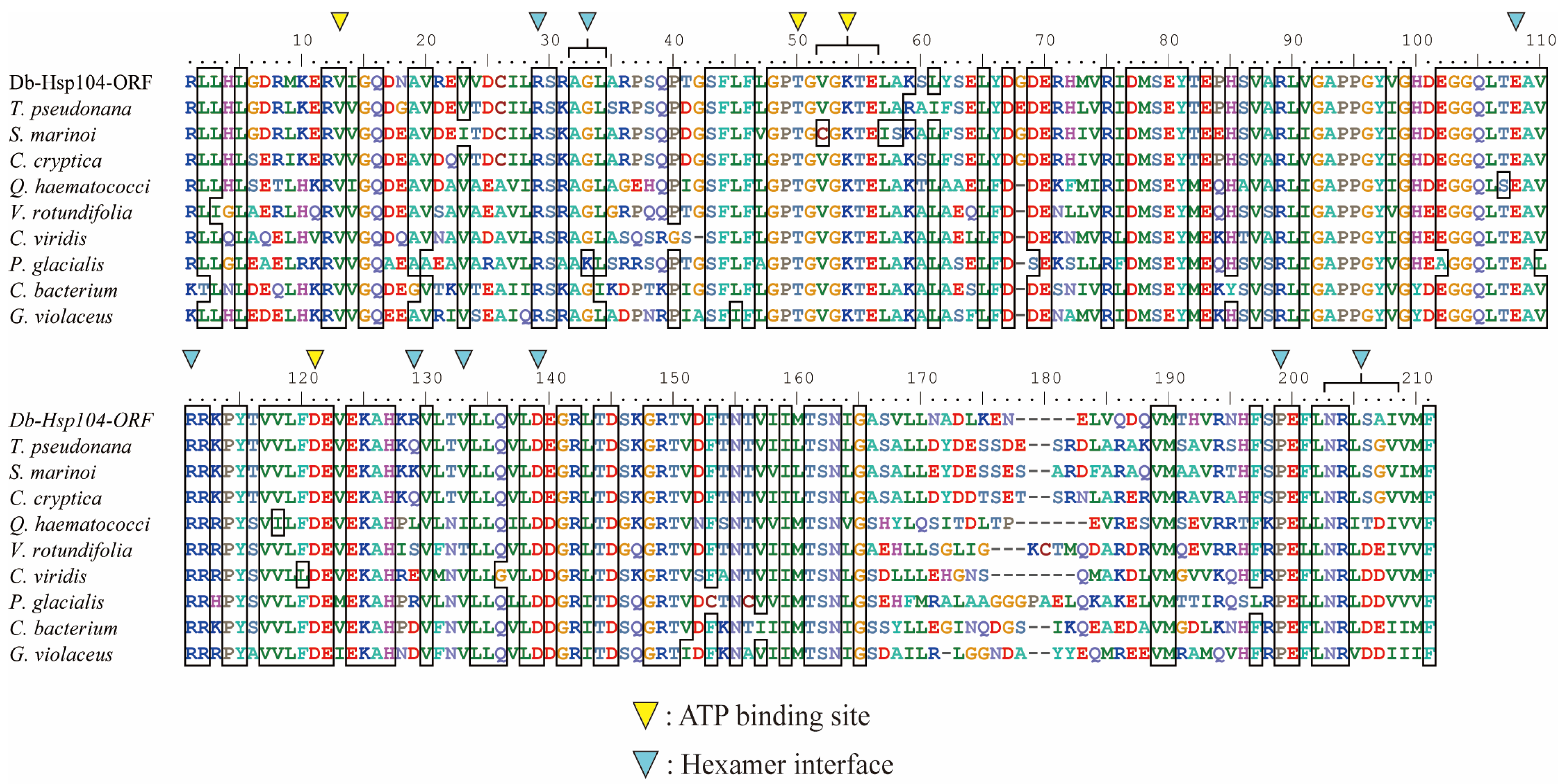
TargetP-2.0 detected no signal peptide in the N-terminal of DbHsp104, and DeepTMHMM-1.0 predicted no transmembrane helices (Figure S1). The protein comprised a full-length ClpA family domain (ATP-dependent Clp protease ATP-binding subunit; COG0542) extending from residue 1 to 874 aa (Figure 1B). A Clp repeat domain (Clp R; 2–157 aa) and two P-loops that included nucleoside triphosphate hydrolase domains (P-loop NTPase; 168–535 and 571–787 aa) were identified. A multiple aa sequences of DbHsp104 and another Hsp104/ClpB were aligned (Figure 2). An ATPase AAA-type core domain (IPR003959; 192–347 aa) was located within NBD1, spanning residues 214–343 aa and containing several ATP-binding sites [EPGVGKTA218–225, D289, T326]. A conserved arginine finger [R343], Walker A motifs [GEPGVGKT217–224], and Walker B motifs [ILFVDE285–290] were also identified. Within the second loop NTPase domain, a chaperone protein ClpB/Hsp104 subfamily domain (cd19499; 580–785 aa) was found, and it contained hexamer interface residues [R608, AGL611–613, E687, RR690–691, R708, V712, D718, SPE772–774, NRLSAI777–782] and ATP-binding sites [V592, T629, VGKTE631–635, D700].

3.3. Phylogenetic Relevance and Motif Characteristic of DbHsp104

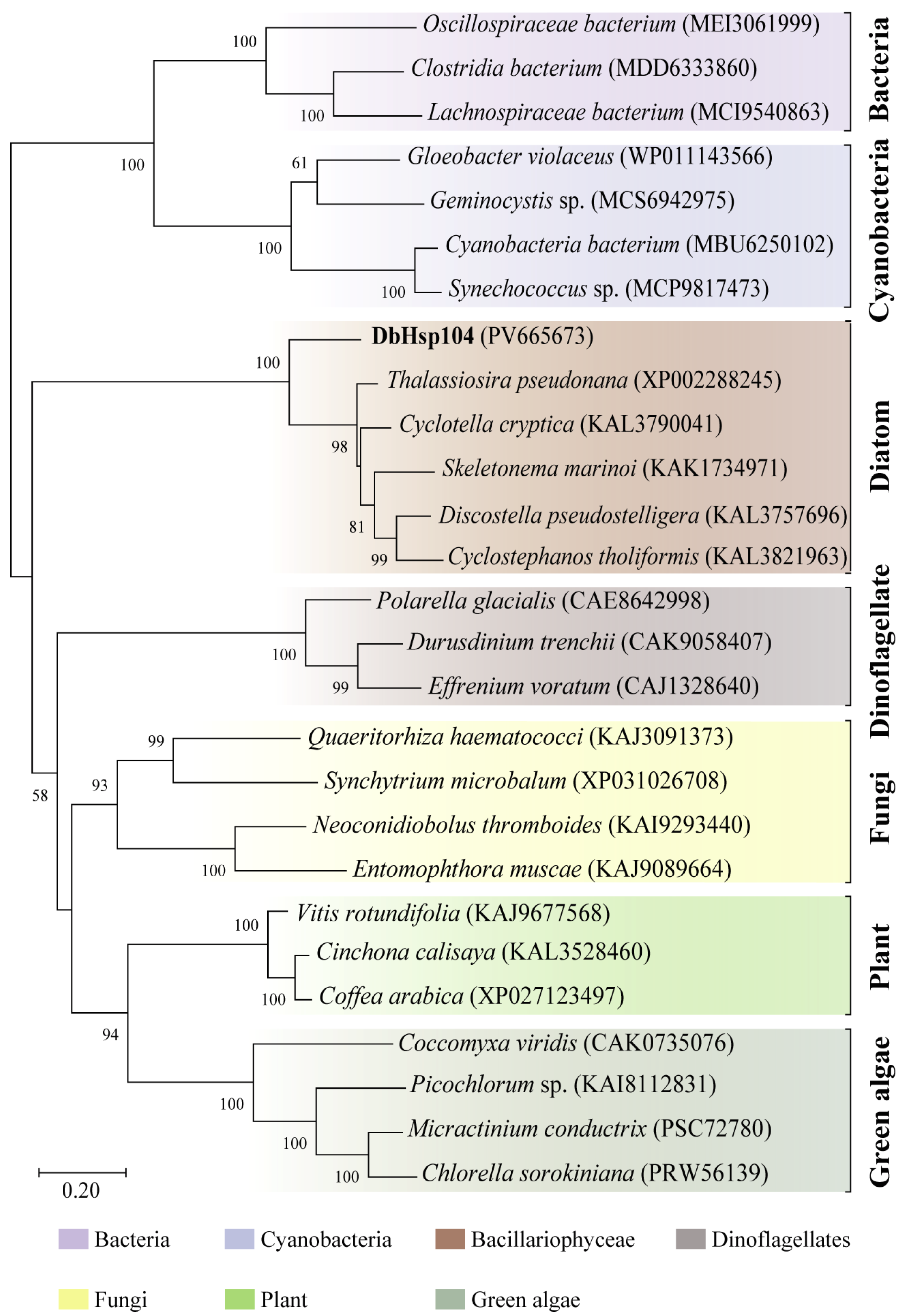
The ML phylogeny conducted using the LG + G model showed that DbHsp104 clustered together with the Hsp104 identified in other diatoms (Figure 3). The diatom Hsp104 formed a single clade with a maximal bootstrap value (100%), while the enzyme showed phylogenetic distance from those of other diatoms (98%). Diatom Hsp104s formed a sister clade with other eukaryotic clades (green algae, dinoflagellate, fungi, and plants; 60%). Meanwhile, eukaryotic Hsp104s and prokaryotic homologs were clearly separated, and bacteria and cyanobacterial Hsp104/Clp formed a distinct outgroup.

3.4. Structural Features of DbHsp104

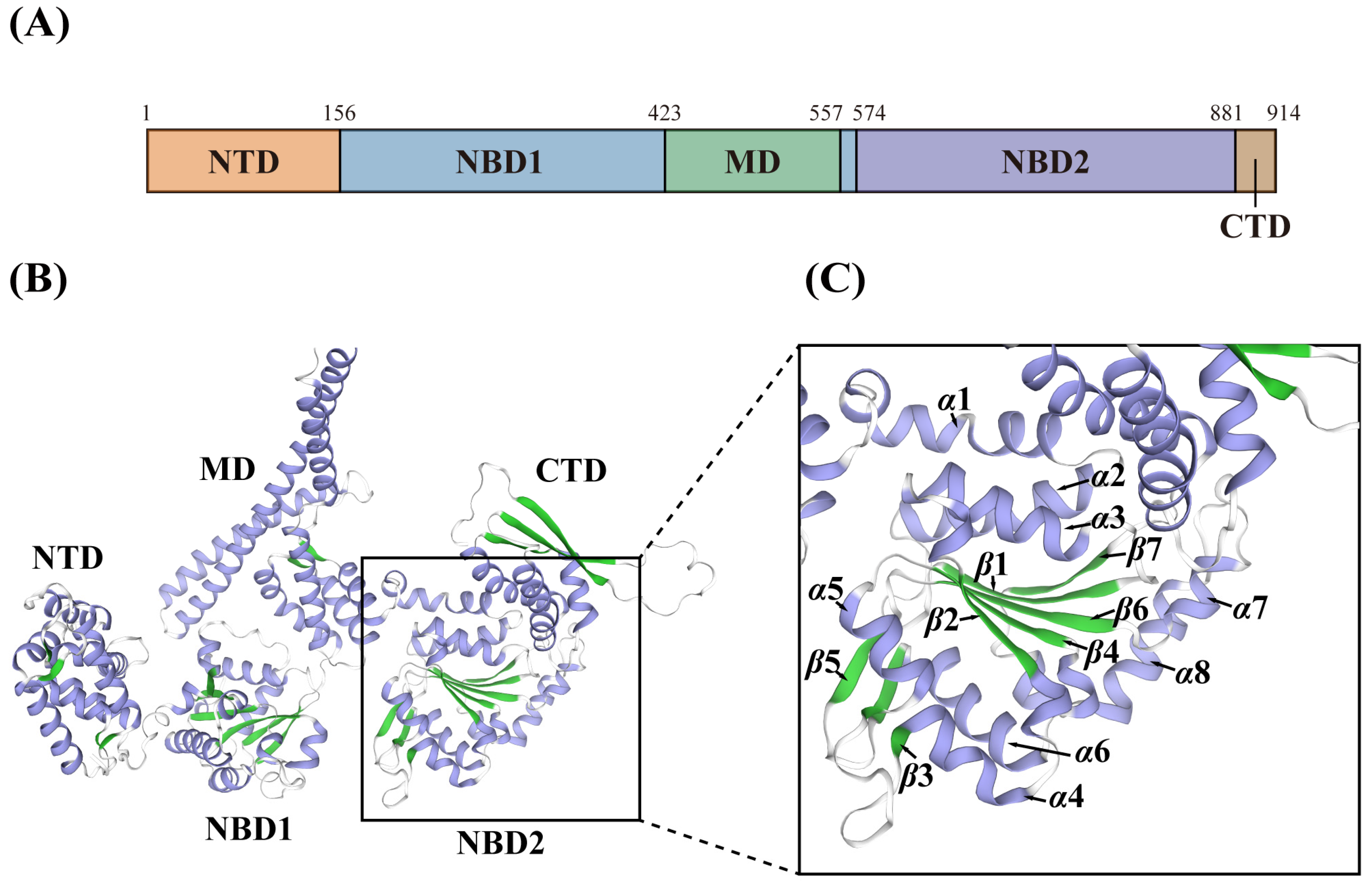
We constructed a 3D structure model of DbHsp104 using AlphaFold2 in ColabFold, and the model was colored based on the five functional domains (Figure 4 and Figure S2). Given that DbHsp104 is a single polypeptide AAA+ chaperone that assembles into a functional homohexamer, the reliability score was determined using a predicted confidence metric (pLDDT). The top five models for DbHsp104 were provided with ranks from 1 to 5 in the order of highest reliability (Figure S3). The following graph illustrates the pLDDT value of each model. The reliability of each model was determined by measuring its performance in this particular way, and the 3D structure of the most reliable model (rank_1: model_3) was confirmed.

3.5. Transcriptional Changes in Dbhsp104 Under Temperature Stress

The gene expression levels of DbHsp104 were evaluated after 0, 12, 24, and 48 h of exposure to three different temperatures (20, 25, and 30 °C) (Figure 5). Under the control condition (20 °C), no significant changes in DbHsp104 were observed, while the gene was significantly up-regulated under thermal stress. The gene expression levels at 25 °C increased 1.73-fold at 24 h and 3.85-fold at 48 h (p < 0.001), and they were also significantly up-regulated at 12, 24, and 48 h, with 1.77-, 3.37-, and 4.87-fold increases at 30 °C, respectively (p < 0.001). Taken together, the gene responses were enhanced with both temperature and exposure time.

3.6. Effect of Pollutants on DbHsp104 Expression

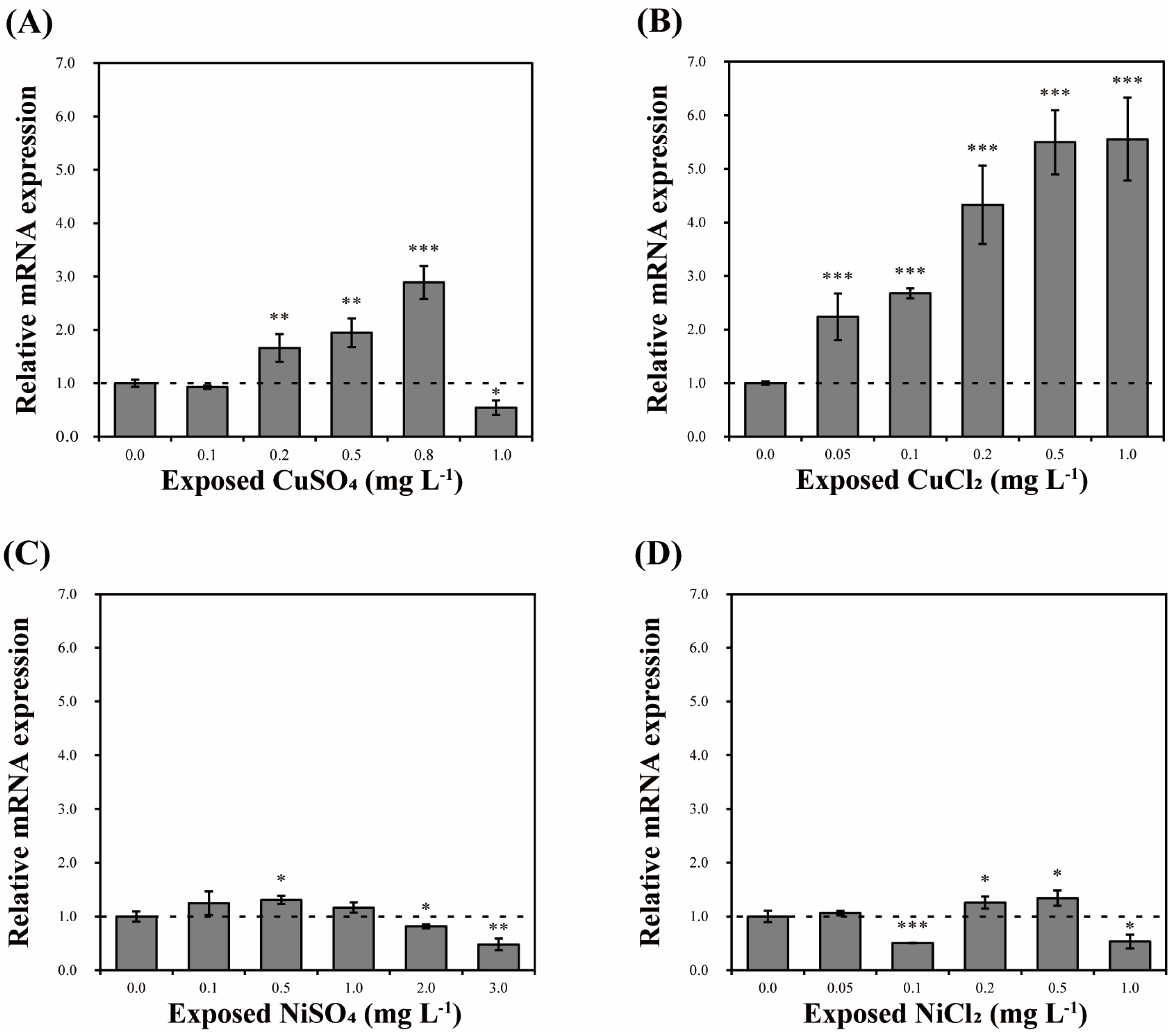
Cells of D. brightwellii were exposed for 24 h to five concentrations of heavy metals (CuSO4, CuCl2, NiSO4, and NiCl2) and EDCs (BPA, PCB, and EDS) to evaluate their effects on DbHsp104 expression. The five concentrations were chosen based on the EC50 values of each chemical (Table 3). In the heavy metal assays, copper and nickel showed distinct patterns (Figure 6). For example, CuSO4 significantly up-regulated DbHsp104 compared to the control (0.0 mg L−1), peaking at 0.8 mg L−1 (2.89-fold; p < 0.001); its expression rose to 0.8 mg L−1 and then dropped at 1.0 mg L−1 (0.54-fold; p < 0.05). CuCl2 showed the most significant increase in DbHsp104 by concentration from a 2.24- to 5.55-fold increase (p < 0.001). In contrast, there was no notable pattern of increase or decrease in DbHsp104 under the NiSO4 and NiCl2 treatments. The maximum increases were 1.31- and 1.34-fold at 0.5 mg L−1 (p < 0.05), and the greatest decreases were 0.48- and 0.51-fold at 3.0 mg L−1 of NiSO4 (p < 0.005) and 0.1 mg L−1 of NiCl2 (p < 0.001).
Overall, EDC treatments lead to no significant changes in DbHsp104 gene expression (Figure 7). BPA caused a slight up-regulation at 0.05 mg L−1 (1.19-fold; p < 0.05) and down-regulation at 0.25 mg L−1 (0.78-fold; p < 0.05) compared to the control (0.0 mg L−1). PCB reduced its expression at 0.05 mg L−1 (0.75-fold; p < 0.05), and no significant changes were observed under EDS treatments.

4. Discussion

4.1. Molecular Features of Hsp and Hsp104

Heat shock proteins (Hsps) are a family of molecular chaperones that protect cells from diverse stress conditions [48]. They play central roles in protein folding and repairing damage caused by environmental changes or pollutant exposures [21,49]. Accordingly, ecotoxicological studies have employed Hsp expression patterns as biomarkers in aquatic organisms [50,51]. Among those, the Hsp104/ClpB family shows a distinct class of ATP-dependent lyase that restores proteins from their aggregated states [52]. This function is essential for maintaining proteostasis and ensuring cellular survival under conditions that cause extensive protein denaturation.
Hsp104 has been extensively studied in the model yeast Saccharomyces cerevisiae, where it is vital for the induction of thermotolerance [53,54]. It was found that Hsp104 cooperates with co-chaperones Hsp40 and Hsp70 within a chaperone network to mediate protein refolding and resolubilizing aggregated proteins [54,55,56,57,58,59,60]. Structurally, Hsp104 assembles into an ATP-dependent hexameric complex that acts as a lyase and folds the aggregated polypeptide back through the central channel [61,62,63,64]. Bacterial Hsp104/ClpB proteins also show a conserved mechanism of ATP-driven protein disaggregation [34,65,66]. For example, the Escherichia coli ClpB is well-documented for its essential role in survival under heat shock [67,68]. In the case of microalgae, members of the Hsp104/ClpB family were identified in cyanobacteria and green algae [69,70,71]. However, studies on the structure and function of microalgal Hsp104, especially in diatoms, remain limited. Therefore, in the present study, we identified and characterized the DbHsp104 of the marine diatom D. brightwellii and investigated its transcriptional responses to diverse stressors to elucidate its role in cellular stress adaptation mechanisms.

4.2. Structural Features of Eukaryotic DbHsp104

Bioinformatic analysis of DbHsp104 revealed that it possesses the typical molecular features of canonical Hsp104/ClpB proteins. DbHsp104 contains two highly conserved nucleotide-binding domains (NBD1 and NBD2), which include the Walker A and B motifs that function as ATP-binding and hydrolysis sites. In addition, a notable structural feature of the distal loop was found between Helix A and Helix B of the middle domain (MD). The loop enables cooperation between Hsp70 and Hsp100 and contributes to thermal resistance in vivo [30]. The distal loop sequence of DbHsp104 [ALGREKDKASKDRRK] showed a high degree of similarity (93.3%) to that of diatom T. pseudonana Hsp104 (GenBank accession no. XP002288245). It also shared 60% and 52.9% similarity with that of Hsp104 of the fungus Calcarisporiella thermophila (PDB ID: 6D00) and yeast S. cerevisiae (5KNE), respectively. DbHsp104 even showed about 53.3% similarity with bacterial homologs from E. coli (4D2U) and Thermus thermophilus (1QVR). These results suggest that the distal loop region is evolutionarily conserved across diverse taxa and plays a critical role in the functional activity of Hsp104/ClpB.
There was no predicted signal peptide and transmembrane domain in DbHsp104, which implies that it is a soluble non-membrane protein. In addition, it comprises five domains: NTD, NBD1, MD, NBD2, and the C-terminal domain (CTD). Structural analysis showed that MD exhibited similarity to other Hsp104/ClpB proteins. MD located between NBD1 and NBD2 serves as an important interface for interaction with the Hsp70/DnaK system, where it catalyzes substrate transfer and refolding [54,72]. Additionally, CTD is a unique feature of eukaryotic Hsp104, which is not present in bacterial ClpB orthologs [34]. Although the precise role of the CTD is not clearly defined, it is known to be related to substrate specificity and regulatory control [35]. Furthermore, phylogenetic analysis of the Hsp104/ClpB family also supports that DbHsp104 is closer to eukaryotic Hsp104 rather than bacterial ClpB. These molecular features suggest that DbHsp104 is a eukaryotic-type Hsp104 family.

4.3. Effect of Thermal Stress on DbHsp104 Gene Expression

Thermal stress poses a major threat to proteostasis and enzyme activity, and aquatic organisms are particularly vulnerable to such temperature fluctuations [73,74,75]. To cope with these stresses, microalgae rely on various HSPs that assist in maintaining protein homeostasis and enhancing thermotolerance [76,77,78,79]. Among those, the Hsp104/ClpB family plays a vital role in restoring protein function through resolubilization and refolding of aggregated proteins [34]. Consistently, transcriptional levels of DbHsp104 were significantly induced by heat stress in a time- and temperature-dependent manner, showing the conserved rapid-response kinetics of the Hsp104/ClpB family. For example, Hsp104 expression in S. cerevisiae yeast peaks within 1 h of exposure to approximately 39 °C [80,81]. Moreover, Yamamoto et al. [82] reported that Hsp104 transcription and protein levels increased very rapidly after heat shock, and peaked within 15 to 30 min. Similarly, clpB transcripts in E. coli and other bacteria are also rapidly up-regulated upon heat shock [83,84,85]. Taken together, our findings suggest that DbHsp104 acts as a molecular chaperone that rapidly responds to thermal stress, participates in maintaining protein homeostasis, and confers thermotolerance.
As mentioned above, Hsp104 cooperates with Hsp40/70 to mitigate proteotoxic damage and sustain cellular viability under thermal stress [54,72,86]. In our previous study, expression levels of Hsp70 and Hsp90 were significantly up-regulated under heat stress and peaked at 12 h at 25 °C and 48 h at 30 °C in D. brightwellii [87]. The results indicate a high correlation of Hsp70 and Hsp104 in their ability to repair damaged proteins caused by heat stress. These are also supported by a number of studies on Hsp responses under thermal stress in microalgae. For example, in the diatom T. pseudonana, both Hsp70/90 showed significant increases at 25 °C and 30 °C [77]. Similarly, Hsp40 (StHsp40) was significantly up-regulated in response to heat stress in dinoflagellate Scrippsiella trochoidea [88]. Indeed, Santos et al. [71] reported that the mRNA levels of Hsp70 and Hsp100 were similarly induced in green algae Tetraselmis suecica under combined heat and salinity stress, which is expected as a coordinated response for thermotolerance. At the same time, Sathasivam & Ki [78] reported that qRT-PCR analysis of Hsp70 and Hsp100 from T. suecica revealed no significant changes in their expression under thermal stress. Taken together, Hsp70 and Hsp100 appear to regulate their expression in response to heat stress, with expression levels, peak timing, and duration varying depending on the species and strains. Across strains, their expression patterns often show similarities, which suggest coordinated regulation among Hsps.

4.4. Effects of Acute Toxicity on Hsp104 Transcription

A number of studies have reported that Hsps play an essential role in response to diverse environmental and ecotoxicological stresses to protect the cells and regulate the oxidative stress in microorganisms [89,90]. There are many candidates for environmental pollutants, such as heavy metals and persistent organic pollutants (POPs), including EDCs, herbicides, and antibiotics. Among those, the release of heavy metals and EDCs into the aquatic environment has been recognized as a major concern due to their long-term persistence and bioaccumulation potential [91,92]. Numerous studies have reported that such pollutants can induce oxidative stress, DNA damage, and endocrine disruption in various aquatic organisms [93,94,95]. These environmental stressors can therefore cause severe physiological and molecular disturbances, ultimately resulting in irreversible damage to aquatic life.
Copper is an essential trace metal for cell growth and is involved in respiratory electron transfer, cell wall formation, cellular defense, and diverse metabolisms in photosynthetic organisms [96]. However, it is known to cause toxicity at even low concentrations in plants and algae. In detail, excess copper inhibits growth and causes disorders in photosynthesis (chlorophyll reduction, damage in the mineral system, etc.) in its early stage [97,98]. Numerous studies of stress induced by copper also showed common phenomena of growth, chlorophyll, and photosynthesis reduction and reactive oxygen species (ROS) accumulation induced by cellular damage; green algae Closterium ehrenbergii [99,100,101], dinoflagellate Cochlodinium polykrikoides [102,103], Prorocentrum minimum [104,105,106], and Palatinus apiculatus [107], and diatoms D. brightwellii [45], T. pseudonana [77], Nitzschia closterium [108], Skeletonema costatum [109], and Navicula sp. [109]. The estimated EC50 of CuSO4 (0.406 mg L−1) was likely lowest among the heavy metal treatments, followed by NiCl2, CuCl2, and NiSO4 in D. brightwellii (Table 3). These results show that copper induces rapid and lethal toxicity in microalgae, specifically causing damage to photosynthesis-related proteins [110,111].
Such proteostasis stress induces the activation of a molecular chaperone system based on the Hsp families [112]. Consistently, DbHsp104 was significantly induced by copper treatments, and Hsp70/90 was also up-regulated by metal toxicity in D. brightwellii [87]. This is in good agreement with the role of Hsp104 as a chaperone that cooperates with Hsp40/70 to remodel and recover damaged proteins [54,58]. In this regard, Sathasivam & Ki [78] reported that expression of Hsp70/100 was significantly induced in green algae T. suecica, while Hsp20 decreased. Moreover, mRNA levels of Hsp70 were up-regulated in dinoflagellate P. minimum and green algae C. ehrenbergii under copper exposures [46,79]. Notably, Hsp70 shows strong induction in acute damage, involved in the early stages of protein repair [113]. Collectively, the results suggest that DbHsp104 responds rapidly to acute stress and is likely to cooperate with Hsp70 to manage initial proteostasis recovery.

4.5. Minor Effects of EDCs on Hsp104 System

EDCs are substances that interfere with the transmission of hormonal signals [114]. Surprisingly, a number of studies have reported that EDCs can inhibit the growth of non-target microorganisms [114,115,116]. Although the mechanisms of reaction and sensitivity vary depending on the compounds, the three tested EDCs have been shown to be able to delay growth by interfering with the cell cycle [115]. In addition, BPA [114] and EDS [116] are reported to induce oxidative stress and membrane/permeability disruption in microalgae. Similarly, Wang et al. [115] reported that PCBs did not increase the expression level of Hsp90 in the dinoflagellate P. minimum, while they decreased the expression of Hsp90 in the dinoflagellate C. polykrikoides [117]. In addition, neither the Hsp90 of green algae C. ehrenbergii [79] nor the hsp70/83 of fruit fly Drosophila melanogaster [118] reacted to EDS. In line with these, EDCs yielded higher EC50 values compared to the heavy metals and did not show an acute toxic signature or fatal mortality. Consistently, DbHsp104 showed no significant transcriptional induction under exposure to any of the three EDCs. These results support the finding that EDCs induce subacute stress related to cell division, leading to damage that is less dependent on Hsp104.

4.6. DbHsp104 as a Potential Biomarker

The distinct expression profile of DbHsp104 highlights it as a potential biomarker for environmental stresses, especially acute toxicity. It showed a rapid and dose-dependent transcriptional response to key stressors, heat and copper, both common and potent water pollutants [91,119]. Unlike common biomarkers that respond to multiple stress factors, the observed response of DbHsp104 suggests it may have potential in more specific diagnostic applications [120]. Monitoring DbHsp104 and Hsp104/clpB from other photosynthetic organisms thus provides a sensitive and early warning of water contamination, enabling more rapid monitoring and active management [120,121]. Future studies would benefit from extending more diverse Hsp104 candidates to test their responses to diverse metals (e.g., cadmium, lead, zinc) and contaminants to establish their applications.

5. Conclusions

The present study extends the previous study by Lee et al. [45] regarding the high sensitivity of Hsp in diatom D. brightwellii to various toxic chemicals. To our knowledge, this is the first report identifying the molecular features of DbHsp104 and evaluating its transcriptional responses in diatoms. Phylogeny and domain organization showed the distinctive features of the eukaryotic Hsp104/ClpB family. Transcriptional responses exhibited significant induction under heat stress and copper exposure. There was no change in response to nickel or the tested EDCs. Collectively, the results indicate a selective and distinct responsiveness to acute thermal and copper stress, suggesting that DbHsp104 may contribute to the rapid restoration of proteostasis. These findings suggest that Hsp104 may be a useful molecular biomarker for environmental monitoring using diatoms. Future work should integrate an analysis of Hsp104 at the protein level.

Supplementary Materials

The following supporting information can be downloaded at https://www.mdpi.com/article/10.3390/genes16121408/s1. Table S1: Species and accession numbers of Hsp104/ClpB amino acid sequences used for the alignments; Table S2: Taxon, species, and accession numbers of Hsp104/ClpB amino acid sequences used for phylogenetic tree analysis; Figure S1: (A) TargetP-2.0 prediction for the signal peptide and (B) TMHHM-2.0 prediction for transmembrane helices of the DbHsp104 identified from the marine diatom D. brightwellii; Figure S2: (A) Domain organization and (B) three-dimensional structure (3D) of the DbHsp104 identified from the marine diatom Ditylum brightwellii. Three-dimensional structure of DbHsp104 color-coded by the domains; N-terminal domain (NTD, orange), nucleotide-binding domain 1 (NBD1, blue), middle domain (MD, green), NBD2 (purple), and C-terminal domain (CTD, brown); Figure S3: Residue confidence profiles for the five top-ranked AlphaFold2 models of DbHsp104. The plot shows predicted IDDT (pLDDT) values for residues 1–915 aa on the x-axis and scores from 0 to 100 on the y-axis (≥90, very high; 70–90, high; <70, moderate–low confidence). Curves for rank 1–5 (blue, orange, green, red, purple) overlap across the sequence; Figure S4: Conserved residues mapped onto the three-dimensional (3D) structure of DbHsp104. (A) Overview of residues of nucleotide-binding domain 1 (NBD1). (B) Walker A motif (orange), Walker B motif (blue), and conserved arginine finger (yellow). (C) Overview of residues of NBD2. (D) Hexamer interface (red). (E) Overview of ATP-binding sites. (F) Specific ATP-binding site (black).

Author Contributions

Conceptualization, J.-S.K.; validation, H.-S.K., J.-W.L. and J.-S.K.; formal analysis, J.-W.L.; investigation, H.-S.K. and J.-W.L.; data curation, H.-S.K.; writing—original draft preparation, H.-S.K. and J.-W.L.; writing—review and editing, H.-S.K., J.-W.L. and J.-S.K.; visualization, J.-W.L.; supervision, J.-S.K.; project administration, J.-S.K.; funding acquisition, J.-S.K. All authors have read and agreed to the published version of the manuscript.

Funding

This research was funded by the National Research Foundation of Korea (NRF) grant funded by the Korea government (MSIT) (RS-2024-00354842 and RS-2024-00359501).

Institutional Review Board Statement

Not applicable.

Informed Consent Statement

Not applicable.

Data Availability Statement

The data presented in this study are available on request from the corresponding author.

Conflicts of Interest

The authors declare no conflicts of interest.

Abbreviations

The following abbreviations are used in this manuscript:
HSPheat shock protein
CuSO4copper sulfate
CuCl2copper chloride
NiSO4nickel sulfate
BPAbisphenol A
PCBpolychlorinated biphenyl
EDSendosulfan
EDCsendocrine-disrupting chemicals
P-loop NTPaseP-loop containing nucleoside triphosphate hydrolase
NTDN-terminal domain
NBDnucleotide-binding domains
CTDC-terminal domain
CLPR Clp repeat domain
MLMaximum likelihood
ANOVAOne-way analysis of variance

References

  1. Hildebrand, M. Diatoms, biomineralization processes, and genomics. Chem. Rev. 2008, 108, 4855–4874. [Google Scholar] [CrossRef] [PubMed]
  2. Armbrust, E.V. The life of diatoms in the world’s oceans. Nature 2009, 459, 185–192. [Google Scholar] [CrossRef] [PubMed]
  3. Brzezinski, M.A.; Dickson, M.L.; Nelson, D.M.; Sambrotto, R. Ratios of Si, C, and N uptake by microplankton in the Southern Ocean. Deep Sea Res. Part II Top. Stud. Oceanogr. 2003, 50, 619–633. [Google Scholar] [CrossRef]
  4. Sommers, M.D.; Naselli-Flores, L.; Padisák, J. Ecosystem services provided by freshwater and marine diatoms: Silica cell wall and high reproduction rate as key traits. Hydrobiologia 2022, 849, 123–140. [Google Scholar] [CrossRef]
  5. Fritz, C.; Masouras, A.; Pinheiro, P. Diatoms as bioindicators for health assessments of ephemeral freshwater ecosystems: A comprehensive review. Sci. Total Environ. 2024, 904, 166766. [Google Scholar] [CrossRef]
  6. Stauber, J.L.; Davies, C.M. Use and limitations of microbial bioassays for assessing copper bioavailability in the aquatic environment. Environ. Rev. 2000, 8, 255–301. [Google Scholar] [CrossRef]
  7. Gerringa, L.J.A.; Rijstenbil, J.W.; Poortvliet, T.C.W.; Van Drie, J.; Schot, M.C. Speciation of copper and responses of the marine diatom Ditylum brightwellii upon increasing copper concentrations. Aquat. Toxicol. 1995, 31, 77–90. [Google Scholar] [CrossRef]
  8. Costa, A.P.T.; Schneck, F. Diatoms as indicators in running waters: Trends of studies on biological assessment and monitoring. Environ. Monit. Assess. 2022, 194, 695. [Google Scholar] [CrossRef]
  9. Blanco, S. What do diatom indices indicate? Modeling the specific pollution sensitivity index. Environ. Sci. Pollut. Res. 2024, 31, 29449–29459. [Google Scholar] [CrossRef]
  10. Moreno-Garrido, I.; Lubián, L.M.; Soares, A.M. Influence of cellular density on determination of EC50 in microalgal growth inhibition tests. Ecotoxicol. Environ. Saf. 2000, 47, 112–116. [Google Scholar] [CrossRef]
  11. Carvalho, R.N.; Bopp, S.K.; Lettieri, T. Transcriptomics responses in marine diatom Thalassiosira pseudonana exposed to the polycyclic aromatic hydrocarbon benzo[a]pyrene. PLoS ONE 2011, 6, e26985. [Google Scholar] [CrossRef]
  12. Hirst, H.; Jüttner, I.; Ormerod, S.J. Comparing the responses of diatoms and macro-invertebrates to metals in upland streams of Wales and Cornwall. Freshw. Biol. 2002, 47, 1752–1765. [Google Scholar] [CrossRef]
  13. Blanco, S.; Bécares, E. Are biotic indices sensitive to river toxicants? A comparison of metrics based on diatoms and macro-invertebrates. Chemosphere 2010, 79, 18–25. [Google Scholar] [CrossRef]
  14. Morin, S.; Cordonier, A.; Lavoie, I.; Arini, A.; Blanco, S.; Duong, T.T.; Sabater, S. Consistency in diatom response to metal-contaminated environments. In Emerging and Priority Pollutants in Rivers: Bringing Science into River Management Plans; Springer: Berlin/Heidelberg, Germany, 2012; pp. 117–146. [Google Scholar]
  15. Kim Tiam, S.; Lavoie, I.; Doose, C.; Hamilton, P.B.; Fortin, C. Morphological, physiological and molecular responses of Nitzschia palea under cadmium stress. Ecotoxicology 2018, 27, 675–688. [Google Scholar] [CrossRef]
  16. Rijstenbil, J.W.; Derksen, J.W.M.; Gerringa, L.J.A.; Poortvliet, T.C.W.; Sandee, A.; Van den Berg, M.; Wijnholds, J.A. Oxidative stress induced by copper: Defense and damage in the marine planktonic diatom Ditylum brightwellii, grown in continuous cultures with high and low zinc levels. Mar. Biol. 1994, 119, 583–590. [Google Scholar] [CrossRef]
  17. Horvatić, J.; Peršić, V. The effect of Ni2+, Co2+, Zn2+, Cd2+ and Hg2+ on the growth rate of marine diatom Phaeodactylum tricornutum Bohlin: Microplate growth inhibition test. Bull. Environ. Contam. Toxicol. 2007, 79, 494–498. [Google Scholar] [CrossRef] [PubMed]
  18. Liping, W.; Zheng, B. Toxic effects of fluoranthene and copper on marine diatom Phaeodactylum tricornutum. J. Environ. Sci. 2008, 20, 1363–1372. [Google Scholar] [CrossRef] [PubMed]
  19. Janknegt, P.J.; Van de Poll, W.H.; Visser, R.J.; Rijstenbil, J.W.; Buma, A.G. Oxidative stress responses in the marine Antarctic diatom Chaetoceros brevis (Bacillariophyceae) during photoacclimation. J. Phycol. 2008, 44, 957–966. [Google Scholar] [CrossRef]
  20. Dulle, J.E.; Stein, K.C.; True, H.L. Regulation of the Hsp104 middle domain activity is critical for yeast prion propagation. PLoS ONE 2014, 9, e87521. [Google Scholar] [CrossRef] [PubMed]
  21. Feder, M.E.; Hofmann, G.E. Heat-shock proteins, molecular chaperones, and the stress response: Evolutionary and ecological physiology. Annu. Rev. Physiol. 1999, 61, 243–282. [Google Scholar] [CrossRef]
  22. Åkerfelt, M.; Morimoto, R.I.; Sistonen, L. Heat shock factors: Integrators of cell stress, development and lifespan. Nat. Rev. Mol. Cell Biol. 2010, 11, 545–555. [Google Scholar] [CrossRef] [PubMed]
  23. Padmini, E. Physiological adaptations of stressed fish to polluted environments: Role of heat shock proteins. In Reviews of Environmental Contamination and Toxicology; Springer: New York, NY, USA, 2010; Volume 206, pp. 1–27. [Google Scholar] [CrossRef]
  24. Gupta, S.C.; Sharma, A.; Mishra, M.; Mishra, R.K.; Chowdhuri, D.K. Heat shock proteins in toxicology: How close and how far? Life Sci. 2010, 86, 377–384. [Google Scholar] [CrossRef] [PubMed]
  25. Sanchez, Y.; Taulien, J.; Borkovich, K.A.; Lindquist, S. Hsp104 is required for tolerance to many forms of stress. EMBO J. 1992, 11, 2357–2364. [Google Scholar] [CrossRef] [PubMed]
  26. Lee, G.; Kim, R.S.; Lee, S.B.; Lee, S.; Tsai, F.T. Deciphering the mechanism and function of Hsp100 unfoldases from protein structure. Biochem. Soc. Trans. 2022, 50, 1725–1736. [Google Scholar] [CrossRef]
  27. Lee, J.; Sung, N.; Mercado, J.M.; Hryc, C.F.; Chang, C.; Lee, S.; Tsai, F.T. Overlapping and specific functions of the Hsp104 N domain define its role in protein disaggregation. Sci. Rep. 2017, 7, 11184. [Google Scholar] [CrossRef]
  28. Schirmer, E.C.; Glover, J.R.; Singer, M.A.; Lindquist, S. Hsp104/ClpB proteins: A common mechanism explains diverse functions. Trends Biochem. Sci. 1996, 21, 289–296. [Google Scholar] [CrossRef]
  29. Sweeny, E.A.; Shorter, J. Prion proteostasis: Hsp104 meets its supporting cast. Prion 2008, 2, 135–140. [Google Scholar] [CrossRef]
  30. DeSantis, M.E.; Shorter, J. The elusive middle domain of Hsp104 and ClpB: Location and function. Biochim. Biophys. Acta Mol. Cell Res. 2012, 1823, 29–39. [Google Scholar] [CrossRef]
  31. Shorter, J.; Southworth, D.R. Spiraling in control: Structures and mechanisms of the Hsp104 disaggregase. Cold Spring Harb. Perspect. Biol. 2019, 11, a034033. [Google Scholar] [CrossRef]
  32. Barnett, M.E.; Zolkiewska, A.; Zolkiewski, M. Structure and activity of ClpB from Escherichia coli: Role of the amino- and carboxyl-terminal domains. J. Biol. Chem. 2000, 275, 37565–37571. [Google Scholar] [CrossRef]
  33. Cashikar, A.G.; Schirmer, E.C.; Hattendorf, D.A.; Glover, J.R.; Ramakrishnan, M.S.; Ware, D.M.; Lindquist, S.L. Defining a pathway of communication from the C-terminal peptide binding domain to the N-terminal ATPase domain in a AAA protein. Mol. Cell 2002, 9, 751–760. [Google Scholar] [CrossRef]
  34. Doyle, S.M.; Wickner, S. Hsp104 and ClpB: Protein disaggregating machines. Trends Biochem. Sci. 2009, 34, 40–48. [Google Scholar] [CrossRef]
  35. Mackay, R.G.; Helsen, C.W.; Tkach, J.M.; Glover, J.R. The C-terminal extension of Saccharomyces cerevisiae Hsp104 plays a role in oligomer assembly. Biochemistry 2008, 47, 1918–1927. [Google Scholar] [CrossRef]
  36. Banwait, J.K.; Lucius, A.L. Quantitative insights into processivity of an Hsp100 protein disaggregase on folded protein substrates. Biophys. J. 2024, 124, 753–764. [Google Scholar] [CrossRef] [PubMed]
  37. Lee, U.; Rioflorido, I.; Hong, S.W.; Larkindale, J.; Waters, E.R.; Vierling, E. The Arabidopsis ClpB/Hsp100 family of proteins: Chaperones for stress and chloroplast development. Plant J. 2007, 49, 115–127. [Google Scholar] [CrossRef] [PubMed]
  38. Queraltó, C.; Álvarez, R.; Ortega, C.; Díaz-Yáñez, F.; Paredes-Sabja, D.; Gil, F. Role and regulation of Clp proteases: A target against gram-positive bacteria. Bacteria 2023, 2, 21–36. [Google Scholar] [CrossRef]
  39. Canterford, G.S.; Canterford, D.R. Toxicity of heavy metals to the marine diatom Ditylum brightwellii (West) Grunow: Correlation between toxicity and metal speciation. J. Mar. Biol. Assoc. UK 1980, 60, 227–242. [Google Scholar] [CrossRef]
  40. Ryther, J.H.; Guillard, R.R.L. Studies of marine planktonic diatoms: II. Use of Cyclotella nana Hustedt for assays of vitamin B12 in sea water. Can. J. Microbiol. 1962, 8, 437–445. [Google Scholar] [CrossRef]
  41. Gasteiger, E.; Hoogland, C.; Gattiker, A.; Duvaud, S.E.; Wilkins, M.R.; Appel, R.D.; Bairoch, A. Protein identification and analysis tools on the ExPASy server. In The Proteomics Protocols Handbook; Walker, J.M., Ed.; Humana Press: Totowa, NJ, USA, 2005; pp. 571–607. [Google Scholar]
  42. Jumper, J.; Evans, R.; Pritzel, A.; Green, T.; Figurnov, M.; Ronneberger, O.; Hassabis, D. Highly accurate protein structure prediction with AlphaFold. Nature 2021, 596, 583–589. [Google Scholar] [CrossRef]
  43. Tamura, K.; Stecher, G.; Kumar, S. MEGA11: Molecular evolutionary genetics analysis version 11. Mol. Biol. Evol. 2021, 38, 3022–3027. [Google Scholar] [CrossRef]
  44. Masindi, V.; Muedi, K.L. Environmental contamination by heavy metals. Heavy Met. 2018, 10, 115–133. [Google Scholar]
  45. Lee, M.A.; Guo, R.; Ki, J.S. Different transcriptional responses of heat shock protein 20 in the marine diatom Ditylum brightwellii exposed to metals and endocrine-disrupting chemicals. Environ. Toxicol. 2014, 29, 1379–1389. [Google Scholar] [CrossRef] [PubMed]
  46. Guo, R.; Ki, J.S. Evaluation and validation of internal control genes for studying gene expression in the dinoflagellate Prorocentrum minimum using real-time PCR. Eur. J. Protistol. 2012, 48, 199–206. [Google Scholar] [CrossRef] [PubMed]
  47. Pfaffl, M.W. A new mathematical model for relative quantification in real-time RT–PCR. Nucleic Acids Res. 2001, 29, e45. [Google Scholar] [CrossRef] [PubMed]
  48. Hartl, F.U.; Bracher, A.; Hayer-Hartl, M. Molecular chaperones in protein folding and proteostasis. Nature 2011, 475, 324–332. [Google Scholar] [CrossRef]
  49. Sanders, B.M. Stress proteins in aquatic organisms: An environmental perspective. Crit. Rev. Toxicol. 1993, 23, 49–75. [Google Scholar] [CrossRef]
  50. Bukau, B.; Weissman, J.; Horwich, A. Molecular chaperones and protein quality control. Cell 2006, 125, 443–451. [Google Scholar] [CrossRef]
  51. Moreira-de-Sousa, C.; de Souza, R.B.; Fontanetti, C.S. HSP70 as a biomarker: An excellent tool in environmental contamination analysis—A review. Water Air Soil Pollut. 2018, 229, 264. [Google Scholar] [CrossRef]
  52. Kirstein, J.; Molière, N.; Dougan, D.A.; Turgay, K. Adapting the machine: Adaptor proteins for Hsp104/ClpB and AAA+ proteases. Nat. Rev. Microbiol. 2009, 7, 589–599. [Google Scholar] [CrossRef]
  53. Sanchez, Y.; Lindquist, S.L. HSP104 required for induced thermotolerance. Science 1990, 248, 1112–1115. [Google Scholar] [CrossRef]
  54. Glover, J.R.; Lindquist, S. Hsp104, Hsp70, and Hsp40: A novel chaperone system that rescues previously aggregated proteins. Cell 1998, 94, 73–82. [Google Scholar] [CrossRef]
  55. Parsell, D.A.; Kowal, A.S.; Singer, M.A.; Lindquist, S. Protein disaggregation mediated by heat-shock protein Hsp104. Nature 1994, 372, 475–478. [Google Scholar] [CrossRef] [PubMed]
  56. Lum, R.; Tkach, J.M.; Vierling, E.; Glover, J.R. Evidence for an unfolding/threading mechanism for protein disaggregation by Saccharomyces cerevisiae Hsp104. J. Biol. Chem. 2004, 279, 29139–29146. [Google Scholar] [CrossRef] [PubMed]
  57. Mosser, D.D.; Ho, S.; Glover, J.R. Saccharomyces cerevisiae Hsp104 enhances the chaperone capacity of human cells and inhibits heat stress-induced proapoptotic signaling. Biochemistry 2004, 43, 8107–8115. [Google Scholar] [CrossRef] [PubMed]
  58. Kaimal, J.M.; Kandasamy, G.; Gasser, F.; Andréasson, C. Coordinated Hsp110 and Hsp104 activities power protein disaggregation in Saccharomyces cerevisiae. Mol. Cell. Biol. 2017, 37, e00027-17. [Google Scholar] [CrossRef]
  59. Oguchi, Y.; Kummer, E.; Seyffer, F.; Berynskyy, M.; Anstett, B.; Zahn, R.; Bukau, B. A tightly regulated molecular toggle controls AAA+ disaggregase. Nat. Struct. Mol. Biol. 2012, 19, 1338–1346. [Google Scholar] [CrossRef]
  60. Mogk, A.; Kummer, E.; Bukau, B. Cooperation of Hsp70 and Hsp100 chaperone machines in protein disaggregation. Front. Mol. Biosci. 2015, 2, 22. [Google Scholar] [CrossRef]
  61. Parsell, D.A.; Kowal, A.S.; Lindquist, S. Saccharomyces cerevisiae Hsp104 protein: Purification and characterization of ATP-induced structural changes. J. Biol. Chem. 1994, 269, 4480–4487. [Google Scholar] [CrossRef]
  62. Parsell, D.A.; Sanchez, Y.; Stitzel, J.D.; Lindquist, S. Hsp104 is a highly conserved protein with two essential nucleotide-binding sites. Nature 1991, 353, 270–273. [Google Scholar] [CrossRef]
  63. Sweeny, E.A.; Jackrel, M.E.; Go, M.S.; Sochor, M.A.; Razzo, B.M.; DeSantis, M.E.; Shorter, J. The Hsp104 N-terminal domain enables disaggregase plasticity and potentiation. Mol. Cell 2015, 57, 836–849. [Google Scholar] [CrossRef]
  64. Gates, S.N.; Yokom, A.L.; Lin, J.; Jackrel, M.E.; Rizo, A.N.; Kendsersky, N.M.; Southworth, D.R. Ratchet-like polypeptide translocation mechanism of the AAA+ disaggregase Hsp104. Science 2017, 357, 273–279. [Google Scholar] [CrossRef]
  65. Lee, S.; Sowa, M.E.; Watanabe, Y.H.; Sigler, P.B.; Chiu, W.; Yoshida, M.; Tsai, F.T. The structure of ClpB: A molecular chaperone that rescues proteins from an aggregated state. Cell 2003, 115, 229–240. [Google Scholar] [CrossRef] [PubMed]
  66. Bösl, B.; Grimminger, V.; Walter, S. The molecular chaperone Hsp104—A molecular machine for protein disaggregation. J. Struct. Biol. 2006, 156, 139–148. [Google Scholar] [CrossRef] [PubMed]
  67. Eriksson, M.J.; Clarke, A.K. The Escherichia coli heat shock protein ClpB restores acquired thermotolerance to a cyanobacterial clpB deletion mutant. Cell Stress Chaperones 2000, 5, 255. [Google Scholar] [CrossRef] [PubMed]
  68. Alam, A.; Bröms, J.E.; Kumar, R.; Sjöstedt, A. The role of ClpB in bacterial stress responses and virulence. Front. Mol. Biosci. 2021, 8, 668910. [Google Scholar] [CrossRef]
  69. Eriksson, M.J.; Schelin, J.; Miskiewicz, E.; Clarke, A.K. Novel form of ClpB/HSP100 protein in the cyanobacterium Synechococcus. J. Bacteriol. 2001, 183, 7392–7396. [Google Scholar] [CrossRef]
  70. Kreis, E.; Niemeyer, J.; Merz, M.; Scheuring, D.; Schroda, M. CLPB3 is required for the removal of chloroplast protein aggregates and thermotolerance in Chlamydomonas. J. Exp. Bot. 2023, 74, 3714–3728. [Google Scholar] [CrossRef]
  71. Santos, T.F.; Pereira, H.; Schüler, L.; Maia, I.B.; Jacinto, R.; Bombo, G.; Varela, J. Enhancement of heat tolerance by salt stress in Tetraselmis striata CTP4: Impacts on HSP gene expression, pigments, and proximal composition. J. Appl. Phycol. 2025, 37, 287–301. [Google Scholar] [CrossRef]
  72. Sielaff, B.; Tsai, F.T. The M-domain controls Hsp104 protein remodeling activity in an Hsp70/Hsp40-dependent manner. J. Mol. Biol. 2010, 402, 30–37. [Google Scholar] [CrossRef]
  73. Oakley, C.A.; Durand, E.; Wilkinson, S.P.; Peng, L.; Weis, V.M.; Grossman, A.R.; Davy, S.K. Thermal shock induces host proteostasis disruption and endoplasmic reticulum stress in the model symbiotic cnidarian Aiptasia. J. Proteome Res. 2017, 16, 2121–2134. [Google Scholar] [CrossRef]
  74. Xing, C.; Li, J.; Yuan, H.; Yang, J. Physiological and transcription level responses of microalgae Auxenochlorella protothecoides to cold and heat induced oxidative stress. Environ. Res. 2022, 211, 113023. [Google Scholar] [CrossRef]
  75. Tian, X.; Lin, X.; Xie, Q.; Liu, J.; Luo, L. Effects of temperature and light on microalgal growth and nutrient removal in turtle aquaculture wastewater. Biology 2024, 13, 901. [Google Scholar] [CrossRef] [PubMed]
  76. Guo, R.; Ebenezer, V.; Ki, J.S. Transcriptional responses of heat shock protein 70 (Hsp70) to thermal, bisphenol A, and copper stresses in the dinoflagellate Prorocentrum minimum. Chemosphere 2012, 89, 512–520. [Google Scholar] [CrossRef] [PubMed]
  77. Leung, P.T.; Yi, A.X.; Ip, J.C.; Mak, S.S.; Leung, K.M. Photosynthetic and transcriptional responses of the marine diatom Thalassiosira pseudonana to the combined effect of temperature stress and copper exposure. Mar. Pollut. Bull. 2017, 124, 938–945. [Google Scholar] [CrossRef]
  78. Sathasivam, R.; Ki, J.S. Heat shock protein genes in the green alga Tetraselmis suecica and their role against redox and non-redox active metals. Eur. J. Protistol. 2019, 69, 37–51. [Google Scholar] [CrossRef]
  79. Abassi, S.; Wang, H.; Ki, J.S. Molecular cloning of heat shock protein 70 and HOP from the freshwater green alga Closterium ehrenbergii and their responses to stress. Cell Stress Chaperones 2020, 25, 1117–1123. [Google Scholar] [CrossRef]
  80. Halladay, J.T.; Craig, E.A. A heat shock transcription factor with reduced activity suppresses a yeast HSP70 mutant. Mol. Cell. Biol. 1995, 15, 4890–4897. [Google Scholar] [CrossRef]
  81. Seppä, L.; Hänninen, A.L.; Makarow, M. Upregulation of the Hsp104 chaperone at physiological temperature during recovery from thermal insult. Mol. Microbiol. 2004, 52, 217–225. [Google Scholar] [CrossRef]
  82. Yamamoto, N.; Maeda, Y.; Ikeda, A.; Sakurai, H. Regulation of thermotolerance by stress-induced transcription factors in Saccharomyces cerevisiae. Eukaryot. Cell 2008, 7, 783–790. [Google Scholar] [CrossRef]
  83. Kitagawa, M.; Wada, C.; Yoshioka, S.; Yura, T. Expression of ClpB, an analog of the ATP-dependent protease regulatory subunit in Escherichia coli, is controlled by a heat shock sigma factor (sigma 32). J. Bacteriol. 1991, 173, 4247–4253. [Google Scholar] [CrossRef]
  84. Ingmer, H.; Vogensen, F.K.; Hammer, K.; Kilstrup, M. Disruption and analysis of the clpB, clpC, and clpE genes in Lactococcus lactis: ClpE, a new Clp family in gram-positive bacteria. J. Bacteriol. 1999, 181, 2075–2083. [Google Scholar] [CrossRef]
  85. de Oliveira, N.E.M.; Abranches, J.; Gaca, A.O.; Laport, M.S.; Damaso, C.R.; Bastos, M.D.C.D.F.; Giambiagi-deMarval, M. clpB, a class III heat-shock gene regulated by CtsR, is involved in thermotolerance and virulence of Enterococcus faecalis. Microbiology 2011, 157, 656–665. [Google Scholar] [CrossRef] [PubMed]
  86. Miot, M.; Reidy, M.; Doyle, S.M.; Hoskins, J.R.; Johnston, D.M.; Genest, O.; Wickner, S. Species-specific collaboration of heat shock proteins (Hsp) 70 and 100 in thermotolerance and protein disaggregation. Proc. Natl. Acad. Sci. USA 2011, 108, 6915–6920. [Google Scholar] [CrossRef] [PubMed]
  87. Guo, R.; Lee, M.A.; Ki, J.S. Different transcriptional responses of heat shock protein 70/90 in the marine diatom Ditylum brightwellii exposed to metal compounds and endocrine-disrupting chemicals. Chemosphere 2013, 92, 535–543. [Google Scholar] [CrossRef] [PubMed]
  88. Deng, Y.; Hu, Z.; Shang, L.; Chai, Z.; Tang, Y.Z. Transcriptional responses of the heat shock protein 20 (Hsp20) and 40 (Hsp40) genes to temperature stress and alteration of life cycle stages in the harmful alga Scrippsiella trochoidea (Dinophyceae). Biology 2020, 9, 408. [Google Scholar] [CrossRef]
  89. Blaby, I.K.; Blaby-Haas, C.E.; Pérez-Pérez, M.E.; Schmollinger, S.; Fitz-Gibbon, S.; Lemaire, S.D.; Merchant, S.S. Genome-wide analysis on Chlamydomonas reinhardtii reveals the impact of hydrogen peroxide on protein stress responses and overlap with other stress transcriptomes. Plant J. 2015, 84, 974–988. [Google Scholar] [CrossRef]
  90. Yu, A.; Li, P.; Tang, T.; Wang, J.; Chen, Y.; Liu, L. Roles of Hsp70s in stress responses of microorganisms, plants, and animals. Biomed. Res. Int. 2015, 2015, 510319. [Google Scholar] [CrossRef]
  91. Ali, H.; Khan, E.; Ilahi, I. Environmental chemistry and ecotoxicology of hazardous heavy metals: Environmental persistence, toxicity, and bioaccumulation. J. Chem. 2019, 2019, 6730305. [Google Scholar] [CrossRef]
  92. Fan, J.J.; Wang, S.; Tang, J.P.; Zhao, J.L.; Wang, L.; Wang, J.X.; Yang, Y. Bioaccumulation of endocrine disrupting compounds in fish with different feeding habits along the largest subtropical river, China. Environ. Pollut. 2019, 247, 999–1008. [Google Scholar] [CrossRef]
  93. Hong, Y.J.; Liao, W.; Yan, Z.F.; Bai, Y.C.; Feng, C.L.; Xu, Z.X.; Xu, D.Y. Progress in the research of the toxicity effect mechanisms of heavy metals on freshwater organisms and their water quality criteria in China. J. Chem. 2020, 2020, 9010348. [Google Scholar] [CrossRef]
  94. Santhosh, K.; Kamala, K.; Ramasamy, P.; Musthafa, M.S.; Almujri, S.S.; Asdaq, S.M.B.; Sivaperumal, P. Unveiling the silent threat: Heavy metal toxicity devastating impact on aquatic organisms and DNA damage. Mar. Pollut. Bull. 2024, 200, 116139. [Google Scholar] [CrossRef]
  95. Devi, A.; De Silva, Y.S.K.; Tyagi, L.; Aaryashree. The individual and combined effects of microplastics and heavy metals on marine organisms. Microplastics 2025, 4, 38. [Google Scholar] [CrossRef]
  96. Yruela, I. Copper in plants: Acquisition, transport and interactions. Funct. Plant Biol. 2009, 36, 409–430. [Google Scholar] [CrossRef]
  97. Kumar, K.S.; Dahms, H.-U.; Lee, J.S.; Kim, H.C.; Lee, W.C.; Shin, K.H. Algal photosynthetic responses to toxic metals and herbicides assessed by chlorophyll a fluorescence. Ecotoxicol. Environ. Saf. 2014, 104, 51–71. [Google Scholar] [CrossRef]
  98. Chen, G.; Li, J.; Han, H.; Du, R.; Wang, X. Physiological and molecular mechanisms of plant responses to copper stress. Int. J. Mol. Sci. 2022, 23, 12950. [Google Scholar] [CrossRef]
  99. Wang, H.; Sathasivam, R.; Ki, J.S. Physiological effects of copper on the freshwater alga Closterium ehrenbergii Meneghini (Conjugatophyceae) and its potential use in toxicity assessments. Algae 2017, 32, 131–137. [Google Scholar] [CrossRef]
  100. Abassi, S.; Wang, H.; Ponmani, T.; Ki, J.S. Small heat shock protein genes of the green algae Closterium ehrenbergii: Cloning and differential expression under heat and heavy metal stresses. Environ. Toxicol. 2019, 34, 1013–1024. [Google Scholar] [CrossRef] [PubMed]
  101. Wang, H.; Ki, J.S. Molecular identification, differential expression and protective roles of iron/manganese superoxide dismutases in the green algae Closterium ehrenbergii against metal stress. Eur. J. Protistol. 2020, 74, 125689. [Google Scholar] [CrossRef] [PubMed]
  102. Ebenezer, V.; Lim, W.A.; Ki, J.S. Effects of the algicides CuSO4 and NaOCl on various physiological parameters in the harmful dinoflagellate Cochlodinium polykrikoides. J. Appl. Phycol. 2014, 26, 2357–2365. [Google Scholar] [CrossRef]
  103. Guo, R.; Wang, H.; Suh, Y.S.; Ki, J.S. Transcriptomic profiles reveal the genome-wide responses of the harmful dinoflagellate Cochlodinium polykrikoides when exposed to the algicide copper sulfate. BMC Genom. 2016, 17, 29. [Google Scholar] [CrossRef]
  104. Guo, R.; Ki, J.S. Differential transcription of heat shock protein 90 (HSP90) in the dinoflagellate Prorocentrum minimum by copper and endocrine-disrupting chemicals. Ecotoxicology 2012, 21, 1448–1457. [Google Scholar] [CrossRef] [PubMed]
  105. Kim, H.; Wang, H.; Abassi, S.; Ki, J.S. The herbicide alachlor severely affects photosystem function and photosynthetic gene expression in the marine dinoflagellate Prorocentrum minimum. J. Environ. Sci. Health Part B 2020, 55, 620–629. [Google Scholar] [CrossRef] [PubMed]
  106. Wang, H.; Ki, J.S. Identification of a metacaspase gene in the bloom-forming dinoflagellate Prorocentrum minimum and its putative function involved in programmed cell death. Curr. Microbiol. 2021, 78, 3577–3585. [Google Scholar] [CrossRef] [PubMed]
  107. Bui, Q.T.N.; Kim, H.S.; Ki, J.S. Two chloroplast superoxide dismutase genes (MnSOD and CuZnSOD) in the freshwater dinoflagellate Palatinus apiculatus and their high-level gene expression in response to algicidal agents. J. Appl. Phycol. 2025, 14, 1–13. [Google Scholar] [CrossRef]
  108. Florence, T.M.; Stauber, J.L. Toxicity of copper complexes to the marine diatom Nitzschia closterium. Aquat. Toxicol. 1986, 8, 11–26. [Google Scholar] [CrossRef]
  109. Kim, T.; Byeon, H.; An, Y.; Rayamajhi, V.; Lee, J.; Lee, J.D.; Jung, S.M. Acute toxicity of antifouling agents CuSO4, ZnPT, and CuPT on marine diatoms Skeletonema costatum and Navicula sp. Mar. Environ. Res. 2025, 207, 107084. [Google Scholar] [CrossRef]
  110. Laporte, D.; Rodríguez, F.; González, A.; Zúñiga, A.; Castro-Nallar, E.; Sáez, C.A.; Moenne, A. Copper-induced concomitant increases in photosynthesis, respiration, and C, N and S assimilation revealed by transcriptomic analyses in Ulva compressa (Chlorophyta). BMC Plant Biol. 2020, 20, 25. [Google Scholar] [CrossRef]
  111. Luengluetham, P.; Chotikarn, P.; Nopparat, J.; Buapet, P. Ecotoxicological assessment of copper and zinc in a common aquatic plant Ceratophyllum demersum: Physiological effects and biomarker responses. Aquat. Bot. 2023, 188, 103678. [Google Scholar] [CrossRef]
  112. Santiago, A.M.; Gonçalves, D.L.; Morano, K.A. Mechanisms of sensing and response to proteotoxic stress. Exp. Cell Res. 2020, 395, 112240. [Google Scholar] [CrossRef]
  113. Colunga Biancatelli, R.M.; Solopov, P.A.; Day, T.; Gregory, B.; Osei-Nkansah, M.; Dimitropoulou, C.; Catravas, J.D. HSP70 is a critical regulator of HSP90 inhibitor’s effectiveness in preventing HCl-induced chronic lung injury and pulmonary fibrosis. Int. J. Mol. Sci. 2024, 25, 1920. [Google Scholar] [CrossRef]
  114. Azizullah, A.; Khan, S.; Gao, G.; Gao, K. The interplay between bisphenol A and algae—A review. J. King Saud Univ. Sci. 2022, 34, 102050. [Google Scholar] [CrossRef]
  115. Wang, H.; Guo, R.; Ki, J.S. 6.0 K microarray reveals differential transcriptomic responses in the dinoflagellate Prorocentrum minimum exposed to polychlorinated biphenyl (PCB). Chemosphere 2018, 195, 398–409. [Google Scholar] [CrossRef]
  116. Menone, M.L.; Pesce, S.F.; Díaz, M.P.; Moreno, V.J.; Wunderlin, D.A. Endosulfan induces oxidative stress and changes on detoxication enzymes in the aquatic macrophyte Myriophyllum quitense. Phytochemistry 2008, 69, 1150–1157. [Google Scholar] [CrossRef]
  117. Guo, R.; Youn, S.H.; Ki, J.S. Heat shock protein 70 and 90 genes in the harmful dinoflagellate Cochlodinium polykrikoides: Genomic structures and transcriptional responses to environmental stresses. Int. J. Genom. 2015, 2015, 484626. [Google Scholar] [CrossRef] [PubMed]
  118. Sharma, S.; Rohilla, M.S.; Tiwari, P.K. Developmental and hyperthermia-induced expression of the heat shock proteins HSP60 and HSP70 in tissues of the housefly Musca domestica: An in vitro study. Genet. Mol. Biol. 2007, 30, 159–168. [Google Scholar] [CrossRef]
  119. Liu, Y.; Wang, H.; Cui, Y.; Chen, N. Removal of copper ions from wastewater: A review. Int. J. Environ. Res. Public Health 2023, 20, 3885. [Google Scholar] [CrossRef] [PubMed]
  120. Mukhopadhyay, I.; Nazir, A.; Saxena, D.K.; Chowdhuri, D.K. Heat shock response: Hsp70 in environmental monitoring. J. Biochem. Mol. Toxicol. 2003, 17, 249–254. [Google Scholar] [CrossRef]
  121. Mitra, T.; Mahanty, A.; Ganguly, S.; Purohit, G.K.; Mohanty, S.; Parida, P.K.; Mohanty, B.P. Expression patterns of heat shock protein genes in Rita rita from natural riverine habitat as biomarker response against environmental pollution. Chemosphere 2018, 211, 535–546. [Google Scholar] [CrossRef]
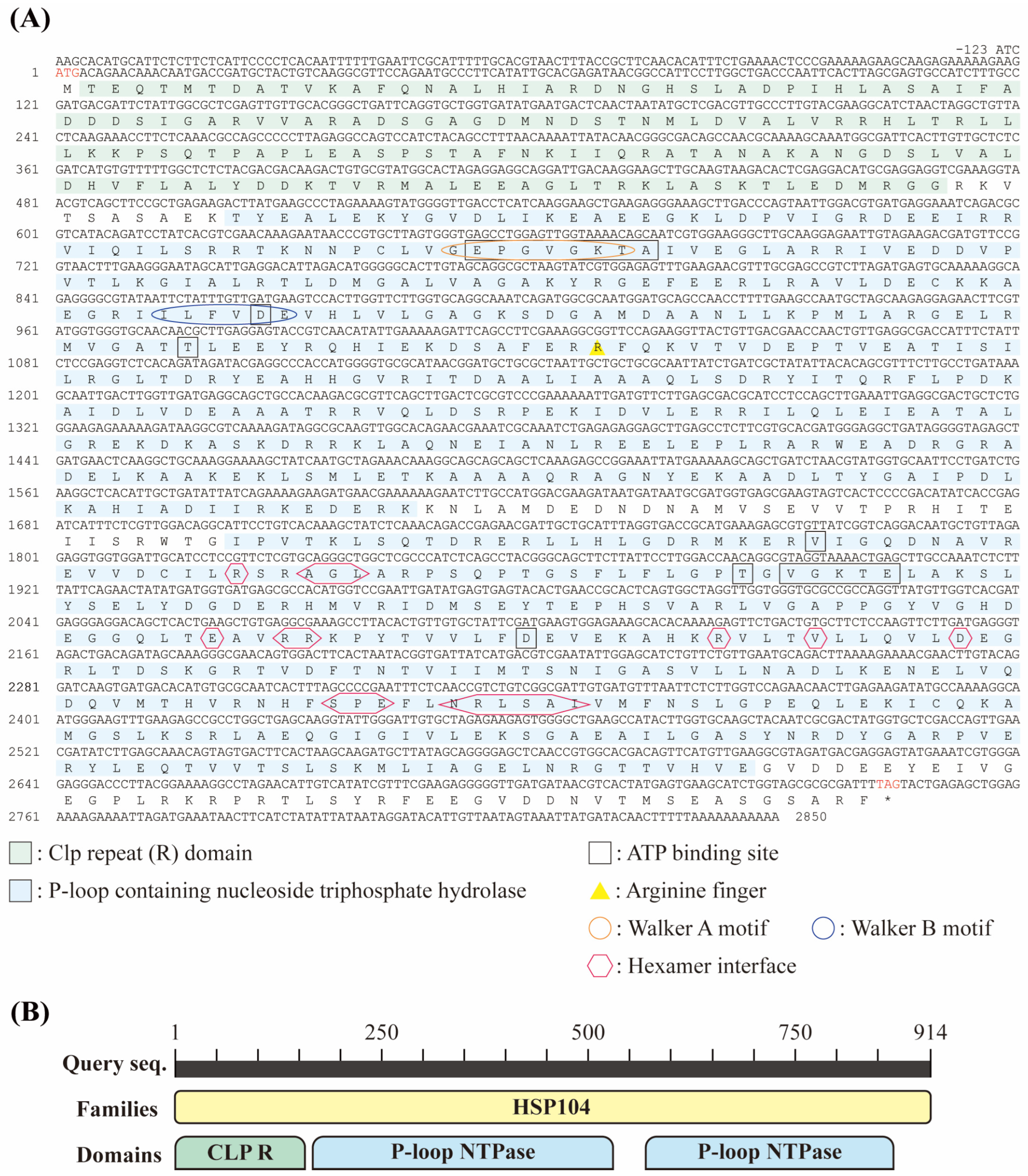
Figure 1. The molecular features of DbHsp104 were determined in the marine diatom D. brightwellii. (A) Nucleotide and deduced amino acid (aa) sequences of DbHsp104. The start (ATG) and stop (TAG, *) codons are shown in red letters. Clp repeat domain (CLP R) and P-loop-containing nucleoside triphosphate hydrolase (P-loop NTPase) domain are highlighted in green and light blue, respectively. ATP-binding sites and arginine fingers are marked in a black box and a yellow triangle, respectively. Walker A and Walker B motifs are encircled in orange and blue, respectively. The hexamer interface is marked by a red octagon. (B) Schematic representation of the DbHsp104 showing the Hsp104 family domain (yellow), comprising the Clp repeat domain (green) and P-loop NTPase domain (light blue).
Genes 16 01408 g001
Figure 2. Multiple amino acid (aa) sequence alignment of DbHsp104 and the Hsp104/ClpB family. The black box represents the conserved residues among the alignments. The ATP-binding sites and hexamer interfaces are marked in yellow and blue inverted triangles, respectively. The protein sequences were taken from the GenBank database, and the details are listed in Supplementary Table S1.
Genes 16 01408 g002
Figure 3. Maximum likelihood tree of DbHsp104 with other Hsp104/ClpB proteins. DbHsp104 is marked in bold. The bootstrap values under 50% were not shown. The protein sequences were taken from the GenBank database, and details are listed in Supplementary Table S2.
Genes 16 01408 g003
Figure 4. Schematic diagram of DbHsp104 domain organization (A). N-terminal domain (NTD), nucleotide-binding domains (NBD1 and NBD2), middle domain (MD), and C-terminal domain (CTD) are colored in orange, blue, green, blue, and brown, respectively. Three-dimensional (3D) protein structure of DbHsp104 (B). α-helices and β-strands are shown in purple and green, respectively (C).
Genes 16 01408 g004
Figure 5. Effects of heat stress on the expression of DbHsp104. The gene expression levels were analyzed after exposure to three different temperatures (20, 25, and 30 °C) for four time intervals (0, 12, 24, and 48 h). Significant differences between the control and samples were determined by one-way ANOVA and are indicated as follows: *** p < 0.001 (n = 3). Error bars and dotted lines represent the mean standard deviation (SD) and baseline (1.0-fold) expression level of the control.
Genes 16 01408 g005
Figure 6. Effect of heavy metals on the expression of DbHsp104. D. brightwellii cells were exposed to various concentrations of copper sulfate (CuSO4, (A)), copper chloride (CuCl2, (B)), nickel sulfate (NiSO4, (C)), and nickel chloride (NiCl2, (D)) for 24 h. Significant differences between the control and samples were determined by one-way ANOVA and are indicated as follows: * p < 0.05, ** p < 0.01, *** p < 0.001 (n = 3). Error bars and dotted line represent mean standard deviation (SD) and baseline (1.0-fold) expression level of the control.
Genes 16 01408 g006
Figure 7. Effect of endocrine-disrupting chemicals (EDCs) on the expression of DbHsp104. D. brightwellii cells were treated with various concentrations of bisphenol A (BPA, (A)), polychlorinated biphenyl (PCB, (B)), and endosulfan (EDS, (C)) for 24 h. Significant differences between the control and samples were determined by one-way ANOVA and are indicated as follows: * p < 0.05, ** p < 0.01 (n = 3). Error bars and the dotted line represent the mean standard deviation (SD) and baseline (1.0-fold) expression level of the control.
Genes 16 01408 g007
Table 2. The chemicals used in the exposure experiments and their treatment concentrations. The tested substances included four heavy metals [copper sulfate (CuSO4), copper chloride (CuCl2), nickel sulfate (NiSO4), and nickel chloride (NiCl2)] and three endocrine-disrupting chemicals [bisphenol A (BPA), polychlorinated biphenyl (PCB), and endosulfan (EDS)].
ChemicalsTreatments (mg L−1)
CuSO40.00.10.20.50.81.0
CuCl20.00.050.10.20.51.0
NiSO40.00.10.51.02.03.0
NiCl20.00.050.10.20.51.0
BPA0.00.050.250.51.02.0
PCB0.00.0010.0050.010.050.1
EDS0.00.00010.00020.00050.0010.002
Table 3. The half-maximal effective concentrations (EC50) of four heavy metals [copper sulfate (CuSO4), copper chloride (CuCl2), nickel sulfate (NiSO4), and nickel chloride (NiCl2)] and three endocrine-disrupting chemicals [bisphenol A (BPA), polychlorinated biphenyl (PCB), and endosulfan (EDS)] were determined from the marine diatom D. brightwellii.
ChemicalsEC50 (mg L−1)Reference
CuSO40.406[45]
CuCl21.455
NiSO43.468
NiCl20.720
BPA0.039
PCB0.002
EDS0.001
Disclaimer/Publisher’s Note: The statements, opinions and data contained in all publications are solely those of the individual author(s) and contributor(s) and not of MDPI and/or the editor(s). MDPI and/or the editor(s) disclaim responsibility for any injury to people or property resulting from any ideas, methods, instructions or products referred to in the content.