Article

Phylogenetic Analysis of Some Species of the Anopheles hyrcanus Group (Diptera: Culicidae) in China Based on Complete Mitochondrial Genomes

1
Department of Pathogen Biology, College of Basic Medical, Naval Medical University, Shanghai 200433, China
2
College of Naval Medicine, Naval Medical University, Shanghai 200433, China
*
Authors to whom correspondence should be addressed.
These authors contributed equally to this work.
Genes 2023, 14(7), 1453; https://doi.org/10.3390/genes14071453
Submission received: 29 June 2023 / Revised: 13 July 2023 / Accepted: 13 July 2023 / Published: 16 July 2023

Abstract

:
Some species of the Hyrcanus group are vectors of malaria in China. However, the member species are difficult to identify accurately by morphology. The development of sequencing technologies offers the possibility of further studies based on the complete mitochondrial genome. In this study, samples of mosquitoes of the Hyrcanus group were collected in China between 1997 and 2015. The mitochondrial genomes of ten species of the Hyrcanus group were analyzed, including the structure and base composition, codon usage, secondary structure of tRNA, and base difference sites in protein coding regions. Phylogenetic analyses using maximum-likelihood and Bayesian inference were performed based on mitochondrial genes and complete mitochondrial genomes The mitochondrial genome of 10 Hyrcanus group members ranged from 15,403 bp to 15,475 bp, with an average 78.23% (A + T) content, comprising of 13 PCGs (protein coding genes), 22 tRNAs, and 2 rRNAs. Site differences between some closely related species in the PCGs were small. There were only 36 variable sites between Anopheles sinensis and Anopheles belenrae for a variation ratio of 0.32% in all PCGs. The pairwise interspecies distance based on 13 PCGs was low, with an average of 0.04. A phylogenetic tree constructed with the 13 PCGs was consistent with the known evolutionary relationships. Some phylogenetic trees constructed by single coding regions (such as COI or ND4) or combined coding regions (COI + ND2 + ND4 + ND5 or ND2 + ND4) were consistent with the phylogenetic tree constructed using the 13 PCGs. The phylogenetic trees constructed using some coding genes (COII, ND5, tRNAs, 12S rRNA, and 16S rRNA) differed from the phylogenetic tree constructed using PCGs. The difference in mitochondrial genome sequences between An. sinensis and An. belenrae was very small, corresponding to intraspecies difference, suggesting that the species was in the process of differentiation. The combination of all 13 PCG sequences was demonstrated to be optimal for phylogenetic analysis in closely related species.

1. Introduction

Malaria is an infectious disease transmitted by Anopheles mosquitoes that seriously threatens human health. According to the World Malaria Report by the World Health Organization, 241 million cases and 627,000 deaths were reported globally in 2020; both numbers increased compared with 2019. In China, malaria once presented a serious risk to people’s health, causing a large number of illnesses and deaths. After years of effective prevention and control, China was certified malaria-free by the World Health Organization in 2021. However, there is still a large number of imported malaria cases in China every year, with 2673, 1085, and 798 cases in 2019–2021 [1,2,3]. In addition, the Anopheles vector mosquitoes are still widespread in China, leading to a high risk of local secondary transmission caused by imported cases. Continued monitoring of the malaria vector Anopheles is important to prevent re-emergence of malaria in China [4].
An. sinensis, An. lesteri, An. dirus, and An. minimus are the most important malaria vectors in China [5]. The distributions of An. dirus and An. minimus are relatively limited, while An. sinensis and An. lesteri have much wider distribution ranges [6]. Both An. sinensis and An. lesteri belong to the Hyrcanus group. This group comprises 25 member species with valid scientific names, some of which are important malaria vectors [7]. Owing to the similarity in morphology, it is difficult to accurately identify some species in the group. These are cryptic species, similar to other malaria vectors as in the An. gambiae complex, the An. minimus group, and the An. funestus group [8,9,10]. In the Hyrcanus group there are cryptic species such as An. sinensis, An. kleini, and An. belenrae that are unable to be distinguished by morphology and can only be identified by gene sequence [11].
Moreover, the phylogenetic relationships between some member species remain controversial. Mitochondrial gene fragment sequences and internally transcribed spacer (ITS) sequences have been used for phylogenetic analysis of the Hyrcanus group [12,13]. In 2005, Rueda summarized the research results concerning the Hyrcanus group in Korea based on the ITS2 sequences and described two new species discovered by Li et al. named An. kleini and An. belenrae [14]. Before this, all were considered to be An. sinensis. Another example is Anopheles kunmingensis and Anopheles liangshanensis, which are similar in morphology, and laboratory mating experiments showed no reproductive isolation [15]. However, 13 fixed differences in the ITS2 region were found between the two, supporting the hypothesis that they are independent species [16]. In addition, An. hyrcanus and An. pseudopictus were considered to be two species, but the results of Ponçon et al. indicated that the genetic distances of the ITS2, COI, and COII sequences between the species are in the range of intraspecific differences [17]. Subsequently, Djadid et al. compared the ITS2 sequence differences between An. hyrcanus and An. pseudopictus in Iran, and the results also supported the two being synonymous species [18]. The taxonomic status of An. lesteri and An. paralia is also controversial. Hybrid cross experiments showed that they were genetically compatible, producing viable progeny, and the interspecific genetic distances of the ITS2, COI, and COII sequences were very small, indicating that they were the same species [19].
At present, most of the mitochondrial genome sequences of Hyrcanus group have been reported, including COI, COII, ND5, CytB, and rRNA sequences. Although some phylogenetic issues have been clarified using mitochondrial genome fragment sequences, research on the complete mitochondrial genome of the Hyrcanus group has special value for further exploration [20]. The complete mitochondrial genomes should contribute more to the phylogenetic analysis than gene fragments [21,22]. However, to our knowledge, the complete mitochondrial genome sequences have only reported in a few species [23,24,25].
In this study, we sequenced the complete mitochondrial genomes of ten species in the Hyrcanus group distributed in China using next-generation sequencing (NGS) to characterize the mitochondrial genome of the Hyrcanus group, and we evaluated the significance of the complete mitochondrial genomes in phylogenetic analysis of the closely related Anopheles species.

2. Materials and Methods

2.1. Specimen Collection and Species Identification

A total of ten species in the Hyrcanus group distributed in China were collected; these were An. belenrae, An. crawfordi, An. junlianensis, An. kleini, An. kunmingensis, An. lesteri, An. liangshanensis, An. peditaeniatus, An. sinensis, and An. yatsushiroensis. The specimens were collected from ten sites in seven provinces in China between 1997 and 2015. Adult mosquitoes were collected by mosquito light traps (Houji Dianzi, Shen Zhen, China) or mosquito aspirators (Table 1). The collected adult mosquitoes were killed with chloroform vapor, dried, and stored separately in 1.5 mL centrifuge tubes with a desiccant. Mosquitoes were brought back to the laboratory and identified based on morphological characteristics as Hyrcanus group species and stored at −80 °C until genome extraction. After extraction of genomic DNA, the species identifications of all samples were confirmed by amplifying the ITS2 sequence using PCR [26]. The reaction mixture is 20 μL containing genomic DNA as templates 10 μL premix Taq (Aidlab, Beijing, China), 0.5 μL forward and reverse primers each, 1 μL genomic DNA and 8 μL ddH2O. The PCR reaction was conducted in a ProFlex PCR System (Applied Biosystems, Thermo Fisher Scientific, Waltham, MA, USA), and the cycling conditions were 94 °C for 2 min, followed by 30 cycles of 94 °C/30 s, 45 °C/30 s, 72 °C/30 s, with a final extension at 72 °C for 5 min.

2.2. Genomic DNA Extraction, Library Construction, and Next-Generation Sequencing

A single mosquito was placed in a 1.5 mL centrifuge tube and was thoroughly ground after adding liquid nitrogen. Then, the adult mosquito genomic DNA was extracted using a DNeasy Blood Tissue Kit (Qiagen, Hilden, Germany) according to the manufacturer’s instructions. The genomic DNA concentration of the extracted samples was measured by the Qubit method, and OD260/OD280 was detected by Nanodrop (Thermo Scientific, USA). Genomic DNA samples were stored at −80 °C for further procedures. The DNA was broken into 300–500 bp fragments by the instrument Covaris M220 (Covaris, Woburn, MA, USA). Libraries were then constructed using a TruSeq RNA sample Prep Kit (Illumina, San Diego, CA, USA) and quantified using TBS380 Picogreen (Invitrogen, Carlsbad, CA, USA) reagent. Subsequently, the library was recovered using Certified Low Range Ultra Agarose (Bio-Rad, Richmond, VA, USA). The products were amplified and enriched using a cBot TruSeq PE Cluster Kit v3-cBot-HS (Illumina, San Diego, USA) reagent through PCR reaction, and the cycling conditions were 95 °C for 3 min, followed by 8 cycles of 98 °C/20 s, 60 °C/15 s, 72 °C/30 s, with a final extension at 72 °C for 5 min. Finally, the products obtained were sequenced using a TruSeq SBS Kit (Illumina, San Diego, USA) and in a run of 300 cycles in Illumina Novaseq platform (Illumina, San Diego, USA).

2.3. Genomic Assembly and Annotation

The original image data obtained by Illumina sequencing were converted to FASTQ format sequence data through base calling to obtain the original sequencing data file. In total, the raw data reads ranging from 57,068,184 (An. lesteri) to 147,214,292 (An. belenrae). To ensure the accuracy of subsequent bioinformatics analysis, the Fastp v.19.4 software (https://github.com/OpenGene/fastp/, accessed on 16 May 2022) with default parameters was used to filter the original sequencing data to obtain high-quality data [27]. The specific process is as follows: The adapter sequence was removed from reads. Then, the bases containing non-AGCT at the 5′ end were removed. The ends of reads with lower sequencing quality (sequencing quality value less than Q20) were trimmed. Reads with 10% N content were also removed. Small segments less than 50bp in length after adapter and trimming were discarded. After data filtering, statistics of the sequence reads were counted by cutadapt v1.16 software (http://cutadapt.readthedocs.io/, accessed on 16 May 2022). Using FastQC v.0.11.4 (https://www.bioinformatics.babraham.ac.uk/projects/fastqc/, accessed on 20 May 2022) with the default parameters, the raw sequencing data of each sample were evaluated for quality, including base content distribution and base quality distribution statistics and to further obtain quality control and validation data. The mitochondrial genome reads were extracted with An. sinensis mitochondrial genome sequence as reference. The de novo method was used to splice the sequenced read sequences for multiple iterations using the NOVO Plasty (https://github.com/ndierckx/NOVOPlasty, accessed on 20 May 2022) online software [28]. The sequencing depth ranged from 83× (An. junlianensis) to 7656× (An. sinensis) (Table 1). Based on the de novo assembled mitochondrial genome sequences, gene annotation was performed by the annotate module of mitoZ (https://github.com/linzhi2013/MitoZ) and visualized by the visualize module [29]. Finally, all mitochondrial genome sequences were manually verified by Geneious v.11.0 software.

2.4. Overall Analysis of Mitochondrial Genome Sequences

The nucleotide composition of each mitochondrial genome was evaluated by MEGA 11 software [30]. According to the nucleotide composition of each sample genome, the A–T skew and G–C skew of each gene were calculated using the following formulas: AT-skew = (A% − T%)/(A% + T%), and GC-skew = (G% − C%)/(G% + C%). The secondary structures of tRNAs were predicted by the online tool MITOS webserver (http://mitos.bioinf.uni-leipzig.de/index.py, accessed on 8 September 2022) [31]. The relative uniform codon usage (RSCU) of each mitochondrial genome coding region was calculated using MEGA 11 software.

2.5. Analysis of Variable Sites in PCGs

Based on previous research reports, six pairs of closely related species in the Hyrcanus group were selected to analyze the variation among sites; these were An. sinensis and An. belenrae, An. sinensis and An. kleini, An. belenrae and An. kleini, An. kunmingensis and An. liangshanensis, An. junlianensis and An. yatsushiroensis, and An. crawfordi and An. peditaeniatus. Mitochondrial genome sequences of another two An. lesteri were also obtained to analyze intraspecific variation. All three samples were collected from Sichuan Province, China. (Table S1). The 13 coding regions of the mitochondrial genome were aligned in pairs using the MEGA 11 software, and the variable sites in nucleotide bases and amino acids were calculated.

2.6. Phylogenetic Analysis

The mitochondrial genome PCGs, tRNA, and rRNA coding regions were extracted and summarized using Geneious v.11.0. The gene analysis combination was aligned using the Clustal w algorithm in MEGA 11 software and merged into separate files. Pairwise p-distances of species were calculated based on 13 PCGs using MEGA 11 software. The following two methods were used to construct phylogenetic trees based on mitochondrial genes and complete mitochondrial genomes. Under the Akaike information criterion (AIC), JModel Test v2.1.10 was used to select the optimal nucleotide substitution model [32]. Afterward, a phylogenetic tree was constructed using MEGA 11 software based on the maximum-likelihood method with bootstrapping values defined from 1000 repetitions. In addition, Bayesian inference was performed using MrBayes v.3.2.7 in two independent and simultaneous runs established for 1,000,000 generations, with four chains including one cold chain and three hot chains sampled every 500 generations, with a relative burn-in of 25% [33]. The topological structures obtained by the two methods were viewed and visualized with MEGA 11 software, and the graphs were edited with the online tool ITOL (https://itol.embl.de/itol.cgi, accessed on 17 October 2022) [34].

3. Results

3.1. Overall Composition and Structure of the Mitochondrial Genome

The full-length mitochondrial genomes of ten species members in the Hyrcanus group were obtained by NGS. Among these ten sequences, those of seven species (An. belenrae (GenBank accession no. OP311320), An. crawfordi (GenBank accession no. OP311321), An. junlianensis (GenBank accession no. OP311322), An. kleini (GenBank accession no. OP311323), An. kunmingensis (GenBank accession no. OP311324), An. liangshanensis (GenBank accession no. OP311326) and An. yatsushiroensis (GenBank accession no. OP311329)) are reported here for the first time. These mitochondrial genomic DNAs were tightly arranged double-stranded circular molecules with lengths ranging from 15,403 bp (A. kunmingensis) to 15,475 bp (An. liangshanensis). All mitochondrial genomes contained 13 protein-coding regions, 22 tRNA coding regions, and two rRNA coding regions arranged on the J chain (forward) and N chain (reverse) (Figure 1 and Figure S1).
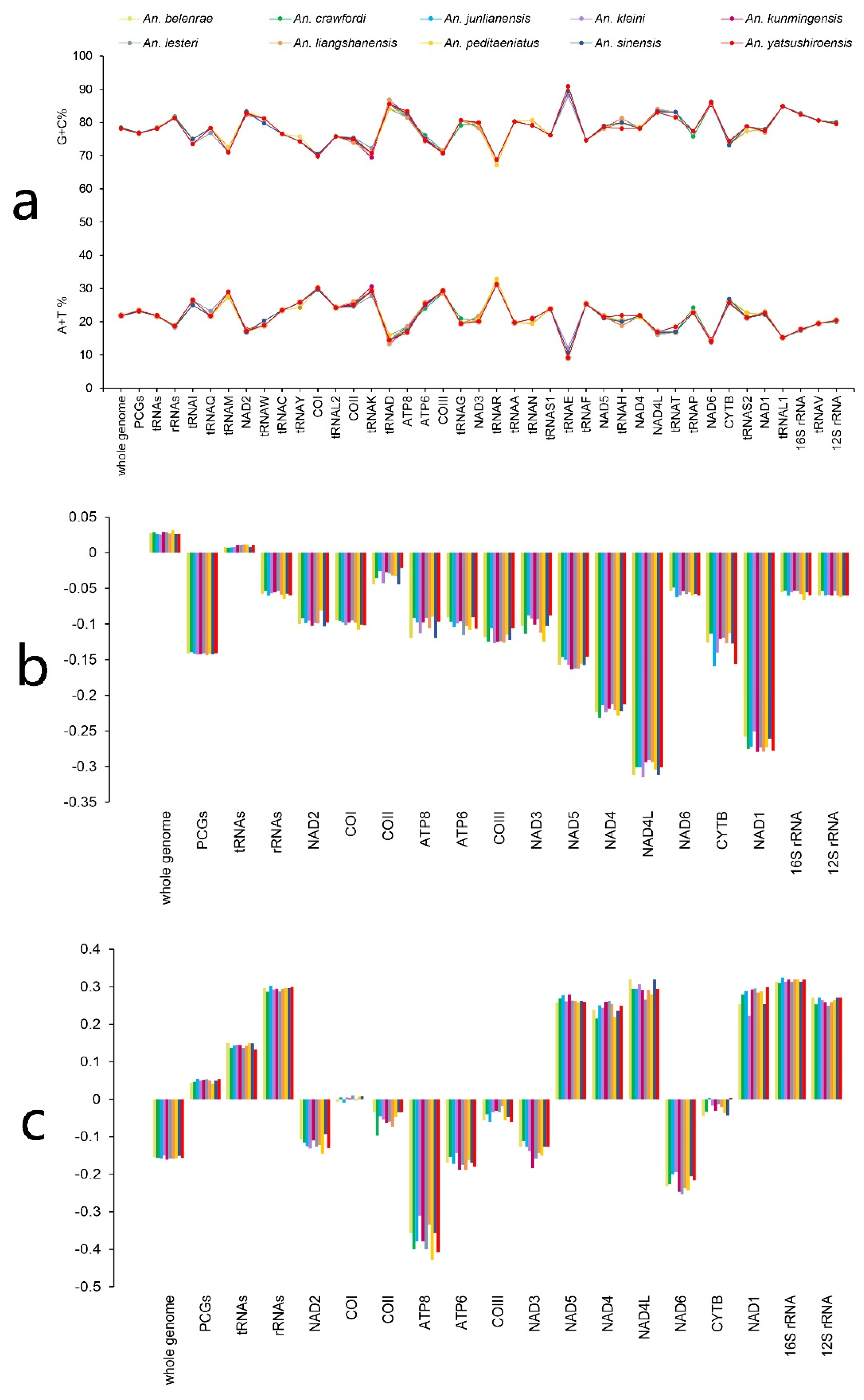
The percentages of (A + T) content in the mitochondrial genomes of 10 species in An. hyrcanus group ranged from 78.14% (An. junlianensis) to 78.41% (An. crawfordi), and the average (A + T) content was 78.23%. The average (A + T) contents of PCGs, tRNA, and rRNA coding regions were 76.78%, 78.25%, and 81.43%, respectively. The mitochondrial genomes of the ten species in Hyrcanus group had positive A–T skew values, with a mean value of 0.028, and G–C skew values were all negative, with a mean value of −0.156 (Figure 2a–c, Table S2), close to values for other mosquitoes [35,36,37].

3.2. Structural Analysis of PCGs

3.2.1. Composition and Structure of PCGs

The total length of the 13 protein-coding region genes was 11,224 bp; these were ATP6 (681 bp), ATP8 (162 bp), COI (537 bp), COII (685 bp), COIII (787 bp), CYTB (1137 bp), ND1 (945 bp), ND2 (1026 bp), ND3 (354 bp), ND4 (1342 bp), ND4L (300 bp), ND5 (1743 bp), and ND6 (525 bp). The percentages of (A + T) content of the protein-coding region genes were from 76.52% (An. liangshanensis) to 76.92% (An. kunmingensis). The average AT content of the ND6 gene was the highest (85.83%) in PCGs, while the lowest AT content was 70.09% for the COI gene. All A–T skew values of the protein-coding regions were negative. Most of the protein-coding regions had negative G–C skew values, except in the following cases: all G–C skew values of ND1, ND5, ND4, ND4L genes were positive; the COI gene G–C skew values of An. crawfordi, An. kleini, An. kunmingensis, An. lesteri, An. peditaeniatus and An. sinensis were positive, while the COI gene G–C skew value of An. yatsushiroensis was 0; and the CYTB gene G–C skew values of An. junlianensis and An. yatsushiroensis were positive. It is worth noting that all of the COI genes G–C skew values of the protein-coding regions were relatively small, and the average absolute value of the G–C skew was only 0.006. These compositional characteristics have been reported for Anophelinae and other insects [38,39].

3.2.2. Codon Usage in the Protein-Coding Regions

Among the 13 protein-coding regions, nine genes (ATP6, ATP8, COI, COII, COIII, CYTB, ND2, ND3, and ND6) were located on the J chain (forward), and the remaining four genes (ND1, ND4, ND4L, and ND5) were located on the N chain (reverse). The initiation and stop codons of the protein-coding regions of the ten species in Hyrcanus group were the same. Specifically, ATG was used as the initiation codon of ATP6, COII, COIII, CYTB, ND4, and ND4L; ATT was used as the initiation codon of ATP8, ND1, ND2, and ND6. In addition, TCG, ATA, and GTG were the initiation codons in COI, ND3, and ND5, respectively. For the stop codons, TAA was used as the stop codon of ATP6, ATP8, ND1, ND2, ND3, ND4L, ND5, ND6, and CYTB. However, the single-nucleotide T was used as the stop codon of COI, COII, COIII, and ND4. The recording of incomplete stop codons (T/TA) is common in insects, where the TAA stop codon may be completed by the addition of 3′ A residues to the mRNA after transcription [40,41].
Except for stop codons, all ten species had 3731 codons in the mitochondrial genome PCGs. The relative synonymous codon usage (RSCU) analysis showed that almost all codons were used, except for CUG (L) and AGG (S) codons, which were not used in any of the coding regions. The frequency of codon usage was different among mosquito species. The CUC (L) codon was only used in An. kleini, An. lesteri, and An. liangshanensis with an average RSCU value of 0.01; CCG (P) codons were only used in An. sinensis, An. belenrae, An. crawfordi, and An. peditaeniatus with an average RSCU value of 0.03; and CGC(R) codons were only used in An. liangshanensis, An. kunmingensis and An. peditaeniatus with an average RSCU value of 0.07. The GGC(G) codon was unused only in An. belenrae and An. peditaeniatus, while the GCG(A) codon was unused only in An. sinensis.
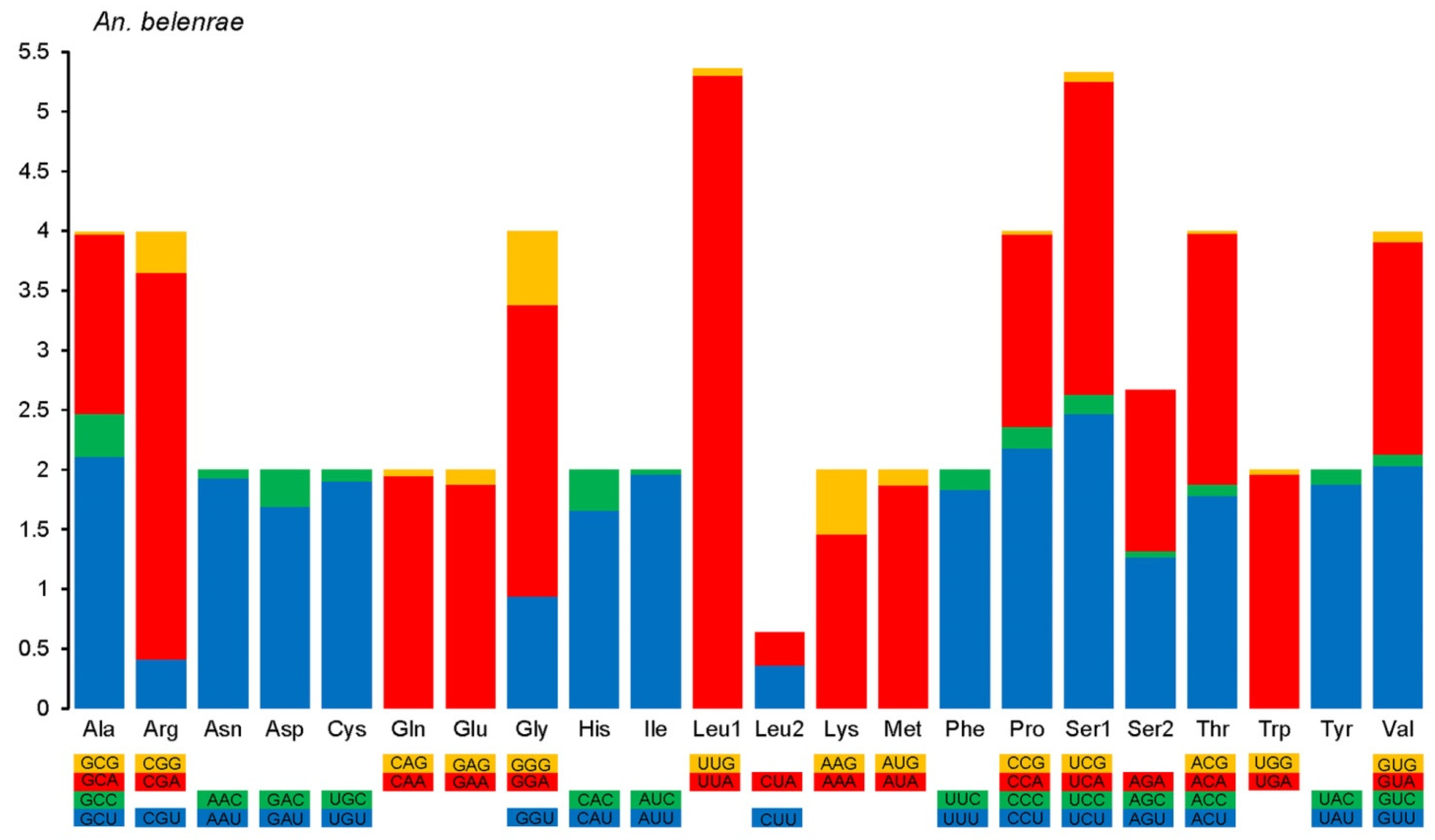
The use of the third base in synonymous codons favored adenine and thymine (uracil) over guanine and cytosine. For example, the most frequently used codon for leucine was UUA with an average RSCU value of 5.27, while the average RSCU value of UUG, which also codes for leucine, was only 0.32, and another CUG codon was not used at all. In terms of codon usage frequency, the highest was UUA (L, average RSCU 5.27), followed by CGA (R, 3.28), UCA (S, 2.55), UCU (S, 2.54), and GCU (A, 2.21), while the lowest were ACG (T, 0.03), ACU (I, 0.03), and GUC (V, 0.06) (Figure 3). The RSCU value was greater than 1 in all NNA and NNU types. This fixed distribution pattern was similar to those reported in other mosquito species [42].

3.3. tRNA Structure Analysis

The full-length tRNAs in the mitochondrial genomes of all ten species of Anopheles mosquitoes ranged from 1474 bp to 1476 bp, and the average length of individual tRNA genes ranged from 64 bp to 72 bp. Among the 22 tRNA gene coding regions in ten mitochondrial genomes, 21 predicted tRNA secondary structures that were typical cloverleaf stem-loop structures with four arms and one loop. Only the predicted secondary structure of the tRNAser1 gene was atypical, lacking a DHU arm and instead forming a DHU loop (Figure S2). This pattern has been reported in other insects [36,37].

3.4. Differential Site Analysis

Differential site analysis was performed on all mitochondrial genome PCGs between six pairs of cryptic species in the An. hyrcanus group, An. sinensis and An. belenrae, An. sinensis and An. kleini, An. belenrae and An. kleini, An. liangshanensis and An. kunmingensis, An. yatsushiroensis and An. junlianensis, and An. crawfordi and An. peditaeniatus. The difference in sites between An. sinensis and An. belenrae was the least (36, 0.32%). The ratio of transitions to transversions was 33:3. This was followed by An. junlianensis and An. yatsushiroensis (81, 0.72%, 73:8), An. kunmingensis and An. liangshanensis (123, 1.10%, 102:21), An. sinensis and An. kleini (190, 1.69%, 157:33), An. belenrae and An. kleini (192, 1.71%, 160:32), and An. crawfordi and An. peditaeniatus (513, 4.57%, 312:201). The comparison between single PCGs showed that some gene sequences were the same, including the ATP6, ATP8, COII, ND3, and ND4L genes of An. sinensis and An. belenrae, the ND3 and ND4L genes of An. junlianensis and An. yatsushiroensis, and the ND4L gene of An. kunmingensis and An. liangshanensis.
PCGs with relatively large differences included ND4 (33, 2.46%, 31:2) and ND1 (26, 2.75%, 21:5) between An. sinensis and An. kleini, ND4 (32, 2.38%, 30:2) and ND1 (26, 2.75%, 22:4) between An. belenrae and An. kleini, ND5 (25, 1.43%, 22:3) and ND1 (15, 1.59%, 15:0) between An. kunmingensis and An. liangshanensis, COI (20, 1.30%, 20:0) between An. junlianensis and An. yatsushiroensis, and ND5 (83, 4.76%, 52:31) and ATP6 (41, 6.02%, 21:20) between An. crawfordi and An. peditaeniatus. Differential sites were mostly on the third base of the codon encoding the amino acid, thus not changing the encoded amino acid, while non-synonymous variation in the encoded amino acids was mostly due to the variation in the first bases of the codons (Table 2; Figure S3).
In order to understand the level of intraspecific variation in the mitochondrial genome of Anopheles, the PCGs of three mitochondrial genomes of An. lesteri were compared. There were total 16 different sites (0.14%) among three samples of An. lesteri, showing less variation compared to interspecific variation. The variation sites were distributed in coding gene with five in COI, four in ND4, three in ND2 and ND5, two in ND1, and one in ATP8, COII, and CYTB. No variation site was detected in ATP6, COIII, ND3, ND4L, and ND6.

3.5. Phylogenetic Analysis

3.5.1. Genetic Distance

In addition to the ten species in the Hyrcanus group mentioned above, the mitochondrial genomes of four other species of Anopheles were also included in the phylogenetic analysis, An. lindesayi, An. barbirostris, An. dirus, and An. minimus. The complete mitochondrial genomes of An. lindesayi (GenBank accession no. OP324636) and An. barbirostris (GenBank accession no. OP324637) were sequenced in our lab. The An. dirus (GenBank accession no. JX219731) and An. minimus (GenBank accession no. KT895423) belonging to the subgenus Cellia were downloaded from the GenBank database. All of the PCGs of 14 Anopheles were aligned using MEGA 11 software. Nucleotide genetic distances between species were calculated based on the aligned data. The results showed that the smallest interspecies distance existed between An. sinensis and An. belenrae with a p-distance of 0.003. Other small distances existed between An. junlianensis and An. yatsushiroensis (p = 0.007), An. kunmingensis and An. liangshanensis (p = 0.011), and An. sinensis and An. kleini (p = 0.017). In addition, the pairwise p-distances between the subgenus Cellia and subgenus Anopheles were all larger than the p-distances within the subgenera (Table 3).

3.5.2. Phylogenetic Analysis Based on Sequences of 13 PCGs

The J model test was used to analyze the data to select the most suitable nucleotide substitution model; this was determined to be GTR + I + G by the AIC standard test. Based on the sequences of 13 PCGs of the mitochondrial genome, phylogenetic trees were constructed using the maximum-likelihood method (Figure 4a) and the Bayesian method (Figure 4b). The topological shapes of the two phylogenetic trees were completely consistent. The 14 species of Anopheles were divided into two clades. The An. dirus and An. minimus belonging to subgenus Cellia together formed a clade. The other clade comprised the subgenus Anopheles, including the 10 species in the An. hyrcanus group, and An. lindesayi and An. barbirostris. In the clade of subgenus Anopheles, the ten species in the An. hyrcanus group clustered together, then clustered with An. barbirostris, and finally clustered with An. lindesayi. The clustering of the An. hyrcanus group was divided into two subgroups. One subgroup contained An. crawfordi and An. peditaeniatus, and the other contained the other eight species. Among the latter, An. sinensis and An. belenrae, An. liangshanensis and An. kunmingensis, and An. yatsushiroensis and An. junlianensis were the most closely related pairs (Figure 4).

3.5.3. Phylogenetic Analysis Based on Single or Combined Coding Regions

The phylogenetic trees based on single protein-coding regions were constructed using the maximum-likelihood method as above. The results showed that the topological structures of the phylogenetic trees constructed based on only COI and ND4 genes were consistent with the phylogenetic tree constructed using the 13 PCGs, but the bootstrap values were relatively low (Figure S4a,b). Moreover, phylogenetic trees constructed based on other genes were different in topology, for example, COII and ND5 (Figure S4c,d). In addition to the sequence of a single protein-coding region, we also selected some highly discriminative coding region combinations (using COI, ND2, ND4, ND5, and ND6) to construct the phylogenetic tree. The results showed that the topological structure of the phylogenetic trees constructed based on the combination of COI + ND2 + ND4 + ND5 genes and ND2 + ND4 genes were consistent with the topological structure of the phylogenetic tree constructed using the 13 PCGs, but the bootstrap values of some branches were low (Figure S5). The phylogenetic trees were also constructed based on the full-length sequence of the tRNA and rRNA coding regions, and the topology was quite different from the phylogenetic tree constructed using the 13 PCGs (Figure S6). These sequences were clearly not suitable for analyzing the phylogenetic relationships among the members of the An. hyrcanus group.

3.5.4. Phylogenetic Analysis Based on the Complete Mitochondrial Genome Sequence

Based on the full-length mitochondrial genome sequence, including all coding and non-coding regions, we also constructed a phylogenetic tree, and the topology was consistent with the phylogenetic tree constructed using the 13 PCGs (Figures S7 and S8).

4. Discussion

In this study, we collected samples of the An. hyrcanus group distributed in different regions of China, and the species were identified by morphological and molecular characteristics. After extracting the genomic DNA of single mosquitoes, the complete mitochondrial genome sequences of ten species in the An. hyrcanus group were obtained using next-generation sequencing. The complete mitochondrial genomes of seven species were reported for the first time. The results showed that the composition and structure of mitochondrial genomes of ten species in the An. hyrcanus group were the same, and their total length and nucleotide composition characteristics were highly similar. In addition, RSCU analysis of protein coding regions showed similar features. These results were consistent with the reported characteristics of mitochondrial genomes in Anopheles mosquitoes, confirming the reliability of the sequences determined in this study.
Owing to the high morphological similarity, some species in the Hyrcanus group are taxonomically controversial [43]. Therefore, molecular methods have become an important basis for accurate identification of similar species [44]. At present, the molecular identification markers of species in the Hyrcanus group include internal transcribed spacer 2 (ITS2) and COI and COII genes of the mitochondrial genome. Ma et al. analyzed the phylogenetic relationships of 12 species in the Hyrcanus group based on ITS2 sequences [45]. Zhu et al. discussed the phylogenetic relationships of six species of the Hyrcanus group based on mitochondrial genes COI + tRNA + COII, ATP6 + COIII, ND1, and 16S rRNA genes [46]. Fang et al. studied the phylogenetic relationships of 18 species of the Hyrcanus group based on COI sequences [47]. Zhang et al. compared ITS2 and COII fragments in constructing phylogenetic trees and suggested that ITS2 sequences could be a better molecular marker to distinguish closely related mosquito species [48]. However, the lengths of ITS2 sequence fragments are different, and thus it is difficult to compare them in the reconstruction of phylogenetic relationships. It has also been suggested that the analysis of phylogenetic relationships may lose some information by using fragment reconstruction. The ITS2 sequence can be used to identify some species in the Hyrcanus group. However, the inconsistent length of sequences in various species, large intraspecific variations especially in Anopheles, and limited evolutionary information provided by a single gene sequence make it not effective for phylogenetic relationship analysis. Overall, ITS2 is suitable for molecular identification of similar species, while the 13 PCGs of the mitochondrial genome are effective for phylogenetic analysis.
Pairwise nucleotide differences in the mitochondrial genome between An. sinensis and An. belenrae were very small. In fact, An. sinensis and An. belenrae were also very similar in ITS2 sequences. Considering their high similarity in morphological and molecular characteristics and overlapping distribution ranges, we believe that An. belenrae and An. Sinensis were in the process of differentiation. However, An. kleini is different in molecular characteristics from An. sinensis and An. belenrae, and the distribution range is more inclined to high latitudes, supporting it being an independent species.
The differences between An. junlianensis and An. yatsushiroensis, and An. kunmingensis and An. liangshanensis were 0.72% and 1.10%, respectively. Some researchers believe that the two groups of Anopheles are synonyms [13]. The results of this study partially support this view, as their level of differentiation was between interspecific and intraspecific. Therefore, the two may still be in the process of species differentiation, and more samples should be collected to further clarify the differentiation level. However, the difference between An. crawfordi and An. peditaeniatus was relatively high at 4.57%. They are clearly two different species.
From the multiple phylogenetic trees constructed above, the topological structure of the phylogenetic tree constructed based on the 13 protein-coding regions was stable and consistent with the known evolution of the Hyrcanus group. Most phylogenetic trees based on single genes of mitochondrial genomes cannot distinguish well the evolutionary relationships between Hyrcanus group species, and are very different from morphological classification results. Therefore, phylogenetic relationship analysis based on single gene should be treated with caution. In addition, tRNA and rRNA coding regions were shown to be unsuitable for phylogenetic analysis. The full-length sequence contains PCGs, tRNA, and rRNA coding regions as well as non-coding regions, leading to the reduction in the discrimination compared with the 13 PCGs evolutionary tree. Certain gene combinations can be selected to construct a phylogenetic tree that is very close to the 13 PCGs phylogenetic tree. However, considering that much basic work is needed to verify the best fragment combination, we believe that 13 PCGs should be the best choice for phylogenetic analysis of the species in the Hyrcanus group [49,50].
However, we were not able to collect all species in the Hyrcanus group distributed in China. For some species, the quality of the extracted genomic DNA was insufficient for next-generation sequencing, partly because of the long storage time. In recent years, the rapid progress of urbanization in China has also led to dramatic changes in the habitat environment of Anopheles mosquitoes, making it difficult to collect some species. Moreover, there are gene polymorphisms in the mitochondrial genome, and the variation among An. hyrcanus group species is common. Differential loci analysis of three An. lesteri samples showed only 16 different sites in the whole mitochondrial genome, which indicated a low intraspecific difference. However, due to sample limitations, one sample per mosquito species was sequenced in this study, except for An. lesteri, and intraspecific differences were not considered. In the future, we will strive to collect more species and expand the sample size to further improve the mitochondrial genome data of the species in the Hyrcanus group.
In conclusion, the complete mitochondrial genome sequences of ten species in the Hyrcanus group distributed in China were obtained, seven of which were reported for the first time. The difference in mitochondrial genome sequences between An. sinensis and An. belenrae was very small, attributed to intraspecies difference, suggesting that the species was in the process of differentiation. The combination of all 13 PCG sequence was shown to be optimal for phylogenetic analysis of closely related species. The results of this study contribute to the accurate identification of the vector species of Anopheles and provide scientific basis for the prevention and control of mosquito-borne diseases.

Supplementary Materials

The following supporting information can be downloaded at: https://www.mdpi.com/article/10.3390/genes14071453/s1, Table S1: Collection and sequencing information of Anopheles lesteri in this study; Table S2: Composition of mitochondrial genome obtained in this study; Figure S1: Schematic map of collection sites for Hyrcanus group in China; Figure S2: Structures of mitochondrial DNA of the Hyrcanus group obtained in this study. The genes arranged in the inner circle are located on the N strand (Reverse), and the genes arranged in the outer circle are located on the J strand (Forward); Figure S3: Secondary structures of the Hyrcanus group tRNAs obtained in this study; Figure S4: Loci differences among 13PCGs of 10 Hyrcanus group species obtained in this study; Figure S5: Phylogenetic reconstruction by maximum likelihood based on single subunits concatenated of the species sequenced in this study and two other taxa with data available from GenBank. (a) COI subunit, (b) ND4 subunit, (c) COII subunit, (d) ND5 subunit. The values of bootstrapping support are shown to the left of the branch point; Figure S6: Phylogenetic reconstruction by maximum likelihood based on combined protein-coding subunits concatenated of the species sequenced in this study and two other taxa with data available from GenBank. (a) The combination of ND2 and ND4 subunits. (b) The combination of COI, ND2, ND4 and ND5 subunits. The values of bootstrapping support are shown to the left of the branch point; Figure S7: Phylogenetic reconstruction by maximum likelihood based on tRNA and rRNA coding region concatenated of the species sequenced in this study and two other taxa with data available from GenBank. (a) tRNAs. (b) 12S rRNA. c) 16S rRNA. The values of bootstrapping support are shown to the left of the branch point; Figure S8: Phylogenetic reconstruction by maximum likelihood based on the complete mitochondrial genome sequence concatenated of the species sequenced in this study and two other taxa with data available from GenBank. The values of bootstrapping support are shown to the left of the branch point.

Author Contributions

Conceptualization: H.P. and Y.M.; data curation: H.D., H.Y. and X.Y.; formal analysis: H.D., H.Y. and X.Y.; funding acquisition: H.P. and Y.M.; investigation: H.D., H.Y., X.Y., W.S., Q.Z., F.T., C.Z., J.B. and X.L.; project administration: H.P. and Y.M.; supervision: H.P. and Y.M.; writing—original draft: H.D. and H.Y.; writing—review and editing: H.D., H.Y., Y.M. and H.P. All authors have read and agreed to the published version of the manuscript.

Funding

This work was funded by grants from the National Natural Sciences Foundation of China (No. 31970445 to YM) and the Natural Sciences Foundation of Shanghai (No. 19ZR1469600 to HP).

Institutional Review Board Statement

Not applicable.

Informed Consent Statement

Not applicable.

Data Availability Statement

All relevant data are within the manuscript and its Supporting Information files. The sequences used and/or analyzed are available in the GenBank (OP311320–OP311329, OP324636–OP324637).

Acknowledgments

We thank MaoQing Gong from Shandong Institute of Parasitic Diseases, XueShu Dong and HongNing Zhou from Yunnan Institute of Parasitic Diseases, XinTian Lei from Sichuan Center for Disease Control and Prevention, HaiYan Huang and GuoQiang Zhang from Suifenghe Center for Disease Control and Prevention, LinHai Zen from Hainan Center for Disease Control and Prevention, Yu Ha from Jiuzhaigou Center for Disease Control and Prevention, TongShan Cao and TingTing Zhang from Donggang Center for Disease Control and Prevention, who provided field assistance in the study.

Conflicts of Interest

The authors declare no conflict of interest.

References

  1. Zhang, L.; Feng, J.; Xia, Z.; Zhou, S. Epidemiological characteristics of malaria and progress on its elimination in China in 2019. Chin. J. Parasitol. Parasit. Dis. 2020, 38, 133–138. [Google Scholar]
  2. Zhang, L.; Feng, J.; Tu, H.; Yin, J.; Xia, Z. Malaria epidemiology in China in 2020. Chin. J. Parasitol. Parasit. Dis. 2021, 39, 195–199. [Google Scholar]
  3. Zhang, L.; Yi, B.; Xia, Z.; Yin, J. Epidemiological characteristics of malaria in China, 2021. Chin. J. Parasitol. Parasit. Dis. 2022, 40, 135–139. [Google Scholar]
  4. Feng, X.; Levens, J.; Zhou, X. Protecting the gains of malaria elimination in China. Infect. Dis. Poverty 2020, 9, 43. [Google Scholar] [CrossRef] [Green Version]
  5. Zhou, Z. The malaria situation in the People’s Republic of China. Bull. World Health Organ. 1981, 59, 931–936. [Google Scholar] [PubMed]
  6. Feng, X.; Feng, J.; Zhang, L.; Tu, H.; Xia, Z. Vector control in China, from malaria endemic to elimination and challenges ahead. Infect. Dis. Poverty 2022, 11, 54. [Google Scholar] [CrossRef]
  7. Harbach, R. The classification of genus Anopheles (Diptera: Culicidae): A working hypothesis of phylogenetic relationships. Bull. Èntomol. Res. 2004, 94, 537–553. [Google Scholar] [CrossRef]
  8. van Bortel, W.; Trung, H.D.; Roelants, P.; Harbach, R.E.; Backeljau, T.; Coosemans, M. Molecular identification of Anopheles minimus s.l. beyond distinguishing the members of the species complex. Insect Mol. Biol. 2000, 9, 335–340. [Google Scholar] [CrossRef]
  9. Cohuet, A.; Toto, J.C.; Simard, F.; Kengne, P.; Fontenille, D.; Coetzee, M. Species identification within the Anopheles funestus group of malaria vectors in Cameroon and evidence for a new species. Am. J. Trop. Med. Hyg. 2003, 69, 200–205. [Google Scholar] [CrossRef] [PubMed]
  10. Tennessen, J.A.; Ingham, V.A.; Toé, K.H.; Guelbéogo, W.M.; Sagnon, N.; Kuzma, R.; Ranson, H.; Neafsey, D.E. A population genomic unveiling of a new cryptic mosquito taxon within the malaria-transmitting Anopheles gambiae complex. Mol. Ecol. 2021, 30, 775–790. [Google Scholar] [CrossRef]
  11. Ma, Y.; Xu, J. Progress of taxonomic study on the Anopheline mosquitoes in China. Chin. J. Vector Biol. Control 2015, 26, 433–438. [Google Scholar]
  12. Ma, Y.; Xu, J. The Hyrcanus group of Anopheles (Anopheles) in China (Diptera: Culicidae): Species discrimination and phylogenetic relationships inferred by ribosomal DNA internal transcribed spacer 2 sequences. J. Med. Entomol. 2005, 42, 610–619. [Google Scholar] [CrossRef]
  13. Hwang, U.W. Revisited ITS2 phylogeny of Anopheles (Anopheles) Hyrcanus group mosquitoes: Reexamination of unidentified and misidentified ITS2 sequences. Parasitol. Res. 2007, 101, 885–894. [Google Scholar] [CrossRef] [PubMed]
  14. Rueda, L.M. Two new species of Anopheles (Anopheles) Hyrcanus Group (Diptera: Culicidae) from the Republic of South Korea. Zootaxa 2005, 941, 1–26. [Google Scholar] [CrossRef]
  15. Kang, W.; Cao, Z.; Yang, Y.; Xi, Y.; Chen, H. Interspecific relationship between Anopheles kunmingensis and Anopheles liangshanensis. Sichuan J. Zool. 1992, 11, 30–31. [Google Scholar]
  16. Ma, Y.; Lei, X. Comparison of rDNA-ITS2 sequences and morphological characters of Anopheles kunmingensis and Anopheles langshanensis in China, with discussion on taxonomic status. Chin. J. Parasitol. Parasit. Dis. 2000, 18, 65–68. [Google Scholar]
  17. Ponçon, N.; Toty, C.; Kengne, P.; Alten, B.; Fontenille, D. Molecular evidence for similarity between Anopheles hyrcanus (Diptera: Culicidae) and Anopheles pseudopictus (Diptera: Culicidae), sympatric potential vectors of malaria in France. J. Med. Entomol. 2008, 45, 576–580. [Google Scholar] [CrossRef]
  18. Djadid, N.D.; Jazayeri, H.; Gholizadeh, S.; Rad, S.P.; Zakeri, S. First record of a new member of Anopheles Hyrcanus Group from Iran: Molecular identification, diagnosis, phylogeny, status of kdr resistance and Plasmodium infection. J. Med. Entomol. 2009, 46, 1084–1093. [Google Scholar] [CrossRef]
  19. Taai, K.; Baimai, V.; Saeung, A.; Thongsahuan, S.; Min, G.-S.; Otsuka, Y.; Park, M.-H.; Fukuda, M.; Somboon, P.; Choochote, W. Genetic compatibility between Anopheles lesteri from Korea and Anopheles paraliae from Thailand. Mem. Inst. Oswaldo Cruz 2013, 108, 312–320. [Google Scholar] [CrossRef] [Green Version]
  20. Beebe, N.W. DNA barcoding mosquitoes: Advice for potential prospectors. Parasitology 2018, 145, 622–633. [Google Scholar] [CrossRef] [Green Version]
  21. Jones, C.M.; Lee, Y.; Kitchen, A.; Collier, T.; Pringle, J.C.; Muleba, M.; Irish, S.; Stevenson, J.C.; Coetzee, M.; Cornel, A.J.; et al. Complete Anopheles funestus mitogenomes reveal an ancient history of mitochondrial lineages and their distribution in southern and central Africa. Sci. Rep. 2018, 8, 9054. [Google Scholar] [CrossRef] [Green Version]
  22. da Silva, A.F.; Machado, L.C.; de Paula, M.B.; Vieira, C.J.D.S.P.; Bronzoni, R.V.D.M.; Santos, M.A.V.D.M.; Wallau, G.L. Culicidae evolutionary history focusing on the Culicinae subfamily based on mitochondrial phylogenomics. Sci. Rep. 2020, 10, 18823. [Google Scholar] [CrossRef]
  23. Chen, B.; Zhang, Y.-J.; He, Z.; Li, W.; Si, F.; Tang, Y.; He, Q.; Qiao, L.; Yan, Z.; Fu, W.; et al. De novo transcriptome sequencing and sequence analysis of the malaria vector Anopheles sinensis (Diptera: Culicidae). Parasites Vectors 2014, 7, 314. [Google Scholar] [CrossRef] [PubMed] [Green Version]
  24. Guo, J.; Yan, Z.; Fu, W.; Yuan, H.; Li, X.; Chen, B. Complete mitogenomes of Anopheles peditaeniatus and Anopheles nitidus and phylogenetic relationships within the genus Anopheles inferred from mitogenomes. Parasites Vectors 2021, 14, 452. [Google Scholar] [CrossRef]
  25. Liu, W.-P.; Zhang, P.-Y.; Xu, S.; Tang, J.-X.; Zhou, H.-Y.; Li, J.-L.; Wang, D.-J.; Liu, Y.-Y.; Fang, Q.; Xia, H.; et al. The complete mitochondrial genome of major malaria vector Anopheles anthropophagus (Diptera: Culicidae) in China. Mitochondrial DNA Part B 2022, 7, 482–484. [Google Scholar] [CrossRef] [PubMed]
  26. Bang, W.J.; Kim, H.C.; Ryu, J.; Lee, H.S.; Lee, S.Y.; Kim, M.S.; Chong, S.T.; Klein, T.A.; Choi, K.S. Multiplex PCR assay for the identification of eight Anopheles species belonging to the Hyrcanus, Barbirostris and Lindesayi groups. Malar. J. 2021, 20, 287. [Google Scholar] [CrossRef] [PubMed]
  27. Chen, S.; Zhou, Y.; Chen, Y.; Gu, J. fastp: An ultra-fast all-in-one FASTQ preprocessor. Bioinformatics 2018, 34, i884–i890. [Google Scholar] [CrossRef] [PubMed]
  28. Dierckxsens, N.; Mardulyn, P.; Smits, G. NOVOPlasty: De novo assembly of organelle genomes from whole genome data. Nucleic Acids Res. 2016, 45, e18. [Google Scholar] [CrossRef] [Green Version]
  29. Meng, G.; Li, Y.; Yang, C.; Liu, S. MitoZ: A toolkit for animal mitochondrial genome assembly, annotation and visualization. Nucleic Acids Res. 2019, 47, e63. [Google Scholar] [CrossRef] [PubMed] [Green Version]
  30. Tamura, K.; Stecher, G.; Kumar, S. MEGA11: Molecular Evolutionary Genetics Analysis Version 11. Mol. Biol. Evol. 2021, 38, 3022–3027. [Google Scholar] [CrossRef] [PubMed]
  31. Bernt, M.; Donath, A.; Jühling, F.; Externbrink, F.; Florentz, C.; Fritzsch, G.; Pütz, J.; Middendorf, M.; Stadler, P.F. MITOS: Improved de novo metazoan mitochondrial genome annotation. Mol. Phylogenetics Evol. 2013, 69, 313–319. [Google Scholar] [CrossRef] [PubMed]
  32. Darriba, D.; Taboada, G.L.; Doallo, R.; Posada, D. jModelTest 2: More models, new heuristics and parallel computing. Nat. Methods 2012, 9, 772. [Google Scholar] [CrossRef] [PubMed] [Green Version]
  33. Ronquist, F.; Teslenko, M.; van der Mark, P.; Ayres, D.L.; Darling, A.; Höhna, S.; Larget, B.; Liu, L.; Suchard, M.A.; Huelsenbeck, J.P. MrBayes 3.2: Efficient Bayesian Phylogenetic Inference and Model Choice across a Large Model Space. Syst. Biol. 2012, 61, 539–542. [Google Scholar] [CrossRef] [Green Version]
  34. Letunic, I.; Bork, P. Interactive Tree of Life (iTOL) v5: An online tool for phylogenetic tree display and annotation. Nucleic Acids Res. 2021, 49, W293–W296. [Google Scholar] [CrossRef]
  35. Hao, Y.; Zou, Y.; Ding, Y.; Xu, W.; Yan, Z.; Li, X.; Fu, W.; Li, T.; Chen, B. Complete mitochondrial genomes of Anopheles stephensi and An. dirus and comparative evolutionary mitochondriomics of 50 mosquitoes. Sci. Rep. 2017, 7, 7666. [Google Scholar] [CrossRef] [PubMed]
  36. Martinez-Villegas, L.; Assis-Geraldo, J.; Koerich, L.; Collier, T.C.; Lee, Y.; Main, B.; Rodrigues, N.B.; Orfano, A.S.; Pires, A.C.A.M.; Campolina, T.B.; et al. Characterization of the complete mitogenome of Anopheles aquasalis, and phylogenetic divergences among Anopheles from diverse geographic zones. PLoS ONE 2019, 14, e0219523. [Google Scholar] [CrossRef] [Green Version]
  37. do Nascimento, B.L.S.; da Silva, F.S.; Nunes-Neto, J.P.; de Almeida Medeiros, D.B.; Cruz, A.C.R.; da Silva, S.P.; da Silva ESilva, L.H.; de Oliveira Monteiro, H.A.; Dias, D.D.; Vieira, D.B.R.; et al. First Description of the Mitogenome and Phylogeny of Culicinae Species from the Amazon Region. Genes 2021, 12, 1983. [Google Scholar] [CrossRef]
  38. Oliveira, T.M.P.; Foster, P.G.; Bergo, E.S.; Nagaki, S.S.; Sanabani, S.S.; Marinotti, O.; Marinotti, P.N.; Sallum, M.A.M. Mitochondrial Genomes of Anopheles (Kerteszia) (Diptera: Culicidae) From the Atlantic Forest Brazil. J. Med. Entomol. 2016, 53, 790–797. [Google Scholar] [CrossRef]
  39. da Silva, F.S.; Cruz, A.C.R.; Medeiros, D.B.D.A.; da Silva, S.P.; Nunes, M.R.T.; Martins, L.C.; Chiang, J.O.; Lemos, P.D.S.; Cunha, G.M.; de Araujo, R.F.; et al. Mitochondrial genome sequencing and phylogeny of Haemagogus albomaculatus, Haemagogus leucocelaenus, Haemagogus spegazzinii, and Haemagogus tropicalis (Diptera: Culicidae). Sci. Rep. 2020, 10, 16948. [Google Scholar] [CrossRef] [PubMed]
  40. Chen, K.; Wang, Y.; Li, X.-Y.; Peng, H.; Ma, Y.-J. Sequencing and analysis of the complete mitochondrial genome in Anopheles sinensis (Diptera: Culicidae). Infect. Dis. Poverty 2017, 6, 149. [Google Scholar] [CrossRef] [Green Version]
  41. Chu, H.; Li, C.; Guo, X.; Zhang, H.; Luo, P.; Wu, Z.; Wang, G.; Zhao, T. The phylogenetic relationships of known mosquito (Diptera: Culicidae) mitogenomes. Mitochondrial DNA Part A 2018, 29, 31–35. [Google Scholar] [CrossRef] [PubMed]
  42. da Silva E Silva, L.H.; Da Silva, F.S.; Medeiros, D.B.D.A.; Cruz, A.C.R.; da Silva, S.P.; Aragão, A.D.O.; Dias, D.D.; Sena Do Nascimento, B.L.; Júnior, J.W.R.; Vieira, D.B.R.; et al. Description of the mitogenome and phylogeny of Aedes spp. (Diptera: Culicidae) from the Amazon region. Acta Trop. 2022, 232, 106500. [Google Scholar] [CrossRef] [PubMed]
  43. Paredes-Esquivel, C.; Harbach, R.E.; Townson, H. Molecular taxonomy of members of the Anopheles hyrcanus group from Thailand and Indonesia. Med. Vet. Èntomol. 2011, 25, 348–352. [Google Scholar] [CrossRef] [PubMed]
  44. Chan, A.; Chiang, L.; Hapuarachchi, H.C.; Tan, C.; Pang, S.; Lee, R.; Lee, K.; Ng, L.; Lam-Phua, S. DNA barcoding: Complementing morphological identification of mosquito species in Singapore. Parasites Vectors 2014, 7, 569. [Google Scholar] [CrossRef]
  45. Ma, Y.; Ma, Y.; Lin, L.; Wang, Y. Phylogenetic relationship among some species of genus Anopheles subgenus Anopheles (Diptera: Culicidae) in China: Based on rDNA-ITS2 sequences. Chin. J. Vector Biol. Control 2013, 24, 382–388. [Google Scholar]
  46. Zhu, H.; Luo, S.; Gao, M.; Tao, F.; Gao, J.; Chen, H.; Li, X.; Peng, H.; Ma, Y. Phylogeny of certain members of Hyrcanus group (Diptera: Culicidae) in China based on mitochondrial genome fragments. Infect. Dis. Poverty 2019, 8, 91. [Google Scholar] [CrossRef] [PubMed] [Green Version]
  47. Fang, Y.; Shi, W.-Q.; Zhang, Y. Molecular phylogeny of Anopheles hyrcanus group (Diptera: Culicidae) based on mtDNA COI. Infect. Dis. Poverty 2017, 6, 61. [Google Scholar] [CrossRef] [PubMed] [Green Version]
  48. Zhang, C.; Yang, R.; Wu, L.; Luo, C.; Guo, X.; Deng, Y.; Zhou, H.; Zhang, Y. Molecular phylogeny of the Anopheles hyrcanus group (Diptera: Culicidae) based on rDNA–ITS2 and mtDNA–COII. Parasites Vectors 2021, 14, 454. [Google Scholar] [CrossRef] [PubMed]
  49. Foster, P.G.; de Oliveira, T.M.P.; Bergo, E.S.; Conn, J.E.; Sant’ana, D.C.; Nagaki, S.S.; Nihei, S.; Lamas, C.E.; González, C.; Moreira, C.C.; et al. Phylogeny of Anophelinae using mitochondrial protein coding genes. R. Soc. Open Sci. 2017, 4, 170758. [Google Scholar] [CrossRef] [Green Version]
  50. Morón-López, J.; Vergara, K.; Sato, M.; Gajardo, G.; Ueki, S. Intraspecies variation of the mitochondrial genome: An evaluation for phylogenetic approaches based on the conventional choices of genes and segments on mitogenome. PLoS ONE 2022, 17, e0273330. [Google Scholar] [CrossRef]
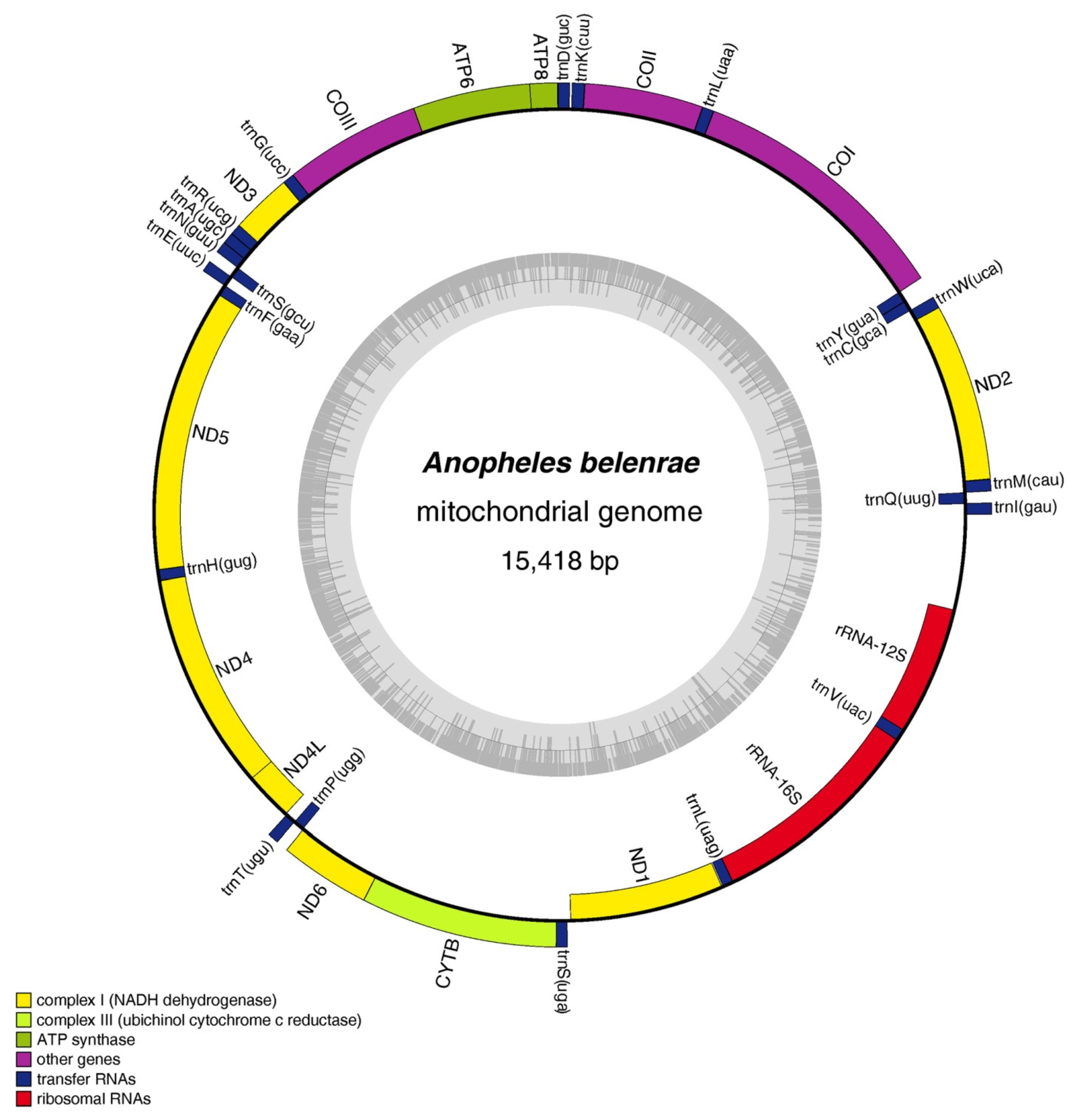
Figure 1. Structures of mitochondrial DNA of An. belenrae in the hyrcanus group obtained in this study. The mitochondrial genome structures of the other nine Anopheles species in this study were similar to that of An. belenrae and are listed in Figure S1. The grey inner ring represents the GC content pattern.
Genes 14 01453 g001
Figure 2. Information on AT/GC contents and AT/GC-skew of the obtained mitochondrial DNAs. (a) A + T and G + C contents (%). (b) A–T skew. (c) G–C skew.
Genes 14 01453 g002
Figure 3. Relative synonymous codon usage (RSCU) of An. belenrae mitochondrial DNA. RSCU values are represented on the y-axis, and families of synonymous codons and their respective amino acids are on the x-axis. Colored squares indicate the difference in the third base of the codon: orange for G, red for A, green for C, and blue for U (T).
Genes 14 01453 g003
Figure 4. Phylogenetic reconstruction by maximum-likelihood and Bayesian inference based on the 13 PCGs concatenated of the species sequenced in this study and 2 other taxa with data available from GenBank. (a) Maximum-likelihood tree. The values of bootstrapping support are shown to the left of the branch point. (b) Bayesian inference tree. The values of Bayesian probabilities are shown on the left in each node.
Genes 14 01453 g004
Table 1. Collection and sequencing information of Hyrcanus group in this study.
SpeciesCollection SiteYearCoordinateRaw Data ReadsClean Data ReadsMapped ReadsMean Depth
An. belenrae *Jining City, Shandong Province, China1999116.50° E, 35.32° N147,214,292143,104,848162,9475755
An. crawfordi *Puer City, Yunnan Province, China2005101.08° E, 22.77° N90,487,88285,354,442826,3147538
An. junlianensis *Junlian City, Sichuan Province, China1997104.50° E, 28.15° N119,378,626114,520,300997183
An. kleini *Suifenhe City, Heilongjiang Province, China2018131.15° E, 44.41° N87,311,92887,130,712163,5121438
An. kunmingensis *Kunming City, Yunnan Province, China1997102.61° E, 24.80° N88,922,52086,777,074475,5104387
An. lesteriPujiang City, Sichuan Province, China2006103.51° E, 30.20° N57,068,18456,937,040266,7952553
An. liangshanensis *Zhaojue City, Sichuan Province, China1997102.80° E, 28.01° N80,987,41880,314,882714,7086716
An. peditaeniatusMeilan District, Hainan Province, China2013110.51° E, 19.99° N101,979,70891,150,57448,331396
An. sinensisJiuzhaigou City, Sichuan Province, China2015104.28° E, 33.24° N121,676,660120,939,1421,180,1777656
An. yatsushiroensis *Donggang City, Liaoning Province, China2000124.15° E, 39.86° N117,486,358114,193,620162,9471511
* means the mitochondrial genome sequence was first reported in this study.
Table 2. Loci differences between 6 pairs of closely related species in Hyrcanus group.
ATP6ATP8COICOIICOIIICYTBND1ND2ND3ND4ND4LND5ND6PCGs
Total number of sites 6811621537685787113794510263541342300174352511,224
Single base substitution
(transitions: transversions)
SIN and BELNN7:2N2:05:02:17:0N5:0N3:02:033:3
SIN and KLE10:20:123:54:39:020:421:514:21:131:20:121:73:0157:33
KLE and BEL10:20:125:54:39:019:422:417:21:130:20:120:73:0160:32
JUN and YAT2:12:020:02:24:08:33:07:0N9:1N13:13:073:8
KUN and LIA6:21:010:33:27:37:115:012:23:015:5N22:31:0102:21
PED and CRA21:205:435:2421:1119:1340:2824:2029:86:542:287:352:3111:5312:201
Total amino acid number 227545122282623793153421184471005811753741
Amino acid mutation
(synonymous: non-synonymous)
SIN and BELNN8:1N2:05:03:05:2N5:0N3:02:033:3
SIN and KLE11:10:128:07:08:124:024:114:22:030:11:028:03:0180:7
KLE and BEL11:10:129:17:08:123:024:119:02:029:11:027:03:0183:6
JUN and YAT3:02:020:04:04:011:03:06:1N10:0N14:03:080:1
KUN and LIA8:00:113:05:010:08:015:014:03:018:2N24:11:0119:4
PED and CRA39:19:059:032:030:267:042:237:010:170:09:179:414:2497:13
N means no differences between pairwise data. BEL = Anopheles belenrae, CRA = Anopheles crawfordi, JUN = Anopheles junlianensis, KLE = Anopheles kleini, KUN = Anopheles kunmingensis, LIA = Anopheles liangshanensis, PED = Anopheles peditaeniatus, SIN = Anopheles sinensis, YAT = Anopheles yatsushiroensis.
Table 3. The pairwise interspecific p-distances of mitochondrial DNA of the 14 Anopheles species calculated by 13PCGs.
BELCRAJUNKLEKUNLESLIAPEDSINYATBARLINDIR
CRA0.048
JUN0.0400.049
KLE0.0170.0490.042
KUN0.0420.0510.0350.041
LES0.0410.0480.0370.0420.037
LIA0.0440.0530.0360.0420.0110.036
PED0.0490.0460.0490.0490.0500.0500.052
SIN0.0030.0480.0390.0170.0410.0410.0430.048
YAT0.0400.0500.0070.0420.0360.0360.0380.0490.040
BAR0.0770.0750.0760.0780.0780.0780.0810.0750.0770.076
LIN0.0880.0860.0900.0900.0900.0900.0910.0890.0880.0910.090
DIR0.0960.0950.0970.0990.1000.1000.1010.0990.0960.0970.0970.101
MIN0.0950.0930.0960.0980.0970.0960.0980.0960.0950.0960.0900.0980.098
BEL = Anopheles belenrae, CRA = Anopheles crawfordi, JUN = Anopheles junlianensis, KLE = Anopheles kleini, KUN = Anopheles kunmingensis, LES = Anopheles lesteri, LIA = Anopheles liangshanensis, PED = Anopheles peditaeniatus, SIN = Anopheles sinensis, YAT = Anopheles yatsushiroensis, BAR = Anopheles barbirostris, LIN = Anopheles lindesayi, DIR = Anopheles dirus, MIN = Anopheles minimus.
Disclaimer/Publisher’s Note: The statements, opinions and data contained in all publications are solely those of the individual author(s) and contributor(s) and not of MDPI and/or the editor(s). MDPI and/or the editor(s) disclaim responsibility for any injury to people or property resulting from any ideas, methods, instructions or products referred to in the content.