Article

A Novel Transcriptome Approach to the Investigation of the Molecular Pathology of Vitreous and Retinal Detachment


1
John van Geest Centre for Brain Repair, Cambridge CB2 0PY, UK
2
Department of Pathology, University of Cambridge, Cambridge, CB2 1TN, UK
*
Author to whom correspondence should be addressed.
Genes 2022, 13(10), 1885; https://doi.org/10.3390/genes13101885
Submission received: 8 August 2022 / Revised: 11 October 2022 / Accepted: 13 October 2022 / Published: 18 October 2022
(This article belongs to the Special Issue Genetics in Stickler Syndrome)

Abstract

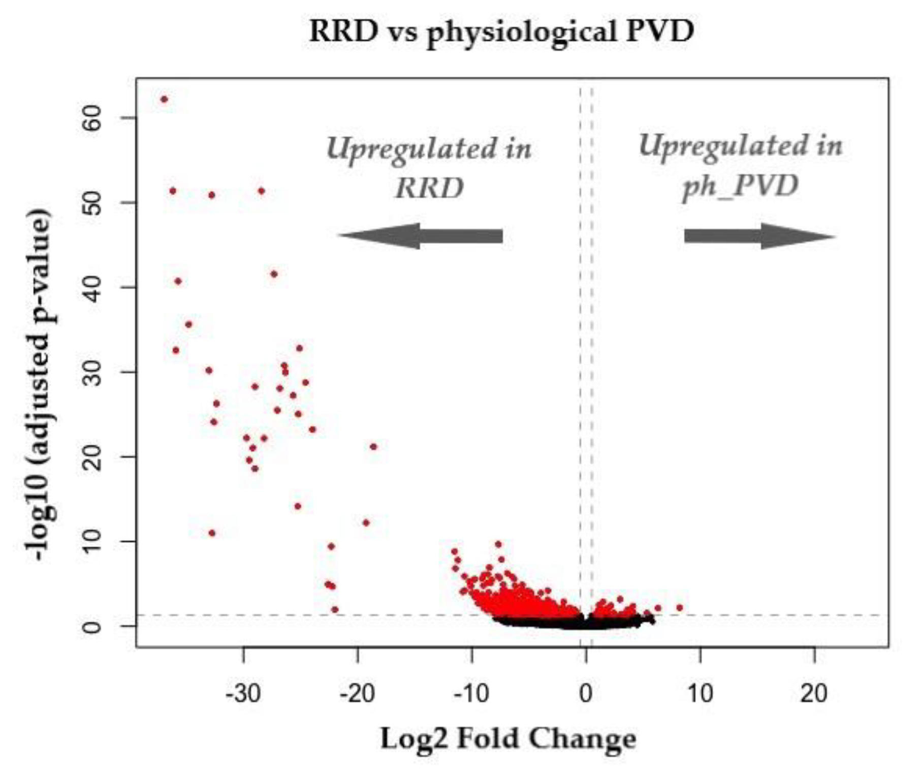
Retinal detachment (RD) is one of the most common, sight-threatening ocular conditions requiring emergency intervention. Posterior vitreous detachment (PVD) occurs in the majority of an aging population whereby the vitreous body separates from the retina. It is well established that PVD is the common precursor to the most common forms of RD; however, it remains unknown why in most individuals PVD will cause no/few complications (physiological PVD) but in a small percentage will cause retinal tears and detachment (pathological PVD). Despite over 100 years of scientific research, the anatomical definitions of PVD and its pathogenesis remain controversial. Recent research has identified a novel cell population (laminocyte), present at significantly higher numbers in pathological PVD when compared to physiological PVD. We review and summarise the seven distinct clinical sub-groups of retinal breaks and focus on the role of the laminocyte in those secondary to PVD and the transcriptomic profile of this unique cell. Provisional whole transcriptome analysis using bulk RNA-Seq shows marked differentially expressed genes when comparing physiological PVD with PVD associated with RD. The limitations of bulk RNA-Seq are considered and the potential to address these using spatial transcriptomics are discussed. Understanding the pathogenesis of PVD-related retinal tears will provide a baseline for the development of novel therapeutic targets and prophylactic treatments.
Keywords: posterior vitreous detachment (PVD); retinal detachment (RD); rhegmatogenous retinal detachment (RRD); posterior hyaloid membrane (PHM); laminocyte; RNA sequencing (RNA-seq); spatial transcriptomics posterior vitreous detachment (PVD); retinal detachment (RD); rhegmatogenous retinal detachment (RRD); posterior hyaloid membrane (PHM); laminocyte; RNA sequencing (RNA-seq); spatial transcriptomics