Article

Spatial and Temporal Expression Characteristics of the HBB Gene Family in Six Different Pig Breeds

1
Laboratory of Genetic Breeding, Reproduction and Precision Livestock Farming, School of Animal Science and Nutritional Engineering, Wuhan Polytechnic University, Wuhan 430023, China
2
Institute of Animal Sciences, Chinese Academy of Agricultural Sciences, Beijing 100193, China
3
Genome Analysis Laboratory of the Ministry of Agriculture and Rural Affairs, Agricultural Genomics Institute at Shenzhen, Chinese Academy of Agricultural Sciences, Shenzhen 518124, China
*
Author to whom correspondence should be addressed.
Genes 2022, 13(10), 1822; https://doi.org/10.3390/genes13101822
Submission received: 31 August 2022 / Revised: 28 September 2022 / Accepted: 3 October 2022 / Published: 9 October 2022
(This article belongs to the Topic Animal Models of Human Disease)

Abstract

:

Simple Summary

β-Thalassemia is one of the most prevalent inherited diseases in China. It is important to develop animal models to accurately simulate human β-thalassemia and there are unique advantages to studying β-thalassemia in pigs. However, there are only few reports on the systematic analysis of the β-thalassemia-related genes and their expression pattern in pigs so far. Therefore, in this study, we firstly predicted 11 porcine hemoglobin-encoding genes and found that there was no HBG gene in pigs, indicating that the globin switches might not exist in pigs. A new hemoglobin-encoding gene, ‘HBB-like’, was found in pigs, which showed high conservation in its amino sequences between pigs and humans. Then, we studied the evolutionary relationship of hemoglobin-encoding genes in human, pig and mouse. The results showed that the β-chain structure of pig and human was highly similar. In addition, we analyzed the hemoglobin-encoding gene expressions by using the iswine database and qPCR. Our results showed significant differences in the spatiotemporal expression patterns among the four genes (HBA, HBB, HBB-like and HBE) in three developmental stages of six different pig breeds. Our study provides an important theoretical basis for further construction of a gene-edited β-thalassemia miniature pig model to assess β--thalassemia treatments.

Abstract

β-Thalassemia induces hemolytic anemia caused by mutations in the β-chain gene locus. As humans progress from embryo to adulthood, hemoglobin recombines twice. To test whether similar hemoglobin reassembly occurs in pigs, bioinformatics tools were used to predict the pig hemoglobin-encoding gene. We then systematically analyzed the expression patterns of the HBB gene family in three developmental stages (weaning, sexual maturity and physical maturity) of six different pig breeds (Landrace, Yorkshire, Wuzhishan, Songliao black, Meishan and Tibetan). The results showed that the new hemoglobin coding gene ‘HBB-like’ was found in pigs, while the HBG gene did not exist in pigs, indicating that human-like reassembly might not exist in pigs. The HBB and HBB-like genes shared highly similar amino acid sequences and gene sequences. The genes on the β-chain were highly similar between humans and pigs and the amino acid sequences of human and pig HBB genes at position 26 and positions 41–42 were identical. qPCR results showed that there were significant differences in the spatiotemporal expression patterns of the four genes (HBA, HBB, HBB-like and HBE) across breeds. Our results provide a foundation for follow-up studies assessing the relationship between the gene-encoding hemoglobin and β-thalassemia disease, as well as the construction of a gene-edited β-thalassemia miniature pig model to assess β-thalassemia treatments.

1. Introduction

Thalassemias are common autosomal recessive disorders, especially in populations in the Mediterranean region, the Indian subcontinent, Southeast Asia and West Africa [1,2]. Although there are many forms of thalassemia, β-thalassemia is the most common form, characterized by microcytosis and hypochromic anemia, resulting from mutations in the human β-globin locus [3,4]. Nearly 80 to 90 million with minor β-thalassemia and 60–70 thousand affected infants are born annually worldwide [5]. Haemoglobins (Hb) have a tetrameric structure, consisting of two α-like (α or ζ) and two β-like (ε, γ, δ or β) globin chains, each linked to a heme group [6]. Normal hemoglobins include Hb Portland (ζ2γ2), Hb Gower 1 (ζ2ε2), Hb Gower 2 (α2ε2), HbF (α2γ2), HbA1 (α2β2) and HbA2 (α2δ2) [7]. As an individual develops, the globin subunits making up hemoglobin undergo reassembly during switching [8]. The erythrocytes in an embryo contain Hb Portland, Hb Gower 1 and Hb Gower 2; the erythrocytes in a fetus contain predominantly fetal HbF; the erythrocytes in adults contain two adult HbA1 and HbA2 [9,10].
The genes involved in forming hemoglobin at the α-globin locus are ζ-globin gene (HBZ) and α-globin gene (HBA). The β-globin gene locus includes ε-globin gene (HBE), -globin gene (HBG2), -globin gene (HBG1), δ-globin gene (HBD) and β-globin gene (HBB) [11]. The β-globin genes (i.e., fetal ɤ-globin and adult β-globin) are expressed in a perfectly tuned way only at specific developmental stages. There are two globin switches in humans, which are HBE to HBG1/HBG2, occurring in the transition from primitive to definitive erythropoiesis and HBG1/HBG2 to HBB occurring in definitive erythropoiesis around the time of birth [12].
To date, more than 900 β-globin variants have been recorded worldwide. These gene alterations often cause either a decrease in β-globin synthesis or completely block synthesis [13]. For example, HbE/β-thalassemia is a GA substitution at the 26 position of the β-globin gene and replaces Glu with Lys, resulting in a decrease in the β-globin chain [14,15,16,17]. As such, 1–3% of the people in Southern China carry a β-thalassemia allele and over 20 β-thalassemia mutations have been reported in the Chinese population [18]. The most common type of β-thalassemia gene mutation in Southern China is CD41–42 (−CTTT), which accounts for approximately 40% of total β-thalassemia mutations in China. It is caused by the deletion of 4-bp in the HBB gene due to a frameshift mutation [19,20]. With widespread population migration, β-thalassemia diseases present an increasing challenge to health services in developing countries [21,22].
Animal models recapitulating both the phenotype and genotype of human disease are valuable in the exploration of pathophysiology and for in vivo evaluation of novel therapeutic treatments. Therefore, it is important to develop animal models to accurately simulate human β-thalassemia for gene therapy or for other more effective therapies. Pig is the optimal experimental animal model for performing clinical research on human disease [23,24,25,26]. Moreover, pigs have unique advantages in studying β-thalassemia and other related blood diseases, compared with mice and rabbits [27,28]. However, there are only few reports on the systematic analysis of the β-thalassemia-related genes and their expression pattern in pigs so far. In this study, we used bioinformatics tools to predict the pig hemoglobin-encoding genes to test whether similar hemoglobin reassembly occurs in pigs. We also systematically analyzed the spatiotemporal expression patterns of the four genes (HBA, HBB, HBB-like and HBE) in three developmental stages (weaning, sexual maturity and physical maturity) of six different pig breeds (Landrace, Yorkshire, Wuzhishan, Songliao black, Meishan and Tibetan). Our results will provide a foundation for further studies assessing the relationship between the gene-encoding pig hemoglobin and β-thalassemia disease, as well as the construction of a gene-edited β-thalassemia miniature pig model to assess β-thalassemia treatments.

2. Materials and Methods

2.1. The Identification of Hemoglobin-Encoding Genes in Pigs and Mice

We downloaded the human hemoglobin-encoding genes sequences from NCBI database to search against the hemoglobin-encoding gene sequences in pig and mouse using the BLASTP program with an e-value of 1 × 1050 as the threshold. We preliminarily identified the hemoglobin-encoding genes in pig and mouse by analyzing the results from HMM and BLASTP. We then used the NCBI-CDD web server (http://www.ncbi.nlm.nih.gov/Struc-ture/cdd/wrpsb.cgi, accessed on 23 November 2021) to confirm the prediction [29].

2.2. Evolutionary Analysis and Motif Prediction of Human, Mouse and Pig Hemoglobin-Encoding Genes

The amino acid sequences of the hemoglobin-encoding genes in human, pig and mouse were downloaded from the NCBI website. Multiple alignments of the amino acid sequences were performed using ClustalW [30]. The phylogenetic analysis was constructed based on the amino acid sequences of the hemoglobin-encoding genes form human, pig and mouse using a neighbor-joining (NJ) method with 1000 bootstrap replicates and visualized with MEGA5 software. Protein motifs were predicted by using Multiple Em for Motif Elicitation (MEME) (https://meme-suite.org/meme/tools/meme, accessed on 27 April 2022) and protein domain functions were analyzed from Uniprot (https://www.uniprot.org/, accessed on 27 April 2022).

2.3. Primer Design

The primers were designed using Primer 5.0 software (Palo Alto, CA, USA) and were synthesized by Sangon Bioengineering Co., Ltd. (Shanghai, China) (Table 1). The forward primer (HBB-2F) for the quantitative detection of the HBB copy number did not bind to the HBB-like gene.

2.4. Reverse Transcription Quantitative Real-Time PCR (RT-qPCR)

RT-qPCR analysis of the four genes (HBB, HBB-like, HBE and HBA) in five tissues (heart, liver, spleen, lung and kidney) of six pig breeds (Landrace, Yorkshire, Wuzhishang, Songliao black, Meishang, Tibetan) at different development stages was performed. Total RNA was extracted using an RNA extraction kit (MagaBio plus, Hangzhou, Chian). cDNA was synthesized using a RevertAid First Strand cDNA Synthesis Kit (Thermo Scientific, USA). qPCR was performed on an ABI 7500 machine using the SYBR Premix Ex Taq Kit (Vazyme, Nanjing, China) and 18S ribosomal ribonucleic acid (18S rRNA) was used as an endogenous control gene. Relative mRNA expression levels were calculated using the Lg2-ΔΔCt method [31]; the 2−∆∆Ct method was reviewed in [32].

2.5. Statistics

The experimental data were analyzed using Prism software (Graphpad prism 8.0). Multiple comparisons of data from multiple groups were performed using analysis of variance (ANOVA). The data are presented as the mean ± standard error of mean (SEM). The level of significance is p < 0.05.

3. Results

3.1. Identification of the Pig Hemoglobin-Encoding Gene Family

In this study, a total of 11 pig hemoglobin-encoding genes and 10 mouse hemoglobin-encoding genes were obtained. Genome-wide hemoglobin-encoding genes in humans, pigs and mice were listed in Table 2. Human hemoglobin-encoding genes were located on chromosomes 11 and 16, while pig hemoglobin-encoding genes were mainly located on chromosomes 9 and 3 and mouse hemoglobin-encoding genes were located on chromosomes 7 and 11. In the prediction of the pig HBB gene family, the HBB-like gene was also found on chromosome 9. In the NCBI database, the three genes on pig chromosome 9 were sequenced from upstream to downstream as HBE, the HBB-like and HBB gene. The four genes on pig chromosome 3 were sequenced from upstream to downstream as HBZ, HBM, HBA and HBQ gene (date were not shown). No new gene HBG(γ) was found and there might not be a double switch similar in pigs to that in humans on the β-globin chain. No HBD and HBM genes were predicted in mice.

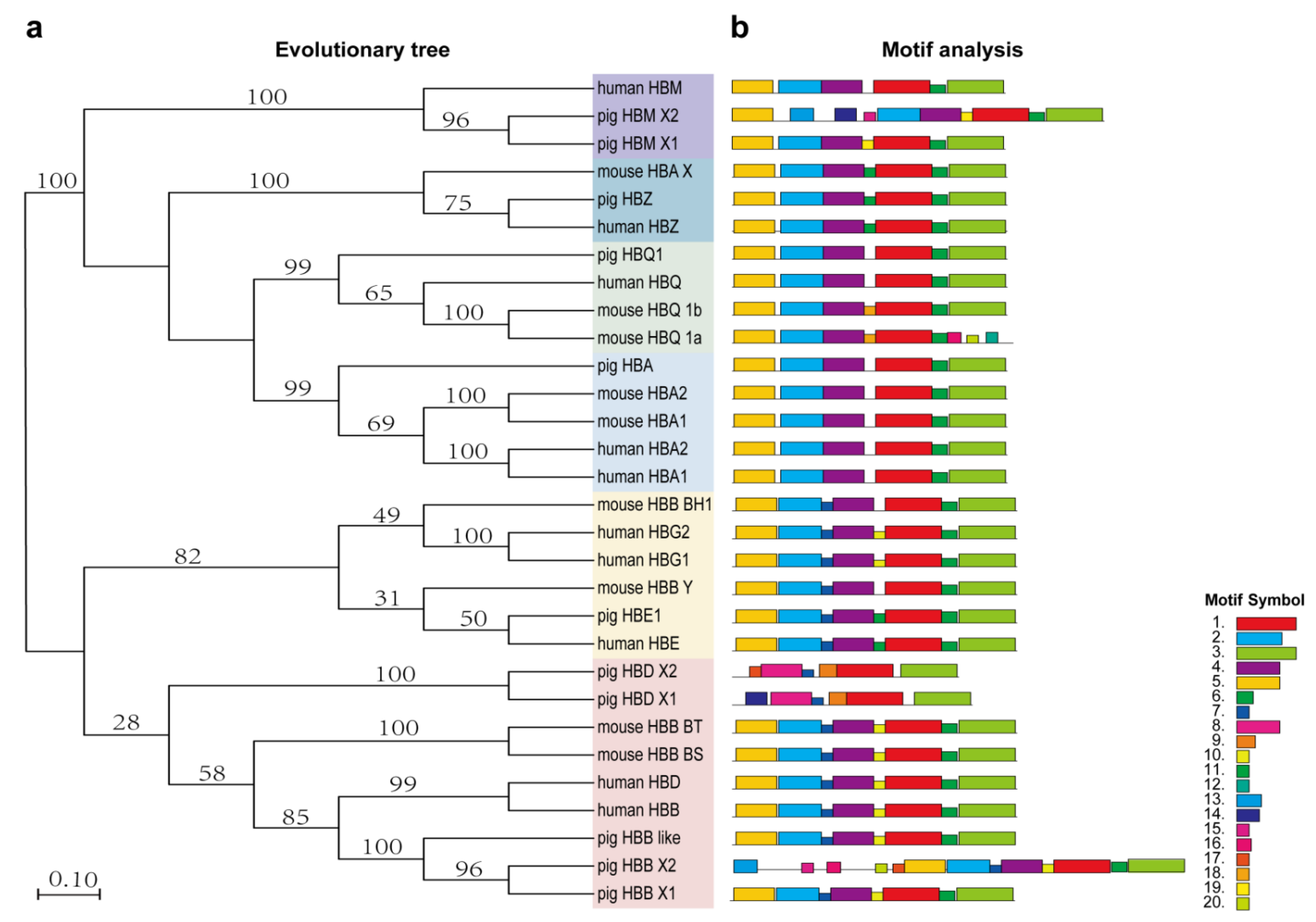
3.2. Evolutionary Analysis of Human, Pig and Mouse Hemoglobin-Encoding Genes

To analyze the evolutionary relationship of hemoglobin-encoding genes in human, pig and mouse, a phylogenetic tree was constructed using amino acid sequences. A total of 10 human sequences, 11 pig sequences and 10 mouse sequences of hemoglobin-encoding genes were evaluated in the phylogenetic tree (Figure 1a). The hemoglobin-encoding genes were divided into two outgroups, α-globin and β-globin. Among them, the mouse HBB-BH2 gene was the LCR of the mouse 5′ regulatory region and were not displayed in the phylogenetic tree. From an evolutionary point of view, HBM and HBZ on the pig α-globin chain and HBE on the β-globin chain were more similar to humans. Human HBB and HBD genes were evolutionally closely related to the pig HBB gene but were distantly related to pig HBD gene. According to the MEM motif, we analyzed and constructed a schematic diagram of the coding hemoglobin structure of pig, mouse and human (Figure 1b; Supplementary Figure S1) and identified a total of 20 different conserved motifs (Figure 1). Most coding genes contained the first six motifs, while the pig HBD gene lacked motif 2 and motif 5, which were not involved in the expression of the hemoglobin gene. In addition, the HBB-like gene was similar to that of humans in the phylogenetic tree (Figure 1a) and the motifs were similar to the HBB structure in pig and human (Figure 2b).

3.3. HBB and HBB-like Gene Sequence Analysis

The CDS sequences and protein sequences of human HBB gene, porcine HBB gene and HBB-like gene were downloaded from the NCBI website and compared by Blast through the EMBL-EBI online website. The results demonstrated that the pig HBB and human HBB proteins had the same length and were highly conserved with 85.03% amino acid sequence identity. Especially in the mutant regions (Codon41–42 and Codon26), their amino acid was identical (Figure 2a). Pig and human HBB gene CDS sequences had high alignment similarity (percent identity, 84.68%), with only one base difference in the coding sequences at the Codon41-42, while the human and pig HBB gene had the same coding sequence at the Codon26 locus (Figure 2b). The protein sequence encoded by HBB-like gene was highly similar to HBB gene in pig, suggesting the two genes might have similar biological functions (Figure 2a). The 30bp upstream and 200bp downstream sequences of Codon 41–42 sequences in CDS of pig HBB and HBB-like genes were completely identical (Figure 2b). Analysis of the Ensembl database demonstrated that their 250 bp genome sequences upstream and downstream of Codon41–42 were almost identical (percent identity, 96.77%).

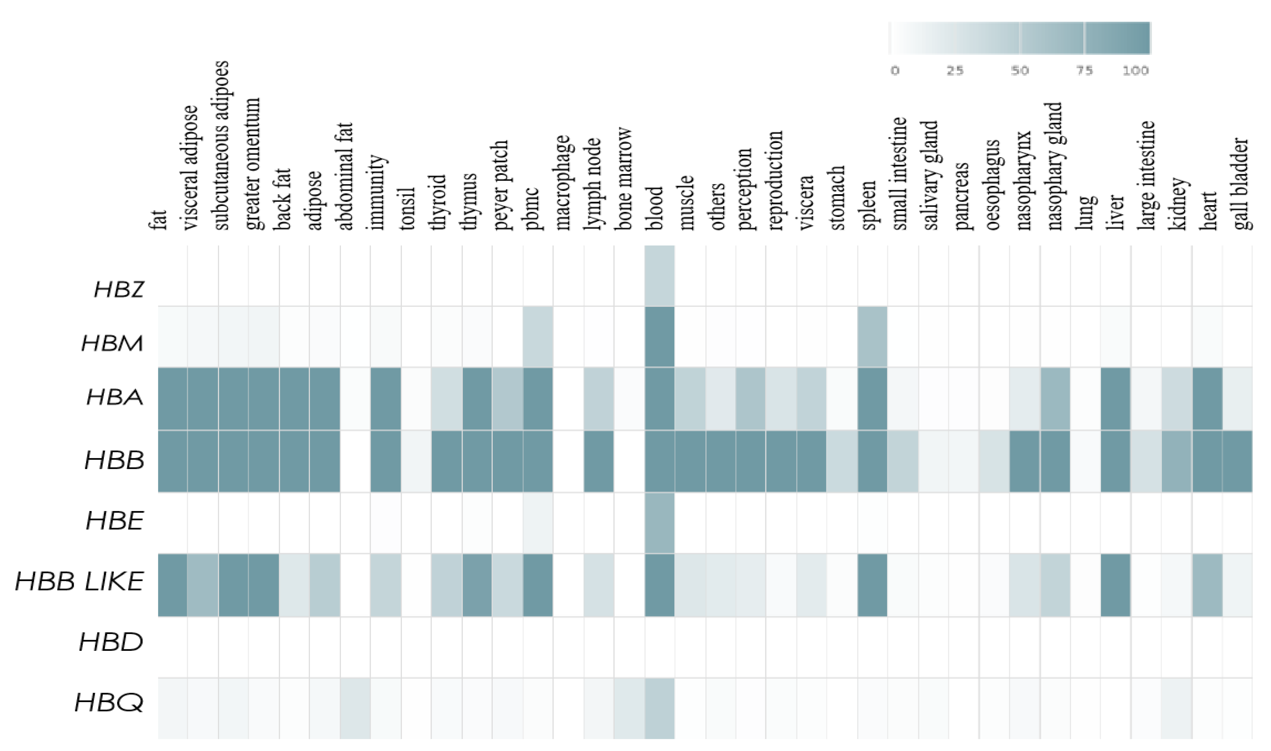
3.4. Expression Levels of the HBB Gene Family in Different Tissues

We analyzed the expression information of eight porcine hemoglobin-encoding genes (HBZ, HBM, HBA, HBB, HBE, HBB-like, HBD and HBQ) in the iswine database (http://iswine.iomics.pro, accessed on 27 April 2022). A tissue expression heatmap of the eight genes was shown in Figure 3. According to the data results, HBA, HBB and HBB-like genes were abundantly expressed in multiple tissues, such as adipose tissue, greater omentum, thymus, blood tissue, spleen, liver and heart. However, HBB and HBB-like genes showed obviously different expression levels in some tissues, such as back fat, lymph node, muscle and thymus. While, HBZ, HBM and HBE genes were only expressed in a few tissues according to the iswine database. In addition, there was no tissue expression profile information of HBD gene in the iswine database.

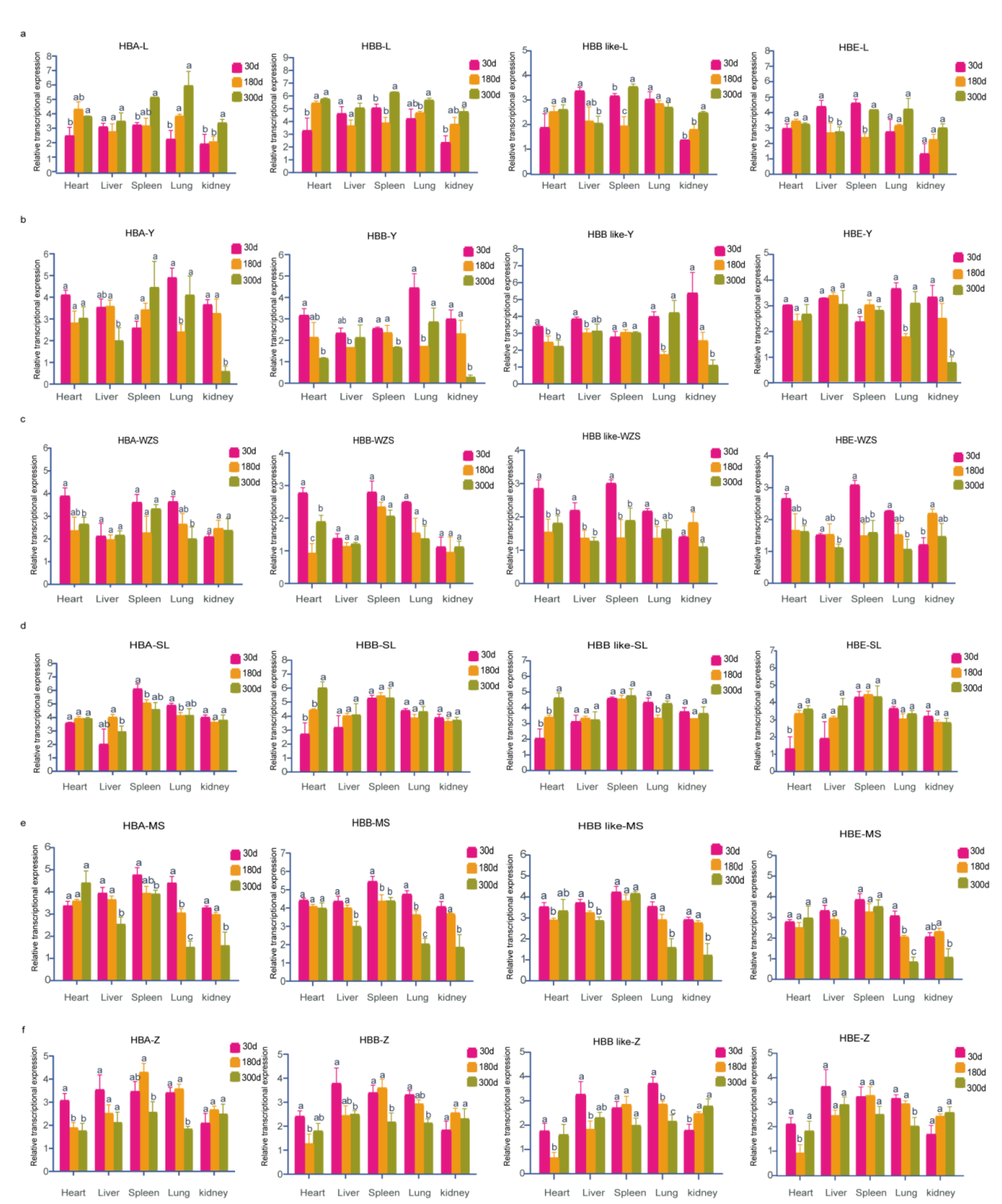
3.5. Spatiotemporal Expression Patterns of Hemoglobin-Encoding Genes in Adult Pigs

We selected three time points (30 d, 180 d and 300 d) to detect the four gene expressions in different tissues (heart, liver, spleen, lung and kidney) of six different pig breeds. The results were shown in Figure 4. Gene expression results showed that HBA and HBB genes were up-regulated in the heart, spleen, lung and kidney tissues derived from the Landrace pig, while HBE and HBB-like genes were down-regulated in liver with the increase in age. In addition, the expressions of HBE and HBB-like genes were significantly lower at 180 days than at any other age in spleen tissue of the Landrace pig. In heart tissue of Yorkshire pigs, the expression levels of HBB and HBB-like genes were down-regulated with the increase in age. In lung tissue of Yorkshire, the expressions of the four genes had no significant differences between 30 days and 300 days and the expression levels were significantly higher than that at 180 days. In kidney tissue, the expression levels of the four genes decreased with the increase in age and reached the lowest level at 300 days. In Wuzhishan pigs, HBA and HBB genes were down-regulated with the increase in age in heart and lung tissues, while HBE and HBB-like genes were down-regulated with age in heart, liver, spleen and lung tissues. In Songliao Black pigs, the expressions of HBB, HBE and HBB-like genes increased in the heart tissue. In heart tissue of the Meishan pigs, there were no significant differences in the expressions of HBA, HBB and HBE genes in the three periods, but the expression of HBB-like gene decreased at first but then increased with the increase in age. In liver, lung and kidney tissues from the Meishan pigs, the four genes were significantly down-regulated at 300 days of age. Moreover, the expressions of HBA and HBB genes decreased with the increase in age, but the expressions of HBB-like and HBE genes had no significant difference in spleen tissue form the Meishan pigs. In heart tissue of the Tibetan pigs, the expression of HBA gene decreased at 180 and 300 days, but the other three genes were only deceased at 180 days. In the Tibetan pig lung tissue, the expression levels of the four genes showed a significant decrease at 300 days of age.

4. Discussion

There are many kinds of mutations in β-thalassemia. The most common type of thalassemia in Asia is the HbE/β-type, while the most common mutation in China is in Codons 41/42 (−CTTT). These two β-thalassemia mutations both occur in the HBB gene [33,34]. Because reproductive cycle in pig is relatively short and pigs have similar metabolism and immunity to humans, it is important to perform disease-related genetic research on pigs. In this study, we predicted 10 and 11 hemoglobin-encoding genes in mouse and pig according to human hemoglobin-encoding genes sequences, respectively, and constructed a phylogenetic tree to analyze their conserved domains. Our results showed that the conserved regions of the β-chain in pigs were highly similar except for the HBD gene, while the HBE genes of humans and pigs were more closely related and the consistency of the conserved regions was high. The two transitions from embryonic to postnatal globin exist in human [35]. The absence of the pig hemoglobin-encoding gene HBG indicated that the two transitions might not exist in pigs. In addition, a new HBB-like gene was discovered in the gene-encoding pig hemoglobin. A comparison of protein and gene sequences among human and pig HBB genes and pig HBB-like gene demonstrated that their protein sequences had similar lengths and high similarity. In humans and pigs, Codons 41/42 (−CTTT) differed by only one base in the HBB gene, while Codons 26 were identical. These results suggested that a β-thalassemia disease model could be constructed around the HBB gene in pigs.
Due to the presence of HBF expression during the fetal period, there are no obvious symptoms of anemia at birth, but by six months of age, if the synthesis of β chains continues to decrease, symptoms of thalassemia will appear [36,37]. Therefore, in this study, we detected the key gene (HBA, HBB, HBB-like and HBE) expressions in some tissues (heart, liver, spleen, lung and kidney) of different pig breeds at 30, 180 and 300 days of age. We found that the temporal and spatial expression patterns of these genes were significantly different in various tissues of the six pig breeds. For example, the expressions of HBA, HBB and HBB-like gene increased in the kidney of Landrace pigs at 300 days of age but decreased in the kidney of Yorkshire and Meishan pigs. However, the expression patterns of these genes in different tissues of the same pig species were similar. Interestingly, the expression of HBB and HBB-like genes were not consistent in different pig breeds and different tissues, suggesting that the HBB-like gene might have other potential functions. Next, we will perform functional detection experiments around the HBB-like gene.

5. Conclusions

In this study, we predicted 11 hemoglobin-encoding genes in pigs. According to our prediction results, we further constructed a phylogenetic tree to analyze their conserved domains among humans, mice and pigs. Our results showed that the β-chain structures between pigs and humans were highly similar. Interestingly, we did not find a porcine HBG gene, indicating that the globin switches might not exist in pigs. A new hemoglobin-encoding gene, ‘HBB-like’, was found in pigs, with its encoding protein sequence sharing high similarity with the HBB gene. Nevertheless, the spatiotemporal expression patterns of HBB and HBB-like genes showed significant difference across breeds. Together, our data provide an insight into the basic reference for construction of a gene-edited β-thalassemia pig model in the future.

Supplementary Materials

The following supporting information can be downloaded at: https://www.mdpi.com/article/10.3390/genes13101822/s1. Supplementary Figure S1. Analysis and distribution of conserved motifs in human, pig and mouse hemoglobin-encoding genes.

Author Contributions

Conceptualization, Z.L., Y.M. and K.L.; Data curation, X.G.; Investigation, X.G.; Supervision, L.H., Z.L., Y.M. and J.Z.; Visualization, X.G., L.H. and J.Z.; Writing—original draft, X.G.; Writing—review and editing, X.G., J.Z. and K.L. All authors have read and agreed to the published version of the manuscript.

Funding

This work was financially supported by the Guangdong Basic and Applied Basic Research Foundation (2020B1515120016) and the National Natural Science Foundation of China (31702203).

Institutional Review Board Statement

Not applicable.

Informed Consent Statement

Not applicable.

Data Availability Statement

The data that support the findings of this study are available from the corresponding author upon reasonable request.

Acknowledgments

This work was supported by funds from the Guangdong Basic and Applied Basic Research Foundation (2020B1515120016) and the National Natural Science Foundation of China (31702203). All individuals have consented to the acknowledgement.

Conflicts of Interest

The authors declare no competing financial interests.

References

  1. Wu, H.; Huang, Q.; Yu, Z.; Zhong, Z. Molecular analysis of alpha- and beta-thalassemia in Meizhou region and comparison of gene mutation spectrum with different regions of southern China. J. Clin. Lab. Anal. 2021, 35, e24105. [Google Scholar] [CrossRef] [PubMed]
  2. Xu, X.; Wu, X. Epidemiology and treatment of beta thalassemia major in China. Pediatr. Investig. 2019, 4, 43–47. [Google Scholar] [CrossRef] [PubMed] [Green Version]
  3. Cao, A.; Kan, Y.W. The prevention of thalassemia. Cold Spring Harb. Perspect. Med. 2013, 3, a011775. [Google Scholar] [CrossRef] [PubMed]
  4. Yang, Z.; Cui, Q.; Zhou, W.; Qiu, L.; Han, B. Comparison of gene mutation spectrum of thalassemia in different regions of China and Southeast Asia. Mol. Genet. Genomic Med. 2019, 7, e680. [Google Scholar] [CrossRef] [Green Version]
  5. Mahdieh, N.; Rabbani, B. Beta thalassemia in 31,734 cases with HBB gene mutations: Pathogenic and structural analysis of the common mutations; Iran as the crossroads of the Middle East. Blood Rev. 2016, 30, 493–508. [Google Scholar] [CrossRef]
  6. Marengo-Rowe, A.J. Structure-function relations of human hemoglobins. Bayl. Univ. Med. Cent. Proc. 2006, 19, 239–245. [Google Scholar] [CrossRef]
  7. Zittersteijn, H.A.; Harteveld, C.L.; Klaver-Flores, S.; Lankester, A.C.; Hoeben, R.C.; Staal, F.J.T.; Gonçalves, M.A.F.V. A small key for a heavy door: Genetic therapies for the treatment of hemoglobinopathies. Front. Genome Ed. 2021, 4, 617780. [Google Scholar] [CrossRef]
  8. Cao, A.; Moi, P. Regulation of the globin genes. Pediatr. Res. 2002, 51, 415–421. [Google Scholar] [CrossRef] [Green Version]
  9. Huehns, E.R.; Flynn, F.V.; Butler, E.A.; Beaven, G.H. Two new haemoglobin variants in a very young human embryo. Nature 1961, 189, 496–497. [Google Scholar] [CrossRef]
  10. Oneal, P.A.; Gantt, N.M.; Schwartz, J.D.; Bhanu, N.V.; Lee, Y.T.; Moroney, J.W.; Reed, C.H.; Schechter, A.N.; Luban, N.L.; Miller, J.L. Fetal hemoglobin silencing in humans. Blood 2006, 108, 2081–2086. [Google Scholar] [CrossRef]
  11. Sripichai, O.; Fucharoen, S. Fetal hemoglobin regulation in β-thalassemia: Heterogeneity, modifiers and therapeutic approaches. Expert. Rev. Hematol. 2016, 9, 1129–1137. [Google Scholar] [CrossRef] [PubMed]
  12. Peschle, C.; Mavilio, F.; Migliaccio, G.; Migliaccio, A.R.; Russo, G.; Mastroberardino, G.; Marinucci, M. Erythropoietic development and hemoglobin switching in human embryos: Cellular and molecular aspects. Prog. Clin. Biol. Res. 1985, 191, 383–396. [Google Scholar] [PubMed]
  13. Su, Q.; Chen, S.; Wu, L.; Tian, R.; Yang, X.; Huang, X.; Chen, Y.; Peng, Z.; Chen, J. Severe Thalassemia Caused by Hb Zunyi [β147(HC3)Stop→Gln; HBB: C.442T>C)] on the β-Globin Gene. Hemoglobin 2019, 43, 7–11. [Google Scholar] [CrossRef]
  14. Lithanatudom, P.; Smith, D.R. Analysis of protein profiling studies of β-thalassemia/Hb E disease. Proteom. Clin. Appl. 2016. [CrossRef] [PubMed]
  15. Galanello, R.; Origa, R. Beta-thalassemia. Orphanet. J. Rare Dis. 2010, 5, 11. [Google Scholar] [CrossRef] [PubMed] [Green Version]
  16. Azman, N.F.; Abdullah, W.Z.; Hanafi, S.; Diana, R.; Bahar, R.; Johan, M.F.; Zilfalil, B.A.; Hassan, R. Genetic polymorphisms of HbE/beta thalassemia related to clinical presentation: Implications for clinical diversity. Ann. Hematol. 2020, 99, 729–735. [Google Scholar] [CrossRef]
  17. Munkongdee, T.; Tongsima, S.; Ngamphiw, C.; Wangkumhang, P.; Peerapittayamongkol, C.; Hashim, H.B.; Fucharoen, S.; Svasti, S. Predictive SNPs for β-thalassemia/HbE disease severity. Sci. Rep. 2021, 11, 10352. [Google Scholar] [CrossRef]
  18. He, X.; Sheng, M.; Xu, M.; Xiong, C.; Ren, Z. Rapid identification of common β-thalassemia mutations in the Chinese population using duplex or triplex amplicon genotyping by high-resolution melting analysis. Genet. Test. Mol. Biomarkers 2010, 14, 851–856. [Google Scholar] [CrossRef]
  19. Zhang, W.; Cai, W.W.; Zhou, W.P.; Li, H.P.; Li, L.; Yan, W.; Deng, Q.K.; Zhang, Y.P.; Fu, Y.X.; Xu, X.M. Evidence of gene conversion in the evolutionary process of the codon 41/42 (-CTTT) mutation causing beta-thalassemia in southern China. J. Mol. Evol. 2008, 66, 436–445. [Google Scholar] [CrossRef]
  20. He, S.; Li, J.; Li, D.M.; Yi, S.; Lu, X.; Luo, Y.; Liang, Y.; Feng, C.; Chen, B.; Zheng, C.; et al. Molecular characterization of α- and β-thalassemia in the Yulin region of Southern China. Gene 2018, 655, 61–64. [Google Scholar] [CrossRef]
  21. Weatherall, D.J. Keynote address: The challenge of thalassemia for the developing countries. Ann. N. Y. Acad. Sci. 2005, 1054, 11–17. [Google Scholar] [CrossRef] [PubMed]
  22. Weatherall, D.J. Thalassemia as a global health problem: Recent progress toward its control in the developing countries. Ann. N. Y. Acad. Sci. 2010, 1202, 17–23. [Google Scholar] [CrossRef] [PubMed]
  23. Cooper, D.K.; Hara, H.; Yazer, M. Genetically engineered pigs as a source for clinical red blood cell transfusion. Clin. Lab. Med. 2010, 30, 365–380. [Google Scholar] [CrossRef] [PubMed]
  24. Cooper, D.K.C.; Ezzelarab, M.; Iwase, H.; Hara, H. Perspectives on the optimal genetically engineered pig in 2018 for initial clinical trials of kidney or heart xenotransplantation. Transplantation 2018, 102, 1974–1982. [Google Scholar] [CrossRef]
  25. Yamamoto, T.; Bikhet, M.H.; Marques, M.B.; Nguyen, H.Q.; Cui, Y.; Javed, M.; Raza, S.S.; Ayares, D.; Iwase, H.; Cooper, D.K.C.; et al. Initial experimental experience of triple-knockout pig red blood cells as potential sources for transfusion in alloimmunized patients with sickle cell disease. Transfusion 2021, 61, 3104–3118. [Google Scholar] [CrossRef]
  26. Rogers, C.S. Genetically engineered livestock for biomedical models. Transgenic Res. 2016, 25, 345–359. [Google Scholar] [CrossRef]
  27. Ou, Z.; Niu, X.; He, W.; Chen, Y.; Song, B.; Xian, Y.; Fan, D.; Tang, D.; Sun, X. The combination of CRISPR/Cas9 and iPSC technologies in the gene therapy of human β-thalassemia in mice. Sci. Rep. 2016, 6, 32463. [Google Scholar] [CrossRef] [Green Version]
  28. Yang, Y.; Kang, X.; Hu, S.; Chen, B.; Xie, Y.; Song, B.; Zhang, Q.; Wu, H.; Ou, Z.; Xian, Y.; et al. CRISPR/Cas9-mediated β-globin gene knockout in rabbits recapitulates human β-thalassemia. J. Biol. Chem. 2021, 296, 100464. [Google Scholar] [CrossRef]
  29. Marchler-Bauer, A.; Bo, Y.; Han, L.; He, J.; Lanczycki, C.J.; Lu, S.; Chitsaz, F.; Derbyshire, M.K.; Geer, R.C.; Gonzales, N.R. CDD/SPARCLE: Functional classification of proteins via subfamily domain architectures. Nucleic. Acids. Res. 2016, 45, D200–D203. [Google Scholar] [CrossRef] [Green Version]
  30. Hung, J.H.; Weng, Z. Sequence Alignment and Homology Search with BLAST and ClustalW. Cold Spring Harb. Protoc. 2016, 11, 10–1101. [Google Scholar] [CrossRef]
  31. Li, D.; Xu, R.; Lv, D.; Zhang, C.; Yang, H.; Zhang, J.; Wen, J.; Li, C.; Tan, X. Identification of the core pollen-specific regulation in the rice OsSUT3 promoter. Int. J. Mol. Sci. 2020, 21, 1909. [Google Scholar] [CrossRef] [PubMed] [Green Version]
  32. Livak, K.J.; Schmittgen, T.D. Analysis of relative gene expression data using real-time quantitative PCR and the 2−∆∆CT method. Methods 2001, 25, 402–408. [Google Scholar] [CrossRef] [PubMed]
  33. Olivieri, N.F. Treatment strategies for hemoglobin E beta-thalassemia. Blood Rev. 2012, 26, S28–S30. [Google Scholar] [CrossRef]
  34. Bao, X.; Zhang, X.; Wang, L.; Wang, Z.; Huang, J.; Zhang, Q.; Ye, Y.; Liu, Y.; Chen, D.; Zuo, Y.; et al. Epigenetic inactivation of ERF reactivates γ-globin expression in β-thalassemia. Am. J. Hum. Genet 2021, 108, 709–721. [Google Scholar] [CrossRef]
  35. Zakaria, N.A.; Islam, M.A.; Abdullah, W.Z.; Bahar, R.; Mohamed Yusoff, A.A.; Abdul Wahab, R.; Shamsuddin, S.; Johan, M.F. Epigenetic insights and potential modifiers as therapeutic targets in β-thalassemia. Biomolecules 2021, 11, 755. [Google Scholar] [CrossRef]
  36. Muncie, H.L., Jr.; Campbell, J. Alpha and beta thalassemia. Am. Fam. Physician 2009, 80, 339–344. [Google Scholar]
  37. Yin, X.L.; Wu, Z.K.; He, Y.Y.; Zhou, T.H.; Zhou, Y.L.; Zhang, X.H. Treatment and complications of thalassemia major in Guangxi, Southern China. Pediatr. Blood Cancer 2011, 57, 1174–1178. [Google Scholar] [CrossRef]
Figure 1. Phylogenetic relationships and motif composition of human, mouse and pig hemoglobin-encoding genes. (a) Represents the phylogenetic tree constructed by MEGA 5.0 software and different gene colors represent different clades; (b) represents the motifs constructed from human, mouse and pig protein sequences. Different colored boxes represent different motifs.
Genes 13 01822 g001
Figure 2. Sequence alignment of human HBB gene, pig HBB gene and pig HBB-like gene. (a) Sequence alignment of human HBB, pig HBB and HBB-like proteins; (b) The CDS sequence alignment of human HBB gene, pig HBB gene and pig HBB-like gene.
Genes 13 01822 g002
Figure 3. Expression patterns of the eight porcine hemoglobin-encoding genes in the iswine database.
Genes 13 01822 g003
Figure 4. Expression patterns of hemoglobin-encoding genes in adult pig different tissues. (af) represent expression of HBA, HBB, the HBB-like and HBE gene in Landrace (L), Yorkshire (Y), Wuzhishan (WZS), Songliao black (SL), Meishan (MS) and Tibetan (Z), respectively. Data are shown as mean ± SEM (n = 3 to 6), student’s t-test. Relative expression was calculated as: Lg2-ΔΔCt. p < 0.05 was considered to be statistically significant. significant a, b, c indicate significant difference.
Genes 13 01822 g004
Table 1. HBB gene family primer sequences.
PrimersSequence (5′–3′)Product Length, bp
SUS-Qhbb1-Faatgtggacgaagttggtggt270
SUS-Qhbb1-Rgttgcccaggagcctgaagt
SUS-HBB-like-Ftcggctgctgtcctcggcctgca303
SUS-HBB-like-Rgttgcccaggagcctgaagt
SUS-HBE1-Ftcctggtggtctacccttgg233
SUS-HBE1-Rgttgcccaggagcctgaagt
SUS-HBA-Fagaggccctggaaagaatgt278
SUS-HBA-Rggttgaaatcatcggggtgg
18S-Fgtaacccgttgaaccccatt151
18S-Rccatccaatcggtagtagcg
Table 2. Human, pig and mouse hemoglobin-encoding genes.
SpeciesGene SymbolProtein IDChromosome
Homo sapiensHBBENST0000033529511
HBDENST0000029290111
HBE1ENST0000039689511
HBG1ENST0000033059711
HBG2ENST0000033690611
HBA1ENST0000032086816
HBA2ENST0000025159516
HBMENST0000035681516
HBQ1ENST0000019970816
HBZENST0000025295116
Sus scrofaHBB X1ENSSSCP000000675219
HBB X2ENSSSCP000150219059
HBB-likeENSSSCP000000156499
HBD X1ENSSSCP000000590429
HBD X2ENSSSCP000000457579
HBE1ENSSSCP000000156489
HBAENSSSCP000000289443
HBM X1ENSSSCP000000639033
HBM X2ENSSSCP000000085163
HBQ1ENSSSCP000000295233
HBZENSSSCP000000085143
Mus MusculusHBA-xENSMUSP0000002053111
HBQ 1aENSMUSP0000002053511
HBQ 1bENSMUSP0000009893611
HBA a1ENSMUSP0000009089711
HBA a2ENSMUSP0000009089511
HBB bsENSMUSP000000239347
HBB yENSMUSP000000332297
HBB bh1ENSMUSP000000648657
HBB btENSMUSP000000957947
HBB bh2ENSMUSP000001024797
Publisher’s Note: MDPI stays neutral with regard to jurisdictional claims in published maps and institutional affiliations.

