Article

Establishment of an Efficient In Vitro Propagation of Cnidium officinale Makino and Selection of Superior Clones through Flow Cytometric Assessment of DNA Content

1
Department of Horticultural Science, Chungbuk National University, Cheongju 28644, Korea
2
Korea Disease Control and Prevention Agency, Osong-eup, Cheongju 28159, Korea
3
Department of Forest Genetic Resources, National Institute of Forest Science, 39 Onjeong-ro, Suwon 16631, Korea
4
Department of Botany, Karnatak University, Dharwad 580003, India
5
Food Science R&D Center, Kolmar BNH Co., Seocho-gu, Seoul 30003, Korea
*
Author to whom correspondence should be addressed.
Genes 2022, 13(10), 1815; https://doi.org/10.3390/genes13101815
Submission received: 19 September 2022 / Revised: 6 October 2022 / Accepted: 6 October 2022 / Published: 8 October 2022
(This article belongs to the Special Issue Phylogenetics, Genetics, and Breeding of Medicinal Plants)

Abstract

:
Cnidium officinale is a valuable medicinal plant cultivated in Asia for its rhizomes. This study reports the in vitro regeneration of Cnidium officinale plants and the induction of rhizomes from microshoots. The rhizomatous buds of Cnidium officinale induced multiple shoots on Murashige and Skoog (MS) medium supplemented with 0.5 mg L−1 BA, which led to the regeneration of plants within four weeks of culture. After four weeks of culture, the plants were assessed for fresh weight, the number of leaves, the number of roots, and the length of roots to compare the performance of the different clones. The clones with good growth characteristics were selected with the aid of a flow cytometric analysis of 2C nuclear DNA content. The plants bearing high DNA values showed better growth characteristics. Various factors, namely, sucrose concentration (30, 50, 70, and 90 g L−1), ABA (0, 0.5, 1.0, and 2.0 mg L−1), the synergistic effects of BA (1.0 mg L−1) + NAA (0.5 mg L−1) and BA (1.0 mg L−1) + NAA (0.5 mg L−1) + ABA (1.0 mg L−1) with or without activated charcoal (1 g L−1), and light and dark incubation were tested on rhizome formation from microshoots. The results of the above experiments suggest that MS medium supplemented with 50 g L−1 sucrose, 1.0 mg L−1 ABA, and 1 g L−1 AC is good for the induction of rhizomes from the shoots of Cnidium officinale. Plantlets with rhizomes were successfully transferred to pots, and they showed 100% survival.

1. Introduction

Cnidium officinale Makino is an important herbaceous medicinal plant that belongs to the Apiaceae family. It is widely cultivated in China, Japan, and Korea for its rhizomes. Dried rhizomes are used in Chinese, Japanese, and Korean medicine as a tonic to improve blood circulation, overcome the problems of inflammation, and treat women’s menstrual problems [1]. The rhizomes of Cnidium officinale have been reported to possess several bioactive compounds, including polysaccharides [2,3,4] and volatile alkylphthalide derivatives [5,6]. Rhizome extracts and isolated compounds have been reported to exhibit various pharmacological activities, including antiangiogenic [7], anticancer [8,9,10], antidiabetic [11], anti-inflammatory [12,13], immunomodulatory [2], analgesic [14], and antimicrobial [15,16] effects. Yang et al. [17] demonstrated that the ethanolic rhizome extracts of Cnidium officinale are usable in the treatment of ischemic injury and, thus, that rhizomes could be used as recovery medicine after plastic surgery, for the disturbance of peripheral blood circulation, and for overcoming postmenopausal problems. Kim [18] and Cha [19] showed the positive effects of Cnidium officinale extracts on the repression of melanogenesis and in decreasing hyperpigmentation, as well as its use as a cosmetic ingredient.
Cnidium officinale does not set seeds; therefore, the multiplication of this plant only occurs through the division of rhizomes, and there is a high risk of virus dissemination affecting yield and quality during cultivation [1]. Therefore, in vitro propagation methods have been developed for this species using shoot tip culture and the induction of somatic embryogenesis using inflorescence and flowers [1,20,21]. However, plant regeneration efficacy is low and inconsistent. Further, Cnidium officinale is a rhizomatous plant, and the in vitro induction of rhizomes from regenerated shoots is desirable. In vitro rhizome induction has been reported in many bulbous plants, such as Kaempferia galanga, Kaempfria rotunda [22], Curcuma longa [23], and Bambosa bambos var. gigantea [24]. The in vitro production of rhizomes has commercial potential for the large-scale generation of pathogen-free seed material for propagation and utilization by the pharmaceutical industry. Further, in vitro rhizome production is also useful for storage, transport, and germplasm preservation. Therefore, in the present study, an efficient plant regeneration protocol was developed in Cnidium officinale using rhizome bud explants. The regenerated plants/clones were assessed for their DNA content using the flow cytometric method. Additionally, the DNA content of the regenerated clones was correlated with growth characteristics. In addition, a reliable method for in vitro rhizome induction from regenerated shoots was established.

2. Materials and Methods

2.1. Preparation of Plant Material and Induction of Microshoots from Rhizome Buds

The rhizomes of Cnidium officinale Makino growing in the greenhouse of Chungbuk National University, Korea, were used in the experiment. For the establishment of the in vitro culture of Cnidium officinale, buds (~1.5 cm long) collected from the rhizome were washed in running tap water for 1 h, and the bacteria and fungi were primarily removed by immersing them in a sterilizer containing 1000 ppm Physan 20 (Maril, Tustin, CA, USA) for 20 s. Subsequently, surface sterilization was conducted for 20 s with 70% ethanol and for 10 min in a solution with added Tween 20, surfactant (ThermoFisher, Seoul, Korea), and 2% NaOCl. The explants were thoroughly washed five to six times with sterile double-distilled water, and they were cultured on Murashige and Skoog [25] medium with 0.5 mg L−1 benzyl adenine (BA), 30 g L−1 sucrose, and 2.4 g L−1 Gelrite (Duchefa Biochimie, Haarlem, the Netherlands). The cultures were incubated at 25 ± 1 °C under a 16 h photoperiod under the white fluorescent light of 35 µ m−2 s−1. Subculture was conducted at four week intervals. The cultured explants developed shoots, simultaneously developing roots at the base of the shoots. Plantlets were maintained in MS medium with 0.5 mg L−1 BA through repeated subculturing. We compared the regeneration rate of ten different clones, which were available in our germplasm collection, and calculated the shoot proliferation rate in subsequent subcultures for up to three cycles.

2.2. Flow Cytometry

Flow cytometry was performed to determine the amount of 2C DNA in the in vitro-grown Cnidium officinale plants. Briefly, a fresh staining solution was prepared on the day of analysis by mixing the staining buffer with PI solution and RNase solution. Leaf discs (0.5 cm2) were prepared from the leaves of Cnidium officinale and Nicotiana tabacum cv. xanthi (internal standard) plants and placed in a Petri dish containing 200 µL of extraction buffer. Nuclei were isolated from the leaf discs using a CyStain PI Absolute P Kit (Partec, Görlitz, Germany), according to the manufacturer’s instructions. The solution was filtered through a 50 µm nylon mesh. Then, 800 µL of staining solution was added to the filtered solution in order to label the DNA with a fluorescent dye. The samples were incubated in the staining solution on a stationary surface in the dark for 20 min and then analyzed using CytoFLEX (Beckman Coulter Inc., Fullerton, CA, USA). At least 5000 nuclei were analyzed per sample.

2.3. Assessment of in Vitro Growth of Regenerated Plants

Ten plants regenerated in vitro from each clone of Cnidium officinale were selected, and they were re-cultured on regeneration medium (MS + 0.5 mg L−1 benzyl adenine, 30 g L−1 sucrose, and 2.4 g L−1 Gelrite) and maintained four weeks. After four weeks of culture, the plants were assessed for their plant height, their fresh weight, the number of leaves, and the number and length of roots. Subsequently, the plants were re-cultured for another passage of a four-week cycle, and the number of plantlets in each cycle and the shoot proliferation rate (shoot proliferation rate = the total number of shoots after subculture vs. the total number of shoots before subculture) were estimated.

2.4. In Vitro Induction of Rhizome

The microshoots (2 cm in length) obtained after the four passages of re-culturing were harvested and transferred to MS media supplemented with 30, 50, 70, and 90 g L−1 sucrose, or 0, 0.5, 1.0, and 2.0 mg L−1 abscisic acid (ABA) and 50 g L−1 sucrose for rhizome induction. Similarly, the effect of 0.5 mg L−1 naphthalene acetic acid (NAA) in combination with 1.0 mg L−1 BA alone, or in combination with 1.0 mg L−1 ABA with or without 1 g L−1 activated charcoal (AC) was also tested on the rhizome induction. In these treatments, the medium was supplied with 50 g L−1 sucrose. Depending on the experiment, the cultures were either kept in continuous dark conditions or in 12 h light (35 µmol m−2 s−1) and 8 h dark conditions at 25 ± 1 °C temperature. The cultures were maintained for up to eight weeks, and data were collected.

2.5. Acclimatization

One day before acclimatization, the lid of the culture vessel was opened by ~20% for adaptation to the ex vitro conditions. Then, the plants with rhizomes regenerated in vitro were harvested from the vessel, carefully washed with distilled water, and planted in wet Growfoam (Horticubes, Smithers-Oasis, Kent, OH, USA). The plants in Growfoam were placed in a plastic container (25 × 25 × 3 cm), and the water was changed every week. The plants were fertilized with 1.0 g L−1 Hyponex solution once per month. The transplanted plants showed 100% survival. After 3 months of growing ex vitro, the plants were transplanted and cultivated in plastic pots (diameter 10 cm) filled with an artificial soil mixture (peat moss 1:1 perlite and vermiculite 1). The potted plants were fertilized with 1.0 g·L−1 Hyponex solution (N20:P20:K20) once per month.

2.6. Statistical Analysis

A one-way analysis of variance (ANOVA) was performed to determine significant differences between the results of shoot/plant regeneration, flow cytometric data, the assessment of growth characteristics, and the in vitro rhizome induction experiments. The statistical significance of the differences between the mean values was then assessed using Duncan’s multiple range test at p < 0.05. All statistical analyses were performed using SAS 9.4 software (SAS Institute Inc., Cary, NC, USA).

3. Results

3.1. Establishment of Culture

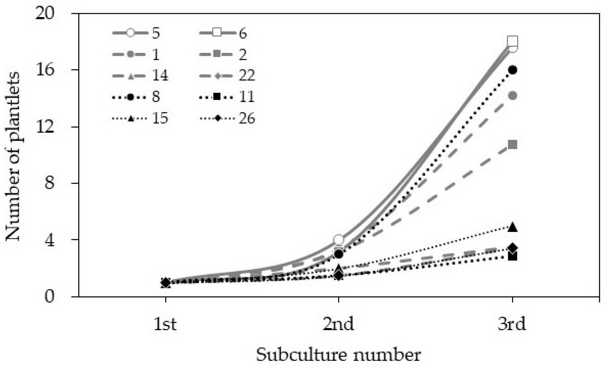
The Cnidium officinale rhizome buds (Figure 1a) were cultured on MS medium, with 0.5 mg L−1 BA involved in swelling one week after inoculation, and produced multiple shoots in subsequent weeks (Figure 1b). The roots simultaneously formed at the basal shoots on the same medium, and plantlets with well-developed shoots and roots were obtained after 4 weeks of incubation (Figure 1c). The shoot proliferation rate varied from 1.5 to 3.4 times across the different clones. Among the ten clones used (clone nos. 1, 2, 5, 6, 8, 11, 14, 15, 22, and 25), clones 5 and 6 performed well and regenerated 16–18 shoots per explant after the third cycle (Figure 2).

3.2. Flow Cytometric Analysis of DNA Content in Different Clones of Cnidium officinale and Selection of Superior Clones with the Highest Regeneration Potential

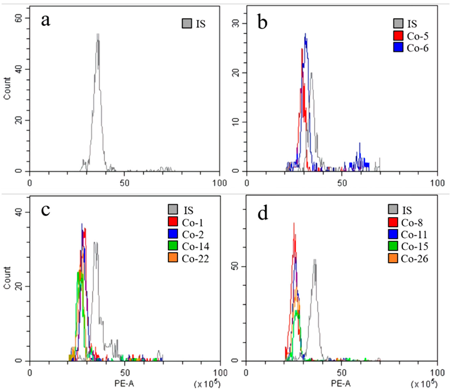
According to the chromosome count database (CCDB, http://ccdb.tau.ac.il/ (accessed on 19 September 2022) [26], the chromosome number of Cnidium officinale populations/clones (germplasm) is highly variable, and the chromosome numbers of 2n = 22, 33, 44 have been reported for this species. The chromosome count of the available Cnidium officinale germplasm/clones was not known; therefore, we carried out a flow cytometric analysis of the 2C DNA content of the different clones used in the current experiment and selected the superior clones for further regeneration experiments and utilization. The flow cytometric analysis data of the Cnidium officinale clones used in the current experiment are presented in Figure 3 and Table 1. The DNA values varied across the different clones, ranging from 7.68 to 8.60 pg/2C (Table 1). Based on such data, the clones were classified into clones with a high DNA content (HD, clones 5 and 6), clones with a moderate DNA content (MD, clones 1, 2, 14, and 22), clones with a low DNA content (LD, clones 8, 11, 15, and 26). The clones with HD, MD, and LD were compared based on their regeneration potential on MS medium with 0.5 mg L−1 BA by using rhizome bud cultures. The clones with a higher DNA content (clones 5 and 6) showed the highest regeneration potential in terms of the regeneration rate (Figure 2).

3.3. Comparison of Growth Characteristics of Different Clones

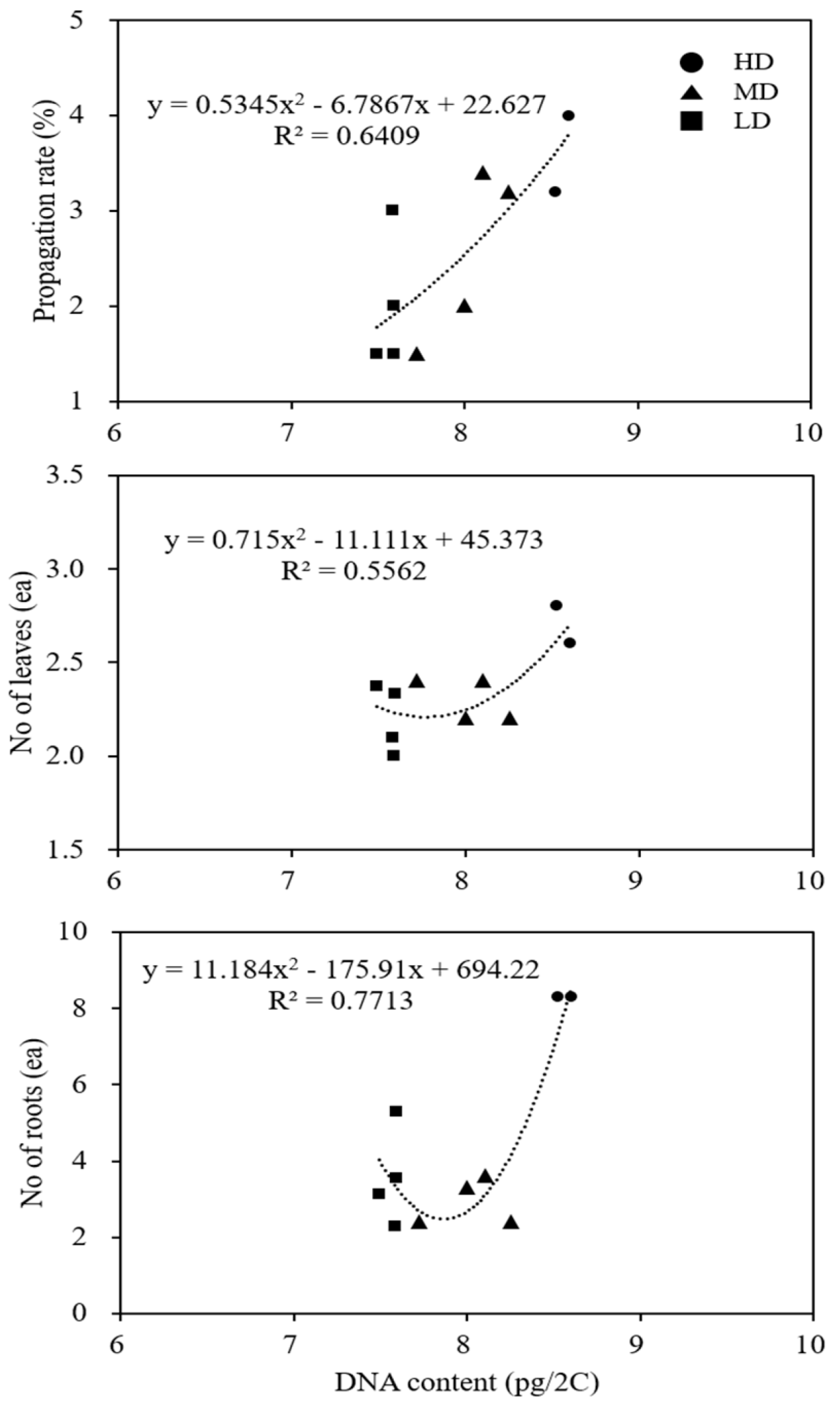
The microshoots of several clones with known 2C DNA values were subcultured on MS medium with 0.5 mg L−1 BA and maintained for four weeks in culture to assess their growth characteristics and performance during in vitro propagation; the data are presented in Table 2. In terms of overall growth performance, i.e., fresh weight, the number of leaves, and the number of roots, the clones with higher DNA values performed well, and clone 5 was the best performer. A correlation analysis of the several clones with different DNA amounts with growth characteristics revealed that there was a positive correlation between the clones with the highest DNA content and propagation rate, the number of leaves, and the number of roots (Figure 4).

3.4. In Vitro Induction of Rhizome

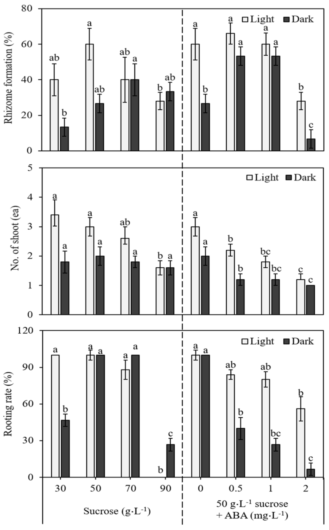
The results obtained for rhizome formation on media with different concentrations of sucrose and ABA, as well as for the light and dark incubation of the cultures, are presented in Figure 5. After two weeks of incubation, rhizome induction was evident at the base of the cultured shoots. Initially, the swelling of the shoot bases was observed, followed by the appearance of small rhizomes at the base within four weeks of incubation. Subsequently, the rhizomes were involved in the shoots’ growth, and roots simultaneously developed in the nodal regions of the rhizomes. Rhizome induction from the shoots was influenced by the levels of sucrose in the medium. The medium supplemented with 30 g L−1 to 50 g L−1 sucrose favored rhizome development (Figure 5). However, with a further increase in the concentration of sucrose from 50 g L−1 onwards (70 and 90 g L−1), there was a marked decrease in the percentage of rhizome formation and the rooting rate. Rhizomes formed on the medium in the presence of ABA, and the 0.5 mg L−1 and 1.0 mg L−1 ABA treatments were better in terms of the percentage of rhizome formation, the number of rhizomes forming shoots, and the rooting rate. A comparison of the light and dark treatments revealed that light incubation is stimulatory in inducing rhizomes from the shoots (Figure 5).
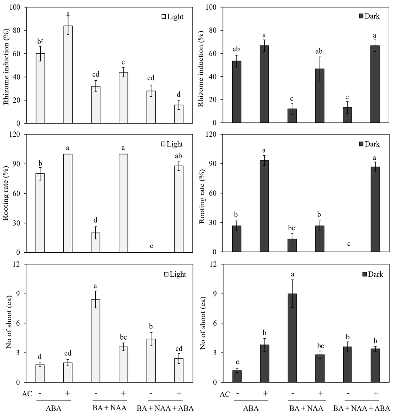
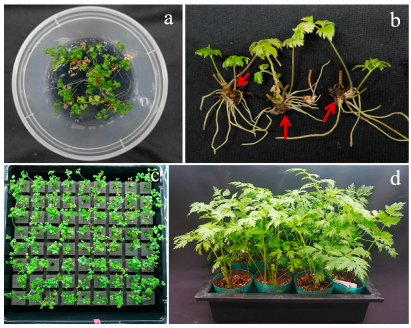
In another set of experiments, we compared the effect of ABA (1.0 mg L−1) and the interaction of BA (1.0 mg L−1) + NAA (0.5 mg L−1) and BA (1.0 mg L−1) + NAA (0.5 mg L−1) + ABA (1.0 mg L−1) on rhizome induction for the in vitro-cultured shoots. At the same time, we also tested the effect of the light and dark treatments, along with hormonal combinations, with and without the addition of activated charcoal (1 g L−1), and the results are presented in Figure 6. Among these different treatments, the cultures that received light treatment performed better than those that received dark treatment, and again, the ABA treatment was far better than the BA + NAA and BA + NAA + ABA treatments. The results of the above experiments suggest that MS medium supplemented with 50 g L−1 sucrose, 1.0 mg L−1 ABA, and 1 g L−1 AC is good for the induction of rhizomes from the shoots of Cnidium officinale (Figure 7a,b).

3.5. Acclimatization

The in vitro-grown plants of Cnidium officinale after rhizome formation and rooting were acclimatized by planting them on Growfoam (Figure 7c), and the acclimatization rate of Cnidium officinale was 100%. After 6 months, the plants were transferred to plastic pots containing vermiculite, and, again, the survival rate was 100% (Figure 7d).

4. Discussion

Cnidium officinale is an important medicinal herb that is utilized in the traditional medicinal systems of China, Japan, and Korea. This plant is propagated by splitting rhizomes since it does not set seeds in nature; however, this method of propagation is responsible for the transmission of viral particles, which results in a loss of the yield and the quality of the rhizomes harvested. In the current study, we developed a successful plant regeneration method by using rhizome buds and by employing an MS medium containing 0.5 mg∙L−1 BA. Multiple shoots regenerated from the rhizome buds and were involved in rooting within four weeks of culture. This simple method was useful in regenerating plants, resulting in several clones of Cnidium officinale, which were maintained in a germplasm repository. The shoot propagation rate varied from 1.5 to 3.4 times across the different clones. Among the ten clones used (clone nos. 1, 2, 5, 6, 8, 11, 14, 15, 22, and 25), clones 5 and 6 performed well and regenerated 16–18 shoots per explant after the third cycle. Lee et al. [1] reported induction somatic embryogenesis in Cnidium officinale from immature flower cultures on MS medium supplemented with 1.0 mg L−1 2,4-D and 0.1 mg L−1 BAP. The embryos that developed were converted into plantlets on MS medium containing 0.1 mg L−1 BAP and 3.0 mg L−1 BAP GA3. Similarly, Adil et al. [27] described recurrent somatic embryogenesis in Cnidium officinale on MS medium containing 1.5 mg L−1 2,4-D and 0.5 mg L−1 BAP. The above results suggest that the induction of both organogenesis and somatic embryogenesis is feasible for the micropropagation of Cnidium officinale.
The flow cytometric estimation of nuclear DNA content is a useful tool in the assessment of medicinal plants regenerated in vitro [28]. In the current study, the clones containing a high amount of DNA, i.e., clones 5 and 6, showed better growth characteristics in terms of fresh weight, the number of leaves, and the number of roots; they were also better performers in terms of the propagation rate. DNA content variations and associated divergence in biological characteristics have also been reported in the natural populations and field-grown plants of Erianthus arundinaceus [29] and Rauvolfia sepentina [30].
The induction of rhizomes during in vitro propagation is an ideal system that helps in the early establishment of plants in fields. An in vitro-induced rhizome produces both shoots and roots on germination, giving rise to a complete plantlet and, thus, acting as a seed for the large-scale propagation of rhizomatous plants [24]. Plant growth regulators, sucrose, and light and dark conditions are known to affect the in vitro induction of rhizomes in many rhizomatous plants, including Curcuma longa [23], Bambusa bambos var. gigantea [24], Kaempferia galanga, and K. rotunda [22].
The results of the present experiments revealed that the supplementation of medium with a suitable sucrose concentration (50 g L−1) is suitable for rhizome induction in Cnidium officinale. Similar to the current results, 50 g L−1 has been found to be suitable for the induction of rhizomes from the in vitro-raised shoots of Bambosa bambos var. gigantea [24]. Sucrose acts as a carbon source and as an osmotic agent, and since storage organs are abundant in carbohydrates, the appropriate concentration of sucrose is essential for the induction and growth of rhizomes in vitro [31,32]. The supplementation of ABA (0.5 and 1.0 mg L−1) favored the induction and development of rhizomes from the microshoots of Cnidium officinale; however, an increased concentration of ABA (2.0 mg L−1) was not beneficial for the induction and growth of rhizomes. Similar to the current observations, Yang et al. [33] reported the promotive effect of ABA on rhizome induction in Nelumbo nucifera. In the current study, light favored rhizome induction from microshoots, whereas the negative effect of the dark treatment was evident. The effect of light on rhizome formation observed in the present study is comparable to that observed in the study conducted by Sakamura et al. [34], where the requirement of continuous light during rhizome formation in Zingiber officinale was reported. The influence of the combined effect of BA, NAA, and ABA did not favor the formation of rhizomes from the microshoots of Cnidium officinale. In contrast to the current results, BA and NAA have been shown to influence the process of in vitro rhizome formation in Cymbidium goeringii [35].
The plantlets with well-developed rhizomes and roots showed 100% survival upon transplantation to Growfoam and subsequently to vermiculite. The protocol for the in vitro induction of multiple shoots and subsequent rhizome formation in Cnidium officinale can be utilized by commercial growers for the production of disease-free clones on a large scale.

Author Contributions

Conceptualization, S.-Y.P.; data curation, H.-E.K.; formal analysis, J.-E.H.; funding acquisition, H.-J.K., G.-M.L.; investigation, H.-E.K. and J.-E.H.; methodology, H.-E.K. and J.-E.H.; project administration, S.-Y.P.; resources, H.-J.K., G.-M.L.; supervision, H.L. and S.-Y.P.; validation, H.L., H.N.M. and S.-Y.P.; writing—original draft, H.-E.K.; writing—review and editing, H.L., H.N.M. and S.-Y.P. All authors have read and agreed to the published version of the manuscript.

Funding

This work was supported by a grant from Kolmar BNH, Korea, and partially supported by the Industrial Strategic Technology Development Program (Grant number P0018148) funded by the Ministry of Trade, Industry & Energy (MOTIE, Korea).

Institutional Review Board Statement

Not applicable.

Informed Consent Statement

Not applicable.

Data Availability Statement

Not applicable.

Acknowledgments

Hosakatte Niranjana Murthy is thankful for the ‘Brain Pool (BP)’ program Grant No. 2022H1D3A2A02056665. This research was supported by Kolmar BNH, Korea.

Conflicts of Interest

The authors declare no conflict of interest.

References

  1. Lee, C.Y.; Kim, Y.K.; Kim, Y.S.; Suh, S.Y.; Lee, S.Y.; Park, S.U. Somatic embryogenesis and plant regeneration in Cnidium officinale Makino. J. Med. Plant Res. 2009, 3, 96–100. [Google Scholar]
  2. Tomoda, M.; Ohara, N.; Gonda, R.; Shimizu, N.; Takada, K.; Satoh, Y.; Shirai, S. An acidic polysaccharide having immunological activities from the rhizome of Cnidium officinale. Chem. Pharm. Bull. 1992, 40, 3025–3029. [Google Scholar] [CrossRef] [Green Version]
  3. Tomoda, M.; Ohara, N.; Shimizu, N.; Gonda, R. Characterization of a novel glucan, which exhibits reticuloendothelial system-potentiating and anti-complementary activities, from the rhizome of Cnidium officinale. Chem. Pharm. Bull. 1994, 42, 630–633. [Google Scholar] [CrossRef] [Green Version]
  4. Tomoda, M.; Ohara, N.; Shimizu, N.; Gonda, R. Characterization of a novel heteroglucan from the rhizome of Cnidium officinale exhibiting high reticuloendothelial system-potentiating and anti-complementary activities. Biol. Pharm. Bull. 1994, 17, 973–976. [Google Scholar] [CrossRef] [Green Version]
  5. Kobayashi, M.; Fujita, M.; Mitsuhashi, H. Components of Cnidium officinale Makino: Occurrence of pregneolone, coniferyl ferulate, and hydoxyphthlides. Chem. Pharm. Bull. 1984, 32, 3779. [Google Scholar] [CrossRef] [PubMed] [Green Version]
  6. Choi, H.S.; Kim, M.S.L.; Sawamura, M. Constituents of the essential oil of Cnidium officinale Makino, a Korean medicinal plant. Flavour Frag. J. 2001, 17, 49–53. [Google Scholar] [CrossRef]
  7. Kwak, D.H.; Kim, J.K.; Kim, J.Y.; Jeong, H.Y.; Keum, K.S.; Han, S.H.; Rho, Y.I.; Woo, W.H.; Jung, K.Y.; Choi, B.K.; et al. Anti-angiogenic activities of Cnidium officinale Makino and Tabanus bovinus. J. Ethnopharmacol. 2002, 81, 373–379. [Google Scholar] [CrossRef]
  8. Lee, K.Y.; Kim, J.H.; Kim, E.Y.; Yeom, M.; Jung, H.S.; Sohn, Y. Water extract of Cnidii Rhizoma suppresses RANKL-induced osteoclastogenesis in RAW 264.7 cell by inhibiting NFATc1/c-Fos signaling and prevents ovariectomized bone loss in SD-rat. BMC Complement. Altern. Med. 2019, 19, 207–219. [Google Scholar] [CrossRef]
  9. Cruz, J.D.L.; Kim, D.J.; Hwang, S.G. Anti-cancer effects of Cnidium officinale Makino extract mediated through apoptosis and cell cycle arrest in the HT-29 human colorectal cancer cell line. Asian Pac. J. Cancer Prev. 2014, 15, 5117–5121. [Google Scholar] [CrossRef]
  10. Hong, H.; Cheolan, J.; Curz, J.F.D.L.; Hwang, S.G. Cnidium officinale Makino extract induces apoptosis through activation of caspase-3 and p53 in human liver cancer HepG2 cells. Exp. Ther. Med. 2017, 14, 3191–3197. [Google Scholar] [CrossRef] [Green Version]
  11. Jeong, S.I.; Kwak, D.H.; Lee, S.; Choo, Y.K.; Woo, W.H.; Keum, K.S.; Choi, B.K.; Jung, K.Y. Inhibitory effects of Cnidium officinale Makino and Tabanus fulvas Meigan on the high glucose-induced proliferation of glomerular mesangial cells. Phytomedicine 2005, 12, 648–655. [Google Scholar] [CrossRef]
  12. Lee, S.H.; Lee, J.H.; Oh, E.Y.; Kim, G.Y.; Choi, B.T.; Kim, C.; Choi, Y.H. Ethanol extract of Cnidium officinale exhibits anti-inflammatory effects in BV2 microglial cells by suppressing NF-kB nuclear translocation and activation of the PI3K/Kkt singling pathway. Int. J. Mol. Med. 2013, 32, 876–882. [Google Scholar] [CrossRef] [PubMed] [Green Version]
  13. Tran, H.N.K.; Cao, T.Q.; Kim, J.A.; Youn, U.Y.; Kim, S.; Woo, M.H.; Min, B.S. Anti-inflammatory activity of compounds from the rhizome of Cnidium officinale. Arch. Pharm. Res. 2018, 41, 977–985. [Google Scholar] [CrossRef]
  14. Lim, E.Y.; Kim, J.G.; Lee, J.; Lee, C.; Shim, J.; Kim, Y.T. Analgesic effect of Cnidium officinale extracts on postoperative, neuropathic, and menopausal pain in rat models. Evid.-Based Complement. Altern. Med. 2019, 2019, 9698727. [Google Scholar] [CrossRef] [PubMed] [Green Version]
  15. Lee, M.J.; Shim, Y.S.; An, S.Y.; Kang, M.K. Surface characterization, biocompatibility and antifungal efficacy of a denature-lining material containing Cnidium officinale extracts. Molecules 2021, 26, 1440. [Google Scholar] [CrossRef]
  16. Lee, M.J.; Kang, M.K. Analysis of the antimicrobial, cytotoxic and antioxidant activities of Cnidium officinale extracts. Plants 2020, 9, 988. [Google Scholar] [CrossRef]
  17. Yang, H.; Jung, D.H.; Lee, H.W. Therapeutic effect of Cnidium officinale Makino extract on ovariectomized hand-limb ischemic mice. Int. Med. Res. 2019, 8, 107–115. [Google Scholar]
  18. Kim, Y.J. Inhibition effect of Cnidium officinale Makino extracts on MMP1 expression in human dermal fibroblasts. Asian J. Beauty Cosmetol. 2018, 16, 131–138. [Google Scholar] [CrossRef]
  19. Cha, H.J. Cnidium officinale Makino extracts inhibit α-MSH-induced melanogenesis in B16F10 mouse melanoma cells. Asian J. Beauty Cosmetol. 2018, 16, 122–130. [Google Scholar] [CrossRef]
  20. Cho, D.Y.; Lee, E.K.; Soh, W.Y. Cotyledon structure and germinability if somatic embryos formed from inflorescence explants of Cnidium officinale M. Korean J. Plant Tissue Cult. 2000, 27, 137–142. [Google Scholar]
  21. Pant, B.; Kohda, H.; Namera, A. Clonal propagation of Cnidium officinale by shoot tip culture. Planta Med. 1996, 62, 281–283. [Google Scholar] [CrossRef] [PubMed]
  22. Chirangini, P.; Sinha, S.K.; Sharma, G.J. In vitro propagation and mirorhizome induction in Kaempferia galanga Linn. and K. rotunda Linn. Ind. J. Biotechnol. 2005, 4, 404–408. [Google Scholar]
  23. Nayak, S.; Naik, P.K. Factors affecting in vitro mirorhizome formation and growth in Curcuma longa L. and improved field performance of micropropagated plants. ScienceAsia 2006, 32, 31–37. [Google Scholar] [CrossRef]
  24. Kapoor, P.; Usha Rao, I. In vitro rhizome induction and plantlet formation from multiple shoots in Bambusa bambos var. gigantea Bennet and Gaur by using growth regulators and sucrose. Plant Cell Tiss. Organ Cult. 2006, 85, 211–217. [Google Scholar] [CrossRef]
  25. Murashige, T.; Skoog, F. A revised medium for rapid growth and bioassays with tobacco tissue cultures. Physiol. Planta. 1962, 15, 473–497. [Google Scholar] [CrossRef]
  26. Rice, A.; Glick, L.; Abadi, S.; Einhorn, M.; Kopelaman, N.M.; Salman-Minkov, A.; Mayzel, J.; Mayrose, I. The Chromosome Counts Database (CCDB)—A community resource of plant chromosome numbers. New Phytol. 2015, 206, 19–26. [Google Scholar] [CrossRef] [PubMed]
  27. Adil, M.; Kang, D.I.; Jeong, B.R. Data on recurrent somatic embryogenesis and in vitro micropropagtion of Cnidium officinale Makino. Data Br. 2018, 19, 2311–2314. [Google Scholar] [CrossRef] [PubMed]
  28. Sliwinska, E.; Thiem, B. Genome size stability in six medicinal plants species propagated in vitro. Biol. Plant. 2007, 51, 556–558. [Google Scholar] [CrossRef]
  29. Yan, J.; Zhang, J.; Sun, K.; Chang, D.; Bai, S.; Shen, Y.; Huang, L.; Zhang, J.; Zhang, Y.; Dong, Y. Ploidy level and DNA content of Erianthus arundinaceus as determined by flow cytometry and the association with biological characteristics. PLoS ONE 2016, 11, e0151948. [Google Scholar] [CrossRef] [PubMed]
  30. Zafer, N.; Mujib, A.; Aali, M.; Tonk, D.; Gulzar, B.; Malik, M.; Sayeed, R.; Mamgain, J. Genome size analysis of field grown and tissue culture regenerated Rauvolfia serpentina (L.) by flow cytometry: Histology and scanning electron microscopic study for in vitro morphogenesis. Ind. Corps Prod. 2019, 128, 545–555. [Google Scholar] [CrossRef]
  31. Sharma, T.R.; Singh, B.M. In vitro microrhizome production in Zingiber officinale Rosc. Plant Cell Rep. 1995, 15, 274–277. [Google Scholar] [CrossRef] [PubMed]
  32. Gopal, J.; Minocha, J.L.; Dhaliwal, H.S. Microtuerization in potato (Solanum tuberosum L.). Plant Cell Rep. 1998, 17, 794–798. [Google Scholar] [CrossRef] [PubMed]
  33. Yang, M.; Zhu, L.; Pan, C.; Xu, L.; Liu, Y.; Ke, W.; Yang, P. Transcriptomic analysis of the regulation of rhizome formation in temperate and tropical lotus (Nelumbo nucifera). Sci. Rep. 2015, 5, 13059. [Google Scholar] [CrossRef] [PubMed] [Green Version]
  34. Sakamura, F.; Ogihara, K.; Suga, T.; Taniguchi, K.; Tanaka, R. Volatile constituents of Zingiber officinalae rhizomes produced by in vitro shoot tip culture. Phytochemistry 1986, 25, 1333–1335. [Google Scholar] [CrossRef]
  35. Shimasaki, K.; Uemoto, S. Rhizome induction and plantlet regeneration of Cymbidium goeringii from flower bud cultures in vitro. Plant Cell Tiss. Organ Cult. 1991, 25, 49–52. [Google Scholar] [CrossRef]
Figure 1. In vitro regeneration of Cnidium officinale plants on MS medium with 0.5 mg∙L−1 benzyl adenine (BA) and 30 g∙L−1 sucrose: (a) rhizome, (b) shoot development (arrows denote multiple shoots), and (c) plantlets.
Genes 13 01815 g001
Figure 2. The number of plants regenerated from different clones of Cnidium officinale on MS medium with 5 mg∙L−1 benzyl adenine (BA) and 30 g∙L−1 sucrose over three 4-week passages of subculture.
Genes 13 01815 g002
Figure 3. Analysis of 2C DNA content in different clones of Cnidium officinale plants using flow cytometry: histograms showing the DNA content of Nicotiana tabacum cv. xanthi for internal standard (IS) (a); clones with high DNA content (HD) (b); clones with moderate DNA content (MD) (c); and clones with low DNA content (LD) (d).
Genes 13 01815 g003
Figure 4. Correlation between DNA content and growth characteristics of ten clones of Cnidium officinale: HD, cones with high DNA content; MD, clones with moderate DNA content; and LD, clones with low DNA content.
Genes 13 01815 g004
Figure 5. Effect of sucrose and ABA on rhizome induction of Cnidium officinale and under the same treatment (light vs. dark). Different letters in each bar significantly differ from each other as per DMRT (p < 0.05).
Genes 13 01815 g005
Figure 6. Effect of ABA, BA, and NAA with or without activated charcoal (AC) and under light or dark conditions on rhizome induction of Cnidium officinale. Different letters in each bar significantly differ from each as per DMRT (p < 0.05).
Genes 13 01815 g006
Figure 7. Rhizomes of Cnidium officinale developed with the in vitro-grown plantlets (a,b); (c) four-week-old plants growing with Growfoam; and (d) six-month-old plants growing in pots containing vermiculite.
Genes 13 01815 g007
Table 1. DNA content of ten in vitro-grown clones of Cnidium officinale.
GroupCloneMedianCVDNA Content z
(pg/2C)
HD y52,988,019.065.9%8.60 ± 0.15a x
HD62,961,047.906.0%8.52 ± 0.20ab
MD12,816,142.065.8%8.10 ± 0.10abc
MD22,869,026.085.8%8.26 ± 0.41abc
MD142,779,891.166.0%8.00 ± 0.24abc
MD222,683,071.826.8%7.72 ± 0.28bc
LD82,634,563.666.8%7.58 ± 0.23c
LD112,602,247.306.3%7.49 ± 0.20c
LD152,638,274.266.7%7.59 ± 0.38c
LD262,636,395.846.1%7.68 ± 0.18c
z Internal standard for calculating DNA content is Nicotiana tabacum cv. xanthi (10.07 pg/2C). y HD: high DNA content, MD: moderate DNA content, LD: low DNA content. x Different letters indicate significantly different values at p < 0.05 according to Duncan’s multiple range test (n = 5).
Table 2. Growth characteristics of ten in vitro-grown clones of Cnidium officinale.
ClonePlant Height
(mm)
Fresh Weight
(mg/Plantlet)
No. of Leaves
(ea/Plantlet)
No. of Roots
(ea/Plantlet)
Root Length
(mm)
124.5 ± 0.9bc z168.0 ± 12.1ab2.4 ± 0.33.6 ± 0.4bc11.1 ± 1.3a
218.9 ± 0.7ef160.0 ± 18.5ab2.2 ± 0.22.4 ± 0.3c4.6 ± 0.3d
521.4 ± 1.2cde230.0 ± 23.3a2.6 ± 0.58.3 ± 0.3a6.4 ± 0.9cd
622.1 ± 1.0bcde198.0 ± 18.6ab2.8 ± 0.28.3 ± 1.0a10.3 ± 0.6ab
819.9 ± 0.9def206.0 ± 14.6a2.1 ± 0.22.3 ± 0.6c5.9 ± 1.7cd
1117.6 ± 0.8f202.0 ± 13.4a2.4 ± 0.23.1 ± 0.9bc6.3 ± 0.5cd
1425.3 ± 1.6ab204.0 ± 23.4a2.2 ± 0.33.3 ± 0.5bc7.2 ± 1.3cd
1520.8 ± 1.2def210.0 ± 29.4a2.3 ± 0.23.6 ± 0.5bc8.0 ± 0.7bc
2227.9 ± 1.2a208.0 ± 15.3a2.4 ± 0.32.4 ± 0.3c8.2 ± 1.0abc
2622.4 ± 1.3bcd134.0 ± 15.1b2.0 ± 0.15.3 ± 0.8b7.9 ± 1.2bc
z Different letters indicate significantly different values at p < 0.05 according to Duncan’s multiple range test (n = 5).
Publisher’s Note: MDPI stays neutral with regard to jurisdictional claims in published maps and institutional affiliations.