Review

cROStalk for Life: Uncovering ROS Signaling in Plants and Animal Systems, from Gametogenesis to Early Embryonic Development

1
Reproductive and Developmental Biology Laboratory, Department of Health, Animal Science and Food Safety (VESPA), Università degli Studi di Milano, 20133 Milan, Italy
2
Department of Environmental Science and Policy, Università degli Studi di Milano, 20133 Milan, Italy
3
Department of Biosciences, Università degli Studi di Milano, 20133 Milan, Italy
*
Author to whom correspondence should be addressed.
The authors are contributed equally to this work.
Genes 2021, 12(4), 525; https://doi.org/10.3390/genes12040525
Submission received: 14 February 2021 / Revised: 29 March 2021 / Accepted: 1 April 2021 / Published: 3 April 2021
(This article belongs to the Special Issue New Insights into Plant Development and Signal Transduction)

Abstract

:
This review explores the role of reactive oxygen species (ROS)/Ca2+ in communication within reproductive structures in plants and animals. Many concepts have been described during the last years regarding how biosynthesis, generation products, antioxidant systems, and signal transduction involve ROS signaling, as well as its possible link with developmental processes and response to biotic and abiotic stresses. In this review, we first addressed classic key concepts in ROS and Ca2+ signaling in plants, both at the subcellular, cellular, and organ level. In the plant science field, during the last decades, new techniques have facilitated the in vivo monitoring of ROS signaling cascades. We will describe these powerful techniques in plants and compare them to those existing in animals. Development of new analytical techniques will facilitate the understanding of ROS signaling and their signal transduction pathways in plants and mammals. Many among those signaling pathways already have been studied in animals; therefore, a specific effort should be made to integrate this knowledge into plant biology. We here discuss examples of how changes in the ROS and Ca2+ signaling pathways can affect differentiation processes in plants, focusing specifically on reproductive processes where the ROS and Ca2+ signaling pathways influence the gametophyte functioning, sexual reproduction, and embryo formation in plants and animals. The study field regarding the role of ROS and Ca2+ in signal transduction is evolving continuously, which is why we reviewed the recent literature and propose here the potential targets affecting ROS in reproductive processes. We discuss the opportunities to integrate comparative developmental studies and experimental approaches into studies on the role of ROS/ Ca2+ in both plant and animal developmental biology studies, to further elucidate these crucial signaling pathways.

1. Introduction

In response to environmental stimuli, plants are able to produce reactive oxygen species (ROS) to control many and different physiological processes, such as responses to biotic stresses, activation of programmed cell death (PCD), germination, regulation of stomatal opening, sexual reproduction, or flowering time (see the vast literature available [1,2,3,4,5,6,7,8,9]). These are just some examples of the distinct processes controlled by ROS metabolism. Nevertheless, ROS by-products are generated in almost all cellular compartments, ranging from chloroplasts to mitochondria and peroxisomes, but also in the cell walls and plasma membrane, apoplast, cytosol, and glyoxysomes (for a complete overview, see [10]). Being ubiquitous, ROS reactions induce fast and dramatic effects on the growth of multiple plant tissues and organs in order to modify patterns of growth and morphogenesis. In this review, we provide a general view on ROS and Ca2+ signal transduction in plants, both at the subcellular, cellular, and organ level. We also provide a trans-kingdom comparative view, discussing recent advances in ROS studies in animal cells, and a comparative perspective on sexual reproduction and embryo development in both lineages. In plant and animal science, during the last decades, specific sensors have been developed that allow the in vivo detection of ROS and Ca2+ dynamics. We describe how these powerful techniques can provide novel perspectives in developmental studies. We discuss how the evolution of these techniques and also omics data can facilitate understanding ROS signaling and signal transduction. Comparative studies in plant and animal reproductive processes will give us a fruitful, comparative, and mechanistic view, taking into the account the similarities present between them. We discuss recent advances in both lineages, and propose novel key players affecting ROS and Ca2+ signal transduction, which are to be explored in the future.

2. ROS and Physiological and Cellular Responses in Plants

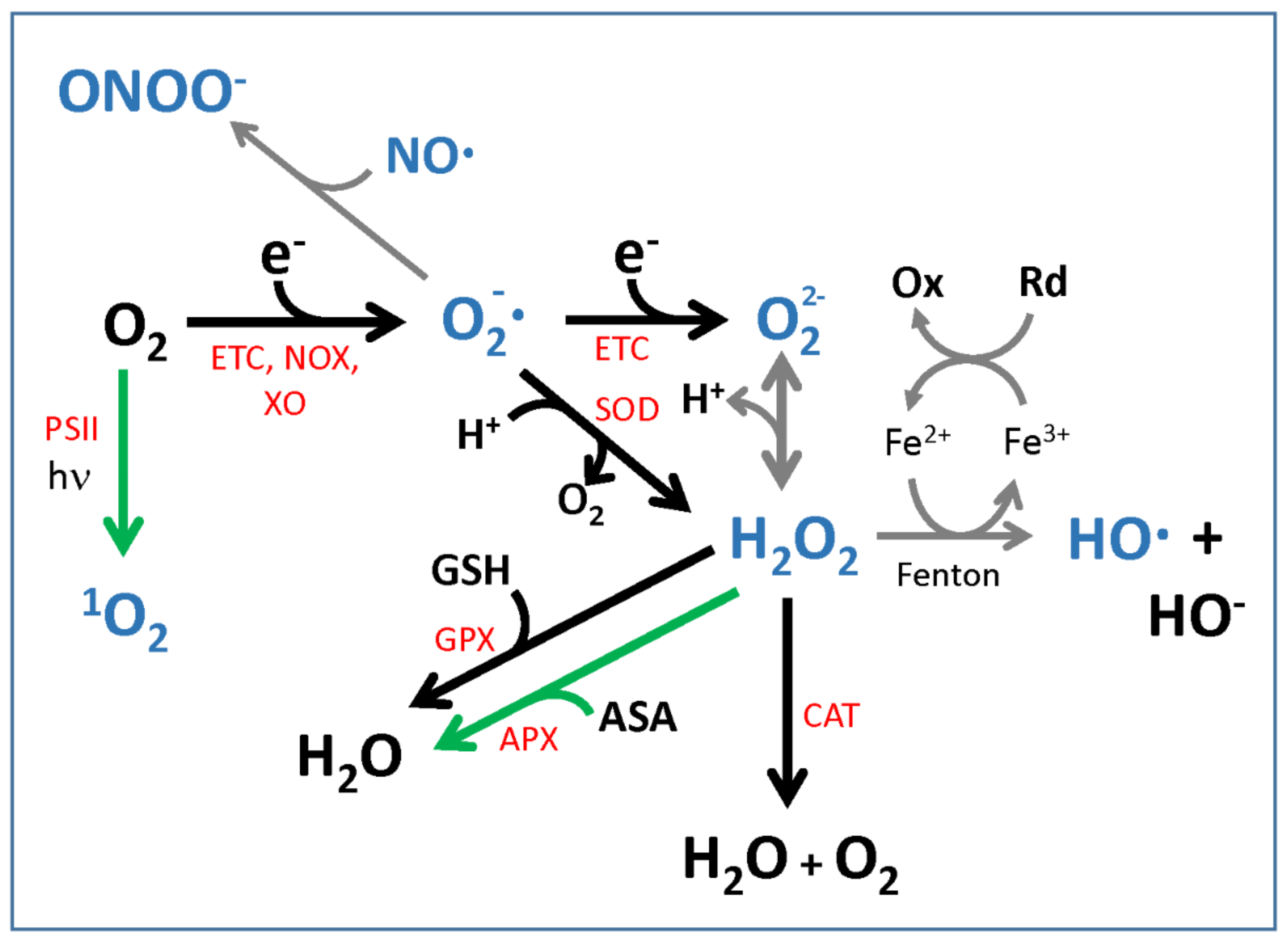
2.1. Reactive Oxygen Species: Production and Detoxification

Several ROS exist in biological systems, with a different reactivity and in different concentrations. They are reactive molecules and are therefore potentially toxic. In this section, we first consider the main ones that are produced in plant and animal cells, the antioxidant systems dedicated to their detoxification, and their role as signals.
ROS are oxygen-derived chemical species (by energy or electron transfer) that are more reactive than oxygen itself and include superoxide anion (O2), hydroxyl radical (HO.), singlet oxygen (1O2), hydrogen peroxide (H2O2), and the related deprotonated anionic forms (HO2 and O22−) [11,12,13] (Figure 1). Nitric oxide (NO) and peroxynitrite (ONOO), though being reactive oxygen-containing chemical species, are not considered ROS and are normally referred to as reactive nitrogen species (RNS), whose metabolism is, however, intertwined with ROS metabolism (Figure 1); for this reason, they are included in this review, with a primary focus on those species able to act as signaling molecules. The hydroxyl radical OH. reacts at a diffusion-limited rate and its half-life is in the order of ns [12,14]; this ROS species can therefore hardly act as a signaling molecule. Singlet oxygen 1O2 has also a very limited half-life, i.e., around 1–4 µs [12]; nevertheless, its stress-signaling activity has been well documented [15,16]. The lifetime of superoxide anion is in the µs–ms range, while that of H2O2 is in the ms–s range, depending on the activities of the scavenging systems [12,17,18].
ROS are routine byproducts of cellular metabolism, in physiological conditions. Both superoxide and H2O2 are indeed produced by the respiratory electron transport chain (ETC), in plant and animal mitochondria; their production rates, however, increase, when ATP consumption slows down or when transport in the respiratory chain is inhibited. Similarly, singlet oxygen and superoxide anions are produced by the photosynthetic ETC in chloroplasts on the PSII side [14,15,19]; H2O2 is produced in various subcellular compartments in plants [20]. Besides mitochondria and chloroplasts [21,22], ROS also can be produced in various other cellular compartments, such as peroxisomes [23], the plasma membrane, plant cell wall, etc. [10].
One ROS may generate other ROS, either through enzymatic or spontaneous reactions. Examples of this are the production of H2O2 from superoxide catalyzed by superoxide dismutases (SOD), the reaction of H2O2 with Fe(II) to produce the hydroxyl radical, or of the superoxide anion with Fe(III) to produce Fe(II), which, in turn, can react with H2O2 (for a review on ROS chemistry, see [13]). These reactions often lead to the generation of more active, and therefore more damaging, ROS [17]; however, their impact also depends on the presence of free iron (Fe2+/Fe3+) or other ROS/RNS [24,25]. One ROS may even stimulate signal transduction pathways leading to further ROS generation, for instance through the respiratory-burst NADPH oxidases present in both animal and plant cells [18,26]. Some ROS are able to cross membranes, either by diffusion or by facilitated transport through aquaporins [27]. They can therefore act as signals of the oxidative status to compartments other than the one where they are produced [17,18].
Ozone (O3) is also a relevant ROS for both plants and animals, mainly in polluted environments. O3 sensing, its effects, reaction products, and plant responses are quite complex [28,29,30,31,32] and go beyond the goal of the present review.
Although ROS are constitutive by-products of essential functions, such as respiration in mitochondria, photosynthesis, and photorespiration in chloroplasts/peroxisomes [33], their production nevertheless increases during exposure to biotic and abiotic stresses, which therefore are referred to as “oxidative stresses” [34]. In particular, the sessile nature of plants forces them to heavily rely on stress resilience strategies rather than stress avoidance [35]. Therefore, plant cells, more than animal cells, must keep steady-state levels of the various ROS under strict control, to avoid any ROS damaging effect on the macromolecules (lipids, DNA, and proteins). This control is achieved through a well-equipped antioxidant system that adjusts, in a coordinated and finely regulated manner, the concentration of various ROS species to avoid ROS noxious effects. For that, two main classes of antioxidant defenses come into play: enzymes and small metabolites (both water and lipid soluble).
In plant cells, glutathione (the tripeptide γ-glutamyl-cysteinyl-glycine, GSH) [12] and ascorbic acid (ASA), also known as vitamin C (vit. C), are the major metabolites impacting the control of the redox status [12,36,37,38,39,40]. ASA is present in all subcellular compartments of both plants and animals [41,42,43], i.e., the cytosol, nucleus, chloroplasts, mitochondria, peroxisomes, vacuoles, and also in the extracellular fluids of animals and cell wall and apoplast of plants; whereas, GSH accumulates in the cytosol, nucleus, chloroplasts, mitochondria, and peroxisomes ([36] and references therein). The ASA biosynthetic pathway mainly proceeds, in higher plants, through the L-galactono lactone intermediate; this pathway is known as the L-galactose or the Wheeler–Smirnoff pathway [44,45,46,47]. The L-gulono lactone pathway also has been investigated as a potential alternative route of ASA synthesis in plants [48,49]; however, in vivo relevance of this latter pathway could not be confirmed, so far [43,50]. The GSH biosynthetic pathway consists of two enzymes, namely γ-glutamylcysteine synthetase (γ-ECS) and glutathione synthase GSHS [51]. Besides ASA and GSH, other metabolites also can act as antioxidant molecules, such as carotenoids [52], tocopherol, and flavonoids ([36] and references therein). All the enzymes involved in the biosynthesis of these antioxidant metabolites can be considered, though indirectly, as part of the antioxidant enzymatic system. Moreover, various enzymes are directly involved in ROS scavenging, such as those taking part in the ascorbate–glutathione pathway, also known as the “Foyer–Halliwell–Asada’’ pathway [14,39], i.e., superoxide dismutases (SOD) and ascorbate peroxidases (APX) dismutate superoxide into H2O2 and O2 and reduce H2O2 to H2O, respectively; the enzymes monodehydroascorbate reductases (MDAR) and dehydroascorbate reductases (DHAR) replenish, in such a pathway, the pool of reduced ASA (from oxidized ASA, i.e., from monodehydroascorbic acid MDHA and dehydroascorbic acid DHA), whereas glutathione reductases restore the pool of glutathione in its reduced state GSH [14,36,39]. Plants cells are equipped with an array of other antioxidant enzymes, such as peroxiredoxins (PRX) and thioredoxins (TRX); a detailed list is discussed in [36].
ROS are therefore constantly removed by specific enzymes and metabolites. These antioxidant systems reduce the toxic effects of ROS, consisting of the oxidation of sugars, nitrogen bases, lipids, and amino acids; the antioxidant systems therefore prevent extensive damage to DNA, RNA, protein, and membrane function. On the other side, ROS, and similar species, can function as signals of stress and induce specific responses in cells.

2.2. ROS Chemistry and Implications for Signaling

The difference between production and scavenging determines the overall result: damage, protection, or signaling [17,18]. The ability of a given ROS to act as a signal between or within compartments depends on its physico-chemical characteristics (for general issues on the chemistry of ROS and the implications for signaling, the reader is referred to recent reviews [18,53,54,55]). For instance, O3 and singlet oxygen are not produced in plant mitochondria, while in animals, singlet oxygen is produced only at specific sites, such as in the retina, or during the respiratory burst of neutrophils and eosinophils [56]; so, these ROS cannot be signals of general relevance. On the other hand, superoxide, HO., and H2O2 are almost ubiquitously produced, but the short half-life of the hydroxyl radical and the lack of known scavengers make it an unlikely signal among compartments. Superoxide, a poorly oxidizing agent in general, is produced in almost every compartment, but is either quickly removed by SOD or it reacts with NO, when available, to form peroxynitrite, which is extremely reactive and able to modify several types of molecules (transition metal centers in proteins, several amino acids (Cys, Tyr, Met, Trp, and His), lipids, and nucleic acids) [24]. SOD-mediated removal of superoxide prevents its reaction with NO [57]. H2O2 has characteristics that make it ideal for signaling: first, it has a relatively long half-life (1 ms or longer [58]) because it is less reactive than most of the other ROS. H2O2 reacts mainly with special Cys residues (peroxidatic Cys) within certain proteins, which act as H2O2 sensors. Second, at room temperature, H2O2 rapidly equilibrates within a compartment because of its high diffusion rate, and can move between compartments. Even if biomembranes are less permeable to H2O2 than to water, specific aquaporins transport it across membranes, both in animals and in plants [59,60].
There are roughly 200 different post-translational modifications possible in higher eukaryotes, 60 of which are ROS-dependent, many of which have been detected in plant mitochondrial proteins [61,62]; Cys can undergo oxidation and nitrosylation (also called S-nitrosation); Arg, Lys, Pro, Thr, and Trp can undergo carbonylation; while Trp and Met undergo oxidation. For instance, cysteine thiol groups of proteins are modified under oxidative stress in different ways. Recent studies identified more than 100 sulfenylated proteins in plastids challenged with H2O2 [63,64]. The Cys residue is oxidized first to sulfenic acid (-SOH), a modification that may be reversed by reduced thioredoxin and glutaredoxin or by reaction with glutathione [65]. Further reaction of sulfenic acid with ROS produces sulfinic (SO2H) and sulfonic acid (SO3H), which are regarded as irreversible [66]. Around 35 proteins become S-nitrosylated (the -SH group reacts with NO and becomes -SNO) in guard cells after exposure to flg22 [67,68]. Tyr nitration has been detected for many proteins and it is involved in the regulation of their activity [69,70]. Tyr nitration is due first to the interaction of Tyr residues with strong oxidizing agents (hydroxyl and carbonate radicals) derived mainly from peroxynitrite, to generate a tyrosyl radical. These can subsequently react with nitrogen dioxide radical, also deriving from peroxynitrite, to form nitrated Tyr residues. There is a physiological level of Tyr nitration that is intensified upon stress [68,69]. Nitration usually inactivates the protein, but it may also affect signal transduction by competing with Tyr phosphorylation. Another direct effect of NO is the nitrosation of the transition metals (Fe, Cu, and Zn) of the protein prosthetic groups.
Each time a ROS modifies proteins and other molecules, thus affecting their activities, it indeed generates a signal. In those cases, the signal is the produced modification (i.e., Tyr nitration or Cys oxidation to sulfenic or sulfinic acid), as such a modification might change the enzymatic activity of the modified protein. If the activity is inhibited, catalysis will slow down, leading to an increase in substrate(s) and a decrease in product(s). Many enzymes, especially in central metabolism, have been shown to be inhibited by oxidative modifications [8,53,69,70,71]. In other cases, the ROS molecule itself will be the signal, because it can diffuse from one compartment to the other and hence modify the redox status in compartments distant from the point where that ROS have been generated. H2O2 as well as NO and peroxynitrite, for instance, fall in this category. Interestingly, feed-back effects are foreseeable, as also some enzymes involved in ROS metabolism are affected by ROS, altering their scavenging capacity. For instance, Tyr nitration by peroxynitrite affects the activity of some SOD isoforms in both plants and animals [72], implying that it tends to boost its own production.

2.3. Redox State Communication among Compartments

The redox status of one compartment is also reverberated in other compartments by means of simple metabolite shuttles. For instance, cytosol, mitochondria, plastids, and peroxisomes share reducing equivalents through the malate–oxaloacetate shuttle. Triose-phosphate (TP)/3-phosphogylcerate (PGA) allows additional exchange of reducing equivalents between the plastid and cytosol in plants, while, in animals, the glycerol phosphate shuttle involving glycerol 3-phosphate and dihydroxyacetone phosphate allows transfer of reducing equivalents from the cytosol to mitochondria [73]. The development of genetically encoded in vivo redox sensors (which are discussed more in detail in Section 3, together with some other sensors) allow visualization of such movements. For instance, recent studies with NADH sensors [74,75] reveal that (i) there are large variations in the NADH/NAD+ ratio among tissues and conditions (for instance, upon sugar supplementation, elicitor exposure, and illumination of seedlings/leaves); and (ii) inhibiting plastidial or mitochondrial ETCs is quite readily reverberated in a change of the ratio in the cytosol, either directly through external NADH dehydrogenases or indirectly through the various metabolite shuttles. A parallel analysis of cytosolic NADH/NAD+, ATP, and pH revealed that there is a rapid transfer of reducing equivalents and protons, but not of ATP, from the chloroplast to cytosol upon illumination [76]. Altering the capacity of the malate valves by knocking out the mitochondrial malate dehydrogenases (mMDH1 or mMDH2), or the NADP+-dependent plastidic isoform (cpNADP-MDH), did not change the capacity of exporting reducing equivalents or its kinetics in the light. However, the cytosolic NAD pool was more reduced in the dark in all three mutants [76]. It is easy to understand how a reduction in capacity for malate oxidation in mitochondria may lead to a higher NADH accumulation in the cytosol. That the same effect is obtained through a reduction in the NADP+-dependent plastidic isoform is more difficult to interpret, but it could mean that a possible increase in NADPH in plastids is relayed to NADH more efficiently in the cytosol via other shuttles.
Thus, altering the redox status by mutation in one compartment can change the balance in other compartments. This is exemplified by the Mosaic Death 1 (MOD1) mutant of Arabidopsis. MOD1 is an enoyl-acyl carrier protein (ACP) reductase involved in plastidial fatty acid synthesis. This enzyme consumes NADH and its absence triggers cell death by increasing the export of reducing equivalents from the chloroplast to mitochondria, mediated by the malate shuttle [77]. Indeed, secondary mutations in malate oxidation in mitochondria or NAD+ import into mitochondria suppress the MOD1 phenotype [77,78,79]. Another example is the gr1 mutant of Arabidopsis, impaired in cytosolic glutathione reductase 1. The mutant shows a more oxidized status in the cytosol, which was reverberated in the mitochondria, as measured through cytosolic and mitochondrial redox sensors (roGFP2-Orp1).

3. Analytical Techniques to Monitor In Vivo Ca2+ and Redox Signaling in Plants: Past and Present Research

In almost every organism, increases in cytosolic Ca2+ concentration ([Ca2+]cyt) work as a key component in different signal transduction pathways. Depending on the stimulus, cytosolic Ca2+ rises can display the form of a single transient or repetitive Ca2+ oscillations and are commonly designated as “Ca2+ signatures” [80,81,82,83,84]. Generation and shaping of cytosolic Ca2+ signatures depend on fine-tuning of the Ca2+ influxes and effluxes occurring at both the plasma membrane (PM) and membranes of the different subcellular compartments. In fact, not only cytosol but also the other subcellular compartments (e.g., chloroplasts, mitochondria, endoplasmic reticulum, etc.) experience Ca2+ transients, hence putatively participating in the cellular Ca2+ homeodynamics and potentially in the shaping of the Ca2+ signature, thus ultimately modulating the signal transduction pathways (reviewed [85,86]). Historically, there have been a few elected cellular systems where scientists have investigated the role of Ca2+ in signaling processes. Because of the fundamental function in plant drought-stress responses, stomata guard cells represent one of the best model systems from where to investigate the roles of Ca2+ signaling [87]. We do cite guard cells because in this system it emerged that, aside from Ca2+, other actors were involved in the ABA-induced stomatal closure, with a prominent role played by ROS (i.e., H2O2) and by their crosstalk with Ca2+ [88,89,90]. In this scenario, a milestone discovery was the identification of H2O2-activated Ca2+ currents in the PM of Arabidopsis thaliana guard cells, which is responsible for cytosolic Ca2+ elevations that contribute to the activation of the anion efflux channels required for the ABA-induced stomatal closure [90,91,92]. Indeed, an impaired activation of these currents in response to ABA strongly reduces the stomata closure, thus affecting the plant’s fitness in drought conditions [93,94]. Surprisingly, even though this discovery was made 20 years ago, the molecular identity of the H2O2-activated Ca2+-permeable channel(s) is still unknown. For this reason, their genetic identification is of utmost importance to decipher the mechanisms involved in plant responses to a changing environment. Nevertheless, further research demonstrated that Ca2+ and H2O2 crosstalk reactions occur in different cell types and at different levels with several feedback mechanisms and self-amplifying loops [95]. One of the most striking evidence pieces in support of this is offered by plant NADPH oxidases, known as respiratory burst oxidase homologues (RBOHs) [96,97]. RBOHs are PM-localized enzymes that produce in the apoplast superoxide anions that are then converted to H2O2 by dismutation [97]. Notably, through RBOHs, ABA induces in guard cells the production of H2O2, thus activating Ca2+ currents across the PM [90]. A very recent discovery ascertained that the H2O2 activation of the Ca2+ currents depends on the activity of the H2O2 receptor HPCA1, a member of the leucine-rich-repeat receptor kinase family [98]. An interesting feature of RBOHs is that their activity is boosted by Ca2+ through direct interaction with Ca2+ (through EF hands) or by Ca2+-dependent phosphorylation [99,100]. So, a self-amplifying Ca2+–H2O2 loop exists in guard cells. Noteworthily, RBOHs are also regulated by other mechanisms, including cysteine persulfidation (H2S). A recent work demonstrated that ABA stimulates, through the activity of L-cysteine desulfhydrase 1, the persulfidation of the RBOHD at Cys825 and Cys890, enhancing its ability to produce ROS, thus positively regulating the ABA-induced stomatal closure [101].
Whereas guard cells represent a peculiar cell system, there is a wealth of examples clearly reporting the existence of a Ca2+–ROS crosstalk in other cell types and in response to different environmental challenges. In particular, in leaf tissues, cells respond to pathogen attack with a quick production of ROS by NADPH oxidases [100]. In this interaction, key players are the pathogen-associated molecular patterns (PAMPs), which are, for example, released by invading bacteria (e.g., flg22 from Pseudomonas syringae) and recognized by pattern recognition receptors (PRRs) [100]. Interestingly, the PRRs’ binding of bacterial PAMPs triggers, at first, a cytosolic Ca2+ increase, followed by the rapid and strong production of ROS [100]. Of note is the demonstration that such a PAMP-induced ROS production is also dependent on RBOHD activity, which, as reported above, is the same isoform involved in the ABA-induced stomatal closure [88]. So, pioneer studies carried out in guard cells have revealed the existence of key basic mechanisms that are shared with other cell types. Indeed, the NADPH oxidase-dependent Ca2+–ROS crosstalk is a model that has been predicted to play a pivotal role also in developmental programs such as pollen tubes and root hair growth [102,103,104,105,106]. The Ca2+–ROS crosstalk model has been predicted to sustain and permit long-distance signaling that occur in plants in response to biotic and abiotic stresses [107,108,109,110]. Systemic wound responses (SWRs) and systemic acquired acclimation (SAA) to abiotic stress are triggered by rapid waves of Ca2+ and H2O2 traveling over a long distance (from leaf to leaf and from leaf to flowers) [95,110,111]. One of the plausible models working in long-distance signaling is therefore represented by a repetition of the Ca2+–H2O2 module in adjacent cells, where the H2O2 produced by a cell can trigger a cytosolic Ca2+ increase in the neighboring cell, which, in turn, stimulates the synthesis of H2O2 via RBOHs in a self-amplifying loop [95,110,112]. Whether this model may be active also during developmental processes, such as embryo development and differentiation, is not currently known. However, it is interesting to note that, in Arabidopsis thaliana, the glutamate receptor-like AtGLR3.5 (a predicted Ca2+-permeable channel) is expressed in germinating seeds and is involved in the regulation of the cytosolic Ca2+ concentration that counteracts the effect of ABA to promote germination [113]. Moreover, GLR3.1 and GLR3.5 in guard cells are predicted to maintain the basal cytosolic Ca2+ levels required for RBOH activity [114]. Interestingly, ROS have been reported to play a pivotal role in the regulation of seed germination and dormancy but how they exert their roles is far from being understood (discussed later, reviewed in [115])
Monitoring Ca2+/H2O2 crosstalk during seed development and germination could be an interesting aspect to study, for instance, to understand the role of these two second messengers during embryo and seed development. In this light, powerful tools like the use of emerging modern imaging technologies are providing new insights in the field. As a proof of concept [116], by using a fluorescence plate reader and Arabidopsis plants expressing MgATP2−, a team sensor demonstrated that seeds, within minutes after imbibition, rapidly accumulated ATP [117]. This piece of evidence paves the way to perform similar experiments to look at Ca2+ and H2O2 in intact seeds. It must be said that since the H2O2 production might be transient, and that it can exert its role through the modification of the cysteine residues of the target proteins [118], one could also consider monitoring the redox status of antioxidant pools, such as glutathione.
To precisely study in vivo and non-invasively the levels of Ca2+, H2O2, and the redox status of the cellular components, we suggest the use of genetically encoded sensors, and in particular ratiometric ones. This suggestion is primarily based on the assumption that the readout of ratiometric sensors is strongly independent of their amount in the cell. This property can also be important when samples are imaged by microscopy, since it allows the correction of focus changes or moving artefacts. For these reasons, we feel comfortable to propose the use of ratiometric genetically encoded sensors for the measurement of Ca2+, H2O2, and the redox status of GSH. There are different genetically encoded ratiometric sensors for the in vivo measurement of these parameters (for this reason, we redirect the interested readers to recent reviews [86,119,120]). However, here we review some tools that are currently used in our lab and that could be used for reliable analyses during embryo and seed development. A schematic representation of their structure and function is presented in Figure 2.

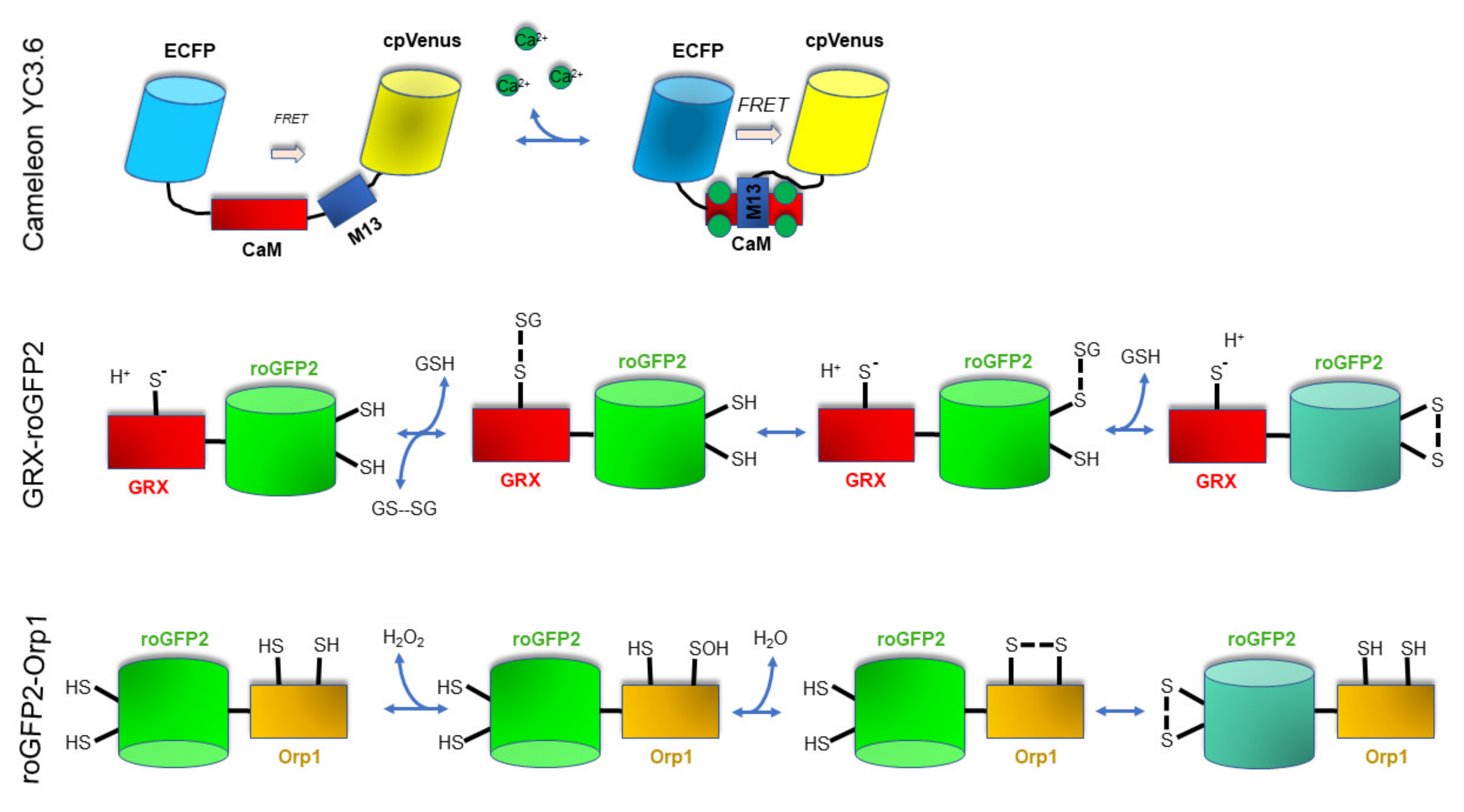
3.1. Imaging Techniques to Monitor In Vivo Ca2+ Levels and Dynamics

Based on our direct experience, the Yellow Cameleon YC3.6 [121] represents an excellent tool for measuring in vivo Ca2+ levels and dynamics in entire organs and tissues [122,123,124,125,126,127]. Specifically, the development of the YC3.6 has greatly advanced the ability to analyze Ca2+ dynamics in vivo with an unprecedented spatial and temporal resolution [128]. YC3.6 is a FRET-based indicator protein. The FRET phenomenon consists of an excitation energy exchange between two fluorescent molecules, an acceptor and a donor, whose absorption spectra partially overlap. This energy transfer occurs when the two molecules are at distances that vary between 2 and 10 nm. In the YC3.6 biosensor, two green fluorescent protein (GFP) variants, cyan fluorescent protein (CFP) and the circularly permuted Venus (cpVenus), are linked together by the Ca2+-binding protein calmodulin and the calmodulin-binding peptide M13 [129]. Binding of Ca2+ to the calmodulin of YC leads to a conformational change in the indicator, bringing CFP and cpVenus into proximity and allowing an enhanced FRET. The efficiency of FRET allows quantitative measurements of the [Ca2+] variations to be made by recording the ratio shifts along a time-course [128]. The higher the Ca2+ concentration, the higher the cpVenus/CFP ratio. Traditional wide-field and confocal fluorescence microscopes able to specifically excite CFP and collect the emissions from the CFP and cpVenus can be efficiently used to measure the CFP/cpVenus FRET, as well as a fluorescent plate reader [126,130]. Arabidopsis thaliana lines expressing the Cameleon YC3.6 targeted to cytosol as well as to different subcellular compartments are available and can be used in entire organs [131] (reviewed in [86,132]).
The imaging of Ca2+ in plants essentially adopts techniques developed for analyses in animal cells; therefore, the use of Cameleon has been largely exploited to monitor the Ca2+ levels and dynamics in the cytosol, nucleus, ER, Golgi apparatus, or endosomes of single living cells ([133] and references therein).

3.2. Imaging Techniques to Monitor In Vivo the 2GSH/GSSG Redox Status

Biochemical techniques have been largely used to study the redox status of the most important antioxidant pools or to study the activity of ROS scavenging enzymes. However, in most cases the biochemical assays require sample destruction and tissue homogenization, which dramatically reduces the sensitivity of the analysis and can also introduce artefacts due to sample manipulation [118,119]. To overcome these issues, in the last 15 years, biologists started to use new in vivo technologies that rely on the use of genetically encoded sensors. This enables real-time monitoring of thiol redox dynamics and possibly of the production of specific ROS.
The most used redox genetically encoded sensors in plants are roGFP1 and roGFP2. These two sensors are modified GFP where two cysteine residues have been inserted in adjacent β-strands on the surface of the protein β-barrel [119,134,135]. This modification renders the proteins sensitive to a change in the cellular redox potential; in fact, the cysteine residues being positioned near the chromophore, they can form a disulfide bond that leads to a structural change that influences protein fluorescence. Similar to Cameleon, roGFPs are ratiometric sensors, since the disulfide bond formation or its breaking changes the quantum yields (QY) of the two main absorption peaks (at 405 and 488 nm, respectively) in an opposite way, leading to a ratiometric response [136]. The higher the oxidation status (hence the disulfide bridges formation), the higher the 405/488 nm ratio. Based on extensive research carried out in animals and plant cells, it has been clearly demonstrated that roGFPs are in a redox potential equilibrium with the 2GSH/GSSG couple in a reaction catalyzed by glutaredoxin (Grx) (reviewed in [119]). Both the GSH pool and Grx are present in the cytosol and in the subcellular compartments [119]; however, since in vivo the roGFPs equilibrium with the 2GSH/GSSG couple is mediated by Grxs, it may be assumed that the roGFPs oxidation/reduction is limited by the availability of endogenous Grxs. To overcome this problem, the human Grx1 was fused to the roGFP2, making a 1:1 proportion between the probe and the catalyst, hence de facto eliminating any possible issue [137]. Arabidopsis lines expressing the Grx1–roGFP2 targeted to cytosol as well as different subcellular compartments are available and can be used in entire organs [126,136].
As in plants, both chemically and genetically encoded probes are available for the detection of redox-mediated signaling and oxidative stress in animal cells and organisms. Advancements in imaging techniques and available probes have been intensively reviewed [138,139,140,141]. Importantly, transgenic animals—both “lower” vertebrates’ models, such as zebrafish, and mice, stably expressing tissue-specific, genetically encoded probes, such as roGFP—are being used to address important biomedical questions (for example, see [139,142]).

3.3. Imaging Techniques to Monitor In Vivo H2O2 Levels and Dynamics

Some peroxidases have an intrinsic and powerful capacity to act as H2O2-dependent protein thiol oxidases when they are recruited into proximity of oxidizable target proteins [137]. Hence, the Yeast Orp1 peroxidase was fused to the roGFP2 to generate an H2O2-dependent roGFP2 oxidation sensor: the roGFP2–-Orp1 [137]. The way roGFP2–Orp1 is used is the same as that one of roGFP2 and Grx1–roGFP2, but the difference is that the oxidation is driven primarily by H2O2 and not by other oxidants. Nevertheless, an important aspect of roGFP2–Orp1 is that its oxidized form is reversibly reduced in vivo, most probably by the Grx and/or the Trx system that directly reduces Orp1 [74]. Because roGFP2–Orp1 undergoes a specific H2O2-dependent oxidation, but can also be reduced, its redox state is influenced not only by the level of the oxidant (H2O2) but also by the reductants themselves, such as GSH and thioredoxin. This makes the use of this probe not completely straightforward for the definition of the H2O2 levels [119]. However, roGFP2-Orp1 has been successfully expressed in Arabidopsis plants, allowing to monitor in the cytosol and mitochondria the intracellular H2O2 dynamics in response to exposure to elicitors such as flg22 and chitosan [74].
A new promising H2O2 ratiometric sensor is the genetically encoded fluorescent protein HyPer7 [143]. HyPer7 is a modified fluorescent sensor composed of a circularly permuted GFP integrated into the OxyR domain from Neisseria meningitis, which is sensitive to very low H2O2 concentrations. This allowed the detection of subtle changes in H2O2 concentrations in the cytosol and mitochondria of animal cells [143], but it also might represent a new useful tool for similar studies in plants.

4. ROS/Ca2+ and the Impact in Differentiation and Developmental Processes in Plants

ROS are end products of multiple and fundamental cellular processes, such as aerobic respiration and photosynthesis, for plant life. As previously described, ROS play key roles in the physiological reprogramming required for plant development [144]. ROS, for instance, act as potent oxidants affecting cell wall cross-linking and extensibility [145,146,147,148], as well as signaling molecules controlling a wide range of biological processes [12,149,150,151,152,153,154].
In response to biotic and abiotic stresses, after mechanical wounding and during developmental processes, ROS are generated by reduction of O2 [155]. At the subcellular level, ROS are produced in several organelles (e.g., chloroplasts, peroxisomes, and mitochondria), but ROS also can be produced in plant cell walls and plasma membranes [7,148,156]. The cell wall is fundamental for the fine control of developmental processes and also to activate specific responses to biotic and abiotic stresses [157,158,159,160]. Indeed, the existence of enzymatic machinery specifically devoted to produce ROS in the cell wall suggests the importance of spatially and temporally regulated ROS production for the control of differentiation processes and, importantly, to trigger fast, local, and active defense mechanisms in response to mechanical injury [148,161,162,163]. In the following section, we describe some of the recent advances uncovering the role of ROS in signal transduction affecting plant developmental processes (see Table 1).

4.1. Seeds, ROS, and Germination

The ability of seeds to survive in harsh environments preserves their longevity. Seeds eventually germinate when the environmental conditions favor the survival and the thriving of the emerging seedlings. These are certainly among the most fascinating and challenging attributes of seed plants [191]. The germination process, which is characterized by rupture of the integument, initial protrusion of the radicle, and the elongation of the embryonic axis, is certainly triggered by favorable conditions of light, temperature, and humidity; however, these conditions might be not sufficient to trigger seed germination in dormant seeds. Dormancy is indeed a protective mechanism aimed at preventing the pre-harvest sprouting; dormancy also prevents simultaneous germination of all the seeds and the competition among them that such a situation would imply [192,193,194]. Interestingly, ROS take part both in dormancy and germination processes [195,196]; they are produced during the drying of the seeds, as well as during imbibition, and represent key players in both seed dormancy and longevity [195,197]. In particular, seeds can germinate when the ROS levels are within a given range; such a range is known as the “ROS concentration window”, and below that concentration seeds would not germinate, whereas above that concentration seeds would suffer from the toxic effects of ROS and their longevity would be compromised [197,198,199]. The existence of a “ROS oxidative window”, needed for seed germination to occur, has been demonstrated by exogenous application of oxidants, by pharmacological or genetic means [12]. For example, A. thaliana plants overexpressing thylakoidal ascorbate peroxidase, tAPX, which scavenges H2O2 (with ascorbate as electron donor), show increased resistance to paraquat-induced photooxidative stress and to nitric oxide-induced cell death; seeds of the transgenic A. thaliana line with the highest tAPX overexpression germinated later than the control seeds and also later than other tAPX transgenic lines with moderate overexpression [200]. However, at that time, the connection between the ROS levels and dormancy went unnoticed. Nonetheless, these results, revisited in light of the “ROS oxidative window”, would suggest that ROS depletion in tAPX plants might also affect developing seeds, and hence, in turn, also their dormancy, which was prolonged in the tAPX overexpressing line. This shift in the “ROS oxidative window” would indeed explain the enhanced longevity observed in 26–30 months after-ripened tAPX seeds, when compared to their wild type line [201]. More recently, measurements of the H2O2 levels in barley seed embryos after imbibition showed higher values in non-dormant seeds than in dormant seeds [202]. These authors reported that activity of NADPH oxidase (producing ROS) and of catalase were, respectively, lower and higher in non-dormant seeds than in dormant ones [202]. The action of H2O2 during germination is multifaceted and it involves endosperm loosening and oxidation of the reserve proteins as well as of the mRNAs, together with the signaling crosstalk with ABA and GA, the two hormones acting in an antagonistic way in the regulation of the germination process, and nitric oxide (NO) [12,203,204,205]. It should be underlined that the ROS status of the cells are dependent on their Fe status, and in particular, on the concentration of uncomplexed, redox-active Fe ions, due to their role in catalyzing the Haber–Weiss reaction [206]; indeed, the Fe nutritional status of a mother plant also affects the dormancy of progeny seeds [191].

4.2. ROS Fine Tune Control on Stem and Root Differentiation in Plants

ROS have multiple roles in plant stem cell regulation, as several works have shown dynamic changes in the expression pattern of ROS biosynthetic enzymes during stem cell differentiation [207]. As elegantly demonstrated by Zeng and collaborators, the fine-tuned balance between superoxide and H2O2 works as a key developmental activator that allows the transition from stem cell maintenance into stem cell differentiation by antagonistically regulating the expression of the transcription factor WUSCHEL [207]. H2O2 is accumulated specifically in the differentiating peripheral region in order to promote stem cell differentiation and antagonistically regulate superoxide accumulation in stem cells by inhibiting key enzymes in superoxide metabolism. The differentiation program involves the repression of SOD and the concomitant activation of peroxidases, establishing the high superoxide/low H2O2 ratio in plant stem cells. During the last years, multiple studies have demonstrated the fundamental role of ROS in root differentiation [8,166,168,208]. The superoxide/H2O2 ratio varies along the roots: while the concentration of superoxide decreases gradually from the meristem to the transition zone, the concentration of H2O2 decreases gradually from the differentiation zone to the elongation zone in the root [209,210]. This balance has been shown to be controlled by UPBEAT1, a basic helix–loop–helix (bHLH) transcription factor, which regulates the expression of a set of peroxidases that control the ROS gradient in the root meristem [168,210]. The transcriptional cascade controlling ROS during root differentiation has been further investigated, demonstrating that hormones such as brassinosteroids define root meristem activity by controlling the ROS balance [211]. Multiple transcriptional factors, such as APP1, ABO8, PHB3, and RITF1, are involved in the brassinolide signaling pathway that controls the ROS signal regulating root stem cell maintenance [166]. Modulation of ROS homeostasis, and its possible involvement in hormone regulation via IAA degradation, has been recently reported to control meristem maintenance in roots [8]. A connection between ROS balance and other hormones, such as brassinosteroids, in the control of root meristem also has been highlighted recently [211].

4.3. ROS Are Crucial in Different Steps during Sexual Plant Reproduction

The process of pollination, which is fundamental for sexual reproduction in almost all plants, has provided excellent examples on how ROS regulation controls developmental processes [1,212]. Different developmental steps are required for successful pollination in plants [213]. Once pollen grains reach the stigma, the pollen adheres and hydrates, and then germinates on it, creating an elongating pollen tube. The pollen tubes grow over a long distance (for instance in maize it can be more than 30 cm) from the stigma toward the ovary in order to reach the female gametophyte. Upon arrival at the female gametophyte, the pollen tube releases two sperms in a degenerated synergid cell for fertilization. Throughout this process, there are continuous interactions, mediated by ROS, between the pollen structure, maternal tissues (stigma, style, and ovule), and female gametophyte, which are necessary for proper pollen hydration and germination, pollen tube growth through the transmitting tract of the pistil, guidance to the ovule, reception of the female gametophyte, and sperm–egg cell fusion (see the recent review by [106]).
We will first review the current literature on ROS and Ca2+ signal transduction in sexual reproduction in flowering plants and animals (see later). Although they constitute different biological systems, embryogenesis in plants and animals has common aspects, such as the male–female gamete interaction and subsequent events of cell proliferation, differentiation, morphogenesis, and embryo development in a maternal-containing environment (see schematic Figure 3 for a compared developmental schematic overview of embryogenesis in plants and mammals).
In most angiosperms, the female gametophyte develops within the ovule and is formed by two antipodal cells, one central cell, two synergid cells, and one egg cell. The cells present in the embryo sac are highly polarized. For instance, while the egg cell nucleus is located toward the chalazal end of the embryo sac, the synergid and central cells have the opposite localization. During gametophyte formation, ROS levels are finely controlled, and they vary depending on the gametophyte cell type. For instance, it has been reported that superoxide and peroxide levels are present in central cells but not in antipodal cells, which later undergo cell death in mature embryo sacs [175]. The central cell has been reported to regulate antipodal cells lifespan. Notably, the accumulation of ROS in the central cell mitochondria might work as a signal to trigger antipodal cell death in a non-cell autonomous way [175]. ROS generated by mitochondria have an important role in regulating cell fate and embryo sac polarity [175]. In Arabidopsis, several studies focused on the role of ROS in female gametophyte development, for instance, via SOD [156]. Most plants have different types of SOD: iron SOD (FeSOD), manganese SOD (MnSOD), and copper–zinc SOD (Cu/ZnSOD). This is considered the first line of cellular defense against superoxide. Its importance was demonstrated, for instance, by the characterization of the Arabidopsis oiwa mutant, a female gametophytic mutant impaired in mitochondrial manganese SOD (MSD1). In this mutant, high levels of peroxide and mitochondrial superoxide accumulate in the central cell as well as the micropylar cells. Interestingly, a range of female gametophyte phenotypes accompanied these alterations; for instance, the mitotic arrest during megagametogenesis or the partial central cell identity determination [175]. Another example is the developmental characterization of the athemn1 mutant defective in tetrapyrrole biosynthesis, which accumulates more ROS in developing embryo sacs and has defects in female gametophyte development, with embryo sacs displaying unfused polar nuclei [176]. Interestingly, central cell differentiation is impaired in the athemn1 mutant. Upon fertilization, its seeds present defective endosperm development and embryo developmental arrest [176]. The role of mitochondrial ROS for proper embryo sac development in female gametophyte development has been demonstrated in several works. This link has been shown in multiple Arabidopsis mutants and excellent reviews are available [214]. Interestingly, a potential role of oxidative stress in the balance between sexual megasporogenesis and apomixis program in facultative apomictic plants also has been proposed recently [215,216]. Further development of research in this context could provide interesting clues in order to understand how environmental factors influence apomixis via ROS-. This has a major impact for breeding programs aiming at introducing apomictic traits to avoid genetic variability associated with sexual reproduction.
However, the role of many players affecting ROS in each cell has yet to be discovered. Single-cell transcriptome data are now available for specific plant structures, such as the female gametophyte [217]. One can thus explore the variation in the level of specific transcripts involved in ROS metabolism among cell types in female gametophyte development. In order to provide candidates for future research, we analyzed this dataset (see Table 2). Interestingly, there are multiple members of genes involved in ROS metabolism, such as SODs, peroxidases, glutathione peroxidases, ascorbate oxidases, and glutathione peroxidases, which are differentially expressed in specific female gametophyte organs such as synergids, egg cells, or the central cells. We found very few genes substantially expressed in the synergids, especially at a high level (e.g., Catalase1). In the central cell, some genes (Catalase1, Glutathione Peroxidase 2 and 6) were expressed at a high level and many at an intermediate level (Glutathione Peroxidase1, 3, and 8). An intermediate situation is taking place for the egg cell, where there are fewer genes expressed compared to the central cell, but usually at a moderate level. Some genes represent interesting candidates involving the cross talk among the cell types and the tissues derived from them (embryo/endosperm). For instance, Glutathione Peroxidase 2 seems to be expressed solely in the central cell, while isoform 6 is apparently expressed in all three cell types, although at different levels. For example, looking at the genes of the canonical VitC biosynthetic pathway, AT3G02870 (VTC4) is apparently expressed in both the egg cell and central cell, while AT5G55120 (VTC5) and AT3G47930 (GalLDH) are expressed only—or almost exclusively—in the central cell. It might be worth looking at the developmental defects in preliminary developmental steps around fertilization or at early embryo development in the respective mutants. The hypothetical altered enzymatic activities of genes that act as scavengers (or that are involved in the production of scavengers) of ROS are likely to locally affect the ROS levels in specific cell types in which they are expressed, and might impact primarily the functionality of these cells. Future advances will provide knowledge on the role of those specific ROS-related regulators in plant reproduction.
The role of ROS in mature female gametophyte function has also been investigated in depth during the last years and there is plenty of literature. Upon pollination, pollen grains land on the stigma of a flower. After crossing the stigmatic barrier, the pollen tube begins to grow through the style and the transmitting tract and it navigates through the funiculus, the structure that attaches the ovule to the placenta. At this stage the pollen tube exits out of the intercellular space and grows along the funiculus, reaching for the micropylar aperture of the ovule. The pollen tube enters the ovule through the micropylar region and bursts to release two sperm cells into the female gametophyte. The double fertilization in angiosperms thus requires two sexual fusions; one sperm fuses with the egg cell and leads to the diploid embryo formation, the other one fuses with the central cell, forming the triploid endosperm. The role of ROS in pollen tube growth and pollen-pistil interaction has been excellently reviewed in detail [106]. One of the key functions performed by ROS in the female gametophyte takes place during pollen tube–synergid cell interaction. Synergid cell death is critical in three key steps of the proper angiosperm fertilization process [218]. First, synergid cell death is necessary for pollen tube entry into the ovary. Secondly, the degenerated state of synergid cells is required for tube growth cessation and release of pollen tube contents. Third, synergid degeneration accompanies the cytoskeletal reorganization necessary to facilitate migration of the two sperm cells to the egg and central cells. Thus, the synergid cells are necessary for successful fertilization by communicating with the pollen tube. They secrete small cysteine-rich peptides LUREs in order to guide the pollen tube growth to reach the ovule [219,220,221]. Pollen tube receptors sense LURE peptides secreted from the synergid cells to orientate pollen tube growth into the ovules. After being attracted to the ovule, signals from the synergid are again perceived by the pollen tube. These signals trigger changes in the pollen tube tip, leading to pollen tube burst and delivery of the sperm cells to the female gamete [221]. It has been proposed that an oxidative environment at the female gametophyte is required for pollen tube reception at the synergids [175]. H2O2 was detected in a specific temporal window specifically in synergid cells; after pollen arrival onto the stigma but before pollen tubes reach the ovule [175]. Supporting the importance of ROS in pollen tube attraction, high ROS levels were detected in the ovule micropyle, the specific region that allows pollen tube penetration into the ovule [222]. Chemical application of ROS scavengers to excised pistils to highlight the role of ROS in pollen tube attraction was performed by Duan and collaborators. They found that pollen tubes were attracted to ovules but they continued to grow and failed to burst and release the sperm, thus confirming that local ROS changes are critical for proper pollen timing on the tube attraction necessary for double fertilization [222]. Consistent with these inhibitors assays, recent evidence supports the role of ROS in the process by studying the synergid-specific receptor-like kinases (RLKs) involved in pollen tube reception: FERONIA (FER), HERKULES1 (HERK1), and ANJEA (ANJ) [180]. It was recently shown that fer mutants also display a pollen tube overgrowth phenotype and do not accumulate ROS in the micropyles of ovules, providing evidence that micropylar ROS levels is an important determinant controlling pollen tube reception [180].
The role of Ca2+ signaling in synergid cells during pollen tube reception was also investigated [186,187,223]. Studies performed with the Arabidopsis synergid-specific gene NORTIA (NTA) have shown how Ca2+ oscillation in synergids is fundamental for proper pollen tube reception. nta mutants display a pollen tube overgrowth phenotype [224]. A recent study reported that also the Ca2+ signal is required for proper pollen tube sensitivity to ovule signals in Arabidopsis thaliana; MLO5 and MLO9 can recruit a Ca2+-specific channel at the plasma membrane via SNARE proteins in order to fine-tune and modulate the Ca2+ gradients in the pollen tube [186,187].
The last step of pollen tube reception involves pollen tube bursting in order to release the sperm cells. Pollen tube discharge triggers synergid degeneration in a coordinated way, involving a pollen tube–synergid interaction [225]. The role of ROS triggering synergid cell degeneration by initiating a programmed cell response (PCD) has been proposed, but these mechanisms are still unknown. The connection between micropylar ROS and synergid control of pollen tube behavior may be more complex than previously thought. Higher resolution imaging of ROS dynamics and Ca2+ sensors during pollen tube arrival to the synergid cells in specific mutants will be essential in order to clarify the role of ROS and Ca2+-waves during reproduction in plants. The use of specific sensors targeted in different cell types appears as a promising strategy for better understanding the role of ROS in the process (as discussed previously).
Several studies focused on the involvement of ROS in pollen functioning. Upon landing on the stigma, pollen grains undergo adhesion and hydration and trigger a germination program in order to generate the pollen tube [213]. For instance, in Arabidopsis it was shown that the NADPH-oxidase homologues, RbohH and RbohJ, are the source for most ROS produced at the pollen tube apex [103]. The relative double mutant presented low ROS generation and concomitantly exhibits bursting in vitro and delayed growth in the pistil [185]. Those studies suggest that Rboh are critical for proper pollen tube growth and are involved in maintenance of cell-wall integrity [103,185]. It was shown that in the null mutant of Arabidopsis KINβγ, a plant-specific subunit of the SNF1-related protein kinase 1 complex, the ROS levels of the pollen grains were reduced and concomitantly pollen adhesion to the stigmatic surface was impaired [226]. Referring to the pollen–pistil interaction, it has been suggested that the ROS signals originating at the pollen grains mediate the interaction with the stigma by controlling the expression of the inward shaker K+ channel SPIK in pollen [227]. Pollen–stigma signaling is involved in the different mechanisms of self-incompatibility in multiple plant species [214,228]. ROS regulation is important for the control of self-incompatibility processes in plants, as reported in many species, although specific strategies might differ between plant systems and developmental stages, including defense functions, signaling, and senescence [229]. It has been shown that ROS triggers programmed cell death (PCD) in self-incompatible response [230]. More examples of the role of ROS controlling the pollen–pistil interaction was reported recently in ornamental plants such as kale (Brassica oleracea var. acephala), a self-incompatible species. The decreased ROS levels in kale stigma exposed to exogenous flavonoid treatment had a negative impact on the attachment and successive germination of the compatible pollen. In recent years, multiple works have investigated ROS signaling in pollen–stigma communication—we apologize for not discussing them because of space limitations [212,214,229,231].

5. ROS/Ca2+ Crosstalk in Mammalian Embryonic Development

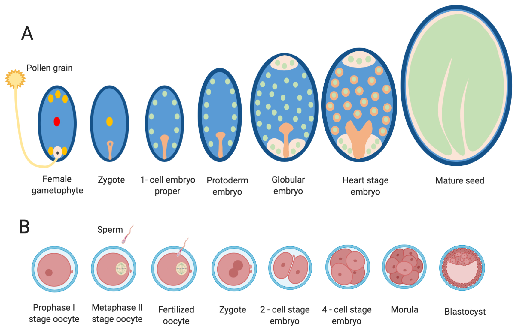
5.1. Overview of Gametogenesis and Early Embryonic Development in Mammals

In mammals, the female gamete, the oocyte, is the largest cell of the body and has the intrinsic ability to initiate embryonic development once it has undergone a process known as activation, while the paternal gamete, the spermatozoon, has developed the ability for motion and penetration of the oocyte’s investments. A schematic representation of fertilization and early embryonic development is shown in Figure 3B.
Oocyte and sperm development are complex processes that initiate very early during development of a new individual (for comprehensive description, please refer to [232]). Once the primordial germ cells (PGC) migrate to the forming gonad, they develop into oogonia and spermatogonia, which are the pluripotent cells from which the male and the female gametes originate. Oogonia and spermatogonia undergo further proliferation before they enter gametogenesis that in turn includes meiosis and cytodifferentiation. These processes largely occur in the gonads in cooperation with their somatic cell components.
In the female, oogenesis starts in the fetal ovary and terminates only after puberty is reached and fertilization eventually occurs. In the fetal ovary, oogonia differentiate into cells called primary oocytes, which duplicate their DNA and then enter the prophase of meiosis I, when they become arrested at the diplotene stage. Primary oocytes are covered by ovarian somatic cells (called follicular cells), thus forming the primordial follicles, which constitute a pool from which the female will recruit follicles for growth and ovulation for the rest of the reproductive life. Follicular growth occurs when follicles are recruited from the primordial pool (follicle activation) and develop into primary, secondary, and tertiary follicles. Concomitant to follicle activation, the enclosed primary oocytes reactivate RNA synthesis and are engaged in a phase of growth during which a large amount of new molecules are produced and stored in the oocyte cytoplasm. Important morphological and functional changes, such as the synthesis of a protective glycoprotein shell—the zona pellucida—and the development of specialized cytoplasmic organelles, occur during the growth phase. At the end of the growth phase, in the tertiary follicle, transcriptional activity is again globally silenced, and the oocytes, now referred to as “fully grown” oocytes, are ready to resume meiosis. Meiotic resumption, which is cyclically triggered only after puberty by hormonal stimulation, is followed by progression to the metaphase, anaphase, and telophase of meiosis I. Completion of meiosis I is followed by an asymmetric cell division in which half of the homologous chromosomes are extruded in a small cell, called the polar body. Then, the large oocyte enters meiosis II, the chromosomes are arranged in the metaphase II plate, and meiotic progression arrests again until fertilization eventually occurs. Progression from the prophase I to the metaphase II stage is referred to as “oocyte maturation”. In most species, oocyte maturation occurs within the ovarian follicle and the metaphase II stage oocyte is released from the ovarian follicle into the oviduct, the anatomical site of fertilization, by a process called ovulation.
In the male, spermatogenesis begins after puberty and includes three main steps: spermacytogenesis, meiosis, and spermiogenesis. Spermacytogenesis leads to the generation of primary spermatocytes by several mitotic divisions. Primary spermatocytes then undergo and complete meiosis, which produces haploid spermatids. Spermiogenesis follows the meiotic division and includes a series of morphological and functional changes that ultimately re-structure the round-shaped spermatids into differentiated spermatozoa. Final functional maturation of the spermatozoa, including acquisition of motility, capacitation, and acrosomal reaction, occur in the male and female genital tract.
When the fertilizing sperm penetrates the oocyte, the maternal and paternal genomes are united in a single cell: the fertilized ovum, forming the zygote. It is well documented that fertilization triggers Ca2+ waves that lead to the prevention of polyspermy, oocyte activation, and meiotic resumption. Specifically, the oocyte resumes and completes meiosis II: half of the sister chromatids are segregated into the small second polar body, while the remaining chromatids that are retained in the large ovum decondense and form the female pronucleus. The chromosomes contained in the sperm head decondense and form the male pronucleus.
After DNA replication, the maternal and paternal genomes reconstitute the diploid status in a process called syngamy, chromosomes condense, and the first mitotic division of the new individual commences. Firstly, the zygote develops into the 2-cell embryo. Thereafter, the embryonic cells (the blastomeres) form a small cluster of cells referred to as the morula, which soon undergoes a process called “compaction”, forming a more uniform surface of the embryo. The outer cells develop into the trophectoderm. Subsequently, during the process of blastulation, a fluid-filled cavity, the blastocyst cavity, develops inside the trophectoderm, and the inner cells, forming the inner cell mass (ICM), gather at one pole of the embryo, which is now known as a blastocyst. The trophectoderm will participate in placenta formation while the ICM gives rise to the embryo proper. The blastocyst expands, hatches from the zona pellucida, and later, towards the end of blastulation, the ICM forms an internal and external cell layer—the hypoblast and epiblast, respectively—to establish the embryonic disc. Implantation and placental development in the uterus occur in a species-specific manner.

5.2. Overview of the Ca2+ and ROS Signaling Interplay in Animal Cells

In animals, as in plants, a mutual interplay between Ca2+ and ROS signaling has emerged in the last decades: Ca2+ signaling is essential for ROS production and, on the other hand, ROS can affect cellular Ca2+ signaling. Likewise, it is widely accepted that physiological (i.e., subtoxic) levels of ROS act as signaling molecules by oxidizing proteins, lipids, and nucleotides [233]. One of the main effects of ROS is the partially reversible oxidation of cysteines, leading to the formation of disulfide bonds, as well as further oxidation products, such as sulfenic, sulfinic, and sulfonic acids, which in turn modulate protein conformation and activity. However, a further increase in ROS concentration affects cellular homeostasis, leading to oxidative stress response and cellular damage. The specific roles of Ca2+ and ROS, as well as their interplay in physiological and pathological conditions, have been extensively reviewed (for example, see [234,235,236,237,238,239,240]). Here, we present a brief overview of the relevant concepts in animal cells, in order to introduce the current view on Ca2+/ROS interplay during early embryonic development in mammals.
Ca2+ ions can allosterically regulate the activity of many proteins and enzymes. Therefore, as already discussed for plants, Ca2+ ions are implicated in a high variety of functions in animal cells. Ca2+ signaling pathways are key in many crucial biological processes, such as cell survival, proliferation and death, contraction, secretion, metabolism, and regulation of gene expression. Animal cells have evolved complex mechanisms to control the fluctuations of cytosolic Ca2+ ions and, in turn, Ca2+-elicited cellular functions. The concentration of cytosolic Ca2+ can be altered by the controlled mobilization from two Ca2+ sources, the extracellular microenvironment, and the intracellular stores, namely, the endoplasmic reticulum (ER)—which is the main Ca2+ store—the Golgi apparatus, and the mitochondria. Animal cells exert this function through the coordinated activity of pumps and channels as well as Ca2+-binding signaling molecules, enzymes, and buffering proteins. One of the most common pathways for mammalian cells to trigger Ca2+ signaling is initiated by ligand-dependent G protein-coupled receptors, causing the synthesis of 1,4,5-inositol trisphosphate (IP3) and IP3-dependent opening of Ca2+ channels of the IP3 receptor (IP3R) family at the ER membrane (for a recent review, see [241,242,243]). Typically, resting animal cells have a low concentration of cytosolic Ca2+ (around 100 nM), while a rise (up to 2–3 µM) in its concentration stimulates specific cellular function (reviewed in [239,243]). Once Ca2+ ions have carried out their signaling functions, they are rapidly removed from the cytosol by extrusion to the extracellular space or by intracellular compartmentalization [243]. Interestingly, a comparative analysis of Ca2+ ion transport proteins in plants and animals revealed that the Ca2+ elements with basic functions in cell responses (CNGC, iGlu receptor, Ca2+ATPase, and Ca2+/Na+-K+ ion exchanger) are basically conserved between plants and animals, while the genes specific for muscle and nerve Ca2+ signal transduction systems (VDCC, IP3 receptor, ryanodine receptor) are very different [244].
Many ROS-generating systems in a cell can be modulated by Ca2+, including those active in mitochondria and those that operate extra-mitochondrially, such as NADPH oxidases, uncoupled nitric oxide synthase, cytochrome P450s, cyclooxygenases, and many others [245].
For the purpose of this review, the role of mitochondria in the control of Ca2+ homeostasis is particularly relevant [234,236,238,246,247] as mitochondria are an important source of ROS in the gonads and their functionality is pivotal in gametes and embryos [248,249]. After the discovery that mitochondria accumulate Ca2+ [250,251], a series of studies revealed that the intra-mitochondrial Ca2+ concentration remains low in resting animal cells, but it rapidly increases upon cell stimulation, and that this is possibly due to the close proximity of mitochondria with Ca2+ channels in the ER that elicits the rise in cytosolic Ca2+ [252,253,254,255] (reviewed in [239]). The mitochondrial Ca2+ uniporter (MCU) and the voltage dependent anion channels (VDACs) are primarily responsible for the function that this organelle exerts in Ca2+ homeostasis (reviewed in [239]).
In mitochondria, Ca2+ uptake is in turn involved in controlling the rate of ATP production by stimulating enzymes of the Krebs cycle and oxidative phosphorylation. Nevertheless, Ca2+ control of ROS production is very complex, and the interactions depend on the cell and tissue types. Since mitochondrial ROS are produced at different sites of the electron transport chain (mostly complex I and III), it is logical to think that high metabolic rates are associated with high ROS production [256]. Nevertheless, it must be highlighted that, within a certain level of Ca2+, the metabolic state (i.e., membrane potential) of the mitochondria itself seems to determine the effects of Ca2+ uptake on the mitochondrial ROS level. However, in other circumstances, when mitochondria are overloaded with Ca2+, ROS production is no longer dependent upon the mitochondrial metabolic state. Furthermore, in the presence of other factors (such as oxidative stress, high phosphate concentrations, and low adenine nucleotide concentration), excessive accumulation of mitochondrial Ca2+ has negative consequences: ATP production is impaired, and the production and sustained opening of the high mitochondrial permeability transition pore (mPTP) is induced [257]; this, in turn, induces a dramatic increase in mitochondrial membrane permeability, its depolarization, and ultimately mitochondrial swelling. As a consequence, a further increase in mROS production, the release of cytochrome c, and, ultimately, apoptosis occur [239,257] (reviewed in [239]).
In conclusion, emission of ROS from mitochondria is the net result of ROS production at the electron transport chain (ETC) and their elimination by antioxidative enzymes [246] and the mutual interplay between Ca2+ and ROS, which is quite complex in animal cells. This is also reflected in the relevance of Ca2+/ROS crosstalk in the physiopathology of the heart, skeletal muscle, neurons, and, eventually, in aging and cancer (reviewed in [239,258]).

5.3. ROS/Ca2+ Signaling in Mammalian Early Embryonic Development

5.3.1. Ca2+ Signaling

Ca2+ exerts a pivotal role in animal gametogenesis, fertilization, and embryonic development (for a recent review, see [259]). Initiation of the development is triggered by a Ca2+-activating signal, which has been broadly studied in both mammalian and non-mammalian species [260,261,262]. All of the knowledge in mammals derives from studies in mice. However, pioneering studies in animal models, such as in fish and sea urchin, have been fundamental to our current knowledge on the role of Ca2+ waves in mammalian development (reviewed in [260]).
As anticipated, in mammals, the oocyte acquires the capability to be fertilized during oocyte growth and subsequent maturation, when the immature prophase I-arrested oocyte resumes meiosis, completes the first meiotic division by extruding the first polar body, and arrests again at the metaphase stage of the second meiotic division (MII), when fertilization will eventually occur (see schematic Figure 3B for a developmental schematic overview of embryogenesis in mammals). Although the immature oocyte is capable of generating Ca2+ waves, which persist for a few hours and cease when the nuclear envelope breaks down after meiotic resumption [259,263,264,265], the precise biological significance of this phenomenon is still unclear (albeit it could relate to mitochondrial activity, see below). Interestingly, several studies in rat oocytes indicate that a moderate increase in Ca2+ and ROS, together with a transient decrease in cyclic AMP content, destabilize MPF, thus promoting meiotic resumption of the prophase I-arrested oocyte [266,267,268], which occur spontaneously (i.e., independently from the hormonal stimuli) when the oocytes are isolated from ovarian follicles. Moreover, during maturation, the cytoplasm of the oocyte is remodeled. Experimental evidence suggest that the precise machinery required to develop the characteristic Ca2+-dependent series of events that occur at fertilization (see below) is acquired during maturation [264]. These changes include the reorganization of the ER—the major Ca2+ store in the oocyte—the increase in intracellular Ca2+ stores, the changes in IP3R 1 expression pattern, as well as redistribution of the Ca2+-binding proteins [259,264,265,269,270,271].
At fertilization, when the sperm and the MII stage-arrested oocyte (egg) fuse, the sperm delivers the diffusible phospholipase C ζ (PLC ζ) into the egg cytosol. PLC ζ then hydrolyzes phosphatidylinositol 4,5-bisphosphate (PIP2) into IP3, which binds its receptor, IP3R1, in the ER, which, in turn, triggers Ca2+ release into the cytosol [272,273]. The Ca2+ concentration then oscillates periodically, generating Ca2+ waves, which last for several hours after sperm entry [262]. Activation of several pathways and enzymes by Ca2+ oscillation leads to several fundamental events in the fertilized egg, which are collectively referred as to oocyte activation, such as meiotic resumption (i.e., exit from the MII arrest), completion of meiosis II, formation of the pronuclei, and exocytosis of the cortical granules to prevent polyspermy. As in other mammalian cells, the Ca2+ concentration returns to its baseline by extrusion through the plasma membrane and/or re-loading in the ER, through the activity of the Ca2+ ATPases (PMCAs) and/or Na+/Ca2+ exchanger (NCX) at the plasma membrane, and the sarco-ER Ca2+ATPases (SERCAs) in the ER, respectively (see Figure 1 of [265]). Mitochondria and the Golgi apparatus also contribute through the activity of the mitochondrial Ca2+ uniporter (MCU) and the secretory-pathway Ca2+-transport ATPases (SPCAs), which take up Ca2+ into these organelles. Nevertheless, a role as a Ca2+ store does not seem to be the main function of mitochondria in fertilized eggs, but rather mitochondrial Ca2+ uptake seems more related to energy production [274,275]. The precise molecular mechanisms that drive all the processes described above are starting to be elucidated and are the subject of intense investigation (reviewed in [259,265]).
Clearly, the control of Ca2+ waves in fertilized oocytes has wide implications in mammalian fertility. Very recently, CRISPR/Cas9 technology was used to generate PLCζ KO mice. These studies provided the definitive evidence that PLCζ is the physiological trigger of the Ca2+ oscillations in mammals [276,277]. In addition, studies in humans identified a mutation in the gene encoding for PLCζ, which led to a reduced ability to generate Ca2+ oscillations in fertilized eggs [278,279,280].

5.3.2. ROS Signaling

For many years, ROS have been considered almost exclusively as detrimental compounds in the field of mammalian reproductive biology. Intense research has been conducted on this subject, and nowadays almost 10,000 publications are found on PubMed when the database is mined for the keywords “oxidative stress and mammalian reproduction”. Still today, even though physiological roles of ROS are widely recognized [249], their detrimental effect is a very hot topic among reproductive scientists [281,282]. This is mainly because oxidative stress, caused by elevated ROS production and/or a decreased antioxidative system, is considered one of the predominant causes of both male and female infertility. Moreover, oxidative stress profoundly impacts the efficiency of assisted reproductive technologies (ART), which are widely used for the treatment of human infertility, in animal breeding, as well as for the preservation of endangered species. ROS production is stimulated during all the steps of in vitro embryo production, such as in vitro oocyte maturation, fertilization, and early embryo culture. Moreover, gametes and embryo freezing are largely used in ART, where ROS generation is particularly challenging. As a consequence, a plethora of studies have focused on the assessment of the effects of antioxidant compounds on in vitro embryo development, and to find the most suitable culture conditions to recapitulate fertilization and early embryonic development in vitro. Just as an example, early embryos are usually cultured under low oxygen tension (5%) to minimize oxidative stress. However, excess antioxidants were found to be not only ineffective but sometimes deleterious. This may be attributed to a disturbance in the signaling functions of ROS by the excess of antioxidants [249].
Nevertheless, the physiological milieu in which the female gametes are generated and then fertilized is, per se, rich in ROS. As a matter of fact, the ovary is an endocrine organ that generates ROS during steroidogenesis. Ovulation, the process by which the mature egg is released in the oviduct for fertilization, is recognized as an inflammatory-like reaction and many proinflammatory agents, such as interleukins and tumor necrosis factor, have physiological roles in the ovarian follicle [283]. Thus, antioxidant processes are set in place in the ovary to protect gametes from oxidative compounds. Many studies have demonstrated that when the function of the antioxidant machinery is diminished, such as in ageing or when antioxidative enzymes are depleted experimentally, fertility is impaired [284]. Furthermore, studies in mice have shown that, in the oocyte, ROS are produced by mitochondria and by oocyte oxygenase [285]. Notably, defective mitochondrial functions are generally associated with poor oocyte developmental competence and meiotic errors. Thus, much is known on the “bad side of ROS” in the ovary and in female egg development. On the contrary, ROS-mediated signaling events in oocytes are less known, one of the limitations being the high amount of ROS that are administered/generated in vitro and the difficulties to study and visualize this phenomenon in living organisms.
In males, spermatogenesis is also prone to the damaging effects of ROS. A sophisticated array of antioxidant systems that include both enzymes and free radical scavengers is set in place in testis to limit the detrimental effects of ROS [249,286,287]. However, the physiological functions for ROS have been reported also in the testis (reviewed in [288]).
ROS are essential signals involved in the regulation of spermatogenesis, steroidogenesis, and sperm function. Specifically, in the sperm, they trigger morphological changes required for sperm maturation and modulate crucial processes involved in the attainment of sperm fertilizing ability, such as capacitation, hyperactivation, acrosome reaction, and sperm–oocyte fusion (reviewed in [288]). For example, physiological levels of ROS are crucial for sperm maturation that occur in the epididymis, the anatomical site where spermatozoa acquire motility, and where chromatin condensation and plasma membrane remodeling occur. During spermiogenesis, histones in chromatin are replaced by protamines that are rich in Cys residues; in the epididymis, H2O2 and antioxidants, such as glutathione peroxidase 4 and 5 (GPX4, GPX5) and peroxiredoxin 6 (PRDX6), contribute to the formation of sulfhydryl bridges between the cysteine residues of the protamines, resulting in chromatin condensation in the sperm head (reviewed in [288]).
Recently, the knowledge on the role of H2O2 in metazoan development has been revisited in the frame of the morphogenetic process during embryogenesis, regeneration, and stem cell differentiation [7]. What emerges is that “redox signaling interacts directly or indirectly with most of the signaling pathways that control embryonic development”. However, as the authors conclude, “We are only starting to perceive the tip of the iceberg”. Similarly, ROS signaling during early mammalian embryogenesis (i.e., before implantation) is far from being deciphered. As already mentioned, oxidative stress is detrimental for early embryos. It has been shown that oxidative stress associated with in vitro culture induces developmental arrest (i.e., block of the cell cycle) and cell death. Studies in SOD KO mice suggest that, under oxidative stress conditions, ROS act by regulating the expression of genes of the cell cycle machinery [249]. Nevertheless, studies in mouse and bovine early embryos have shown increased concentration of ROS at critical stages of development, and the assessment of embryo metabolism confirmed that ROS are key at fertilization and early embryo development [285,289,290,291]. In bovine, in particular, the assessment of oxygen consumption and ROS production at the time of fertilization and cell cleavage have suggested that both processes have a regulatory role. Importantly, these studies, together with the ones that have established a correlation between oxygen consumption and Ca2+ waves [274,275,290,292], led to the suggestion that mitochondrial activity is stimulated by Ca2+ oscillation at fertilization in mammals [291].

5.3.3. Ca2+/ROS Signaling Interplay in Gametes and Early Embryos

Very little is known about the Ca/ROS interplay during gametogenesis and early development, especially mechanistically. However, two very recent studies clearly indicate that the available technologies might be finally able to answer many unresolved questions, and likely accelerate the discoveries of new key processes in development.
The first study, conducted in a mouse model, demonstrated that constitutive IP3R1-mediated Ca2+ release reduces Ca2+ store content and stimulates mitochondrial metabolism in prophase I immature oocytes [259]. As already anticipated, the immature oocyte is capable of generating Ca2+ waves, which persist for a few hours and cease when the nuclear envelope breaks down after meiotic resumption [259,263,264,275]. By using technology, such as the microinjection of fluorescent probes targeted at mitochondria and advanced imaging systems to detect Ca2+ oscillation using the Fura-2 probe, Wakai and Fissore found that Ca2+ stored in the ER of immature oocytes constitutively “leaks” through the IP3R1 [265]. The Ca2+ “leak” ceases around the time when the oocyte resumes meiosis and the nuclear envelope breaks down, which is when the accumulation of Ca2+ in the cellular stores, in preparation for fertilization, is firstly seen. Strikingly, during Ca2+ oscillation, Ca2+ is transferred to the mitochondria, where it stimulates metabolisms and increases the levels of ATP [259]. Clearly, this study opens a new area of investigation on the possible role of the Ca2+/ROS interplay in female gametogenesis.
The second study took advantage of a transgenic Xenopus laevis that ubiquitously expressed the H2O2 sensor HyPer, including the eggs of transgenic females [293]. Using this model, the authors demonstrated that, at fertilization, the Ca2+ waves induced by the sperm–egg fusion triggers a rapid increase in ROS levels, which in turn oscillate with each cell division. Furthermore, using inhibitors of complexes II, III, or IV of the electron transport chain, they also demonstrate that these enzymes are responsible for ROS generation. Importantly, their data also suggest that Ca2+-induced ROS signaling might control the onset of cell division, as inhibition of mitochondrial ROS production in early embryos results in cell cycle arrest, in part, via ROS-dependent regulation of Cdc25C activity. This is important in that cdc25 activates the cyclin B/cyclin-dependent kinase complex that plays a key role in cell cycle regulation to induce entry into mitosis [293]. Since the Ca2+/ROS interplay has been suggested also in mammalian fertilization (see paragraph 4.3.2) and early embryos, this study sets the stage for a new area of research in mammalian early development.

6. Conclusions and Future Remarks

In this review, we have revisited the literature concerning ROS signal transduction in sexual reproduction in plants and animals. Although these two systems are, apparently, biologically far different, common strategies concerning sexual reproductive patterns are shared among them. Probably this is due to the fact that sexual reproduction evolved in eukaryotes well before the divergence of plants and animals [294]. As a matter of fact, transcriptome analysis performed of plant female gametophyte gene expression revealed common molecular pathways affecting gamete (syngamy) and nuclear fusion (karyogamy) between those lineages [295]. Several reproductive patterns of angiosperms evolved similar to those in mammals, for instance, the embryo development surrounded by a maternal environment providing nutrients, the programmed arrest of the mature gamete before fertilization event, the presence of common parental imprinting evolved in both groups, and a selection based on male–male competition [35,296,297,298,299,300,301,302]. In animals, ROS is involved in sperm activation and in egg activation and fertilization [214,284,303,304]. In plants, ROS burst is necessary for proper pollen development, pollen tube rupture, and sperm release [174,222,305]. A role for Ca2+ in sperm production and maturation as well as for female–male gamete interaction has been well documented both in animal and plant systems [261,306,307,308,309,310]. As described in this review, new analytical techniques are being developed during the last years—and are still evolving—such as the use of ratiometric genetically encoded sensors for the measurement of Ca2+, H2O2 and sensors for testing the redox status of GSH [86,119,120]. As discussed, most are already available for animal cells, which allow the in vivo measurements of these parameters in them. In plants, the role of most of the players affecting ROS homeostasis in sexual reproduction and embryo development has yet to be elucidated. As in mammals, single-cell and other tissue specific transcriptome data are now available for specific reproductive structures of plants, such as the female gametophyte and the embryo [217,311,312,313,314,315]. Comparative studies in plant and animal developmental processes, exploring the vast mass of information, would help to build comparative mechanistic models that take into account the similarities shared by both lineages, which will elucidate the novel key players affecting ROS and Ca2+ signaling. Further advances and comparative studies on the role of ROS/Ca2+ should provide new clues into the communication mechanisms between zygotic and maternal tissues in plants and animals, which are relatively poorly described in both lineages.
In agriculture, the elucidation of the complex signaling pathways triggered by plant exposure to various environmental stresses and involving ROS and Ca2+ could support the identification of strategies to improve plant resilience to both biotic and abiotic stresses. Climate changes and global warming are posing new unprecedented challenges to crop production; efforts to understand plant resilience to environmental stresses are currently being made by different research groups [158,316]. As an example, the priming effects of high CO2 on plant defenses through redox signaling pathways have been recently uncovered [317]. Advancements in the understanding of the actions and signaling of ROS in developing and germinating seeds, and their aging process [318,319], also might support existing protocols applied in the various seed banks and enhance success rates in seed preservation and germination.

Supplementary Materials

The following are available online at https://www.mdpi.com/article/10.3390/genes12040525/s1, Table S1: Full datasets supplementary to the data which are presented in Table 2. Original data from [217].

Author Contributions

Conceptualization, V.L., P.M. and I.E.; data curation, V.L., P.M. and I.E.; writing—review and editing V.L., P.M., A.C., I.M. and I.E.; supervision, V.L., P.M., A.C., I.M. and I.E.; writing—original draft preparation, V.L., P.M. and I.E. All authors have read and agreed to the published version of the manuscript.

Funding

This research was funded by Università degli Studi di Milano, Project “cROSstalk” Linea 3-SEED-2020 and Piano di Sviluppo di Ateneo 2019 (Università degli Studi di Milano) (to AC and IE).

Institutional Review Board Statement

Not applicable.

Informed Consent Statement

Not applicable.

Data Availability Statement

Data sharing not applicable.

Conflicts of Interest

The authors declare no conflict of interest.

References

  1. Podolyan, A.; Maksimov, N.; Breygina, M. Redox-regulation of ion homeostasis in growing lily pollen tubes. J. Plant Physiol. 2019, 243. [Google Scholar] [CrossRef]
  2. Tola, A.J.; Jaballi, A.; Germain, H.; Missihoun, T.D. Recent development on plant aldehyde dehydrogenase enzymes and their functions in plant development and stress signaling. Genes 2021, 12, 1–18. [Google Scholar]
  3. Gill, S.S.; Tuteja, N. Reactive oxygen species and antioxidant machinery in abiotic stress tolerance in crop plants. Plant Physiol. Biochem. 2010, 48, 909–930. [Google Scholar] [CrossRef] [PubMed]
  4. Ishibashi, Y.; Kasa, S.; Sakamoto, M.; Aoki, N.; Kai, K.; Yuasa, T.; Hanada, A.; Yamaguchi, S.; Iwaya-Inoue, M. A role for Reactive oxygen species produced by NADPH oxidases in the embryo and aleurone cells in barley seed germination. PLoS ONE 2015, 10. [Google Scholar] [CrossRef] [PubMed]
  5. Baek, D.; Cha, J.Y.; Kang, S.; Park, B.; Lee, H.J.; Hong, H.; Chun, H.J.; Kim, D.H.; Kim, M.C.; Lee, S.Y.; et al. The Arabidopsis a zinc finger domain protein ARS1 is essential for seed germination and ROS homeostasis in response to ABA and oxidative stress. Front. Plant Sci. 2015, 6. [Google Scholar] [CrossRef] [PubMed] [Green Version]
  6. Schippers, J.H.M.; Foyer, C.H.; van Dongen, J.T. Redox regulation in shoot growth, SAM maintenance and flowering. Curr. Opin. Plant Biol. 2016, 29, 121–128. [Google Scholar] [CrossRef]
  7. Rampon, C.; Volovitch, M.; Joliot, A.; Vriz, S. Hydrogen peroxide and redox regulation of developments. Antioxidants 2018, 7, 159. [Google Scholar] [CrossRef] [PubMed] [Green Version]
  8. Huang, H.; Ullah, F.; Zhou, D.X.; Yi, M.; Zhao, Y. Mechanisms of ROS regulation of plant development and stress responses. Front. Plant Sci. 2019, 10, 800. [Google Scholar] [CrossRef] [PubMed]
  9. Breygina, M.; Klimenko, E. ROS and ions in cell signaling during sexual plant reproduction. Int. J. Mol. Sci. 2020, 21, 9476. [Google Scholar] [CrossRef]
  10. Janku, M.; Luhová, L.; Petrivalský, M. On the origin and fate of reactive oxygen species in plant cell compartments. Antioxidants 2019, 8, 105. [Google Scholar] [CrossRef] [Green Version]
  11. Schieber, M.; Chandel, N.S. ROS function in redox signaling and oxidative stress. Curr. Biol. 2014, 24, R453–R462. [Google Scholar] [CrossRef] [PubMed] [Green Version]
  12. Mhamdi, A.; Van Breusegem, F. Reactive oxygen species in plant development. Development 2018, 145, dev164376. [Google Scholar] [CrossRef] [Green Version]
  13. Halliwell, B. Reactive species and antioxidants. Redox biology is a fundamental theme of aerobic life. Plant Physiol. 2006, 141, 312–322. [Google Scholar] [CrossRef] [Green Version]
  14. Asada, K. The water-water cycle in chloroplasts: Scavenging of active oxygens and dissipation of excess photons. Annu. Rev. Plant Biol. 1999, 50, 601–639. [Google Scholar] [CrossRef]
  15. Dmitrieva, V.A.; Tyutereva, E.V.; Voitsekhovskaja, O.V. Singlet oxygen in plants: Generation, detection, and signaling roles. Int. J. Mol. Sci. 2020, 21. [Google Scholar] [CrossRef]
  16. Laloi, C.; Stachowiak, M.; Pers-Kamczyc, E.; Warzych, E.; Murgia, I.; Apel, K. Cross-talk between singlet oxygen- and hydrogen peroxide-dependent signaling of stress responses in Arabidopsis thaliana. Proc. Natl. Acad. Sci. USA 2007, 104, 672–677. [Google Scholar] [CrossRef] [PubMed] [Green Version]
  17. Mittler, R. ROS Are Good. Trends Plant Sci. 2017, 22, 11–19. [Google Scholar] [CrossRef] [PubMed] [Green Version]
  18. Waszczak, C.; Carmody, M.; Kangasjärvi, J. Reactive Oxygen Species in Plant Signaling. Annu. Rev. Plant Biol. 2018, 69, 209–236. [Google Scholar] [CrossRef] [Green Version]
  19. Khorobrykh, S.; Havurinne, V.; Mattila, H.; Tyystjärvi, E. Oxygen and ROS in photosynthesis. Plants 2020, 9, 91. [Google Scholar] [CrossRef] [PubMed] [Green Version]
  20. Smirnoff, N.; Arnaud, D. Hydrogen peroxide metabolism and functions in plants. New Phytol. 2019, 221, 1197–1214. [Google Scholar] [CrossRef]
  21. Huang, S.; Van Aken, O.; Schwarzländer, M.; Belt, K.; Millar, A.H. The roles of mitochondrial reactive oxygen species in cellular signaling and stress response in plants. Plant Physiol. 2016, 171, 1551–1559. [Google Scholar] [CrossRef] [Green Version]
  22. Foyer, C.H.; Wilson, M.H.; Wright, M.H. Redox regulation of cell proliferation: Bioinformatics and redox proteomics approaches to identify redox-sensitive cell cycle regulators. Free Radic. Biol. Med. 2018, 122, 137–149. [Google Scholar] [CrossRef]
  23. Del Río, L.A.; López-Huertas, E. ROS generation in peroxisomes and its role in cell signaling. Plant Cell Physiol. 2016, 57, 1364–1376. [Google Scholar] [CrossRef]
  24. Pacher, P.; Beckman, J.S.; Liaudet, L. Nitric oxide and peroxynitrite in health and disease. Physiol. Rev. 2007, 87, 315–424. [Google Scholar] [CrossRef] [Green Version]
  25. Sharpe, M.A.; Robb, S.J.; Clark, J.B. Nitric oxide and Fenton/Haber-Weiss chemistry: Nitric oxide is a potent antioxidant at physiological concentrations. J. Neurochem. 2003, 87, 386–394. [Google Scholar] [CrossRef] [Green Version]
  26. Zandalinas, S.I.; Mittler, R. ROS-induced ROS Release in Plant and Animal Cells. Available online: https://www.sciencedirect.com/science/article/abs/pii/S0891584917312327 (accessed on 2 February 2021).
  27. Bienert, G.P.; Chaumont, F. Aquaporin-facilitated transmembrane diffusion of hydrogen peroxide. Biochim. Biophys. Acta Gen. Subj. 2014, 1840, 1596–1604. [Google Scholar] [CrossRef] [PubMed]
  28. Grulke, N.E.; Heath, R.L. Ozone effects on plants in natural ecosystems. Plant Biol. 2020, 22, 12–37. [Google Scholar] [CrossRef] [PubMed]
  29. Emberson, L. Effects of ozone on agriculture, forests and grasslands: Improving risk assessment methods for O3. Philos. Trans. R. Soc. A Math. Phys. Eng. Sci. 2020, 378, 20190327. [Google Scholar] [CrossRef] [PubMed]
  30. Baier, M.; Kandlbinder, A.; Golldack, D.; Dietz, K.J. Oxidative stress and ozone: Perception, signalling and response. Plantcell Environ. 2005, 28, 1012–1020. [Google Scholar] [CrossRef]
  31. Vainonen, J.P.; Kangasjärvi, J. Plant signalling in acute ozone exposure. Plantcell Environ. 2015, 38, 240–252. [Google Scholar] [CrossRef]
  32. Gandin, A.; Dizengremel, P.; Jolivet, Y. Integrative role of plant mitochondria facing oxidative stress: The case of ozone. Plant Physiol. Biochem. 2021, 159, 202–210. [Google Scholar] [CrossRef] [PubMed]
  33. Foyer, C.H.; Noctor, G. Stress-triggered redox signalling: What’s in pROSpect? Plant Cell Environ. 2016, 39, 951–964. [Google Scholar] [CrossRef]
  34. Demidchik, V. Mechanisms of oxidative stress in plants: From classical chemistry to cell biology. Environ. Exp. Bot. 2015, 109, 212–228. [Google Scholar] [CrossRef]
  35. Hörandl, E.; Hadacek, F. Oxygen, life forms, and the evolution of sexes in multicellular eukaryotes. Heredity 2020, 125. [Google Scholar] [CrossRef]
  36. Foyer, C.H.; Noctor, G. Redox Homeostasis and Signaling in a Higher-CObinf2einf World. Annu. Rev. Plant Biol. 2020, 71, 157–182. [Google Scholar] [CrossRef]
  37. Noctor, G.; Reichheld, J.P.; Foyer, C.H. ROS-related redox regulation and signaling in plants. Semin. Cell Dev. Biol. 2018, 80, 3–12. [Google Scholar] [CrossRef] [PubMed] [Green Version]
  38. Foyer, C.H.; Noctor, G. Oxidant and antioxidant signalling in plants: A re-evaluation of the concept of oxidative stress in a physiological context. Plantcell Environ. 2005, 28, 1056–1071. [Google Scholar] [CrossRef]
  39. Foyer, C.H.; Noctor, G. Ascorbate and glutathione: The heart of the redox hub. Plant Physiol. 2011, 155, 2–18. [Google Scholar] [CrossRef] [PubMed] [Green Version]
  40. Foyer, C.H.; Noctor, G. Redox regulation in photosynthetic organisms: Signaling, acclimation, and practical implications. Antioxid. Redox Signal. 2009, 11, 861–905. [Google Scholar] [CrossRef]
  41. Zechmann, B.; Stumpe, M.; Mauch, F. Immunocytochemical determination of the subcellular distribution of ascorbate in plants. Planta 2011, 233, 1–12. [Google Scholar] [CrossRef] [PubMed] [Green Version]
  42. Smirnoff, N. Ascorbic acid metabolism and functions: A comparison of plants and mammals. Free Radic. Biol. Med. 2018, 122, 116–129. [Google Scholar] [CrossRef] [PubMed]
  43. Bánhegyi, G.; Benedetti, A.; Margittai, É.; Marcolongo, P.; Fulceri, R.; Németh, C.E.; Szarka, A. Subcellular compartmentation of ascorbate and its variation in disease states. Biochim. Biophys. Acta Mol. Cell Res. 2014, 1843, 1909–1916. [Google Scholar] [CrossRef] [PubMed] [Green Version]
  44. Murray, C.J.L.; Vos, T.; Lozano, R.; Naghavi, M.; Flaxman, A.D.; Michaud, C.; Ezzati, M.; Shibuya, K.; Salomon, J.A.; Abdalla, S.; et al. Disability-adjusted life years (DALYs) for 291 diseases and injuries in 21 regions, 1990-2010: A systematic analysis for the Global Burden of Disease Study 2010. Lancet 2012, 380, 2197–2223. [Google Scholar] [CrossRef]
  45. Dowdle, J.; Ishikawa, T.; Gatzek, S.; Rolinski, S.; Smirnoff, N. Two genes in Arabidopsis thaliana encoding GDP-L-galactose phosphorylase are required for ascorbate biosynthesis and seedling viability. Plant J. 2007, 52, 673–689. [Google Scholar] [CrossRef] [PubMed]
  46. Wheeler, G.; Ishikawa, T.; Pornsaksit, V.; Smirnoff, N. Evolution of alternative biosynthetic pathways for vitamin C following plastid acquisition in photosynthetic eukaryotes. Elife 2015, 2015. [Google Scholar] [CrossRef] [PubMed] [Green Version]
  47. Lim, B.; Smirnoff, N.; Cobbett, C.S.; Golz, J.F. Ascorbate-deficient VTC2 mutants in arabidopsis do not exhibit decreased growth. Front. Plant Sci. 2016, 7. [Google Scholar] [CrossRef] [Green Version]
  48. Aboobucker, S.; Suza, W.; Lorence, A. Characterization of Two Arabidopsis L-Gulono-1,4-lactone Oxidases, AtGulLO3 and AtGulLO5, Involved in Ascorbate Biosynthesis. React. Oxyg. Species 2017. [Google Scholar] [CrossRef]
  49. Maruta, T.; Ichikawa, Y.; Mieda, T.; Takeda, T.; Tamoi, M.; Yabuta, Y.; Ishikawa, T.; Shigeoka, S. The contribution of Arabidopsis homologs of L-gulono-1,4-lactone oxidase to the biosynthesis of ascorbic acid. Biosci. Biotechnol. Biochem. 2010, 74, 1494–1497. [Google Scholar] [CrossRef] [Green Version]
  50. Ivanov Kavkova, E.; Blöchl, C.; Tenhaken, R. The Myo-inositol pathway does not contribute to ascorbic acid synthesis. Plant Biol. 2019, 21, 95–102. [Google Scholar] [CrossRef] [Green Version]
  51. Hasanuzzaman, M.; Nahar, K.; Anee, T.I.; Fujita, M. Glutathione in plants: Biosynthesis and physiological role in environmental stress tolerance. Physiol. Mol. Biol. Plants 2017, 23, 249–268. [Google Scholar] [CrossRef]
  52. Havaux, M. Carotenoid oxidation products as stress signals in plants. Plant J. 2014, 79, 597–606. [Google Scholar] [CrossRef]
  53. Corpas, F.J.; González-Gordo, S.; Palma, J.M. Nitric oxide and hydrogen sulfide modulate the NADPH-generating enzymatic system in higher plants. J. Exp. Bot. 2021, 72, 830–847. [Google Scholar] [CrossRef] [PubMed]
  54. D’Autréaux, B.; Toledano, M.B. ROS as signalling molecules: Mechanisms that generate specificity in ROS homeostasis. Nat. Rev. Mol. Cell Biol. 2007, 8, 813–824. [Google Scholar] [CrossRef] [PubMed]
  55. Winterbourn, C.C. Biological chemistry of superoxide radicals. ChemTexts 2020, 6. [Google Scholar] [CrossRef]
  56. Tarr, M.; Valenzeno, D.P. Singlet oxygen: The relevance of extracellular production mechanisms to oxidative stress in vivo. Photochem. Photobiol. Sci. 2003, 2, 355–361. [Google Scholar] [CrossRef] [PubMed]
  57. Indo, H.P.; Yen, H.C.; Nakanishi, I.; Matsumoto, K.I.; Tamura, M.; Nagano, Y.; Matsui, H.; Gusev, O.; Cornette, R.; Okuda, T.; et al. A mitochondrial superoxide theory for oxidative stress diseases and aging. J. Clin. Biochem. Nutr. 2015, 56, 1–7. [Google Scholar] [CrossRef] [PubMed] [Green Version]
  58. Reth, M. Hydrogen peroxide as second messenger in lymphocyte activation. Nat. Immunol. 2002, 3, 1129–1134. [Google Scholar] [CrossRef]
  59. Rodrigues, O.; Reshetnyak, G.; Grondin, A.; Saijo, Y.; Leonhardt, N.; Maurel, C.; Verdoucq, L. Aquaporins facilitate hydrogen peroxide entry into guard cells to mediate ABA- and pathogen-triggered stomatal closure. Proc. Natl. Acad. Sci. USA 2017, 114, 9200–9205. [Google Scholar] [CrossRef] [Green Version]
  60. Medraño-Fernandez, I.; Bestetti, S.; Bertolotti, M.; Bienert, G.P.; Bottino, C.; Laforenza, U.; Rubartelli, A.; Sitia, R. Stress Regulates Aquaporin-8 Permeability to Impact Cell Growth and Survival. Antioxid. Redox Signal. 2016, 24, 1031–1044. [Google Scholar] [CrossRef] [PubMed] [Green Version]
  61. Shigeto, J.; Tsutsumi, Y. Diverse functions and reactions of class III peroxidases. New Phytol. 2016, 209, 1395–1402. [Google Scholar] [CrossRef] [Green Version]
  62. Møller, I.M.; Igamberdiev, A.U.; Bykova, N.V.; Finkemeier, I.; Rasmusson, A.G.; Schwarzländer, M. Matrix redox physiology governs the regulation of plant mitochondrial metabolism through posttranslational protein modifications. Plant Cell 2020, 32, 573–594. [Google Scholar] [CrossRef] [PubMed]
  63. Huang, J.; Willems, P.; Wei, B.; Tian, C.; Ferreira, R.B.; Bodra, N.; Martínez Gache, S.A.; Wahni, K.; Liu, K.; Vertommen, D.; et al. Mining for protein S-sulfenylation in Arabidopsis uncovers redox-sensitive sites. Proc. Natl. Acad. Sci. USA 2019, 116, 20256–20261. [Google Scholar] [CrossRef] [Green Version]
  64. De Smet, B.; Willems, P.; Fernandez-Fernandez, A.D.; Alseekh, S.; Fernie, A.R.; Messens, J.; Van Breusegem, F. In vivo detection of protein cysteine sulfenylation in plastids. Plant J. 2019, 97, 765–778. [Google Scholar] [CrossRef] [Green Version]
  65. Pan, J.; Carroll, K.S. Chemical biology approaches to study protein cysteine sulfenylation. Biopolymers 2014, 101, 165–172. [Google Scholar] [CrossRef] [PubMed] [Green Version]
  66. Gurrieri, L.; Distefano, L.; Pirone, C.; Horrer, D.; Seung, D.; Zaffagnini, M.; Rouhier, N.; Trost, P.; Santelia, D.; Sparla, F. The Thioredoxin-Regulated α-Amylase 3 of Arabidopsis thaliana Is a Target of S-Glutathionylation. Front. Plant Sci. 2019, 10. [Google Scholar] [CrossRef] [PubMed] [Green Version]
  67. Lawrence, S.R.; Gaitens, M.; Guan, Q.; Dufresne, C.; Chen, S. S-nitroso-proteome revealed in stomatal guard cell response to flg22. Int. J. Mol. Sci. 2020, 21. [Google Scholar] [CrossRef] [Green Version]
  68. Sánchez-Vicente, I.; Fernández-Espinosa, M.G.; Lorenzo, O. Nitric oxide molecular targets: Reprogramming plant development upon stress. J. Exp. Bot. 2019, 70, 4441–4460. [Google Scholar] [CrossRef] [PubMed] [Green Version]
  69. Kolbert, Z.; Feigl, G.; Bordé, Á.; Molnár, Á.; Erdei, L. Protein tyrosine nitration in plants: Present knowledge, computational prediction and future perspectives. Plant Physiol. Biochem. 2017, 113, 56–63. [Google Scholar] [CrossRef] [Green Version]
  70. Astier, J.; Lindermayr, C. Nitric oxide-dependent posttranslational modification in plants: An update. Int. J. Mol. Sci. 2012, 13, 15193–15208. [Google Scholar] [CrossRef] [Green Version]
  71. Pan, L.; De Smet, I. Expanding the Mitogen-Activated Protein Kinase (MAPK) Universe: An Update on MAP4Ks. Front. Plant Sci. 2020, 11, 1220. [Google Scholar] [CrossRef]
  72. Holzmeister, C.; Gaupels, F.; Geerlof, A.; Sarioglu, H.; Sattler, M.; Durner, J.; Lindermayr, C. Differential inhibition of Arabidopsis superoxide dismutases by peroxynitrite-mediated tyrosine nitration. J. Exp. Bot. 2015, 66, 989–999. [Google Scholar] [CrossRef] [PubMed] [Green Version]
  73. Feher, J. ATP Production I. In Quantitative Human Physiology; Elsevier: Amsterdam, The Netherlands, 2017; pp. 218–226. [Google Scholar]
  74. Nietzel, T.; Elsässer, M.; Ruberti, C.; Steinbeck, J.; Ugalde, J.M.; Fuchs, P.; Wagner, S.; Ostermann, L.; Moseler, A.; Lemke, P.; et al. The fluorescent protein sensor roGFP2-Orp1 monitors in vivo H2O2 and thiol redox integration and elucidates intracellular H2O2 dynamics during elicitor-induced oxidative burst in Arabidopsis. New Phytol. 2019, 221, 1649–1664. [Google Scholar] [CrossRef] [PubMed] [Green Version]
  75. Steinbeck, J.; Fuchs, P.; Negroni, Y.L.; Elsässer, M.; Lichtenauer, S.; Stockdreher, Y.; Feitosa-Araujo, E.; Kroll, J.B.; Niemeier, J.O.; Humberg, C.; et al. In vivo nadh/nad1 biosensing reveals the dynamics of cytosolic redox metabolism in plants. Plant Cell 2020, 32, 3324–3345. [Google Scholar] [CrossRef]
  76. Elsässer, M.; Feitosa-Araujo, E.; Lichtenauer, S.; Wagner, S.; Fuchs, P.; Giese, J.; Kotnik, F.; Hippler, M.; Meyer, A.J.; Maurino, V.G.; et al. Photosynthetic activity triggers pH and NAD redox signatures across different plant cell compartments. bioRxiv 2020. [Google Scholar] [CrossRef]
  77. Luo, L.; He, Y.; Zhao, Y.; Xu, Q.; Wu, J.; Ma, H.; Guo, H.; Bai, L.; Zuo, J.; Zhou, J.M.; et al. Regulation of mitochondrial NAD pool via NAD+ transporter 2 is essential for matrix NADH homeostasis and ROS production in Arabidopsis. Sci. China Life Sci. 2019, 62, 991–1002. [Google Scholar] [CrossRef]
  78. Zhao, Y.; Luo, L.; Xu, J.; Xin, P.; Guo, H.; Wu, J.; Bai, L.; Wang, G.; Chu, J.; Zuo, J.; et al. Malate transported from chloroplast to mitochondrion triggers production of ROS and PCD in Arabidopsis thaliana. Cell Res. 2018, 28, 448–461. [Google Scholar] [CrossRef] [Green Version]
  79. Zhao, Y.; Yu, H.; Zhou, J.M.; Smith, S.M.; Li, J. Malate Circulation: Linking Chloroplast Metabolism to Mitochondrial ROS. Trends Plant Sci. 2020, 25, 446–454. [Google Scholar] [CrossRef] [Green Version]
  80. Clapham, D.E. Calcium Signaling. Cell 2007, 131, 1047–1058. [Google Scholar] [CrossRef] [Green Version]
  81. Sanders, D.; Pelloux, J.; Brownlee, C.; Harper, J.F. Calcium at the crossroads of signaling. Plant Cell 2002, 14. [Google Scholar] [CrossRef] [Green Version]
  82. Dodd, A.N.; Kudla, J.; Sanders, D. The language of calcium signaling. Annu. Rev. Plant Biol. 2010, 61, 593–620. [Google Scholar] [CrossRef]
  83. Batistič, O.; Kudla, J. Calcium: Not just another ion. Plant Cell Monogr. 2010, 17, 17–54. [Google Scholar] [CrossRef]
  84. Kudla, J.; Becker, D.; Grill, E.; Hedrich, R.; Hippler, M.; Kummer, U.; Parniske, M.; Romeis, T.; Schumacher, K. Advances and current challenges in calcium signaling. New Phytol. 2018, 218, 414–431. [Google Scholar] [CrossRef] [PubMed]
  85. Stael, S.; Wurzinger, B.; Mair, A.; Mehlmer, N.; Vothknecht, U.C.; Teige, M. Plant organellar calcium signalling: An emerging field. J. Exp. Bot. 2012, 63, 1525–1542. [Google Scholar] [CrossRef] [Green Version]
  86. Costa, A.; Navazio, L.; Szabo, I. The contribution of organelles to plant intracellular calcium signalling. J. Exp. Bot. 2018, 69, 4175–4193. [Google Scholar] [CrossRef] [PubMed] [Green Version]
  87. Schroeder, J.I.; Allen, G.J.; Hugouvieux, V.; Kwak, J.M.; Waner, D. Guard cell signal transduction. Annu. Rev. Plant Biol. 2001, 52, 627–658. [Google Scholar] [CrossRef] [PubMed] [Green Version]
  88. Kwak, J.M.; Mori, I.C.; Pei, Z.M.; Leonhard, N.; Angel Torres, M.; Dangl, J.L.; Bloom, R.E.; Bodde, S.; Jones, J.D.G.; Schroeder, J.I. NADPH oxidase AtrbohD and AtrbohF genes function in ROS-dependent ABA signaling in arabidopsis. EMBO J. 2003, 22, 2623–2633. [Google Scholar] [CrossRef]
  89. Sierla, M.; Waszczak, C.; Vahisalu, T.; Kangasjärvi, J. Reactive oxygen species in the regulation of stomatal movements. Plant Physiol. 2016, 171, 1569–1580. [Google Scholar] [CrossRef] [Green Version]
  90. Pel, Z.M.; Murata, Y.; Benning, G.; Thomine, S.; Klüsener, B.; Allen, G.J.; Grill, E.; Schroeder, J.I. Calcium channels activated by hydrogen peroxide mediate abscisic acid signalling in guard cells. Nature 2000, 406, 731–734. [Google Scholar] [CrossRef]
  91. Mori, I.C.; Murata, Y.; Yang, Y.; Munemasa, S.; Wang, Y.F.; Andreoli, S.; Tiriac, H.; Alonso, J.M.; Harper, J.F.; Ecker, J.R.; et al. CDPKs CPK6 and CPK3 function in ABA regulation of guard cell S-type anion- and Ca2+- permeable channels and stomatal closure. PLoS Biol. 2006, 4, 1749–1762. [Google Scholar] [CrossRef] [Green Version]
  92. Vahisalu, T.; Kollist, H.; Wang, Y.F.; Nishimura, N.; Chan, W.Y.; Valerio, G.; Lamminmäki, A.; Brosché, M.; Moldau, H.; Desikan, R.; et al. SLAC1 is required for plant guard cell S-type anion channel function in stomatal signalling. Nature 2008, 452, 487–491. [Google Scholar] [CrossRef]
  93. Murata, Y.; Pei, Z.-M.; Mori, I.C.; Schroeder, J. Abscisic Acid Activation of Plasma Membrane Ca 2+ Channels in Guard Cells Requires Cytosolic NAD(P)H and Is Differentially Disrupted Upstream and Downstream of Reactive Oxygen Species Production in abi1-1 and abi2-1 Protein Phosphatase 2C Mutants. Plant Cell 2001, 13, 2513–2523. [Google Scholar] [CrossRef] [PubMed] [Green Version]
  94. Laanemets, K.; Wang, Y.F.; Lindgren, O.; Wu, J.; Nishimura, N.; Lee, S.; Caddell, D.; Merilo, E.; Brosche, M.; Kilk, K.; et al. Mutations in the SLAC1 anion channel slow stomatal opening and severely reduce K+ uptake channel activity via enhanced cytosolic [Ca2+] and increased Ca2+ sensitivity of K+ uptake channels. New Phytol. 2013, 197, 88–98. [Google Scholar] [CrossRef] [PubMed] [Green Version]
  95. Johns, S.; Hagihara, T.; Toyota, M.; Gilroy, S. The Fast and The Furious: Rapid long-range signaling in plants. Plant Physiol. 2021. [Google Scholar] [CrossRef] [PubMed]
  96. Torres, M.A.; Dangl, J.L.; Jones, J.D.G. Arabidopsis gp91phox homologues Atrbohd and Atrbohf are required for accumulation of reactive oxygen intermediates in the plant defense response. Proc. Natl. Acad. Sci. USA 2002, 99, 517–522. [Google Scholar] [CrossRef] [PubMed] [Green Version]
  97. Sagi, M.; Fluhr, R. Production of reactive oxygen species by plant NADPH oxidases. Plant Physiol. 2006, 141, 336–340. [Google Scholar] [CrossRef] [Green Version]
  98. Wu, F.; Chi, Y.; Jiang, Z.; Xu, Y.; Xie, L.; Huang, F.; Wan, D.; Ni, J.; Yuan, F.; Wu, X.; et al. Hydrogen peroxide sensor HPCA1 is an LRR receptor kinase in Arabidopsis. Nature 2020, 578, 577–581. [Google Scholar] [CrossRef] [PubMed]
  99. Kobayashi, M.; Ohura, I.; Kawakita, K.; Yokota, N.; Fujiwara, M.; Shimamoto, K.; Doke, N.; Yoshioka, H. Calcium-dependent protein kinases regulate the production of reactive oxygen species by potato NADPH oxidase. Plant Cell 2007, 19, 1065–1080. [Google Scholar] [CrossRef] [Green Version]
  100. Kadota, Y.; Shirasu, K.; Zipfel, C. Regulation of the NADPH Oxidase RBOHD during Plant Immunity. Plant Cell Physiol. 2015, 56, 1472–1480. [Google Scholar] [CrossRef] [Green Version]
  101. Shen, J.; Zhang, J.; Zhou, M.; Zhou, H.; Cui, B.; Gotor, C.; Romero, L.C.; Fu, L.; Yang, J.; Foyer, C.H.; et al. Persulfidation-based modification of cysteine desulfhydrase and the NADPH oxidase RBOHD controls guard cell abscisic acid signaling. Plant Cell 2020, 32, 1000–1017. [Google Scholar] [CrossRef]
  102. Monshausen, G.B.; Messerli, M.A.; Gilroy, S. Imaging of the Yellow Cameleon 3.6 indicator reveals that elevations in cytosolic Ca2+ follow oscillating increases in growth in root hairs of Arabidopsis. Plant Physiol. 2008, 147, 1690–1698. [Google Scholar] [CrossRef] [Green Version]
  103. Kaya, H.; Nakajima, R.; Iwano, M.; Kanaoka, M.M.; Kimura, S.; Takeda, S.; Kawarazaki, T.; Senzaki, E.; Hamamura, Y.; Higashiyama, T.; et al. Ca2+-activated reactive oxygen species production by Arabidopsis RbohH and RbohJ is essential for proper pollen tube tip growth. Plant Cell 2014, 26, 1069–1080. [Google Scholar] [CrossRef] [Green Version]
  104. Wudick, M.M.; Feijó, J.A. At the intersection: Merging Ca2+ and ROS signaling pathways in pollen. Mol. Plant 2014, 7, 1595–1597. [Google Scholar] [CrossRef] [PubMed] [Green Version]
  105. Mangano, S.; Juárez, S.P.D.; Estevez, J.M. ROS regulation of polar growth in plant cells. Plant Physiol. 2016, 171, 1593–1605. [Google Scholar] [CrossRef] [PubMed] [Green Version]
  106. Zhang, M.J.; Zhang, X.S.; Gao, X.Q. ROS in the Male–Female Interactions During Pollination: Function and Regulation. Front. Plant Sci. 2020, 11, 177. [Google Scholar] [CrossRef] [PubMed] [Green Version]
  107. Dubiella, U.; Seybold, H.; Durian, G.; Komander, E.; Lassig, R.; Witte, C.P.; Schulze, W.X.; Romeis, T. Calcium-dependent protein kinase/NADPH oxidase activation circuit is required for rapid defense signal propagation. Proc. Natl. Acad. Sci. USA 2013, 110, 8744–8749. [Google Scholar] [CrossRef] [PubMed] [Green Version]
  108. Choi, W.G.; Toyota, M.; Kim, S.H.; Hilleary, R.; Gilroy, S. Salt stress-induced Ca2+ waves are associated with rapid, long-distance root-to-shoot signaling in plants. Proc. Natl. Acad. Sci. USA 2014, 111, 6497–6502. [Google Scholar] [CrossRef] [Green Version]
  109. Evans, M.J.; Choi, W.G.; Gilroy, S.; Morris, R.J. A ROS-assisted calcium wave dependent on the AtRBOHD NADPH oxidase and TPC1 cation channel propagates the systemic response to salt stress. Plant Physiol. 2016, 171, 1771–1784. [Google Scholar] [CrossRef] [Green Version]
  110. Gilroy, S.; Białasek, M.; Suzuki, N.; Górecka, M.; Devireddy, A.R.; Karpiński, S.; Mittler, R. ROS, calcium, and electric signals: Key mediators of rapid systemic signaling in plants. Plant Physiol. 2016, 171, 1606–1615. [Google Scholar] [CrossRef]
  111. Gaupels, F.; Durner, J.; Kogel, K.H. Production, amplification and systemic propagation of redox messengers in plants? The phloem can do it all! New Phytol. 2017, 214, 554–560. [Google Scholar] [CrossRef] [Green Version]
  112. Choi, W.G.; Hilleary, R.; Swanson, S.J.; Kim, S.H.; Gilroy, S. Rapid, Long-Distance Electrical and Calcium Signaling in Plants. Annu. Rev. Plant Biol. 2016, 67, 287–307. [Google Scholar] [CrossRef]
  113. Kong, D.; Ju, C.; Parihar, A.; Kim, S.; Cho, D.; Kwak, J.M. Arabidopsis glutamate receptor homolog3.5 modulates cytosolic Ca2+ level to counteract effect of abscisic acid in seed germination. Plant Physiol. 2015, 167, 1630–1642. [Google Scholar] [CrossRef] [PubMed] [Green Version]
  114. Kong, D.; Hu, H.C.; Okuma, E.; Lee, Y.; Lee, H.S.; Munemasa, S.; Cho, D.; Ju, C.; Pedoeim, L.; Rodriguez, B.; et al. L-Met Activates Arabidopsis GLR Ca2+ Channels Upstream of ROS Production and Regulates Stomatal Movement. Cell Rep. 2016, 17, 2553–2561. [Google Scholar] [CrossRef]
  115. Bailly, C. The signalling role of ROS in the regulation of seed germination and dormancy. Biochem. J. 2019, 476, 3019–3032. [Google Scholar] [CrossRef] [PubMed]
  116. Nietzel, T.; Mostertz, J.; Ruberti, C.; Née, G.; Fuchs, P.; Wagner, S.; Moseler, A.; Müller-Schüssele, S.J.; Benamar, A.; Poschet, G.; et al. Redox-mediated kick-start of mitochondrial energy metabolism drives resource-efficient seed germination. Proc. Natl. Acad. Sci. USA 2020, 117, 741–751. [Google Scholar] [CrossRef]
  117. De Col, V.; Fuchs, P.; Nietzel, T.; Elsässer, M.; Voon, C.P.; Candeo, A.; Seeliger, I.; Fricker, M.D.; Grefen, C.; Møller, I.M.; et al. ATP sensing in living plant cells reveals tissue gradients and stress dynamics of energy physiology. Elife 2017, 6. [Google Scholar] [CrossRef]
  118. Zaffagnini, M.; Fermani, S.; Marchand, C.H.; Costa, A.; Sparla, F.; Rouhier, N.; Geigenberger, P.; Lemaire, S.D.; Trost, P. Redox Homeostasis in Photosynthetic Organisms: Novel and Established Thiol-Based Molecular Mechanisms. Antioxid. Redox Signal. 2019, 31, 155–210. [Google Scholar] [CrossRef]
  119. Schwarzländer, M.; Dick, T.P.; Meyer, A.J.; Morgan, B. Dissecting redox biology using fluorescent protein sensors. Antioxid. Redox Signal. 2016, 24, 680–712. [Google Scholar] [CrossRef]
  120. Walia, A.; Waadt, R.; Jones, A.M. Genetically Encoded Biosensors in Plants: Pathways to Discovery. Annu. Rev. Plant Biol. 2018, 69, 497–524. [Google Scholar] [CrossRef]
  121. Nagai, T.; Yamada, S.; Tominaga, T.; Ichikawa, M.; Miyawaki, A. Expanded dynamic range of fluorescent indicators for Ca2+ by circularly permuted yellow fluorescent proteins. Proc. Natl. Acad. Sci. USA 2004, 101, 10554–10559. [Google Scholar] [CrossRef] [Green Version]
  122. Beneloujaephajri, E.; Costa, A.; L’Haridon, F.; Métraux, J.P.; Binda, M. Production of reactive oxygen species and wound-induced resistance in Arabidopsis thaliana against Botrytis cinerea are preceded and depend on a burst of calcium. BMC Plant Biol. 2013, 13, 160. [Google Scholar] [CrossRef] [Green Version]
  123. Benikhlef, L.; L’Haridon, F.; Abou-Mansour, E.; Serrano, M.; Binda, M.; Costa, A.; Lehmann, S.; Métraux, J.P. Perception of soft mechanical stress in Arabidopsis leaves activates disease resistance. BMC Plant Biol. 2013, 13, 133. [Google Scholar] [CrossRef] [PubMed] [Green Version]
  124. Doccula, F.G.; Luoni, L.; Behera, S.; Bonza, M.C.; Costa, A. In vivo analysis of calcium levels and glutathione redox status in Arabidopsis epidermal leaf cells infected with the hypersensitive response-inducing bacteria Pseudomonas syringae pv. tomato AvrB (PstAvrB). In Methods in Molecular Biology; Humana Press Inc.: Totowa, NJ, USA, 2018; Volume 1743, pp. 125–141. [Google Scholar]
  125. Storti, M.; Costa, A.; Golin, S.; Zottini, M.; Morosinotto, T.; Alboresi, A. Systemic calcium wave propagation in physcomitrella patens. Plant Cell Physiol. 2018, 59, 1377–1384. [Google Scholar] [CrossRef]
  126. Wagner, S.; Steinbeck, J.; Fuchs, P.; Lichtenauer, S.; Elsässer, M.; Schippers, J.H.M.; Nietzel, T.; Ruberti, C.; Van Aken, O.; Meyer, A.J.; et al. Multiparametric real-time sensing of cytosolic physiology links hypoxia responses to mitochondrial electron transport. New Phytol. 2019, 224, 1668–1684. [Google Scholar] [CrossRef] [PubMed]
  127. Hilleary, R.; Paez-Valencia, J.; Vens, C.; Toyota, M.; Palmgren, M.; Gilroy, S. Tonoplast-localized Ca2+ pumps regulate Ca2+ signals during pattern-triggered immunity in Arabidopsis thaliana. Proc. Natl. Acad. Sci. USA 2020, 117, 18849–18857. [Google Scholar] [CrossRef] [PubMed]
  128. Behera, S.; Kudla, J. Live Cell Imaging of Cytoplasmic Ca2+ Dynamics in Arabidopsis Guard Cells. Cold Spring Harb. Protoc. 2013, 8, 665–669. [Google Scholar] [CrossRef]
  129. Miyawaki, A.; Llopis, J.; Heim, R.; Michael McCaffery, J.; Adams, J.A.; Ikura, M.; Tsien, R.Y. Fluorescent indicators for Ca2+ based on green fluorescent proteins and calmodulin. Nature 1997, 388, 882–887. [Google Scholar] [CrossRef] [PubMed]
  130. Costa, A.; Candeo, A.; Fieramonti, L.; Valentini, G.; Bassi, A. Calcium Dynamics in Root Cells of Arabidopsis thaliana Visualized with Selective Plane Illumination Microscopy. PLoS ONE 2013, 8. [Google Scholar] [CrossRef]
  131. Krebs, M.; Held, K.; Binder, A.; Hashimoto, K.; Den Herder, G.; Parniske, M.; Kudla, J.; Schumacher, K. FRET-based genetically encoded sensors allow high-resolution live cell imaging of Ca2+ dynamics. Plant J. 2012, 69, 181–192. [Google Scholar] [CrossRef]
  132. Vigani, G.; Costa, A. Harnessing the new emerging imaging technologies to uncover the role of Ca2+ signalling in plant nutrient homeostasis. Plant Cell Environ. 2019, 42, 2885–2901. [Google Scholar] [CrossRef] [Green Version]
  133. Bischof, H.; Burgstaller, S.; Waldeck-Weiermair, M.; Rauter, T.; Schinagl, M.; Ramadani-Muja, J.; Graier, W.F.; Malli, R. Live-Cell Imaging of Physiologically Relevant Metal Ions Using Genetically Encoded FRET-Based Probes. Cells 2019, 8, 492. [Google Scholar] [CrossRef] [Green Version]
  134. Dooley, C.T.; Dore, T.M.; Hanson, G.T.; Jackson, W.C.; Remington, S.J.; Tsien, R.Y. Imaging dynamic redox changes in mammalian cells with green fluorescent protein indicators. J. Biol. Chem. 2004, 279, 22284–22293. [Google Scholar] [CrossRef] [Green Version]
  135. Meyer, A.J.; Dick, T.P. Fluorescent protein-based redox probes. Antioxid. Redox Signal. 2010, 13, 621–650. [Google Scholar] [CrossRef]
  136. Meyer, A.J.; Brach, T.; Marty, L.; Kreye, S.; Rouhier, N.; Jacquot, J.P.; Hell, R. Redox-sensitive GFP in Arabidopsis thaliana is a quantitative biosensor for the redox potential of the cellular glutathione redox buffer. Plant J. 2007, 52, 973–986. [Google Scholar] [CrossRef]
  137. Gutscher, M.; Pauleau, A.L.; Marty, L.; Brach, T.; Wabnitz, G.H.; Samstag, Y.; Meyer, A.J.; Dick, T.P. Real-time imaging of the intracellular glutathione redox potential. Nat. Methods 2008, 5, 553–559. [Google Scholar] [CrossRef] [PubMed]
  138. Dikalov, S.I.; Harrison, D.G. Methods for detection of mitochondrial and cellular reactive oxygen species. Antioxid. Redox Signal. 2014, 20, 372–382. [Google Scholar] [CrossRef] [Green Version]
  139. Wagener, K.C.; Kolbrink, B.; Dietrich, K.; Kizina, K.M.; Terwitte, L.S.; Kempkes, B.; Bao, G.; Müller, M. Redox Indicator Mice Stably Expressing Genetically Encoded Neuronal roGFP: Versatile Tools to Decipher Subcellular Redox Dynamics in Neuropathophysiology. Antioxid. Redox Signal. 2016, 25, 41–58. [Google Scholar] [CrossRef] [PubMed] [Green Version]
  140. Zhang, X.; Gibhardt, C.; Cappello, S.; Zimmermann, K.; Vultur, A.; Bogeski, I. Measuring Mitochondrial ROS in Mammalian Cells with a Genetically Encoded Protein Sensor. Bio Protoc. 2018, 8. [Google Scholar] [CrossRef] [Green Version]
  141. Abo, M.; Weerapana, E. Chemical Probes for Redox Signaling and Oxidative Stress. Antioxid. Redox Signal. 2019, 30, 1369–1386. [Google Scholar] [CrossRef]
  142. Breckwoldt, M.O.; Wittmann, C.; Misgeld, T.; Kerschensteiner, M.; Grabher, C. Redox imaging using genetically encoded redox indicators in zebrafish and mice. Biol. Chem. 2015, 396, 511–522. [Google Scholar] [CrossRef]
  143. Pak, V.V.; Ezeriņa, D.; Lyublinskaya, O.G.; Pedre, B.; Tyurin-Kuzmin, P.A.; Mishina, N.M.; Thauvin, M.; Young, D.; Wahni, K.; Martínez Gache, S.A.; et al. Ultrasensitive Genetically Encoded Indicator for Hydrogen Peroxide Identifies Roles for the Oxidant in Cell Migration and Mitochondrial Function. Cell Metab. 2020, 31, 642–653.e6. [Google Scholar] [CrossRef]
  144. Kärkönen, A.; Kuchitsu, K. Reactive oxygen species in cell wall metabolism and development in plants. Phytochemistry 2015, 112, 22–32. [Google Scholar] [CrossRef]
  145. Muller, K.; Linkies, A.; Vreeburg, R.A.M.; Fry, S.C.; Krieger-Liszkay, A.; Leubner-Metzger, G. In vivo cell wall loosening by hydroxyl radicals during cress seed germination and elongation growth. Plant Physiol. 2009, 150, 1855–1865. [Google Scholar] [CrossRef] [Green Version]
  146. Novo-Uzal, E.; Fernández-Pérez, F.; Herrero, J.; Gutiérrez, J.; Gómez-Ros, L.V.; Bernal, M.Á.; Díaz, J.; Cuello, J.; Pomar, F.; Pedreño, M.Á. From Zinnia to Arabidopsis: Approaching the involvement of peroxidases in lignification. J. Exp. Bot. 2013, 64, 3499–3518. [Google Scholar] [CrossRef] [Green Version]
  147. Voiniciuc, C.; Yang, B.; Schmidt, M.H.W.; Günl, M.; Usadel, B. Starting to gel: How arabidopsis seed coat epidermal cells produce specialized secondary cell walls. Int. J. Mol. Sci. 2015, 16, 3452–3473. [Google Scholar] [CrossRef] [Green Version]
  148. Prasad, A.; Sedlářová, M.; Balukova, A.; Rác, M.; Pospíšil, P. Reactive Oxygen Species as a Response to Wounding: In Vivo Imaging in Arabidopsis thaliana. Front. Plant Sci. 2020, 10, 1660. [Google Scholar] [CrossRef]
  149. Petrov, V.D.; Van Breusegem, F. Hydrogen peroxide-a central hub for information flow in plant cells. Aob Plants 2012, 12, pls014. [Google Scholar] [CrossRef] [Green Version]
  150. Baxter, A.; Mittler, R.; Suzuki, N. ROS as key players in plant stress signalling. J. Exp. Bot. 2014, 65, 1229–1240. [Google Scholar] [CrossRef]
  151. Suzuki, N. ROS as key players of abiotic stress responses in plants. In Reactive Oxygen Species and Oxidative Damage in Plants Under Stress; Springer International Publishing: Berlin/Heidelberg, Germany, 2015; pp. 57–82. ISBN 9783319204215. [Google Scholar]
  152. Ezquer, I.; Li, J.; Ovecka, M.; Baroja-Fernández, E.; Muñoz, F.J.; Montero, M.; Díaz De Cerio, J.; Hidalgo, M.; Sesma, M.T.; Bahaji, A.; et al. Microbial volatile emissions promote accumulation of exceptionally high levels of starch in leaves in Mono- and dicotyledonous plants. Plant Cell Physiol. 2010, 51, 1674–1693. [Google Scholar] [CrossRef] [Green Version]
  153. Li, J.; Ezquer, I.; Bahaji, A.; Montero, M.; Ovecka, M.; Baroja-Fernández, E.; José Muñoz, F.; Mérida, Á.; Almagro, G.; Hidalgo, M.; et al. Microbial Volatile-Induced Accumulation of Exceptionally High Levels of Starch in Arabidopsis Leaves is a Process Involving NTRC and Starch Synthase Classes III and IV. Available online: https://pubmed.ncbi.nlm.nih.gov/21649509/ (accessed on 2 February 2021).
  154. Dumanović, J.; Nepovimova, E.; Natić, M.; Kuča, K.; Jaćević, V. The Significance of Reactive Oxygen Species and Antioxidant Defense System in Plants: A Concise Overview. Available online: https://www.ncbi.nlm.nih.gov/pmc/articles/PMC7815643/ (accessed on 2 February 2021).
  155. Suzuki, N.; Koussevitzky, S.; Mittler, R.; Miller, G. ROS and redox signalling in the response of plants to abiotic stress. Plantcell Environ. 2012, 35, 259–270. [Google Scholar] [CrossRef]
  156. Das, K.; Roychoudhury, A. Reactive oxygen species (ROS) and response of antioxidants as ROS-scavengers during environmental stress in plants. Front. Environ. Sci. 2014, 2, 53. [Google Scholar] [CrossRef] [Green Version]
  157. Vaahtera, L.; Schulz, J.; Hamann, T. Cell wall integrity maintenance during plant development and interaction with the environment. Nat. Plants 2019, 5, 924–932. [Google Scholar] [CrossRef] [PubMed]
  158. Ezquer, I.; Salameh, I.; Colombo, L.; Kalaitzis, P. Plant cell walls tackling climate change: Insights into plant cell wall remodeling, its regulation, and biotechnological strategies to improve crop adaptations and photosynthesis in response to global warming. Plants 2020, 9, 212. [Google Scholar] [CrossRef] [PubMed] [Green Version]
  159. Hasegawa, K.; Kamada, S.; Takehara, S.; Takeuchi, H.; Nakamura, A.; Satoh, S.; Iwai, H. Rice Putative Methyltransferase Gene OsPMT16 Is Required for Pistil Development Involving Pectin Modification. Front. Plant Sci. 2020, 11. [Google Scholar] [CrossRef] [PubMed]
  160. Gigli-Bisceglia, N.; Engelsdorf, T.; Hamann, T. Plant cell wall integrity maintenance in model plants and crop species-relevant cell wall components and underlying guiding principles. Cell. Mol. Life Sci. 2020, 77, 2049–2077. [Google Scholar] [CrossRef] [PubMed] [Green Version]
  161. Cona, A.; Rea, G.; Angelini, R.; Federico, R.; Tavladoraki, P. Functions of amine oxidases in plant development and defence. Trends Plant Sci. 2006, 11, 80–88. [Google Scholar] [CrossRef] [PubMed]
  162. Prasad, A.; Pospíšil, P.; Tada, M. Editorial: Reactive oxygen species (ros) detection methods in biological system. Front. Physiol. 2019, 10, 1316. [Google Scholar] [CrossRef] [Green Version]
  163. Seifert, G.J. The FLA4-FEI pathway: A unique and mysterious signaling module related to cell wall structure and stress signaling. Genes 2021, 12, 145. [Google Scholar] [CrossRef]
  164. Tsukagoshi, H.; Busch, W.; Benfey, P.N. Transcriptional regulation of ROS controls transition from proliferation to differentiation in the root. Cell 2010, 143, 606–616. [Google Scholar] [CrossRef] [Green Version]
  165. del Pozo, J.C. Reactive Oxygen Species: From Harmful Molecules to Fine-Tuning Regulators of Stem Cell Niche Maintenance. PLoS Genet. 2016, 12. [Google Scholar] [CrossRef] [Green Version]
  166. Lee, Y. Redox Control on Stem Cell Fate and Maintenance in the Root. J. Plant Biol. 2019, 62, 320–328. [Google Scholar] [CrossRef]
  167. Yu, Q.; Tian, H.; Yue, K.; Liu, J.; Zhang, B.; Li, X.; Ding, Z. A P-Loop NTPase Regulates Quiescent Center Cell Division and Distal Stem Cell Identity through the Regulation of ROS Homeostasis in Arabidopsis Root. PLoS Genet. 2016, 12. [Google Scholar] [CrossRef]
  168. Zhou, X.; Xiang, Y.; Li, C.; Yu, G. Modulatory Role of Reactive Oxygen Species in Root Development in Model Plant of Arabidopsis thaliana. Front. Plant Sci. 2020, 11, 1. [Google Scholar] [CrossRef] [PubMed]
  169. Yang, L.; Zhang, J.; He, J.; Qin, Y.; Hua, D.; Duan, Y.; Chen, Z.; Gong, Z. ABA-Mediated ROS in Mitochondria Regulate Root Meristem Activity by Controlling PLETHORA Expression in Arabidopsis. PLoS Genet. 2014, 10, e1004791. [Google Scholar] [CrossRef] [Green Version]
  170. Kong, X.; Tian, H.; Yu, Q.; Zhang, F.; Wang, R.; Gao, S.; Xu, W.; Liu, J.; Shani, E.; Fu, C.; et al. PHB3 Maintains Root Stem Cell Niche Identity through ROS-Responsive AP2/ERF Transcription Factors in Arabidopsis. Cell Rep. 2018, 22, 1350–1363. [Google Scholar] [CrossRef] [PubMed] [Green Version]
  171. Wang, Y.; Ries, A.; Wu, K.; Yang, A.; Crawford, N.M. The arabidopsis prohibitin gene phb3 functions in nitric oxide-mediated responses and in hydrogenperoxide-induced nitric oxide accumulation. Plant Cell 2010, 22, 249–259. [Google Scholar] [CrossRef] [PubMed] [Green Version]
  172. Yamada, M.; Han, X.; Benfey, P.N. RGF1 controls root meristem size through ROS signalling. Nature 2020, 577, 85–88. [Google Scholar] [CrossRef]
  173. Yamada, M.; Hsiao, Y.C. The roles of peptide hormones and their receptors during plant root development. Genes 2021, 12, 1–13. [Google Scholar]
  174. Martin, M.V.; Distéfano, A.M.; Zabaleta, E.J.; Pagnussat, G.C. New insights into the functional roles of reactive oxygen species during embryo sac development and fertilization in Arabidopsis thaliana. Plant Signal. Behav. 2013, 8. [Google Scholar] [CrossRef] [Green Version]
  175. Victoria Martin, M.; Fernando Fiol, D.; Sundaresan, V.; Julián Zabaleta, E.; Pagnussat, G.C. Oiwa, a female gametophytic mutant impaired in a mitochondrial manganese-superoxide dismutase, reveals crucial roles for reactive oxygen species during embryo sac development and fertilization in Arabidopsis. Plant Cell 2013, 25, 1573–1591. [Google Scholar] [CrossRef] [Green Version]
  176. Pratibha, P.; Singh, S.K.; Srinivasan, R.; Bhat, S.R.; Sreenivasulu, Y. Gametophyte development needs mitochondrial coproporphyrinogen III oxidase function. Plant Physiol. 2017, 174, 258–275. [Google Scholar] [CrossRef] [Green Version]
  177. Takeuchi, H.; Higashiyama, T. A Species-Specific Cluster of Defensin-Like Genes Encodes Diffusible Pollen Tube Attractants in Arabidopsis. PLoS Biol. 2012, 10, e1001449. [Google Scholar] [CrossRef] [PubMed] [Green Version]
  178. Zhang, X.; Liu, W.; Nagae, T.T.; Takeuchi, H.; Zhang, H.; Han, Z.; Higashiyama, T.; Chai, J. Structural basis for receptor recognition of pollen tube attraction peptides. Nat. Commun. 2017, 8. [Google Scholar] [CrossRef] [PubMed] [Green Version]
  179. Takeuchi, H.; Higashiyama, T. Tip-localized receptors control pollen tube growth and LURE sensing in Arabidopsis. Nature 2016, 531, 245–248. [Google Scholar] [CrossRef] [PubMed]
  180. Galindo-Trigo, S.; Blanco-Touriñán, N.; DeFalco, T.A.; Wells, E.S.; Gray, J.E.; Zipfel, C.; Smith, L.M. Cr RLK 1L receptor-like kinases HERK 1 and ANJEA are female determinants of pollen tube reception. EMBO Rep. 2020, 21. [Google Scholar] [CrossRef] [PubMed]
  181. Escobar-Restrepo, J.M.; Huck, N.; Kessler, S.; Gagliardini, V.; Gheyselinck, J.; Yang, W.C.; Grossniklaus, U. The Feronia receptor-like kinase mediates male-female interactions during pollen tube reception. Science 2007, 317, 656–660. [Google Scholar] [CrossRef] [PubMed]
  182. Kessler, S.A.; Shimosato-Asano, H.; Keinath, N.F.; Wuest, S.E.; Ingram, G.; Panstruga, R.; Grossniklaus, U. Conserved molecular components for pollen tube reception and fungal invasion. Science 2010, 330, 968–971. [Google Scholar] [CrossRef]
  183. Guo, H.; Ye, H.; Li, L.; Yin, Y. A family of receptor-like kinases are regulated by BES1 and involved in plant growth in Arabidopsis thaliana. Plant Signal. Behav. 2009, 4, 784–786. [Google Scholar] [CrossRef] [Green Version]
  184. Boisson-Dernier, A.; Lituiev, D.S.; Nestorova, A.; Franck, C.M.; Thirugnanarajah, S.; Grossniklaus, U. ANXUR Receptor-Like Kinases Coordinate Cell Wall Integrity with Growth at the Pollen Tube Tip Via NADPH Oxidases. PLoS Biol. 2013, 11, e1001719. [Google Scholar] [CrossRef] [Green Version]
  185. Boisson-Dernier, A.; Roy, S.; Kritsas, K.; Grobei, M.A.; Jaciubek, M.; Schroeder, J.I.; Grossniklaus, U. Disruption of the pollen-expressed FERONIA homologs ANXUR1 and ANXUR2 triggers pollen tube discharge. Development 2009, 136, 3279–3288. [Google Scholar] [CrossRef] [Green Version]
  186. Meng, J.G.; Liang, L.; Jia, P.F.; Wang, Y.C.; Li, H.J.; Yang, W.C. Integration of ovular signals and exocytosis of a Ca2+ channel by MLOs in pollen tube guidance. Nat. Plants 2020, 6, 143–153. [Google Scholar] [CrossRef]
  187. Ju, Y.; Kessler, S.A. Keeping pollen tubes on track. Nat. Plants 2020, 6, 51–52. [Google Scholar] [CrossRef] [PubMed]
  188. Jones, D.S.; Yuan, J.; Smith, B.E.; Willoughby, A.C.; Kumimoto, E.L.; Kessler, S.A. MILDEW RESISTANCE LOCUS O function in pollen tube reception is linked to its oligomerization and subcellular distribution. Plant Physiol. 2017, 175, 172–185. [Google Scholar] [CrossRef] [PubMed] [Green Version]
  189. Kaya, H.; Iwano, M.; Takeda, S.; Kanaoka, M.M.; Kimura, S.; Abe, M.; Kuchitsu, K. Apoplastic ros production upon pollination by rbohH and RbohJ in arabidopsis. Plant Signal. Behav. 2015, 10. [Google Scholar] [CrossRef] [PubMed] [Green Version]
  190. Hartung, F.; Wurz-Wildersinn, R.; Fuchs, J.; Schubert, I.; Suer, S.; Puchta, H. The catalytically active tyrosine residues of both SPO11-1 and SPO11-2 are required for meiotic double-strand break induction in Arabidopsis. Plant Cell 2007, 19, 3090–3099. [Google Scholar] [CrossRef] [Green Version]
  191. Murgia, I.; Morandini, P. Iron deficiency prolongs seed dormancy in Arabidopsis plants. Front. Plant Sci. 2017, 8. [Google Scholar] [CrossRef] [Green Version]
  192. Veasey, E.A.; Karasawa, M.G.; Santos, P.P.; Rosa, M.S.; Mamani, E.; Oliveira, G.C.X. Variation in the loss of seed dormancy during after-ripening of wild and cultivated rice species. Ann. Bot. 2004, 94, 875–882. [Google Scholar] [CrossRef] [Green Version]
  193. Willis, C.G.; Baskin, C.C.; Baskin, J.M.; Auld, J.R.; Venable, D.L.; Cavender-Bares, J.; Donohue, K.; de Casas, R.R.; Bradford, K.; Burghardt, L.; et al. The evolution of seed dormancy: Environmental cues, evolutionary hubs, and diversification of the seed plants. New Phytol. 2014, 203, 300–309. [Google Scholar] [CrossRef]
  194. Longo, C.; Holness, S.; De Angelis, V.; Lepri, A.; Occhigrossi, S.; Ruta, V.; Vittorioso, P. From the outside to the inside: New insights on the main factors that guide seed dormancy and germination. Genes 2021, 12, 52. [Google Scholar] [CrossRef]
  195. Rajjou, L.; Duval, M.; Gallardo, K.; Catusse, J.; Bally, J.; Job, C.; Job, D. Seed germination and vigor. Annu. Rev. Plant Biol. 2012, 63, 507–533. [Google Scholar] [CrossRef] [Green Version]
  196. Née, G.; Xiang, Y.; Soppe, W.J. The release of dormancy, a wake-up call for seeds to germinate. Curr. Opin. Plant Biol. 2017, 35, 8–14. [Google Scholar] [CrossRef] [Green Version]
  197. Ventura, L.; Donà, M.; Macovei, A.; Carbonera, D.; Buttafava, A.; Mondoni, A.; Rossi, G.; Balestrazzi, A. Understanding the molecular pathways associated with seed vigor. Plant Physiol. Biochem. 2012, 60, 196–206. [Google Scholar] [CrossRef] [PubMed]
  198. Bailly, C.; El-Maarouf-Bouteau, H.; Corbineau, F. From intracellular signaling networks to cell death: The dual role of reactive oxygen species in seed physiology. Comptes Rendus Biol. 2008, 331, 806–814. [Google Scholar] [CrossRef]
  199. Leymarie, J.; Vitkauskaité, G.; Hoang, H.H.; Gendreau, E.; Chazoule, V.; Meimoun, P.; Corbineau, F.; El-Maarouf-Bouteau, H.; Bailly, C. Role of reactive oxygen species in the regulation of arabidopsis seed dormancy. Plant Cell Physiol. 2012, 53, 96–106. [Google Scholar] [CrossRef] [Green Version]
  200. Murgia, I.; Tarantino, D.; Vannini, C.; Bracale, M.; Carravieri, S.; Soave, C. Arabidopsis thaliana plants overexpressing thylakoidal ascorbate peroxidase show increased resistance to Paraquat-induced photooxidative stress and to nitric oxide-induced cell death. Plant J. 2004, 38, 940–953. [Google Scholar] [CrossRef]
  201. Murgia, I.; Giacometti, S.; Balestrazzi, A.; Paparella, S.; Pagliano, C.; Morandini, P. Analysis of the transgenerational iron deficiency stress memory in Arabidopsis thaliana plants. Front. Plant Sci. 2015, 6. [Google Scholar] [CrossRef] [PubMed] [Green Version]
  202. Ishibashi, Y.; Aoki, N.; Kasa, S.; Sakamoto, M.; Kai, K.; Tomokiyo, R.; Watabe, G.; Yuasa, T.; Iwaya-Inoue, M. The interrelationship between abscisic acid and reactive oxygen species plays a key role in barley seed dormancy and germination. Front. Plant Sci. 2017, 8. [Google Scholar] [CrossRef] [Green Version]
  203. Bazin, J.; Langlade, N.; Vincourt, P.; Arribat, S.; Balzergue, S.; El-Maarouf-Bouteau, H.; Bailly, C. Targeted mRNA oxidation regulates sunflower seed dormancy alleviation during dry after-ripening. Plant Cell 2011, 23, 2196–2208. [Google Scholar] [CrossRef] [Green Version]
  204. El-Maarouf-Bouteau, H.; Sajjad, Y.; Bazin, J.; Langlade, N.; Cristescu, S.M.; Balzergue, S.; Baudouin, E.; Bailly, C. Reactive oxygen species, abscisic acid and ethylene interact to regulate sunflower seed germination. Plant Cell Environ. 2015, 38, 364–374. [Google Scholar] [CrossRef] [PubMed]
  205. Wojtyla, Ł.; Lechowska, K.; Kubala, S.; Garnczarska, M. Different modes of hydrogen peroxide action during seed germination. Front. Plant Sci. 2016, 7, 66. [Google Scholar] [CrossRef] [Green Version]
  206. Ravet, K.; Pilon, M. Copper and iron homeostasis in plants: The challenges of oxidative stress. Antioxid. Redox Signal. 2013, 19, 919–932. [Google Scholar] [CrossRef] [Green Version]
  207. Zeng, J.; Dong, Z.; Wu, H.; Tian, Z.; Zhao, Z. Redox regulation of plant stem cell fate. Embo J. 2017, 36, 2844–2855. [Google Scholar] [CrossRef] [PubMed]
  208. Negin, B.; Shemer, O.; Sorek, Y.; Williams, L.E. Shoot stem cell specification in roots by the WUSCHEL transcription factor. PLoS ONE 2017, 12. [Google Scholar] [CrossRef] [PubMed] [Green Version]
  209. Dunand, C.; Crèvecoeur, M.; Penel, C. Distribution of superoxide and hydrogen peroxide in Arabidopsis root and their influence on root development: Possible interaction with peroxidases. New Phytol. 2007, 174, 332–341. [Google Scholar] [CrossRef] [PubMed]
  210. Wells, D.M.; Wilson, M.H.; Bennett, M.J. Feeling UPBEAT about Growth: Linking ROS Gradients and Cell Proliferation. Dev. Cell 2010, 19, 644–646. [Google Scholar] [CrossRef] [PubMed] [Green Version]
  211. Lv, B.; Tian, H.; Zhang, F.; Liu, J.; Lu, S.; Bai, M.; Li, C.; Ding, Z. Brassinosteroids regulate root growth by controlling reactive oxygen species homeostasis and dual effect on ethylene synthesis in Arabidopsis. PLoS Genet. 2018, 14. [Google Scholar] [CrossRef]
  212. Breygina, M.A.; Abramochkin, D.V.; Maksimov, N.M.; Yermakov, I.P. Hydrogen peroxide affects ion channels in lily pollen grain protoplasts. Plant Biol. 2016, 18, 761–767. [Google Scholar] [CrossRef]
  213. Zheng, Y.Y.; Lin, X.J.; Liang, H.M.; Wang, F.F.; Chen, L.Y. The long journey of pollen tube in the pistil. Int. J. Mol. Sci. 2018, 19. [Google Scholar] [CrossRef] [Green Version]
  214. Sankaranarayanan, S.; Ju, Y.; Kessler, S.A. Reactive Oxygen Species as Mediators of Gametophyte Development and Double Fertilization in Flowering Plants. Available online: https://www.ncbi.nlm.nih.gov/pmc/articles/PMC7419745/ (accessed on 14 December 2020).
  215. Klatt, S.; Hadacek, F.; Hodač, L.; Brinkmann, G.; Eilerts, M.; Hojsgaard, D.; Hörandl, E. Photoperiod Extension Enhances Sexual Megaspore Formation and Triggers Metabolic Reprogramming in Facultative Apomictic Ranunculus auricomus. Front. Plant Sci. 2016, 7, 278. [Google Scholar] [CrossRef] [Green Version]
  216. Mateo de Arias, M.; Gao, L.; Sherwood, D.A.; Dwivedi, K.K.; Price, B.J.; Jamison, M.; Kowallis, B.M.; Carman, J.G. Whether Gametophytes Are Reduced or Unreduced in Angiosperms Might Be Determined Metabolically. Genes 2020, 11, 1449. [Google Scholar] [CrossRef]
  217. Song, Q.; Ando, A.; Jiang, N.; Ikeda, Y.; Chen, Z.J. Single-cell RNA-seq analysis reveals ploidy-dependent and cell-specific transcriptome changes in Arabidopsis female gametophytes. Genome Biol. 2020, 21. [Google Scholar] [CrossRef]
  218. Sandaklie-Nikolova, L.; Palanivelu, R.; King, E.J.; Copenhaver, G.P.; Drews, G.N. Synergid cell death in Arabidopsis is triggered following direct interaction with the pollen tube. Plant Physiol. 2007, 144, 1753–1762. [Google Scholar] [CrossRef] [Green Version]
  219. Okuda, S.; Tsutsui, H.; Shiina, K.; Sprunck, S.; Takeuchi, H.; Yui, R.; Kasahara, R.D.; Hamamura, Y.; Mizukami, A.; Susaki, D.; et al. Defensin-like polypeptide LUREs are pollen tube attractants secreted from synergid cells. Nature 2009, 458, 357–361. [Google Scholar] [CrossRef]
  220. Higashiyama, T.; Takeuchi, H. The mechanism and key molecules involved in pollen tube guidance. Annu. Rev. Plant Biol. 2015, 66, 393–413. [Google Scholar] [CrossRef]
  221. Kanaoka, M.M.; Higashiyama, T. Peptide signaling in pollen tube guidance. Curr. Opin. Plant Biol. 2015, 28, 127–136. [Google Scholar] [CrossRef] [Green Version]
  222. Duan, Q.; Kita, D.; Johnson, E.A.; Aggarwal, M.; Gates, L.; Wu, H.M.; Cheung, A.Y. Reactive oxygen species mediate pollen tube rupture to release sperm for fertilization in Arabidopsis. Nat. Commun. 2014, 5, 3129. [Google Scholar] [CrossRef] [PubMed]
  223. Ngo, Q.A.; Vogler, H.; Lituiev, D.S.; Nestorova, A.; Grossniklaus, U. A calcium dialog mediated by the FERONIA signal transduction pathway controls plant sperm delivery. Dev. Cell 2014, 29, 491–500. [Google Scholar] [CrossRef] [Green Version]
  224. Yuan, J.; Ju, Y.; Jones, D.S.; Zhang, W.; Lucca, N.; Staiger, C.J.; Kessler, S.A. Pollen tube-triggered accumulation of NORTIA at the filiform apparatus facilitates fertilization in Arabidopsis thaliana. bioRxiv 2019. [Google Scholar] [CrossRef]
  225. Leydon, A.R.; Tsukamoto, T.; Dunatunga, D.; Qin, Y.; Johnson, M.A.; Palanivelu, R. Pollen tube discharge completes the process of synergid degeneration that is initiated by pollen tube-synergid interaction in arabidopsis. Plant Physiol. 2015, 169, 485–496. [Google Scholar] [CrossRef] [Green Version]
  226. Gao, X.Q.; Liu, C.Z.; Li, D.D.; Zhao, T.T.; Li, F.; Jia, X.N.; Zhao, X.Y.; Zhang, X.S. The Arabidopsis KINβγ Subunit of the SnRK1 Complex Regulates Pollen Hydration on the Stigma by Mediating the Level of Reactive Oxygen Species in Pollen. PLoS Genet. 2016, 12. [Google Scholar] [CrossRef] [PubMed] [Green Version]
  227. Li, D.D.; Guan, H.; Li, F.; Liu, C.Z.; Dong, Y.X.; Zhang, X.S.; Gao, X.Q. Arabidopsis shaker pollen inward K+ channel SPIK functions in SnRK1 complex-regulated pollen hydration on the stigma. J. Integr. Plant Biol. 2017, 59, 604–611. [Google Scholar] [CrossRef] [Green Version]
  228. Manrique, S.; Friel, J.; Gramazio, P.; Hasing, T.; Ezquer, I.; Bombarely, A. Genetic Insights into the Modification of the Pre-fertilization Mechanisms during Plant Domestication. Available online: https://academic.oup.com/jxb/article-abstract/70/11/3007/5509875 (accessed on 14 January 2021).
  229. Zafra, A.; Rejón, J.D.; Hiscock, S.J.; de Dios Alché, J. Patterns of ROS accumulation in the stigmas of angiosperms and visions into their multi-functionality in plant reproduction. Front. Plant Sci. 2016, 7. [Google Scholar] [CrossRef] [PubMed] [Green Version]
  230. Serrano, I.; Romero-Puertas, M.C.; Sandalio, L.M.; Olmedilla, A. The role of reactive oxygen species and nitric oxide in programmed cell death associated with self-incompatibility. J. Exp. Bot. 2015, 66, 2869–2876. [Google Scholar] [CrossRef] [PubMed] [Green Version]
  231. Hiscock, S.J.; Allen, A.M. Diverse cell signalling pathways regulate pollen-stigma interactions: The search for consensus. New Phytol. 2008, 179, 286–317. [Google Scholar] [CrossRef] [PubMed]
  232. Hyttel, P.; Sinowatz, F.; Vejlsted, M. Essentiels of Domestic Animal Embryology; Saunders Ltd.: Nottingham, UK, 2010; Volume 1, pp. 68–103. [Google Scholar]
  233. Holmström, K.M.; Finkel, T. Cellular mechanisms and physiological consequences of redox-dependent signalling. Nat. Rev. Mol. Cell Biol. 2014, 15, 411–421. [Google Scholar] [CrossRef]
  234. Duchen, M.R. Mitochondria and calcium: From cell signalling to cell death. J. Physiol. 2000, 529, 57–68. [Google Scholar] [CrossRef]
  235. Hidalgo, C.; Donoso, P. Crosstalk between calcium and redox signaling: From molecular mechanisms to health implications. Antioxid. Redox Signal. 2008, 10, 1275–1312. [Google Scholar] [CrossRef]
  236. Feissner, R.F.; Skalska, J.; Gaum, W.E.; Sheu, S.S. Crosstalk signaling between mitochondrial Ca2+ and ROS. Front. Biosci. 2009, 14, 1197–1218. [Google Scholar] [CrossRef] [PubMed] [Green Version]
  237. Görlach, A.; Bertram, K.; Hudecova, S.; Krizanova, O. Calcium and ROS: A mutual interplay. Redox Biol. 2015, 6, 260–271. [Google Scholar] [CrossRef] [Green Version]
  238. Bertero, E.; Maack, C. Calcium signaling and reactive oxygen species in Mitochondria. Circ. Res. 2018, 122, 1460–1478. [Google Scholar] [CrossRef]
  239. Feno, S.; Butera, G.; Reane, D.V.; Rizzuto, R.; Raffaello, A. Crosstalk between calcium and ROS in pathophysiological conditions. Oxid. Med. Cell. Longev. 2019, 2019, 9324018. [Google Scholar] [CrossRef] [Green Version]
  240. Milkovic, L.; Cipak Gasparovic, A.; Cindric, M.; Mouthuy, P.A.; Zarkovic, N. Short Overview of ROS as Cell Function Regulators and Their Implications in Therapy Concepts. Cells 2019, 8, 793. [Google Scholar] [CrossRef] [Green Version]
  241. Jain, R.; Watson, U.; Vasudevan, L.; Saini, D.K. ERK Activation Pathways Downstream of GPCRs. In International Review of Cell and Molecular Biology; Elsevier Inc.: Amsterdam, The Netherlands, 2018; Volume 338, pp. 79–109. ISBN 9780128137727. [Google Scholar]
  242. Prole, D.L.; Taylor, C.W. Structure and function of ip3 receptors. Cold Spring Harb. Perspect. Biol. 2019, 11. [Google Scholar] [CrossRef] [PubMed] [Green Version]
  243. Marchi, S.; Giorgi, C.; Galluzzi, L.; Pinton, P. Ca2+ Fluxes and Cancer. Mol. Cell 2020, 78, 1055–1069. [Google Scholar] [CrossRef] [PubMed]
  244. Nagata, T.; Iizumi, S.; Satoh, K.; Ooka, H.; Kawai, J.; Carninci, P.; Hayashizaki, Y.; Otomo, Y.; Murakami, K.; Matsubara, K.; et al. Comparative analysis of plant and animal calcium signal transduction element using plant full-length cDNA data. Mol. Biol. Evol. 2004, 21, 1855–1870. [Google Scholar] [CrossRef] [PubMed]
  245. Forrester, S.J.; Kikuchi, D.S.; Hernandes, M.S.; Xu, Q.; Griendling, K.K. Reactive oxygen species in metabolic and inflammatory signaling. Circ. Res. 2018, 122, 877–902. [Google Scholar] [CrossRef]
  246. Nickel, A.; Kohlhaas, M.; Maack, C. Mitochondrial reactive oxygen species production and elimination. J. Mol. Cell. Cardiol. 2014, 73, 26–33. [Google Scholar] [CrossRef]
  247. Duchen, M.R. Mitochondria and Ca2+ in cell physiology and pathophysiology. Cell Calcium 2000, 28, 339–348. [Google Scholar] [CrossRef]
  248. Ramalho-Santos, J.; Varum, S.; Amaral, S.; Mota, P.C.; Sousa, A.P.; Amaral, A. Mitochondrial functionality in reproduction: From gonads and gametes to embryos and embryonic stem cells. Hum. Reprod. Update 2009, 15, 553–572. [Google Scholar] [CrossRef] [Green Version]
  249. Tsunoda, S.; Kimura, N.; Fujii, J. Oxidative stress and redox regulation of gametogenesis, fertilization, and embryonic development. Reprod. Med. Biol. 2014, 13, 71–79. [Google Scholar] [CrossRef]
  250. Deluca, H.F.; Engstrom, G.W. Calcium uptake by rat kidney mitochondria. Proc. Natl. Acad. Sci. USA 1961, 47, 1744–1750. [Google Scholar] [CrossRef] [Green Version]
  251. Vasington, F.D.; Murphy, J.V. Ca++ Uptake by Rat Kidney Mitochondria and Its Dependence on Respiration and Phosphorylation. J. Biol. Chem. 1962, 237, 2670–2677. [Google Scholar] [CrossRef]
  252. Rizzuto, R.; Brini, M.; Pizzo, P.; Murgia, M.; Pozzan, T. Chimeric green fluorescent protein as a tool for visualizing subcellular organelles in living cells. Curr. Biol. 1995, 5, 635–642. [Google Scholar] [CrossRef] [Green Version]
  253. Rizzuto, R.; Pinton, P.; Carrington, W.; Fay, F.S.; Fogarty, K.E.; Lifshitz, L.M.; Tuft, R.A.; Pozzan, T. Close contacts with the endoplasmic reticulum as determinants of mitochondrial Ca2+ responses. Science 1998, 280, 1763–1766. [Google Scholar] [CrossRef]
  254. Csordás, G.; Thomas, A.P.; Hajnóczky, G. Quasi-synaptic calcium signal transmission between endoplasmic reticulum and mitochondria. EMBO J. 1999, 18, 96–108. [Google Scholar] [CrossRef] [PubMed] [Green Version]
  255. Rizzuto, R.; Simpson, A.W.M.; Brini, M.; Pozzan, T. Rapid changes of mitochondrial Ca2+ revealed by specifically targeted recombinant aequorin. Nature 1992, 358, 325–327. [Google Scholar] [CrossRef] [PubMed]
  256. Brookes, P.S.; Yoon, Y.; Robotham, J.L.; Anders, M.W.; Sheu, S.S. Calcium, ATP, and ROS: A mitochondrial love-hate triangle. Am. J. Physiol. Cell Physiol. 2004, 287, C817–C833. [Google Scholar] [CrossRef] [PubMed]
  257. Di Lisa, F.; Bernardi, P. A CaPful of mechanisms regulating the mitochondrial permeability transition. J. Mol. Cell. Cardiol. 2009, 46, 775–780. [Google Scholar] [CrossRef]
  258. Madreiter-Sokolowski, C.T.; Thomas, C.; Ristow, M. Interrelation between ROS and Ca2+ in aging and age-related diseases. Redox Biol. 2020, 36, 101678. [Google Scholar] [CrossRef]
  259. Wakai, T.; Mehregan, A.; Fissore, R.A. Ca2+ signaling and homeostasis in mammalian oocytes and eggs. Cold Spring Harb. Perspect. Biol. 2019, 11. [Google Scholar] [CrossRef] [Green Version]
  260. Whitaker, M. Calcium at fertilization and in early development. Physiol. Rev. 2006, 86, 25–88. [Google Scholar] [CrossRef]
  261. Horner, V.L.; Wolfner, M.F. Transitioning from egg to embryo: Triggers and mechanisms of egg activation. Dev. Dyn. 2008, 237, 527–544. [Google Scholar] [CrossRef] [PubMed]
  262. Miyazaki, S. Thirty years of calcium signals at fertilization. Semin. Cell Dev. Biol. 2006, 17, 233–243. [Google Scholar] [CrossRef] [PubMed]
  263. Carroll, J.; Swann, K. Spontaneous cytosolic calcium oscillations driven by inositol trisphosphate occur during in vitro maturation of mouse oocytes. J. Biol. Chem. 1992, 267, 11196–11201. [Google Scholar] [CrossRef]
  264. Ajduk, A.; Małagocki, A.; Maleszewski, M. Cytoplasmic maturation of mammalian oocytes: Development of a mechanism responsible for sperm-induced Ca2+ oscillations. Reprod. Biol. 2008, 8, 3–22. [Google Scholar] [CrossRef]
  265. Wakai, T.; Fissore, R.A. Constitutive IP3R1-mediated Ca2+ release reduces Ca2+ store content and stimulates mitochondrial metabolism in mouse GV oocytes. J. Cell Sci. 2019, 132. [Google Scholar] [CrossRef] [Green Version]
  266. Tiwari, M.; Prasad, S.; Shrivastav, T.G.; Chaube, S.K. Calcium Signaling During Meiotic Cell Cycle Regulation and Apoptosis in Mammalian Oocytes. J. Cell. Physiol. 2017, 232, 976–981. [Google Scholar] [CrossRef]
  267. Pandey, A.N.; Chaube, S.K. A Moderate Increase of Hydrogen Peroxide Level Is Beneficial for Spontaneous Resumption of Meiosis from Diplotene Arrest in Rat Oocytes Cultured In Vitro. Biores. Open Access 2014, 3, 183–191. [Google Scholar] [CrossRef]
  268. Cheong, Y.H.; Pandey, G.K.; Grant, J.J.; Batistic, O.; Li, L.; Kim, B.G.; Lee, S.C.; Kudla, J.; Luan, S. Two calcineurin B-like calcium sensors, interacting with protein kinase CIPK23, regulate leaf transpiration and root potassium uptake in Arabidopsis. Plant J. 2007, 52, 223–239. [Google Scholar] [CrossRef]
  269. Wakai, T.; Fissore, R.A. Ca2+ homeostasis and regulation of ER Ca2+ in mammalian oocytes/eggs. Cell Calcium 2013, 53, 63–67. [Google Scholar] [CrossRef] [Green Version]
  270. Wakai, T.; Vanderheyden, V.; Yoon, S.Y.; Cheon, B.; Zhang, N.; Parys, J.B.; Fissore, R.A. Regulation of inositol 1,4,5-trisphosphate receptor function during mouse oocyte maturation. J. Cell. Physiol. 2012, 227, 705–717. [Google Scholar] [CrossRef] [Green Version]
  271. Wakai, T.; Zhang, N.; Vangheluwe, P.; Fissore, R.A. Regulation of endoplasmic reticulum Ca2+ oscillations in mammalian eggs. J. Cell Sci. 2013, 126, 5714–5724. [Google Scholar] [CrossRef] [Green Version]
  272. Lee, B.; Vermassen, E.; Yoon, S.Y.; Vanderheyden, V.; Ito, J.; Alfandari, D.; De Smedt, H.; Parys, J.B.; Fissore, R.A. Phosphorylation of IP3R1 and the regulation of [Ca2+]i responses at fertilization: A role for the MAP kinase pathway. Development 2006, 133, 4355–4365. [Google Scholar] [CrossRef] [PubMed] [Green Version]
  273. Yoon, S.Y. Role of Type 1 Inositol 1,4,5-triphosphate Receptors in Mammalian Oocytes. Dev. Reprod. 2019, 23, 1–9. [Google Scholar] [CrossRef]
  274. Dumollard, R.; Marangos, P.; Fitzharris, G.; Swann, K.; Duchen, M.; Carroll, J. Sperm-triggered [Ca2+] oscillations and Ca2+ homeostasis in the mouse egg have an absolute requirement for mitochondrial ATP production. Development 2004, 131, 3057–3067. [Google Scholar] [CrossRef] [Green Version]
  275. Dumollard, R.; Campbell, K.; Halet, G.; Carroll, J.; Swann, K. Regulation of cytosolic and mitochondrial ATP levels in mouse eggs and zygotes. Dev. Biol. 2008, 316, 431–440. [Google Scholar] [CrossRef] [Green Version]
  276. Hachem, A.; Godwin, J.; Ruas, M.; Lee, H.C.; Buitrago, M.F.; Ardestani, G.; Bassett, A.; Fox, S.; Navarrete, F.; De Sutter, P.; et al. Plcζ is the physiological trigger of the Ca2+ oscillations that induce embryogenesis in mammals but conception can occur in its absence. Development 2017, 144, 2914–2924. [Google Scholar] [CrossRef] [PubMed] [Green Version]
  277. Nozawa, K.; Satouh, Y.; Fujimoto, T.; Oji, A.; Ikawa, M. Sperm-borne phospholipase C zeta-1 ensures monospermic fertilization in mice. Sci. Rep. 2018, 8. [Google Scholar] [CrossRef]
  278. Escoffier, J.; Lee, H.C.; Yassine, S.; Zouari, R.; Martinez, G.; Karaouzène, T.; Coutton, C.; Kherraf, Z.E.; Halouani, L.; Triki, C.; et al. Homozygous mutation of PLCZ1 leads to defective human oocyte activation and infertility that is not rescued by the WW-binding protein PAWP. Hum. Mol. Genet. 2016, 25, 878–891. [Google Scholar] [CrossRef] [Green Version]
  279. Nomikos, M.; Kashir, J.; Lai, F.A. The role and mechanism of action of sperm PLC-zeta in mammalian fertilisation. Biochem. J. 2017, 474, 3659–3673. [Google Scholar] [CrossRef] [PubMed] [Green Version]
  280. Nomikos, M.; Stamatiadis, P.; Sanders, J.R.; Beck, K.; Calver, B.L.; Buntwal, L.; Lofty, M.; Sideratou, Z.; Swann, K.; Lai, F.A. Male infertility-linked point mutation reveals a vital binding role for the C2 domain of sperm PLCζ. Biochem. J. 2017, 474, 1003–1016. [Google Scholar] [CrossRef]
  281. Lu, J.; Wang, Z.; Cao, J.; Chen, Y.; Dong, Y. A novel and compact review on the role of oxidative stress in female reproduction. Reprod. Biol. Endocrinol. 2018, 16, 80. [Google Scholar] [CrossRef]
  282. Von Mengden, L.; Klamt, F.; Smitz, J. Redox Biology of Human Cumulus Cells: Basic Concepts, Impact on Oocyte Quality, and Potential Clinical Use. Antioxid. Redox Signal. 2020, 32, 522–535. [Google Scholar] [CrossRef] [PubMed] [Green Version]
  283. Duffy, D.M.; Ko, C.; Jo, M.; Brannstrom, M.; Curry, T.E. Ovulation: Parallels with inflammatory processes. Endocr. Rev. 2019, 40, 369–416. [Google Scholar] [CrossRef] [Green Version]
  284. Aitken, R.J. Impact of oxidative stress on male and female germ cells: Implications for fertility. Reproduction 2020, 159, R189–R201. [Google Scholar] [CrossRef] [PubMed] [Green Version]
  285. Nasr-esfahani, M.; Johnson, M.H.; Aitken, R.J. The effect of iron and iron chelators on the in-vitro block to development of the mouse preimplantation embryo: BAT6 a new medium for improved culture of mouse embryos in vitro. Hum. Reprod. 1990, 5, 997–1003. [Google Scholar] [CrossRef]
  286. Fujii, J.; Imai, H. Redox reactions in mammalian spermatogenesis and the potential targets of reactive oxygen species under oxidative stress. Spermatogenesis 2014, 4, e979108. [Google Scholar] [CrossRef] [PubMed] [Green Version]
  287. Guerriero, G.; Trocchia, S.; Abdel-Gawad, F.K.; Ciarcia, G. Roles of reactive oxygen species in the spermatogenesis regulation. Front. Endocrinol. 2014, 5, 56. [Google Scholar] [CrossRef] [PubMed] [Green Version]
  288. Baskaran, S.; Finelli, R.; Agarwal, A.; Henkel, R. Reactive oxygen species in male reproduction: A boon or a bane? Andrologia 2021, 53, e13577. [Google Scholar] [CrossRef]
  289. Morales, H.; Tilquin, P.; Rees, J.F.; Massip, A.; Dessy, F.; Van Langendonckt, A. Pyruvate prevents peroxide-induced injury of in vitro preimplantation bovine embryos. Mol. Reprod. Dev. 1999, 52, 149–157. [Google Scholar] [CrossRef]
  290. Dumollard, R.; Ward, Z.; Carroll, J.; Duchen, M.R. Regulation of redox metabolism in the mouse oocyte and embryo. Development 2007, 134, 455–465. [Google Scholar] [CrossRef] [Green Version]
  291. Lopes, A.S.; Lane, M.; Thompson, J.G. Oxygen consumption and ROS production are increased at the time of fertilization and cell cleavage in bovine zygotes. Hum. Reprod. 2010, 25, 2762–2773. [Google Scholar] [CrossRef] [Green Version]
  292. Dumollard, R.; Carroll, J.; Duchen, M.R.; Campbell, K.; Swann, K. Mitochondrial function and redox state in mammalian embryos. Semin. Cell Dev. Biol. 2009, 20, 346–353. [Google Scholar] [CrossRef] [PubMed]
  293. Han, Y.; Ishibashi, S.; Iglesias-Gonzalez, J.; Chen, Y.; Love, N.R.; Amaya, E. Ca2+ -Induced Mitochondrial ROS Regulate the Early Embryonic Cell Cycle. Cell Rep. 2018, 22, 218–231. [Google Scholar] [CrossRef] [PubMed] [Green Version]
  294. Hörandl, E.; Speijer, D. How oxygen gave rise to eukaryotic sex. Proc. R. Soc. B Biol. Sci. 2018, 285, 20172706. [Google Scholar] [CrossRef] [Green Version]
  295. Wuest, S.E.; Vijverberg, K.; Schmidt, A.; Weiss, M.; Gheyselinck, J.; Lohr, M.; Wellmer, F.; Rahnenführer, J.; von Mering, C.; Grossniklaus, U. Arabidopsis Female Gametophyte Gene Expression Map Reveals Similarities between Plant and Animal Gametes. Curr. Biol. 2010, 20, 506–512. [Google Scholar] [CrossRef] [Green Version]
  296. Bernasconi, G.; Ashman, T.L.; Birkhead, T.R.; Bishop, J.D.D.; Grossniklaus, U.; Kubli, E.; Marshall, D.L.; Schmid, B.; Skogsmyr, I.; Snook, R.R.; et al. Evolutionary Ecology of the Prezygotic Stage. Science 2004, 303, 971–975. [Google Scholar] [CrossRef] [PubMed] [Green Version]
  297. Kleene, K.C. Sexual selection, genetic conflict, selfish genes, and the atypical patterns of gene expression in spermatogenic cells. Dev. Biol. 2005, 277, 16–26. [Google Scholar] [CrossRef] [Green Version]
  298. Márton, M.L.; Dresselhaus, T. A comparison of early molecular fertilization mechanisms in animals and flowering plants. Sex. Plant Reprod. 2008, 21, 37–52. [Google Scholar] [CrossRef]
  299. Randerson, J.P.; Hurst, L.D. A comparative test of a theory for the evolution of anisogamy. Proc. R. Soc. B Biol. Sci. 2001, 268, 879–884. [Google Scholar] [CrossRef] [Green Version]
  300. Cox, P.A.; Sethian, J.A. Gamete Motion, Search, and the Evolution of Anisogamy, Oogamy, and Chemotaxis. Am. Nat. 1985, 125, 74–101. [Google Scholar] [CrossRef] [Green Version]
  301. Blute, M. The Evolution of Anisogamy: More Questions than Answers. Biol. Theory 2013, 7, 3–9. [Google Scholar] [CrossRef]
  302. Miller, C.W. VII.5 Sexual Selection: Male-Male Competition. Princet. Guide Evol. 2013, 2013, 641–646. [Google Scholar]
  303. de Lamirande, E.; O’Flaherty, C. Sperm activation: Role of reactive oxygen species and kinases. Biochim. Biophys. Acta Proteins Proteom. 2008, 1784, 106–115. [Google Scholar] [CrossRef] [PubMed]
  304. Ford, W.C.L. Regulation of sperm function by reactive oxygen species. Hum. Reprod. Update 2004, 10, 387–399. [Google Scholar] [CrossRef] [PubMed]
  305. Yu, S.X.; Feng, Q.N.; Xie, H.T.; Li, S.; Zhang, Y. Reactive oxygen species mediate tapetal programmed cell death in tobacco and tomato. BMC Plant Biol. 2017, 17. [Google Scholar] [CrossRef] [PubMed] [Green Version]
  306. Cai, X.; Wang, X.; Patel, S.; Clapham, D.E. Insights into the early evolution of animal calcium signaling machinery: A unicellular point of view. Cell Calcium 2015, 57, 166–173. [Google Scholar] [CrossRef] [Green Version]
  307. Cao, S.S.; Kaufman, R.J. Endoplasmic reticulum stress and oxidative stress in cell fate decision and human disease. Antioxid. Redox Signal. 2014, 21, 396–413. [Google Scholar] [CrossRef]
  308. Ge, L.L.; Xie, C.T.; Tian, H.Q.; Russell, S.D. Distribution of calcium in the stigma and style of tobacco during pollen germination and tube elongation. Sex. Plant Reprod. 2009, 22, 87–96. [Google Scholar] [CrossRef] [Green Version]
  309. Denninger, P.; Bleckmann, A.; Lausser, A.; Vogler, F.; Ott, T.; Ehrhardt, D.W.; Frommer, W.B.; Sprunck, S.; Dresselhaus, T.; Grossmann, G. Male-female communication triggers calcium signatures during fertilization in arabidopsis. Nat. Commun. 2014, 5, 1–12. [Google Scholar] [CrossRef] [Green Version]
  310. Li, L.G.; Hui, Q.T.; Russell, S.D. Calcium function and distribution during fertilization in angiosperms. Am. J. Bot. 2007, 94, 1046–1060. [Google Scholar] [CrossRef] [Green Version]
  311. Leng, L.; Sun, J.; Huang, J.; Gong, F.; Yang, L.; Zhang, S.; Yuan, X.; Fang, F.; Xu, X.; Luo, Y.; et al. Single-Cell Transcriptome Analysis of Uniparental Embryos Reveals Parent-of-Origin Effects on Human Preimplantation Development. Cell Stem Cell 2019, 25, 697–712.e6. [Google Scholar] [CrossRef]
  312. Zhu, P.; Guo, H.; Ren, Y.; Hou, Y.; Dong, J.; Li, R.; Lian, Y.; Fan, X.; Hu, B.; Gao, Y.; et al. Single-cell DNA methylome sequencing of human preimplantation embryos. Nat. Genet. 2018, 50, 12–19. [Google Scholar] [CrossRef] [PubMed]
  313. He, P.; Williams, B.A.; Trout, D.; Marinov, G.K.; Amrhein, H.; Berghella, L.; Goh, S.T.; Plajzer-Frick, I.; Afzal, V.; Pennacchio, L.A.; et al. The changing mouse embryo transcriptome at whole tissue and single-cell resolution. Nature 2020, 583, 760–767. [Google Scholar] [CrossRef] [PubMed]
  314. Sladitschek, H.L.; Fiuza, U.M.; Pavlinic, D.; Benes, V.; Hufnagel, L.; Neveu, P.A. MorphoSeq: Full Single-Cell Transcriptome Dynamics Up to Gastrulation in a Chordate. Cell 2020, 181, 922–935.e21. [Google Scholar] [CrossRef] [PubMed]
  315. Hofmann, F.; Schon, M.A.; Nodine, M.D. The embryonic transcriptome of Arabidopsis thaliana. Plant Reprod. 2019, 32, 77–91. [Google Scholar] [CrossRef] [Green Version]
  316. Dhankher, O.P.; Foyer, C.H. Climate resilient crops for improving global food security and safety. Plant Cell Environ. 2018, 41, 877–884. [Google Scholar] [CrossRef] [PubMed]
  317. Mhamdi, A.; Noctor, G. High CO2 primes plant biotic stress defences through redox-linked pathways. Plant Physiol. 2016, 172, 929–942. [Google Scholar] [CrossRef] [Green Version]
  318. Liu, U.; Cossu, T.A.; Davies, R.M.; Forest, F.; Dickie, J.B.; Breman, E. Conserving orthodox seeds of globally threatened plants ex situ in the Millennium Seed Bank, Royal Botanic Gardens, Kew, UK: The status of seed collections. Biodivers. Conserv. 2020, 29, 2901–2949. [Google Scholar] [CrossRef]
  319. Bailly, C.; Kranner, I. Analyses of reactive oxygen species and antioxidants in relation to seed longevity and germination. In Methods in Molecular Biology; Humana Press: Totowa, NJ, USA, 2011; Volume 773, pp. 343–367. ISBN 9781617792304. [Google Scholar]
Figure 1. Simplified scheme of cellular (reactive oxygen species/reactive nitrogen species) ROS/RNS metabolism. Reactive species are in blue, and the other reactants in black. Black arrows represent the enzyme-catalyzed reactions, green arrows are essentially plant-specific, grey arrows represent non-enzymatic (spontaneous) reactions, and the enzyme or enzyme complexes are in red. The spontaneous Fenton reaction involves Fe3+/Fe2+ (or Cu2+/Cu+, not included) to produce the hydroxyl radical. hν represents the energy of a photon. NOX, NADPH oxidase (which includes plant respiratory burst oxidase homologue (RBOH); XO, xanthine oxidase; Ox and Rd represent any redox couple able to oxidize Fe2+; other abbreviations are as in the main text.
Genes 12 00525 g001
Figure 2. Schematic structure of a Ca2+ sensor (Cameleon YC3.6, (top)), a redox sensor (GRX-roGFP2, (middle)), and H2O2 sensor (roGFP2-Orp1, (bottom)). Ca2+ binding to the CaM domain rearranges the structure and brings the ECFP close to the cpVenus, thereby augmenting the efficiency of FRET between the two fluorescent protein domains. In the redox sensor, the oxidized glutathione dimers, accumulating during oxidative stress, react with a sensitive Cys residue of the GRX, forming a disulfide bond. The bond is transferred to the roGFP2 moiety (right) and eventually affects fluorescence emission. When reduced GSH prevails, the sensor is brought back to the reduced form. The ROS sensor reacts with H2O2 (left), forming a sulfenic group. This undergoes dehydration, leading to disulfide bridge formation, which is transferred to the roGFP2 moiety (right), altering fluorescence emission. Dashed lines between the sulfur atoms represent disulfide bonds.
Genes 12 00525 g002
Figure 3. Schematic representation of fertilization and embryonic development in flowering plants (A) and mammals (B). Created with BioRender.com (https://biorender.com/, accessed on 28 January 2021). (A) In angiosperms, once the pollen grain attaches on the tip of the pistil, the pollen tube grows through the pistil and reaches the ovule located in the ovary. Upon fertilization, the ovule transforms into a seed. The ovule contains the female gametophyte, which consists of different specific cell types: 1 egg cell, 1 central cell, 2 synergid cells, and 3 antipodal cells. The two synergid cells attract the pollen tube in the direction of the ovule. When the pollen tube reaches the interior of the female gametophyte (embryo sac), the pollen tube delivers the two sperm cells inside the ovule. One of the sperm cells fuses with the egg cell, forming a diploid embryo, while the other sperm cell fuses with the nuclei of the central cell, forming a triploid endosperm. Once the zygote has been constituted, it undergoes a first asymmetric cell division. This mass of cells that constitute the embryo will be visible after several days of active division. The structures created in the process of double fertilization generate thus a diploid embryo and a triploid endosperm. The endosperm tissue surrounds and nourishes the embryo and it is typical in angiosperm seeds. In some species, it is completely absorbed at maturity (non-persistent endosperm) while, in others, like in most of the cereals, the endosperm is present until germination (persistent endosperm), and the cotyledons serve to absorb the stored nutrients delivered from the endosperm upon germination. At maturity, the maternal seed coat fully develops around the embryo and the endosperm. At the end of the process, a mature seed is formed containing a protected embryo that, after germination, can develop into a young plant. (B) In animals, the Prophase I arrested oocyte undergoes a process called oocyte maturation during which the oocyte completes meiosis I, extrudes the first polar body, and progresses to the metaphase stage of meiosis II. The fertilizing sperm penetrates the MII stage oocyte and forms the paternal pronucleus. The oocyte, in turn, completes meiosis II, extruding the second polar body and forming the maternal pronucleus. After DNA replication, the maternal and paternal genomes reconstitute the diploid status in a process called syngamy, the chromosome condense, and the first mitotic division of the zygote occurs. Thereafter, the blastomeres undergo consecutive mitotic divisions forming the blastocyst. In most mammals, oocyte maturation occurs in the ovarian follicle, while fertilization and early embryonic development occur in the oviduct. Afterwards, the embryo reaches the uterus where implantation occurs.
Genes 12 00525 g003
Table 1. The genes affecting ROS and Ca2+ signaling cited in this work.
Gene IDFull Gene NamePlant Structure
Affected
ReferenceImpact on ROS and Development
At2g47270UPBEAT1 (UPB1), a transcription factor with a bHLH domainRoot differentiation[164,165]Regulates the expression of a set of peroxidases that modulate the balance of ROS between the zones of cell proliferation and the zone of cell elongation where differentiation begins. Disruption of UPB1 activity alters this ROS balance, leading to a delay in the onset of differentiation.
At5g53540P-loop NTPase APP1Root differentiation[166,167] Encodes a P-loop NTPase APP1. The disruption of APP1 is accompanied by a reduction in ROS level, a rise in the rate of cell division in the quiescent center (QC) and the promotion of root distal stem cell (DSC) differentiation.
At4g11690ABA OVERLY SENSITIVE MUTANT(ABO8), a pentatricopeptide repeat (PPR) proteinRoot differentiation[168,169]Abo8 mutants accumulate more ROS in root tips than the wild type.
At5g40770PROHIBITIN 3 (PHB3)Root differentiation[168,170,171] PHB3 coordinates cell division and differentiation in the root apical meristem via ROS-dependent signaling.
At2g12646RGF1-INDUCIBLE TRANSCRIPTION FACTOR 1 (RITF1)Root meristem[172,173]Controls root meristem size through ROS signaling.
At3g10920MANGANESE SUPEROXIDE DISMUTASE (MSD1)Pollen[174,175]Female gametophytic mutant impaired in mitochondrial manganese-superoxide dismutase (MSD1) displays high levels of ROS detectable in the central cell and micropylar cells.
At5g63290ATHEMN1, HEMN1Female gametophyte[175,176]athemn1 mutant defective in tetrapyrrole biosynthesis which had increased ROS accumulation in developing embryo sacs and defects in female gametophyte development with embryo sacs displaying unfused polar nuclei.
At5g43285LURE1.1Synergid cell at female gametophyte[177,178,179] Encodes a cysteine-rich peptide that acts as a pollen tube attractant guiding pollen tubes to the ovular micropyle.
At4g08869LURE1.7Pollen tube[177,178,179] Encodes a defensin-like family protein. Pollen tube emergence accelerator that favors conspecific pollen over pollen from other species and thus promotes reproductive isolation.
At4g08875LURE1.8Pollen tube[177,178,179] Encodes a defensin-like family protein. Pollen tube emergence accelerator that favors conspecific pollen over pollen from other species and thus promotes reproductive isolation.
At3g51550FER, FERONIASynergid cell at female gametophyte[180,181,182]Receptor-like kinase involved in pollen tube reception.
At3g46290HERCULES RECEPTOR KINASE 1 (HERK1)Pollen tube[180,183]Receptor-like kinase involved in pollen tube reception.
At3g04690ANXUR1 (ANX1)Pollen tube growth[184,185]Receptor-like kinase involved in pollen tube reception.
At2g17430NORTIA (NTA, MLO7)Pollen tube reception[182]Studies performed with the Arabidopsis synergid-expressed gene NORTIA have shown how calcium oscillation in synergids is fundamental to proper pollen tube reception. nta mutants affected in Ca2+ display the pollen tube overgrowth phenotype.
At2g33670MILDEW RESISTANCE LOCUS O 5 (MLO5)Stigma, anther, and pollen grains[186]MLO5 and MLO9 selectively recruit Ca2+ channel CNGC18-containing vesicles to the plasma membrane through the R-SNARE proteins in order to modify Ca2+ gradients in the pollen tube.
At1g42560MILDEW RESISTANCE LOCUS O 9 (MLO9)Pollen[186,187,188]MLO5 and MLO9 selectively recruit Ca2+ channel CNGC18-containing vesicles to the plasma membrane through the R-SNARE proteins in order to modify Ca2+ gradients in the pollen tube.
At2g44110MILDEW RESISTANCE LOCUS O 15 (MLO15)Seedlings, root tips, and flower structure[186,187,188]MLOs; together with MLO5 and MLO9, MLO15 is required for proper pollen tube sensitivity to ovule signals in Arabidopsis thaliana.
At5g60010RESPIRATORY BURST OXIDASE HOMOLOG H (RBOHH)Pollen[189]ROS production by RbohH and RbohJ is essential for proper pollen tube tip growth. Double mutant pollen tubes cease their growth and burst in vitro and fail to reach the female gametophytes in vivo.
At3g45810RESPIRATORY BURST OXIDASE HOMOLOG J (RBOHJ)Pollen[189]ROS production by RbohH and RbohJ is essential for proper pollen tube tip growth. Double mutant pollen tubes stop growth, burst in vitro and fail to reach the female gametophytes in vivo.
AT1G63990SPORULATION 11 (SPO11)Floral organs[35,190]SPO11 is a DNA topoisomerase whose activity is exposed to redox regulation. SPO11 is required for meiotic recombination. Plants homozygous for atspo11-2 exhibit a strong sterility phenotype and this is associated with severe defects in synapsis during the first meiotic division and reduced meiotic recombination.
Table 2. Expression of the genes involved in ROS metabolism in Arabidopsis female gametophyte cells; the data for selected genes were extracted from Table S1 of reference [217] and presented as Supplementary Table S1. The average gene expression value was calculated for each cell type. Red, high expression level; yellow, medium expression level; white, no expression detected; on a relative scale.
Gene IDShort_DescriptionEgg CellCentral CellSynergid Cell
At1g20630Catalase 152.044757.34265.66
At4g35090Catalase 20.00495.800.00
At1g20620Catalase 39.72177.360.00
At1g08830Copper/Zinc Superoxide Dismutase 16.62911.3125.49
At1g09090Respiratory Burst Oxidase Homolog B0.002.850.00
At1g12520Copper Chaperone For SOD15.026.810.00
At1g14920GAI /GRAS Family Transcription Factor Family Protein3.141.100.00
At1g32230WWE Protein-Protein Interaction Domain Protein Family20.53258.703.41
At1g66350RGA-Like 10.001.010.00
At1g17020Senescence-Related Gene 119.503.590.00
At4g12420Cupredoxin Superfamily Protein10.867.810.00
At4g39830Plant L-Ascorbate Oxidase0.0016.790.00
At5g21100Plant L-Ascorbate Oxidase0.002.260.00
At5g21105Plant L-Ascorbate Oxidase3.207.968.67
At2g25080Glutathione Peroxidase 113.18272.940.00
At2g31570Glutathione Peroxidase 21.825543.760.00
At2g43350Glutathione Peroxidase 310.49358.55102.44
At2g48150Glutathione Peroxidase 40.0025.730.00
At3g63080Glutathione Peroxidase 55.1883.6266.53
At4g11600Glutathione Peroxidase 6153.484766.73206.43
At4g31870Glutathione Peroxidase 70.0012.640.00
At1g63460Glutathione Peroxidase 813.60259.4212.65
At2g30860Glutathione S-Transferase PHI 90.000.000.00
At5g27380Glutathione Synthetase 2 GSH21.82402.090.00
At4g23100Glutamate-Cysteine Ligase GSH165.251411.020.00
At4g33670Gal-DH NAD(P)-Linked Oxidoreductase Superfamily Protein0.00273.910.00
At3g55590Glucose-1-Phosphate Adenylyltransferase Family Protein3.42131.8253.29
At2g39770GDP-Mannose VTC12.18326.290.00
At3g02870Inositol Monophosphatase Family Protein VTC4124.00616.270.00
At5g55120Galactose-1-Phosphate Guanylyltransferase VTC52.88139.2416.42
At4g26850Mannose-1-Phosphate Guanylyltransferase VTC26.67118.310.00
At3g47930GalDH L-galactono-1,4-lactone dehydrogenase0.00204.955.26
At5g56490D-arabinono-1,4-lactone oxidase GulLO1.26182.313.41
At5g28840GDP-D-mannose 3’,5’-epimerase GME86.90395.340.00
At1g32300D-arabinono-1,4-lactone oxidase family protein0.000.000.00
At4g39120IMPL2 myo-inositol monophosphatase like 24.97163.220.00
At1g31190IMPL1 myo-inositol monophosphatase like 12.17212.950.00
At2g46740D-arabinono-1,4-lactone oxidase family protein0.0013.040.00
At2g46750D-arabinono-1,4-lactone oxidase family protein0.000.0016.42
At2g46760D-arabinono-1,4-lactone oxidase family protein0.000.000.00
At5g11540D-arabinono-1,4-lactone oxidase family protein0.00784.190.00
At5g56470FAD-dependent oxidoreductase family protein2.934.910.00
At5g56490D-arabinono-1,4-lactone oxidase family protein1.26182.313.41
At4g32770Tocopherol cyclase / vitamin E deficient 1 (VTE1)0.00175.020.00
At3g63410S-adenosyl-L-methionine-dependent methyltransferase0.00414.270.00
At1g64970Gamma-tocopherol methyltransferase0.000.000.00
At2g18950Homogentisate phytyltransferase 10.000.000.00
At3g11945Homogentisate prenyltransferase0.00180.245.89
At1g06570Phytoene desaturation 18.5550.435.26
Publisher’s Note: MDPI stays neutral with regard to jurisdictional claims in published maps and institutional affiliations.

