Review

Genomic Islands in Mycoplasmas

Interactions Hôtes-Agents Pathogènes (IHAP), Université de Toulouse, INRAE, ENVT, 31300 Toulouse, France
*
Authors to whom correspondence should be addressed.
Genes 2020, 11(8), 836; https://doi.org/10.3390/genes11080836
Submission received: 30 June 2020 / Revised: 17 July 2020 / Accepted: 20 July 2020 / Published: 22 July 2020
(This article belongs to the Special Issue Genomic Islands)

Abstract

:
Bacteria of the Mycoplasma genus are characterized by the lack of a cell-wall, the use of UGA as tryptophan codon instead of a universal stop, and their simplified metabolic pathways. Most of these features are due to the small-size and limited-content of their genomes (580–1840 Kbp; 482–2050 CDS). Yet, the Mycoplasma genus encompasses over 200 species living in close contact with a wide range of animal hosts and man. These include pathogens, pathobionts, or commensals that have retained the full capacity to synthesize DNA, RNA, and all proteins required to sustain a parasitic life-style, with most being able to grow under laboratory conditions without host cells. Over the last 10 years, comparative genome analyses of multiple species and strains unveiled some of the dynamics of mycoplasma genomes. This review summarizes our current knowledge of genomic islands (GIs) found in mycoplasmas, with a focus on pathogenicity islands, integrative and conjugative elements (ICEs), and prophages. Here, we discuss how GIs contribute to the dynamics of mycoplasma genomes and how they participate in the evolution of these minimal organisms.

1. Common and Specific Features of Mycoplasma Genomes

Members of the Mycoplasma genus belong to the Mollicutes, a large class of bacteria characterized by the lack of a cell-wall. Mycoplasmas also distinguish themselves by their small, pleomorphic cell, the use of UGA as tryptophan codon instead of the universal stop, and their simplified metabolic pathways. These features are all directly linked to the small-size and limited-content of their genomes (580–1840 Kbp; 482–2050 CDS) that have nevertheless retained the full capacity to synthesize DNA, RNA, and all the proteins required to sustain a parasitic life-style. Indeed, the Mycoplasma genus encompasses over 200 species, all colonizing and living in close contact with their hosts, including man and a wide range of animals. Still, most species are able to grow in rich media under laboratory conditions, making some of these bacteria the smallest and simplest known organisms capable of autonomous replication [1].
Due to their general simplicity, mycoplasmas were first considered as primitive organisms from which more complex bacteria emerged during evolution. This assumption was abandoned in the 1980s, following Carl Woese’s phylogenetic analyses based on 16S rRNA—Mollicutes derived from a common ancestor to Gram-positive bacteria with a low GC content [2]. Phylogenomics more recently supported these data and confirmed the monophyletic origin of Mollicutes within the Firmicutes [3]. Mollicutes were further divided into 4 main related phylogenetic clades, three of which (Hominis, Pneumoniae, and Spiroplasma) contain members of the Mycoplasma genus, with some being positioned on some of the longest branches of the universal phylogenetic tree [3]. This finding points towards mycoplasmas as some of the fastest evolving bacteria [4].
The small-size of the mycoplasma genome (under 1 Mbp) is well suited for whole genome sequencing (WSG) and the circular, 580-Kbp chromosome of the human pathogen, Mycoplasma genitalium was one of the first bacterial genomes to be fully decrypted [5]. Currently, genome sequences in public databases are available for over 60% of the known mycoplasma species and for more than 280 strains, of which approximately half are provided as a single circularized chromosome. These numbers are increasing rapidly but the already existing data offer a valuable source for mining the pan mycoplasma genome. Indeed, comparative genome analyses combined with saturation transposon mutagenesis have been conducted for more than 20 years with the aim (i) of exploiting mycoplasmas for the study of the minimal cell concept and identifying the set of essential genes required to sustain life and (ii) for identifying virulence genes in pathogenic species. Depending on the species considered and the method, the estimated number of essential genes varied from 256 to 422 [6,7,8]. An important part of the remaining gene pool turned out to encode hypothetical proteins with little homology to virulence factors identified in other, more typical bacteria.
Since the 1990s, studies combining comparative genomics to classical microbiology approaches showed that several mycoplasma species, most specifically those infecting ruminants, exchanged significant parts of their genomes during evolution and still retained the ability to conjugate [9,10,11]. Horizontal gene transfer (HGT) is one of the main drivers of microbial innovation and is responsible for the exchange of large gene clusters, known as genomic islands (GIs) among bacteria. This review summarizes our current knowledge regarding GIs found in mycoplasmas and discusses their particular features with regards to those found in bacteria with larger genomes. Several types of mobile genetic elements fall into the broad definition of GIs and the focus here is on pathogenicity islands, integrative and conjugative elements (ICEs), and prophages (integrated phages).

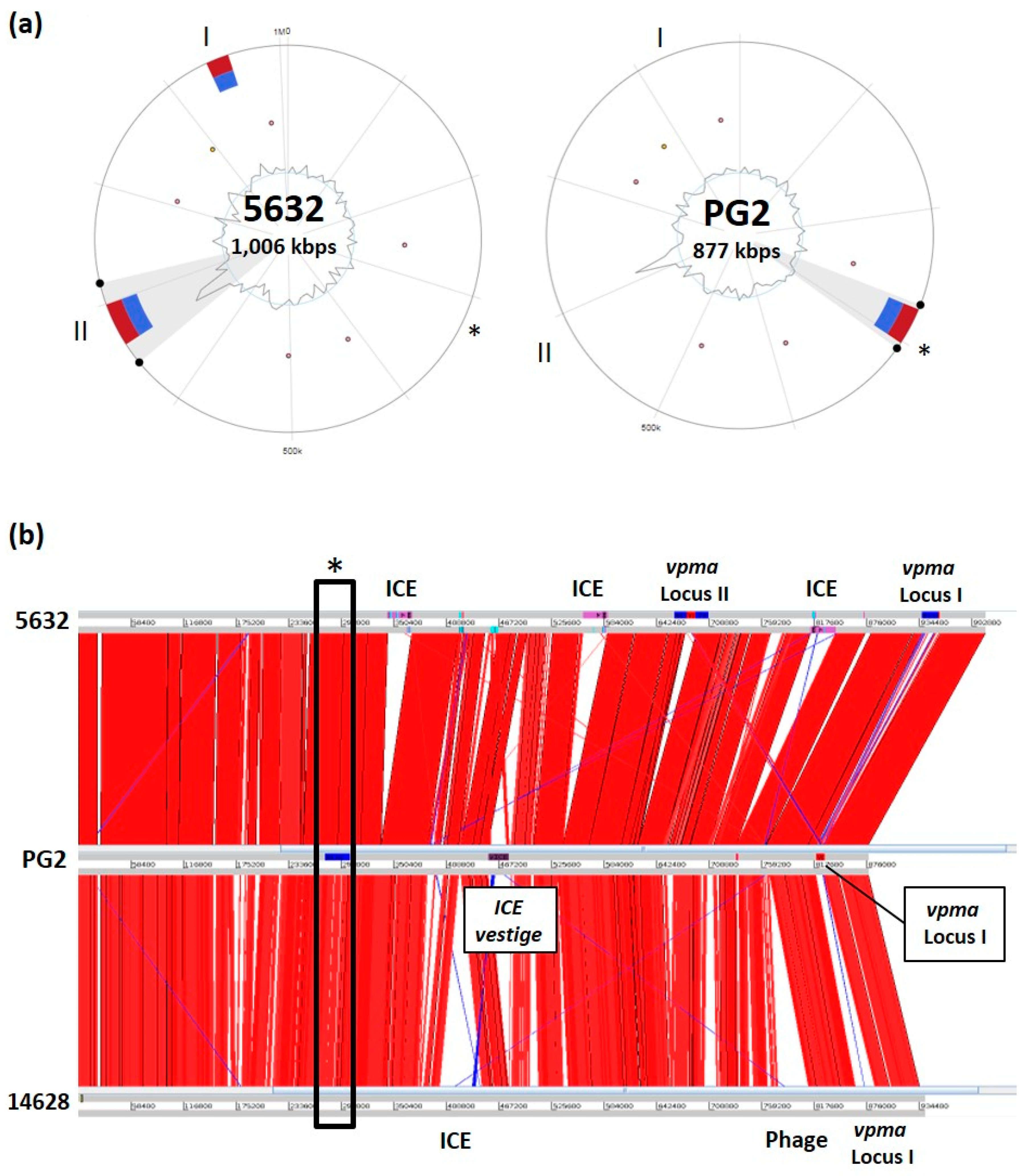
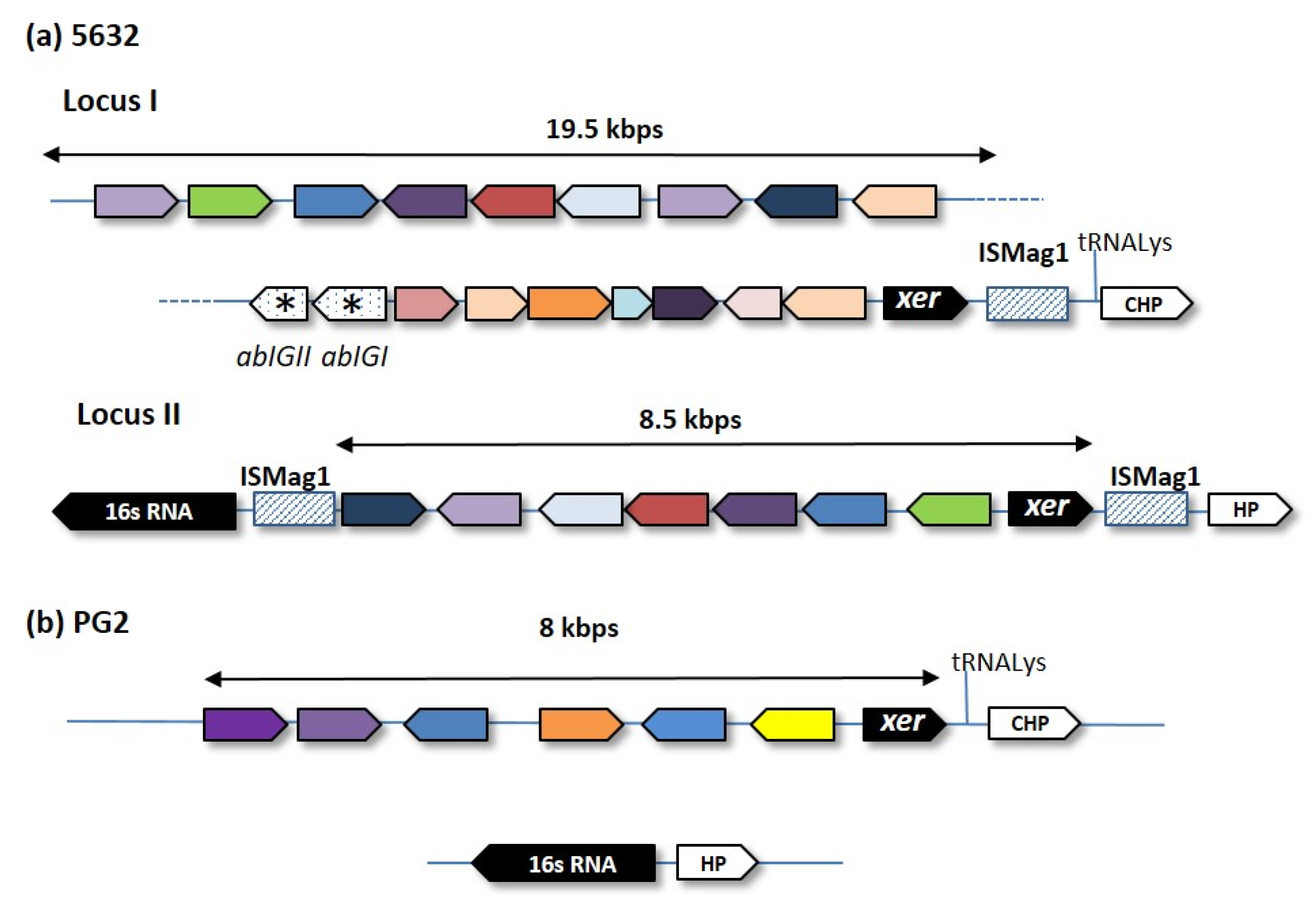
2. Genomic Islands and the Mycoplasma Flexible Gene Pool

Comprehensive analyses of bacterial genomes revealed the presence of GIs as part of the flexible gene pool [12]. Present in pathogenic or nonpathogenic bacteria, these are usually gene clusters spanning several Kbp that have potentially been acquired by HGT, as reflected by their GC content or codon usages, which might differ from the rest of the core genome. GIs often encode virulence or adaptive traits and are frequently associated with tRNA or integrase genes located at one end of the island. Most GIs derived from mobile genetic elements have lost their mobility genes through evolution but some have retained the ability to disseminate from cell-to-cell via conjugation or transduction. These are respectively known as ICEs and prophages.
Until the 1990s, the occurrence of GIs and HGT in mycoplasma was not considered, due to the small size of their genome and the prevailing evolutionary scenario that was only based on successive gene losses [13]. Yet, as more strains from a single mycoplasma species were being sequenced, important discoveries were made that challenged this paradigm [4,14,15]. One relates to the occurrence of ICEs in several mycoplasma species (see below), with their distribution being variable among strains of the same species. The second was based on comparative genomics across strains and species, revealing that HGT has shaped the genome of ruminant mycoplasmas raising the prospect that in addition to ICEs, mycoplasma genomes might harbor other GIs. Current tools for GI prediction are unfortunately often not reliable when using mycoplasma genomes. These prediction tools, which are based on sequence composition or comparative genomics, might be less suited (i) to the low GC-content and biased genetic code of the mycoplasma genome, and (ii) to the low homology existing between mycoplasmal genes and classical virulence factors or mobile genetic elements.
Current GI predictive tools can however provide hints for further expert analyses. For instance, Island Viewer 4 detected two loci in the M. agalactiae strain 5632 [16] (Figure 1), a small ruminant pathogen for which the circular genome is available [17]. Close examination revealed that these loci carry a cluster of related genes, each encoding for surface proteins of the Vpma family [18]. The vpma gene organization of strain 5632 is depicted in Figure 2 and showed that these loci harbor several features characteristic of GIs, as described for other bacteria. More specifically, the two loci span several Kbp (19 Kbp for 5632-locus I), contain a recombinase gene at one end, are in close proximity of mobile insertion sequence (IS) elements, and vary in their vpma content among strains [19]. In addition, the largest cluster, locus I, is closed to a tRNALys gene and harbors two unrelated genes, abiGI and abiGII, whose products present similarities to the abortive infection system AbiG found in more classical bacteria.
Discovered in the 90s, the M. agalactiae vpma loci were shown to encode phase-variable surface proteins that are key in allowing host-colonization and immune-escape [21,22]. These GIs could match the definition of a pathogenicity island (PAI), a GI involved in virulence. The M. agalactiae PG2 type strain carries a single vpma locus whose location corresponds to locus I of 5632 but was not detected by the prediction tool. Rather, Island Viewer 4 points towards a region encompassing 20 genes, distant from the vpma cluster, which is present in both 5632 and PG2 but was only detected in PG2 (Figure 2). Whether this also represents a GIs was less clear but close examination of this locus revealed the presence of coding sequences (CDSs) encoding several surface proteins, including the P40, which was involved in adhesion to host cell [23] and that of a CDS with homology to RumC recombinases.
The M. agalactiae vpma family had a counterpart in its close relative M. bovis [24], a pathogen of cattle, and is, to our knowledge, the only pathogenicity island (PAI) described as such in mycoplasmas. Several gene clusters, some encoding virulence factors, have undergone HGT among mycoplasmas [9], but lacked some features found in classical GIs.

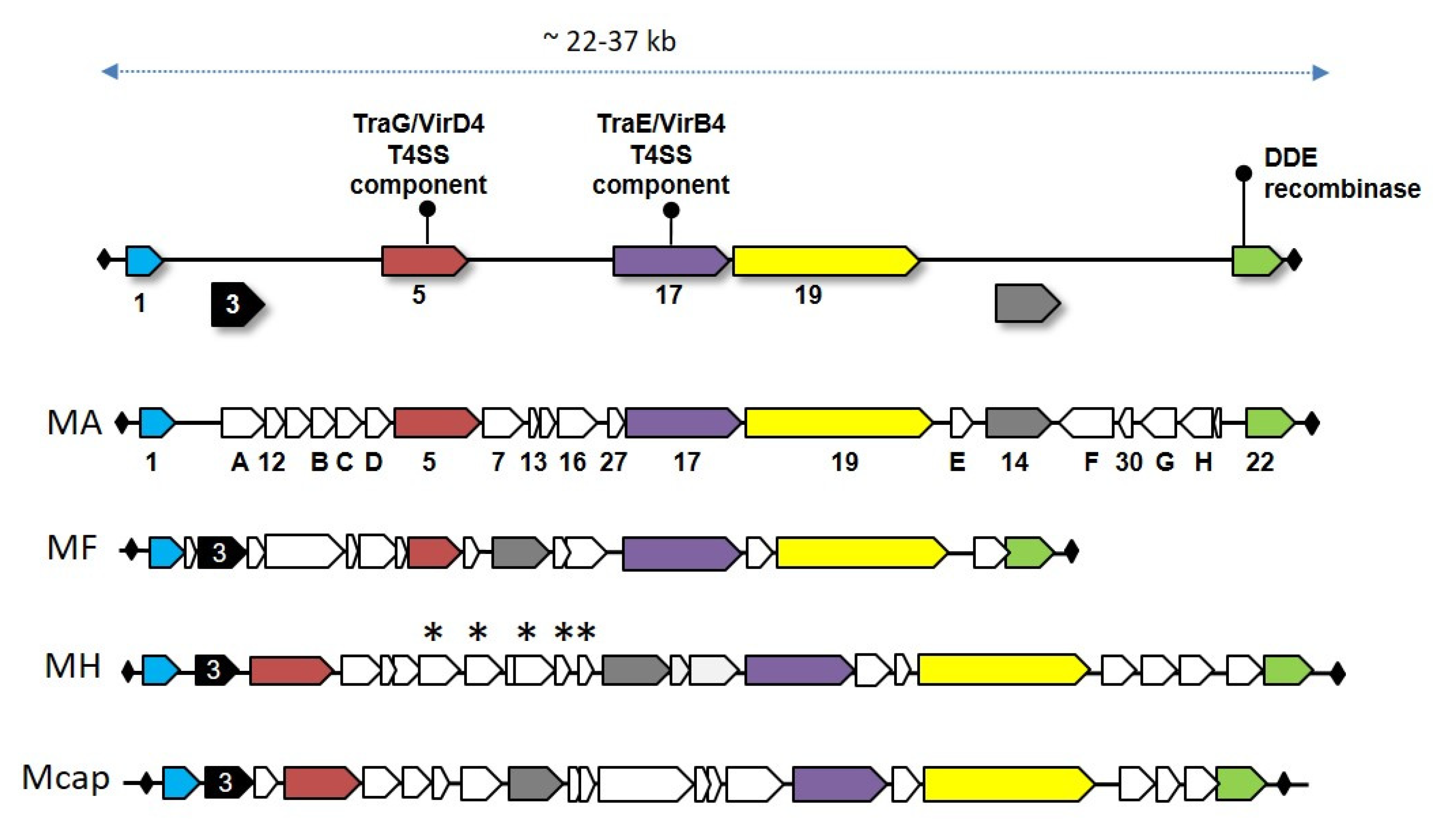
3. Mycoplasma Integrative Conjugative Elements

Mycoplasma ICEs (MICEs) are self-transmissible elements that play a crucial role in the unconventional chromosomal transfer described in M. agalactiae; their contribution toward HGT in mycoplasma was reviewed in 2018 [14], and here the focus is on their genetic organization and specific features.
MICEs were initially discovered in M. fermentans and M. agalactiae that were respectively isolated from human and ruminants [25,26]. In these two mycoplasma species, sequences with localized homologies to two gene-encoding proteins with DNA mobility-related functions in other bacteria [27], namely traG and traE, were detected in a chromosomal region having a GC-content slightly different than flanking genomic sequences [25]. Thus far, data mining of the mycoplasma genomes retrieved MICEs in 14 different mycoplasma species isolated from ruminants, swine, or human that belonged to two phylogenetic clades, namely Hominis and Spiroplasma. Recurrent attempts to automatically detect MICEs in sequenced mycoplasma genomes failed when using dedicated software (i.e., using ICEfinder tool of ICEberg 2.0 [28]), leaving manual search based on sequence similarities of conserved MICE genes the only reliable approach. While this situation reflects the little overall homology existing between MICE and ICE from other, more classical bacteria (see below), it is expected that the growing number of MICE sequences in databases should improve their future detection.
MICEs are large modular chromosomal regions of 22 to 37 Kbp that encode for about 20 structural genes flanked by two inverted repeats (IR), which are juxtaposed in the free circular form (Figure 3 and Table 1). One of their hallmarks is the occurrence at their 3′ end of a structural gene, CDS22, which encodes a DDE recombinase belonging to a family of prokaryotic DDE transposases, themselves related to eukaryotic Mutator-like transposases [29]. In 2014, Guerillot et al. systematically searched for genetic elements carrying DDE transposases and showed that they were associated with large integrative elements in streptococci (TnGBS) and with MICEs, which clustered in one large family designated p-MULT 5 [29]. TnGBS preferentially insert upstream streptococci promoters and this particular insertion was associated with a specific signature not found in CDS22 [30,31]. Indeed, MICEs belong to the few conjugative transposons which integrate at random in their host chromosome, with the excision-integration process being driven by the DDE recombinase encoded by CDS22. This finding was supported by several direct and indirect evidences, including the monitoring of the integration of a mini-artificial ICE construct in M. agalactiae, genome sequencing of MICE flanking sequences and comparative genomics of MICEs in different species [30,31].
As identified by transposon mutagenesis or BLAST searches, a large portion of MICE genes are involved in the horizontal self-dissemination of the element [46]. MICEs also carry several hypothetical genes with no predicted function or no homology, with some being specific to a single mycoplasma species. For instance, M. hominis ICEs all possess a 4.0–5.1 Kbp cluster of five to six CDSs (Figure 3) with no homolog in other MICEs and whose synteny slightly differs among strains [42]. Except for one, all of these were predicted to share structural similarities to DNA interacting/modifying proteins, with some sharing common structural similarities with transcription activator-like (TAL) effectors. In symbiotic bacteria, TAL effectors are involved in polynucleotide recognition and signal transduction. Interestingly, M. hominis colonizes the human urogenital tract, where it might occur as an endosymbiont of Trichomonas vaginalis. Whether TAL effectors encoded by M. hominis ICEs contribute to the interaction of the mycoplasma with T. vaginalis by impacting gene expression is an interesting hypothesis that remains to be addressed [42].
Entire, functional MICEs often occur in multiple copies in a single mycoplasma genome, along with MICE vestiges, as a result of MICE erosion (Table 1) [17]. This raised the question of the cost imposed by these large, multi-copy elements on the fitness of the small mycoplasma genome. We recently showed that the M. agalactiae strain 5632 that harbors 3 MICEs copies is more fit under laboratory conditions than the PG2 strain that only has a single MICE vestige (Figure 1) [47]. Following mating experiments, the transfer of one MICE from 5632 into the PG2 genome resulted in fitness loss. This finding suggests that strain 5632 is able to counterbalance the fitness cost of MICEs, while PG2 is not. Comparative genome analyses showed that the two strains encode syntenic genomes but differ in several SNPs and the size of their variable gene repertoires, such as the vpma (see above and Figure 2) [17]. Yet, the factors responsible for 5632 adaptation to MICEs cost remain to be explored. Of note, MICE deletion in M. mycoides subsp. capri strain GM12T did not affect the in vitro growth [48].
As already mentioned, all MICEs described above belong to a new family of self-transmissible elements that have so far not been found outside of Mollicutes and thus appear to be specific to this class. Yet, MICEs are not the only large mobile genetic elements (MGE) circulating in mycoplasmas. Indeed, an excision-competent composite transposon was identified in M. hominis, which derived from a larger uncharacterized transposon of Streptococcus agalactiae and carried the tet(M) determinant, along with Tn916 sequences [43]. Tn916 and Tn916-like conjugative transposons encode the tet(M) determinant that confers resistance to tetracycline. These were found in several bacterial genera but have seldom been detected in Mollicutes. Thus, the finding of this composite transposon in M. hominis indicates that ICEs from two different origins circulate in this mycoplasma species and that tetracycline resistance might disseminate via HGT.

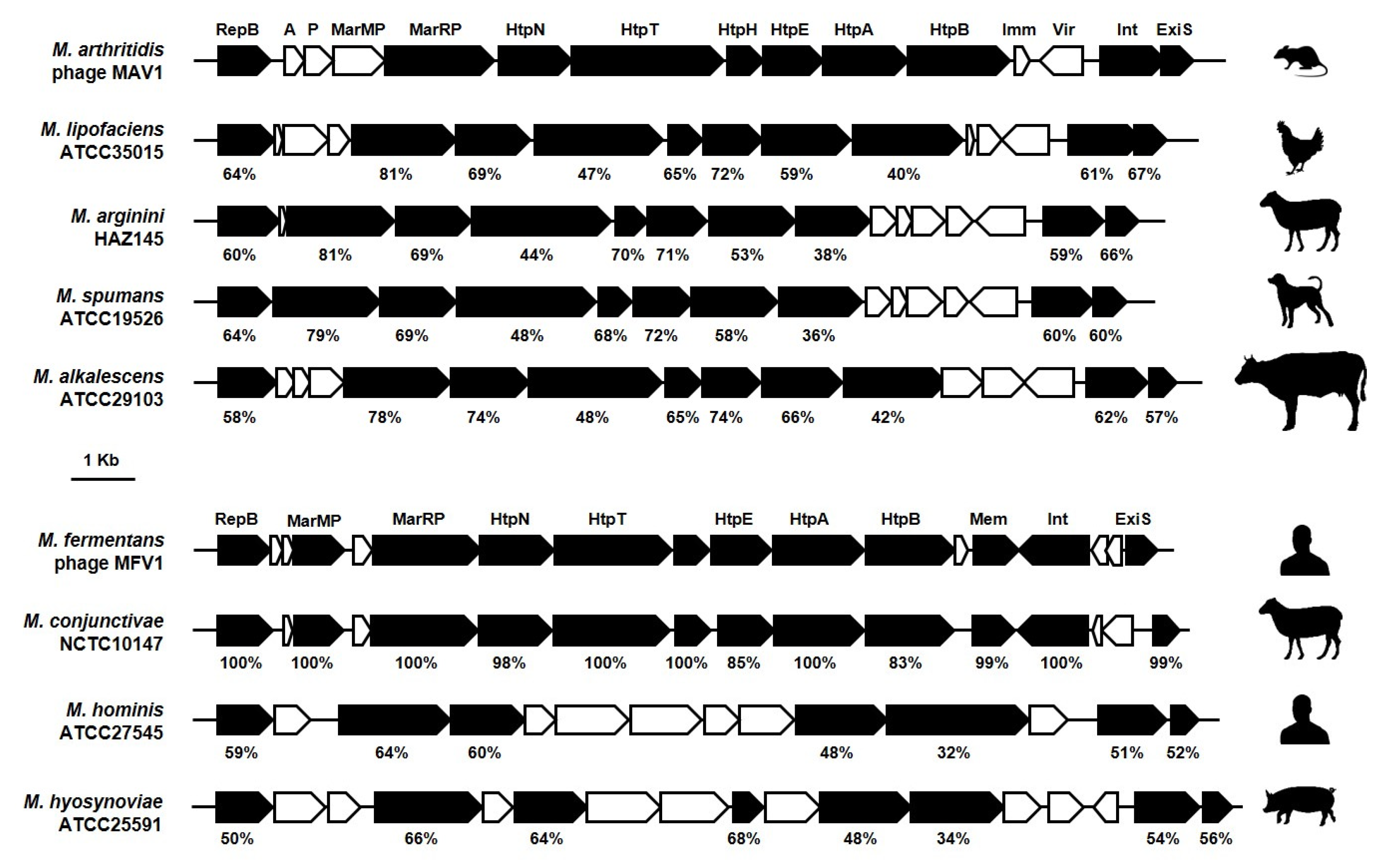
4. Mycoplasma Viruses and Prophages

The identification of prophage sequences in a growing number of mycoplasma species indicates that GIs in Mollicutes are not limited to MICEs and raises questions about the influence of these particular elements on mycoplasma genomic evolution and environmental adaptation.
The first direct evidence of a viral attack in Mollicutes was provided by the isolation of a plaque producing phage in Acholeplasma laidlawii [49]. Since then, several groups of viruses were documented in the genus Acholeplasma, and this repertoire quickly extended to phylogenetically distant genera, including Spiroplasma and Mycoplasma [50,51,52,53]. Acholeplasma (Acholeplasmataceae) and Spiroplasma (Spiroplasmataceae) viruses were extensively characterized. These viruses were classified into several groups with specific morphological and genomic features [51,52]. Remarkably, the L2 group belonged to the Plasmaviridae, a unique family of enveloped viruses only represented by the Acholeplasma virus L2. The genome of this virus was encoded by a circular double-stranded DNA molecule that could be found integrated into the host chromosome at tRNA genes. The other groups belonged to Inoviridae and Podoviridae, two major viral families that unlike Plasmaviridae encompass an important number of Gram-positive and Gram-negative targeting bacteriophages. Yet, many of these viruses still had an uncertain taxonomic classification.
With more than 200 species, members of the Mycoplasma genus are associated with a considerable number of chronic infections in humans and a broad range of animal hosts. In contrast to the many Acholeplasma and Spiroplasma viruses identified, only four Mycoplasma viruses (MV) were successfully isolated so far (Table 2). MVBr1 was isolated from M. bovirhinis, a commensal species frequently found associated with the upper respiratory tract in cattle [54,55]. With its isometric head and long tail, MVBr1 has the typical bacteriophage structure and was classified in the family Myoviridae. This bacteriophage contains a linear dsDNA molecule of about 11.7 Kbp. Three other viruses were isolated from pathogenic mycoplasma species (Table 2), but their physicochemical properties were not fully characterized and thus had no taxonomic status.
As mycoplasma genome sequences became available in public databases, a growing number of genomic elements were identified as prophage genomes or phage-like sequences [56,60,61,63,64,66,70]. Consistent with our previous observations with MICEs (see above), most existing computational tools dedicated to identifying prophage sequences in bacterial genomes are poorly efficient in distinguishing viral patterns from mycoplasma sequences [71]. Yet, as suggested below by BLASTP analyses using available mycoplasma viral sequences (Table 2), the number of detected prophages in published mycoplasma sequences is most likely underestimated. Thus, the dogmatic view suggesting that mycoplasmas were subjected to the elimination of prophage genomes during their evolution might have to be revisited [72].
Based on their overall genetic organization and sequence similarity, mycoplasma prophage genomes can be divided into two groups (see below). The first group comprises prophages with a small genome in size (genome size ca. 16 Kbp) represented by M. arthritidis φMAV1 and M. fermentans φMFV1 that have a similar genetic organization and a high degree of synteny [53,63]. Both genomes exhibit characteristic features of mobile genetic elements, including a compact organization, an almost unidirectional CDS orientation, and the occurrence of genes involved in their integration and excision from the host chromosome [53,63]. BLASTP analyses revealed that 10 of the 18 CDSs identified in φMFV1 exhibited some similarity with their counterparts in φMAV1 [63]. Here, database searches of available genomes revealed prophage sequences that closely resemble φMAV1 or φMFV1 and were broadly distributed among human and animal mycoplasma species (Figure 4) These included the M. hominis φMHoV1 [64], as well as several M. hyosynoviae-derived phages [66]. In this highly conserved genomic framework are several regions of higher heterogeneity (Figure 4). Among those are the φMFV1 mem and the φMAV1 vir products, each encoding a unique membrane-anchored surface protein [58,59,63,73]. Mem displays features of coiled-coil proteins and occurs predominantly as an integral, membrane-associated product anchored by its N-terminal transmembrane domain. The expression of this phage product can vary among clonal populations of M. fermentans, without affecting the viability of mycoplasma cells [63]. The φMAV1-specific vir gene was found to encode a lipoprotein expressed at the surface of M. arthritidis infected cells and to confer resistance to phage superinfection [73]. Interestingly, φMAV1 was reported to be required for arthritogenesis in rodents, and the phage-encoded Vir lipoprotein was identified as a putative virulence factor [53,59,74]. However, attempts to confirm this hypothesis were unsuccessful, suggesting a complex interaction between φMAV1 and M. arthritidis in the infected host [53,73,75]. The biological impact of phage-encoded surface proteins is still unclear, but these studies illustrate the possible role of prophage-associated genes in the acquisition of new heritable phenotypic traits in mycoplasmas.
The second category of mycoplasma prophages displays a larger genome (size ca. 34 Kb), which were identified in several ruminant mycoplasma species, including M. agalactiae, M. conjunctivae, and M. bovigenitalium [41,56]. Their genetic architecture is an assemblage of several regions, each characterized by a similar number of CDSs sharing the same orientation. In contrast to φMAV1 and φMFV1, sequences with a high degree of similarity are limited to several common phage products (Figure 5). BLASTP analyses using several of these typical phage products, such as the prohead protein, portal, and terminase of the M. agalactiae prophage [56], not only confirmed the occurrence of a similar prophage region in M. conjunctivae and M. bovigenitalium but also identified new host species, including the ruminant pathogen M. bovis, as well as several mycoplasmas isolated from the respiratory tract of dogs and minks (Figure 5). The M. agalactiae prophage, hereafter designated as φMAgV1, was reported in the genome of an atypical strain associated with a mortality episode of Alpine ibexes in France [56].
Apart from these two prophage groups, an important number of phage-like protein sequences were recently documented in a 54 Kbp chromosomal region of M. bovirhinis strain HAZ141_2 suggesting the possible occurrence of a unique prophage in this ruminant mycoplasma species [60,61]. Proteins with significant similarity to known phage proteins were also detected in the genome of several human urogenital Mycoplasma species [70]. The origin of these sequences, detected by using the PHASTER web server [76], is largely unknown, but their clustering within one or two chromosomal regions raises the possibility of a common origin. While the extraordinary diversity of prophage-like sequences in mycoplasmas has only started to emerge, BLASTP analysis might also be useful to identify possible footprints of viral attacks in mycoplasmas. This was observed in the draft genome sequence of M. bovis strain 3308MB, which was found to harbor sequences with some similarity to the M. pulmonis virus P1 (Figure 6). Interestingly, these sequences were found within a single contig of the draft genome with an overall organization similar to M. pulmonis virus P1 and no sequence homology with Mollicutes. The viral origin of this single contig sequence remains to be confirmed, but it might indicate that the mycoplasma isolate used for genome sequencing was facing a viral attack.
Finally, the isolation of plaque-forming viral particles for φMAV1 [59], the detection of extrachromosomal forms of φMFV1 and φMAgV1 [56,63], together with the identification of multiple chromosomal integration sites [63], demonstrated the remarkable mobility of these prophage genomes and their contribution to genotypic variation.

5. Distribution of MICE and Prophages in Mycoplasma Species

The prevalence of MICE among strains depends on the species:98% of M. bovis strains tested so far carry MICE sequences of the minimal backbone, that is, the set of MICE genes common to all (Figure 3), for only 34% in the closely related M. agalactiae species or 45% in M. hominis clinical isolates [30]. Notably, some Mycoplasma species with several strains sequenced seemed to be deprived of MICE. This is the cases of M. genitalium, M. pneumoniae, or M. gallisepticum, which have different genome size and colonize different hosts—the two first ones colonize the human genital and respiratory tracts, respectively, and have genomes of 580 and 820 Kbp; the third one is a respiratory pathogen of birds and has a genome size of about 990 Kbp, similar to that of M. agalactiae 5632 (ca. 1000 Kbp). These 3 species belong to the phylogenetic Pneumoniae clade and MICEs were only identified so far in mycoplasmas of the Spiroplasma and Hominis clades. Whether there is a link between the occurrence of MICEs in a given species and its phylogenetic clustering, remains to be explored, but there are evidences of MICE dissemination across the Spiroplasma and Hominis clades [30].
As for MICEs, the occurrence of mycoplasma prophages varied within species. For instance, the M. agalactiae φMAgV1 was detected in most but not all ibex isolates and was absent from most ovine strains [56]. This phage was inserted in a region that might have undergone horizontal gene transfer with members of the mycoides cluster, a remote group of ruminant species, suggesting that this prophage might be directly or indirectly associated with genome dynamics. Another example of the intricate link between prophage and genome dynamics is the occurrence of several φMFV1 regions in one copy of M. fermentans MICE in strain PG18 [26,63]. This illustrates the possibility of gene exchange between these regions and suggests that MICE might contribute to the horizontal dissemination of prophage genes.
Several restriction-modification, bacteriophage abortive infection mechanisms (Abi), and CRISPR systems were identified in mycoplasmas. These are known to restrict HGT, and in mycoplasmas, their repertoire varies within species [77,78,79]. Whether the strain-specificity of these systems correlates with the distribution of MICEs and prophages within a single species is yet to be fully addressed but there is so far no apparent correlation.

6. Conclusions

Since the 1990s, the main scenario proposed to explain Mollicutes’ evolution was based on gene losses. The discovery of MICEs had two main impacts: toa break away from the general idea that small mycoplasma genomes were deprived of complex mobile genetic elements and to stimulate research on mycoplasma horizontal gene transfers. Based on comparative genomics and mating experiments, it is now clear that several Mycoplasma species retained a form of sexual competence and that their evolution was also driven by gene gain. The mechanism underlying HGT in ruminant Mycoplasma species hasstarted to emerge and may challenge the definition of GIs in these bacteria. Indeed, in these organisms in silico and experimental data points towards the entire genome being mobile, with large genomic chromosomic regions being exchanged.
This review showed that several mycoplasma genomes are populated with MICEs and prophages, and most likely, also contain pathogenicity islands. The identification of these particular GIs might be difficult in certain species because of the unconventional mechanism of HGT that shuffle their genomes. In addition, the specificity of the mycoplasma genetic code most likely limits the acquisition of GIs from donor bacteria outside of Mollicutes.
This review also points towards the heterogeneity of the species pan-genome, underlying the need for exploring multiple strains even in bacteria with small genomes.
Overall, a new picture has started to emerge since the last 10 years in which mycoplasma genomes are more dynamic than first thought.

Author Contributions

C.C. and E.B. contributed equally to this work. Conceptualization, writing and editing C.C., E.B. and L.-X.N.; Drawings C.C., E.B., and L.-X.N.; Data collection C.C., E.B., L.-X.N., E.D.-F., and M.F. All authors have read and agreed to the published version of the manuscript.

Funding

This work was supported by financial supports from INRAE and ENVT.

Conflicts of Interest

The authors declare no conflict of interest.

References

  1. Citti, C.; Blanchard, A. Mycoplasmas and their host: Emerging and re-emerging minimal pathogens. Trends Microbiol. 2013, 21, 196–203. [Google Scholar] [CrossRef]
  2. Woese, C.R.; Maniloff, J.; Zablen, L.B. Phylogenetic analysis of the mycoplasmas. Proc. Natl. Acad. Sci. USA 1980, 77, 494–498. [Google Scholar] [CrossRef] [PubMed] [Green Version]
  3. Ciccarelli, F.D.; Doerks, T.; von Mering, C.; Creevey, C.J.; Snel, B.; Bork, P. Toward automatic reconstruction of a highly resolved tree of life. Science 2006, 311, 1283–1287. [Google Scholar] [CrossRef] [PubMed] [Green Version]
  4. Sirand-Pugnet, P.; Citti, C.; Barré, A.; Blanchard, A. Evolution of mollicutes: Down a bumpy road with twists and turns. Res. Microbiol. 2007, 158, 754–766. [Google Scholar] [CrossRef]
  5. Fraser, C.M.; Gocayne, J.D.; White, O.; Adams, M.D.; Clayton, R.A.; Fleischmann, R.D.; Bult, C.J.; Kerlavage, A.R.; Sutton, G.; Kelley, J.M.; et al. The minimal gene complement of Mycoplasma genitalium. Science 1995, 270, 397–403. [Google Scholar] [CrossRef] [PubMed]
  6. Mushegian, A.R.; Koonin, E.V. A minimal gene set for cellular life derived by comparison of complete bacterial genomes. Proc. Natl. Acad. Sci. USA 1996, 93, 10268–10273. [Google Scholar] [CrossRef] [Green Version]
  7. Hutchison, C.A.; Peterson, S.N.; Gill, S.R.; Cline, R.T.; White, O.; Fraser, C.M.; Smith, H.O.; Venter, J.C. Global transposon mutagenesis and a minimal mycoplasma genome. Science 1999, 286, 2165–2169. [Google Scholar] [CrossRef] [Green Version]
  8. Dybvig, K.; Lao, P.; Jordan, D.S.; Simmons, W.L. Fewer essential genes in mycoplasmas than previous studies suggest. FEMS Microbiol. Lett. 2010, 311, 51–55. [Google Scholar] [CrossRef] [Green Version]
  9. Sirand-Pugnet, P.; Lartigue, C.; Marenda, M.; Jacob, D.; Barré, A.; Barbe, V.; Schenowitz, C.; Mangenot, S.; Couloux, A.; Segurens, B.; et al. Being pathogenic, plastic, and sexual while living with a nearly minimal bacterial genome. PLoS Genet. 2007, 3, e75. [Google Scholar] [CrossRef]
  10. Dordet-Frisoni, E.; Sagné, E.; Baranowski, E.; Breton, M.; Nouvel, L.X.; Blanchard, A.; Marenda, M.S.; Tardy, F.; Sirand-Pugnet, P.; Citti, C. Chromosomal transfers in mycoplasmas: When minimal genomes go mobile. mBio 2014, 5, e01958. [Google Scholar] [CrossRef] [Green Version]
  11. Dordet-Frisoni, E.; Faucher, M.; Sagné, E.; Baranowski, E.; Tardy, F.; Nouvel, L.X.; Citti, C. Mycoplasma chromosomal transfer: A distributive, conjugative process creating an infinite variety of mosaic genomes. Front. Microbiol. 2019, 10, 2441. [Google Scholar] [CrossRef] [PubMed] [Green Version]
  12. Dobrindt, U.; Hochhut, B.; Hentschel, U.; Hacker, J. Genomic islands in pathogenic and environmental microorganisms. Nat. Rev. Microbiol. 2004, 2, 414–424. [Google Scholar] [CrossRef] [PubMed]
  13. Razin, S.; Yogev, D.; Naot, Y. Molecular biology and pathogenicity of mycoplasmas. Microbiol. Mol. Biol. Rev. 1998, 62, 1094–1156. [Google Scholar] [CrossRef] [PubMed] [Green Version]
  14. Citti, C.; Dordet-Frisoni, E.; Nouvel, L.X.; Kuo, C.H.; Baranowski, E. Horizontal gene transfers in mycoplasmas (Mollicutes). Curr. Issues Mol. Biol. 2018, 29, 3–22. [Google Scholar] [CrossRef] [Green Version]
  15. Lo, W.-S.; Gasparich, G.E.; Kuo, C.-H. Convergent evolution among ruminant-pathogenic mycoplasma involved extensive gene content changes. Genome Biol. Evol. 2018, 10, 2130–2139. [Google Scholar] [CrossRef]
  16. Bertelli, C.; Laird, M.R.; Williams, K.P.; Lau, B.Y.; Hoad, G.; Winsor, G.L.; Brinkman, F.S. IslandViewer 4: Expanded prediction of genomic islands for larger-scale datasets. Nucleic Acids Res. 2017, 45, W30–W35. [Google Scholar] [CrossRef]
  17. Nouvel, L.X.; Sirand-Pugnet, P.; Marenda, M.S.; Sagné, E.; Barbe, V.; Mangenot, S.; Schenowitz, C.; Jacob, D.; Barré, A.; Claverol, S.; et al. Comparative genomic and proteomic analyses of two Mycoplasma agalactiae strains: Clues to the macro- and micro-events that are shaping mycoplasma diversity. BMC Genom. 2010, 11, 86. [Google Scholar] [CrossRef] [Green Version]
  18. Nouvel, L.-X.; Marenda, M.; Sirand-Pugnet, P.; Sagné, E.; Glew, M.; Mangenot, S.; Barbe, V.; Barré, A.; Claverol, S.; Citti, C. Occurrence, plasticity, and evolution of the vpma gene family, a genetic system devoted to high-frequency surface variation in Mycoplasma agalactiae. J. Bacteriol. 2009, 191, 4111–4121. [Google Scholar] [CrossRef] [Green Version]
  19. Nouvel, L.-X.; Marenda, M.S.; Glew, M.D.; Sagné, E.; Giammarinaro, P.; Tardy, F.; Poumarat, F.; Rosengarten, R.; Citti, C. Molecular typing of Mycoplasma agalactiae: Tracing European-wide genetic diversity and an endemic clonal population. Comp. Immunol. Microbiol. Infect. Dis. 2012, 35, 487–496. [Google Scholar] [CrossRef]
  20. Glew, M.D.; Marenda, M.; Rosengarten, R.; Citti, C. Surface diversity in Mycoplasma agalactiae is driven by site-specific DNA inversions within the vpma multigene locus. J. Bacteriol. 2002, 184, 5987–5998. [Google Scholar] [CrossRef] [Green Version]
  21. Chopra-Dewasthaly, R.; Spergser, J.; Zimmermann, M.; Citti, C.; Jechlinger, W.; Rosengarten, R. Vpma phase variation is important for survival and persistence of Mycoplasma agalactiae in the immunocompetent host. PLoS Pathog. 2017, 13, e1006656. [Google Scholar] [CrossRef] [Green Version]
  22. Glew, M.D.; Papazisi, L.; Poumarat, F.; Bergonier, D.; Rosengarten, R.; Citti, C. Characterization of a multigene family undergoing high-frequency DNA rearrangements and coding for abundant variable surface proteins in Mycoplasma agalactiae. Inf. Immun. 2000, 68, 4539–4548. [Google Scholar] [CrossRef] [PubMed] [Green Version]
  23. Fleury, B.; Bergonier, D.; Berthelot, X.; Peterhans, E.; Frey, J.; Vilei, E.M. Characterization of P40, a cytadhesin of Mycoplasma agalactiae. Infect. Immun. 2002, 70, 5612–5621. [Google Scholar] [CrossRef] [PubMed] [Green Version]
  24. Lysnyansky, I.; Sachse, K.; Rosenbusch, R.; Levisohn, S.; Yogev, D. The vsp locus of Mycoplasma bovis: Gene organization and structural features. J. Bacteriol. 1999, 181, 5734–5741. [Google Scholar] [CrossRef] [PubMed] [Green Version]
  25. Marenda, M.; Barbe, V.; Gourgues, G.; Mangenot, S.; Sagne, E.; Citti, C. A new integrative conjugative element occurs in Mycoplasma agalactiae as chromosomal and free circular forms. J. Bacteriol. 2006, 188, 4137–4141. [Google Scholar] [CrossRef] [PubMed] [Green Version]
  26. Calcutt, M.J.; Lewis, M.S.; Wise, K.S. Molecular genetic analysis of ICEF, an integrative conjugal element that is present as a repetitive sequence in the chromosome of Mycoplasma fermentans PG18. J. Bacteriol. 2002, 184, 6929–6941. [Google Scholar] [CrossRef] [Green Version]
  27. Alvarez-Martinez, C.E.; Christie, P.J. Biological diversity of prokaryotic type IV secretion systems. Microbiol. Mol. Biol. Rev. 2009, 73, 775–808. [Google Scholar] [CrossRef] [Green Version]
  28. Liu, M.; Li, X.; Xie, Y.; Bi, D.; Sun, J.; Li, J.; Tai, C.; Deng, Z.; Ou, H.-Y. ICEberg 2.0: An updated database of bacterial integrative and conjugative elements. Nucleic Acids Res. 2019, 47, D660–D665. [Google Scholar] [CrossRef]
  29. Guérillot, R.; Siguier, P.; Gourbeyre, E.; Chandler, M.; Glaser, P. The diversity of prokaryotic DDE transposases of the mutator superfamily, insertion specificity, and association with conjugation machineries. Genome Biol. Evol. 2014, 6, 260–272. [Google Scholar] [CrossRef] [Green Version]
  30. Tardy, F.; Mick, V.; Dordet-Frisoni, E.; Marenda, M.S.; Sirand-Pugnet, P.; Blanchard, A.; Citti, C. Integrative conjugative elements are widespread in field isolates of Mycoplasma species pathogenic for ruminants. Appl. Environ. Microbiol. 2015, 81, 1634–1643. [Google Scholar] [CrossRef] [Green Version]
  31. Dordet Frisoni, E.; Marenda, M.S.; Sagné, E.; Nouvel, L.X.; Guérillot, R.; Glaser, P.; Blanchard, A.; Tardy, F.; Sirand-Pugnet, P.; Baranowski, E.; et al. ICEA of Mycoplasma agalactiae: A new family of self-transmissible integrative elements that confers conjugative properties to the recipient strain. Mol. Microbiol. 2013, 89, 1226–1239. [Google Scholar] [CrossRef] [PubMed]
  32. Calderon-Copete, S.P.; Wigger, G.; Wunderlin, C.; Schmidheini, T.; Frey, J.; Quail, M.A.; Falquet, L. The Mycoplasma conjunctivae genome sequencing, annotation and analysis. BMC Bioinform. 2009, 10, S7. [Google Scholar] [CrossRef] [PubMed] [Green Version]
  33. Vasconcelos, A.T.R.; Ferreira, H.B.; Bizarro, C.V.; Bonatto, S.L.; Carvalho, M.O.; Pinto, P.M.; Almeida, D.F.; Almeida, L.G.P.; Almeida, R.; Alves-Filho, L.; et al. Swine and poultry pathogens: The complete genome sequences of two strains of Mycoplasma hyopneumoniae and a strain of Mycoplasma synoviae. J. Bacteriol. 2005, 187, 5568–5577. [Google Scholar] [CrossRef] [PubMed] [Green Version]
  34. Liu, W.; Feng, Z.; Fang, L.; Zhou, Z.; Li, Q.; Li, S.; Luo, R.; Wang, L.; Chen, H.; Shao, G.; et al. Complete genome sequence of Mycoplasma hyopneumoniae strain 168. J. Bacteriol. 2011, 193, 1016–1017. [Google Scholar] [CrossRef] [PubMed] [Green Version]
  35. Minion, F.C.; Lefkowitz, E.J.; Madsen, M.L.; Cleary, B.J.; Swartzell, S.M.; Mahairas, G.G. The genome sequence of Mycoplasma hyopneumoniae strain 232, the agent of swine Mycoplasmosis. J. Bacteriol. 2004, 186, 7123–7133. [Google Scholar] [CrossRef] [Green Version]
  36. Rechnitzer, H.; Brzuszkiewicz, E.; Strittmatter, A.; Liesegang, H.; Lysnyansky, I.; Daniel, R.; Gottschalk, G.; Rottem, S. Genomic features and insights into the biology of Mycoplasma fermentans. Microbiology 2011, 157, 760–773. [Google Scholar] [CrossRef]
  37. Shu, H.-W.; Liu, T.-T.; Chan, H.-I.; Liu, Y.-M.; Wu, K.-M.; Shu, H.-Y.; Tsai, S.-F.; Hsiao, K.-J.; Hu, W.S.; Ng, W.V. Genome sequence of the repetitive-sequence-rich Mycoplasma fermentans strain M64. J. Bacteriol. 2011, 193, 4302–4303. [Google Scholar] [CrossRef] [Green Version]
  38. Wise, K.S.; Calcutt, M.J.; Foecking, M.F.; Röske, K.; Madupu, R.; Methé, B.A. Complete genome sequence of Mycoplasma bovis type strain PG45 (ATCC 25523). Inf. Immun. 2011, 79, 982–983. [Google Scholar] [CrossRef] [Green Version]
  39. Li, Y.; Zheng, H.; Liu, Y.; Jiang, Y.; Xin, J.; Chen, W.; Song, Z. The complete genome sequence of Mycoplasma bovis strain Hubei-1. PLoS ONE 2011, 6, e20999. [Google Scholar] [CrossRef]
  40. Qi, J.; Guo, A.; Cui, P.; Chen, Y.; Mustafa, R.; Ba, X.; Hu, C.; Bai, Z.; Chen, X.; Shi, L.; et al. Comparative geno-plasticity analysis of Mycoplasma bovis HB0801 (Chinese Isolate). PLoS ONE 2012, 7, e38239. [Google Scholar] [CrossRef]
  41. Manso-Silván, L.; Tardy, F.; Baranowski, E.; Barré, A.; Blanchard, A.; Breton, M.; Couture, C.; Citti, C.; Dordet-Frisoni, E.; Dupuy, V.; et al. Draft genome sequences of Mycoplasma alkalescens, Mycoplasma arginini, and Mycoplasma bovigenitalium, three species with equivocal pathogenic status for cattle. Genome Announc. 2013, 1, e00348-13. [Google Scholar] [CrossRef] [Green Version]
  42. Meygret, A.; Peuchant, O.; Dordet-Frisoni, E.; Sirand-Pugnet, P.; Citti, C.; Bébéar, C.; Béven, L.; Pereyre, S. High prevalence of integrative and conjugative elements encoding transcription activator-like effector repeats in Mycoplasma hominis. Front. Microbiol. 2019, 10, 2385. [Google Scholar] [CrossRef] [PubMed]
  43. Calcutt, M.J.; Foecking, M.F. An Excision-Competent and Exogenous Mosaic Transposon Harbors the tetM Gene in Multiple Mycoplasma hominis Lineages. Antimicrob. Agents Chemother. 2015, 59, 6665–6666. [Google Scholar] [CrossRef] [PubMed] [Green Version]
  44. Thiaucourt, F.; Manso-Silvan, L.; Salah, W.; Barbe, V.; Vacherie, B.; Jacob, D.; Breton, M.; Dupuy, V.; Lomenech, A.M.; Blanchard, A.; et al. Mycoplasma mycoides, from “mycoides Small Colony” to “capri”. A microevolutionary perspective. BMC Genom. 2011, 12, 114. [Google Scholar] [CrossRef]
  45. Dupuy, V.; Sirand-Pugnet, P.; Baranowski, E.; Barré, A.; Breton, M.; Couture, C.; Dordet-Frisoni, E.; Gaurivaud, P.; Jacob, D.; Lemaitre, C.; et al. Complete genome sequence of Mycoplasma putrefaciens strain 9231, one of the agents of contagious Agalactia in goats. Genome Announc. 2013, 1, e00354-13. [Google Scholar] [CrossRef] [Green Version]
  46. Baranowski, E.; Dordet-Frisoni, E.; Sagné, E.; Hygonenq, M.-C.; Pretre, G.; Claverol, S.; Fernandez, L.; Nouvel, L.X.; Citti, C. The Integrative Conjugative Element (ICE) of Mycoplasma agalactiae: Key elements involved in horizontal dissemination and influence of coresident ICEs. mBio 2018, 9, e00873-18. [Google Scholar] [CrossRef] [Green Version]
  47. Faucher, M.; Nouvel, L.-X.; Dordet-Frisoni, E.; Sagné, E.; Baranowski, E.; Hygonenq, M.-C.; Marenda, M.-S.; Tardy, F.; Citti, C. Mycoplasmas under experimental antimicrobial selection: The unpredicted contribution of horizontal chromosomal transfer. PLoS Genet. 2019, 15, e1007910. [Google Scholar] [CrossRef] [Green Version]
  48. Jores, J.; Ma, L.; Ssajjakambwe, P.; Schieck, E.; Liljander, A.; Chandran, S.; Stoffel, M.H.; Cippa, V.; Arfi, Y.; Assad-Garcia, N.; et al. Removal of a subset of non-essential genes fully attenuates a highly virulent Mycoplasma strain. Front. Microbiol. 2019, 10, 664. [Google Scholar] [CrossRef] [Green Version]
  49. Gourlay, R.N. Isolation of a virus infecting a strain of Mycoplasma laidlawii. Nature 1970, 225, 1165. [Google Scholar] [CrossRef]
  50. Gourlay, R.N. Mycoplasma viruses: Isolation, physicochemical, and biological properties. CRC Crit. Rev. Microbiol. 1974, 3, 315–331. [Google Scholar] [CrossRef]
  51. Maniloff, J.; Haberer, K.; Gourlay, R.N.; Das, J.; Cole, R. Mycoplasma viruses. Intervirology 1982, 18, 177–188. [Google Scholar] [CrossRef]
  52. Maniloff, J. Mycoplasma viruses. Crit. Rev. Microbiol. 1988, 15, 339–389. [Google Scholar] [CrossRef] [PubMed]
  53. Dybvig, K.; Tu, A.-H.; Clapper, B. Mycoplasma phages. Phages 2005, 223–237. [Google Scholar] [CrossRef]
  54. Howard, C.J.; Gourlay, R.N.; Wyld, S.G. Isolation of a virus, MVBr1, from Mycoplasma bovirhinis. FEMS Microbiol. Lett. 1980, 7, 163–165. [Google Scholar] [CrossRef] [Green Version]
  55. Gourlay, R.N.; Wyld, S.G.; Garwes, D.J. Some properties of mycoplasma virus Br 1. Arch. Virol. 1983, 75, 1–15. [Google Scholar] [CrossRef] [PubMed]
  56. Tardy, F.; Baranowski, E.; Nouvel, L.-X.; Mick, V.; Manso-Silvàn, L.; Thiaucourt, F.; Thébault, P.; Breton, M.; Sirand-Pugnet, P.; Blanchard, A.; et al. Emergence of atypical Mycoplasma agalactiae strains harboring a new prophage and associated with an alpine wild ungulate mortality episode. Appl. Environ. Microbiol. 2012, 78, 4659–4668. [Google Scholar] [CrossRef] [Green Version]
  57. Voelker, L.L.; Weaver, K.E.; Ehle, L.J.; Washburn, L.R. Association of lysogenic bacteriophage MAV1 with virulence of Mycoplasma arthritidis. Infect. Immun. 1995, 63, 4016–4023. [Google Scholar] [CrossRef] [Green Version]
  58. Voelker, L.L.; Dybvig, K. Characterization of the lysogenic bacteriophage MAV1 from Mycoplasma arthritidis. J. Bacteriol. 1998, 180, 5928–5931. [Google Scholar] [CrossRef] [Green Version]
  59. Voelker, L.L.; Dybvig, K. Sequence analysis of the Mycoplasma arthritidis bacteriophage MAV1 genome identifies the putative virulence factor. Gene 1999, 233, 101–107. [Google Scholar] [CrossRef]
  60. Hata, E.; Nagai, K.; Murakami, K. Complete genome sequence of Mycoplasma bovirhinis strain HAZ141_2 from bovine nasal discharge in Japan. Genome Announc. 2017, 5. [Google Scholar] [CrossRef] [Green Version]
  61. Chen, S.; Hao, H.; Zhao, P.; Liu, Y.; Chu, Y. Genome-wide analysis of Mycoplasma bovirhinis GS01 reveals potential virulence factors and Phylogenetic relationships. G3 Genes Genomes Genet. 2018, 8, 1417–1424. [Google Scholar] [CrossRef] [PubMed] [Green Version]
  62. Zhu, X.; Dordet-Frisoni, E.; Gillard, L.; Ba, A.; Hygonenq, M.-C.; Sagné, E.; Nouvel, L.X.; Maillard, R.; Assié, S.; Guo, A.; et al. Extracellular DNA: A nutritional trigger of Mycoplasma bovis Cytotoxicity. Front. Microbiol. 2019, 10, 2753. [Google Scholar] [CrossRef]
  63. Röske, K.; Calcutt, M.J.; Wise, K.S. The Mycoplasma fermentans prophage phiMFV1: Genome organization, mobility and variable expression of an encoded surface protein. Mol. Microbiol. 2004, 52, 1703–1720. [Google Scholar] [CrossRef] [Green Version]
  64. Calcutt, M.J.; Foecking, M.F. Analysis of the complete Mycoplasma hominis LBD-4 genome sequence reveals strain-variable Prophage insertion and distinctive repeat-containing surface protein arrangements. Genome Announc. 2015, 3. [Google Scholar] [CrossRef] [PubMed] [Green Version]
  65. Gourlay, R.N.; Wyld, S.G.; Poulton, M.E. Some characteristics of mycoplasma virus Hr 1, isolated from and infecting Mycoplasma hyorhinis. Brief report. Arch. Virol. 1983, 77, 81–85. [Google Scholar] [CrossRef] [PubMed]
  66. Bumgardner, E.A.; Kittichotirat, W.; Bumgarner, R.E.; Lawrence, P.K. Comparative genomic analysis of seven Mycoplasma hyosynoviae strains. Microbiologyopen 2015, 4, 343–359. [Google Scholar] [CrossRef]
  67. Dybvig, K.; Liss, A.; Alderete, J.; Cole, R.M.; Cassell, G.H. Isolation of a virus from Mycoplasma pulmonis. ISR J. Med. Sci. 1987, 23, 418–422. [Google Scholar] [PubMed]
  68. Zou, N.; Park, K.; Dybvig, K. Mycoplasma virus P1 has a linear, double-stranded DNA genome with inverted terminal repeats. Plasmid 1995, 33, 41–49. [Google Scholar] [CrossRef]
  69. Tu, A.H.; Voelker, L.L.; Shen, X.; Dybvig, K. Complete nucleotide sequence of the mycoplasma virus P1 genome. Plasmid 2001, 45, 122–126. [Google Scholar] [CrossRef]
  70. Roachford, O.S.E.; Nelson, K.E.; Mohapatra, B.R. Comparative genomics of four Mycoplasma species of the human urogenital tract: Analysis of their core genomes and virulence genes. Int. J. Med. Microbiol. 2017, 307, 508–520. [Google Scholar] [CrossRef]
  71. Hurwitz, B.L.; Ponsero, A.; Thornton, J.; U’Ren, J.M. Phage hunters: Computational strategies for finding phages in large-scale ‘omics datasets. Virus Res. 2018, 244, 110–115. [Google Scholar] [CrossRef] [PubMed]
  72. Canchaya, C.; Proux, C.; Fournous, G.; Bruttin, A.; Brüssow, H. Prophage genomics. Microbiol. Mol. Biol. Rev. 2003, 67, 238–276. [Google Scholar] [CrossRef] [PubMed] [Green Version]
  73. Clapper, B.; Tu, A.-H.T.; Elgavish, A.; Dybvig, K. The vir gene of bacteriophage MAV1 confers resistance to phage infection on Mycoplasma arthritidis. J. Bacteriol. 2004, 186, 5715–5720. [Google Scholar] [CrossRef] [PubMed] [Green Version]
  74. Tu, A.-H.T.; Lindsey, J.R.; Schoeb, T.R.; Elgavish, A.; Yu, H.; Dybvig, K. Role of bacteriophage MAV1 as a mycoplasmal virulence factor for the development of arthritis in mice and rats. J. Infect. Dis. 2002, 186, 432–435. [Google Scholar] [CrossRef] [PubMed] [Green Version]
  75. Clapper, B.; Tu, A.-H.T.; Simmons, W.L.; Dybvig, K. Bacteriophage MAV1 is not associated with virulence of Mycoplasma arthritidis. Infect. Immun. 2004, 72, 7322–7325. [Google Scholar] [CrossRef] [Green Version]
  76. Arndt, D.; Grant, J.R.; Marcu, A.; Sajed, T.; Pon, A.; Liang, Y.; Wishart, D.S. PHASTER: A better, faster version of the PHAST phage search tool. Nucleic Acids Res. 2016, 44, W16–W21. [Google Scholar] [CrossRef] [Green Version]
  77. Thomas, C.M.; Nielsen, K.M. Mechanisms of, and barriers to, horizontal gene transfer between bacteria. Nat. Rev. Microbiol. 2005, 3, 711–721. [Google Scholar] [CrossRef]
  78. Ipoutcha, T.; Tsarmpopoulos, I.; Talenton, V.; Gaspin, C.; Moisan, A.; Walker, C.A.; Brownlie, J.; Blanchard, A.; Thebault, P.; Sirand-Pugnet, P. Multiple origins and specific evolution of CRISPR/Cas9 systems in minimal bacteria (Mollicutes). Front. Microbiol. 2019, 10, 2701. [Google Scholar] [CrossRef]
  79. Popa, O.; Dagan, T. Trends and barriers to lateral gene transfer in prokaryotes. Curr. Opin. Microbiol. 2011, 14, 615–623. [Google Scholar] [CrossRef]
Figure 1. Genomic island (GIs) predictions and locations in three sequenced M. agalactiae strains. (a) GI predictions using Island Viewer 4 and the two circular genomes of M. agalactiae strain 5632 (left) and type strain PG2T (right). The circle represents a single chromosome, with the outermost red bars indicating locations of all predicted GIs by integrating the four detection methods included in IslandViewer 4. Within the circle, GIs predictions by the software IslandPath-DIMOB are shown as blue. SIGI-HMM, IslandPick, and Islander did not give any result. Homologs to microbial resistance genes and pathogen-associated genes are indicated as circular glyphs inside the circles. (b) Genome comparison using the Artemis Comparison Tool and the M. agalactiae genome from 3 strains, 5632 (NCBI RefSeq NC_013948.1), PG2 (NCBI RefSeq NC_009497.1), and 14628 (WGS SPQY01000001: SPQY01000015). The vpma loci are labeled I and II and are detailed in Figure 2; the asterisk designates the locus detected in PG2T by Island Viewer 4. M. agalactiae ICEs present in 5632 and 14628 are indicated as well as the position of an ICE vestige in PG2. A prophage identified only in 14628 is shown, which was not detected by Island Viewer 4 or other dedicated prediction tools. Of note, panel (a) was generated using genomes annotated with the NCBI Prokaryotic Genome Annotation Pipeline (PGAP) (NC_009497.1 for PG2 and NC_013948.1 for 5632), when expert annotation was used, one ICE was detected in strain 5632, in addition to the vpma loci and a single locus in PG2, which was not detected before (CU179680.1 for PG2; FP671138.1 for 5632).
Genes 11 00836 g001
Figure 2. Comparison of M. agalactiae vpma loci of strain 5632 (a) with that of the PG2T type strain (b). In PG2T, the counterpart of 5632 locus-II is deprived of vpma genes. Large filled arrows represent Vpma CDSs, with each color representing individual Vpmas whose genes might occur in the two loci. The two non-Vpma-related CDSs (abiGI and abiGI) only found in 5632 are indicated by an asterisk. ISMag1 elements are indicated by hatched boxes. Schematics were approximately drawn to scale. HP—hypothetical protein; CHP—conserved hypothetical protein. Locus I of 5632 was split in two parts to fit into a portrait format but were indeed collinear, as indicated by the dotted lines. xer: genes encoding the tyrosine recombinase [20].
Genes 11 00836 g002
Figure 3. Overall gene organization of Mycoplasma Integrative and Conjugative Element (MICE). MICEs are large GIs whose sizes range from 22–37 kbp and display a set of highly conserved genes across MICEs. These are represented as colored filled arrows with some, less conserved, being represented below the line. Inverted repeats that flank the MICE and are juxtaposed in the circular form are represented by diamonds. Direct repeats (not shown here) are generated upon MICE random integration in the host chromosome. For illustration, representative MICEs of the M. agalactiae strain 5632 (MA), M. fermentans strain M64 (MF), M. hominis strain 4788 (MH), and M. mycoides subsp. capri strain GM12T (Mcap) are shown. Asterisks represent MICE genes which are specific to M. hominis ICEs.
Genes 11 00836 g003
Figure 4. Comparison of φMAV1 and φMFV1 genomic regions with prophage sequences identified in mycoplasma species of the Hominis phylogenetic group. φMAV1-like and φMFV1-like sequences were identified by BLASTP analyses using CDSs highlighted in black and a sequence database consisting of non-redundant protein sequences restricted to Mollicutes (taxid:31969). No similarity was identified outside of the Hominis phylogenetic group. CDS products in φMAV1 and φMFV1 are indicated. For each CDS, the percentage of global similarity with φMAV1 or φMFV1 is indicated. This value was determined by using the EMBOSS Needle alignment tool. Animal icons are used to illustrate the host tropism of each strain. These illustrations are limited to a single prophage sequence per chromosome, and a single strain per species. The overall organization φMAV1 is highly conserved among φMAV1-like sequences, which mainly differ at putative repressor (imm) and virulence (vir) genes and CDSs located at close proximity. A remarkable feature of φMFV1 genomic region is its proximity with the animal Mycoplasma species M. conjunctivae.
Genes 11 00836 g004
Figure 5. Genome map of M. agalactiae φMAgV1 from strain 14628 and comparison with MAgV1-like sequences identified in Mycoplasma species of the Hominis phylogenetic group. The locations, sizes, and orientations of the CDSs are indicated by arrows. MAgV1-like sequences were identified by BLASTP analyses using φMAgV1 CDSs highlighted in black and a sequence database consisting of non-redundant protein sequences restricted to Mollicutes (taxid:31969). No similarity was identified outside of the Hominis phylogenetic group. The letter code in black arrows refers to CDS products: H, helicase; Pol, DNA polymerase; D, DNA primase; C, prohead protein; P, portal; T, terminase; X, Xer. For each CDS, the percentage of the global similarity with φMAgV1 is indicated. This value was determined by using the EMBOSS Needle alignment tool. Animal icons are used to illustrate the host tropism of each strain. The overall organization of φMAgV1 is conserved in M. bovis RM16 and M. bovigenitalium 51080. A particular feature of MAgV1-like sequences in M. molare ATCC 27746 and M. mustelae ATCC 35214 is the central position of the recombinase gene (xer). The genome of M. conjunctivae HRC/581T is characterized by an important reorganization of the MAgV1-like sequence and the absence of DNA polymerase and recombinase genes.
Genes 11 00836 g005
Figure 6. Genomic organization of M. pulmonis phage P1 and putative fingerprint of a P1-like phage attack in M. bovis 3308MB. Black arrows indicate CDSs in phage P1 displaying some similarity with sequences of the 3308MB draft genome. The percentage of global similarity was determined by using the EMBOSS Needle alignment tool. The letter code in black arrows refers to the CDS products. Pol, DNA polymerase GP16, DNA encapsidation protein. Animal icons are used to illustrate the host tropism of M. pulmonis and M. bovis.
Genes 11 00836 g006
Table 1. Mycoplasma integrative conjugative elements (MICE).
Phylogenic GroupSpeciesHosts 1StrainICE OccurrenceICE DesignationCopy NumberSize (Kbp)CDS NumberReference
HominisM. conjunctivaeSmall RHRC/581 T+ICECJ228.523[32]
M. hyopneumoniaeSwine7448+ICEH122.314[33]
168+ICEH123.419[34]
232+ICEH122.519[35]
M. fermentansHumanPG18 T+ICEF423.222[26]
JER+ICEF224.423[36]
M64+ICEF722.7–26.022–26[37]
M. agalactiaeSmall RPG2 Tvestigevestige1ndnd[9]
5632+ICEA327.223[25]
Wild R14628+ICEA127.223[30]
M. bovisCattlePG45 T+ICEB137.122[38]
Hubeivestigevestigeun 4ndnd[39]
HB0801vestigevestigeunndnd[40]
M. aurisSmall R15026D 2nd 3unndnd[31]
M. bovigenitaliumCattle51080Dndunndnd[41]
M. alkalescensCattle14918Dndunndnd[41]
M. hominisHuman4788+ICEHo129.125[42]
4235+ICEHo230.525[42]
35+ICEHo229.1; 30.325[42]
Sprott+TetM mosaic transposon125.211[43]
SpiroplasmaM. mycoides subsp. capriSmall RGM12 T+ICEM129.621[30]
95010+ICEMx2230.021[44]
M. capricolum subsp. capricolumSmall RCK T+ICEC123.817GenBank CP000123.1
M. putrefaciensSmall R9231Dndunndnd[45]
M. yeatsiiSmall R13926Dndunndnd[31]
M. feriruminatorisWild R14/OD_0492DndunndTraE detectedGenBank LR739237.1
8756-13DndunndTraE detectedGenBank LR739235.1
1 Small R: Small ruminant; Wild R: Wild ruminant. 2 D: CDSs of the MICE backbone were detected. 3 nd: not determined. 4 un: unknown. T: type strain.
Table 2. Mycoplasma phages and prophages.
Phylogenetic GroupSpeciesHost 1Strain 2Phage NameMorpho-logy 3Genome (Size in Kbp)Reference
HominisM. agalactiaeWild R14628MAgV1 4nk 6dsDNA (34)[56]
M. alkalescensCattleATCC 29103MAV1-likenknkThis study
M. argininiWHRHAZ145MAV1-likenknkThis study
M. arthritidisRodentPG61 *MAV1nkdsDNA (16)[57,58,59]
M. bovigenitaliumCattle51080MAgV1-likenknk[41,56]
M. bovirhinisCattlendBr1PH, LTnk[54,55]
HAZ141_2nd 5nkdsDNA (54)[60,61]
M. bovisCattleRM16MAgV1-likenknkThis study, [62]
3308MBP1-likenknkThis study
M. conjunctivaeSmall RHRC/581 TMAgV1-likenknk[32,56]
NCTC 10147MFV1-likenknkThis study
M. fermentansHumanPG18 T*MFV1nkdsDNA (16)[63]
M. hominisHumanLBD4MHoV1nkdsDNA (16)[64]
M. hyorhinisSwineGDL-1Hr1PH, STnk[65]
M. hyosynoviaeSwineNPL3 *MFV1-likenknk[66]
M. lipofaciensAvianATCC 35015MAV1-likenknkThis study
M. molareCanineATCC 27746MAgV1-likenknkThis study
M. mustelaeMinkATCC 35214MAgV1-likenknkThis study
M. pulmonisRodentndP1PH, STdsDNA (12)[67,68,69]
M. spumansCanineATCC 19526MAV1-likenknkThis study
1 R: ruminants; WHR: Wide host range. 2 The asterisk indicates that similar prophage-sequences were been reported in other strains. 3 PH: Polyhedral head; LT: long tail; ST: short tail. 4 MAgV1 was initially designated as prophage 14628 [56]. 5 nd: not determined. 6 nk: not known.

