Article

The Transcriptome of Verticillium dahliae Responds Differentially Depending on the Disease Susceptibility Level of the Olive (Olea europaea L.) Cultivar

by
Jaime Jiménez-Ruiz
1,
María de la O Leyva-Pérez
1,
Carmen Gómez-Lama Cabanás
2,
Juan B. Barroso
1,
Francisco Luque
1 and
Jesús Mercado-Blanco
2,*
1
Center for Advanced Studies in Olive Grove and Olive Oils, Department of Experimental Biology, University of Jaén, 23071 Jaén, Spain
2
Department of Crop Protection, Institute for Sustainable Agriculture, Agencia Estatal Consejo Superior de Investigaciones Científicas (CSIC), Campus ‘Alameda del Obispo’, Avenida Menéndez Pidal s/n, 14004 Córdoba, Spain
*
Author to whom correspondence should be addressed.
Genes 2019, 10(4), 251; https://doi.org/10.3390/genes10040251
Submission received: 22 February 2019 / Revised: 22 March 2019 / Accepted: 22 March 2019 / Published: 27 March 2019
(This article belongs to the Section Microbial Genetics and Genomics)

Abstract

:
Among biotic constraints affecting olive trees cultivation worldwide, the soil-borne fungus Verticillium dahliae is considered one of the most serious threats. Olive cultivars display differential susceptibility to the disease, but our knowledge on the pathogen’s responses when infecting varieties differing in susceptibility is scarce. A comparative transcriptomic analysis (RNA-seq) was conducted in olive cultivars Picual (susceptible) and Frantoio (tolerant). RNA samples originated from roots during the first two weeks after inoculation with V. dahliae defoliating (D) pathotype. Verticillium dahliae mRNA amount was overwhelmingly higher in roots of the susceptible cultivar, indicating that proliferation of pathogen biomass is favored in ‘Picual’. A significant larger number of V. dahliae unigenes (11 fold) were only induced in this cultivar. Seven clusters of differentially expressed genes (DEG) were identified according to time-course expression patterns. Unigenes potentially coding for niche-adaptation, pathogenicity, virulence and microsclerotia development were induced in ‘Picual’, while in ‘Frantoio’ expression remained negligible or null. Verticillium dahliae D pathotype transcriptome responses are qualitatively and quantitatively different, and depend on cultivar susceptibility level. The much larger V. dahliae biomass found in ‘Picual’ roots is a consequence of both host and pathogen DEG explaining, to a large extent, the higher aggressiveness exerted over this cultivar.

1. Introduction

The soil-borne, hemibiotrophic pathogen Verticillium dahliae Kleb. infects hundreds of plant species, causing vascular diseases (Verticillium wilts) in many high-value crops worldwide [1,2,3]. A broad host range, the production of dormant, long-term enduring structures (i.e., microsclerotia) in soil and infected plant debris, and the systemic nature of infections are factors explaining the extreme difficulty for the effective control of this pathogen [4]. Verticillium dahliae, and especially the highly virulent defoliating (D) pathotype (lineage 1A) [5], severely affects cultivated olive (Olea europaea L. subsp. europaea var. europaea) in many regions where it is cultivated, particularly in the Mediterranean Basin where this tree species has unquestionable economic and social importance [6,7,8]. Since no control measure has proved to be successful when implemented individually, an integrated disease management strategy to control Verticillium wilt of olive (VWO) is recommended [9].
The olive–V. dahliae interaction has been investigated by different methodological approaches (e.g., microscopy, histochemistry, transcriptomics, etc.), enabling a good knowledge on relevant aspects of this pathosystem such as the pathogen’s colonization process [10], the effect of specific abiotic factors (i.e., soil temperature) on the host response to VWO [11], the triggering of defence-related systemic responses upon V. dahliae infection [12], and histological and biochemical responses taking place in host tissues during pathogen’s attack [13,14,15,16,17].
Our understanding of plant–microbe interactions is rapidly increasing by whole-genome and whole-transcriptome sequencing approaches. In the particular case of V. dahliae, RNA-seq has been used to analyze gene expression in microsclerotia e.g., [18,19], or to study the interaction of the pathogen with hosts such as cotton [20,21], tobacco [22], tomato [23] and olive [24].
Whole-transcriptome studies aiming to unravel the underlying genetic bases of plant–microbe interactions are normally focused on the transcriptome of one of the partners of the interaction, either the host or the pathogen as in the examples mentioned above. Only in a few cases, global transcriptomic responses are studied for both organisms during their interaction e.g., [24]. In this latter study, a co-transcriptome for olive and V. dahliae D pathotype was generated. The olive transcriptome (‘Oleup’) was further refined and augmented (generating the so-called ‘PicFra’ transcriptome) after a comparative transcriptomic study aiming to disentangle early olive responses related to tolerance/susceptibility upon V. dahliae infection [25]. Remarkably, comparative transcriptomic studies dealing with interactions involving a pathogen and host genotypes differing in susceptibility level are overwhelmingly, if not exclusively, focused on gene expression changes occurring in the host. Indeed, an increasing body of knowledge is available on global genetic responses of host genotypes showing differential susceptibility to the attack of pathogens such as Phytophthora nicotianae in Citrus spp. [26], ‘Candidatus Liberibacter asiaticus’ in Citrus hystrix [27], Fusarium fujikuroi in rice [28], Xanthomonas perforans Race T3 in tomato [29], or Xylella fastidiosa in olive [30], just to name a few recent examples. For Verticillium spp., comparative transcriptomics have provided relevant information on the genetic basis underlying host tolerance/susceptibility in the pathosystems V. dahliae-olive [25] or V. nonalfalfae-hop (Humulus lupulus L.) [31]. However, whole transcriptome responses of a pathogen during the interaction with host genotypes which display differential susceptibility are rather unknown.
In a previous study, the V. dahliae transcriptome ‘Vedah’ was generated in addition to the above-mentioned olive transcriptome [24]. It consisted of nearly 38,000 V. dahliae unigenes (more than 52,000 isoforms/transcripts). Furthermore, differentially expressed genes (DEGs) of V. dahliae overexpressed in infected olive roots of the susceptible cultivar (cv.) Picual were mostly related to general metabolism. However, the induction of genes potentially coding for the reactive oxygen species (ROS) defence response was also observed. Interestingly, this response took place earlier in V. dahliae than in olive roots, where a ROS response induction was also detected [24]. It thus seemed that a ROS protective response was quickly induced in V. dahliae hyphae upon interaction with olive roots, whilst root tissues developed a response to the ROS stress at later stages. Moreover, the response to ROS stress was much stronger in the susceptible cv. Picual than in tolerant ‘Frantoio’ plants, as recently demonstrated by Leyva-Pérez and co-workers [25]. Thus, a still open question is whether the ROS protective response in V. dahliae also differs depending on the VWO tolerance level of the cultivar. Furthermore, V. dahliae DEGs coding for four chitin-binding proteins likely involved in inactivation of the plant endochitinase PR4 protein, two Ace1-like DEGs that could indirectly mediate avirulence [32], and the MiniChromosome Maintenance (mcm1) gene, described as a virulence factor in V. dahliae [33], were also found to be expressed during the interaction [24]. It would be interesting to assess whether these and others pathogen effectors, as well as pathogenicity and virulence factors and other genes relevant for colonization and infection show differential expression patterns depending on the infected olive genotype.
While analysis of whole-transcriptome olive responses in roots showed clear differences depending on the VWO susceptibility/tolerance level of the cultivar, our knowledge on whether the transcriptome of V. dahliae also responds differentially depending on this host phenotype is null. Within this framework, and considering the above-mentioned antecedents, we aimed here to unravel V. dahliae transcriptome responses upon infection of susceptible (‘Picual’) and tolerant (‘Frantoio’) olive cultivars. We will examine global transcriptomic responses from the pathogen’s perspective and from the same experimental scenario from which host responses were previously scrutinized. The objectives of this work are: (i) to compare V. dahliae (D pathotype, linage 1A) whole-transcriptome responses and to assess whether time-course differences occur upon interaction with olive cultivars displaying different tolerance to the pathogen, and (ii) to identify differential expression of ad hoc targeted V. dahliae genes related to effectors, pathogenicity, virulence and niche adaptation, in order to determine whether the pathogen uses different strategies to infect olive cultivars differing in susceptibility to VWO. The hypothesis to-be-tested is that differences in tolerance are not only due to the host responses but also to an overall differential response of the V. dahliae transcriptome.

2. Materials and Methods

2.1. Verticillium dahliae–olive bioassays

The V. dahliae isolate V937I [34], representative of the highly-virulent, D pathotype (linage 1A) [35] was used. The V937I inoculum (conidia suspension) was prepared as described by Jiménez-Ruiz and co-workers [24]. All tissue samples originated from bioassays performed and described in our previous works [24,25]. These experiments were carried out to assess, at the whole-transcriptome level, the interaction under non-gnotobiotic conditions between olive cultivars differing in susceptibility to VWO and the D pathotype of V. dahliae. Therefore, for this present study, the same infected tissue samples generated by Leyva-Pérez and co-workers [25] were analysed. Briefly, olive plants (cvs. Frantoio and Picual; four-month-old) obtained from a commercial nursery located at Córdoba province (southern Spain) were inoculated by immersing their roots systems in a conidial suspension (1 × 10⁷ conidia mL−1 for 30 min) of the isolate V937I as previously described [36]. ‘Frantoio’ is tolerant to VWO, while ‘Picual’ is very susceptible to D isolates of V. dahliae [9,12,25]. Inoculated plants were then individually transplanted into polypropylene pots filled-in with an ad hoc prepared autoclaved sandy substrate [37]. Culturing conditions were: random distribution of pots in a growth chamber adjusted to 24 °C (day)/21 °C (night), 60% relative humidity, 14-h photoperiod of fluorescent light (360 µE m−2 s−1) during 15 days (for RNA sampling) or two months (for VWO symptoms observation). The photoperiod was gradually increased to alleviate plant stress after manipulation, inoculation and transplanting [12,24]. Roots of each plant were harvested at 0, 2, 7 and 15 days (three plants/time point) after V937I inoculation (DAI). Tissue samples were immediately frozen in liquid nitrogen and kept at −80 °C until total RNA extraction. A group of ‘Picual’ and ‘Frantoio’ V. dahliae-inoculated and non-inoculated plants were kept to verify actual VWO symptoms development.

2.2. Preparation of RNA Samples and Sequencing

Groups of randomly-selected plants (3 for each cultivar) per treatment (inoculated and non-inoculated) and time point (see above) were defined in order to reduce plant-to-plant variability, as described by Leyva-Pérez and co-workers [25]. Total RNA samples from roots sampled at the time-points indicated above were extracted using a ‘Spectrum Plant Total RNA kit’ (Sigma-Aldrich, St Louis, MO, USA), following the manufacturer’s indications. Contaminant DNA was removed with on-column DNase digestion (Roche, Basel, Switzerland). The Agilent 2100 bioanalyser (Agilent Technologies, Santa Clara, CA, USA) using an RNA 6000 Pico assay kit (Agilent Technologies) was employed for RNA quality tests. Then, equimolar amounts of RNA from each group were pooled, cDNA libraries were prepared, and NGS sequencing was performed at Sistemas Genómicos S.L. (Paterna, Valencia, Spain) using an Illumina HiSeq 1000 sequencer. Two replicates per sample were sequenced on different lanes in the flow cell [25].

2.3. RNA-seq Data Pre-Processing

Data pre-processing was carried-out as described by Leyva-Pérez and co-workers [25]. Briefly, Fastqmcf [38] was used to first pre-process raw Illumina RNA-seq reads, discarding primers and reads with adaptors, unknown nucleotides and poor-quality or short-length reads, thereby increasing the Qscore to >30 for all the libraries and length >50 bp (Q30L50). A thorough quality control of sequencing was carried out twice with FastQC software (Version 0.10.1) (University of New South Wales, UNSW, Sydney, Australia) [39] to provide a summary and compare statistics files, before and after pre-processing.

2.4. Gene expression and Differentially Expressed Genes

Gene expression was carried out with RNA-Seq by Arraystar Lasergene 13, using a 95% False Discovery Rate (FDR) and validated by Jiménez-Ruiz and co-workers [24]. Reference transcriptome was obtained from the V. dahliae (‘Vedah’) transcriptome consisting of 37,425 unigenes (52,119 isoforms/transcripts) described in Jiménez-Ruiz and co-workers [24]. The most specific Gene Ontology (GO)-term-enriched analysis was performed with Blast2GO [40]. Results obtained by the RNA-seq analysis were already validated by qPCR, and details have been previously reported [24,25].

2.5. Data and Materials Availability

The Illumina sequenced read data reported in this study have been deposited in the National Center for Biotechnology Information (NCBI) Sequence Read Archive and are available under the following Accession Numbers: SRR5330848, SRR5330847, SRR5330842, SRR5330843, SRR5330844, SRR5330845 [25], and SRR6760519, SRR6760520 (this work). The Transcriptome project has been deposited under the accession number PRJNA378602.

3. Results

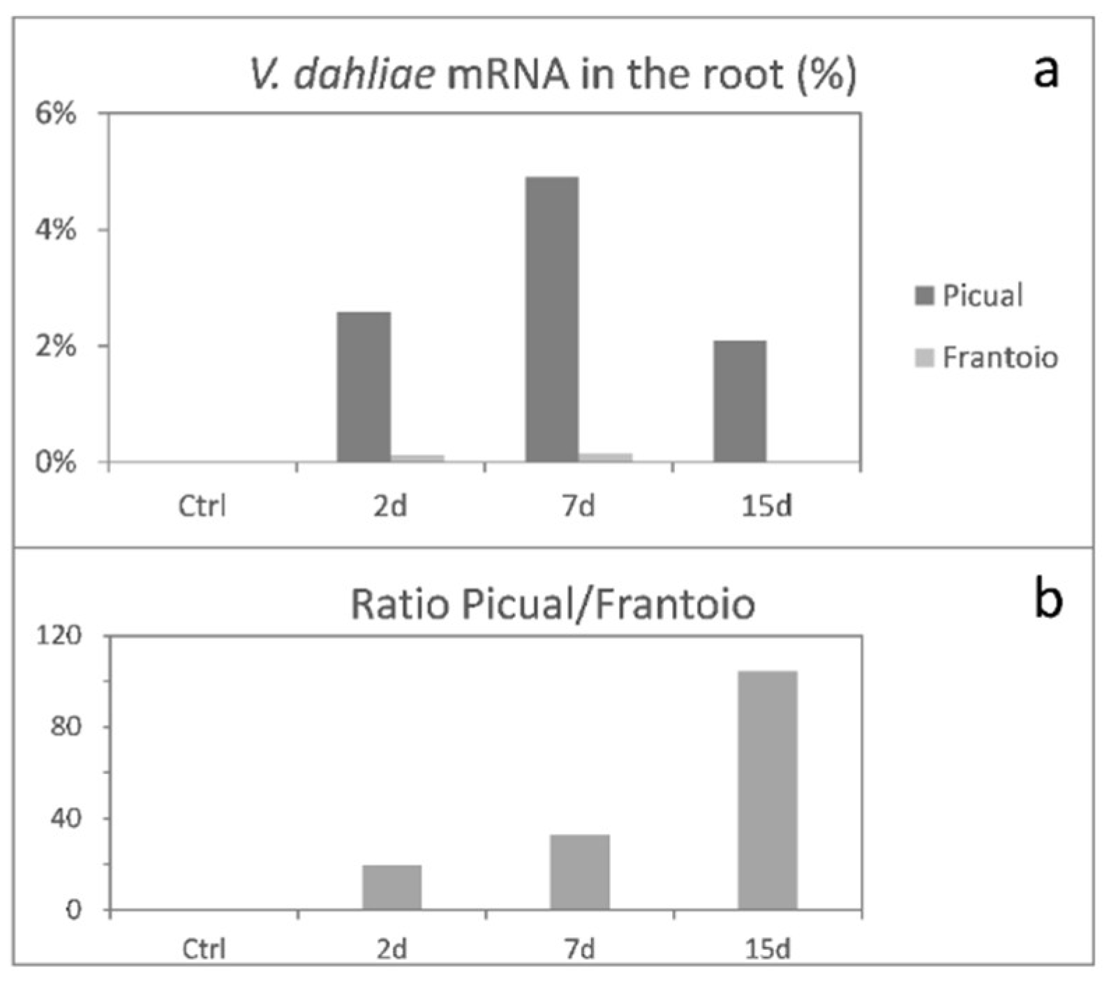
3.1. Global mRNA Amount of Verticillium Dahliae D pathotype is Strikingly Higher in ‘Picual’ (VWO-Susceptible) Than in ‘Frantoio’ (VWO-Tolerant) Roots

In order to measure V. dahliae growth during the olive root infection process, the percentage of total paired-end reads unequivocally aligning both ends with V. dahliae genes and at the correct distance was obtained for each time point (Figure 1a). As mRNAs are short half-life molecules, the RNAseq data of fungal mRNA paired-end reads present on/in root tissues at a given sampling time-point constitute a direct estimation of the amount of metabolically active fungal biomass. Maximum accumulation of V. dahliae mRNA was detected at 7 DAI in both cultivars. Remarkably, the percentage of pathogen transcripts was a lot higher in ‘Picual’ than in ‘Frantoio’ roots, indicating that the VWO-susceptible cultivar significantly favored the proliferation of V. dahliae biomass in contrast to the mycelial growth observed in ‘Frantoio’ roots. Moreover, the Picual/Frantoio ratio of the percentage of reads increased over time (Figure 1b).

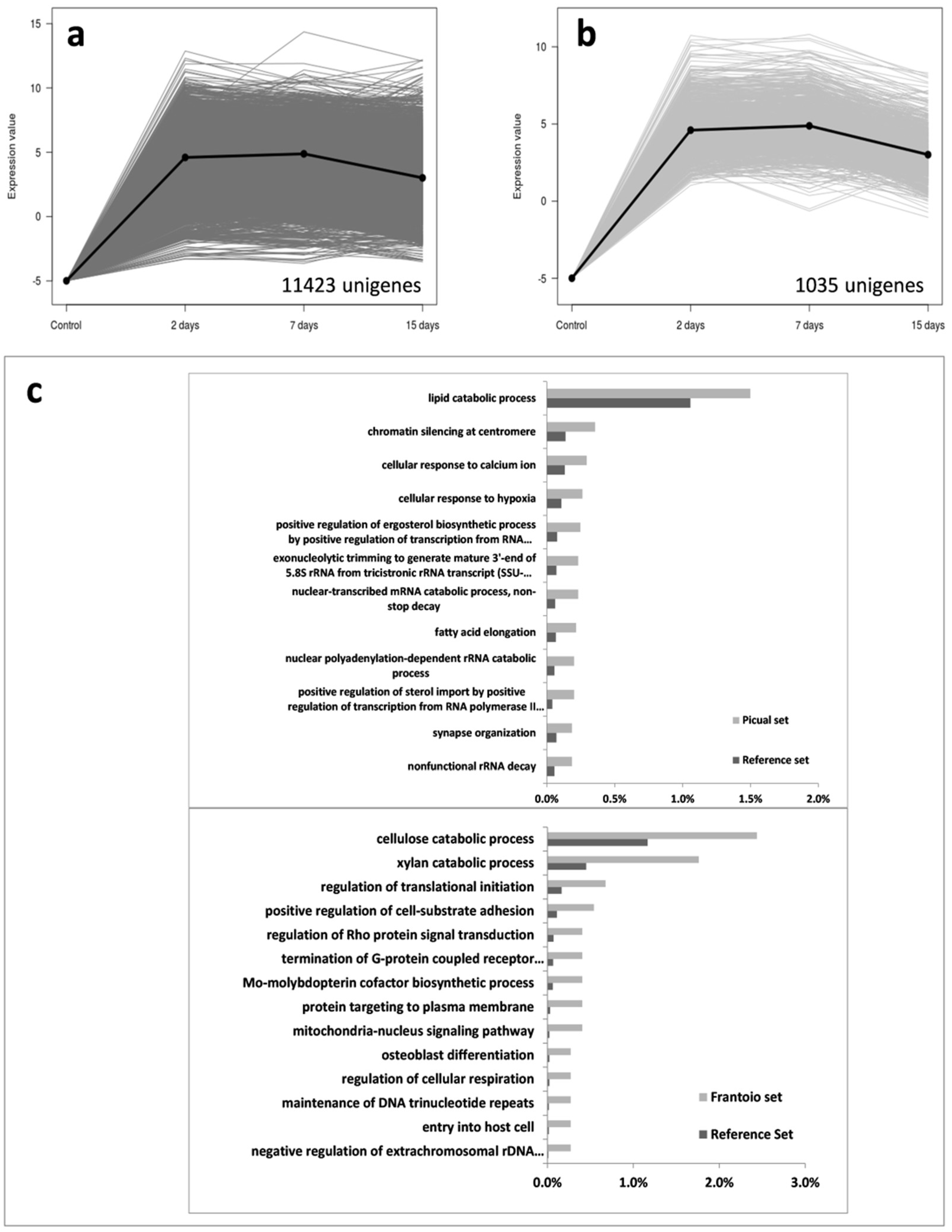
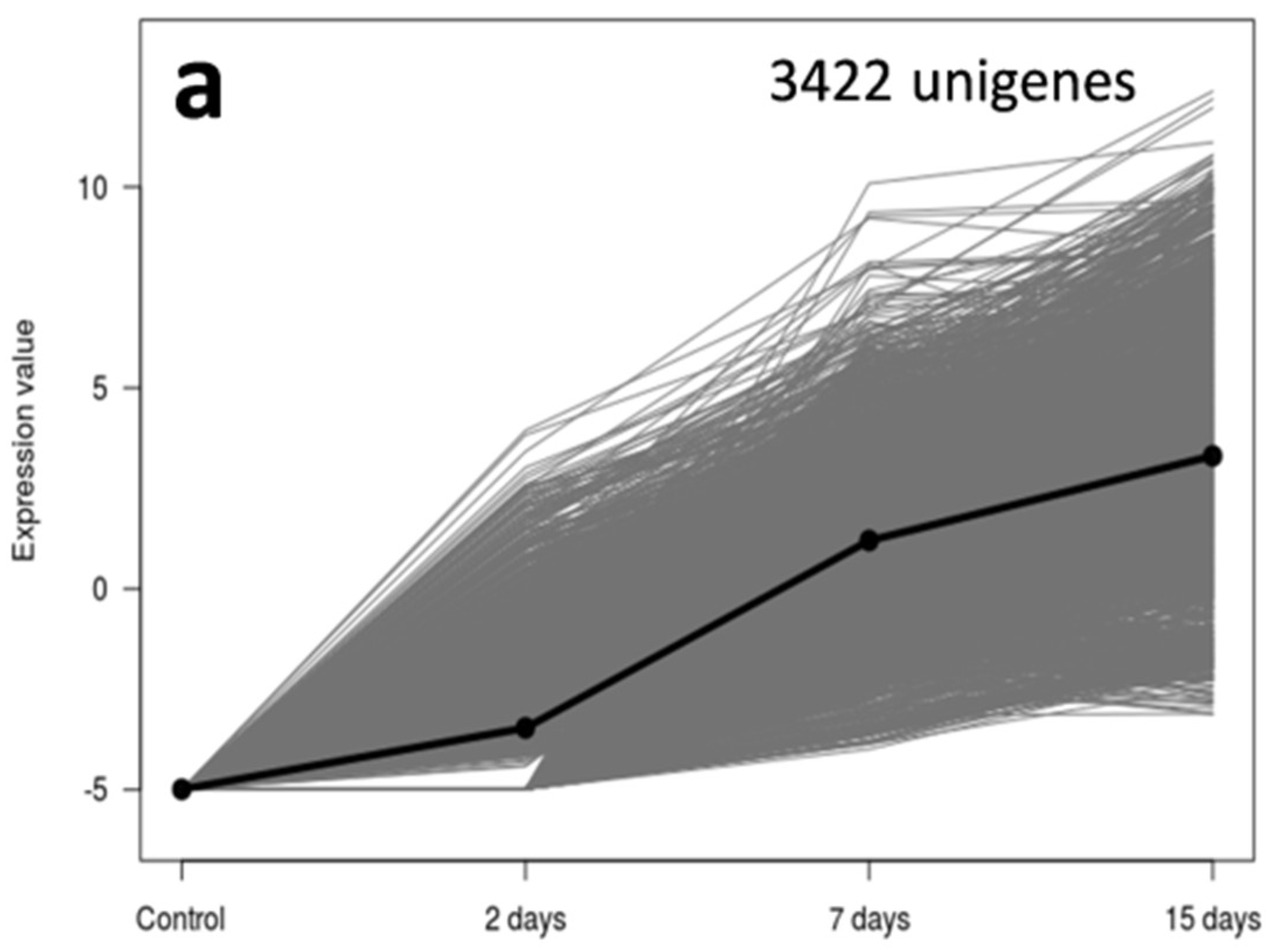
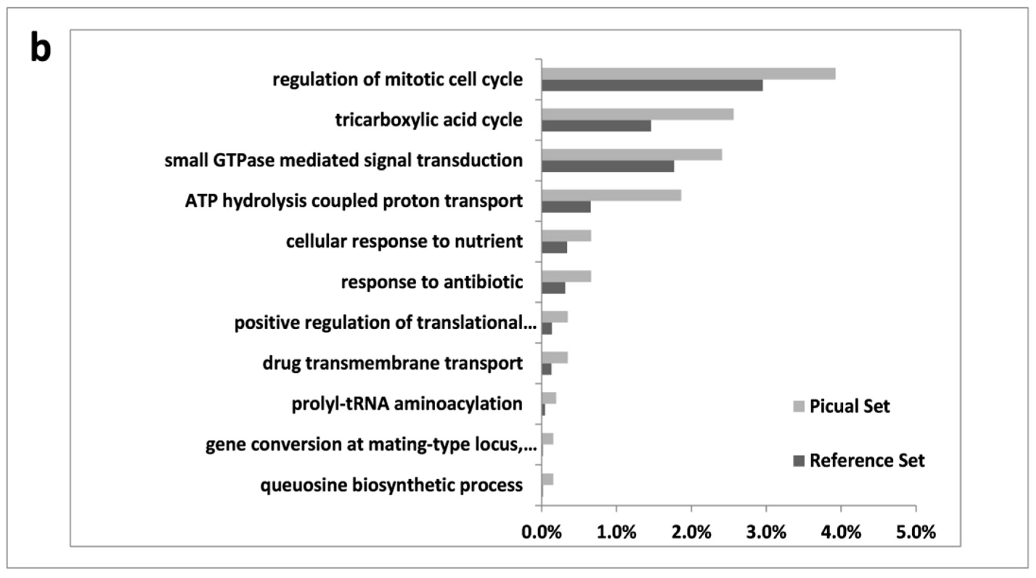
3.2. An Overwhelming Number of Verticillium Dahliae Unigenes Are Only Induced in Roots of the Susceptible Cultivar Picual

Whole-transcriptome responses of V. dahliae D pathotype clearly differed between ‘Picual’ and ‘Frantoio’ roots. Furthermore, RNA-seq analysis of V. dahliae transcripts showed a much higher number of unigenes induced in roots of the VWO-susceptible cultivar than in ‘Frantoio’. Thus, using and eightfold change over the almost null data of non-inoculated control plants and a 95% FDR, 30,240 V. dahliae unigenes were found to be over expressed in ‘Picual’ roots, but only 5641 in ‘Frantoio’ roots.
Verticillium dahliae transcript expression profiles obtained during the time-course infection process here under study (0–15 DAI), enabled us to cluster over-expressed unigenes in 7 distinctive groups. Group 1 consisted in pathogen’s unigenes highly induced along the entire period, 11,423 being over-expressed in ‘Picual’ but only 1035 in ‘Frantoio’ (Figure 2). This remarkable difference was not merely quantitative but also concerned the type of V. dahliae genes induced in each cultivar. Indeed, while in ‘Picual’ roots the most specific enriched GO terms were diverse and related to different processes such as lipid metabolism, response to calcium or hypoxia, chromatin silencing or other processes related to gene expression, in ‘Frantoio’ roots V. dahliae was essentially enriched in unigenes of cellulose and xylan catabolic processes (Figure 2c).
Group 2 corresponded to V. dahliae unigenes that were induced at early moments of the infection (2 DAI), but eventually were repressed at 15 DAI (Figure 3), coinciding with the timing in which fungal biomass decreased in both cultivars (Figure 1). This group consisted of 2775 unigenes in ‘Picual’ and 3816 unigenes in ‘Frantoio’. The most specific enriched GO terms again differed in both olive cultivars. Remarkably, unigenes related to pseudohyphal growth, diverse biosynthetic processes, and mitotic GO terms decayed at 15 DAI in ‘Picual’ roots. This was again consistent with the decrease of fungal growth (i.e., percentage of V. dahliae transcripts in roots) observed at that time point (Figure 1). However, most specific enriched GO terms in ‘Frantoio’ roots were related to a wide range of processes, some of them previously found in ‘Picual’ Group 1 (Figure 2c), like cellular response to hypoxia or fatty acid elongation (Figure 3c).
Groups 3, 4 and 5 corresponded to transient (at 2, or 7, or 2/15 DAI, respectively) induction of V. dahliae unigenes with no significant feature to be highlighted after conducting most specific enriched GO terms analysis (Figures S1–S3). Fluctuating expression patterns of these transcripts along time are difficult to interpret. The only relevant observation is that, once again, a much higher number of pathogen unigenes were expressed in ‘Picual’ roots compared to that in ‘Frantoio’ roots. Moreover, transient induction at 7 DAI of a large number of genes (5524) was only detected in ‘Picual’ roots (Figure S3).
These were also the cases for V. dahliae transcripts allocated to Groups 6 and 7. Croup 6 corresponded to unigenes that were induced only in ‘Picual’ at a late stage of the root infection process (15 DAI; Figure 4). This group is comprised by 4370 unigenes, cellulose and xylan catabolic processes being highly represented according to the most specific GO term enrichment analysis (Figure 4b). Interestingly enough, these unigenes were early induced in ‘Frantoio’ roots (i.e., Group 1; Figure 2), but only over-expressed in ‘Picual’ at 15 DAI. In addition, other catabolic processes are induced at this time point, as well as conidia formation (Figure 4b). Finally, Group 7 (only present in ‘Picual’ roots) consisted of 3422 V. dahliae unigenes showing a steady increase of gene expression along time (Figure 5). The most specific enriched GO terms of this Group showed prevalence of GO terms related to energy generation, regulation of mitotic cell cycle and cellular response to nutrients.

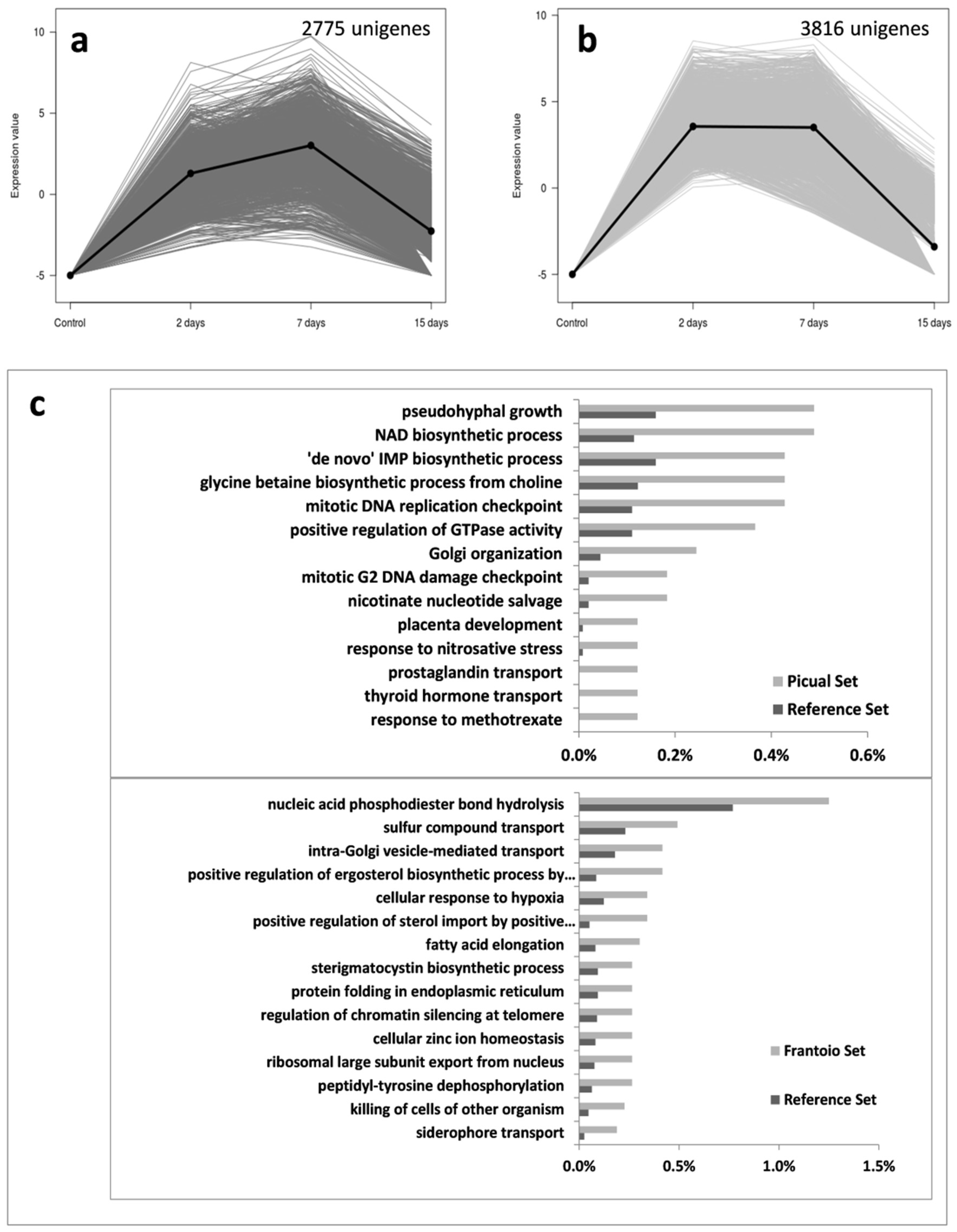
3.3. Targeting Verticillium Dahliae Niche Adaptation-Related Genes and Their Differential Expression Patterns in Cultivars Differing in VWO Susceptibility

We examined the time-course gene expression patterns of a number of DEGs related to niche adaptation (some of them also related to other processes; see next epigraphs). To avoid potential misinterpretation of results, particularly considering differences in relative transcripts abundance observed between ‘Picual’ and Frantoio’ (Figure 1), a basal level of log2 RPKM (Reads Per Kilobase Million) values >4 was ad hoc established. Therefore, unigenes with expression below this threshold in both varieties were considered as too low expressed to draw any conclusion. This also applies for unigenes described in the following sections.
Concerning carbon and nitrogen metabolism the most relevant finding concerned one unigene potentially coding for SNF1 (kinase complex involved in catabolite de-repression of carbohydrate active enzymes, CAZyme), that showed a significantly higher expression in ‘Picual’ at 15 DAI compared to the null expression observed in ‘Frantoio’ (Vedah_comp442094_c0) (Figure 6). In addition, two transcripts coding for chorismate synthases (Vedah_comp445065_c0 and Vedah_comp461778_c0) were found to be highly induced in the susceptible cultivar at 15 DAI (Figure 6). Besides, unigenes potentially coding for endoglucanase EG-1 (another CAZyme), SSP1 (Peptidyl-prolyl cis-trans isomerase/Serine/threonine-protein kinase), and CPC1 (cross-pathway control protein 1) detected in the V937-I transcriptome showed either no changes in gene expression (occasionally erratic along time) or an overall higher induction in the susceptible cv. Picual (data not shown), that was assumed to be due to the difference in relative transcripts abundance observed between cultivars (see above; Figure 1).
Verticillium dahliae must also cope with other stressing conditions found within the xylem vessels. Unigenes potentially coding for the high-affinity glucose transporter RGT2 were found but no relevant difference in their expression patterns was observed (data not shown). In addition, BLAST analysis did not reveal the presence of a GT2 (proteobacteria type 2 glucosyltransferase) homolog in V937-I. Stress due to reactive oxidative species (ROS) must also be confronted by V. dahliae when proliferating within the xylem. Again, differences were found neither for unigenes potentially coding for catalase-peroxidases nor for proteins of the Thi4 family involved in the biosynthesis of thiazole (precursor of thiamine, vitamin B1), including putative thiamine thiazole synthase transcripts (data not shown). The same could be concluded for unigenes coding for putative α-N-arabinofuranosidase (data not shown). The only remarkable finding related to stress tolerance was the differential up-regulation in ‘Picual’ of four unigenes coding for PRX1 (mitochondrial peroxiredoxin). One of them only showed a transient (2 DAI) significant up regulation in Picual roots (Vedah_comp377200_c0) (data not shown), while the other three (Vedah_comp377255_c0, Vedah_comp455648_c0 and Vedah_comp463413_c0) displayed a steady induction over time reaching maximum expression levels at 15 DAI. In contrast, expression of these unigenes in ‘Frantoio’ remained unaltered and always at a negligible level (Figure 6).

3.4. Verticillium Dahliae Effector-Coding Unigenes Do Not Show Differential Expression Patterns in ‘Picual’ and ‘Frantoio’ Roots

The disease progress is facilitated by the production of effectors (pathogen-secreted molecules) which either suppress host defenses or protect the pathogen against their action. The expression of unigenes potentially coding for effectors was also investigated. Results showed that, overall, no noticeable differences were found for unigenes coding for LysM, NLP (necrosis- and ethylene-inducing-like proteins), isochorismatase, and secreted, small cysteine-rich proteins found in the V937-I transcriptome, regardless the olive cultivar infected by the D pathotype (data not shown).

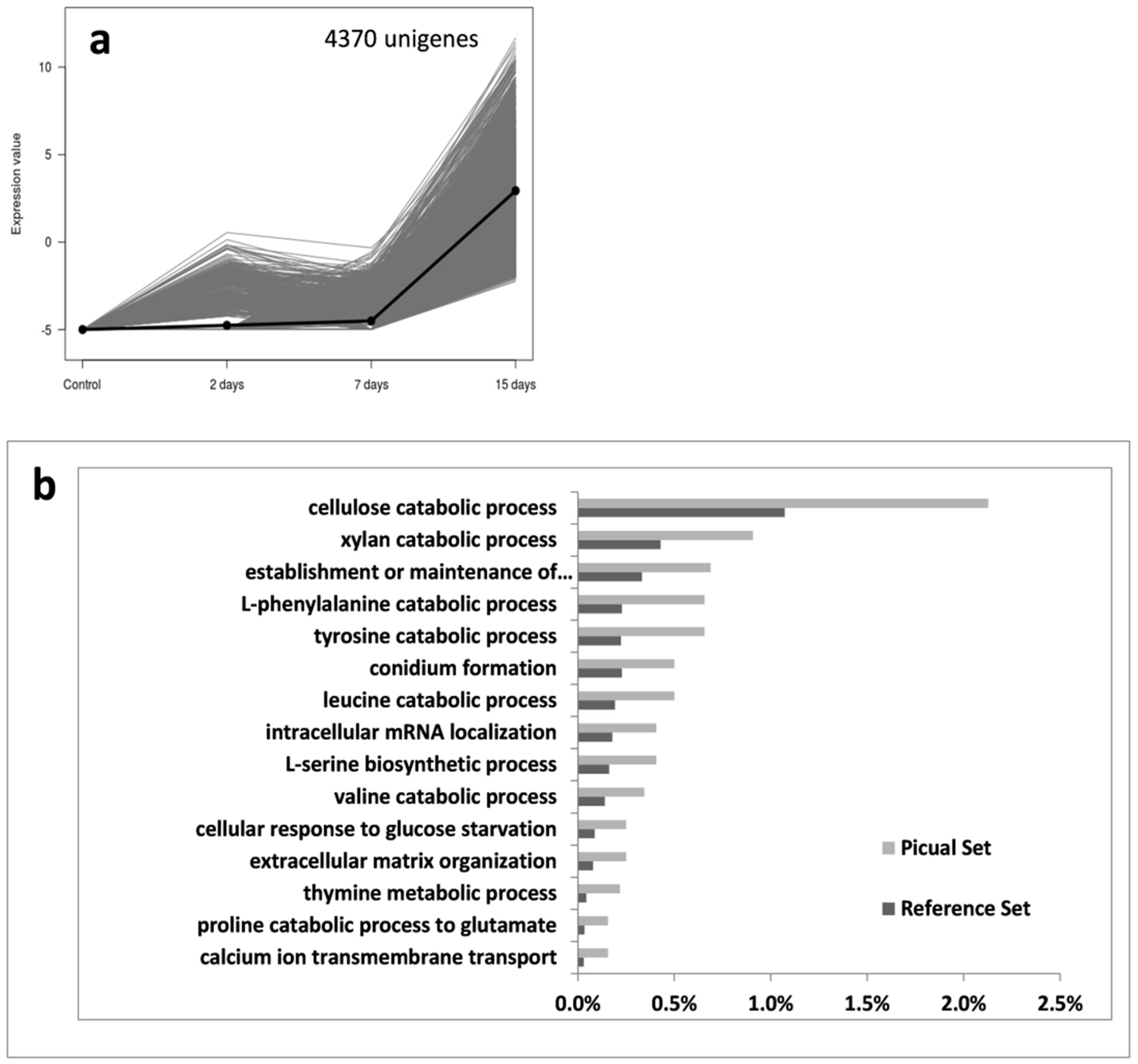
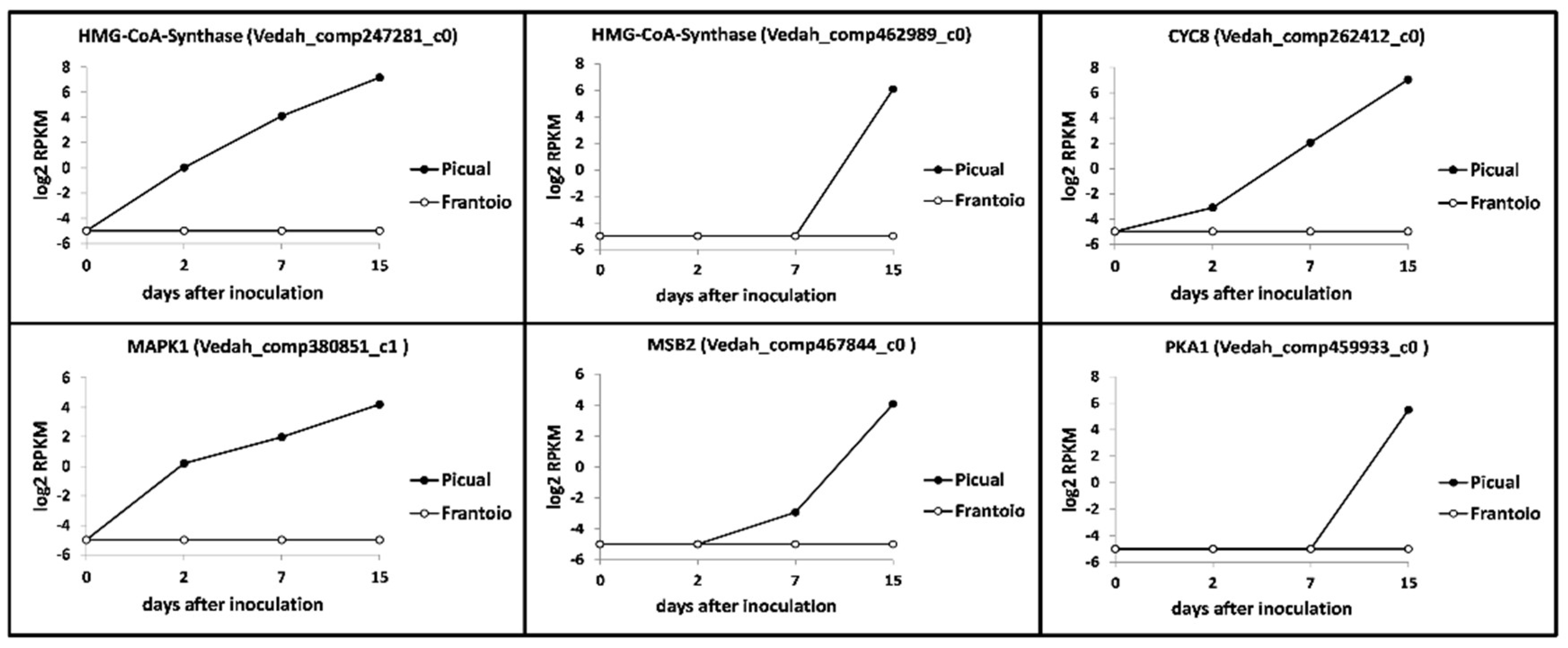
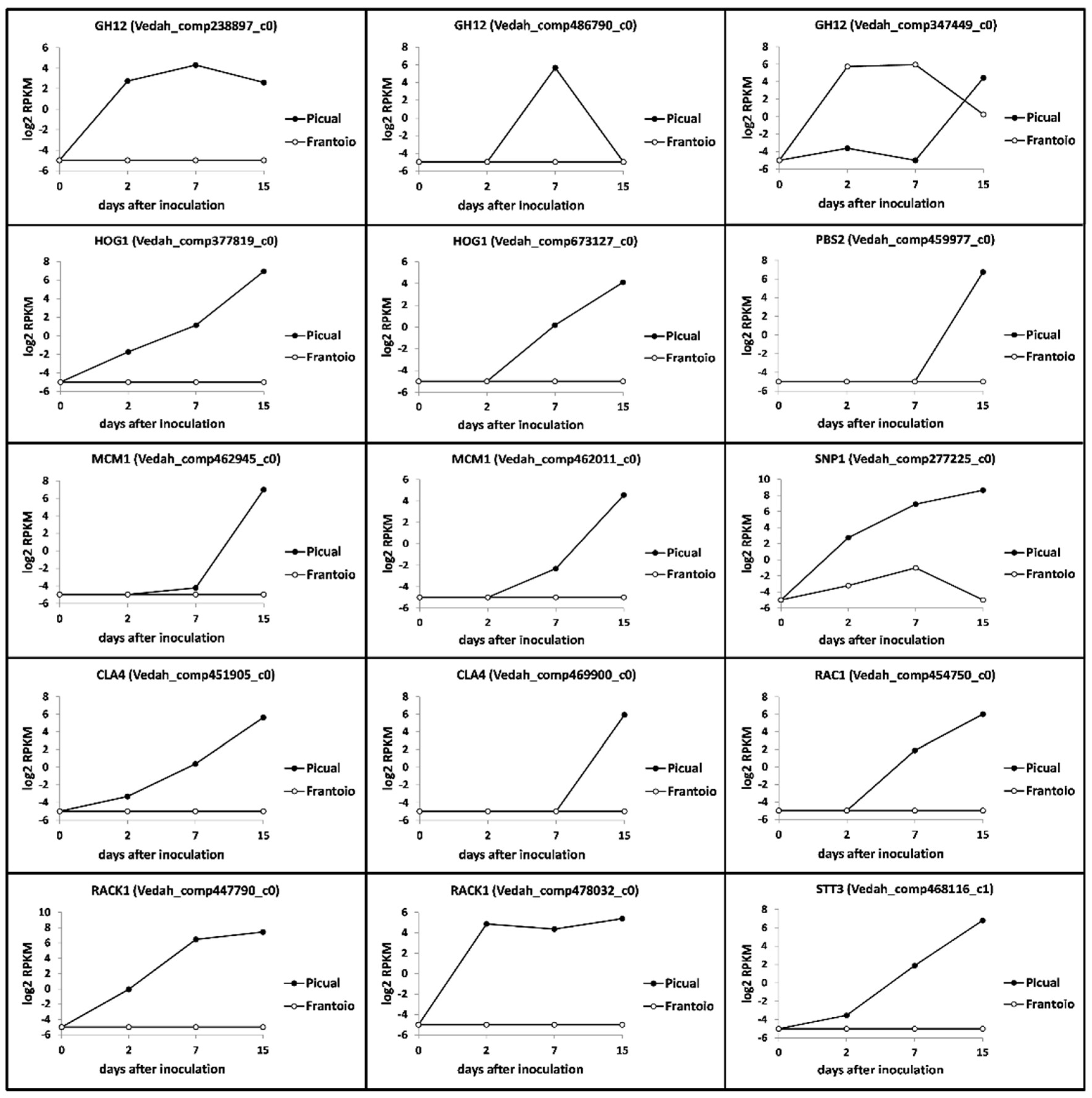
3.5. Verticillium Dahliae D pathotype Unigenes Involved in Pathogenicity, Virulence and Microsclerotia Development Showing Differential Expression Patterns

Verticillium dahliae pathogenicity, virulence and microsclerotia production pathways are often interconnected. Relevant results on unigenes potentially coding for these traits and that showed significantly-different expression patterns are now summarized. Two transcripts coding for hydroxymethyl-glutaryl (HMG)-CoA synthase (Vedah_comp247281_c0 and Vedah_comp462989_c0) were significantly up-regulated at late times after inoculation in the susceptible cultivar, while remained at null expression level in ‘Frantoio’ (Figure 7). Several unigenes potentially coding for CYC8 (glucose repression mediator protein) were found in the V937-I transcriptome. Only one of them (Vedah_comp262412_c0) showed a steady increased expression over time in ‘Picual’, reaching a log2 RPKM value > 6 at 15 DAI. In contrast, no expression was detected for this unigene in ‘Frantoio’ roots (Figure 7). The same behaviour was observed for a transcript coding for a mitogen-activated protein kinase MAPK1 (Vedah_comp380851_c1) (Figure 7). A transcript (Vedah_comp467844_c0) coding for a MSB2 protein (transmembrane mucin highly conserved in the MAPK signal pathway) was also found showing a clearly different expression pattern in ‘Picual’ (log2 RPKM value > 4 at 15 DAI) compared to that observed in ‘Frantoio (null expression) roots (Figure 7). Finally, several unigenes potentially coding for cAMP-dependent protein kinase A (PKA) were also found. Only one of them (Vedah_comp459933_c0) showed the same behaviour consistently reported in this section (log2 RPKM value > 4 at 15 DAI in ‘Picual’ and null in ‘Frantoio’) (Figure 7).

3.6. Multi-Task Verticillium Dahliae Unigenes Displaying Differential Expression Patterns

Several unigenes putatively coding for GH12 (Glycoside hydrolase 12) proteins (virulence factors and pathogen-associated molecular patterns [PAMPs] in oomycetes) were found in the V937-I transcriptome. Three transcripts showed significantly-different expression patterns that are worth mentioning. Two of them did not express at all in ‘Frantoio’ while they did in ‘Picual’, either consistently over time (Vedah_comp238897_c0) or just transiently at 7 DAI (Vedah_comp486790_c0) (Figure 8). Remarkably, one transcript (Vedah_comp347449_c0) behaved distinctly to the other putative GH12, displaying up regulation at 2 and 7 DAI in ‘Frantoio’ while in ‘Picual’ roots was fully repressed until 15 DAI, when a clear induction was observed in contrast to ‘Frantoio’ (Figure 8).
Unigenes coding for another MAPK, high-osmolarity glycerol (HOG1), were also found but only two of them (Vedah_comp377819_c0 and Vedah_comp673127_c0) showed significant expression levels in ‘Picual’, mainly at 15 DAI, and null in ‘Frantoio’ (Figure 8). Interestingly, a transcript (Vedah_comp459977_c0) potentially coding for PBS2 (a MAPK kinase) and that together with HOG1 are involved in a cascade that regulates microsclerotia formation and virulence, was also found to be significantly up regulated in ‘Picual (but not in Frantoio) at 15 DAI (Figure 8). The same behaviour (strong up regulation at late times; 15 DAI) was observed for two unigenes (Vedah_comp442945_c0 and Vedah_comp462011_c0) potentially coding for MCM1, a MADS-box transcription factor performing pleiotropic functions in V. dahliae (Figure 8). Several transcripts coding for RAC1 (a small GTPase) and its effector CLA4 have been found in the V937-I transcriptome. Among them, only one unigene for RAC1 (Vedah_comp454750_c0) and two for CLA4 (Vedah_comp451905_c0 and Vedah_comp469900_c0) showed rather similar up regulation patterns from 7 DAI, and only in ‘Picual’ roots (Figure 8). Several unigenes coding for RACK1 (receptor for activated C-kinase) were found from which two of them showed log2 RPKM values > 4, one with a consistent pattern along time (Vedah_comp478032_c0) and the other from 7 DAI (Vedah_comp447790_c0), while in ‘Frantoio’ expression was null (Figure 8). Among the transcripts potentially coding for CP1, a member of the SnodProt1 (cerato-platanin protein, CPP) phytotoxin family, some showed log2 RPKM values > 4 but with erratic expression patterns over time in ‘Picual’ (no expression in ‘Frantoio; data not shown). However, one of them (Vedah_comp277225_c0) showed a clear up-regulation trend over time, reaching log2 RPKM values > 8 at 15 DAI in ‘Picual’ in contrast to the nearly negligible expression observed in ‘Frantoio’ roots (Figure 8). Finally, one transcript (Vedah_comp468116_c0) coding for STT3 (catalytic subunit of the multi-subunit oligosaccharyl transferase, OST) is worth mentioning because, in agreement with the consistent expression pattern summarized here for many unigenes of the V937-I transcriptome, a clear up regulation over time only in ‘Picual’ roots was observed, particularly at late times (15 DAI) (Figure 8).

4. Discussion

Results showed that the amount of V. dahliae transcripts present in root tissues positively correlated with the VWO susceptibility level of the cultivar. Differences found were outstanding, ‘Frantoio’ roots harboring much lower amounts of V937-I biomass over time compared to that observed in ‘Picual’. Moreover, considering the ratio of ‘Picual’/’Frantoio’ percentage of reads a steady and significant increase of this ratio was also observed over time, reaching a maximum at 15 DAI. At this time-point V. dahliae is known to be well established in olive root tissues under artificial inoculation conditions [41]. These results agree with Quantitative Real-Time PCR (Q-RT-PCR) data from previous studies in which the amount of V. dahliae DNA in roots was always lower in VWO-tolerant cultivars [41,42,43]. Our results further support this correlation since V. dahliae transcriptional activity was significantly higher when the pathogen colonized and infected ‘Picual’ roots. This finding may be relevant in order to explain the differential susceptibility to V. dahliae reported for these cultivars, and must be linked to the differential whole transcriptome response earlier reported for the host. Therefore, tolerance of ‘Frantoio’ plants is not only a consequence of a complex process in which numerous plant traits are involved [25], but the result of basal and early pathogen-induced transcriptomic responses in the host hindering pathogen’s biomass proliferation and, consequently, limiting its transcriptional mechanism.
Co-transcriptomic studies of plant-pathogen interactions allow to infer global events taking place in both partners of the interaction. However, these studies are difficult because of the complexity for accurate discrimination between transcripts from the host and the pathogen. Therefore, these approaches are not frequent and have mostly focused on individual genes or gene co-expression networks e.g., [44,45,46,47]. Previously, we conducted a study in which olive (cv. Picual) and V. dahliae co-transcriptomes were generated [24]. Later, comparative transcriptomics focused on the differential host responses taking place in olive roots upon V. dahliae D pathotype infection, and their correlation with the VWO susceptibility level of olive cultivars was carried out [25]. One of the main results of this later study was that a much higher number of host unigenes were upregulated in ‘Picual’ roots during the early moments of the interaction (up to 7 DAI). In the present study, the number of V. dahliae induced unigenes was also demonstrated to be overwhelmingly higher (6 fold) in the susceptible variety. Therefore, both the pathogen (this study) and the host [25] displayed a much higher transcriptional activity (overall up regulation) during a compatible interaction. Data obtained are a reflection of the actual situation taking place in the olive roots: V. dahliae proliferating at a much higher rate in ‘Picual’ than in ‘Frantoio’, thereby producing more transcripts and consequently facilitating the detection of low expressed genes in the former. However, the availability of detecting a fair number of unigene transcripts in both cultivars allowed us to perform most specific enriched GO terms analyses, making it possible to observe clear differences between cultivars (Figure 2, Figure 3, Figure 4 and Figure 5 and Figures S1–S3).
According to the expression pattern of V. dahliae transcripts observed during the time-course infection process, seven different clusters of expressed unigenes during the root infection were found. Some of the most relevant findings are now discussed. In Group 1 (i.e., expression during the whole sampling period), differences were not only quantitative (the number of V. dahliae transcripts whose expression was detected in ‘Picual’ was 11-fold higher compared to ‘Frantoio’) but also qualitative. Remarkably, the pathogen was mainly enriched in transcripts related to cellulose and xylan catabolic processes when colonizing the roots of the tolerant cultivar, and this pattern was consistent over time. In contrast, these most specific enriched GO terms were overexpressed in ‘Picual’ roots only at 15 DAI (group 6; Figure 4). Previously, we identified an olive unigene potentially coding for a Dirigent-like protein involved in lignification that showed a significantly higher basal expression in ‘Frantoio’ than in ‘Picual [25], suggesting that ‘Frantoio’ roots can be more lignified as otherwise reported for the VWO-tolerant cv. Sayali [17]. The lignocellulosic biomass is mainly composed of lignin, cellulose and xylan [48]. Early and consistent induction over time of these V. dahliae genes in the interaction with ‘Frantoio’ (but not in ‘Picual’) can be explained by the need to dismantle the higher content of these polymers present in the roots of the tolerant cultivar. The production and involvement of cellulases in pathogenicity and virulence has been demonstrated for different phytopathogenic fungi, including V. dahliae e.g., [49,50,51,52]. Gui and co-workers [53] recently reported two GH12 proteins in V. dahliae Vd991, VdEG1 and VdEG3, with both cellulase activity required for virulence and elicitor activity that induces host-dependent immunity during Nicotiana benthamiana infection. They demonstrated that both proteins acted as PAMPs triggering cell death and PAMP-triggered immunity (PTI), although this function was independent of their enzymatic activity. Ten unigenes coding for GH12 proteins were found in the V-937I transcriptome. Only three of them, potentially coding for xyloglucan-specific endo-β-1,4-glucanase A, displayed significantly-different expression patterns in ‘Picual’ and ‘Frantoio’, although none corresponded to VdEG1 and VdEG3. In fact, VdEG1 matched to transcript Vedah_comp428223_c0 that only displayed significantly different up regulation at 15 DAI in ‘Picual’ compared to the null expression in ‘Frantoio’ (data not shown). Whether any of these GH12 genes may play a similar role in the V-937I-olive interaction to that reported by Gui and co-workers [53] is not known. Moreover, the unique discrepant expression pattern (transcript Vedah_comp347449_c0; Figure 8) compared to the other GH12, and to the overall induction observed for pathogenicity- and virulence-related genes in ‘Picual’ in contrast to the null expression in ‘Frantoio’, deserves to be further studied. Another relevant observation when examining group 6 (as well as groups 5 and 7) was the absence of pathogen’s transcripts in ‘Frantoio’ roots. In addition to the above-mentioned late (15 DAI) induction in ‘Picual’ roots of V. dahliae genes involved in cellulose and xylan catabolic processes, what may be explained by the pathogen colonizing more lignified regions of the ‘Picual’ roots while progressing upwards during the host colonization process, a number of catabolic processes were also induced at this time-point. Moreover, the conidia formation process was actively up regulated in ‘Picual’ in contrast with ‘Frantoio’, which also correlate with their differential VWO susceptibility level. While penetration and internal root colonization in ‘Frantoio’ is severely hampered, massive entrance of V937-I biomass into ‘Picual’ roots seemed to lead to active conidiation thereby favouring pathogen dissemination through the vascular vessels. The rapid upward spread of V. dahliae in the vascular tissue is primarily attributed to conidia translocated by the transpiration stream, a scenario known to take place in trees since long time ago [54]. In fact, D pathotype biomass detection by both microscopy [10] and molecular [36] approaches in aerial organs of ‘Picual’ at early or middle-term times after root inoculation is plausibly explained by conidia production, rapid upwards translocation, and subsequent germination rather than by hyphal proliferation through the vascular system. This scenario is seldom (or never) observed in tolerant cultivars [41,42,43]. To further support the induction of the conidiation process in ‘Picual’, it is worth mentioning the up regulation over time, reaching a log2 RPKM value > 6 at 15 DAI, of one transcript potentially coding for STT3 (catalytic subunit of the multi-subunit OST that plays a key role in glycoprotein modification) in ‘Picual’ but not in ‘Frantoio’. Su and co-workers [55] have recently demonstrated that the expression of the STT3 gene of V. dahliae was increased at the stage of conidia germination, and that mycelial growth, sporulation ability and glycoprotein secretion were impaired, as well as germination ratio and virulence decreased in knockout mutants of this gene. Finally, the abundance of most specific enriched GO terms in groups 5, 6 and 7 related to energy generation, cellular response to nutrients and catabolic processes, among others, indicate that in ‘Picual’ roots the pathogen is actively growing and demanding nutrients, likely more available as a consequence of the active host tissue infection process. This situation does not take place in ‘Frantoio’ roots as demonstrated by the negligible amounts of V-937I biomass and the total absent of transcriptional activity, particularly at 15 DAI.
Effective colonization of a host means that a given pathogen is adapted to an ecological niche that provides specific nutrients, sometimes constituting a growth limiting factor for the invader. Moreover, stress conditions must be confronted by the pathogen within the colonized microhabitat (in this case the xylem vessels during most of V. dahliae’s parasitic phase), as well as defence responses deployed by the host [56]. In order to assess whether the transcriptome of the D isolate V937-I showed differential responses related to niche adaptation and stress condition depending on the infected olive cultivar a number of unigenes were targeted. Overall, the time-course expression patterns of V. dahliae DEGs related to these traits did not show noticeable differences regardless the infected olive cultivar and the ad hoc threshold log2 RPKM basal level value set to be considered as significantly relevant. Some of the unigenes here evaluated could also be assigned to other groups, stressing the fact that many of them can be involved in different processes of the parasitic phase of V. dahliae. For instance, one of the few remarkable differences found was an unigene coding for SNF1 (high expression in ‘Picual’ at 15 DAI but null expression in ‘Frantoio’). This gene, coding for a sucrose nonfermenting 1 enzyme, seems to act as part of a nutrient signalling pathway controlling both microsclerotial development and pathogenicity [56,57], and thus could also be allocated in our study to the pathogenicity/virulence genes group. Verticillium dahliae tomato race 1 mutants in which SNF1 was disrupted showed no expression of cell wall-degrading enzymes in simulated xylem medium, much lower growth when cultivated in medium amended with pectin and galactose, severe reduction in virulence on tomato and eggplant, and defective early colonization of roots [57]. High induction of this gene in V-937I at late times of the root infection in the susceptible olive cultivar can correspond to nutrient response processes exacerbated in ‘Picual’ as consequence of the massive colonization and degradation of root host tissues. Related to this, the up regulation of two (out of 5) unigenes potentially coding for chorismate synthase (key enzyme in the first branch point intermediate of aromatic amino acid biosynthesis) at 15 DAI in ‘Picual but not in Frantoio’ may also relate to the profuse colonization of the xylem in the susceptible cultivar at this time point, a microhabitat poor in nutrients including low concentrations of amino acids [58]. During the infection process, V. dahliae must also confront ROS protective response by the host. Earlier results have revealed that this response takes place in the pathogen (firstly) and the host (at later times) [24]. Moreover, the overall response to ROS stress was clearly stronger in the susceptible cultivar compared to that observed in ‘Frantoio’ roots [25]. In this present study, no remarkable differences were found but the steady up regulation over time (maximum expression reached at 15 DAI) observed for three transcripts potentially coding for PRX1 (a thiol-specific peroxidase) only in ‘Picual’ roots. A PRX1 protein was reported to be abundantly secreted in the xylem sap of hop plants upon infection of V. nonalfalfae. Moreover, in planta expression of the coding gene increased with the pathogen’s colonization process, suggesting a role in virulence [59].
None of the examined transcripts potentially coding for known V. dahliae effectors and detected in the V-937I transcriptome showed noticeable differences in their expression patterns in the two olive cultivars. An effector role in infecting hosts has been recently suggested for a conserved secretory protein VdCP1 that also showed chitin-binding properties, identified as a member of the SnodProt1 phytotoxin family [60]. These authors reported that vdcp1 gene displayed high expression along the V. dahliae infection process, and also suggested that VdCP1 could protect V. dahliae cell wall from enzymatic degradation. Eight unigenes coding for these CPP were found in the V-937I trasncriptome, but only four showed log2 RPKM values > 4. Interestingly, one of them displayed an increasing and steady up regulation overtime in ‘Picual’ in contrast to ‘Frantoio’, suggesting a similar role during the infection process of a susceptible olive cultivar to that reported by Zhang and co-workers [60].
Finally, transcripts potentially coding from pathogenicity and virulence showed the same overall differential expression patterns consistently observed between olive cultivars. For instance, the induction at 15 DAI of a transcript coding for a MSB2 protein in ‘Picual’ but not in ‘Frantoio’ is worth mentioning, since a VdMsb gene has been implicated in the colonization and penetration processes of cotton and Arabidopsis thaliana by V. dahliae strain Vd8 [61]. Similarly, transcripts coding for HMG-CoA synthase, CYC8, MAPK1, PKA1, HOG1 and PBS2 were also found to be induced in ‘Picual’ but not in ‘Frantoio’, and always reaching high log2 RPKM values at 15 DAI. The link between the expression of genes coding for these proteins and processes such as conidiation, pathogenesis, virulence, microsclerotia formation, etc., has been highlighted in a number of studies, e.g., [62,63,64,65]. Altogether, the high induction of genes involved in these processes when infecting ‘Picual’ roots denotes that colonization and proliferation of the pathogen’s biomass in root tissues of the susceptible cultivar is progressing effectively, and that the host defence responses have been overcome. These events were not observed in ‘Frantoio’ roots. Massive proliferation of the D pathotype in ‘Picual’ roots, including production of microsclerotia on the root surface, has been previously demonstrated by microscopy evidence [10].

5. Conclusions

The V. dahliae D pathotype transcriptome responds differently depending on the VWO susceptibility level of the cultivar. Differences are not only qualitative along the time-course of the early root colonization process, but mostly quantitative. While outstanding differences were found regarding the biological processes induced in the pathogen, the much larger V. dahliae biomass found in roots of ‘Picual’ plants, consequently leading to a much higher transcriptional activity, seems to explain to a large extent the higher aggressiveness exerted over this cultivar and why a susceptible cultivar succumb much faster to the disease. The comparative co-transcriptomic studies performed enabled to holistically disentangle both pathogen and host responses in the olive–V. dahliae interaction. VWO susceptibility can thus be explained by the absence of basal and pathogen-induced transcriptomic responses in susceptible varieties [25], which favors the pathogen’s transcriptional mechanism and its massive proliferation, colonization and further dissemination in the host roots.

Supplementary Materials

The following are available online at https://www.mdpi.com/2073-4425/10/4/251/s1, Figure S1: Group 3 unigenes that present a high mRNA level at 2 days and low at 7- and 15-days post-inoculation. Unigenes were selected using an eightfold change and a 95% of False Discovery Rate; mRNA level in ‘Picual’ (a) and ‘Frantoio’ roots (b); Most specific enriched GO terms of over-expressed genes in Verticillium dahliae infecting ‘Picual’ or ‘Frantoio’ roots (c)., Figure S2: Group 4; unigenes that present a high mRNA level at 2- and 15-days but low at 7-days post-inoculation. Unigenes were selected using an eightfold change and a 95% of False Discovery Rate; mRNA level in ‘Picual’ (a) and ‘Frantoio’ roots (b); Most specific enriched GO terms of over-expressed genes in Verticillium dahliae infecting Picual or Frantoio roots (c)., Figure S3: Group 5; unigenes that present a high mRNA level at 7 days but low at 2- and 15-days post-inoculation. Unigenes were selected using an eightfold change and a 95% of False Discovery Rate; mRNA level in ‘Picual’ roots (a) and most specific enriched GO terms of over-expressed genes in Verticillium dahliae infecting ‘Picual’ roots (b).

Author Contributions

Conceptualization, J.B.B., F.L. and J.M.-B.; Data curation, J.J.-R. and M.d.l.O.L.-P.; Formal analysis, J.J.-R., M.d.l.O.L.-P., F.L. and J.M.-B.; Funding acquisition, F.L. and J.M.-B.; Investigation, J.J.-R., M.d.l.O.L.-P. and C.G.-L.C.; Methodology, C.G.-L.C.; Writing—review & editing, F.L. and J.M.-B.

Funding

Supported by grants AGL2016-75729-C2-1-R and AGL2016-75729-C2-2-R from the Spanish Ministerio de Economía, Industria y Competitividad/Agencia Estatal de Investigación, and grant AGR-5948 from Junta de Andalucía (Consejería de Economía, Innovación y Ciencia), co-financed by the European Regional Development Fund (ERDF).

Acknowledgments

Thanks are due to Antonio Valverde-Corredor for his technical assistance. Technical and human support provided by CICT of Universidad de Jaén (UJA, MINECO, Junta de Andalucía, FEDER) is also acknowledged.

Conflicts of Interest

The authors declare no conflict of interest. The funders had no role in the design of the study; in the collection, analyses, or interpretation of data; in the writing of the manuscript, or in the decision to publish the results.

References

  1. Fradin, E.F.; Thomma, B.P.H.J. Physiology and molecular aspects of Verticillium wilt diseases caused by V. dahliae and V. albo-atrum. Mol. Plant Pathol. 2006, 7, 71–86. [Google Scholar] [CrossRef]
  2. Klosterman, S.J.; Atallah, Z.K.; Vallad, G.E.; Subbarao, K.V. Diversity, pathogenicity and management of Verticillium species. Annu. Rev. Phytopathol. 2009, 47, 39–62. [Google Scholar] [CrossRef]
  3. Daayf, F. Verticillium wilts in crop plants: Pathogen invasion and host defense responses. Can. J. Plant Pathol. 2015, 37, 8–20. [Google Scholar] [CrossRef]
  4. Pegg, G.F.; Brady, B.L. Verticillium Wilts; CAB International: Wallingford, UK, 2002. [Google Scholar]
  5. Milgroom, M.G.; Jiménez-Gasco, M.M.; Olivares-García, C.; Jiménez-Díaz, R.M. Clonal expansion and migration of a highly virulent, defoliating lineage of Verticillium dahliae. Phytopathology 2016, 106, 1036–1046. [Google Scholar] [CrossRef]
  6. Dervis, S.; Mercado-Blanco, J.; Erten, L.; Valverde-Corredor, A.; Pérez-Artés, E. Verticillium wilt of olive in Turkey: A survey on disease importance, pathogen diversity and susceptibility of relevant olive cultivars. Eur. J. Plant Pathol. 2010, 127, 287–301. [Google Scholar] [CrossRef]
  7. López-Escudero, F.J.; Mercado-Blanco, J.; Roca, J.M.; Valverde-Corredor, A.; Blanco-López, M.A. Verticillium wilt of olive in the Guadalquivir Valley (southern Spain): Relations with some agronomical factors and spread of Verticillium dahliae. Phytopathol. Mediterr. 2010, 49, 370–380. [Google Scholar]
  8. Triki, M.A.; Krid, S.; Hsairi, H.; Hammemi, I.; Ioos, R.; Gdoura, R.; Rhouma, A. Occurrence of Verticillium dahliae defoliating pathotypes on olive trees in Tunisia. Phytopathol. Mediterr. 2011, 50, 267–272. [Google Scholar]
  9. López-Escudero, F.J.; Mercado-Blanco, J. Verticillium wilt of olive: A case study to implement an integrated strategy to control a soil-borne pathogen. Plant Soil 2011, 344, 1–50. [Google Scholar] [CrossRef]
  10. Prieto, P.; Navarro-Raya, C.; Valverde-Corredor, A.; Amyotte, S.G.; Dobinson, K.F.; Mercado-Blanco, J. Colonization process of olive tissues by Verticillium dahliae and its in planta interaction with the biocontrol root endophyte Pseudomonas fluorescens PICF7. Microb. Biotechnol. 2009, 2, 499–511. [Google Scholar] [CrossRef] [PubMed]
  11. Calderón, R.; Lucena, C.; Trapero-Casas, J.L.; Zarco-Tejada, P.J.; Navas-Cortés, J.A. Soil temperature determines the reaction of olive cultivars to Verticillium dahliae pathotypes. PLoS ONE 2014, 9, e110664. [Google Scholar] [CrossRef] [PubMed]
  12. Gómez-Lama Cabanás, C.; Schilirò, E.; Valverde-Corredor, A.; Mercado-Blanco, J. Systemic responses in a tolerant olive (Olea europaea L.) cultivar upon root colonization by the vascular pathogen Verticillium dahliae. Front. Microbiol. 2015, 6, 928. [Google Scholar]
  13. Baídez, A.G.; Gómez, P.; Del Rio, J.A.; Ortuño, A. Dysfunctionality of the xylem in Olea europaea L. plants associated with the infection process by Verticillium dahliae Kleb. Role of phenolic compounds in plant defense mechanism. J. Agric. Food Chem. 2007, 55, 3373–3377. [Google Scholar] [CrossRef] [PubMed]
  14. Markakis, E.A.; Tjamos, S.E.; Antoniou, P.P.; Roussos, P.A.; Paplomatas, E.J.; Tjamos, E.C. Phenolic responses of resistant and susceptible olive cultivars induced by defoliating and non-defoliating Verticillium dahliae pathotypes. Plant Dis. 2010, 94, 1156–1162. [Google Scholar] [CrossRef] [PubMed]
  15. Bubici, G.; Cirulli, M. Control of Verticillium wilt of olive by resistant rootstocks. Plant Soil 2012, 352, 363–376. [Google Scholar] [CrossRef]
  16. Gharbi, Y.; Barkallah, M.; Bouazizi, E.; Cheffi, M.; Gdoura, R.; Triki, M.A. Differential fungal colonization and physiological defense responses of new olive cultivars infected by the necrotrophic fungus Verticillium dahliae. Acta Physiol. Plant 2016, 38, 242. [Google Scholar] [CrossRef]
  17. Gharbi, Y.; Barkallah, M.; Bouazizi, E.; Hibar, K.; Gdoura, R.; Triki, M.A. Lignification, phenols accumulation, induction of PR proteins and antioxidant-related enzymes are key factors in the resistance of Olea europaea to Verticillium wilt of olive. Acta Physiol. Plant 2017, 39, 43. [Google Scholar] [CrossRef]
  18. Duressa, D.; Anchieta, A.; Chen, D.M.; Klimes, A.; García-Pedrajas, M.D.; Dobinson, K.F.; Klosterman, S.J. RNA-seq analyses of gene expression in the microsclerotia of Verticillium dahliae. BMC Genomics 2013, 14, 607. [Google Scholar] [CrossRef]
  19. Hu, D.; Wang, C.; Tao, F.; Cui, Q.; Xu, X.; Shang, W.; Hu, X. Whole genome wide expression profiles on germination of Verticillium dahliae microsclerotia. PLoS ONE 2014, 9, e100046. [Google Scholar] [CrossRef] [PubMed]
  20. Sun, Q.; Jiang, H.; Zhu, X.; Wang, W.; He, X.; Shi, Y.; Yuan, Y.; Du, X.; Cai, Y. Analysis of sea-island cotton and upland cotton in response to Verticillium dahliae infection by RNA sequencing. BMC Genomics 2013, 14, 852. [Google Scholar] [CrossRef]
  21. Zhang, Y.; Wang, X.F.; Ding, Z.G.; Ma, Q.; Zhang, G.R.; Zhang, S.L.; Li, Z.K.; Wu, L.Q.; Zhang, G.Y.; Ma, Z.Y. Transcriptome profiling of Gossypium barbadense inoculated with Verticillium dahliae provides a resource for cotton improvement. BMC Genomics 2013, 14, 637. [Google Scholar] [CrossRef]
  22. Faino, L.; de Jonge, R.; Thomma, B.P.H.J. The transcriptome of Verticillium dahliae-infected Nicotiana benthamiana determined by deep RNA sequencing. Plant Signal. Behav. 2012, 7, 1065–1069. [Google Scholar] [CrossRef]
  23. Tan, G.; Liu, K.; Kang, J.; Xu, K.; Zhang, Y.; Hu, L.; Zhang, J.; Li, C. Transcriptome analysis of the compatible interaction of tomato with Verticillium dahliae using RNA-sequencing. Front. Plant Sci. 2015, 6, 428. [Google Scholar] [CrossRef]
  24. Jiménez-Ruiz, J.; de la O Leyva-Pérez, M.; Schilirò, E.; Barroso, J.B.; Bombarely, A.; Mueller, L.; Mercado-Blanco, J.; Luque, F. Transcriptomic analysis of the Olea europaea L. roots during the Verticillium dahliae early infection process. Plant Genome 2017, 10. [Google Scholar] [CrossRef]
  25. Leyva-Pérez, M.O.; Jiménez-Ruiz, J.; Gómez-Lama Cabanás, C.; Valverde-Corredor, A.; Barroso, J.B.; Luque, F.; Mercado-Blanco, J. Tolerance of olive (Olea europaea) cv Frantoio to Verticillium dahliae relies on both basal and pathogen-induced differential transcriptomic responses. New Phytol 2018, 217, 671–686. [Google Scholar] [CrossRef]
  26. Ajengui, A.; Bertolini, E.; Ligorio, A.; Chebil, S.; Ippolito, A.; Sanzani, S.M. Comparative transcriptome analysis of two citrus germplasms with contrasting susceptibility to Phytophthora nicotianae provides new insights into tolerance mechanisms. Plant Cell Rep. 2017, 37, 483–499. [Google Scholar] [CrossRef]
  27. Hu, Y.; Zhong, X.; Liu, X.; Lou, B.; Zhou, C.; Wang, X. Comparative transcriptome analysis unveils the tolerance mechanisms of Citrus hystrix in response to ‘Candidatus Liberibacter asiaticus’ infection. PLoS ONE 2017, 12, e0189229. [Google Scholar] [CrossRef]
  28. Matić, S.; Bagnaresi, P.; Biselli, C.; Orru’, L.; Carneiro, G.A.; Siciliano, I.; Valé, G.; Gullino, M.L.; Spadaro, D. Comparative transcriptome profiling of resistant and susceptible rice genotypes in response to the seedborne pathogen Fusarium fujikuroi. BMC Genomics 2016, 17, 608. [Google Scholar] [CrossRef] [PubMed]
  29. Du, H.; Wang, Y.; Yang, J.; Yang, W. Comparative transcriptome analysis of resistant and susceptible tomato Lines in response to infection by Xanthomonas perforans Race T3. Front. Plant Sci. 2015, 6, 1173. [Google Scholar] [CrossRef]
  30. Giampetruzzi, A.; Morelli, M.; Saponari, M.; Loconsole, G.; Chiumenti, M.; Boscia, D.; Savino, V.N.; Martelli, G.P.; Saldarelli, P. Transcriptome profiling of two olive cultivars in response to infection by the CoDiRO strain of Xylella fastidiosa subsp. pauca. BMC Genomics 2016, 17, 475. [Google Scholar] [CrossRef]
  31. Progar, V.; Jakše, J.; Štajner, N.; Radišek, S.; Javornik, B.; Berne, S. Comparative transcriptional analysis of hop responses to infection with Verticillium nonalfalfae. Plant Cell Rep. 2017, 36, 1599–1613. [Google Scholar] [CrossRef] [PubMed]
  32. Stergiopoulos, I.; de Wit, P.J.G.M. Fungal effector proteins. Annu. Rev. Phytopathol. 2009, 47, 233–263. [Google Scholar] [CrossRef]
  33. Xiong, D.; Wang, Y.; Tian, L.; Tian, C. MADS-Box transcription factor VdMcm1 regulates conidiation, microsclerotia formation, pathogenicity, and secondary metabolism of Verticillium dahliae. Front. Microbiol. 2016, 7, 1192. [Google Scholar] [CrossRef]
  34. Maldonado-González, M.M.; Bakker, P.A.H.M.; Prieto, P.; Mercado-Blanco, J. Arabidopsis thaliana as a tool to identify traits involved in Verticillium dahliae biocontrol by the olive root endophyte Pseudomonas fluorescens PICF7. Front. Microbiol. 2015, 6, 266. [Google Scholar] [CrossRef]
  35. Jiménez-Díaz, R.M.; Olivares-García, C.; Trapero-Casas, J.L.; Jiménez-Gasco, M.M.; Navas-Cortés, J.A.; Landa, B.B.; Milgroom, M.G. Variation of pathotypes and races and their correlations with clonal lineages in Verticillium dahliae. Plant Pathol. 2016, 66, 651–666. [Google Scholar] [CrossRef]
  36. Mercado-Blanco, J.; Rodríguez-Jurado, D.; Pérez-Artés, E.; Jiménez-Díaz, R.M. Detection of the defoliating pathotype of Verticillium dahliae in infected olive plants by nested PCR. Eur. J. Plant Pathol. 2002, 108, 1–13. [Google Scholar] [CrossRef]
  37. Prieto, P.; Mercado-Blanco, J. Endophytic colonization of olive roots by the biocontrol strain Pseudomonas fluorescens PICF7. FEMS Microbiol. Ecol. 2008, 64, 297–306. [Google Scholar] [CrossRef] [PubMed]
  38. Aronesty, E. Ea-utils: “Command-Line Tools for Processing Biological Sequencing Data”. 2011. Available online: https://github.com/ExpressionAnalysis/ea-utils (accessed on 10 May 2013).
  39. Schmieder, R.; Edwards, R. Quality control and preprocessing of metagenomic datasets. Bioinformatics 2011, 27, 863–864. [Google Scholar] [CrossRef] [PubMed]
  40. Conesa, A.; Götz, S.; García-Gómez, J.M.; Terol, J.; Talon, M.; Robles, M. Blast2GO: A universal tool for annotation, visualization and analysis in functional genomics research. Bioinformatics 2005, 21, 3674–3676. [Google Scholar] [CrossRef]
  41. Mercado-Blanco, J.; Collado-Romero, M.; Parrilla-Araujo, S.; Rodríguez-Jurado, D.; Jiménez-Díaz, R.M. Quantitative monitoring of colonization of olive genotypes by Verticillium dahliae pathotypes with real-time polymerase chain reaction. Physiol. Mol. Plant Pathol. 2003, 63, 91–105. [Google Scholar] [CrossRef]
  42. Markakis, E.A.; Tjamos, S.E.; Antoniou, P.P.; Paplomatas, E.J.; Tjamos, E.C. Symptom development, pathogen solation and Real-Time qPCR quantification as factors for evaluating the resistance of olive cultivars to Verticillium pathotypes. Eur. J. Plant Pathol. 2009, 124, 603–611. [Google Scholar] [CrossRef]
  43. Trapero, C.; Alcántara, E.; Jiménez, J.; Amaro-Ventura, M.C.; Romero, J.; Koopmann, B.; Karlovsky, P.; von Tiedemann, A.; Pérez-Rodríguez, M.; López-Escudero, F.J. Starch hydrolysis and vessel occlusion related to wilt symptoms in olive stems of susceptible cultivars infected by Verticillium dahliae. Front. Plant Sci. 2018, 9, 72. [Google Scholar] [CrossRef] [PubMed]
  44. Musungu, B.M.; Bhatnagar, D.; Brown, R.L.; Payne, G.A.; O’Brian, G.; Fakhoury, A.M.; Geisler, M. A network approach of gene co-expression in the Zea mays/Aspergillus flavus pathosystem to map host/pathogen interaction pathways. Front. Genet. 2016, 7, 206. [Google Scholar] [CrossRef] [PubMed]
  45. Lanver, D.; Muller, A.N.; Happel, P.; Schweizer, G.; Haas, F.B.; Franitza, M.; Pellegrin, C.; Reissmann, S.; Altmuller, J.; Rensing, S.A.; et al. The biotrophic development of Ustilago maydis studied by RNA-Seq analysis. Plant Cell 2018, 30, 300–323. [Google Scholar] [CrossRef] [PubMed]
  46. McClure, R.S.; Overall, C.C.; Hill, E.A.; Song, H.S.; Charania, M.; Bernstein, H.C.; McDermott, J.E.; Beliaev, A.S. Species-specific transcriptomic network inference of interspecies interactions. ISME J. 2018, 12, 2011–2023. [Google Scholar] [CrossRef] [PubMed]
  47. Nobori, T.; Velasquez, A.C.; Wu, J.; Kvitko, B.H.; Kremer, J.M.; Wang, Y.; He, S.Y.; Tsuda, K. Transcriptome landscape of a bacterial pathogen under plant immunity. Proc. Natl. Acad. Sci. USA 2018, 115, E3064. [Google Scholar] [CrossRef]
  48. Yu, J.; Paterson, N.; Blamey, J.; Millan, M. Cellulose, xylan and lignin interactions during pyrolysis of lignocellulosic biomass. Fuel 2017, 191, 140–149. [Google Scholar] [CrossRef]
  49. Sexton, A.C.; Paulsen, M.; Woestemeyer, J.; Howlett, B.J. Cloning, characterization and chromosomal location of three genes encoding host-cell-wall degrading enzymes in Leptosphaeria maculans, a fungal pathogen of Brassica spp. Gene 2000, 248, 89–97. [Google Scholar] [CrossRef]
  50. Eshel, D.; Miyara, I.; Ailing, T.; Dinoor, A.; Prusky, D. pH regulates endoglucanase expression and virulence of Alternaria alternata in persimmon fruit. Mol. Plant Microbe Interact. 2002, 15, 774–779. [Google Scholar] [CrossRef]
  51. Novo, M.; Pomar, F.; Gayoso, C.; Merino, F. Cellulase activity in isolates of Verticillium dahliae differing in aggressiveness. Plant Dis. 2006, 90, 155–160. [Google Scholar] [CrossRef] [PubMed]
  52. Van Vu, B.; Itoh, K.; Nguyen, Q.B.; Tosa, Y.; Nakayashiki, H. Cellulases belonging to glycoside hydrolase families 6 and 7 contribute to the virulence of Magnaporthe oryzae. Mol. Plant Microbe Interact. 2012, 25, 1135–1141. [Google Scholar] [CrossRef]
  53. Gui, Y.-J.; Chen, J.-Y.; Zhang, D.-D.; Li, N.-Y.; Li, T.-G.; Zhang, W.Q.; Wang, X.Y.; Short, D.P.G.; Li, L.; Guo, W.; et al. Verticillium dahliae manipulates plant immunity by glycoside hydrolase 12 proteins in conjunction with carbohydrate-binding module 1. Environ. Microbiol. 2017, 19, 1914–1932. [Google Scholar] [CrossRef]
  54. Banfield, W.M. Distribution by the sap stream of spores of three fungi that induce vascular wilt diseases of elm. J. Agric. Res. 1941, 62, 637–681. [Google Scholar]
  55. Su, X.; Rehman, L.; Guo, H.; Li, X.; Cheng, H. The oligosaccharyl transferase subunit STT3 mediates fungal development and is required for virulence in Verticillium dahliae. Curr. Genet. 2018, 64, 235–246. [Google Scholar] [CrossRef]
  56. Klimes, A.; Dobinson, K.F.; Thomma, B.P.H.J.; Kosterman, S.J. Genomics spurs rapid advances in our understanding of the biology of vascular wilt pathogens in the genus Verticillium. Annu. Rev. Phytopathol. 2015, 53, 181–198. [Google Scholar] [CrossRef]
  57. Tzima, A.K.; Paplomatas, E.J.; Rauyaree, P.; Ospina-Giraldo, M.D.; Kang, S. VdSNF1, the sucrose nonfermenting protein kinase gene of Verticillium dahliae, is required for virulence and expression of genes involved in cell-wall degradation. Mol. Plant Microbe Interact. 2011, 24, 129–142. [Google Scholar] [CrossRef]
  58. Singh, S.; Braus-Stromeyer, S.A.; Timpner, C.; Tran, V.T.; Lohaus, G.; Reusche, M.; Knüfer, J.; Teichmann, T.; von Tiedemann, A.; Braus, G.H. Silencing of Vlaro2 for chorismate synthase revealed that the phytopathogen Verticillium longisporum induces the cross-pathway control in the xylem. Appl. Microbiol. Biotechnol. 2010, 85, 1961–1976. [Google Scholar] [CrossRef]
  59. Flajsman, M.; Mandelc, S.; Radisek, S.; Stajner, N.; Jakse, J.; Kosmelj, K.; Javornik, B. Identification of novel virulence-associated proteins secreted to xylem by Verticillium nonalfalfae during colonization of hop plants. Mol. Plant Microbe Interact. 2016, 29, 362–373. [Google Scholar] [CrossRef]
  60. Zhang, Y.; Gao, Y.; Liang, Y.; Dong, Y.; Yang, X.; Yuan, J.; Qiu, D. The Verticillium dahliae SnodProt1-like protein VdCP1 contributes to virulence and triggers the plant immune system. Front. Plant Sci. 2017, 8, 1880. [Google Scholar] [CrossRef]
  61. Tian, L.; Xu, J.; Zhou, L.; Guo, W. VdMsb regulates virulence and microsclerotia production in the fungal plant pathogen Verticillium dahliae. Gene 2014, 550, 238–244. [Google Scholar] [CrossRef]
  62. Tzima, A.; Paplomatas, E.J.; Rauyaree, P.; Kang, S. Roles of the catalytic subunit of cAMP-dependent protein kinase a in virulence and development of the soilborne plant pathogen Verticillium dahliae. Fungal Genet. Biol. 2010, 47, 406–415. [Google Scholar] [CrossRef]
  63. Li, Z.-F.; Liu, Y.-J.; Feng, Z.-L.; Feng, H.-J.; Klosterman, S.J.; Zhou, F.-F.; Zhao, L.H.; Shi, Y.Q.; Zhu, H.Q. VdCYC8, encoding CYC8 glucose repression mediator protein, is required for microsclerotia formation and full virulence in Verticillium dahliae. PLoS ONE 2015, 10, e0144020. [Google Scholar] [CrossRef]
  64. Tian, L.; Wang, Y.; Yu, J.; Xiong, D.; Zhao, H.; Tian, C. The mitogen-activated protein kinase kinase VdPbs2 of Verticillium dahliae regulates microsclerotia formation, stress response, and plant infection. Front. Microbiol. 2016, 7, 1532. [Google Scholar] [CrossRef]
  65. Wang, Y.; Tian, L.; Xiong, D.; Klosterman, S.J.; Xiao, S.; Tian, C. The mitogen-activated protein kinase gene, VdHog1, regulates osmotic stress response, microsclerotia formation and virulence in Verticillium dahliae. Fungal Genet. Biol. 2016, 88, 13–23. [Google Scholar] [CrossRef]
Figure 1. Accumulation of Verticillium dahliae transcripts in the roots of inoculated plants during early stage of infection in the susceptible variety ‘Picual’ and tolerant ‘Frantoio’. The percentage of the total paired-end reads that aligned with V. dahliae genes is represented for each time point. Only the paired-end reads that aligned both ends unequivocally at the correct distance with fungal genes are represented (a). Ratio of Picual/Frantoio percentage of reads is represented for each time point (b).
Genes 10 00251 g001
Figure 2. Group 1 unigenes that present a high mRNA level at 2-, 7- and 15-days post-inoculation. Unigenes were selected using an eightfold change and a 95% of False Discovery Rate; mRNA level in ‘Picual’ (a) and ‘Frantoio’ roots (b); Most specific enriched Gene Ontology (GO) terms of over-expressed genes in Verticillium dahliae infecting ‘Picual’ or ‘Frantoio’ roots (c).
Genes 10 00251 g002
Figure 3. Group 2 unigenes that present a high mRNA level at 2- and 7-days but low at 15-days post-inoculation. Unigenes were selected using an eightfold change and a 95% of False Discovery Rate; mRNA level in ‘Picual’ (a) and ‘Frantoio’ roots (b); Most specific enriched GO terms of over-expressed genes in Verticillium dahliae infecting ‘Picual’ or ‘Frantoio’ roots (c).
Genes 10 00251 g003
Figure 4. Group 6 unigenes that present a high mRNA level at 15 days but low at 2- and 7-days post-inoculation. Unigenes were selected using an eightfold change and a 95% of False Discovery Rate; mRNA level in ‘Picual’ roots (a) and most specific enriched GO terms of over-expressed genes in Verticillium dahliae infecting ‘Picual’ roots (b).
Genes 10 00251 g004
Figure 5. Group 7 unigenes that present a continuous increase of the mRNA level from 2 to 15 days post-inoculation. Unigenes were selected using an eightfold change and a 95% of False Discovery Rate; mRNA level in ‘Picual’ roots (a) and most specific enriched GO terms of over-expressed genes in Verticillium dahliae infecting ‘Picual’ roots (b).
Genes 10 00251 g005aGenes 10 00251 g005b
Figure 6. Verticillium dahliae unigenes related to niche adaptation and differentially expressed in ‘Picual’ and ‘Frantoio’ roots during infection. Samples were analyzed by RNAseq (see the Results section for details). RPKM, Reads Per Kilobase Million.
Genes 10 00251 g006
Figure 7. Verticillium dahliae unigenes related to pathogenicity, virulence and microsclerotia development and differentially expressed in ‘Picual’ and ‘Frantoio’ roots during infection. Samples were analyzed by RNAseq (see the Results section for details). RPKM, Reads Per Kilobase Million.
Genes 10 00251 g007
Figure 8. Verticillium dahliae unigenes related to phenotypes important during the parasitic phase and differentially expressed in ‘Picual’ and ‘Frantoio’ roots during infection. Samples were analyzed by RNAseq (see the Results section for details). RPKM, Reads Per Kilobase Million.
Genes 10 00251 g008

