Review

MicroRNA Regulatory Pathways in the Control of the Actin–Myosin Cytoskeleton

Department of Medical Chemistry, Faculty of Medicine, University of Debrecen, 4032 Debrecen, Hungary
*
Authors to whom correspondence should be addressed.
The authors contributed equally to the manuscript.
Cells 2020, 9(7), 1649; https://doi.org/10.3390/cells9071649
Submission received: 11 June 2020 / Revised: 3 July 2020 / Accepted: 7 July 2020 / Published: 9 July 2020
(This article belongs to the Special Issue Actin-Myosin Cytoskeleton Regulation and Function)

Abstract

:
MicroRNAs (miRNAs) are key modulators of post-transcriptional gene regulation in a plethora of processes, including actin–myosin cytoskeleton dynamics. Recent evidence points to the widespread effects of miRNAs on actin–myosin cytoskeleton dynamics, either directly on the expression of actin and myosin genes or indirectly on the diverse signaling cascades modulating cytoskeletal arrangement. Furthermore, studies from various human models indicate that miRNAs contribute to the development of various human disorders. The potentially huge impact of miRNA-based mechanisms on cytoskeletal elements is just starting to be recognized. In this review, we summarize recent knowledge about the importance of microRNA modulation of the actin–myosin cytoskeleton affecting physiological processes, including cardiovascular function, hematopoiesis, podocyte physiology, and osteogenesis.

1. Introduction

Actin–myosin interactions are the primary source of force generation in mammalian cells. Actin forms a cytoskeletal network and the myosin motor proteins pull actin filaments to produce contractile force. All eukaryotic cells contain an actin–myosin network inferring contractile properties to these cells. Interactions between actin and myosin are crucial for normal functions at both the cellular level, including maintenance of cell shape, cell division, cell movement, and cellular response to external stimuli, and the tissue level, including maintenance of tissue integrity, embryonic development, regulation of barrier functions, and muscle contraction [1,2]. In fact, the actin–myosin network is important in a very wide range of functions; therefore, a wide range of diseases arise from defects in the actin–myosin cytoskeleton in both muscle and non-muscle cells. In cardiac muscle cells, defects in the actomyosin cytoskeleton lead to dilated cardiomyopathy and hypertrophic cardiomyopathy [3,4,5]. In vascular smooth muscle cells, disruption in actin–myosin interactions can cause a variety of vascular diseases, including thoracic aneurysms and dissections, coronary artery disease, and stroke [6,7,8,9]. Airway smooth muscle contractile defects contribute to asthma [10,11]. Cytoskeletal abnormalities in intestinal epithelial cells contribute to inflammation-induced gut barrier disruption [12]. Changes in the actin–myosin network in mesenchymal cells influence tumor progression, cell invasion, and metastasis [13].
In light of the wide range and number of diseases influenced by actin–myosin dynamics, understanding how the actin–myosin network is regulated is crucial. Here we focus only on miRNA regulation of the actin–myosin network. We will first give a brief overview of the actin cytoskeleton and myosin heavy chains. Then, we will focus on miRNA regulation of the actin–myosin network, emphasizing regulation in a few cell types, including muscle cells, hematopoietic cells, and malignant cells. We discuss the targets of miRNA regulation, the effects on actomyosin interactions, and the contribution of miRNA regulation on the relevant pathology.

2. Regulation of Actin and Myosin Cytoskeleton

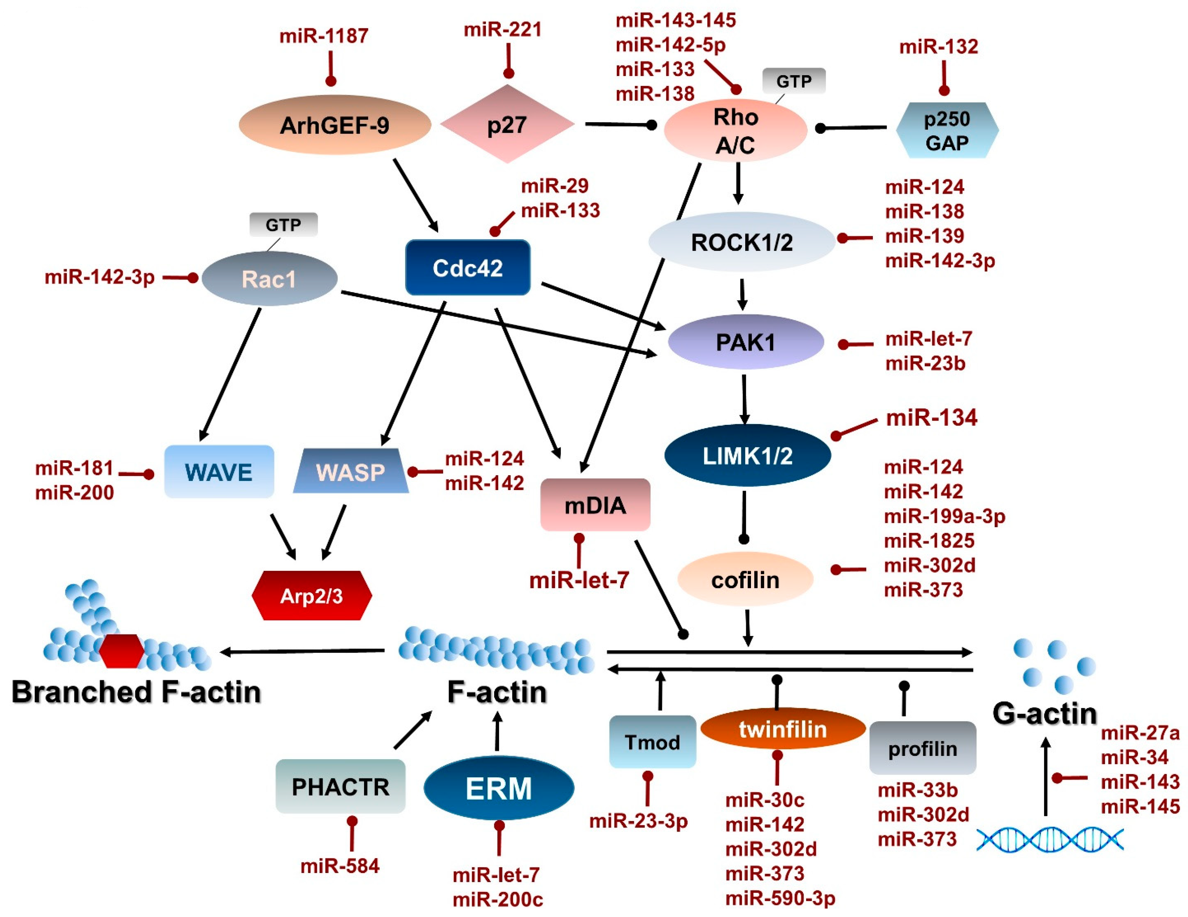
2.1. Actin Cytoskeleton

The actin cytoskeleton consists of actin filaments, along with actin accessory and regulatory proteins. In non-muscle cells, actin cytoskeletal plasticity is necessary for cell motility, differentiation, and division, and movement of intracellular organelles. In muscle cells, the actin cytoskeleton also plays a fundamental role in muscle contraction [14]. Actin filaments are assembled from globular actin (G-actin) into double helical actin filaments (F-actin), which are linear and polar, with a fast-growing plus end and a slow-growing minus end [15,16].
Actin filaments are highly plastic, and assembly and disassembly of actin filaments are highly regulated to respond to the needs of the cell. Regulatory proteins control actin filament dynamics, including nucleation, elongation, and disassembly. While spontaneous nucleation of actin filaments can occur, the process is dampened due to the sequestration or binding of actin monomers by members of the profilin family or thymosin [17]. The actin-related protein (ARP) 2/3 complex mimics actin trimer/tetramers to nucleate actin filaments [18]. Nucleation promoting factors (NPF) activate the ARP2/3 complex [19]. Formin family members can also act as nucleators, stabilizing actin dimers and recruiting profilin–actin complexes [20]. Formin family members, which bind profilin–actin complexes, also participate in elongation of actin filaments [20]. In addition, Ena/VASP family members participate in actin filament elongation [21]. Disassembly of actin filaments is facilitated by coronin, glial maturation factor (GMF), and actin depolymerizing factor (ADF)/cofilin family members [22,23]. Small GTPases also play a major role in the regulation of actin dynamics, as highlighted in a later section.

2.2. Myosin

Molecular motors, such as myosin, kinesin, and dynein, transduce chemical signals into mechanical force. Myosin moves along actin filaments to generate force in a process involving hydrolysis of ATP and sequential changes in the conformation of myosin while releasing and rebinding to actin. There are almost 40 different genes that encode for myosin heavy chains, and most cells express multiple myosin genes [24]. The specific combination of different myosin isoforms in a cell influences the organization of the actin cytoskeleton and, therefore, cell shape and movement and contractile properties, in the case of smooth, cardiac, and skeletal muscle.
Myosins have a three-domain structure: A motor domain, a neck domain, and a tail domain. While the motor and neck domains have a conserved structure, the tail domain at the C-terminal of myosin is highly divergent, enabling the many cellular functions of myosins [25]. The heterogeneity of the C-termini, sometimes described as the cargo-binding site, allows binding to a variety of molecules, including proteins and lipids. For example, myosins bind to Rab proteins for vesicular trafficking [26]. Other myosins bind to lipids to allow interaction with membranes.
All myosin heavy chains have a conserved N-terminal motor domain (also known as the catalytic head domain). The motor domain interacts with actin and the conformation of the motor domain changes with nucleotide binding. In the ADP-bound state or in the absence of nucleotide, the motor domain interacts strongly with actin. The affinity for actin decreases drastically when ATP is bound to the motor domain. In this way, the ATPase cycle regulates the association of myosin with actin filaments, and, thus, the motor function of myosins.
Myosins are classified based on the characteristics of the motor domain. Changes in the expression of myosin heavy chain isoforms can change cellular function. Mammalian myosin isoform nomenclature is based on the recommendations of Rossi et al. [27]. Class 1 myosins (MYO1A-H) have a single tail domain, which links the plasma membrane or vesicles to actin filaments [24]. While some class 1 myosins interact through adaptor proteins, others interact with lipids to modify membrane tension [28]. The MYO1 isoforms perform diverse functions, including cell crawling, actin organization on organelles, cortical tension of the plasma membrane, and endocytosis and exocytosis, and may play a role in metastasis [13,29,30,31,32]. For example, MYO1B regulates actin interactions with post-Golgi carriers and endocytic vesicles and the development of cortical tension at the plasma membrane [29,33,34]. MYO1C regulates endocytosis and exocytosis [31,35].
Class 2 myosins, classified into muscle and non-muscle isoforms, are present in all mammalian cells. Muscle myosin 2 isoforms are responsible for skeletal, smooth, and cardiac muscle contractions and are double-headed, containing 2 heavy chains and 2 light chains. Non-muscle myosin 2 isoforms (2A-C encoded by MYH9, MYH10, and MYH17) form highly dynamic short filaments [36,37] and function in a variety of activities, including cell migration, cell–cell adhesions, cell shape determination, endocytosis, and vesicular trafficking [38].
Next to the motor domain of myosin is the neck domain, sometimes called the lever, which binds myosin light chains (MLC), the essential light chains, and calmodulin and calmodulin-like proteins that stabilize the α-helical structure of this region. In smooth muscle myosin and non-muscle myosins, phosphorylation of the regulatory MLC, induced by binding of calcium to calmodulin, modulates the interaction between the myosin heavy chain and MLC. Phosphorylation of MLC plays a secondary role in skeletal muscle contraction. The phosphorylation of MLC is highly regulated by many kinases and phosphatases. The level of MLC phosphorylation, which can affect the force of contraction, results from a balance between MLC kinase (MYLK) and MLC phosphatase (MLCP). MYLK is predominantly activated by Ca2+/calmodulin, although there is evidence of regulatory phosphorylation of MYLK [39]. MLC phosphatase is a trimer consisting of a catalytic subunit (PP1c), a myosin targeting subunit (MYPT1), and a small subunit. MYPT1 has two inhibitory phosphorylation sites, T696 and T853 (human numbering); phosphorylation at these sites inhibits phosphatase activity of the holoenzyme. Several protein kinases can phosphorylate these 2 inhibitory phosphorylation sites, including Rho-associated protein kinase (ROCK), zipper-interacting protein kinase (ZIPK), and integrin-linked kinase (ILK) [40]. An endogenous inhibitor of MLCP also exists, called CPI17. Phosphorylation of CPI17 induces binding to MLCP, resulting in inhibition of phosphatase activity. Protein kinase C (PKC), ROCK, and ILK all phosphorylate CPI17 [41,42,43].

2.3. Regulation of Actin–Myosin Dynamics by Small GTPases

Small GTPases play crucial roles in regulating actin and myosin dynamics. Small GTPases cycle between the active GTP bound form and the inactive GDP form. Guanine nucleotide exchange factors (GEFs) catalyze the exchange of GDP for GTP to facilitate activation of GTPases, and GTPase-activating proteins (GAPs) promote GTP hydrolysis to inactivate GTPases, although GTPases also have intrinsic phosphatase activity. The effects of RhoA and Rac1 on cytoskeleton rearrangements have been widely established and these small GTPases interact in many cellular functions involving changes in the cytoskeleton [44]. Downstream targets of RhoA and Rac1 are Rho-associated coiled coil kinase 1/1 (ROCK) and p21-activated kinase (PAK1), which both function in actin dynamics and myosin function [44]. For example, ROCK and PAK1 phosphorylate MYPT1 to regulate MLC phosphorylation. In addition, ROCK regulates profilin and cofilin, which are involved in actin polymerization/depolymerization [45].

3. MicroRNA Overview

MicroRNAs (miRNAs) are evolutionarily conserved small regulatory RNA molecules that modulate gene expression by diverse post-transcriptional processes. Their actions depend on sequence-specific interactions of individual miRNAs with the 3′-untranslated regions of their mRNA targets. Perfect Watson–Crick complementarity is observed in 7 consecutive base pairs of the target binding region in most cases. Therefore, a single nucleotide change in this region may cause sufficient disruption in binding to deregulate the target genes. According to the latest release of the miRBase database (v22, http://mirbase.org/cgi-bin/browse.pl), the human genome contains 1917 annotated hairpin precursors and 2654 mature sequences [46]. Alles and coworkers showed that there are 2300 true human mature miRNAs, 1115 of which are annotated in the miRBase (v22) [47]. Based on the latest GENCODE (v34, https://www.gencodegenes.org/human/stats.html) data, there are 60,669 genes in the human genome, indicating that approximately 4% of human genes encode miRNA. This estimation assumes the equal distribution of miRNAs among genes, but note that a gene can code more than one miRNA [48]. Until recently, miRNAs were believed to act solely by negative regulation of target mRNA [49]; however, increasing evidence indicates that miRNAs oscillate between repression and stimulation in response to specific cellular conditions and cofactors [50]. These exciting findings, however, have made it even more difficult to explain how miRNAs regulate gene expression.
MiRNAs can be classified by their localization as intergenic and intragenic [51,52]. Intergenic miRNAs can be found between genes and they are transcribed mostly by RNA polymerase III [53]. On the other hand, intragenic miRNAs are embedded within exons or introns of protein-coding genes and are simultaneously expressed in the same orientation of their host genes by RNA polymerase II [54]. Moreover, a small percentage of miRNAs are found scattered among repetitive elements that are transcribed by RNA polymerase III [55]. The biogenesis of miRNAs starts with RNA polymerase II- or III-dependent transcription of a miRNA gene locus generating a long primary RNA (pri-miRNA).
Pri-miRNAs undergo maturation processes, including 5′-7-methylguanosine capping, splicing, and 3′-polyadenylation. They have an RNA hairpin in which one of the two strands contains the mature miRNA. The hairpin is cleaved from the pri-miRNA in the nucleus by the double-strand-specific ribonuclease, Drosha, forming precursor miRNA (pre-miRNA) [56]. Pre-miRNAs are transported to the cytoplasm with the help of Exportin-5 and further cleaved by Dicer [57] into microRNA (miRNA) [58]. Dicer belongs to the RNase III class. A second cleavage in the cytoplasm produces a double-stranded RNA, where miRNA is the antisense or guide [52]. Dicer acts in cooperation with additional proteins, such as the members of the Argonaute or AGO protein family [59], a protein activator of PKR [60], and the HIV-1 TAR RNA-binding protein [61]. These proteins are necessary components in the formation of the RISC-loading complex that modulates either the specificity or the efficiency of miRNA biogenesis. MiRNAs do not function as naked RNAs; instead, they create complexes with proteins forming a ribonucleoprotein (miRNP) [62].

4. Direct Regulation of Actin and Myosin Gene Expression by miRNAs

MiRNAs are capable of controlling the expression levels of many cytoskeletal molecules and upstream regulatory signaling modules. MiRNAs play accentuated roles in organizing diverse aspects of biochemical pathways that govern normal cellular shape, motility, and contraction. Additionally, altered miRNA expression profiles contribute to the pathogenesis of many human diseases, including cardiovascular diseases, disorders of hematopoietic cell differentiation, and cancer development [63,64]. In this review, we will summarize the current knowledge regarding the miRNAs that directly or indirectly regulate the actin cytoskeleton and the actin–myosin complex, along with their physiological and pathological relevance (Table 1, Table 2 and Table 3).

4.1. Direct Regulation of Actin Gene Expression by miRNAs

In addition to downregulating gene expression, some miRNAs induce translational upregulation [65], such as in the case of the cytoplasmic β-actin gene. Although β–actin is a constitutively and ubiquitously expressed housekeeping gene, its tissue-specific expression was found in mouse neurons. Gosh et al. [66] demonstrated that the β-actin gene (ACTB) generates two alternative transcripts with different UTR regions by alternative polyadenylation in a tissue-specific manner. The expression level of the longer transcript is relatively low but exhibits higher translational efficiency. Moreover, it harbored a conserved mmu-miR-34/34b-5p binding site. MiR-34 upregulates the mRNA encoding β-actin, although the modulatory interaction is complex. A sequence-specific anti-miRNA molecule or mutation in the β-actin miR-34 target site results in reduced expression, which is restored by a mutant miRNA complementary to the mutant target site [66] (Table 1).
One common target of miRNAs is the alpha smooth muscle actin (α-SMA), the main actin isoform in smooth muscle cells. α-SMA is often used as a marker of smooth muscle cell contractile phenotype versus proliferative phenotype and increased expression of α-SMA increases the contractile potential of smooth muscle tissue. Thus, the effects of miRNA on α-SMA are probably the most widely reported in the smooth muscle actin–myosin network (Table 1).
The intracellular domain of neuroregulin-1 (NRG-1) induces α-SMA via miR-548-f. In a complex interaction, the intracellular domain of NRG-1 recruits IKZF1 (a zinc finger protein) to the first intron of the α-SMA gene to induce circular α-SMA (circACTA) formation. CircACTA acts as a sponge for miR-548-f, relieving the repression of α-SMA expression by miR-548-f and upregulating α-SMA expression [81]. In a mouse model, miR-548-f was significantly decreased in renal arteries with intimal hyperplasia, suggesting that the dysregulation of miR-548-f may participate in intimal hyperplasia in response to vascular injury [81].
MiR-19a also influences α-SMA expression. In addition, miR-19a influences the expression of smooth muscle 22α (SM22α), an actin-associated protein, which helps to bundle and stabilize actin filaments [68,69]. Transformation with miR-19a mimics increased expression of α-SMA and SM22α, resulting in increased migration of vascular smooth muscle cells via targeting of RhoB [70], suggesting that miR-19a may play a role in the development of atherosclerosis.
The miRNA, let-7g, regulates α-SMA expression. Let-7g also regulates the expression of calponin, a calcium-binding protein that regulates actin–myosin interactions and consequently, smooth muscle contraction. Wang et al. showed that let-7g increased expression of α-SMA and calponin to maintain vascular smooth muscle contractile phenotype and, therefore, reduce the formation of atherosclerotic plaques in apoE−/− mice [67]. Platelet-derived growth factor-B (PDGF-B) promotes smooth muscle proliferative, rather than a contractile phenotype. Let-7g increases α-SMA and calponin expression via downregulation of PDGF-B and mitogen-activated protein kinase kinase (MEKK1) leading to reduced interaction of KLF4 (a zinc finger protein of the Krüppel-like family) and serum response factor (SRF), which subsequently de-represses myocardin (MYOCD) and increases α-SMA and calponin expression [67].
Micro-RNA regulation of PDGF-B may also play a role in smooth muscle phenotypic switching by the drug teniposide. Teniposide increases expression of miR-21 in vascular smooth muscle cells and partially attenuates the repressive effects of PDGF-B on SM22α and α-SMA expression [71]. Transfection of vascular smooth muscle cells with a miR-21 antagomir prevents teniposide-mediated upregulation of SM22α and α-SMA [71].
The miRNA, miR-27a binds directly to the α-SMA transcript in the 3′-UTR and suppresses α-SMA expression in mouse primary vascular smooth muscle cells [75]. Angiotensin II upregulates miR-27a and downregulates α-SMA expression. The knockdown of miR-27a prevents the angiotensin II-mediated switch of vascular smooth muscle cells from contractile to proliferative phenotype [75]. Interestingly, increased cyclic stretch induces secretion of miR-27a from vascular smooth muscle cells, which may target GRK6 in endothelial cells in a paracrine fashion [76] (Table 1 and Figure 1).

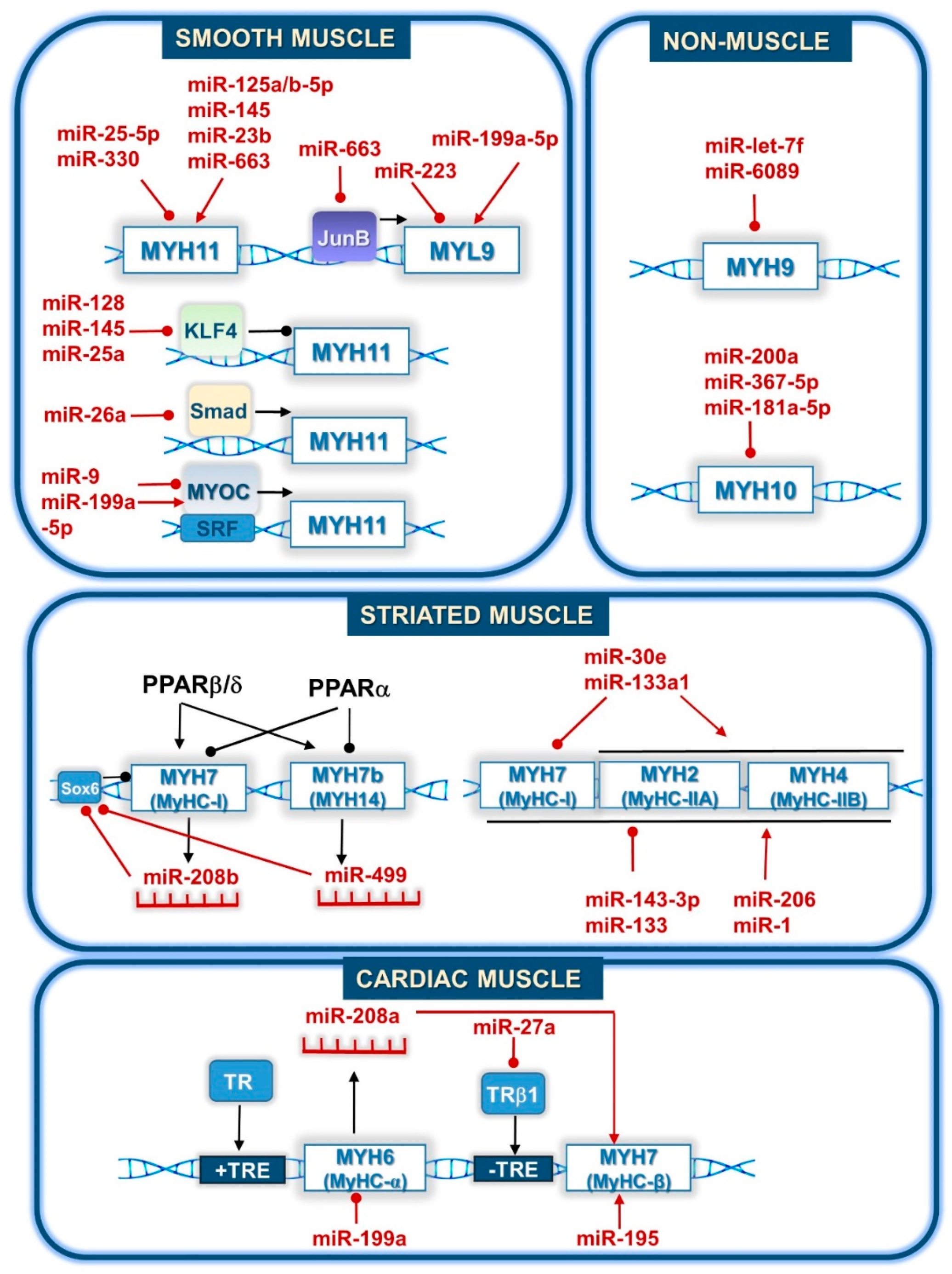
4.2. Direct Regulation of Myosin Gene Expression by miRNAs

Several miRNAs regulate skeletal muscle differentiation [83,84,85], fiber type formation [86,87], and regeneration [88] by directly regulating the expression of different myosin heavy chain (MyHC) isoforms. MiR-1- and miR-133-encoding genes form clusters that can be found on chromosomes 2 and 18 of mice. These miRNAs are absent in undifferentiated muscle cells; however, differentiation can activate their expression. MiR-1 overexpression strongly promotes myogenesis of C2C12 mouse myoblast cells by inducing the expression of MyHC, a late myogenic marker [84]. Furthermore, mutations in the seed sequences of miR-1 abolish its ability to activate myogenic gene expression. In contrast, overexpression of miR-133 inhibits MyHC expression and mutant forms of miR-133 are unable to repress the expression of MyHC [84] (Table 2 and Figure 2).
Additionally, miR-133a is involved in a skeletal muscle fiber type switch in response to exercise training [87]. Normally, this phenomenon can be characterized by an increase in the proportion of slow-twitch type I and fast-twitch type IIA oxidative fibers at the expense of fast-twitch type IIB glycolytic fibers. Nie and coworkers demonstrated that the deletion of miR-133a caused a significant loss of type IIB fibers without a relative increase in type IIX/I and type IIA fibers in mice, resulting in impaired exercise tolerance [87]. MiR-206 is exclusively expressed in skeletal muscle tissue and its expression is induced during differentiation. Transfection of C2C12 cells with double-stranded RNA duplexes, which can mimic miR-206 function, results in increased MyHC expression in the presence or absence of serum. Following 2′-O-methyl antisense oligonucleotide treatment and serum deprivation of C2C12 cells, MyHC levels are diminished due to the inhibition of miR-1 and miR-206 [83]. Both miR-143-3p and miR-30e expression are upregulated during C2C12 cell differentiation. Overexpression of miR-30e promotes C2C12 myoblast differentiation and significantly decreases the expression of myosin heavy chain type I (MyHC-I) by reducing Pgc1α mRNA levels and simultaneously increasing the expression of MyHC-IIA, IIX, and IIB-encoding genes [86]. In addition, miR-143-3p overexpression inhibits the expression of many differentiation markers, especially MyHC, whereas transfection of miR-143-3p inhibitors enhances myotube formation [85]. MiRNAs seem to be important regulators of skeletal muscle regeneration, even though the underlying molecular mechanisms need to be further investigated.
MiR-501 is a muscle-specific miRNA located in an intronic region of the voltage-sensitive chloride channel 5 (Clcn5) gene. Western blot analysis of the embryonic myosin heavy chain (MyHC-emb) revealed a significant reduction in its protein level when miR-501 was silenced. As muscle regeneration proceeds, MyHC-emb is replaced by adult MyHC isoforms, but the latter showed lower abundance due to miR-501 inhibition [88].
Among the stress-inducible miRNAs in the adult mouse heart, miR-208a plays crucial regulatory roles in pathological cardiac remodeling by controlling MyHC-β expression [95,98]. MiR-208a is a cardiac-specific miRNA that is located in an intron of the MyHC-α-encoding gene. Thoracic aortic banding (TAB) causes cardiac hypertrophy by inducing MyHC-β expression and hampering MyHC-α expression. MiR-208a−/− mice were incapable of upregulating MyHC-β in this model. Instead, MyHC-α expression was increased in miR-208a−/− mice as a compensatory mechanism. Consistently, miR-208a overexpressing transgenic mice exhibited marked and specific upregulation of the MyHC-β-encoding gene [95]. Similarly, a significant upregulation of miR-208a in type 2 diabetic mice and the human heart was associated with an increase in MyHC-β expression [98]. MiR-208a also controls the expression of Myh7b (also referred to as MYH14) and its intronic miRNA, miR-499, in a precise, stoichiometric manner, even in the absence of stress. Myh7b/miR-499 expression was decreased by 50% in the hearts of miR-208a+/- mice and was terminated in the hearts of miR-208a−/− mice [99].
MiR-195 also belongs to the stress-inducible miRNAs in the heart. Real-time PCR analysis of miR-195 overexpressing cardiac tissue showed dramatic upregulation of MyHC-β expression in transgenic mice leading to cardiac failure [93]. Additionally, forced expression of miR-199a in rat primary cardiomyocytes led to the downregulation of MyHC-α mRNA levels by 80%, while MyHC-β expression was not affected, indicating that miR-199a may influence cardiac function through the regulation of cardiomyocyte contractile proteins [94].
MiRNAs can also be found in melanocytes and are associated with changes in melanosome transport [92] and melanin secretion [91]. Myosin 5A (MYO5A) forms a tripartite complex together with Rab27a and melanophilin (Mlph) that is responsible for transporting melanosomes along actin filaments. Luciferase reporter assays showed that miR-145 directly binds to MYO5A in mouse and human melanocytes. Overexpression and downregulation of miR-145 reduce and increase the expression of a set of pigmentation genes including MYO5A at the mRNA and protein levels in melan-a cells [92]. MiR-143-5p targets the 3′-UTR of the MYO5A transcript [91]. Upon silencing miR-143-5p with a short tandem target mimic (STTM)-miR-143-5p, MYO5A expression was significantly increased along with Mlph and Rab27a in mouse melanocytes, suggesting a stable interaction between them [91]. Dual-luciferase reporter assays in 293T cells demonstrate that miR-143-5p does not bind to the mutant form of MYO5A as a consequence of reduced complementarity [91]. Based on these data, adequate MYO5A expression is necessary for proper melanosome transport and melanin secretion.
Thyroid hormone (TH) has a fundamental role in determining skeletal muscle fiber type composition. Recently, miRNAs that are targeted by TH in striated muscles have been identified [89,90]. Zhang and coworkers demonstrated that miR-133a1, which controls muscle fiber type specification, is a direct target gene of TH [90]. Overexpression of miR-133a in soleus (SOL) muscles and C2C12 myotubes decreases the expression of MyHC-I with a concomitant increase in MyHC-II, while TEA domain family member 1 (TEAD1) overexpression has the opposite effect. Overexpression of TEAD1 without its 3′ UTR significantly antagonizes the effect of miR-133a mimics. By contrast, TEAD1 with its 3′ UTR shows an attenuated effect, indicating that TEAD1 mediates miR-133a effects in the regulation of muscle fiber type composition [90].
TH can also exert effects through thyroid hormone receptors (THRs). MiR-27a was found to upregulate MyHC-β expression but not MyHC-α expression in neonatal rat ventricular myocytes (NRVMs) via thyroid hormone receptor β1 (THRβ1) [89]. TH treatment along with THRβ1 overexpression enhances the downregulation of MyHC-β, whereas in the absence of TH or the presence of miR-27a decoys, MyHC-α expression does not change. THRβ1 overexpression also leads to the suppression of MyHC-β protein levels under serum-containing conditions, while MyHC-α levels remained unaltered. The same result was obtained using miR-27a decoys, suggesting that miR-27a can regulate MyHC-β expression by targeting THRβ1 [89].
MiR-499 is encoded by the ancient MYH7b gene, and transcriptionally regulates the expression of its host gene via Sox6, a transcriptional repressor [97]. In the presence of miR-499, the activity of a luciferase reporter gene carrying 1 kb of the mouse Sox6 3′ UTR region was reduced by 60% compared to the control. Additionally, endogenous Sox6 mRNA levels were decreased due to miR-499 overexpression in cardiomyocytes. On the other hand, overexpression of Sox6 in C2C12 myoblasts resulted in a decrease in MYH7b expression [97]. Taken together, it can be assumed that miR-499 can control the expression of its host gene and, therefore, its expression, by targeting Sox6.
PPARβ/δ and PPARα exert opposing effects on the type I skeletal muscle fiber program through a downstream regulatory circuitry, which consists of miRNAs [96]. MiR208b, which is encoded by the MYH7 gene, and miR-499, which is encoded by the MYH7b gene, exert their effects on the same mRNAs to downregulate transcriptional repressors of the type I skeletal muscle fiber program, such as Sox6 [97,99]. MiR-208b/MYH7 and miR-499/MYH7b expression are markedly increased in muscle creatine kinase (MCK)-PPARβ/δ muscle but are undetectable or significantly reduced in MCK-PPARα muscle. Antisense-mediated inhibition of miR-208b or miR-499 alone does not affect MyHC expression, although inhibition of both miRNAs causes a dramatic decrease in MYH7 mRNA in wild type (WT) myotubes and blocks the enhancing effects of PPARβ/δ on slow-twitch gene expression in MCK- PPARβ/δ myotubes. Moreover, crossing MCK-miR-499 mice with an MCK-PPARα line leads to the prevention of PPARα-mediated repression of the type I muscle fiber program [96]. Collectively, this evidence suggests that PPAR signaling is upstream of miR-208b/MYH7 and miR-499/MYH7b, and that PPARβ/δ activates, whereas PPARα suppresses their expression in muscle (Table 2 and Figure 2).
Non-muscle myosin II (NMII) functions in cellular organization, motility, cell shape, and polarity, and altered NMII activity contributes to a variety of disease pathologies. Non-muscle myosin IIA (NMIIA) consists of non-muscle myosin heavy chain IIA (NMHCIIA), encoded by the MYH9 gene, and regulatory and essential light chains [100]. Let-7f acts as a tumor suppressor to inhibit invasion and metastasis in gastric cancers via direct targeting of the tumor metastasis-associated gene, MYH9 [101]. MiR-6089 also targets MYH9 and its overexpression suppresses ovarian cancer cell proliferation, migration, invasion, and metastasis in vivo and in vitro, and miR-6089 negatively correlates with MYH9 expression in clinical samples. The proposed mechanism is an miR-6089/MYH9/β-catenin/c-Jun negative feedback loop [102]. Non-muscle myosin IIB (NMIIB) consists of NMHCIIB, encoded by the MYH10 gene, and regulatory and essential light chains. In migratory cells, NMIIB preferentially localizes to the rear of the cell [100]. MiR-200a directly targets NMHCIIB and the overexpression of NMHCIIB partially rescues miR-200a-mediated inhibition of cell migration, as well as cell growth in vitro and in vivo. Moreover, siRNA-mediated silencing of NMHCIIB expression inhibits meningioma tumor growth in mice [103]. MiR-200a also targets and downregulates the MYH10 gene product in nasopharyngeal carcinoma [104]. MyRepress, a new technology that models gene expression of miRNAs in non-conventional binding sites revealed another two miRNAs targeting MYH10: miR-181a-5p targets one non-conventional target site in the coding region of MYH10, while miR-367-5p was identified as a repressor of MYH10 expression in MCF7 cells [105]. Non-muscle myosin IIC, the most recently discovered non-muscle myosin isoform, consists of NMHIIC and is encoded by MYH14 [100]. To our knowledge, no information has been published yet concerning the miRNA regulation of this isoform or about the myosin light chain component of NM.

5. Physiological and Pathological Processes Related to miRNA-Regulated Pathways

The actin–myosin network is present in all eukaryotic cells and is highly plastic, facilitating alterations in response to changing cellular conditions or responses to external stimuli. Because of the wide variety of cellular activities in which the actin–myosin network participates, virtually all physiological processes are affected. A growing body of evidence demonstrates that miRNAs regulate the plasticity of the actin–myosin network under both physiological and pathological conditions. We will review only a few of these processes impacted by miRNA regulation, including smooth muscle function and vascular diseases [72,73,74,77,78,79,80,82,106,107,108,109,110,111,112,113,114,115,116,117,118,119,120,121,122,123,124,125,126,127,128,129], hematopoiesis [130,131,132,133,134,135,136,137], podocyte biology [138,139], osteoblast differentiation [140,141,142,143], cilia assembly [144,145,146,147,148], cancer formation [127,149,150,151,152,153,154,155,156,157,158,159,160,161,162,163,164,165,166,167,168,169,170,171,172,173], leukocytes and lymphocytes in action [174,175,176,177,178], and cardiovascular disorders [93,179,180,181].

5.1. MiRNAs in Smooth Muscle Function and Diseases

In smooth muscle cells, the actin–myosin network plays a crucial role not only in smooth muscle contractile activity, but also in smooth muscle cell motility, intracellular processes, and proliferation. Disruption of these processes can have serious pathological consequences leading to disease development. In vascular smooth muscle, the pathological consequences of disrupting actin–myosin dynamics include vascular restenosis, hypertension, and atherosclerosis [116]. Recently, a number of publications have revealed the role of miRNAs in regulating smooth muscle function via direct (discussed in the previous section) or indirect targeting of the actin–myosin network. Understanding the effects of miRNAs can lead to the identification of new drug targets for treating smooth muscle-related diseases.
In addition to the direct effects of miRNAs on smooth muscle actin and myosin (as described above), miRNAs also affect the expression of proteins that regulate the actin–myosin network in smooth muscle cells. The crucial role of miRNAs in smooth muscle function is demonstrated by Dicer knockouts. If Dicer is deleted during embryogenesis, severe hemorrhage causes 100% lethality at E16.5–E17.5 [108]. Conditional smooth muscle specific deletion of Dicer in adult mice results in decreased contractile function and a profound reduction in blood pressure [107].
Interestingly, not only do miRNAs influence actin–myosin dynamics, but actin–myosin dynamics affect miRNA levels also. In a complex feedback mechanism, actin polymerization is thought to regulate the levels of a number of miRNAs to precisely regulate contractile and cytoskeleton protein levels in response to cellular needs. Myocardin-related transcription factors (MRTFs) can shuttle between the cytoplasm and the nucleus. In the cytoplasm, MRTFs bind to G-actin, preventing nuclear translocation of these transcription factors [119,120]. Upon actin polymerization into F-actin, MRTFs are released and shuttled to the nucleus, where they interact with serum response factor (SRF) [119,120]. Alajbegovic and colleagues showed that in conjunction with MRTF, actin polymerization in human and mouse vascular smooth muscle cells regulates miR-1, miR-22, miR-143, miR-145, and miR-378a [106]. Increased aortic dilation causes actin depolymerization and reduced expression of these miRNAs [106], implying that these miRNAs may play a role in vascular diseases, such as aortic aneurysms.
Much of the effects of miRNAs on smooth muscle function are elicited by driving smooth muscle cells towards either a proliferative/synthetic phenotype or a differentiated/contractile phenotype. Dedifferentiated vascular smooth muscle cells (VSMCs) possess increased rates of proliferation and migration, as well as reduced expression of differentiation markers, such as α-SMA, SM22α, and smooth muscle myosin heavy chain (MYH11). The development of vascular diseases, such as post-angioplasty restenosis, atherosclerosis, and hypertension can be ascribed to pathological smooth muscle cell phenotype switching, which is modulated in numerous ways by miRNAs. The diverse molecular mechanisms regulating SMC differentiation by miRNAs involve the direct or indirect regulation of transcription factors, including serum response factor (SRF), myocardin (MYOCD), myocardin-related transcription factors (MRTFs), and the Krüppel-like zinc finger family (KLF). SRF, MYOCD, and MRTFs induce the differentiated contractile protein expression in smooth muscle cells. In contrast, the Krüppel-like zinc finger family (KLF) induces the proliferative smooth muscle phenotype [118] (Figure 1 and Table 3).
MiR-143/145, which is located in a non-protein coding region, is widely expressed in smooth muscle cells, and, thus, has been extensively studied with respect to the smooth muscle phenotype. Genetic deletion of miR-143/145 results in a more proliferative smooth muscle cell phenotype versus a contractile phenotype, indicating that miR-143/145 favors the contractile phenotype [109]. The miR-143/145 cluster is regulated by MYOCD and SRF [77]. Knockdown of miR-143/145 results in disruption of stress fiber formation, indicating that miR-143/145 plays a crucial role in cytoskeleton assembly [77]. Among the targets for miR-143/145 that disrupt actin assembly are MRTF-B, β-actin, cofilin, KLF5, MYOCD, and ROCK1 [77,78]. Knockdown of miR-143/145 results in reduced neointimal formation and the inability to respond to vascular injury [77]. In addition, miR-145 expression is reduced in vascular smooth muscle cells from patients with atherosclerosis, supporting a role for miR-143/145 in the development of atherosclerosis [78]. The miR-143/145 cluster may also mediate an increased risk of vascular diseases induced by hyperglycemia [114]. Glucose induces the expression of smooth muscle differentiation, including calponin and SM22α, and genetic ablation of miR-143/145 ameliorated these effects [114]. In addition to the effects on vascular smooth muscle cells, miR-145 has similar anti-proliferative effects in corpus cavernosum smooth muscle cells [117].
MiR-23b [72] and miR-125a-5p [73] are highly expressed in VSMCs and are downregulated after vascular injury in vivo. Their overexpression is sufficient to reduce VSMCs proliferation and migration, and to promote the expression of selective VSMCs marker genes, such as α-SMA, MYH11, and SMA22α [72,73]. Over-expression of miR-23b decreases neointimal formation induced by a balloon injury. MiR-125a-5p is downregulated in response to PDGF-BB and targets ETS-1. These studies suggest that regulation of miR-23b and miR-125a-5p contribute to restenosis.
In vitro functional studies showed that overexpression of two miRNAs, miR-330 and miR-125b-5p, have opposing actions on the renin lineage of vascular smooth muscle cells. Under normal conditions, miR-125b-5p was expressed in arteriolar SMCs and in juxtaglomerular (JG) cells, but after reacquisition of the renin phenotype, miR-125b-5p was downregulated in arteriolar SMCs. MiR-330, normally absent, is expressed in JG cells under stress and inhibits contractile characteristics of these cells, favoring their endocrine character. In silico analysis showed that miR-330 and miR-125b-5p have potential binding sites in smoothelin, calbindin 1, MYH11, α-SMA, and renin genes [80].
MiR-182 is downregulated during the dedifferentiation of rat aortic smooth muscle cells in culture [79]. On the other hand, transfection of vascular smooth muscle cells with miR-182 increased the expression of α-SMA, SM22α, and calponin and, consequently, pushed the smooth muscle cells towards the contractile phenotype and inhibited dedifferentiation, proliferation, and migration [79]. MiR-182 targets the 3′-UTR of fibroblast growth factor 9 (FGF9) to downregulate FGF9 expression, which in turn affects the expression of the smooth muscle specific proteins [79,128]. In addition, FGF9 upregulates platelet-derived growth factor receptor B (PDGFRB), which is necessary for smooth muscle cell proliferation and migration. Thus, miR-182 may play a role in vascular proliferative diseases, such as atherosclerosis.
PDGF also pushes VSMCs towards the proliferative phenotype and promotes migration into the neointimal layer after arterial injury via miR-663 and miR-26a. MiR-663 is significantly downregulated in human aortic VSMCs upon PDGF-BB-treatment, whereas miR-663 expression is markedly enhanced during VSMC differentiation. Overexpression of miR-663 results in increased expression of the molecular markers for VSMC differentiation and reduced expression of JunB and myosin light chain 9 (MYL9) [82]. MiR-26a, another regulator of PDGF-BB-mediated VSMC phenotypic switch is significantly increased in the PDGF-BB-stimulated VSMC model and in arteries with neointimal lesion formation. MiR-26a suppresses Smad1 expression, leading to VSMC switching to the proliferative/synthetic phenotype and eventually to vascular remodeling by downregulating VSMC differentiation marker genes [74].
During the formation of atherosclerotic plaques in mice, a parallel increase in expression levels of MYLK and miR-92a was observed. The activation of ROCK/STAT3 and/or MYLK/STAT3 may upregulate miR-92a expression, which subsequently inhibits KLF4 expression and promotes PDGF-BB-mediated proliferation and migration of VSMCs [186].
MiR-128-1 is an intronic miRNA encoded for by two distinct genes, miR-128-1 and miR-128-2, which are located in the introns of R3HDM1 (R3H domain containing 1) and RCS (ARPP-21, cyclic AMP-regulated phosphoprotein, 21 kDa) [110]. MiR-128 modulates the vascular smooth muscle phenotype through targeting KLF4. KLF4 modulates the methylation of MYH11 (encoding for myosin heavy chain 11, a smooth muscle myosin), preventing its transcription [112]. Increased miR-128 expression correlates with the contractile phenotype. In a mouse model of carotid stenosis, lentiviral delivery of miR-128 suppressed intimal hyperplasia [112]. Thus, altered miR-128 expression may also play a role in vascular proliferative diseases and may be a potential target for treating these diseases.
MiR-223 is an evolutionarily conserved miRNA located on the X chromosome [115,124]. In pulmonary artery smooth muscle cells, hypoxia downregulates miR-223, resulting in increased proliferation of the smooth muscle cells [129]. In vivo, a miR-223 antagomir attenuates hypoxia-induced increases in pulmonary artery hypertension [129]. MiR-223 targets RhoB, resulting in decreased RhoB expression, decreased ROCK activity, and, subsequently, inhibition of MYPT1 phosphorylation. In addition, miR-223 targets the 3′-UTR of MLC to inhibit MLC expression [129]. Overall, hypoxia-induced downregulation of miR-223 induces downregulation of the RhoB/ROCK pathway leading to pathological changes in pulmonary artery smooth muscle cells; thus, miR-223 may be a target to treat pulmonary artery hypertension.
Transfection of bronchial smooth muscle cells with a miR-133a antagomir upregulates RhoA expression [111]. Human and mouse RhoA have a putative binding site for miR-133a in the 3′-UTR; thus, miR-133a may directly regulate RhoA in bronchial smooth muscle cells [111]. Interleukin-13 downregulates miR-133a, resulting in upregulation of RhoA and, consequently, the increased bronchial smooth muscle contractility present in allergic asthma [125]. Similarly, hypercapnia also upregulates RhoA in airway smooth muscle cells, and this upregulation was prevented by transfection with a miR-133a mimic [126].
Compared to vascular smooth muscle cells, much less has been published on the role of miRNAs in organ smooth muscle phenotype and regulation of the actin–myosin network. The importance of miRNAs in gastrointestinal smooth muscle function is demonstrated by the deletion of Dicer, which causes downregulation of contractile genes in gastrointestinal smooth muscle cells [123]. Similar to vascular smooth muscle, SRF regulates differentiation of smooth muscle cells via several miRNAs in the gastrointestinal tract, including miR-199a-3p, miR-214, miR-143, and miR-145 [122]. MiR-143 and miR-145 are upregulated in the intestinal smooth muscle of patients with gastroschisis compared to patients with atresia, and are associated with contractile dysfunction [113,121] (Figure 1 and Table 3). In cultured bladder smooth muscle cells, cytoskeleton remodeling and loss of contractility occur in conjunction with the upregulation of miR-199a-5p [113]. WNT2 is targeted by miR-199a-5p, which increases the expression of smoothelin and SM22 via inhibition via WNT signaling [113].

5.2. MiRNAs in Cardiovascular Diseases

The heart responds to both physiological triggers of increased demand, e.g., exercise, and pathological triggers, e.g., hypertension and valvular diseases, by enlarging the myocardium via cardiac muscle cell hypertrophy. Several recent publications demonstrate the role of miRNAs in the development of cardiac hypertrophy. In fact, a signature pattern of miRNAs is upregulated (miR-21, miR-23a, miR-125, miR-195, miR-199, and miR-214) or downregulated (miR-1, miR-29, miR-30, miR-133, and miR-150) during the development of cardiac hypertrophy [93]. The in vitro expression of miR-133 and miR-1 inhibits the development of cardiac hypertrophy, and miR-133 and miR-1 are downregulated in murine models and patients with myocardial hypertrophy [179]. The two major targets of miR-133 relevant to cardiac hypertrophy development are RhoA and Cdc42 [179]. Both RhoA and Cdc42 are associated with cytoskeletal and myofibrillar rearrangements during hypertrophy.
Several miRNAs can induce cardiomyocyte proliferation promoting cardiac regeneration. The most effective miRNAs activate nuclear localization of Yes-associated protein (YAP) transcriptional cofactor and induce expression of YAP-responsive genes. Besides targeting the Hippo pathway by miR-199, several pro-proliferative miRNAs inhibit filamentous actin depolymerization by targeting cofilin-2 [181]. In particular, a shift was observed between F- and G-actin toward the polymerized state in response to overexpression of miR199-3p, miR-302d, miR-373, and miR-33b. Cardiomyocytes showed rounded morphology and formed cortical layers of actin. The miRNAs acted by directly downregulating cofilin-2 [181], which activated YAP nuclear translocation and stimulated cardiomyocyte proliferation. Thus, actin cytoskeleton dynamics are a strong activator of YAP in response to mechanical cues, facilitating adaptation to the extracellular environment [180]. Several other proteins regulating the actin cytoskeleton were predicted by computational algorithms to be regulated by miR590-3p, miR-302d, miR373, miR-199a, miR33b, miR-302d, and miR-373, including twinfilin-1 and-2, thymosin, and profilin-2 [181].

5.3. Regulation of Actin Cytoskeleton by miRNAs in the Hematopoietic System

MiRNAs regulate hematopoiesis and the function of both myeloid and lymphoid progeny by several mechanisms. MiRNAs play a vital role in every stage of hematopoiesis. Our major focus will be those miRNAs that target actin and myosin cytoskeleton affecting the formation, development, and differentiation of blood cells, as well as their pathological relevance [137].
Numerous miRNAs participate in the process of erythropoiesis, but their major targets are the regulators of erythroid homeostasis, γ-globin gene expression, and erythroid markers [137]. Fluid shear stress is one of the major factors that promotes the maturation of erythrocytes by acting on the membrane skeleton, which requires actin cytoskeletal remodeling, creating its physiologically functioning hexagonal topology. MiR-23-3p is suppressed by shear stress leading to the upregulation of erythrocytes tropomodulin of 41 kDa (E-Tmod41), which caps the end of filamentous actin, and induces F-actin cytoskeleton remodeling and erythroid differentiation [136].
MiR-142 also plays a crucial role as a broad hematopoietic pro-differentiation factor and is involved in megakaryogenesis [131], B-cell leukemogenesis [132], and dendritic cell development [135]. Megakaryopoiesis requires distinctive cytoskeletal rearrangements, including the cytoplasmic protoplatelet protrusions to bend and bifurcate, leading to the proper biosynthesis of thrombocytes and their release into the bloodstream [130]. Genetic ablation of miR-142 impairs megakaryocyte maturation, inhibits polyploidization, and induces abnormal proplatelet formation and thrombocytopenia [131]. Based on the study of Hornstein et al., actin cytoskeletal dynamics are disturbed, and more homogenous and circular F-actin structures are formed in miR-142-depleted cells relative to the control. Pivotal actin cytoskeleton-associated proteins were identified as direct targets of miR-142, including twinfilin-1, cofilin-2, integrin alpha V, glucocorticoid receptor DNA binding factor 1, and Wiskott–Aldrich syndrome-like (Wasl) [131] (Figure 1 and Table 3).
The miR-181 family is an important regulator of T cell development, proliferation, and activation [133]. MiR-181c reduces the SCAR/WAVE actin-nucleating complex unit (BRK1) protein expression level. BRK1 is a member of the WAVE protein family. The miR-181-BRK1 axis is required for actin polymerization and, thus, for lamellipodia formation in T lymphocytes. Moreover, overexpression of miR-181c leads to severe impairment of actin polymerization in response to stimulation and a marked reduction in the amount of F-actin at the T cell-B cell contact site, proving its importance in immunological synapse formation [133].
The homeostasis of mast cells of the innate immune system is also modulated by miRNAs. Monticelli and coworkers showed that miR-221 influences the effector functions and actin cytoskeleton of mast cells, but does not affect their differentiation. They proposed a model in which miR-221 has a dual function. In resting cells, miR-221 maintains normal mast cell homeostasis via cell cycle regulation and cytoskeleton targeting of Cyclin-dependent kinase inhibitor 1B (p27Kip1), CD25, and actin. In contrast, upon mast cell stimulation miR-221 influences the extent of degranulation and cytokine production in response to IgE-antigen complexes. Overexpression of miR-221 resulted in a thicker F-actin ring formation compared to control cells. In contrast, the depletion of miR-221 reduced the amount of F-actin [134]. The major target of miR-221 is p27Kip1, which is not only a cell cycle inhibitor but is also important in cell motility, suggesting its synergistic effect on the cytoskeletal changes in mast cells (Figure 1 and Table 3).

5.4. Actin Regulation in Podocyte Biology

Podocytes cover the external surface of the glomerular capillary by a sophisticated web of primary and secondary ramifications. The maintenance of the dynamic actin–myosin cytoskeletal architecture is crucial in the biology of podocytes since not only the shape and function, but the foot process effacement and microvillus transformation, are determined by alterations in actin [138]. Brain-derived neurotrophic factor (BDNF) is a pleiotropic neurotrophin found to bind to the tropomyosin-related kinase B (TrkB) in podocytes. BDNF downregulates miR-134, directly augmenting LIMK1 translation. Upregulated miR-132 reduces p250GAP translation, resulting in an attenuation of the blockade of Rac1. In turn, miR-132 increases LIMK1 expression and phosphorylation, which affects cofilin activity and favors actin polymerization [139].

5.5. MiRNA Regulation of the Actin Cytoskeleton in Osteoblast Differentiation

Bone morphogenic protein II (BMP-II) is a signaling molecule of the TGF- β superfamily that regulates the actin cytoskeleton by acting on miRNAs. BMP plays a crucial role in bone formation and development [141] by binding to BMP receptors (BMPR) followed by activation of SMAD proteins or MAPKs [143]. MiR-1187 is a robustly downregulated miRNA in medicarpin-stimulated osteogenesis. Overexpression of miR-1187 results in decreased actin polymerization, cortical protrusion formation, and osteoblast mineralization. MiR-1187 is a repressor of BMPRII and the Cdc42 guanine nucleotide exchange factor 9 (ARhGEF-9) expression, which is involved in the activation of Cdc42, leading to actin cytoskeleton rearrangement [142]. The suggested mechanism for the negative regulation of osteogenic differentiation is that miR-1187 represses BMPR-II and ArhGEF-9, thereby suppressing the non-SMAD/Cdc42 signaling pathway leading to the inhibition of p21- activated kinase (PAK1) and LIMK phosphorylation and the activation of cofilin, thus inhibiting actin polymerization and cytoskeletal rearrangement [140].

5.6. MiRNAs as Key Regulators in Actin Reorganization in on Cilia Assembly

Another process, in which actin dynamics is highly controlled, is cilia assembly. Cilia assembly is crucial to several biological processes, including fluid flow, cell movement, and sensory perception in organisms ranging from protozoa to mammals [187]. Cilia are microtubule-based, micrometer-scale, whip-like organelles that can form motile cilium on multiciliated cells (MCCs) on the luminal surface of airways, cerebral ventricles, oviducts, and the efferent ducts of testis. Multiciliogenesis is characterized by several steps. First, MCC precursors exit from the cell cycle, followed by multiplication of centrioles, then the apical actin cytoskeleton is reorganized into a cortical meshwork of actin, and, finally, the synthesized centrioles migrate towards the apical pole of the cell, where they anchor to the actin meshwork and mature into basal bodies from which a cilium can be elongated [147]. MiR-34/449 miRNAs are key regulators of vertebrate multiciliogenesis that directly repress the expression of multiple proteins. MiR-34/449 downregulates the expression of several cell cycle-regulated genes and members of the Notch pathway, which enhance miR-449 expression and facilitate entry into MCC differentiation. On the other hand, micro-RNA34/449 appears to be a negative regulator of CP110, which is involved in basal body maturation as a protein-blocking ciliation [148]. Lastly, miR-34/449 controls the apical actin meshwork formation by targeting components of the small GTPase pathways directly repressing the small GTPase R-Ras during multiciliogenesis [146].
Another candidate in the control of multiciliogenesis is miR-129-3p, which represses multiple CP110 and actin regulators critical for cilia formation. Upon overexpression of miR-129-3p, RPE1 cells exhibited a disorganized actin cytoskeleton with amorphous aggregates and speckles compared to control cells with parallel arrays of bundled filamentous actin or stress fibers. MiR-129-3p facilitates basal body formation by repressing CP110 expression. As a parallel function, miR-129-3p inhibits ciliogenesis and axoneme growth by hampering F-actin formation via targeting and downregulating gene expression of ARP2 (core subunit of ARP2/3 complex), TOCA1 (Cdc42 effector), ABLIM1, and ABLIM3 (homologous F-actin binding proteins) [144,145]. This further establishes miR-129-3p as a central player in multiciliogenesis, acting at several distinct levels of this complex physiological process (Figure 1 and Table 3).

5.7. Signaling Pathways of miRNAs-Regulated Cancer Formation

Regulation of the actin cytoskeleton is involved in cancer cell migration, tumor invasion, and metastasis [170]. MiRNA dysregulation is implicated in the development and progression of nearly all tumor types and some miRNAs can function as tumor suppressors (Figure 1 and Table 3).
MiR-124 serves as a tumor suppressor in various cancers, including breast and prostate cancers [164,173]. MiR-124 is also involved in the development of liver cancer; miR-124 suppresses aggressive hepatocellular carcinoma by repressing Rho-associated kinase 2 and the zeste polycomb repressive complex 2 subunit [172]. A systematic analysis of the molecular mechanism of miR-124 revealed that elements of the ROCK signaling pathway are regulated by the overexpression of miR-124 in hepatocarcinoma cells. In addition, miR-124 regulates a total of 17 genes, which are significantly associated with the actin cytoskeleton pathway, including talin, cofilin 2, Wiskott–Aldrich Syndrom (WAS) protein family member 1 (WASF1), and CFL2, an intracellular actin-modulating protein that regulates actin-filament dynamics [168]. The overexpression of miR-124 represses cell migration and invasion of intrahepatic cholangiocarcinoma cells [171]. An et al. showed that miR-124 is downregulated in malignant glioma cells and its target is the ROCK1 gene. Overexpression of miR-124 reduced ROCK1 expression and suppressed glioma cell invasion by affecting actin cytoskeleton rearrangement and reducing cell surface ruffles [65].
Another candidate in the regulation of the actin cytoskeleton is miR-1181, which is downregulated in pancreatic cancer cell lines compared to normal ductal epithelial cells. The overexpression of miR-1181 inhibited the migration, invasion, and proliferation of PANC-1 pancreatic cancer cells and resulted in decreased expression of F-actin and β-tubulin. Furthermore, miR-1181 inhibits the expression of signal transducer and activator of transcription 3 (STAT3) [127].
The development of thyroid carcinoma is also regulated by miRNAs. Has-miR-196a-2 is overexpressed in thyroid carcinoma according to a bioinformatics analysis. A Gene Sets Enrichment analysis suggested that has-miR-196a-2 is enriched not only in the adherens junction, but in the actin cytoskeleton as well [160], and that has-miR-196a-2 expression is closely related to the invasion and migration of thyroid carcinoma cells due to the upregulation of the WNT pathway [151].
Rho superfamily signaling is controlled at the level of expression of downstream effectors of small GTPases, such as RhoA or C. Both the hyperactivation of ROCK and the downregulation of its upstream regulators have been described in numerous cancer types. ROCK is targeted by miR-138 [156] and miR-139 in tongue squamous cell carcinoma (TSCC) and hepatocellular carcinoma (HCC), respectively. Reduced miR-138 levels correlate with increased expression of RhoC and ROCK2, which results in altered, elongated cell morphology, enhanced cell stress fiber formation, and accelerated cell migration and invasion in TSCC [156]. MiR-139 interacts with the 3’ untranslated region of ROCK2 and reduces its expression in HCC cells. Levels of miR-139 inversely correlate with ROCK2 protein in human HCC samples. The expression of miR-139 is reduced in human metastatic HCC samples and correlates with prognosis [169].
Ultimately, signaling by many Rho GTPases via numerous downstream actin-regulating proteins control the dynamics of actin polymerization in cancer. MiR-31 [166] and miR-200 [165] are capable of suppressing the WASP-family protein member 3 (WAVE3). The expression of WAVE3, an actin cytoskeleton remodeling and metastasis promoter protein, showed a clear inverse correlation with miR-31 and miR-200 in invasive versus non-invasive cancer cells [165].
The downregulation of several miRNAs modulates cytoskeletal dynamics in breast cancer. These miRNAs primarily affect the upstream regulators of the actin cytoskeleton and myosin modulating cell spreading, the formation of lamellipodia, and cell invasion. The first miRNA-target mRNA pair to be verified in vivo was let-7 miRNA, which negatively regulates target gene expression by either mRNA cleavage or translational repression. Let-7 is the most frequently and significantly associated miRNA with clinical outcomes in patients with cancer [162]. Let-7b is reduced in patients with metastatic breast cancer correlating with cell migration and invasion by affecting focal adhesion and stress fiber stabilization. Let-7b directly represses multiple genes involved in the actin cytoskeleton pathway, including PAK, radixin (RDX), integrin beta-8 (ITGB8), and diaphanous-related formin 2 (DIAPH2). However, the expression of actin remains the same, suggesting that the actin cytoskeleton modulation occurs through the downregulation of its upstream effectors [155].
Another miRNA that regulates the actin cytoskeleton through the regulation of PAK in breast cancer is miR-23b. Clinically, low miR-23b correlates with the development of metastases in breast cancer. Expression of miR-23b increases F-actin size and cell spreading as well as the formation of lamellipodia. MiR-23b inhibits a large number of cytoskeletal genes, including LIMK2, CFL2, and PIK3R3, by direct interaction with their 3′UTRs. In contrast, miR-23b downregulates PAK2 by an indirect mechanism and decreases the phosphorylation of myosin light chain (MLC) II without modulating its gene expression [163]. Liu et al. identified miR-30c as a potential regulator of the actin cytoskeleton in breast cancer. MiR-30c functions as a tumor suppressor acting directly on twinfilin 1 actin-binding protein and vimentin. Interleukin-11 is a downstream target of twinfilin 1, which regulates STAT3 phosphorylation, causing invasion of cancer cells [149].
A major player in the progression of breast cancer is transforming growth factor- β (TGF- β), which promotes epithelial-to-mesenchymal transition, migration, invasion, and metastasis [161]. TGF- β silences the expression of miR-584, resulting in enhanced protein phosphatase and actin regulator 1 (PHACTR1) expression, leading to actin rearrangement and breast cancer cell migration [150].
One of the most predominant miRNAs regulating the formation of stress fiber formation through diverse signaling pathways is miR-200. The expression of miR-200 is often downregulated in breast metastases and the reduced expression is associated with poor prognosis in breast cancer [152]. MiR-200c downregulates the transcriptional repressors, ZEB1 and SIB/Zeb2 [153], resulting in the upregulation of E-cadherin and the inhibition of cell motility in cancer cells [159]. However, miR-200c also acts on actin remodeling by directly targeting moesin, fibronectin 1 [154], formin homology 2 domain containing 1 (FHOD1), and Mg2+/Mn2+-dependent protein phosphatase 1F (PPM1F) [157]. FHOD1 is an actin-nucleating protein which induces the formation of actin filaments by activating serum response factor (SRF), a key regulator of the actin cytoskeleton and contractile processes, resulting in increased MLC expression. Phosphorylation of MLC also contributes to stress fiber formation through cross-linking actin filaments. FHOD1-stimulated actin nucleation also contributes to stress fiber formation, while PPM1F increases the phosphorylation level of MLC independent of MLC expression [157]. PPM1F modulates not only stress fiber formation but also modulates MLC via inhibition of PAK, provoking increased phosphorylation of MYPT of myosin phosphatase at the inhibitory site [158]. Apart from the previously described targets of miR-200c, Brabletz et al. identified the tyrosine kinase substrate with five SH3 domains (TKS5) and MYLK as further direct targets. Expression of TKS5 and MYLK correlates inversely with miR-200c in cancer cells and breast cancer patients. Both TKS5 and MYLK are necessary for invasion and invadopodia formation [167].

5.8. MiRNAs in Actin Cytoskeleton Regulation: Leukocytes and Lymphocytes in Action

Tuberculosis is a major cause of death worldwide. Mycobacterium tuberculosis, the causative agent in tuberculosis, has evolved an ingenious strategy to survive inside of the host cell. Mycobacterium tuberculosis manipulates the early steps of interaction with macrophages (Mφ) to avoid the activation of microbiocidal mechanisms [175]. MiRNA-142-3p is a key candidate in the regulation of actin dynamics required for phagocytosis. Expression of miR-142-3p is induced in murine Mφ upon mycobacterial infection, accompanied by a temporal inhibition of N-Wasp (WASL) expression. WASL is involved in the regulation of actin dynamics during the invasion of host cells by pathogens. MiR-142-3p induces a significant decrease in intracellular mycobacterial intake of Mφ [174]. In addition to miRNA-142-3p, other miRNAs, including miR-377, miR-32, miR-410, miR-19b, and let-7f may bind to the 3+-UTR sequence of Was1 upon mycobacterial infection [174].
Epidermal wound healing also requires the activation of an orchestra of miRNAs. MiR-142 family members are indispensable for protection against Staphylococcus aureus infection and clearance at wound sites. Healing of S. aureus-infected skin wounds was significantly delayed in miR-142-depleted mice compared with WT mice. Both chemotactic and phagocytic behavior of neutrophils is impaired, including disturbed cell polarity, increased cell motility, altered phagocytosis, and enhanced F-actin assembly. MiR-142 regulates actin cytoskeleton dynamics in neutrophils by downregulating small GTPase translation. MiR-142-3p and miR-142-5p interact with Rac1 and RhoA mRNA 3′-UTRs, downregulating actin polymerization and lamellipodia formation and actomyosin assembly, respectively [178].
MiR-142-3p also regulates the actin cytoskeleton of CD4+ T cells via the previously mentioned mechanism targeting Rac1 and ROCK2 in atherosclerosis. In this chronic immune-inflammatory disease, inflammatory vascular cells release chemokines, such as CXCL12, to attract and activate lymphocytes [176]. CXCL12 inhibits the expression of miR-142-3p in CD4+ T cells, causing the elevated expression of its target genes, RAC1 and ROCK2. Increased RAC1 promotes the assembly of lamellipodia, helping the T cells to protrude toward chemokine signals. On the other hand, ROCK2 activates the actomyosin complex contraction and facilitates the motive power of CD4+ T cells for migration [177].

6. Conclusions

MiRNAs play crucial roles in numerous biological processes in various tissues through the regulation of the actomyosin complex. The majority of miRNAs are conserved across multiple species, indicating the importance of these molecules. Many publications in the last decade demonstrate altered miRNA expression in human diseases and selective modulation of miRNAs through antisense inhibition or replacement could significantly affect disease development. In fact, miRNAs are differentially expressed in many diseases producing miRNA signatures, such as let-7 in various cancer types, miR-145 in vascular diseases, and miR-208 in cardiomyocyte hypertrophy [188]. Thus, miRNAs may be valuable drug targets and biomarkers. As master regulators of the human genome, miRNAs are ideal candidates for therapeutic applications. Moreover, recent technological advances, combined with the ease of administration and highly efficient tissue uptake of miRNA, without the need for developing new formulations, give miRNA therapeutics an extra edge.
Several new strategies have been developed in miRNA therapy and they bear great potential. MiRNA-based therapies act via highly specific binding of oligomers and antagomirs to the complementary mRNA target sites, but this technology still needs to overcome multiple challenges [189]. First, miRNAs might have numerous targets and up- or downregulation of a pathogenic or beneficial miRNA can affect an entire gene network or signaling pathway. Moreover, the tissue-specific regulation of miRNA is another challenge, which can potentially be ameliorated by the addition of targeting components to enhance tissue specificity [190]. Third, to find the most suitable, efficient, and specific delivery system in miRNA replacement therapy is a challenge. The efficacy of miRNA-based therapy is greatly decreased by unsuitable vector size, which may be toxic or inactivated by nonspecific serum proteins [191].
One potential application that targets the miRNAs regulating actin and myosin cytoskeleton is the modulation of vascular phenotypes and the use of differential expression markers, such as smooth muscle α-actin, smooth muscle 22α, and smooth muscle myosin heavy chain. Moreover, targeting miRNAs could be utilized in cardiovascular diseases, such as post-angioplasty restenosis, atherosclerosis, and hypertension, where the pathological phenotype switching could be reversed [192]. Another muscle-related application is the ability to manipulate the processes associated with exercise adaptation, including cardiac and skeletal muscle hypertrophies or angiogenesis, by targeting myosin isoforms to induce isotype switches that modulate the contractile and metabolic features of the muscle.

Funding

This work was supported by grants of the National Research, Development and Innovation Office (F.K. 125,043 to B.L. and K 120,669 to K.U.), EFOP-3.6.2-16-2017-00006, and the University of Debrecen, Faculty of Medicine Research Fund for B.L and K.U.

Conflicts of Interest

The authors have declared that no competing interests exist.

Abbreviations

ABLIM1actin binding LIM protein 1
ABLIM3actin binding LIM protein 3
ACTBbeta-actin
ADF/cofilin familyactin depolymerizing factor/cofilin family
apoEapolipoprotein E
ARhGEF-9Cdc42 guanine nucleotide exchange factor 9
ARP2/3 complexactin-related protein 2/3 complex
ARPP-21cyclic AMP-regulated phosphoprotein of 21 kDa
BDNFbrain-derived neurotropic factor
BMP-IIbone morphogenic protein II
BMPRbone morphogenic protein receptor
BRK1BRICK1 subunit of SCAR/WAVE actin-nucleating complex
CD25interleukin-2 receptor alpha chain
CFL2cofilin 2
circACTAcircular α-actin
Clcn5voltage sensitive chloride channel 5
CP110centriolar coiled-coil protein of 110 kDa
CPI17protein kinase C-potentiated inhibitor protein of 17 kDa
CXCL2chemokine ligand 2
DIAPH2diaphanous-related formin 2
Ena/VASP familyenabled/vasodilator-stimulated phosphoprotein family
E-Tmod41erythrocyte tropomodulin of 41 kDa
ETS-1E-twenty-six family member 1
FGF9fibroblast growth factor 9
FHOD1formin homology 2 domain containing 1
GAPGTPase activating protein
GEFguanine nucleotide exchange factor
GMFglial maturation factor
GRK6G-protein-coupled receptor kinase 6
HCChepatocellular carcinoma
IKZF1Ikaros family zinc finger protein 1
ILKintegrin-linked kinase
ITGB8integrin beta 8
JGjuxtaglomerular
KLF4Krüppel-like factor 4
KLF5Krüppel-like factor 5
LIMK1LIM domain kinase 1
MCCmulticiliated cell
MCKmuscle creatine kinase
MEKK1mitogen-activated protein kinase kinase 1
miRNAmicroRNA
miRNPmiRNA ribonucleoprotein complex
MLCmyosin light chain
MLCPmyosin light chain phosphatase
Mlphmelanophilin
MRTF-Bmyocardin-related transcription factor B
MYH10myosin heavy chain 10
MYH11myosin heavy chain 11
MYH17myosin heavy chain 17
MYH7b (MYH14)myosin heavy chain 7b (myosin heavy chain 14)
MYH9myosin heavy chain 9
MyHCmyosin heavy chain
MyHC-embembryonic myosin heavy chain
MyHC-IIAmyosin heavy chain-IIA
MyHC-IIBmyosin heavy chain-IIB
MyHC-IIXmyosin heavy chain-IIX
MyHC-slow(I)myosin heavy chain slow type I
MyHC-αmyosin heavy chain alpha
MyHC-βmyosin heavy chain beta
MYL9myosin light chain 9
MYLKmyosin light chain kinase
MYO1Amyosin 1A
MYO1Bmyosin 1B
MYO1Cmyosin 1C
MYO5Amyosin 5A
MYOCDmyocardin
MYPT1myosin phosphatase target subunit 1
macrophage
NPFnucleation promoting factor
NRG-1neuroregulin-1
NRVMneonatal rat ventricular myocyte
p250GAPbrain-enriched GTPase-activating protein for Rho family GTPases
p27Kip1cyclin-dependent kinase inhibitor 1B
PAKp21-activated kinase
PDGF-Bplatelet-derived growth factor-B
PDGFRBplatelet-derived growth factor receptor B
Pgc1αperoxisome proliferator-activated receptor gamma coactivator 1 alpha
PHACTR1protein phosphatase and actin regulator 1
PIK3R3phosphoinositide-3-kinase regulatory subunit 3
PKCprotein kinase C
PP1cprotein phosphatase 1 catalytic subunit
PPARαperoxisome proliferator-activated receptor alpha
PPARβ/δperoxisome proliferator-activated receptor beta/delta
PPM1FMg2+/Mn2+-dependent protein phosphatase 1F
pre-miRNAhairpin-like miRNA precursor
pri-miRNAprimary microRNA
R3HDM1R3H domain containing 1
Rab27aRas-related protein Rab27a
Rac1Ras-related C3 botulinum toxin substrate 1
RCSregulator of calmodulin signaling
RDXradixin
RhoARas homolog family member A
RhoBRas homolog family member B
RhoCRas homolog family member C
ROCKRho-associated protein kinase
ROCK2Rho-associated protein kinase 2
R-RasRas-related protein
SM22αsmooth muscle 22α
SMCsmooth muscle cell
SMTNsmoothelin
SOLsoleus muscle
Sox6SRY-related HMG box 6
SRFserum response factor
STAT3signal transducer and activator of transcription 3
STTMshort tandem target mimic
TABthoracic aortic banding
TEAD1TEA domain family member 1
TGF-βtransforming growth factor beta
THthyroid hormone
THRthyroid hormone receptor
THRβ1thyroid hormone receptor beta 1
TKS5tyrosine kinase substrate with 5 SH domains
TrkBtropomyosin-related kinase B
TSCCtongue squamous cell carcinoma
VSMCvascular smooth muscle cell
WASF1WAS protein family member 1
WASLWiscott–Aldrich syndrome protein
WAVE3WASP family protein member 3
WTwild type
YAPYes-associated protein
ZEB1zinc finger e-box binding homeobox 1
ZIPKzipper-interacting protein kinase
α-SMAalpha smooth muscle actin

References

  1. Vicente-Manzanares, M.; Ma, X.; Adelstein, R.S.; Horwitz, A.R. Non-muscle myosin II takes centre stage in cell adhesion and migration. Nat. Rev. Mol. Cell Biol. 2009, 10, 778–790. [Google Scholar] [CrossRef] [PubMed] [Green Version]
  2. Kasza, K.E.; Supriyatno, S.; Zallen, J.A. Cellular defects resulting from disease-related myosin II mutations in Drosophila. Proc. Natl. Acad. Sci. USA 2019, 116, 22205–22211. [Google Scholar] [CrossRef] [PubMed]
  3. Hershberger, R.E.; Hedges, D.J.; Morales, A. Dilated cardiomyopathy: The complexity of a diverse genetic architecture. Nat. Rev. Cardiol. 2013, 10, 531–547. [Google Scholar] [CrossRef]
  4. Seidman, J.G.; Seidman, C. The genetic basis for cardiomyopathy: From mutation identification to mechanistic paradigms. Cell 2001, 104, 557–567. [Google Scholar] [CrossRef] [Green Version]
  5. Van Driest, S.L.; Jaeger, M.A.; Ommen, S.R.; Will, M.L.; Gersh, B.J.; Tajik, A.J.; Ackerman, M.J. Comprehensive Analysis of the Beta-Myosin Heavy Chain Gene in 389 Unrelated Patients With Hypertrophic Cardiomyopathy. J. Am. Coll. Cardiol. 2004, 44, 602–610. [Google Scholar] [CrossRef] [PubMed] [Green Version]
  6. Lu, H.; Fagnant, P.M.; Bookwalter, C.S.; Joel, P.; Trybus, K.M. Vascular disease-causing mutation R258C in ACTA2 disrupts actin dynamics and interaction with myosin. Proc. Natl. Acad. Sci. USA 2015, 112, E4168–E4177. [Google Scholar] [CrossRef] [Green Version]
  7. Regalado, E.S.; Guo, N.-C.; Prakash, S.; Bensend, T.A.; Flynn, K.; Estrera, A.; Safi, H.; Liang, D.; Hyland, J.; Child, A.; et al. Aortic Disease Presentation and Outcome Associated With ACTA2 Mutations. Circ. Cardiovasc. Genet. 2015, 8, 457–464. [Google Scholar] [CrossRef] [Green Version]
  8. Guo, D.-C.; Papke, C.L.; Tran-Fadulu, V.; Regalado, E.S.; Avidan, N.; Johnson, R.J.; Kim, D.H.; Pannu, H.; Willing, M.C.; Sparks, E.; et al. Mutations in Smooth Muscle Alpha-Actin (ACTA2) Cause Coronary Artery Disease, Stroke, and Moyamoya Disease, Along with Thoracic Aortic Disease. Am. J. Hum. Genet. 2009, 84, 617–627. [Google Scholar] [CrossRef] [Green Version]
  9. Ostberg, N.P.; Zafar, M.A.; Ziganshin, B.A.; Elefteriades, J.A. The Genetics of Thoracic Aortic Aneurysms and Dissection: A Clinical Perspective. Biomolecules 2020, 10, 182. [Google Scholar] [CrossRef] [Green Version]
  10. An, S.S.; Bai, T.R.; Bates, J.H.T.; Black, J.L.; Brown, R.H.; Brusasco, V.; Chitano, P.; Deng, L.; Dowell, M.; Eidelman, D.H.; et al. Airway smooth muscle dynamics: A common pathway of airway obstruction in asthma. Eur. Respir. J. 2007, 29, 834–860. [Google Scholar] [CrossRef] [PubMed]
  11. An, S.S.; Fredberg, J.J. Biophysical basis for airway hyperresponsiveness. Can. J. Physiol. Pharmacol. 2007, 85, 700–714. [Google Scholar] [CrossRef] [PubMed] [Green Version]
  12. Ivanov, A.; Parkos, C.A.; Nusrat, A. Cytoskeletal Regulation of Epithelial Barrier Function during Inflammation. Am. J. Pathol. 2010, 177, 512–524. [Google Scholar] [CrossRef] [PubMed]
  13. Ouderkirk, J.L.; Krendel, M. Non-muscle myosins in tumor progression, cancer cell invasion, and metastasis. Cytoskeleton (Hoboken) 2014, 71, 447–463. [Google Scholar] [CrossRef] [PubMed] [Green Version]
  14. Tang, D.D. The Dynamic Actin Cytoskeleton in Smooth Muscle. Adv. Pharmacol. 2018, 81, 1–38. [Google Scholar]
  15. Oda, T.; Iwasa, M.; Aihara, T.; Maéda, Y.; Narita, A. The nature of the globular- to fibrous-actin transition. Nature 2009, 457, 441–445. [Google Scholar] [CrossRef] [PubMed]
  16. Blanchoin, L.; Boujemaa-Paterski, R.; Sykes, C.; Plastino, J. Actin Dynamics, Architecture, and Mechanics in Cell Motility. Physiol. Rev. 2014, 94, 235–263. [Google Scholar] [CrossRef] [Green Version]
  17. Skruber, K.; Read, T.-A.; Vitriol, E.A. Reconsidering an active role for G-actin in cytoskeletal regulation. J. Cell Sci. 2018, 131, jcs203760. [Google Scholar] [CrossRef] [Green Version]
  18. Rotty, J.D.; Wu, C.; Bear, J.E. New insights into the regulation and cellular functions of the ARP2/3 complex. Nat. Rev. Mol. Cell Biol. 2013, 14, 7–12. [Google Scholar] [CrossRef]
  19. Boczkowska, M.; Rebowski, G.; Kast, D.J.; Dominguez, R. Structural analysis of the transitional state of Arp2/3 complex activation by two actin-bound WCAs. Nat. Commun. 2014, 5, 3308. [Google Scholar] [CrossRef]
  20. Breitsprecher, D.; Goode, B.L. Formins at a glance. J. Cell Sci. 2013, 126, 1–7. [Google Scholar] [CrossRef] [PubMed] [Green Version]
  21. Brühmann, S.; Ushakov, D.S.; Winterhoff, M.; Dickinson, R.B.; Curth, U.; Faix, J. Distinct VASP tetramers synergize in the processive elongation of individual actin filaments from clustered arrays. Proc. Natl. Acad. Sci. USA 2017, 114, E5815–E5824. [Google Scholar] [CrossRef] [PubMed] [Green Version]
  22. Chan, C.; Beltzner, C.C.; Pollard, T.D. Cofilin dissociates Arp2/3 complex and branches from actin filaments. Curr. Biol. 2009, 19, 537–545. [Google Scholar] [CrossRef] [PubMed] [Green Version]
  23. Sokolova, O.S.; Chemeris, A.; Guo, S.; Alioto, S.L.; Gandhi, M.; Padrick, S.; Goode, B.L. Structural Basis of Arp2/3 Complex Inhibition by GMF, Coronin, and Arpin. J. Mol. Biol. 2017, 429, 237–248. [Google Scholar] [CrossRef]
  24. Peckham, M. How myosin organization of the actin cytoskeleton contributes to the cancer phenotype. Biochem. Soc. Trans. 2016, 44, 1026–1034. [Google Scholar] [CrossRef] [PubMed] [Green Version]
  25. Thompson, R.F.; Langford, G. Myosin superfamily evolutionary history. Anat. Rec. 2002, 268, 276–289. [Google Scholar] [CrossRef] [PubMed]
  26. Van Gele, M.; Dynoodt, P.; Lambert, J. Griscelli syndrome: A model system to study vesicular trafficking. Pigment. Cell Melanoma Res. 2009, 22, 268–282. [Google Scholar] [CrossRef] [PubMed]
  27. Rossi, A.C.; Mammucari, C.; Argentini, C.; Reggiani, C.; Schiaffino, S. Two novel/ancient myosins in mammalian skeletal muscles: MYH14/7b and MYH15 are expressed in extraocular muscles and muscle spindles. J. Physiol. 2010, 588, 353–364. [Google Scholar] [CrossRef] [Green Version]
  28. McConnell, R.E.; Tyska, M.J. Leveraging the membrane—Cytoskeleton interface with myosin-1. Trends Cell Boil. 2010, 20, 418–426. [Google Scholar] [CrossRef] [Green Version]
  29. Almeida, C.G.; Yamada, A.; Tenza, D.; Louvard, D.; Raposo, G.; Coudrier, E. Myosin 1b promotes the formation of post-Golgi carriers by regulating actin assembly and membrane remodelling at the trans-Golgi network. Nat. Cell Biol. 2011, 13, 779–789. [Google Scholar] [CrossRef]
  30. Krendel, M.; Osterweil, E.K.; Mooseker, M.S. Myosin 1E interacts with synaptojanin-1 and dynamin and is involved in endocytosis. FEBS Lett. 2007, 581, 644–650. [Google Scholar] [CrossRef] [Green Version]
  31. Tokuo, H.; Coluccio, L.M. Myosin-1c regulates the dynamic stability of E-cadherin–based cell–cell contacts in polarized Madin–Darby canine kidney cells. Mol. Boil. Cell 2013, 24, 2820–2833. [Google Scholar] [CrossRef] [PubMed]
  32. Makowska, K.A.; Hughes, R.E.; White, K.J.; Wells, C.M.; Peckham, M. Specific Myosins Control Actin Organization, Cell Morphology, and Migration in Prostate Cancer Cells. Cell Rep. 2015, 13, 2118–2125. [Google Scholar] [CrossRef] [PubMed] [Green Version]
  33. Coluccio, L.M.; Geeves, M.A. Transient Kinetic Analysis of the 130-kDa Myosin I (MYR-1 Gene Product) from Rat Liver. A myosin I designed for maintenance of tension? J. Boil. Chem. 1999, 274, 21575–21580. [Google Scholar] [CrossRef] [Green Version]
  34. Tang, N.; Ostap, E.M. Motor domain-dependent localization of myo1b (myr-1). Curr. Boil. 2001, 11, 1131–1135. [Google Scholar] [CrossRef] [Green Version]
  35. Bose, A.; Robida, S.; Furcinitti, P.S.; Chawla, A.; Fogarty, K.; Corvera, S.; Czech, M.P. Unconventional Myosin Myo1c Promotes Membrane Fusion in a Regulated Exocytic Pathway. Mol. Cell. Boil. 2004, 24, 5447–5458. [Google Scholar] [CrossRef] [Green Version]
  36. Billington, N.; Wang, A.; Mao, J.; Adelstein, R.S.; Sellers, J.R. Characterization of Three Full-length Human Nonmuscle Myosin II Paralogs. J. Boil. Chem. 2013, 288, 33398–33410. [Google Scholar] [CrossRef] [PubMed] [Green Version]
  37. Shutova, M.S.; Spessott, W.A.; Giraudo, C.G.; Svitkina, T.M. Endogenous species of mammalian nonmuscle myosin IIA and IIB include activated monomers and heteropolymers. Curr. Boil. 2014, 24, 1958–1968. [Google Scholar] [CrossRef] [PubMed] [Green Version]
  38. Heissler, S.M.; Manstein, D.J. Nonmuscle myosin-2: Mix and match. Cell. Mol. Life Sci. 2012, 70, 1–21. [Google Scholar] [CrossRef] [PubMed] [Green Version]
  39. Wirth, A.; Schroeter, M.; Kock-Hauser, C.; Manser, E.; Chalovich, J.M.; De Lanerolle, P.; Pfitzer, G. Inhibition of Contraction and Myosin Light Chain Phosphorylation in Guinea-Pig Smooth Muscle by p21-Activated Kinase 1. J. Physiol. 2003, 549, 489–500. [Google Scholar] [CrossRef] [Green Version]
  40. Perrino, B.A. Calcium Sensitization Mechanisms in Gastrointestinal Smooth Muscles. J. Neurogastroenterol. Motil. 2016, 22, 213–225. [Google Scholar] [CrossRef] [PubMed] [Green Version]
  41. Deng, J.T.; Sutherland, C.; Brautigan, D.L.; Eto, M.; Walsh, M.P. Phosphorylation of the myosin phosphatase inhibitors, CPI-17 and PHI-1, by integrin-linked kinase. Biochem. J. 2002, 367 Pt 2, 517–524. [Google Scholar] [CrossRef] [Green Version]
  42. Kitazawa, T.; Eto, M.; Woodsome, T.P.; Khalequzzaman, M.D. Phosphorylation of the myosin phosphatase targeting subunit and CPI-17 during Ca2+ sensitization in rabbit smooth muscle. J. Physiol. 2003, 546 Pt 3, 879–889. [Google Scholar] [CrossRef]
  43. Koyama, M.; Ito, M.; Feng, J.; Seko, T.; Shiraki, K.; Takase, K.; Hartshorne, D.J.; Nakano, T. Phosphorylation of CPI-17, an inhibitory phosphoprotein of smooth muscle myosin phosphatase, by Rho-kinase. FEBS Lett. 2000, 475, 197–200. [Google Scholar] [CrossRef] [Green Version]
  44. Spiering, D.; Hodgson, L. Dynamics of the Rho-family small GTPases in actin regulation and motility. Cell Adhes. Migr. 2011, 5, 170–180. [Google Scholar] [CrossRef] [PubMed] [Green Version]
  45. Pollard, T.D.; Borisy, G.G. Cellular Motility Driven by Assembly and Disassembly of Actin Filaments. Cell 2003, 112, 453–465. [Google Scholar] [CrossRef] [PubMed] [Green Version]
  46. Kozomara, A.; Birgaoanu, M.; Griffiths-Jones, S. miRBase: From microRNA sequences to function. Nucleic Acids Res. 2019, 47, D155–D162. [Google Scholar] [CrossRef] [PubMed]
  47. Alles, J.; Fehlmann, T.; Fischer, U.; Backes, C.; Galata, V.; Minet, M.; Hart, M.; Abu-Halima, M.; A Grässer, F.; Lenhof, H.-P.; et al. An estimate of the total number of true human miRNAs. Nucleic Acids Res. 2019, 47, 3353–3364. [Google Scholar] [CrossRef] [PubMed] [Green Version]
  48. Boivin, V.; Deschamps-Francoeur, G.; Scott, M.S. Protein coding genes as hosts for noncoding RNA expression. Semin. Cell Dev. Boil. 2018, 75, 3–12. [Google Scholar] [CrossRef]
  49. Nilsen, T.W. Mechanisms of microRNA-mediated gene regulation in animal cells. Trends Genet. 2007, 23, 243–249. [Google Scholar] [CrossRef]
  50. Vasudevan, S. Posttranscriptional Upregulation by MicroRNAs. Wiley Interdiscip. Rev. RNA 2012, 3, 311–330. [Google Scholar] [CrossRef] [PubMed]
  51. Issabekova, A.; Berillo, O.; Régnier, M.; Anatoly, I. Interactions of intergenic microRNAs with mRNAs of genes involved in carcinogenesis. Bioinformation 2012, 8, 513–518. [Google Scholar] [CrossRef] [PubMed]
  52. Godnic, I.; Zorc, M.; Skok, D.J.; Calin, G.A.; Horvat, S.; Dovč, P.; Kovač, M.; Kunej, T. Genome-Wide and Species-Wide In Silico Screening for Intragenic MicroRNAs in Human, Mouse and Chicken. PLoS ONE 2013, 8, e65165. [Google Scholar] [CrossRef] [PubMed] [Green Version]
  53. Schanen, B.C.; Li, X. Transcriptional regulation of mammalian miRNA genes. Genomics 2011, 97, 1–6. [Google Scholar] [CrossRef] [PubMed]
  54. Lee, Y.; Kim, M.; Han, J.; Yeom, K.-H.; Lee, S.; Baek, S.H.; Kim, V.N. MicroRNA genes are transcribed by RNA polymerase II. EMBO J. 2004, 23, 4051–4060. [Google Scholar] [CrossRef] [PubMed]
  55. Borchert, G.M.; Lanier, W.; Davidson, B.L. RNA polymerase III transcribes human microRNAs. Nat. Struct. Mol. Boil. 2006, 13, 1097–1101. [Google Scholar] [CrossRef] [PubMed]
  56. Kirino, Y.; Mourelatos, Z. Site-specific crosslinking of human microRNPs to RNA targets. RNA 2008, 14, 2254–2259. [Google Scholar] [CrossRef] [PubMed] [Green Version]
  57. Lund, E.; Guttinger, S.; Calado, A.; Dahlberg, J.E.; Kutay, U. Nuclear Export of MicroRNA Precursors. Science 2004, 303, 95–98. [Google Scholar] [CrossRef] [Green Version]
  58. Koscianska, E.; Starega-Roslan, J.; Krzyzosiak, W.J. The Role of Dicer Protein Partners in the Processing of MicroRNA Precursors. PLoS ONE 2011, 6, e28548. [Google Scholar] [CrossRef] [PubMed] [Green Version]
  59. Maniataki, E.; Mourelatos, Z. A human, ATP-independent, RISC assembly machine fueled by pre-miRNA. Genome Res. 2005, 19, 2979–2990. [Google Scholar] [CrossRef] [Green Version]
  60. Lee, Y.; Hur, I.; Park, S.-Y.; Kim, Y.-K.; Suh, M.R.; Kim, V.N. The role of PACT in the RNA silencing pathway. EMBO J. 2006, 25, 522–532. [Google Scholar] [CrossRef]
  61. Chendrimada, T.P.; Gregory, R.I.; Kumaraswamy, E.; Norman, J.; Cooch, N.; Nishikura, K.; Shiekhattar, R. TRBP recruits the Dicer complex to Ago2 for microRNA processing and gene silencing. Nature 2005, 436, 740–744. [Google Scholar] [CrossRef] [PubMed]
  62. Ladomery, M.R.; Maddocks, D.G.; Wilson, I.D. MicroRNAs: Their discovery, biogenesis, function and potential use as biomarkers in non-invasive prenatal diagnostics. Int. J. Mol. Epidemiol. Genet. 2011, 2, 253–260. [Google Scholar] [PubMed]
  63. Valastyan, S.; A Weinberg, R. Roles for microRNAs in the regulation of cell adhesion molecules. J. Cell Sci. 2011, 124, 999–1006. [Google Scholar] [CrossRef] [PubMed] [Green Version]
  64. Ventura, A.; Jacks, T. MicroRNAs and cancer: Short RNAs go a long way. Cell 2009, 136, 586–591. [Google Scholar] [CrossRef] [Green Version]
  65. An, L.; Liu, Y.; Wu, A.; Guan, Y. MicroRNA-124 Inhibits Migration and Invasion by Down-Regulating ROCK1 in Glioma. PLoS ONE 2013, 8, e69478. [Google Scholar] [CrossRef]
  66. Ghosh, T.; Soni, K.; Scaria, V.; Halimani, M.; Bhattacharjee, C.; Pillai, B. MicroRNA-mediated up-regulation of an alternatively polyadenylated variant of the mouse cytoplasmic β-actin gene. Nucleic Acids Res. 2008, 36, 6318–6332. [Google Scholar] [CrossRef] [Green Version]
  67. Wang, T.; Chen, K.; Hsu, P.; Lin, H.-F.; Wang, Y.-S.; Chen, C.-Y.; Liao, Y.; Juo, S.-H.H. MicroRNA let-7g suppresses PDGF-induced conversion of vascular smooth muscle cell into the synthetic phenotype. J. Cell. Mol. Med. 2017, 21, 3592–3601. [Google Scholar] [CrossRef]
  68. Han, M.; Dong, L.-H.; Zheng, B.; Shi, J.-H.; Wen, J.-K.; Cheng, Y. Smooth muscle 22 alpha maintains the differentiated phenotype of vascular smooth muscle cells by inducing filamentous actin bundling. Life Sci. 2009, 84, 394–401. [Google Scholar] [CrossRef]
  69. Zhang, J.C.L.; Kim, S.; Helmke, B.P.; Yu, W.W.; Du, K.L.; Lu, M.M.; Strobeck, M.; Yu, Q.-C.; Parmacek, M.S. Analysis of SM22α-Deficient Mice Reveals Unanticipated Insights into Smooth Muscle Cell Differentiation and Function. Mol. Cell. Boil. 2001, 21, 1336–1344. [Google Scholar] [CrossRef] [Green Version]
  70. Sun, G.; Song, H.; Wu, S. miR-19a promotes vascular smooth muscle cell proliferation, migration and invasion through regulation of Ras homolog family member B. Int. J. Mol. Med. 2019, 44, 1991–2002. [Google Scholar] [CrossRef] [Green Version]
  71. Han, H.; Yang, S.; Liang, Y.; Zeng, P.; Liu, L.; Yang, X.; Duan, Y.; Han, J.; Chen, Y. Teniposide regulates the phenotype switching of vascular smooth muscle cells in a miR-21-dependent manner. Biochem. Biophys. Res. Commun. 2018, 506, 1040–1046. [Google Scholar] [CrossRef] [PubMed]
  72. Iaconetti, C.; De Rosa, S.; Polimeni, A.; Sorrentino, S.; Gareri, C.; Carino, A.; Sabatino, J.; Colangelo, M.; Curcio, A.; Indolfi, C. Down-regulation of miR-23b induces phenotypic switching of vascular smooth muscle cells in vitro and in vivo. Cardiovasc. Res. 2015, 107, 522–533. [Google Scholar] [CrossRef] [PubMed] [Green Version]
  73. Gareri, C.; Iaconetti, C.; Sorrentino, S.; Covello, C.; De Rosa, S.; Indolfi, C. MiR-125a-5p Modulates Phenotypic Switch of Vascular Smooth Muscle Cells by Targeting ETS-1. J. Mol. Boil. 2017, 429, 1817–1828. [Google Scholar] [CrossRef] [PubMed]
  74. Yang, X.; Dong, M.; Wen, H.; Liu, X.; Zhang, M.; Ma, L.; Zhang, C.; Luan, X.; Lu, H.; Zhang, Y. MiR-26a contributes to the PDGF-BB-induced phenotypic switch of vascular smooth muscle cells by suppressing Smad1. Oncotarget 2017, 8, 75844–75853. [Google Scholar] [CrossRef] [PubMed] [Green Version]
  75. Xu, M.-M.; Deng, H.-Y.; Li, H.-H. MicroRNA-27a regulates angiotensin II-induced vascular smooth muscle cell proliferation and migration by targeting α-smooth muscle-actin in vitro. Biochem. Biophys. Res. Commun. 2019, 509, 973–977. [Google Scholar] [CrossRef]
  76. Wang, L.; Bao, H.; Wang, K.-X.; Zhang, P.; Yao, Q.-P.; Chen, X.-H.; Huang, K.; Qi, Y.-X.; Jiang, Z.-L. Secreted miR-27a Induced by Cyclic Stretch Modulates the Proliferation of Endothelial Cells in Hypertension via GRK6. Sci. Rep. 2017, 7, 41058. [Google Scholar] [CrossRef]
  77. Xin, M.; Small, E.M.; Sutherland, L.B.; Qi, X.; McAnally, J.; Plato, C.F.; Richardson, J.A.; Bassel-Duby, R.; Olson, E.N. MicroRNAs miR-143 and miR-145 modulate cytoskeletal dynamics and responsiveness of smooth muscle cells to injury. Genome Dev. 2009, 23, 2166–2178. [Google Scholar] [CrossRef] [Green Version]
  78. Zhang, Y.N.; Xie, B.D.; Sun, L.; Chen, W.; Jiang, S.L.; Liu, W.; Li, R.K. Phenotypic switching of vascular smooth muscle cells in the ‘normal region’of aorta from atherosclerosis patients is regulated by miR-145. J. Cell Mol. Med. 2016, 20, 1049–1061. [Google Scholar] [CrossRef] [Green Version]
  79. Dong, N.; Wang, W.; Tian, J.; Xie, Z.; Lv, B.; Dai, J.; Li, H. MicroRNA-182 prevents vascular smooth muscle cell dedifferentiation via FGF9/PDGFRbeta signaling. Int. J. Mol. Med. 2017, 39, 791–798. [Google Scholar] [CrossRef] [PubMed] [Green Version]
  80. Medrano, S.; Monteagudo, M.C.; Sequeira-Lopez, M.L.S.; Pentz, E.S.; Gomez, R.A. Two microRNAs, miR-330 and miR-125b-5p, mark the juxtaglomerular cell and balance its smooth muscle phenotype. Am. J. Physiol. Physiol. 2012, 302, F29–F37. [Google Scholar] [CrossRef] [Green Version]
  81. Sun, Y.; Yang, Z.; Zheng, B.; Zhang, X.H.; Zhang, M.L.; Zhao, X.S.; Wen, J.K. A Novel Regulatory Mechanism of Smooth Muscle alpha-Actin Expression by NRG-1/circACTA2/miR-548f-5p Axis. Circ. Res. 2017, 121, 628–635. [Google Scholar] [CrossRef] [PubMed]
  82. Li, P.; Zhu, N.; Yi, B.; Wang, N.; Chen, M.; You, X.; Zhao, X.; Solomides, C.C.; Qin, Y.; Sun, J. MicroRNA-663 regulates human vascular smooth muscle cell phenotypic switch and vascular neointimal formation. Circ. Res. 2013, 113, 1117–1127. [Google Scholar] [CrossRef] [PubMed] [Green Version]
  83. Kim, H.K.; Lee, Y.S.; Sivaprasad, U.; Malhotra, A.; Dutta, A. Muscle-specific microRNA miR-206 promotes muscle differentiation. J. Cell Boil. 2006, 174, 677–687. [Google Scholar] [CrossRef] [PubMed]
  84. Chen, J.-F.; Mandel, E.M.; Thomson, J.M.; Wu, Q.; E Callis, T.; Hammond, S.M.; Conlon, F.L.; Wang, D.-Z. The role of microRNA-1 and microRNA-133 in skeletal muscle proliferation and differentiation. Nat. Genet. 2006, 38, 228–233. [Google Scholar] [CrossRef]
  85. Du, J.; Zhang, Y.; Shen, L.; Luo, J.; Lei, H.; Zhang, P.; Pu, Q.; Liu, Y.; Shuai, S.; Li, Q.; et al. Effect of miR-143-3p on C2C12 myoblast differentiation. Biosci. Biotechnol. Biochem. 2016, 80, 706–711. [Google Scholar] [CrossRef] [Green Version]
  86. Jia, H.; Zhao, Y.; Li, T.; Zhang, Y.; Zhu, D. Mir-30e is negatively regulated by myostatin in skeletal muscle and is functionally related to fiber-type composition. Acta Biochim. Biophys. Sin. (Shanghai) 2017, 49, 392–399. [Google Scholar] [CrossRef] [Green Version]
  87. Nie, Y.; Sato, Y.; Wang, C.; Yue, F.; Kuang, S.; Gavin, T.P. Impaired exercise tolerance, mitochondrial biogenesis, and muscle fiber maintenance in miR-133a–deficient mice. FASEB J. 2016, 30, 3745–3758. [Google Scholar] [CrossRef] [Green Version]
  88. Mizbani, A.; Luca, E.; Rushing, E.J.; Krützfeldt, J. MicroRNA deep sequencing in two adult stem cell populations identifies miR-501 as a novel regulator of myosin heavy chain during muscle regeneration. Development 2016, 143, 4137–4148. [Google Scholar] [CrossRef] [Green Version]
  89. Nishi, H.; Ono, K.; Horie, T.; Nagao, K.; Kinoshita, M.; Kuwabara, Y.; Watanabe, S.; Takaya, T.; Tamaki, Y.; Takanabe-Mori, R.; et al. MicroRNA-27a Regulates Beta Cardiac Myosin Heavy Chain Gene Expression by Targeting Thyroid Hormone Receptor β1 in Neonatal Rat Ventricular Myocytes. Mol. Cell. Boil. 2011, 31, 744–755. [Google Scholar] [CrossRef] [Green Version]
  90. Zhang, D.; Wang, X.; Li, Y.; Zhao, L.; Lu, M.; Yao, X.; Xia, H.; Wang, Y.-C.; Liu, M.-F.; Jiang, J.; et al. Thyroid hormone regulates muscle fiber type conversion via miR-133a1. J. Cell Boil. 2014, 207, 753–766. [Google Scholar] [CrossRef]
  91. Qi, S.; Liu, B.; Zhang, J.; Liu, X.; Dong, C.; Fan, R. Knockdown of microRNA-143-5p by STTM technology affects eumelanin and pheomelanin production in melanocytes. Mol. Med. Rep. 2019, 20, 2649–2656. [Google Scholar] [CrossRef] [PubMed]
  92. Dynoodt, P.; Mestdagh, P.; Van Peer, G.; Vandesompele, J.; Goossens, K.; Peelman, L.J.; Geusens, B.; Speeckaert, R.M.; Lambert, J.L.; Van Gele, M. Identification of miR-145 as a Key Regulator of the Pigmentary Process. J. Investig. Dermatol. 2013, 133, 201–209. [Google Scholar] [CrossRef] [Green Version]
  93. Van Rooij, E.; Sutherland, L.B.; Liu, N.; Williams, A.H.; McAnally, J.; Gerard, R.D.; Richardson, J.A.; Olson, E.N. A signature pattern of stress-responsive microRNAs that can evoke cardiac hypertrophy and heart failure. Proc. Natl. Acad. Sci. USA 2006, 103, 18255–18260. [Google Scholar] [CrossRef] [Green Version]
  94. Song, X.-W.; Li, Q.; Lin, L.; Wang, X.-C.; Li, D.-F.; Wang, G.-K.; Ren, A.-J.; Wang, Y.-R.; Qin, Y.-W.; Yuan, W.-J.; et al. MicroRNAs are dynamically regulated in hypertrophic hearts, and miR-199a is essential for the maintenance of cell size in cardiomyocytes. J. Cell. Physiol. 2010, 225, 437–443. [Google Scholar] [CrossRef] [PubMed]
  95. Van Rooij, E.; Sutherland, L.B.; Qi, X.; Richardson, J.A.; Hill, J.; Olson, E.N. Control of Stress-Dependent Cardiac Growth and Gene Expression by a MicroRNA. Science 2007, 316, 575–579. [Google Scholar] [CrossRef] [PubMed] [Green Version]
  96. Gan, Z.; Rumsey, J.; Hazen, B.C.; Lai, L.; Leone, T.C.; Vega, R.B.; Xie, H.; Conley, K.E.; Auwerx, J.; Smith, S.R.; et al. Nuclear receptor/microRNA circuitry links muscle fiber type to energy metabolism. J. Clin. Investig. 2013, 123, 2564–2575. [Google Scholar] [CrossRef] [Green Version]
  97. Bell, M.L.; Buvoli, M.; Leinwand, L.A. Uncoupling of Expression of an Intronic MicroRNA and Its Myosin Host Gene by Exon Skipping. Mol. Cell. Boil. 2010, 30, 1937–1945. [Google Scholar] [CrossRef] [Green Version]
  98. Rawal, S.; Nagesh, P.T.; Coffey, S.; Van Hout, I.; Galvin, I.F.; Bunton, R.W.; Davis, P.; Williams, M.J.; Katare, R. Early dysregulation of cardiac-specific microRNA-208a is linked to maladaptive cardiac remodelling in diabetic myocardium. Cardiovasc. Diabetol. 2019, 18, 13. [Google Scholar] [CrossRef] [Green Version]
  99. Van Rooij, E.; Quiat, D.; Johnson, B.A.; Sutherland, L.B.; Qi, X.; Richardson, J.A.; Kelm, R.J.; Olson, E.N. A Family of microRNAs Encoded by Myosin Genes Governs Myosin Expression and Muscle Performance. Dev. Cell 2009, 17, 662–673. [Google Scholar] [CrossRef] [Green Version]
  100. Newell-Litwa, K.A.; Horwitz, R.; Lamers, M.L. Non-muscle myosin II in disease: Mechanisms and therapeutic opportunities. Dis. Model. Mech. 2015, 8, 1495–1515. [Google Scholar] [CrossRef] [Green Version]
  101. Liang, S.; He, L.; Zhao, X.; Miao, Y.; Gu, Y.; Guo, C.; Xue, Z.; Dou, W.; Hu, F.; Wu, K.; et al. MicroRNA Let-7f Inhibits Tumor Invasion and Metastasis by Targeting MYH9 in Human Gastric Cancer. PLoS ONE 2011, 6, e18409. [Google Scholar] [CrossRef] [PubMed] [Green Version]
  102. Liu, L.; Ning, Y.; Yi, J.; Yuan, J.; Fang, W.; Lin, Z.; Zeng, Z. MiR-6089/MYH9/beta-catenin/c-Jun negative feedback loop inhibits ovarian cancer carcinogenesis and progression. Biomed. Pharmacother. 2020, 125, 109865. [Google Scholar] [CrossRef] [PubMed]
  103. Senol, O.; Schaaij-Visser, T.B.M.; Erkan, E.P.; Dorfer, C.; Lewandrowski, G.; Pham, T.V.; Piersma, S.R.; Peerdeman, S.M.; Strobel, T.; Tannous, B.; et al. MiR-200a-mediated suppression of non-muscle heavy chain IIb inhibits meningioma cell migration and tumor growth in vivo. Oncogene 2015, 34, 1790–1798. [Google Scholar] [CrossRef]
  104. Liu, W.; Cai, T.; Li, L.; Chen, H.; Chen, R.; Zhang, M.; Zhang, W.; Zhao, L.; Xiong, H.; Qin, P.; et al. MiR-200a Regulates Nasopharyngeal Carcinoma Cell Migration and Invasion by Targeting MYH10. J. Cancer 2020, 11, 3052–3060. [Google Scholar] [CrossRef] [PubMed] [Green Version]
  105. Ghosal, S.; Saha, S.; Das, S.; Sen, R.; Goswami, S.; Jana, S.S.; Chakrabarti, J. MiRepress: Modelling gene expression regulation by microRNA with non-conventional binding sites. Sci. Rep. 2016, 6, 22334. [Google Scholar] [CrossRef] [PubMed] [Green Version]
  106. Alajbegovic, A.; Turczyńska, K.M.; Hien, T.T.; Cidad, P.; Swärd, K.; Hellstrand, P.; Della Corte, A.; Forte, A.; Albinsson, S. Regulation of microRNA expression in vascular smooth muscle by MRTF-A and actin polymerization. Biochim. Biophys. Acta Mol. Cell. Res. 2017, 1864, 1088–1098. [Google Scholar] [CrossRef] [PubMed]
  107. Albinsson, S.; Skoura, A.; Yu, J.; Di Lorenzo, A.; Fernandez-Hernando, C.; Offermanns, S.; Miano, J.M.; Sessa, W.C. Smooth Muscle miRNAs Are Critical for Post-Natal Regulation of Blood Pressure and Vascular Function. PLoS ONE 2011, 6, e18869. [Google Scholar] [CrossRef] [PubMed] [Green Version]
  108. Albinsson, S.; Suarez, Y.; Skoura, A.; Offermanns, S.; Miano, J.M.; Sessa, W.C. MicroRNAs are necessary for vascular smooth muscle growth, differentiation, and function. Arter. Thromb. Vasc. Boil. 2010, 30, 1118–1126. [Google Scholar] [CrossRef]
  109. Boettger, T.; Beetz, N.; Kostin, S.; Schneider, J.; Krüger, M.; Hein, L.; Braun, T. Acquisition of the contractile phenotype by murine arterial smooth muscle cells depends on the Mir143/145 gene cluster. J. Clin. Investig. 2009, 119, 2634–2647. [Google Scholar] [CrossRef] [Green Version]
  110. Bruno, I.G.; Karam, R.; Huang, L.; Bhardwaj, A.; Lou, C.H.; Shum, E.Y.; Song, H.-W.; Corbett, M.A.; Gifford, W.D.; Gecz, J.; et al. Identification of a MicroRNA that Activates Gene Expression by Repressing Nonsense-Mediated RNA Decay. Mol. Cell 2011, 42, 500–510. [Google Scholar] [CrossRef] [Green Version]
  111. Chiba, Y.; Tanabe, M.; Goto, K.; Sakai, H.; Misawa, M. Down-Regulation of miR-133a Contributes to Up-Regulation of RhoA in Bronchial Smooth Muscle Cells. Am. J. Respir. Crit. Care Med. 2009, 180, 713–719. [Google Scholar] [CrossRef] [PubMed]
  112. Farina, F.M.; Hall, I.F.; Serio, S.; Zani, S.M.; Climent, M.; Salvarani, N.; Carullo, P.; Civilini, E.; Condorelli, G.; Elia, L.; et al. MiR-128-3p Is a Novel Regulator of Vascular Smooth Muscle Cell Phenotypic Switch and Vascular Diseases. Circ. Res. 2020, 126. [Google Scholar] [CrossRef] [PubMed]
  113. Gheinani, A.H.; Burkhard, F.C.; Rehrauer, H.; Fournier, C.A.; Monastyrskaya, K. MicroRNA MiR-199a-5p Regulates Smooth Muscle Cell Proliferation and Morphology by Targeting WNT2 Signaling Pathway. J. Boil. Chem. 2015, 290, 7067–7086. [Google Scholar] [CrossRef] [PubMed] [Green Version]
  114. Hien, T.T.; Turczyńska, K.M.; Dahan, D.; Ekman, M.; Grossi, M.; Sjögren, J.; Nilsson, J.; Braun, T.; Boettger, T.; Vaz, E.G.; et al. Elevated Glucose Levels Promote Contractile and Cytoskeletal Gene Expression in Vascular Smooth Muscle via Rho/Protein Kinase C and Actin Polymerization. J. Boil. Chem. 2016, 291, 3552–3568. [Google Scholar] [CrossRef] [PubMed] [Green Version]
  115. Johnnidis, J.B.; Harris, M.H.; Wheeler, R.T.; Stehling-Sun, S.; Lam, M.H.; Kirak, O.; Brummelkamp, T.R.; Fleming, M.D.; Camargo, F.D. Regulation of progenitor cell proliferation and granulocyte function by microRNA-223. Nature 2008, 451, 1125–1129. [Google Scholar] [CrossRef] [PubMed]
  116. Lacolley, P.; Regnault, V.; Nicoletti, A.; Li, Z.; Michel, J.-B. The vascular smooth muscle cell in arterial pathology: A cell that can take on multiple roles. Cardiovasc. Res. 2012, 95, 194–204. [Google Scholar] [CrossRef] [Green Version]
  117. Luo, J.; Liu, L.; Wu, Z.; Chen, G.; Li, E.; Luo, L.; Li, F.; Zhao, S.; Wei, A.; Zhao, Z. The effects of miRNA-145 on the phenotypic modulation of rat corpus cavernosum smooth muscle cells. Int. J. Impot. Res. 2017, 29, 229–234. [Google Scholar] [CrossRef]
  118. McDonald, R.A.; Hata, A.; MacLean, M.R.; Morrell, N.W.; Baker, A.H. MicroRNA and vascular remodelling in acute vascular injury and pulmonary vascular remodelling. Cardiovasc. Res. 2011, 93, 594–604. [Google Scholar] [CrossRef] [Green Version]
  119. Medjkane, S.; Pérez-Sánchez, C.; Gaggioli, C.; Sahai, E.; Treisman, R. Myocardin-related transcription factors and SRF are required for cytoskeletal dynamics and experimental metastasis. Nat. Cell Biol. 2009, 11, 257–268. [Google Scholar] [CrossRef]
  120. Miralles, F.; Posern, G.; Zaromytidou, A.-I.; Treisman, R. Actin Dynamics Control SRF Activity by Regulation of Its Coactivator MAL. Cell 2003, 113, 329–342. [Google Scholar] [CrossRef] [Green Version]
  121. Moore-Olufemi, S.D.; Olsen, A.B.; Hook-Dufresne, D.M.; Bandla, V.; Cox, C.S. Transforming Growth Factor-Beta 3 Alters Intestinal Smooth Muscle Function: Implications for Gastroschisis-Related Intestinal Dysfunction. Dig. Dis. Sci. 2015, 60, 1206–1214. [Google Scholar] [CrossRef] [Green Version]
  122. Park, C.; Hennig, G.W.; Sanders, K.M.; Cho, J.H.; Hatton, W.J.; Redelman, D.; Park, J.K.; Ward, S.M.; Miano, J.M.; Yan, W.; et al. Serum Response Factor–Dependent MicroRNAs Regulate Gastrointestinal Smooth Muscle Cell Phenotypes. Gastroenterology 2011, 141, 164–175. [Google Scholar] [CrossRef] [PubMed] [Green Version]
  123. Park, C.; Yan, W.; Ward, S.M.; Hwang, S.J.; Wu, Q.; Hatton, W.J.; Park, J.; Sanders, K.M.; Ro, S. MicroRNAs Dynamically Remodel Gastrointestinal Smooth Muscle Cells. PLoS ONE 2011, 6, e18628. [Google Scholar] [CrossRef] [PubMed] [Green Version]
  124. Rodríguez, A.E.; Hernández, J.Á.; Benito, R.; Gutiérrez, N.C.; García, J.L.; Hernández-Sánchez, M.; de Coca, A.G. Molecular characterization of chronic lymphocytic leukemia patients with a high number of losses in 13q14. PLoS ONE 2012, 7, e48485. [Google Scholar] [CrossRef] [PubMed] [Green Version]
  125. Sakai, H.; Suto, W.; Kai, Y.; Chiba, Y. Mechanisms underlying the pathogenesis of hyper-contractility of bronchial smooth muscle in allergic asthma. J. Smooth Muscle Res. 2017, 53, 37–47. [Google Scholar] [CrossRef] [PubMed] [Green Version]
  126. Shigemura, M.; Lecuona, E.; Angulo, M.; Homma, T.; Rodríguez, D.A.; Gonzalez-Gonzalez, F.J.; Welch, L.C.; Amarelle, L.; Kim, S.-J.; Kaminski, N.; et al. Hypercapnia increases airway smooth muscle contractility via caspase-7-mediated miR-133a-RhoA signaling. Sci. Transl. Med. 2018, 10, eaat1662. [Google Scholar] [CrossRef] [PubMed] [Green Version]
  127. Wang, J.; Guo, X.-J.; Ding, Y.-M.; Jiang, J.-X. MiR-1181 inhibits invasion and proliferation via STAT3 in pancreatic cancer. World J. Gastroenterol. 2017, 23, 1594–1601. [Google Scholar] [CrossRef]
  128. Yu, B.; Qian, T.; Wang, Y.; Zhou, S.; Ding, G.; Ding, F.; Gu, X. MiR-182 inhibits Schwann cell proliferation and migration by targeting FGF9 and NTM, respectively at an early stage following sciatic nerve injury. Nucleic Acids Res. 2012, 40, 10356–10365. [Google Scholar] [CrossRef] [Green Version]
  129. Zeng, Y.; Zhang, X.; Kang, K.; Chen, J.; Wu, Z.; Huang, J.; Lu, W.; Chen, Y.; Zhang, J.; Wang, Z.; et al. MicroRNA-223 Attenuates Hypoxia-induced Vascular Remodeling by Targeting RhoB/MLC2 in Pulmonary Arterial Smooth Muscle Cells. Sci. Rep. 2016, 6, 24900. [Google Scholar] [CrossRef]
  130. Bender, M.; Eckly, A.; Hartwig, J.H.; Elvers, M.; Pleines, I.; Gupta, S.; Krohne, G.; Jeanclos, E.; Gohla, A.; Gurniak, C.; et al. ADF/n-cofilin–dependent actin turnover determines platelet formation and sizing. Blood 2010, 116, 1767–1775. [Google Scholar] [CrossRef] [Green Version]
  131. Chapnik, E.; Rivkin, N.; Mildner, A.; Beck, G.; Pasvolsky, R.; Metzl-Raz, E.; Birger, Y.; Amir, G.; Tirosh, I.; Porat, Z.; et al. MiR-142 orchestrates a network of actin cytoskeleton regulators during megakaryopoiesis. Elife 2014, 3, e01964. [Google Scholar] [CrossRef] [PubMed]
  132. Gauwerky, C.E.; Huebner, K.; Isobe, M.; Nowell, P.C.; Croce, C.M. Activation of MYC in a masked t(8;17) translocation results in an aggressive B-cell leukemia. Proc. Natl. Acad. Sci. USA 1989, 86, 8867–8871. [Google Scholar] [CrossRef] [PubMed] [Green Version]
  133. Li, Q.-J.; Chau, J.; Ebert, P.J.; Sylvester, G.; Min, H.; Liu, G.; Braich, R.; Manoharan, M.; Soutschek, J.; Skare, P.; et al. MiR-181a Is an Intrinsic Modulator of T Cell Sensitivity and Selection. Cell 2007, 129, 147–161. [Google Scholar] [CrossRef] [Green Version]
  134. Mayoral, R.J.; Dehò, L.; Rusca, N.; Bartonicek, N.; Saini, H.; Enright, A.J.; Monticelli, S. MiR-221 Influences Effector Functions and Actin Cytoskeleton in Mast Cells. PLoS ONE 2011, 6, e26133. [Google Scholar] [CrossRef] [PubMed] [Green Version]
  135. Mildner, A.; Chapnik, E.; Manor, O.; Yona, S.; Kim, K.-W.; Aychek, T.; Varol, D.; Beck, G.; Itzhaki, Z.B.; Feldmesser, E.; et al. Mononuclear phagocyte miRNome analysis identifies miR-142 as critical regulator of murine dendritic cell homeostasis. Blood 2013, 121, 1016–1027. [Google Scholar] [CrossRef] [PubMed] [Green Version]
  136. Mu, W.; Wang, X.; Zhang, X.; Zhu, S.; Sun, D.; Ka, W.; Sung, L.A.; Yao, W. Fluid Shear Stress Upregulates E-Tmod41 via miR-23b-3p and Contributes to F-Actin Cytoskeleton Remodeling during Erythropoiesis. PLoS ONE 2015, 10, e0136607. [Google Scholar] [CrossRef] [PubMed] [Green Version]
  137. Undi, R.B.; Kandi, R.; Gutti, R.K. MicroRNAs as Haematopoiesis Regulators. Adv. Hematol. 2013, 2013, 695754. [Google Scholar] [CrossRef] [PubMed]
  138. Faul, C.; Asanuma, K.; Yanagida-Asanuma, E.; Kim, K.; Mundel, P. Actin up: Regulation of podocyte structure and function by components of the actin cytoskeleton. Trends Cell Boil. 2007, 17, 428–437. [Google Scholar] [CrossRef]
  139. Li, M.; Armelloni, S.; Zennaro, C.; Wei, C.; Corbelli, A.; Ikehata, M.; Berra, S.; Giardino, L.; Mattinzoli, D.; Watanabe, S.; et al. BDNF repairs podocyte damage by microRNA-mediated increase of actin polymerization. J. Pathol. 2015, 235, 731–744. [Google Scholar] [CrossRef] [Green Version]
  140. John, A.A.; Prakash, R.; Kureel, J.; Singh, D. Identification of novel microRNA inhibiting actin cytoskeletal rearrangement thereby suppressing osteoblast differentiation. J. Mol. Med. (Berl.) 2018, 96, 427–444. [Google Scholar] [CrossRef]
  141. Okamoto, H.; Matsumi, Y.; Hoshikawa, Y.; Takubo, K.; Ryoke, K.; Shiota, G. Involvement of MicroRNAs in Regulation of Osteoblastic Differentiation in Mouse Induced Pluripotent Stem Cells. PLoS ONE 2012, 7, e43800. [Google Scholar] [CrossRef] [PubMed] [Green Version]
  142. Wakayama, Y.; Fukuhara, S.; Ando, K.; Matsuda, M.; Mochizuki, N. Cdc42 Mediates Bmp-Induced Sprouting Angiogenesis through Fmnl3-Driven Assembly of Endothelial Filopodia in Zebrafish. Dev. Cell 2015, 32, 109–122. [Google Scholar] [CrossRef] [PubMed] [Green Version]
  143. Wu, M.; Chen, G.; Li, Y.-P. TGF-β and BMP signaling in osteoblast, skeletal development, and bone formation, homeostasis and disease. Bone Res. 2016, 4, 16009. [Google Scholar] [CrossRef] [PubMed]
  144. Barrientos, T.; Frank, D.; Kuwahara, K.; Bezprozvannaya, S.; Pipes, G.C.T.; Bassel-Duby, R.; Richardson, J.A.; Katus, H.A.; Olson, E.N.; Frey, N. Two Novel Members of the ABLIM Protein Family, ABLIM-2 and -3, Associate with STARS and Directly Bind F-actin. J. Boil. Chem. 2007, 282, 8393–8403. [Google Scholar] [CrossRef] [PubMed] [Green Version]
  145. Cao, J.; Shen, Y.; Zhu, L.; Xu, Y.; Zhou, Y.; Wu, Z.; Li, Y.; Yan, X.; Zhu, X. MiR-129-3p controls cilia assembly by regulating CP110 and actin dynamics. Nat. Cell Biol. 2012, 14, 697–706. [Google Scholar] [CrossRef] [PubMed]
  146. Chevalier, B.; Adamiok, A.; Mercey, O.; Revinski, D.R.; Zaragosi, L.E.; Pasini, A.; Marcet, B. MiR-34/449 control apical actin network formation during multiciliogenesis through small GTPase pathways. Nat. Commun. 2015, 6, 8386. [Google Scholar] [CrossRef] [Green Version]
  147. Dawe, H.R.; Farr, H.; Gull, K. Centriole/basal body morphogenesis and migration during ciliogenesis in animal cells. J. Cell Sci. 2007, 120, 7–15. [Google Scholar] [CrossRef] [Green Version]
  148. Mercey, O.; Kodjabachian, L.; Barbry, P.; Marcet, B. MicroRNAs as key regulators of GTPase-mediated apical actin reorganization in multiciliated epithelia. Small GTPases 2016, 7, 54–58. [Google Scholar] [CrossRef] [Green Version]
  149. Bockhorn, J.; Yee, K.; Chang, Y.-F.; Prat, A.; Huo, D.; Nwachukwu, C.; Dalton, R.; Huang, S.; Swanson, K.E.; Perou, C.M.; et al. MicroRNA-30c targets cytoskeleton genes involved in breast cancer cell invasion. Breast Cancer Res. Treat. 2013, 137, 373–382. [Google Scholar] [CrossRef] [Green Version]
  150. Fils-Aimé, N.; Dai, M.; Guo, J.; El-Mousawi, M.; Kahramangil, B.; Neel, J.-C.; Lebrun, J.-J. MicroRNA-584 and the Protein Phosphatase and Actin Regulator 1 (PHACTR1), a New Signaling Route through Which Transforming Growth Factor-β Mediates the Migration and Actin Dynamics of Breast Cancer Cells. J. Boil. Chem. 2013, 288, 11807–11823. [Google Scholar] [CrossRef] [Green Version]
  151. Fu, Y.-T.; Zhang, D.-Q.; Zhou, L.; Li, S.-J.; Sun, H.; Liu, X.-L.; Zheng, H.-B. Has-MiR-196a-2 is up-regulated and acts as an independent unfavorable prognostic factor in thyroid carcinoma. Eur. Rev. Med Pharmacol. Sci. 2018, 22, 2707–2714. [Google Scholar] [PubMed]
  152. Gibbons, D.L.; Lin, W.; Creighton, C.J.; Rizvi, Z.H.; Gregory, P.; Goodall, G.J.; Thilaganathan, N.; Du, L.; Zhang, Y.; Pertsemlidis, A.; et al. Contextual extracellular cues promote tumor cell EMT and metastasis by regulating miR-200 family expression. Genes. Dev. 2009, 23, 2140–2151. [Google Scholar] [CrossRef] [PubMed] [Green Version]
  153. Gregory, P.; Bert, A.G.; Paterson, E.L.; Barry, S.C.; Tsykin, A.; Farshid, G.; Vadas, M.A.; Khew-Goodall, Y.; Goodall, G.J. The miR-200 family and miR-205 regulate epithelial to mesenchymal transition by targeting ZEB1 and SIP1. Nat. Cell Biol. 2008, 10, 593–601. [Google Scholar] [CrossRef] [PubMed]
  154. Howe, E.; Cochrane, D.R.; Richer, J.K. Targets of miR-200c mediate suppression of cell motility and anoikis resistance. Breast Cancer Res. 2011, 13, R45. [Google Scholar] [CrossRef] [PubMed] [Green Version]
  155. Hu, X.; Guo, J.; Zheng, L.; Li, C.; Zheng, T.M.; Tanyi, J.L.; Liang, S.; Benedetto, C.; Mitidieri, M.; Katsaros, D.; et al. The Heterochronic microRNA let-7 Inhibits Cell Motility by Regulating the Genes in the Actin Cytoskeleton Pathway in Breast Cancer. Mol. Cancer Res. 2013, 11, 240–250. [Google Scholar] [CrossRef] [PubMed] [Green Version]
  156. Jiang, L.; Liu, X.; Kolokythas, A.; Yu, J.; Wang, A.; Heidbreder, C.E.; Shi, F.; Zhou, X. Downregulation of the Rho GTPase signaling pathway is involved in the microRNA-138-mediated inhibition of cell migration and invasion in tongue squamous cell carcinoma. Int. J. Cancer 2010, 127, 505–512. [Google Scholar] [CrossRef] [PubMed]
  157. Jurmeister, S.; Baumann, M.; Balwierz, A.; Keklikoglou, I.; Ward, A.; Uhlmann, S.; Zhang, J.D.; Wiemann, S.; Şahin, Ö. MicroRNA-200c represses migration and invasion of breast cancer cells by targeting actin-regulatory proteins FHOD1 and PPM1F. Mol. Cell. Boil. 2012, 32, 633–651. [Google Scholar] [CrossRef] [PubMed] [Green Version]
  158. Koh, C.-G.; Tan, E.-J.; Manser, E.; Lim, L. The p21-activated kinase PAK is negatively regulated by POPX1 and POPX2, a pair of serine/threonine phosphatases of the PP2C family. Curr. Boil. 2002, 12, 317–321. [Google Scholar] [CrossRef] [Green Version]
  159. Kong, D.; Li, Y.; Wang, Z.; Banerjee, S.; Ahmad, A.; Kim, H.-R.C.; Sarkar, F.H. MiR-200 Regulates PDGF-D-Mediated Epithelial-Mesenchymal Transition, Adhesion, and Invasion of Prostate Cancer Cells. Stem Cells 2009, 27, 1712–1721. [Google Scholar] [CrossRef] [Green Version]
  160. Yang, S.; Mo, P. The store-operated calcium channels in cancer metastasis from cell migration, invasion to metastatic colonization. Front. Biosci. (Landmark Ed.) 2018, 23, 1241–1256. [Google Scholar] [CrossRef] [Green Version]
  161. Moore, L.D.; Isayeva, T.; Siegal, G.P.; Ponnazhagan, S. Silencing of transforming growth factor-beta1 in situ by RNA interference for breast cancer: Implications for proliferation and migration in vitro and metastasis in vivo. Clin. Cancer Res. 2008, 14, 4961–4970. [Google Scholar] [CrossRef] [PubMed] [Green Version]
  162. Nair, V.S.; Maeda, L.S.; Ioannidis, J.P. Clinical Outcome Prediction by MicroRNAs in Human Cancer: A Systematic Review. J. Natl. Cancer Inst. 2012, 104, 528–540. [Google Scholar] [CrossRef] [PubMed] [Green Version]
  163. Pellegrino, L.; Stebbing, J.; Braga, V.M.; Frampton, A.E.; Jacob, J.; Buluwela, L.; Jiao, L.R.; Periyasamy, M.; Madsen, C.D.; Caley, M.P.; et al. MiR-23b regulates cytoskeletal remodeling, motility and metastasis by directly targeting multiple transcripts. Nucleic Acids Res. 2013, 41, 5400–5412. [Google Scholar] [CrossRef] [PubMed]
  164. Shi, X.-B.; Xue, L.; Ma, A.-H.; Tepper, C.G.; Gandour-Edwards, R.; Kung, H.-J.; White, R.W.D. Tumor suppressive miR-124 targets androgen receptor and inhibits proliferation of prostate cancer cells. Oncogene 2013, 32, 4130–4138. [Google Scholar] [CrossRef] [PubMed] [Green Version]
  165. Sossey-Alaoui, K.; Bialkowska, K.; Plow, E.F. The miR200 Family of MicroRNAs Regulates WAVE3-dependent Cancer Cell Invasion. J. Boil. Chem. 2009, 284, 33019–33029. [Google Scholar] [CrossRef] [PubMed] [Green Version]
  166. Sossey-Alaoui, K.; Downs-Kelly, E.; Das, M.; Izem, L.; Tubbs, R.; Plow, E.F. WAVE3, an actin remodeling protein, is regulated by the metastasis suppressor microRNA, miR-31, during the invasion-metastasis cascade. Int. J. Cancer 2011, 129, 1331–1343. [Google Scholar] [CrossRef] [PubMed] [Green Version]
  167. Sundararajan, V.; Gengenbacher, N.; Stemmler, M.P.; Kleemann, J.A.; Brabletz, T.; Brabletz, S. The ZEB1/miR-200c feedback loop regulates invasion via actin interacting proteins MYLK and TKS5. Oncotarget 2015, 6, 27083–27096. [Google Scholar] [CrossRef] [PubMed] [Green Version]
  168. Wang, G.; Liu, H.; Wei, Z.; Jia, H.; Liu, Y.; Liu, J. Systematic analysis of the molecular mechanism of microRNA-124 in hepatoblastoma cells. Oncol. Lett. 2017, 14, 7161–7170. [Google Scholar] [CrossRef]
  169. Wong, C.C.-L.; Wong, C.-M.; Tung, E.K.; Au, S.L.; Lee, J.M.; Poon, R.T.; Man, K.; Ng, I.O.-L. The MicroRNA miR-139 Suppresses Metastasis and Progression of Hepatocellular Carcinoma by Down-regulating Rho-Kinase 2. Gastroenterology 2011, 140, 322–331. [Google Scholar] [CrossRef]
  170. Yamaguchi, H.; Condeelis, J. Regulation of the actin cytoskeleton in cancer cell migration and invasion. Biochim. Biophys. Acta 2007, 1773, 642–652. [Google Scholar] [CrossRef] [PubMed] [Green Version]
  171. Zeng, B.; Li, Z.; Chen, R.; Guo, N.; Zhou, J.; Zhou, Q.; Lin, Q.; Cheng, D.; Liao, Q.; Zheng, L.; et al. Epigenetic regulation of miR-124 by Hepatitis C Virus core protein promotes migration and invasion of intrahepatic cholangiocarcinoma cells by targeting SMYD3. FEBS Lett. 2012, 586, 3271–3278. [Google Scholar] [CrossRef] [PubMed] [Green Version]
  172. Zheng, F.; Liao, Y.; Cai, M.-Y.; Liu, Y.-H.; Liu, T.-H.; Chen, S.-P.; Bian, X.-W.; Guan, X.-Y.; Lin, M.C.-M.; Zeng, Y.-X.; et al. The putative tumour suppressor microRNA-124 modulates hepatocellular carcinoma cell aggressiveness by repressing ROCK2 and EZH2. Gut 2012, 61, 278–289. [Google Scholar] [CrossRef] [PubMed]
  173. Zheng, H.; Song, F.; Zhang, L.; Yang, D.; Ji, P.; Wang, Y.; Zhang, W. Genetic variants at the miR-124 binding site on the cytoskeleton-organizing IQGAP1 gene confer differential predisposition to breast cancer. Int. J. Oncol. 2011, 38, 1153–1161. [Google Scholar] [CrossRef]
  174. Bettencourt, P.; Marion, S.; Pires, D.; Santos, L.F.; Lastrucci, C.; Carmo, N.; Blake, J.; Beneš, V.; Griffiths, G.; Neyrolles, O.; et al. Actin-binding protein regulation by microRNAs as a novel microbial strategy to modulate phagocytosis by host cells: The case of N-Wasp and miR-142-3p. Front. Cell Infect. Microbiol. 2013, 3, 19. [Google Scholar] [CrossRef] [PubMed] [Green Version]
  175. Deretic, V.; Vergne, I.; Chua, J.; Master, S.; Singh, S.B.; Fazio, J.; Kyei, G. Endosomal membrane traffic: Convergence point targeted by Mycobacterium tuberculosis and HIV. Cell. Microbiol. 2004, 6, 999–1009. [Google Scholar] [CrossRef] [PubMed]
  176. Lahoute, C.; Herbin, O.; Mallat, Z.; Tedgui, A. Adaptive immunity in atherosclerosis: Mechanisms and future therapeutic targets. Nat. Rev. Cardiol. 2011, 8, 348–358. [Google Scholar] [CrossRef]
  177. Liu, J.; Li, W.; Wang, S.; Wu, Y.; Li, Z.; Wang, W.; Liu, R.; Ou, J.; Zhang, C.; Wang, S. MiR-142-3p Attenuates the Migration of CD4+ T Cells through Regulating Actin Cytoskeleton via RAC1 and ROCK2 in Arteriosclerosis Obliterans. PLoS ONE 2014, 9, e95514. [Google Scholar] [CrossRef] [PubMed] [Green Version]
  178. Tanaka, K.; Kim, S.E.; Yano, H.; Matsumoto, G.; Ohuchida, R.; Ishikura, Y.; Araki, M.; Araki, K.; Park, S.; Komatsu, T.; et al. MiR-142 Is Required for Staphylococcus aureus Clearance at Skin Wound Sites via Small GTPase-Mediated Regulation of the Neutrophil Actin Cytoskeleton. J. Investig. Dermatol. 2017, 137, 931–940. [Google Scholar] [CrossRef] [PubMed]
  179. Caré, A.; Catalucci, D.; Felicetti, F.; Bonci, D.; Addario, A.; Gallo, P.; Bang, M.-L.; Segnalini, P.; Gu, Y.; Dalton, N.D.; et al. MicroRNA-133 controls cardiac hypertrophy. Nat. Med. 2007, 13, 613–618. [Google Scholar] [CrossRef] [PubMed]
  180. Dupont, S.; Morsut, L.; Aragona, M.; Enzo, E.; Giulitti, S.; Cordenonsi, M.; Zanconato, F.; Le Digabel, J.; Forcato, M.; Bicciato, S.; et al. Role of YAP/TAZ in mechanotransduction. Nature 2011, 474, 179–183. [Google Scholar] [CrossRef]
  181. Torrini, C.; Cubero, R.J.; Dirkx, E.; Braga, L.; Ali, H.; Prosdocimo, G.; Gutierrez, M.I.; Collesi, C.; Licastro, D.; Zentilin, L.; et al. Common Regulatory Pathways Mediate Activity of MicroRNAs Inducing Cardiomyocyte Proliferation. Cell Rep. 2019, 27, 2759–2771.e5. [Google Scholar] [CrossRef] [PubMed] [Green Version]
  182. Zhu, S.; Si, M.-L.; Wu, H.; Mo, Y.-Y. MicroRNA-21 Targets the Tumor Suppressor Gene Tropomyosin 1 (TPM1). J. Boil. Chem. 2007, 282, 14328–14336. [Google Scholar] [CrossRef] [PubMed] [Green Version]
  183. Riches, K.; Alshanwani, A.R.; Warburton, P.; O’Regan, D.J.; Ball, S.G.; Wood, I.C.; Porter, K.E. Elevated expression levels of miR-143/5 in saphenous vein smooth muscle cells from patients with Type 2 diabetes drive persistent changes in phenotype and function. J. Mol. Cell. Cardiol. 2014, 74, 240–250. [Google Scholar] [CrossRef] [PubMed] [Green Version]
  184. Li, X.; Zhao, F.; Xin, M.; Li, G.; Luna, C.; Li, G.; Gonzalez, P. Regulation of intraocular pressure by microRNA cluster miR-143/145. Sci. Rep. 2017, 7, 915. [Google Scholar] [CrossRef] [PubMed]
  185. Lim, S.P.; Ioannou, N.; Ramsay, A.; Darling, D.; Gäken, J.; Mufti, G.J. MiR-181c -BRK1 axis plays a key role in actin cytoskeleton-dependent T cell function. J. Leukoc. Boil. 2018, 103, 855–866. [Google Scholar] [CrossRef] [PubMed] [Green Version]
  186. Wang, J.; Zhang, C.; Li, C.; Zhao, D.; Li, S.; Ma, L.; Cui, Y.; Wei, X.; Zhao, Y.; Gao, Y. MicroRNA-92a promotes vascular smooth muscle cell proliferation and migration through the ROCK/MLCK signalling pathway. J. Cell. Mol. Med. 2019, 23, 3696–3710. [Google Scholar] [CrossRef] [PubMed] [Green Version]
  187. Nigg, E.; Raff, J.W. Centrioles, Centrosomes, and Cilia in Health and Disease. Cell 2009, 139, 663–678. [Google Scholar] [CrossRef] [PubMed] [Green Version]
  188. Melo, S.A.; Kalluri, R. Molecular pathways: microRNAs as cancer therapeutics. Clin. Cancer Res. 2012, 18, 4234–4239. [Google Scholar] [CrossRef] [Green Version]
  189. Rupaimoole, R.; Han, H.D.; Lopez-Berestein, G.; Sood, A.K. MicroRNA therapeutics: Principles, expectations, and challenges. Chin. J. Cancer. 2011, 30, 368–370. [Google Scholar] [CrossRef]
  190. Van Rooij, E.; Olson, E.N. MicroRNAs: Powerful new regulators of heart disease and provocative therapeutic targets. J. Clin. Investig. 2007, 117, 2369–2376. [Google Scholar] [CrossRef] [Green Version]
  191. Balacescu, O.; Visan, S.; Baldasici, O.; Balacescu, L.; Vlad, C.; Achimas-Cadariu, P. MiRNA-based therapeutics in oncology, realities, and challenges. In Antisense Therapy; Sharad, S., Kapur, S., Eds.; Intech Open: London, UK, 2019. [Google Scholar]
  192. Bansal, P.; Christopher, A.F.; Kaur, R.P.; Kaur, G.; Kaur, A.; Gupta, V. MicroRNA therapeutics: Discovering novel targets and developing specific therapy. Perspect. Clin. Res. 2016, 7, 68–74. [Google Scholar] [CrossRef] [PubMed]
Figure 1. MicroRNAs (miRNAs) involved in the regulation of signaling pathways modulating the actin cytoskeleton. Globular actin (G-actin) forms filamentous actin (F-actin) by the direct action of cofilin, twinfilin, profilin, and tropomodulin (Tmod). F-actin forms branched F-actin with the help of Arp2/3. These processes are modulated by upstream signaling pathways also regulated by microRNAs (miR). Black arrows and round-headed lines represent activation and inhibition, respectively. Lines show the action of miRNAs (red) and proteins regulated by miRNAs (black).
Cells 09 01649 g001
Figure 2. Regulation of myosin isoforms by microRNAs. In smooth muscle, MYH9 is either regulated directly by microRNAs or through the regulation of their specific transcription factors, such as Krüppel-like factor 4 (KLF4), SMAD, and myocardin (Myoc). In striated muscle, PPARβ/δ and PPARα exert contrary effects on MYH7 and MYH7b genes. MiR208b and miR-499 are encoded by the MYH7 and MYH7b genes, respectively, and they affect the same mRNAs to downregulate Sox6. In cardiac muscle, the transcription of MYH7 is activated by the product of MYH6, namely miR-208a. This process is also promoted by miR-195 and by the inhibitory action of miR-27a on the thyroid receptor beta 1(THRβ1) that binds to an inhibitory thyroid response element (-TRE). MicroRNAs (miR) are labeled in red with their numbers. Black arrows and round-headed lines represent activation and inhibition, respectively. Lines show the action of miRNAs (red) and proteins regulated by miRNAs (black). Myosin genes are labeled in the white boxes as follows: MYH10 (myosin heavy chain 10); MYH11 (myosin heavy chain 11); MYH7b (myosin heavy chain 7b); MYH14 (myosin heavy chain 14); MYH9 (myosin heavy chain 9); MyHC (myosin heavy chain); MYH2 (MyHC-IIA, myosin heavy chain-IIA); MYH4 (MyHC-IIB, myosin heavy chain-IIB); MYH7 (MyHC-slow(I), myosin heavy chain slow type I); MYH6 (MyHC-α, myosin heavy chain alpha); MYH7 (MyHC-β, myosin heavy chain beta); MYL9 (myosin light chain 9).
Cells 09 01649 g002
Table 1. Regulation of actin genes by miRNAs.
miRNA PhysiologyRegulation Regulated TranscriptCitation
miR-let-7gsmooth muscle contractiondownregulationα-SMA[67]
miR-19anormal smooth muscle contractilityupregulation α-SMA[68,69,70]
miR-21smooth muscle contractionupregulationα-SMA[71]
miR-23bvascular smooth muscle contractionupregulationα-SMA[72]
miR-25a-5pvascular smooth muscle contractionupregulationα-SMA[73]
miR-26avascular smooth muscle contractiondownregulationα-SMA[74]
miR-27aprimary vascular smooth muscle cellsdownregulationα-SMA[75,76]
miR-34/34b-5pnormal cytoskeletal functionupregulationα-actin[66]
miR-143/145vascular smooth muscleupregulationα-actin[77,78]
miR-182vascular smooth muscleupregulationα-SMA[79]
miR-330vascular smooth muscle contractiondownregulationα-SMA[80]
miR-548-fnormal smooth muscle contractilityupregulationα-SMA[81]
miR-663vascular smooth muscle contractionupregulationα-SMA[82]
Table 2. Regulation of myosin genes by miRNAs.
miRNA Physiological EffectRegulationRegulated TranscriptCitation
miR-1myoblast differentiationupregulationMYH (MyHC)[84]
miR-23bvascular smooth muscle contractionupregulationMYH11[72]
miR-26avascular smooth muscle contractiondownregulationMYH11[74]
miR-27aheartupregulationMYH7 (MyHC-)[89]
miR-30eskeletal muscle fiber type formationupregulationMYH2, MYH1, MYH4 (MyHC-IIA,IIX,IIB)[86]
miR-125a-5pvascular smooth muscle contractionupregulationMYH11[73]
miR-133myoblast differentiationdownregulationMYH (MyHC)[84]
miR-133askeletal muscle fiber type switchupregulationMYH4 (MyHC-IIB)[87]
miR-133a1skeletal muscle fiber type formationdownregulationMYH7 (MyHC-I)[90]
miR-143-3pmyoblast differentiationdownregulationMYH (MyHC)[85]
miR-143-5pmelanin secretiondownregulationMYO5A (myosin 5A)[91]
miR-145melanosome transportdownregulationMYO5A (myosin 5A)[92]
miR-195cardiac remodelingupregulationMYH7 (MyHC-β)[93]
miR-199acardiomyocyte contractiondownregulationMYH6 (MyHC-α)[94]
miR-206myoblast differentiationupregulationMYH (MyHC)[83]
miR-208acardiac remodelingupregulationMYH7 (MyHC-β)[95]
miR-208bskeletal muscle fiber type and energy metabolismupregulationMYH7 (MyHC-I)[96]
miR-330vascular smooth muscle contractiondownregulationMYH11[80]
miR-499striated muscleupregulationMYH7b/MYH14 (MyHC-14)[97]
miR-501skeletal muscle regenerationupregulationMYH3 (MyHC-emb)[88]
miR-663vascular smooth muscle contractionupregulationMYH11
MYL9
[82]
Table 3. MicroRNAs implicated in the regulation of the actomyosin cytoskeleton.
miRNA Function/PathologyRegulationRegulated TranscriptCitation
miR-1cardiomyocyte hypertrophydownregulationRho A, CDC42[179]
miR-let-7breast cancerdownregulationPAK1, DIAPH2, RDX,ITGB8[155]
miR-let-7gsmooth muscle contractilityupregulationcalponin[67]
miR-19asmooth muscle contractilityupregulationSM22α[68,69]
miR-21smooth muscle contractilitydownregulationSM22α
tropomyosin
[71,182]
miR-23bbreast cancer
vascular smooth muscle
downregulation

upregulation
PAK2, LIMK2

SM22α
[163]
miR-23b-3perythropoiesisdownregulationE-Tmod41[136]
miR-26avascular smooth muscle contractilitydownregulationSM22α[74]
miR-30cbreast cancerdownregulationtwinfilin 1, vimentin[149]
miR-31tumorigenesis, metastasis formationdownregulationRhoA, WAVE3[165,166]
miR-34/449ciliationdownregulationR-Ras
CP110
[146,148]
miR-124liver cancer


glioblastoma
downregulationROCK2, CRL, WARP, cofilin

ROCK1
[168]
[65]
miR-128vascular smooth muscle contractilityupregulationKLF4[112]
miR-129-3pmulticiliogenesis [144,145]
miR-132podocyte formationdownregulationLIMK1[139]
miR-133cardiomyocyte hypertrophydownregulationRhoA, CDC42[179]
miR-133aairway smooth muscleupregulationRhoA, Cdc42

[179]
miR-134podocyteupregulationP250GAP[139]
miR-138tongue squamous cell carcinomaupregulationRho C, ROCK2[156]
miR-139hepatocellular carcinomadownregulation ROCK2[169]
miR-142megakaryopoiesisNeeds to be active for normalCfl-2, Wasl,
twinfilin, integrin α
[131]
miR-142-3pphagocytosis

T cell activation
upregulation

downregulation
WASL, Cfl-2

Rac, ROCK2
[174]
[178]
[177]
miR-142-5pT cell activation upregulationRhoA[178]
miR-143/145vascular smooth muscle
atherosclerosis

glaucoma
upregulationMRTF-B, actin, Cfl-2, KLF5, MYOCD, ROCK1
Myosin VI,
MYLK
[77,78]


[183]
[184]
miR-181cT cell activationdownregulation BRK1[185]
miR-182vascular smooth muscle contractilityupregulationSM22α, calponin[79]
Has-miR-196a-2thyroid cancerupregulationWNT[151]
miR-200cbreast cancer
tumorigenesis
downERM, fibronectin
FHOD1, PPM1F
TKS5, MYLK
WAVE3
ZEB
[154,157,167]
[153,165]
miR-221mast cell homeostasis and stimulationupregulationp27Kip1, CD23[134]
miR-223vascular smooth muscle contractilitydownregulationRhoB[78]
miR-330vascular smooth muscle contractilitydownregulationcalbindin, smoothelin, renin[80]
miR-584breast cancerdownregulationPHACTR[150]
miR-663vascular smooth muscle contractilityupregulationSM22α[82]
miR-1181pancreatic cancerdownregulationβ-tubulin
STAT3
[127]
miR-1187osteoblastdownregulationBMPR-II, ArhGEF-9[140]

