Review

Hyaluronan Mediated Motility Receptor (HMMR) Encodes an Evolutionarily Conserved Homeostasis, Mitosis, and Meiosis Regulator Rather than a Hyaluronan Receptor

1
Department of Pediatrics, University of British Columbia, Vancouver, BC V5Z 4H4, Canada
2
Michael Cuccione Childhood Cancer Research Program, BC Children’s Hospital, Vancouver, BC V5Z 4H4, Canada
*
Author to whom correspondence should be addressed.
Current position: Department of Neuroscience and Physiology, SUNY Upstate Medical University, Syracuse, NY 13210, USA.
Cells 2020, 9(4), 819; https://doi.org/10.3390/cells9040819
Submission received: 3 March 2020 / Revised: 24 March 2020 / Accepted: 25 March 2020 / Published: 28 March 2020
(This article belongs to the Special Issue The Microtubule Cytoskeleton in Chromosome Segregation and Beyond)

Abstract

:
Hyaluronan is an extracellular matrix component that absorbs water in tissues and engages cell surface receptors, like Cluster of Differentiation 44 (CD44), to promote cellular growth and movement. Consequently, CD44 demarks stem cells in normal tissues and tumor-initiating cells isolated from neoplastic tissues. Hyaluronan mediated motility receptor (HMMR, also known as RHAMM) is another one of few defined hyaluronan receptors. HMMR is also associated with neoplastic processes and its role in cancer progression is often attributed to hyaluronan-mediated signaling. But, HMMR is an intracellular, microtubule-associated, spindle assembly factor that localizes protein complexes to augment the activities of mitotic kinases, like polo-like kinase 1 and Aurora kinase A, and control dynein and kinesin motor activities. Expression of HMMR is elevated in cells prior to and during mitosis and tissues with detectable HMMR expression tend to be highly proliferative, including neoplastic tissues. Moreover, HMMR is a breast cancer susceptibility gene product. Here, we briefly review the associations between HMMR and tumorigenesis as well as the structure and evolution of HMMR, which identifies Hmmr-like gene products in several insect species that do not produce hyaluronan. This review supports the designation of HMMR as a homeostasis, mitosis, and meiosis regulator, and clarifies how its dysfunction may promote the tumorigenic process and cancer progression.

1. Introduction

Hyaluronan is an extracellular matrix component that absorbs water in tissues. Due to its hydrating nature, hyaluronan has many commercial uses, including cosmetic applications, but it also regulates the proliferation of certain stem cell populations and may enable certain hallmarks of cancer [1,2]. Hyaluronan receptors, such as Cluster of differentiation 44 (CD44), are highly expressed on stem cells in normal tissues and tumor-initiating cells isolated from neoplastic tissues [2,3,4]. Interestingly, one of few defined receptors for hyaluronan, termed hyaluronan mediated motility receptor (HMMR, also known as RHAMM, XRHAMM, IHABP, or CD168), is a centrosome and microtubule-associated protein that regulates cell growth. HMMR associates with breast cancer risk as well as cancer progression in multiple tumor types. Here, we outline the structure and evolution of HMMR, which supports its designation as a homeostasis, mitosis, and meiosis regulator rather than a hyaluronan receptor. This review helps to clarify how the perversion of HMMR function during cell division may support the tumorigenic process.

2. A Brief History of Hyaluronan Mediated Motility Receptor (HMMR)

HMMR was first identified as a constituent of a novel hyaluronan receptor complex purified from the supernatants of murine cells [5]. In 1992, a partial Hmmr transcript was cloned and the gene product was predicted to be about 58 kDa [5]. Many of the early experiments that described HMMR as a hyaluronan receptor used reagents generated against and/or recognizing proteins (56–75 kDa) that are now known to be much smaller than full-length HMMR (reviewed in [6]); we now know that murine Hmmr contains 18 exons and encodes a 95 kDa protein [6,7]. Often, experiments studied truncated Hmmr cDNA, which lack N-terminal sequence and key domains: for example, RHAMM 1v4, which only encodes exon 6–exon 18, and RHAMM2, which only encodes exon 10–exon 18 (reviewed in [6]). In 1999, five additional exons were cloned at the N-terminus of HMMR; as the gene product was found to be exclusively intracellular, it was suggested that it be renamed Intracellular hyaluronan binding protein (IHABP) [8,9]. This complicated history and the confusing nomenclature (i.e., RHAMM1, RHAMM1v4, RHAMM2, or IHABP) serves to cloud our understanding of the gene product’s function both as a microtubule-associated spindle assembly factor and as a putative hyaluronan receptor.
What’s in a name? That which we call a rose By any other word would smell as sweet.
Romeo and Juliet, 2, 2, 45-46
Juliet’s words are eloquent but the sharing of scientific information does rely upon accurate naming, classification, and ontology. Certain proteins assume unconventional roles during pathological conditions that may render their classification opaque [10,11]. However, an unbiased examination of the evolution of the HMMR gene and the gene product’s structure may provide clarity to the physiological functions and how those functions are perverted during the tumorigenic process.

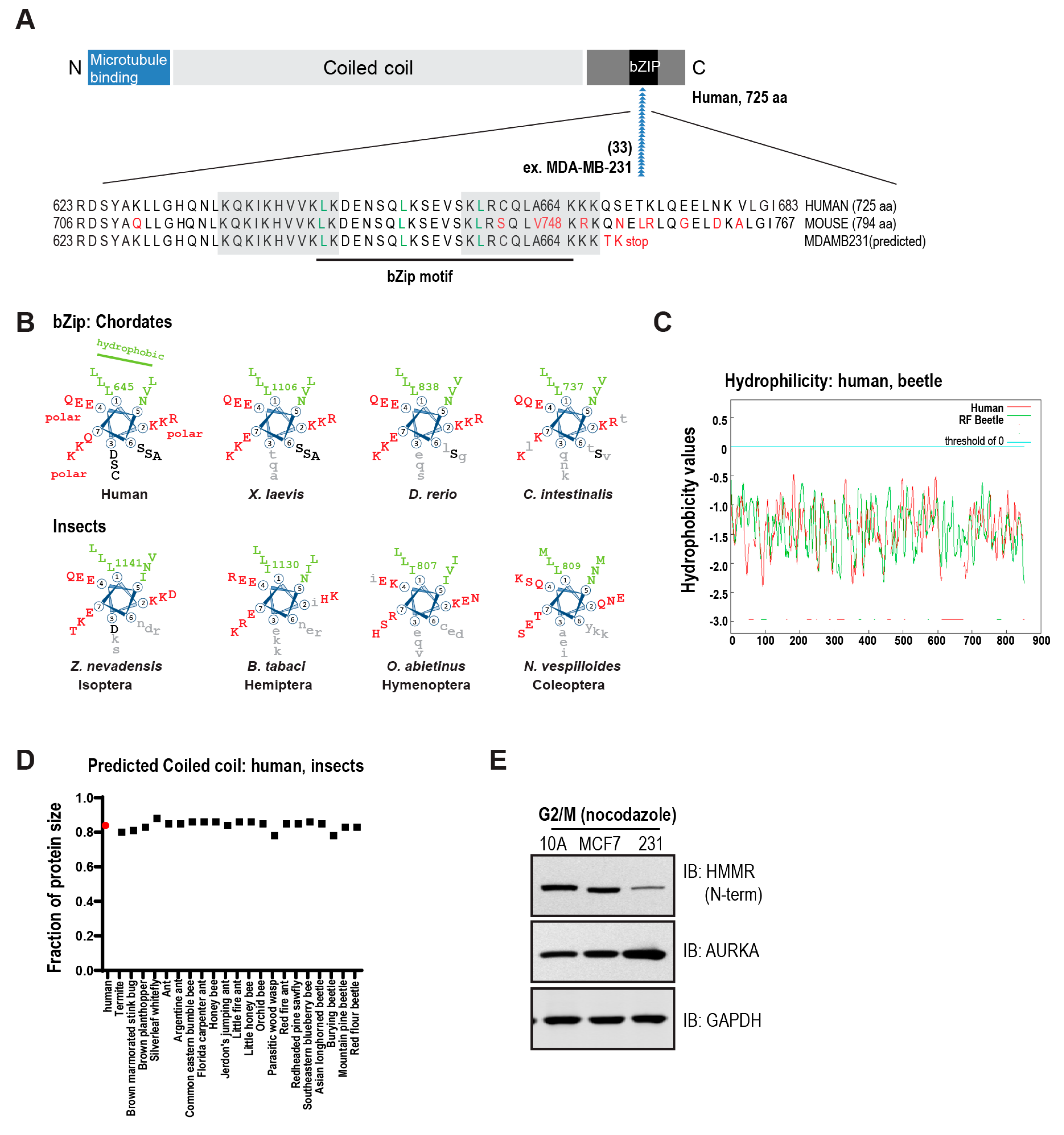
3. The Conserved Basic C-Terminal Domain in HMMR Is a Leucine Zipper Motif

To interact with hyaluronan, CD44 and other link module-based binding proteins use tandem repeat loops with homology to the cartilage link protein [12]. HMMR, however, was shown to interact with hyaluronan in an ionic manner through a basic, 35 amino acid, C-terminal region, which can be further subdivided into two motifs of 10 amino acids and 11 amino acids, respectively [13]. These motifs correspond with human HMMR amino acids 636–646 and amino acids 658–667, which are highly basic regions that became known as Basic (B) (X)7 Basic (B X7 B) motifs [13,14] (Figure 1A). Perhaps unsurprisingly, these basic residues in HMMR are essential for an ionic interaction with hyaluronan as well as heparin [13,14], and these interactions are abolished with increasing concentrations of salt [13].
It is important to note, however, the so-called “X7” residues are in fact highly ordered and evolutionarily conserved. When represented as a heptad repeat, this motif contains a leucine every seven residues (position 1) and conserved hydrophobic (interior) and polar (exterior) residues that enable a predicted coiled-coil structure and a basic leucine zipper (bZip) motif. Importantly, this bZip motif is conserved throughout chordates, including in the sea squirt Ciona intestinalis, as well as in species of insects (Figure 1B). That this highly basic stretch of amino acids binds in an ionic manner to hyaluronic acid in vitro is expected; that an urochordate and several species of insects encode a putative hyaluronan receptor (Hmmr) is remarkable, given these animals lack evidence for hyaluronan anabolism or synthesis, which first appears in amphioxus [15].

3.1. Structural Domains in HMMR

HMMR is predicted to be a hydrophilic protein (Figure 1C) and lacks a hydrophobic signal peptide or potential hydrophobic transmembrane domains. In fact, HMMR lacks the structural properties required for canonical extracellular export. Across chordate and insect species, HMMR is predicted to be a largely coiled-coil protein (Figure 1D, 80–85% of total amino acids) with microtubule binding domains at the N-terminus and a bZip motif [16] and degradation domains [17] at the C-terminus. These structural features—N-terminal microtubule binding, largely coiled-coil structure, C-terminal bZip motif—support its role as a non-motor spindle assembly factor.
The central region of human HMMR is a coiled-coil stalk (amino acids 69–681) that acts as a potential dimerization domain and binding region for other proteins. That is, HMMR interacts with CHICA/FAM83D through amino acids 365–546 [18], while amino acids 574–602 act as a calcium-dependent calmodulin binding domain [19]. Through its interaction with CHICA, HMMR locates dynein light chain 1 (DYNLL1) and CK1alpha to the spindle [20], and HMMR plays a similar role in docking BACH1 at the spindle [21], although the needed interaction domain is unknown. These protein complexes, formed through interactions with the coiled-coil stalk, play critical roles in the correct orientation of the mitotic spindle and the establishment of the cell division axis.
At the N-terminus, human HMMR possesses two microtubule binding subdomains at amino acids 40–59 and amino acids 76–90, respectively, with the latter domain encoded by exon 4 [19]. Through these domains, HMMR binds directly to microtubules [19]. While full-length HMMR is able to locate to centrosomes and bind to microtubules [16], the naturally occurring splice variant HMMR (-exon 4) localizes to the mitotic spindle during mitosis but shows nuclear localization during interphase [16,19]. Localization to the nucleus is also seen for a phosphorylated form of HMMR at Thr703 [22]. Phosphorylation at Thr703 was identified by phospho-proteomics [23] and, through the use of antibodies specific for phospho-Thr703 HMMR, it was revealed that phospho-Thr703 not only directs the protein to the nucleus but also may assist in the regulation of the Ran-dependent nuclear transport of targeting protein for Xklp2 (TPX2) [22].
The very N-terminal residues in HMMR are uniquely similar to those of mira/Miranda [24], a determinant of asymmetric neuroblast divisions in Drosophila. Miranda is a docking protein in neuroblasts that is responsible for the asymmetric cortical localization of key proteins and mRNAs that regulate neuroblast cell fate, including Prospero and brain tumor (Brat) [25,26]. Despite this critical role in Drosophila neurodevelopment, Miranda has no known orthologs outside the order Diptera. Importantly, Miranda and HMMR share significant similarity in their very N-terminal sequence [24]. These N-terminal residues in Miranda are needed for microtubule-dependent asymmetric localization [27]. Moreover, both Miranda and HMMR are largely coiled-coil proteins that bind microtubules but HMMR has not yet been determined to play an orthologous function during asymmetric cell divisions.
The conserved, C-terminal bZip motif in HMMR overlaps with the designated B-X7-B motifs, which bind in an ionic manner to hyaluronan and heparin [13,14]. The bZip motif of HMMR contains 72% homology with a C-terminal bZip motif in kinesin-like protein Kif15/hKlp2 (amino acids 1342–1388) [16]. Importantly, the bZip motif in hKlp2 locates the protein to microtubule minus ends through an interaction with targeting protein for Xklp2 (TPX2) [28]. While TPX2 was first defined by complexing with the bZip motif in hKlp2 and targeting the complex to microtubule minus ends [28,29], TPX2 has now been extensively characterized as both a critical activator for the mitotic kinase Aurora kinase A as well as other functions (reviewed in [30]). For HMMR, the bZip motif locates the protein to the centrosome and HMMR complexes with a significant fraction of TPX2 in a cell cycle-dependent manner [31,32,33,34]. These complexes serve to both regulate mitotic kinase activities [33] and alter Eg5 motor protein processivity [35,36].
Taken together, the structure of HMMR is one of a largely coiled-coil microtubule-associated protein, which can bind to microtubules directly through its N-terminus and localize to the centrosome through its C-terminal bZip motif. HMMR serves as a binding partner for spindle assembly factors, such as TPX2, DYNLL1, and CHICA/FAM83D, to regulate the assembly, stability and positioning of spindle microtubules during mitosis and meiosis.

3.2. Evolution of HMMR

The presence of hmmr orthologs amongst invertebrate animals has not been examined likely because arthropods do not express hyaluronan [37]. By using the conserved bZip motif as a landmark, we identified putative hmmr orthologs in many, but not all, orders of insects (Table S1). The bZip motif in hmmr homologs are most highly conserved in Isoptera, the earliest insect lineage represented (Figure 1B). We found no evidence for hmmr homologs in genomes in the order Diptera, which includes Drosophila.
We discovered that hmmr and mira are mutually exclusive amongst insect species with mira encoded exclusively in Dipterans. Comparison of N-terminal homology regions [24] from 127 known and predicted Miranda and HMMR/Hmmr sequences identified conservation in critical residues (Ala6 and Ala18, [24]), suggesting that mira potentially evolved in Diptera from an ancestral hmmr (Table S1).
It is a striking finding that a defined receptor (Hmmr) is encoded within multiple species that do not produce its putative ligand (hyaluronan). As Hmmr-like orthologs are encoded in species that predate the first appearance of hyaluronan by millions of years (in amphioxus [15]), we must conclude that Hmmr did not originally evolve as a hyaluronan receptor.

3.3. Conserved HMMR-NudC Domain-Containing Protein 2 (NUDCD2) Gene Cluster

The appearance of HMMR in multiple species that lack hyaluronan strongly argues against it playing an evolutionarily-conserved role in hyaluronan binding. Indeed, the conserved gene location of HMMR across multiple species of tetrapods and fish may implicate an evolutionarily-conserved role related to cell cycle and dynein motor activity. That is, HMMR is found in a small four gene cluster, along with NUDCD2 (NudC domain-containing protein 2), CCNG1 (cyclin G1), and MAT2B (methionine adenosyltransferase 2B), at 163.5 MB of human chromosome 5q34 (Figure 2A).
The HMMR-NUDCD2 gene cluster is proximal to a cluster of gamma-aminobutyric acid (GABA) A receptor subunit genes at 162 MB of human chromosome 5q33.3; this region of human chromosome 5q associates with human diseases related to neural development and/or homeostasis, including addiction, schizophrenia, memory, seizures (for 5q33.1), prominent forehead, and mental retardation (for 5q34). The proximity of these two small gene clusters has been maintained through the evolution of jawed vertebrates (Figure 2B): one cluster encodes cell cycle and dynein related gene products (CCNG1-NUDCD2-HMMR) while the other encodes GABAA receptor subunits.
In cartilaginous fish and Ciona, which is potentially the closest invertebrate to humans, HMMR, NUDCD2, GABRB2, and CCNG1 are located at different chromosomal sites (Figure 2B). Initiating with bony fish, however, NUDCD2 and CCNG1 are clustered with GABRA1 and GABRA6 while HMMR is located relatively proximal on chromosome 14 (Figure 2B). In lobe-fin fish, the two clusters separate and their organization and overall structure are maintained through tetrapod evolution (Figure 2B).
It is important to note the conserved orientation of HMMR and NUDCD2 in these small gene clusters. That is, the genes are oriented in opposite directions; for example, on human chromosome 5q, HMMR is in a forward orientation (163,460 kB–163,492 kB) and NUDCD2 is in a reverse orientation (163,446 kB–163,460 kB) so that the 5′ ends of the genes are 530 base-pairs apart. This conserved, close proximity of NUDCD2-HMMR may be a chance of nature. But, importantly, HMMR and NUDCD2 perform similar cell cycle-regulated functions as adaptors for dynein motor proteins and each have critical roles in the process of neural development and homeostasis.

4. HMMR Functions as a Homeostasis, Mitosis, and Meiosis Regulator

4.1. HMMR Is Needed for Tissue Homeostasis and Neural Development

HMMR is expressed in the developing nervous system [38] as well as in the proliferative regions of the adult mouse brain [39]. The mutation or loss of HMMR in vertebrate animal models induces neurodevelopmental defects [7,40,41]. Hmmrtm1a/tm1a mice encode Hmmr with targeted disruption following exon 2 while Hmmrm/m mice encode Hmmr that potentially retain N-terminal HMMR structure due to targeted disruption after exon 10. As a consequence, loss-of Hmmr phenotypes differ in these animals. Hmmrtm1a/tm1a mice suffer neonatal lethality and display heterogeneous cortical sizes, enlarged ventricles, and alterations in neural cell subsets [7]. However, Hmmrm/m mice are viable but, Hmmrm/m embryos undergo transient megalencephaly during development [40]. In both mouse models (Hmmrtm1a/tm1a mice and Hmmrm/m mice), the C-terminus of Hmmr is required to orient neural stem cell divisions [7,40].
A third animal model, Xenopus embryos treated with morpholinos targeting hmmr, also present with neurodevelopmental defects. Hmmr morphant embryos develop brain defects, including defective neural tube closure, narrowed forebrains, loss of hemispheric separation, and smaller olfactory bulbs [41]. In this model, hmmr morphant cells rescued with a mutant lacking the N-terminal 130 amino acids were rounded and displayed web-like microtubule arrays rather than the linear arrays observed in wild-type neural cells [41]. Thus, the N-terminus of Hmmr was required for the polarization of cells in the deep neural layer.
Loss of Hmmr/hmmr phenotypes are reminiscent, but not overlapping with those seen for LIS1 (Lissencephaly 1), an alternate cytoplasmic dynein partner protein; classical lissencephaly, a brain developmental disease characterized by decreased cortical complexity and generally larger brain size, results from mutations in the LIS1 gene [42]. A complex between dynein and the coiled-coil adaptor proteins LIS1, NUDC (nuclear distribution C, dynein complex regulator) and NUDE-like 1 (NDEL1) is required for correct corticogenesis. The loss of this complex, mediated through LIS1 mutations, can result in a cortical malformation disorder associated with severe cognitive impairment and epilepsy [43].
Importantly, NDEL1 relies upon post-translational modification by Aurora kinase A and TPX2 to modify microtubule dynamics, neuronal migration and neurite extension downstream [44,45,46]. Provocatively, NUDCD2 also regulates the LIS1-dynein complex [47], centrosome function [48], and mitotic spindle integrity [49]. Thus, the evolutionally conserved HMMR-NUDCD2 gene cluster encodes two coiled-coil centrosome proteins that complex with dynein and play critical roles in homeostasis, centrosome function, and mitotic spindle integrity.
In mouse models, the mutation or loss of HMMR/Hmmr expression also disrupts the correct development or homeostasis of gonadal tissues [7,50,51]. While the mechanisms responsible are yet to be determined, these phenotypes are likely reflective of the high expression of HMMR in gonadal tissues and the necessary role played by dynein, and its adaptor proteins, during the processes of gametogenesis and spermatogenesis [52,53,54].

4.2. HMMR Regulates Spindle Assembly in Mitotic Cells and Meiotic Extracts.

HMMR is a largely coiled-coil protein that can bind to microtubules directly through its N-terminus [19] and localize to the centrosome through a C-terminal bZip motif [16], which is structurally very similar to the C-terminal bZip motif in Xklp2 that enables an interaction with TPX2 [16,28]. The similarity in these domain structures seeded the hypothesis that HMMR also interacts with TPX2, which has been confirmed experimentally through the study of HMMR/RHAMM in human mitotic cells [16,33,35] and Xenopus RHAMM (XRHAMM) in Xenopus meiotic extracts [32,34,36,55].
The interaction between HMMR and TPX2 is important for Ran-dependent microtubule assembly near chromosomes [32,34], the activity of Aurora kinase A and microtubule nucleation at spindle poles [33], and the regulation of the processivity of the kinesin motor Eg5 [35,36]. The C-terminal bZip motif in Xklp2 regulates its location to microtubule minus ends in a dynein-dependent manner [28]; similarly, the C-terminal bZip motif in HMMR regulates its location to centrosomes and interaction with dynein [31].
HMMR is critical for the orientation of the mitotic spindle in human mitotic cells. HMMR locates DYNLL1 indirectly through an interaction with CHICA/FAM83D [18], which serves to modulate the position of the mitotic spindle through three complementary mechanisms: 1. HMMR localizes DYNLL1 at spindle poles, which dampens local dynein motor activities when brought proximal to the cell cortex [18]; 2. HMMR modulates the local activity of PLK1 and locates Ran-GTP to the spindle pole [7]; and, HMMR binds CHICA/FAM83D to locate and activate the protein kinase CK1alpha [20].
Each of the preceding mitotic and meiotic functions were predicted by the domain structure of HMMR. In addition to such hypothesis-based studies, a number of non-biased, genome-wide analyses also indicate similar mitotic roles of HMMR. The All RNA-Seq and Chip-Seq Sample and Signature Search (ARCHS4) tool, which mines sequencing data from 103,083 mouse and 84,863 human samples [56], predicts HMMR functions in processes related to mitosis and chromosome segregation, kinetochore, and nuclear pore complex assembly, which have all been independently and experimentally validated [16,22,35]. Using this tool [56], HMMR is found to be co-expressed with TOP2A, KIF11, TPX2, ECT2, BUB1, KIF20A, NUSAP1, KIF20B, SMC2, and CCNA2; each of these gene products are involved with cell cycle regulation, spindle organization, and chromosome segregation. A complementary proteome-wide analysis of mitotic substages, which combined specific intracellular immunolabeling protocols and FACS separation of interphase and mitotic cells, identified HMMR as one of 14 proteins that peak in abundance in G2-phase and mitosis along with other cell cycle gene products, such as Aurora A and B, Polo-like kinase 1, and CENPF [57]. Finally, the Mitocheck consortium combined several powerful screening tools, such as RNA interference, time-lapse microscopy and computational image processing that assessed chromosome and nuclear morphology, to profile the roles of ~21,000 gene products during mitosis [58]. HMMR depletion was associated with mitosis-specific phenotypes, such as strange nuclear shape, polylobed nuclei and chromosome segregation errors, leading to HMMR being designated a validated mitotic hit [58].
Given the critical mitotic functions of HMMR, it is unsurprising that HMMR is predicted to be regulated by proliferation associated transcription factors, FOXM1, E2F4, and MYC [56] and, experimentally, by YAP-TEAD of the Hippo pathway [59]. Similarly, it is unsurprising that HMMR expression is transcriptionally downregulated by the tumor suppressor TP53 [60]. Perhaps less predictable are the mechanisms utilized for post-transcriptional regulation of HMMR expression. Along with Bard1 and other Ran-dependent spindle assembly factors, Hmmr was identified as a substrate for the ubiquitin ligase anaphase-promoting complex/cyclosome (APC/C) [17]; the recognition and proteolysis of Hmmr and Bard1 (as well as complexed Brca1) relied upon APC/C recognition motifs, such as destruction (D) box, KEN box, or TEK box [17]. For Hmmr, D, KEN, and TEK boxes were identified in the C-terminal domain [17]. HMMR is also a substrate for the BRCA1/BARD1 E3 ubiquitin ligase [61] and deficient BRCA1 function leads to the stabilization of HMMR [61,62]. The HMMR-BRCA1 interaction influences mammary tumorigenesis as indicated by the association between HMMR polymorphisms and breast cancer risk in carriers of BRCA1 mutations but not BRCA2 mutations [63]. Similarly, a HMMR partner protein, DYNLL1, has been correlated with progression free survival in carriers of BRCA1 mutations but not BRCA2 mutations and shown to modulate PARP inhibitor sensitivity in BRCA1-mutant cells [64]. The potential importance of HMMR-DYNLL1 mediated control of the cell division axis in the context of BRCA1 mutations is not yet known but may help to explain how BRCA1 mutations perturb the differentiation hierarchy present in the normal human mammary gland [65,66,67].

5. HMMR Associates with Breast Cancer risk, Cancer Prognosis, and Progression

The expression of HMMR is cell cycle-regulated with peak expression between late G2 phase and early mitosis [31]. Consistently, HMMR expression is low in most healthy tissues but is elevated in proliferative tissues, such as the testis, spleen, placenta, and thymus [7,8]. Moreover, elevated HMMR expression associates with poor prognosis in a variety of cancers, such as breast cancer [68], colorectal cancer [69], stomach cancer [70], endometrial cancer [71], prostate cancer [72], and multiple myeloma [73]. But, the association between HMMR and cancer is not as simple as high HMMR expression demarcates very mitotic tumors, which correlates with more aggressive tumor growth and poor patient survival.
For several types of cancers, poor patient survival also associates with low HMMR expression. For example, hemizygous deletion of HMMR is present in almost half of malignant peripheral nerve sheath tumors [74,75]; HMMR expression is diminished in 96% of human seminomas [50]; and, germline HMMR variants that associate with lower expression also associate with increased breast cancer risk in some patient cohorts [61,76,77]. Moreover, an HMMR splice variant that loses microtubule binding activity is expressed at an elevated level in a variety of cancers. That is, the N-terminal domain of HMMR binds microtubules, encoded in part by exon 4 [16,19], and the expression of a naturally occurring splice variant that lacks exon 4 (-exon 4) correlates with progression of multiple myeloma [73] and breast cancers [78]. HMMR (-exon 4) expression is also sufficient to promote pancreatic islet tumor growth and metastasis to lymph nodes and liver [79]. As indicated by the cancer cell-line encyclopedia and COSMIC databases, HMMR contains a mutational hotspot encoded by a homopolymeric adenine tract within the C-terminal basic leucine zipper motif (Figure 1A). Indeed, immortal MDA-MB-231 breast cancer cells contain an A664 frameshift mutation (cDNA change c.1992_1993insA), and the expression of HMMR is greatly reduced in lysates from nocodazole-synchronized, G2/M phase lysates generated from MDA-MB-231 cells relative to MCF7 cells or MCF10A cells (Figure 1E). Thus, aggressive cancers may contain either abnormally high or abnormally low expression of HMMR, and tumors are frequently characterized by the expression of a putative loss of function HMMR splice variant (-exon 4) or frameshift mutations in the homopolymeric adenine tract within the C-terminal bZip motif (Figure 1A).
HMMR is a low penetrance breast cancer susceptibility gene [61]; genotyping of three HMMR haplotype-tagging single nucleotide polymorphisms (htSNPs) identified statistically significant associations with risk of breast cancer [61]. Importantly, HMMR htSNPs showed an association with either germline overexpression (rs10515860 SNP; A-C-A haplotype) or germline downregulation of HMMR (G-C-A-T-G haplotype) [61]. HMMR is also a key substrate for the BRCA1-BARD1 E3 ubiquitin ligase during the process of spindle assembly [61]; dysfunction of BRCA1 is well-documented to stabilize HMMR protein expression [50,55,61,62]. Moreover, common HMMR variants modify the risk of developing breast cancer for carriers of BRCA1, but not BRCA2, mutations [63]. Thus, there is evidence to support the hypothesis that HMMR protein abundance must be tightly regulated through transcriptional control by TP53 [60] and the Hippo pathway [59] and post-translational turnover by BRCA1-BARD1 [61] and the anaphase promoting complex [17]. Consistent with this, silencing or elevating HMMR expression disrupts microtubule-based processes during cell division, and results in mitotic spindle abnormalities, genome instability, and changes to the cell division axis and progenitor cell fate.

6. Conclusions

It is also possible, and has not been disproven, that the unconventional export of HMMR during pathological states may enable non-physiological functionalities, such as ionic interaction between the C-terminal bZip motif and acidic hyaluronan or heparin. However, hmmr is encoded in numerous species that lack hyaluronan, which argues strongly that HMMR did not evolve as a hyaluronan receptor; rather, the evolution of HMMR in a small gene cluster with the dynein adaptor NUDCD2 and the structure of the HMMR gene product support the designation of HMMR as a homeostasis, mitosis, and meiosis regulator. Indeed, HMMR is a largely coiled coil gene product with evolutionarily conserved N-terminal and C-terminal domains that target the protein to microtubules and centrosomes, respectively. Consistent with these structural features defining the protein’s physiological function, recent studies indicate necessary roles for HMMR during cell division and microtubule-associated processes related to neural development, control of the cell division axis, and progenitor cell fate in three different vertebrate animal models.

Supplementary Materials

The following are available online at https://www.mdpi.com/2073-4409/9/4/819/s1, Table S1: HMMR and Miranda sequences and domains in insect species.

Author Contributions

Conceptualization, Z.H., M.C., and C.A.M.; formal analysis, M.C.; investigation, Z.H., and M.C.; writing—original draft preparation, Z.H., M.C. and C.A.M.; writing—review and editing, Z.H., L.M., M.C., and C.A.M.; supervision, C.A.M.; funding acquisition, Z.H., M.C., and C.A.M. All authors have read and agreed to the published version of the manuscript.

Funding

This research was funded by: Michael Cuccione Foundation (MCF) brain tumour award (CAM), Canada Institutes for Health Research (CIHR), in partnership with the Avon Foundation for Women, operating grant (CAM), CIHR New Investigator Salary Award (CAM), CFRI scientist level 1 salary award (CAM), University of British Columbia 4-year fellowship (ZH), and MCF fellowship (MC).

Acknowledgments

The authors acknowledge the support of the Michael Cuccione Foundation.

Conflicts of Interest

The authors declare no conflict of interest.

References

  1. Caon, I.; Bartolini, B.; Parnigoni, A.; Carava, E.; Moretto, P.; Viola, M.; Karousou, E.; Vigetti, D.; Passi, A. Revisiting the hallmarks of cancer: The role of hyaluronan. Semin. Cancer Biol. 2019. [Google Scholar] [CrossRef] [PubMed]
  2. Skandalis, S.S.; Karalis, T.; Heldin, P. Intracellular hyaluronan: Importance for cellular functions. Semin. Cancer Biol. 2019. [Google Scholar] [CrossRef] [PubMed]
  3. Morath, I.; Hartmann, T.N.; Orian-Rousseau, V. CD44: More than a mere stem cell marker. Int. J. Biochem. Cell Biol. 2016, 81, 166–173. [Google Scholar] [CrossRef] [PubMed]
  4. Clevers, H. The cancer stem cell: Premises, promises and challenges. Nat. Med. 2011, 17, 313–319. [Google Scholar] [CrossRef] [PubMed]
  5. Hardwick, C.; Hoare, K.; Owens, R.; Hohn, H.P.; Hook, M.; Moore, D.; Cripps, V.; Austen, L.; Nance, D.M.; Turley, E.A. Molecular cloning of a novel hyaluronan receptor that mediates tumor cell motility. J. Cell Biol. 1992, 117, 1343–1350. [Google Scholar] [CrossRef] [PubMed] [Green Version]
  6. Hofmann, M.; Assmann, V.; Fieber, C.; Sleeman, J.P.; Moll, J.; Ponta, H.; Hart, I.R.; Herrlich, P. Problems with RHAMM: A new link between surface adhesion and oncogenesis? Cell 1998, 95, 591–592. [Google Scholar] [CrossRef] [Green Version]
  7. Connell, M.; Chen, H.; Jiang, J.; Kuan, C.W.; Fotovati, A.; Chu, T.L.; He, Z.; Lengyell, T.C.; Li, H.; Kroll, T.; et al. HMMR acts in the PLK1-dependent spindle positioning pathway and supports neural development. eLife 2017, 6, e28672. [Google Scholar] [CrossRef] [Green Version]
  8. Fieber, C.; Plug, R.; Sleeman, J.; Dall, P.; Ponta, H.; Hofmann, M. Characterisation of the murine gene encoding the intracellular hyaluronan receptor IHABP (RHAMM). Gene 1999, 226, 41–50. [Google Scholar] [CrossRef]
  9. Hofmann, M.; Fieber, C.; Assmann, V.; Gottlicher, M.; Sleeman, J.; Plug, R.; Howells, N.; von Stein, O.; Ponta, H.; Herrlich, P. Identification of IHABP, a 95 kDa intracellular hyaluronate binding protein. J. Cell Sci. 1998, 111, 1673–1684. [Google Scholar]
  10. Jiang, J.; Casalegno-Garduno, R.; Chen, H.; Schmitt, A.; Schmitt, M.; Maxwell, C.A. Multifunctional proteins bridge mitosis with motility and cancer with inflammation and arthritis. Sci. World J. 2010, 10, 1244–1257. [Google Scholar] [CrossRef]
  11. Maxwell, C.A.; McCarthy, J.; Turley, E. Cell-surface and mitotic-spindle RHAMM: Moonlighting or dual oncogenic functions? J. Cell Sci. 2008, 121, 925–932. [Google Scholar] [CrossRef] [PubMed] [Green Version]
  12. Goetinck, P.F.; Stirpe, N.S.; Tsonis, P.A.; Carlone, D. The tandemly repeated sequences of cartilage link protein contain the sites for interaction with hyaluronic acid. J. Cell Biol. 1987, 105, 2403–2408. [Google Scholar] [CrossRef] [PubMed]
  13. Yang, B.; Zhang, L.; Turley, E.A. Identification of two hyaluronan-binding domains in the hyaluronan receptor RHAMM. J. Biol. Chem. 1993, 268, 8617–8623. [Google Scholar]
  14. Yang, B.; Hall, C.L.; Yang, B.L.; Savani, R.C.; Turley, E.A. Identification of a novel heparin binding domain in RHAMM and evidence that it modifies HA mediated locomotion of ras-transformed cells. J. Cell. Biochem. 1994, 56, 455–468. [Google Scholar] [CrossRef]
  15. Csoka, A.B.; Stern, R. Hypotheses on the evolution of hyaluronan: A highly ironic acid. Glycobiology 2013, 23, 398–411. [Google Scholar] [CrossRef] [PubMed] [Green Version]
  16. Maxwell, C.A.; Keats, J.J.; Crainie, M.; Sun, X.; Yen, T.; Shibuya, E.; Hendzel, M.; Chan, G.; Pilarski, L.M. RHAMM is a centrosomal protein that interacts with dynein and maintains spindle pole stability. Mol. Biol. Cell 2003, 14, 2262–2276. [Google Scholar] [CrossRef] [PubMed]
  17. Song, L.; Rape, M. Regulated degradation of spindle assembly factors by the anaphase-promoting complex. Mol. Cell 2010, 38, 369–382. [Google Scholar] [CrossRef] [Green Version]
  18. Dunsch, A.K.; Hammond, D.; Lloyd, J.; Schermelleh, L.; Gruneberg, U.; Barr, F.A. Dynein light chain 1 and a spindle-associated adaptor promote dynein asymmetry and spindle orientation. J. Cell Biol. 2012, 198, 1039–1054. [Google Scholar] [CrossRef] [Green Version]
  19. Assmann, V.; Jenkinson, D.; Marshall, J.F.; Hart, I.R. The intracellular hyaluronan receptor RHAMM/IHABP interacts with microtubules and actin filaments. J. Cell Sci. 1999, 112, 3943–3954. [Google Scholar]
  20. Fulcher, L.J.; He, Z.; Mei, L.; Macartney, T.J.; Wood, N.T.; Prescott, A.R.; Whigham, A.J.; Varghese, J.; Gourlay, R.; Ball, G.; et al. FAM83D directs protein kinase CK1alpha to the mitotic spindle for proper spindle positioning. EMBO Rep. 2019, 20, e47495. [Google Scholar] [CrossRef]
  21. Li, J.; Shima, H.; Nishizawa, H.; Ikeda, M.; Brydun, A.; Matsumoto, M.; Kato, H.; Saiki, Y.; Liu, L.; Watanabe-Matsui, M.; et al. Phosphorylation of BACH1 switches its function from transcription factor to mitotic chromosome regulator and promotes its interaction with HMMR. Biochem. J. 2018, 475, 981–1002. [Google Scholar] [CrossRef] [PubMed]
  22. Chu, T.L.H.; Connell, M.; Zhou, L.; He, Z.; Won, J.; Chen, H.; Rahavi, S.M.R.; Mohan, P.; Nemirovsky, O.; Fotovati, A.; et al. Cell Cycle-Dependent Tumor Engraftment and Migration Are Enabled by Aurora-A. Mol. Cancer Res. 2018, 16, 16–31. [Google Scholar] [CrossRef] [PubMed] [Green Version]
  23. Nousiainen, M.; Sillje, H.H.; Sauer, G.; Nigg, E.A.; Korner, R. Phosphoproteome analysis of the human mitotic spindle. Proc. Natl. Acad. Sci. USA 2006, 103, 5391–5396. [Google Scholar] [CrossRef] [PubMed] [Green Version]
  24. Chang, C.W.; Nashchekin, D.; Wheatley, L.; Irion, U.; Dahlgaard, K.; Montague, T.G.; Hall, J.; Johnston, D.S. Anterior-posterior axis specification in Drosophila oocytes: Identification of novel bicoid and oskar mRNA localization factors. Genetics 2011, 188, 883–896. [Google Scholar] [CrossRef] [PubMed] [Green Version]
  25. Ikeshima-Kataoka, H.; Skeath, J.B.; Nabeshima, Y.; Doe, C.Q.; Matsuzaki, F. Miranda directs Prospero to a daughter cell during Drosophila asymmetric divisions. Nature 1997, 390, 625–629. [Google Scholar] [CrossRef] [PubMed]
  26. Shen, C.P.; Jan, L.Y.; Jan, Y.N. Miranda is required for the asymmetric localization of Prospero during mitosis in Drosophila. Cell 1997, 90, 449–458. [Google Scholar] [CrossRef] [Green Version]
  27. Irion, U.; Adams, J.; Chang, C.W.; Johnston, D.S. Miranda couples oskar mRNA/Staufen complexes to the bicoid mRNA localization pathway. Dev. Biol. 2006, 297, 522–533. [Google Scholar] [CrossRef] [Green Version]
  28. Wittmann, T.; Boleti, H.; Antony, C.; Karsenti, E.; Vernos, I. Localization of the kinesin-like protein Xklp2 to spindle poles requires a leucine zipper, a microtubule-associated protein, and dynein. J. Cell Biol. 1998, 143, 673–685. [Google Scholar] [CrossRef] [Green Version]
  29. Boleti, H.; Karsenti, E.; Vernos, I. Xklp2, a novel Xenopus centrosomal kinesin-like protein required for centrosome separation during mitosis. Cell 1996, 84, 49–59. [Google Scholar] [CrossRef] [Green Version]
  30. Garrido, G.; Vernos, I. Non-centrosomal TPX2-Dependent Regulation of the Aurora A Kinase: Functional Implications for Healthy and Pathological Cell Division. Front. Oncol. 2016, 6, 88. [Google Scholar] [CrossRef] [Green Version]
  31. Maxwell, C.A.; Keats, J.J.; Belch, A.R.; Pilarski, L.M.; Reiman, T. Receptor for hyaluronan-mediated motility correlates with centrosome abnormalities in multiple myeloma and maintains mitotic integrity. Cancer Res. 2005, 65, 850–860. [Google Scholar] [PubMed]
  32. Groen, A.C.; Cameron, L.A.; Coughlin, M.; Miyamoto, D.T.; Mitchison, T.J.; Ohi, R. XRHAMM functions in ran-dependent microtubule nucleation and pole formation during anastral spindle assembly. Curr. Biol. 2004, 14, 1801–1811. [Google Scholar] [CrossRef] [PubMed] [Green Version]
  33. Chen, H.; Mohan, P.; Jiang, J.; Nemirovsky, O.; He, D.; Fleisch, M.C.; Niederacher, D.; Pilarski, L.M.; Lim, C.J.; Maxwell, C.A. Spatial regulation of Aurora A activity during mitotic spindle assembly requires RHAMM to correctly localize TPX2. Cell Cycle 2014, 13, 2248–2261. [Google Scholar] [CrossRef] [PubMed] [Green Version]
  34. Scrofani, J.; Sardon, T.; Meunier, S.; Vernos, I. Microtubule nucleation in mitosis by a RanGTP-dependent protein complex. Curr. Biol. 2015, 25, 131–140. [Google Scholar] [CrossRef] [PubMed] [Green Version]
  35. Chen, H.; Connell, M.; Mei, L.; Reid, G.S.D.; Maxwell, C.A. The non-motor adaptor HMMR dampens Eg5-mediated forces to preserve the kinetics and integrity of chromosome segregation. Mol. Biol. Cell 2018, 29, 786–796. [Google Scholar] [CrossRef] [PubMed] [Green Version]
  36. Eibes, S.; Gallisa-Sune, N.; Rosas-Salvans, M.; Martinez-Delgado, P.; Vernos, I.; Roig, J. Nek9 Phosphorylation Defines a New Role for TPX2 in Eg5-Dependent Centrosome Separation before Nuclear Envelope Breakdown. Curr. Biol. 2018, 28, 121–129. [Google Scholar] [CrossRef] [Green Version]
  37. Stern, R. Go Fly a Chitin: The Mystery of Chitin and Chitinases in Vertebrate Tissues. Front. Biosci. 2017, 22, 580–595. [Google Scholar] [CrossRef] [Green Version]
  38. Casini, P.; Nardi, I.; Ori, M. RHAMM mRNA expression in proliferating and migrating cells of the developing central nervous system. Gene Expr. Patterns 2010, 10, 93–97. [Google Scholar] [CrossRef]
  39. Lindwall, C.; Olsson, M.; Osman, A.M.; Kuhn, H.G.; Curtis, M.A. Selective expression of hyaluronan and receptor for hyaluronan mediated motility (Rhamm) in the adult mouse subventricular zone and rostral migratory stream and in ischemic cortex. Brain Res. 2013, 1503, 62–77. [Google Scholar] [CrossRef]
  40. Li, H.; Kroll, T.; Moll, J.; Frappart, L.; Herrlich, P.; Heuer, H.; Ploubidou, A. Spindle Misorientation of Cerebral and Cerebellar Progenitors Is a Mechanistic Cause of Megalencephaly. Stem Cell Rep. 2017, 9, 1071–1080. [Google Scholar] [CrossRef] [Green Version]
  41. Prager, A.; Hagenlocher, C.; Ott, T.; Schambony, A.; Feistel, K. hmmr mediates anterior neural tube closure and morphogenesis in the frog Xenopus. Dev. Biol. 2017, 430, 188–201. [Google Scholar] [CrossRef] [PubMed]
  42. Vallee, R.B.; Tsai, J.W. The cellular roles of the lissencephaly gene LIS1, and what they tell us about brain development. Genes Dev. 2006, 20, 1384–1393. [Google Scholar] [CrossRef] [PubMed] [Green Version]
  43. Toba, S.; Hirotsune, S. A unique role of dynein and nud family proteins in corticogenesis. Neuropathology 2012, 32, 432–439. [Google Scholar] [CrossRef] [PubMed]
  44. Mori, D.; Yamada, M.; Mimori-Kiyosue, Y.; Shirai, Y.; Suzuki, A.; Ohno, S.; Saya, H.; Wynshaw-Boris, A.; Hirotsune, S. An essential role of the aPKC-Aurora A-NDEL1 pathway in neurite elongation by modulation of microtubule dynamics. Nat. Cell Biol. 2009, 11, 1057–1068. [Google Scholar] [CrossRef]
  45. Mori, D.; Yano, Y.; Toyo-oka, K.; Yoshida, N.; Yamada, M.; Muramatsu, M.; Zhang, D.; Saya, H.; Toyoshima, Y.Y.; Kinoshita, K.; et al. NDEL1 phosphorylation by Aurora-A kinase is essential for centrosomal maturation, separation, and TACC3 recruitment. Mol. Cell. Biol. 2007, 27, 352–367. [Google Scholar] [CrossRef] [Green Version]
  46. Yamada, M.; Toba, S.; Yoshida, Y.; Haratani, K.; Mori, D.; Yano, Y.; Mimori-Kiyosue, Y.; Nakamura, T.; Itoh, K.; Fushiki, S.; et al. LIS1 and NDEL1 coordinate the plus-end-directed transport of cytoplasmic dynein. EMBO J. 2008, 27, 2471–2483. [Google Scholar] [CrossRef]
  47. Yang, Y.; Yan, X.; Cai, Y.; Lu, Y.; Si, J.; Zhou, T. NudC-like protein 2 regulates the LIS1/dynein pathway by stabilizing LIS1 with Hsp90. Proc. Natl. Acad. Sci. USA 2010, 107, 3499–3504. [Google Scholar] [CrossRef] [Green Version]
  48. Li, M.; Xu, X.; Zhang, J.; Liu, M.; Wang, W.; Gao, Y.; Sun, Q.; Zhang, J.; Lu, Y.; Wang, F.; et al. NudC-like protein 2 restrains centriole amplification by stabilizing HERC2. Cell Death Dis. 2019, 10, 628. [Google Scholar] [CrossRef]
  49. Yang, Y.; Wang, W.; Li, M.; Gao, Y.; Zhang, W.; Huang, Y.; Zhuo, W.; Yan, X.; Liu, W.; Wang, F.; et al. NudCL2 is an Hsp90 cochaperone to regulate sister chromatid cohesion by stabilizing cohesin subunits. Cell. Mol. Life Sci. 2019, 76, 381–395. [Google Scholar] [CrossRef] [Green Version]
  50. Li, H.; Frappart, L.; Moll, J.; Winkler, A.; Kroll, T.; Hamann, J.; Kufferath, I.; Groth, M.; Taudien, S.; Schutte, M.; et al. Impaired Planar Germ Cell Division in the Testis, Caused by Dissociation of RHAMM from the Spindle, Results in Hypofertility and Seminoma. Cancer Res. 2016, 76, 6382–6395. [Google Scholar] [CrossRef] [Green Version]
  51. Li, H.; Moll, J.; Winkler, A.; Frappart, L.; Brunet, S.; Hamann, J.; Kroll, T.; Verlhac, M.H.; Heuer, H.; Herrlich, P.; et al. RHAMM deficiency disrupts folliculogenesis resulting in female hypofertility. Biol. Open 2015, 4, 562–571. [Google Scholar] [CrossRef] [PubMed] [Green Version]
  52. Whitfield, M.; Thomas, L.; Bequignon, E.; Schmitt, A.; Stouvenel, L.; Montantin, G.; Tissier, S.; Duquesnoy, P.; Copin, B.; Chantot, S.; et al. Mutations in DNAH17, Encoding a Sperm-Specific Axonemal Outer Dynein Arm Heavy Chain, Cause Isolated Male Infertility Due to Asthenozoospermia. Am. J. Hum. Genet. 2019, 105, 198–212. [Google Scholar] [CrossRef] [PubMed] [Green Version]
  53. Ellenbecker, M.; Osterli, E.; Wang, X.; Day, N.J.; Baumgarten, E.; Hickey, B.; Voronina, E. Dynein Light Chain DLC-1 Facilitates the Function of the Germline Cell Fate Regulator GLD-1 in Caenorhabditis elegans. Genetics 2019, 211, 665–681. [Google Scholar] [CrossRef] [Green Version]
  54. Wen, Q.; Tang, E.I.; Lui, W.Y.; Lee, W.M.; Wong, C.K.C.; Silvestrini, B.; Cheng, C.Y. Dynein 1 supports spermatid transport and spermiation during spermatogenesis in the rat testis. Am. J. Physiol. Endocrinol. Metab. 2018, 315, E924–E948. [Google Scholar] [CrossRef] [PubMed] [Green Version]
  55. Joukov, V.; Groen, A.C.; Prokhorova, T.; Gerson, R.; White, E.; Rodriguez, A.; Walter, J.C.; Livingston, D.M. The BRCA1/BARD1 heterodimer modulates ran-dependent mitotic spindle assembly. Cell 2006, 127, 539–552. [Google Scholar] [CrossRef] [PubMed] [Green Version]
  56. Lachmann, A.; Torre, D.; Keenan, A.B.; Jagodnik, K.M.; Lee, H.J.; Wang, L.; Silverstein, M.C.; Ma’ayan, A. Massive mining of publicly available RNA-seq data from human and mouse. Nat. Commun. 2018, 9, 1366. [Google Scholar] [CrossRef] [PubMed] [Green Version]
  57. Ly, T.; Whigham, A.; Clarke, R.; Brenes-Murillo, A.J.; Estes, B.; Madhessian, D.; Lundberg, E.; Wadsworth, P.; Lamond, A.I. Proteomic analysis of cell cycle progression in asynchronous cultures, including mitotic subphases, using PRIMMUS. Elife 2017, 6, e27574. [Google Scholar] [CrossRef] [Green Version]
  58. Neumann, B.; Walter, T.; Heriche, J.K.; Bulkescher, J.; Erfle, H.; Conrad, C.; Rogers, P.; Poser, I.; Held, M.; Liebel, U.; et al. Phenotypic profiling of the human genome by time-lapse microscopy reveals cell division genes. Nature 2010, 464, 721–727. [Google Scholar] [CrossRef]
  59. Wang, Z.; Wu, Y.; Wang, H.; Zhang, Y.; Mei, L.; Fang, X.; Zhang, X.; Zhang, F.; Chen, H.; Liu, Y.; et al. Interplay of mevalonate and Hippo pathways regulates RHAMM transcription via YAP to modulate breast cancer cell motility. Proc. Natl. Acad. Sci. USA 2014, 111, E89–E98. [Google Scholar] [CrossRef] [Green Version]
  60. Sohr, S.; Engeland, K. RHAMM is differentially expressed in the cell cycle and downregulated by the tumor suppressor p53. Cell Cycle 2008, 7, 3448–3460. [Google Scholar] [CrossRef] [Green Version]
  61. Pujana, M.A.; Han, J.D.; Starita, L.M.; Stevens, K.N.; Tewari, M.; Ahn, J.S.; Rennert, G.; Moreno, V.; Kirchhoff, T.; Gold, B.; et al. Network modeling links breast cancer susceptibility and centrosome dysfunction. Nat. Genet. 2007, 39, 1338–1349. [Google Scholar] [CrossRef] [PubMed]
  62. He, Z.; Kannan, N.; Nemirovsky, O.; Chen, H.; Connell, M.; Taylor, B.; Jiang, J.; Pilarski, L.M.; Fleisch, M.C.; Niederacher, D.; et al. BRCA1 controls the cell division axis and governs ploidy and phenotype in human mammary cells. Oncotarget 2017, 8, 32461–32475. [Google Scholar] [CrossRef] [PubMed] [Green Version]
  63. Maxwell, C.A.; Benitez, J.; Gomez-Baldo, L.; Osorio, A.; Bonifaci, N.; Fernandez-Ramires, R.; Costes, S.V.; Guino, E.; Chen, H.; Evans, G.J.; et al. Interplay between BRCA1 and RHAMM regulates epithelial apicobasal polarization and may influence risk of breast cancer. PLoS Biol. 2011, 9, e1001199. [Google Scholar] [CrossRef] [PubMed] [Green Version]
  64. He, Y.J.; Meghani, K.; Caron, M.C.; Yang, C.; Ronato, D.A.; Bian, J.; Sharma, A.; Moore, J.; Niraj, J.; Detappe, A.; et al. DYNLL1 binds to MRE11 to limit DNA end resection in BRCA1-deficient cells. Nature 2018, 563, 522–526. [Google Scholar] [CrossRef]
  65. Liu, S.; Ginestier, C.; Charafe-Jauffret, E.; Foco, H.; Kleer, C.G.; Merajver, S.D.; Dontu, G.; Wicha, M.S. BRCA1 regulates human mammary stem/progenitor cell fate. Proc. Natl. Acad. Sci. USA 2008, 105, 1680–1685. [Google Scholar] [CrossRef] [Green Version]
  66. Lim, E.; Vaillant, F.; Wu, D.; Forrest, N.C.; Pal, B.; Hart, A.H.; Asselin-Labat, M.L.; Gyorki, D.E.; Ward, T.; Partanen, A.; et al. Aberrant luminal progenitors as the candidate target population for basal tumor development in BRCA1 mutation carriers. Nat. Med. 2009, 15, 907–913. [Google Scholar] [CrossRef]
  67. Ding, L.; Su, Y.; Fassl, A.; Hinohara, K.; Qiu, X.; Harper, N.W.; Huh, S.J.; Bloushtain-Qimron, N.; Jovanovic, B.; Ekram, M.; et al. Perturbed myoepithelial cell differentiation in BRCA mutation carriers and in ductal carcinoma in situ. Nat. Commun. 2019, 10, 4182. [Google Scholar] [CrossRef] [Green Version]
  68. Assmann, V.; Gillett, C.E.; Poulsom, R.; Ryder, K.; Hart, I.R.; Hanby, A.M. The pattern of expression of the microtubule-binding protein RHAMM/IHABP in mammary carcinoma suggests a role in the invasive behaviour of tumour cells. J. Pathol. 2001, 195, 191–196. [Google Scholar] [CrossRef]
  69. Zlobec, I.; Baker, K.; Terracciano, L.M.; Lugli, A. RHAMM, p21 combined phenotype identifies microsatellite instability-high colorectal cancers with a highly adverse prognosis. Clin. Cancer Res. 2008, 14, 3798–3806. [Google Scholar] [CrossRef] [Green Version]
  70. Li, H.; Guo, L.; Li, J.W.; Liu, N.; Qi, R.; Liu, J. Expression of hyaluronan receptors CD44 and RHAMM in stomach cancers: Relevance with tumor progression. Int. J. Oncol. 2000, 17, 927–932. [Google Scholar] [CrossRef]
  71. Rein, D.T.; Roehrig, K.; Schondorf, T.; Lazar, A.; Fleisch, M.; Niederacher, D.; Bender, H.G.; Dall, P. Expression of the hyaluronan receptor RHAMM in endometrial carcinomas suggests a role in tumour progression and metastasis. J. Cancer Res. Clin. Oncol. 2003, 129, 161–164. [Google Scholar] [CrossRef] [PubMed]
  72. Gust, K.M.; Hofer, M.D.; Perner, S.R.; Kim, R.; Chinnaiyan, A.M.; Varambally, S.; Moller, P.; Rinnab, L.; Rubin, M.A.; Greiner, J.; et al. RHAMM (CD168) is overexpressed at the protein level and may constitute an immunogenic antigen in advanced prostate cancer disease. Neoplasia 2009, 11, 956–963. [Google Scholar] [CrossRef] [PubMed] [Green Version]
  73. Maxwell, C.A.; Rasmussen, E.; Zhan, F.; Keats, J.J.; Adamia, S.; Strachan, E.; Crainie, M.; Walker, R.; Belch, A.R.; Pilarski, L.M.; et al. RHAMM expression and isoform balance predict aggressive disease and poor survival in multiple myeloma. Blood 2004, 104, 1151–1158. [Google Scholar] [CrossRef] [PubMed]
  74. Mantripragada, K.K.; Spurlock, G.; Kluwe, L.; Chuzhanova, N.; Ferner, R.E.; Frayling, I.M.; Dumanski, J.P.; Guha, A.; Mautner, V.; Upadhyaya, M. High-resolution DNA copy number profiling of malignant peripheral nerve sheath tumors using targeted microarray-based comparative genomic hybridization. Clin. Cancer Res. 2008, 14, 1015–1024. [Google Scholar] [CrossRef] [Green Version]
  75. Mohan, P.; Castellsague, J.; Jiang, J.; Allen, K.; Chen, H.; Nemirovsky, O.; Spyra, M.; Hu, K.; Kluwe, L.; Pujana, M.A.; et al. Genomic imbalance of HMMR/RHAMM regulates the sensitivity and response of malignant peripheral nerve sheath tumour cells to aurora kinase inhibition. Oncotarget 2013, 4, 80–93. [Google Scholar] [CrossRef] [Green Version]
  76. Blanco, I.; Kuchenbaecker, K.; Cuadras, D.; Wang, X.; Barrowdale, D.; de Garibay, G.R.; Librado, P.; Sanchez-Gracia, A.; Rozas, J.; Bonifaci, N.; et al. Assessing associations between the AURKA-HMMR-TPX2-TUBG1 functional module and breast cancer risk in BRCA1/2 mutation carriers. PLoS ONE 2015, 10, e0120020. [Google Scholar] [CrossRef] [Green Version]
  77. Kalmyrzaev, B.; Pharoah, P.D.; Easton, D.F.; Ponder, B.A.; Dunning, A.M.; Team, S. Hyaluronan-mediated motility receptor gene single nucleotide polymorphisms and risk of breast cancer. Cancer Epidemiol. Biomark. Prev. 2008, 17, 3618–3620. [Google Scholar] [CrossRef] [Green Version]
  78. Venables, J.P.; Klinck, R.; Bramard, A.; Inkel, L.; Dufresne-Martin, G.; Koh, C.; Gervais-Bird, J.; Lapointe, E.; Froehlich, U.; Durand, M.; et al. Identification of alternative splicing markers for breast cancer. Cancer Res. 2008, 68, 9525–9531. [Google Scholar] [CrossRef] [Green Version]
  79. Du, Y.C.; Chou, C.K.; Klimstra, D.S.; Varmus, H. Receptor for hyaluronan-mediated motility isoform B promotes liver metastasis in a mouse model of multistep tumorigenesis and a tail vein assay for metastasis. Proc. Natl. Acad. Sci. USA 2011, 108, 16753–16758. [Google Scholar] [CrossRef] [Green Version]
Figure 1. Conserved structural domains in chordate and insect Hyaluronan mediated motility receptor (HMMR). (A). The N-terminal microtubule-binding domain (blue) is separated from a C-terminal basic leucine zipper motif (bZIP, black) by a large predicted coiled-coil domain. The C-terminus targets the protein to the centrosome and regulates ubiquitination. Somatic mutations in HMMR occur at a hotspot in the bZIP motif and give rise to frameshift mutations that truncate or alter the C-terminus (ex. MDA-MB-231). The basic motifs in HMMR that interact with hyaluronan (HA) are boxed in grey. Conserved leucines are indicated in green for the bZIP motif, which is underlined in black. (B). Conservation of the bZip motif in vertebrate and invertebrate animals. Leucines (position 1) and hydrophobic residues (position 5) that comprise the hydrophobic face (green) are conserved. Polar residues (red) are also conserved while positions 3 and 6 show more variability (grey). (C). Hydrophobicity plots (AlignMe) comparing human HMMR (075330) with an Hmmr-like product in Red Flour beetle (D2A2B7, 813 aa). (D). Length represented as a fraction of total protein size for the predicted coiled coil length in human HMMR (red) and Hmmr-like gene products identified in insect species. (E). MDA-MB-231 cells contain an A664 frameshift mutation and express reduced levels of HMMR. The reduced expression for HMMR in MDA-MB-231 cells is not due to cell cycle distribution as levels are reduced in lysates from nocodazole-synchronized G2/M cells. Glyceraldehyde 3-phosphate dehydrogenase (GAPDH) serves as a loading control and Aurora kinase A (AURKA) serves as a control for the expression of a cell cycle-regulated gene product in the cell lysates.
Cells 09 00819 g001
Figure 2. Conserved HMMR-NUDC domain-containing protein 2 (NUDCD2) gene cluster in tetrapods. (A). Chromosome location for the CCNG1-NUDCD2-HMMR and GABAA gene clusters on human chromosome 5q33.1. Importantly, NUDCD2 and HMMR are oriented in opposite directions such that their 5′-ends are separated by only 530 base-pairs. The proximity of these gene clusters is conserved throughout chordates. In humans, this region of chromosome 5q associates with numerous human diseases and phenotypes related to neural development and homeostasis. (B). Chromosome locations for the CCNG1-NUDCD2-HMMR and GABAA gene clusters shows conservation of clusters within tetrapod species and lobe-fin fish species. The orientation of genes is indicated as forward (+) or reverse (−).
Cells 09 00819 g002

