Review

Prognosis, Biology, and Targeting of TP53 Dysregulation in Multiple Myeloma

1
Celgene Corporation, Summit, NJ 07901, USA
2
Celgene Corporation, Celgene Institute for Translational Research Europe, 41092 Sevilla, Spain
3
Celgene Corporation, San Diego, CA 92121, USA
*
Author to whom correspondence should be addressed.
These authors contributed equally to this work.
Cells 2020, 9(2), 287; https://doi.org/10.3390/cells9020287
Submission received: 17 December 2019 / Revised: 10 January 2020 / Accepted: 19 January 2020 / Published: 24 January 2020
(This article belongs to the Special Issue Molecular and Cellular Mechanisms of Cancers: Multiple Myeloma)

Abstract

:
Multiple myeloma (MM) is the second most common hematological cancer and is characterized by genetic features including translocations, chromosomal copy number aberrations, and mutations in key oncogene and tumor suppressor genes. Dysregulation of the tumor suppressor TP53 is important in the pathogenesis of many cancers, including MM. In newly-diagnosed MM patients, TP53 dysregulation occurs in three subsets: monoallelic deletion as part of deletion of chromosome 17p (del17p) (~8%), monoallelic mutations (~6%), and biallelic inactivation (~4%). Del17p is an established high-risk feature in MM and is included in current disease staging criteria. Biallelic inactivation and mutation have also been reported in MM patients but are not yet included in disease staging criteria for high-risk disease. Emerging clinical and genomics data suggest that the biology of high-risk disease is complex, and so far, traditional drug development efforts to target dysregulated TP53 have not been successful. Here we review the TP53 dysregulation literature in cancer and in MM, including the three segments of TP53 dysregulation observed in MM patients. We propose a reverse translational approach to identify novel targets and disease drivers from TP53 dysregulated patients to address the unmet medical need in this setting.

1. Introduction

Multiple myeloma (MM) is a malignancy of fully differentiated B cells and represents the second largest hematological cancer in the US [1]. Over the last two decades, remarkable progress in the development of therapeutics has resulted in approval of novel therapies that include immunomodulatory drugs, proteasome inhibitors, and more recently an anti-CD38 antibody, with significant increases in both progression free and overall survival of patients [2,3]. However, clinical benefit is not uniform, and the disease remains incurable. Patients with high-risk disease are one segment that are underserved by current therapies [3]. The Revised ISS (R-ISS) criteria that is used to risk stratify MM patients at diagnosis includes select cytogenetic abnormalities (CA) to define high-risk MM: the presence of one or more of: 4;14 or 14;16 translocations, or deletion of chromosome 17p (del17p) [4]. While del17p, which includes the TP53 gene, is a known high-risk marker in MM, variability in cytogenetic assay cutoff has resulted in a heterogenous population of patients with this abnormality being designated as high-risk. The Myeloma Genome Project (MGP) has identified high-risk patients using molecular methods to circumvent challenges associated with traditional methods. MGP identified two high-risk patient segments that included TP53 aberrations: Double Hit MM (DHMM) which includes patients with biallelic inactivation of TP53 (a deletion and a mutation) and a second segment of patients harboring del17p in a high cancer cell fraction (CCF) [5,6]. In this review, we discuss the current understanding of P53 in cancer, and the prognosis and biology of patients harboring distinct abnormalities involving TP53: (1) del17p, (2) mutations, and (3) biallelic inactivating events. These TP53 aberrations can be present in newly diagnosed MM (NDMM) but may also be acquired in later stages of the disease following treatment. Emerging data and our ongoing analyses suggest a complex molecular basis of P53 dysregulated high-risk MM. Here, we review TP53 aberrations in cancer, in MM including clinical prognosis in MM, the biology of P53 inactivation, and attempts to target TP53 in drug development. We suggest a path forward for developing new therapies by taking a reverse translational approach to address the unmet need for these patients.

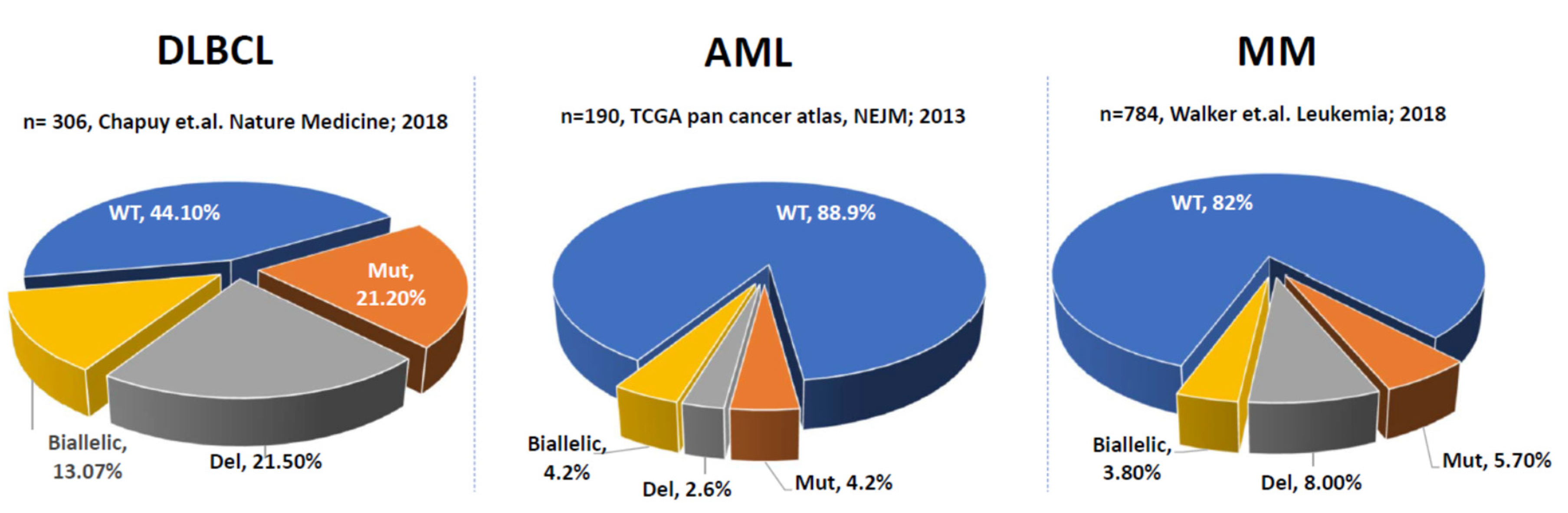
2. P53 Aberrations in Solid Tumors and Hematological Malignancies

TP53 was originally discovered as a binding partner of simian virus 40 large T antigen in virally- transformed cells [7,8,9,10]. Initially it was classified as an oncogene, but later work established its role as a tumor suppressor [11]. A variety of inactivating TP53 mutations have been reported in human cancers and germline mutations in TP53 are a hallmark of Li-Fraumeni syndrome, a hereditary cancer predisposition disorder [11,12,13].
Approximately 50% of human cancers have TP53 alterations [14,15,16]. In The Cancer Genome Atlas (TCGA) dataset that includes 32 distinct studies and over 10,000 cancer cases, the prevalence of TP53 mutations are 15.20%, deletions 15.90%, and biallelic inactivation events are 22.02% of cases [17]. In this dataset, ovarian serous cystadenocarcinoma, uterine, and lung cancers have the highest prevalence of TP53 abnormalities (~90% of cases) while paraganglioma had the fewest at only 0.50% [18]. Other groups have also reported high prevalence of TP53 abnormalities in solid tumors, particularly ovarian, pancreatic, breast, and small cell lung cancer [13,19,20,21]. However, Li and colleagues analyzed data from 7893 patients and found that TP53 mutations were only prognostically relevant in 9 cancer types in the TCGA dataset including lung adenocarcinoma, hepatocellular carcinoma, head and neck squamous cell carcinoma, acute myeloid leukemia (AML) and clear cell renal carcinoma [18]. Approximately 80% of TP53 mutations are missense mutations and are localized in the DNA-binding domain. Eight of these mutations (R175, V157F, Y220C, G245, R248, R249, R273 and R282) account for ~28% of total mutations in TP53 with R5, R248 and R273 being reported in multiple tumor types, suggesting that there is a selection for these mutant alleles in cancer [22].
In addition to single-allele missense mutations, loss of heterozygosity (LOH) in the second allele of TP53 has been reported in multiple solid tumor studies and mutations in this allele were significantly higher (25–37%) than in non-del17P cases [12,15,23,24,25,26]. Analysis of TP53 gene and pathway alterations in 32 tumor types from the TCGA dataset revealed that ~91% of cancers exhibit biallelic inactivation of the TP53 gene. The second allele loss was due to either mutation, chromosomal deletion, or by copy neutral LOH [27]. Gene expression profiling of both cell lines and patient samples suggested that even monoallelic deletion of TP53 can result in significantly lower expression levels [26,28].
Compared to solid tumors, dysregulation of TP53 is less frequent in hematological malignancies, for example, in diffuse large B-cell lymphoma (DLBCL) and AML, ~10–50% of cases have alterations in TP53 (Figure 1). In DLBCL, biallelic inactivation is the most common TP53 aberration (13%) while deletion and mutation are each present in ~20% of cases. In AML, alterations in TP53 are less common with biallelic inactivation and mutation present in ~4% of cases each and deletion reported in only ~3% of cases. There is only one dataset with SNV data from 211 MM patients available in TCGA which lacks copy number variation (CNV) data, thus providing incomplete information about monoallelic versus biallelic inactivation of TP53 in MM [29]. Our analysis from MGP demonstrated that deletion is the most common abnormality at 8%, followed by mutation (~6%) and biallelic inactivation (~4%) (Figure 1). Even though the prevalence of TP53 aberrations is high across multiple tumors, their biological effects are still poorly understood. Various studies have suggested that missense mutations in TP53 are gain of function (GOF) mutations and confer oncogenic functions to P53 [22,30,31,32,33,34,35,36,37]. In contrast, other studies suggest that missense mutations in TP53 are loss of function (LOF) and act through a dominant-negative mechanism affecting oligomerization [38,39,40,41,42]. A recent report conducted a detailed analysis of TP53 hot spot missense mutations in human myeloid malignancies applying genome editing, saturation mutagenesis screening and mouse models [40]. This analysis showed that in AML, TP53 mutations do not confer a neomorphic GOF potential but instead, are dominant negative in nature and affect the tumor suppressor function of the protein [40].

3. Prognosis of Del17p/TP53 Inactivation in Multiple Myeloma

3.1. Deletion of 17p in MM

Chromosomal aberrations, including translocations and CNVs are common in MM. However, heterogeneity in patient populations, detection methods, threshold (defined as the percentage of tumor cells positive, also known as CCF), sample sizes, and treatment regimens have made it challenging to interpret their impact on clinical outcome. The prognostic relevance of del17p was described in an analysis of NDMM patients in the Intergroupe Francophone de Myélome 99 (IFM99) trial [43]. Del17p (identified by fluorescent in situ hybridization [FISH]) in CD138-positive tumor cells was present in 11% of patients (58 out of 532) where patients with the deletion had significantly shorter median event-free survival (EFS; 14.6 vs. 35 mo, p < 0.001) and median overall survival (OS, 22.4 mo vs. not reached [NR], p < 0.001).
Del17p was one of two markers that retained prognostic value for both EFS and OS in a multivariate analysis of a large NDMM patient dataset, and is included in the R-ISS as a standard part of risk assessment in the clinic [4]. However, one source of variability among studies has been the threshold used to determine whether patients are considered positive for del17p. In the IFM99 study, del17p patients had a median of 75% plasma cells positive for the deletion, and those with ≥60% positive cells (CCF >0.6) were considered high-risk. The majority of studies have used a cutoff of >20% to determine a significant impact on clinical outcome and some have shown that a patient should have ~60% cells positive for the deletion in order to be included in the high-risk del17p subgroup [25,43,44,45,46,47,48,49,50]. However, some groups have used thresholds below 20%, including one Phase 3 study that counted a patient positive for del17p if even a single cell was positive by FISH [51,52]. The use of different thresholds/CCFs, different size datasets, as well as different treatment regimens have resulted in discordance in the reported prognosis of del17p patients (Table 1). In NDMM, the median progression free survival (PFS) for patients with del17p has been reported to be approximately 15 months in several studies, with a range from 4 to 26 months (Table 1). Differences in threshold/CCF as well as treatment intensity impact the clinical outcomes of these patients. The study by Chang and colleagues, and that by An and colleagues reported shorter median PFS compared to others in Table 1, but it should be noted that these patients were treated with high-dose chemotherapy or chemotherapy plus either thalidomide or bortezomib.
The MGP systematically evaluated large FISH and genomic datasets for the association of CCF to OS in NDMM patients [6]. This analysis indicated that patients with greater than 55% del17p-positive cells (CCF > 0.55), had poor clinical outcomes, where patients had median PFS and median OS of only 14 and 36 months as compared to the low CCF group (≤0.55) who had median PFS of 24 months and median OS of 84 months (FISH dataset, n = 605) [6]. The CCF > 0.55 threshold to identify high-risk del17p patients (high-risk [HR] del17p, ~7% of NDMM) was validated in a meta-analysis across three datasets, the original test dataset from the IFM, as well as an independent FISH replication dataset (n = 235) and a genomic dataset (n = 108) [6].
The poor outcome of high-CCF del17p was also demonstrated in analysis of multiplex ligation-dependent probe amplification (MLPA) data, a multiplex polymerase chain reaction (PCR) method for simultaneously detecting CNVs at different genomic regions, where patients with MLPA cutoffs corresponding to ≥50% tumor cells positive for del17p had significantly shorter PFS and OS than those with fewer del17p-postive cells [47,53]. These data are consistent with earlier reports suggesting that a CCF of ~60% should be used to identify high-risk patients [43].
Several studies investigated clinical outcomes of relapsed and/or refractory MM (RRMM) patients with del17p (Table 2). Lakshman and colleagues reported data in 76 MM patients who were negative for del17p at diagnosis who later tested positive for del17p. The presence of del17p was detected at a median of 35.6 months after diagnosis (median of 2 lines of therapy), and was associated with short median PFS (30.1 vs. 23 mo; p = 0.032) and median OS (106.1 vs. 68.2 mo; p < 0.001) in comparison to patients without detectable del17p in the same timeframe [44,54]. Other reports of clinical outcomes in del17p-positive patients demonstrate the association of poor clinical outcome with median PFS ranging from 3.4 to 21.4 months, presumably impacted by threshold/CCF used as well as treatment intensity in individual patient cohorts. The recent analysis from Avet-Loiseau and colleagues demonstrates the impact of treatment intensity where patients with >60% del17p-positive cells had a median PFS of 15.47 months in the triplet arm versus only 5.1 months in the doublet arm [55]. Taken together these data demonstrate that patients with del17p have poor clinical outcomes, and that patients with high CCF del17p are at particularly high risk of early progression and death compared to low-CCF patients in both NDMM and RRMM settings. These findings are clinically relevant and CCF should be considered for evaluation of del17p in MM patients and integrated into risk stratification guidelines.

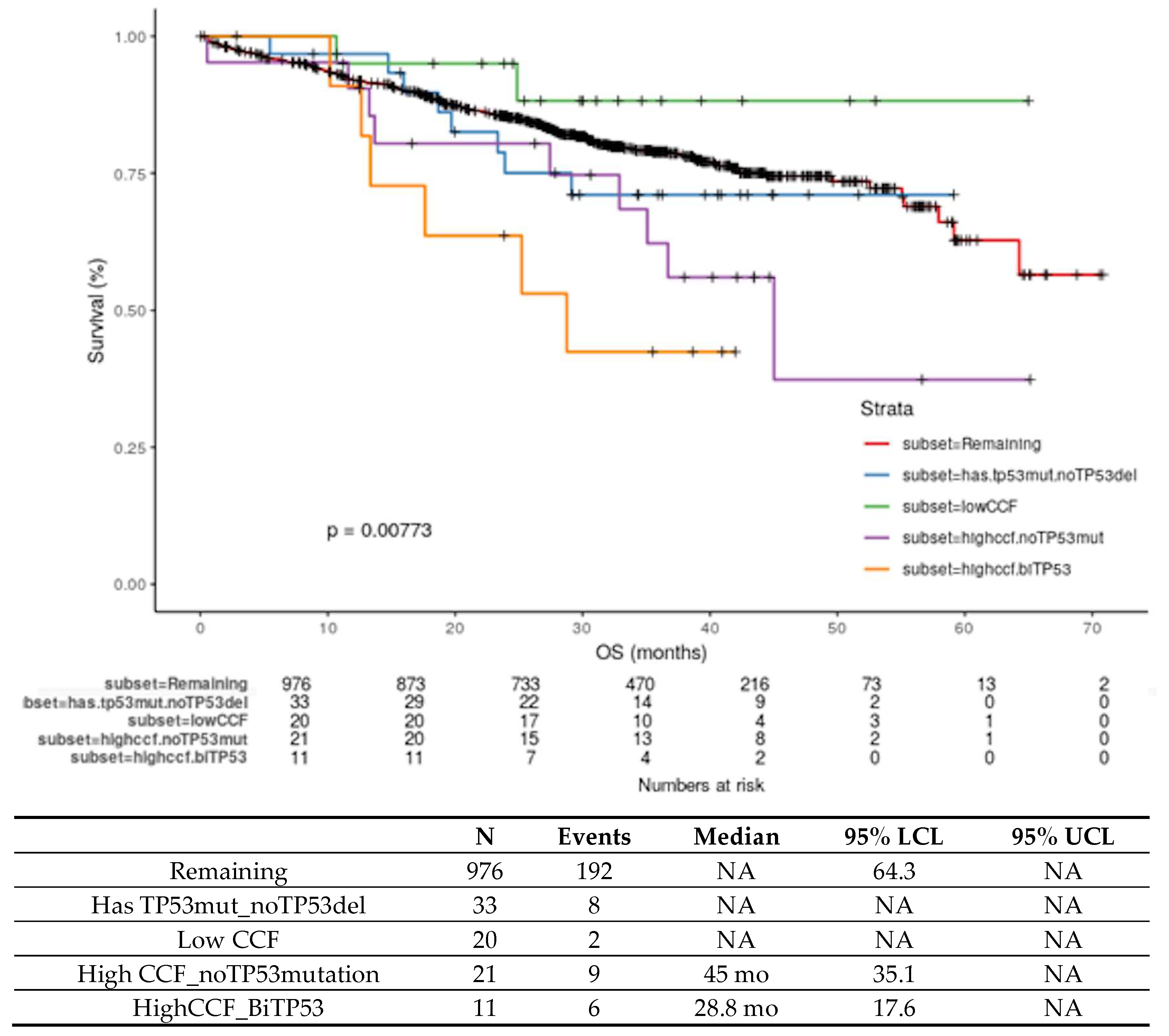
3.2. Biallelic Inactivation of TP53 in MM

Early reports from analyses of small numbers of patients suggested an association between deletion on one allele and mutation on the second allele of chromosome 17p, putatively resulting in complete inactivation of P53 function [25,44,46]. These studies reported clinical outcome of patients with biallelic inactivation as well as del17p alone; however, small numbers limited the ability to differentiate the impact of biallelic versus monoallelic inactivation. In a cohort of 92 NDMM patients, Lodé and colleagues reported that among 54 patients with del17p, 20 (37%) also had a mutation in TP53; however, no differences in survival were observed between patients with complete inactivation of TP53 and those with del17p alone [25]. In a larger NDMM dataset (n = 779) where both TP53 mutation and FISH data were available (n = 72), a significant correlation between mutation and deletion was observed [44]. Patients with biallelic inactivation (n = 7) had significantly shorter PFS and OS than patients with monoallelic inactivation (n = 26, del17p or TP53 mutation alone) where 3-year OS was 29% versus 84% and 3-year PFS was 29% versus 73%. This study did not separately report the outcomes for patients with only a mutation or deletion.
From the MGP, using the largest uniformly-processed genomic dataset from NDMM patients (n = 1273), DHMM was identified and independently validated as a molecularly-defined high-risk group. DHMM is ~6–8% of NDMM and includes two groups of patients, (a) biallelic inactivation of TP53 and (b) amplification (≥4 copies) of 1q21 with ISS3 with median PFS of 15.4 months and median OS of 20.7 months [5,62]. Patients with biallelic inactivation of TP53 had significantly shorter PFS than patients with monoallelic del17p, (18 mo estimate: PFS 36% vs. 76%; OS: 58% vs. 90%) [5]. However, this analysis did not consider the CCF >0.55 cut off for determination of HR del17p.
Using the >0.55 CCF cutoff defined by genomics-based methods, analysis from MGP identified high-risk del17p patients as discussed in Section 3.1. The MGP data also showed a significant association between the presence of CCF >0.55 and of mutation on the second allele of TP53, where 27 of 28 patients with a TP53 mutation had CCF >0.55 [6]. Patients with biallelic inactivation had significantly shorter PFS than patients with one wild-type copy of TP53 [6]. In an updated analysis of this dataset with longer clinical follow-up, patients with HR del17p + TP53 mutation have the shortest OS (Figure 2, orange line, median 28.8 mo, p = 0.008). Figure 2 provides updated OS data for patients with HR del17p (median 45 mo) in the MGP dataset. These data highlight that NDMM patients with either biallelic inactivation of TP53 or high CCF del17p have very poor clinical outcome. Together, these two groups of P53 dysregulated patients comprise approximately 6–7% of the NDMM population (the addition of the DHMM amp1q segment would bring this to ~10% of NDMM).
The timing and order of acquisition of deletion and mutations leading to biallelic inactivation of TP53 in MM patient samples has not been widely studied. A recent preclinical study was performed in isogenic AMO-1 MM cell lines containing monoallelic and biallelic TP53 variants followed by in-vitro competition assays. Interestingly the authors found that biallelic TP53 inactivated cells outcompete monoallelic TP53 variant containing cells. Although this study was limited to one cell line, the data provided some experimental evidence for the acquired proliferation fitness of biallelic clones [63]. Due to the recessive nature of tumor suppressor mutations, biallelic inactivation is generally a prerequisite for acquiring an oncogenic or tumor maintenance phenotype. While biallelic inactivation would result in loss of P53 function, it is unclear what drives the high-risk feature of a higher clonal fraction of del17p cells with respect to P53 function.

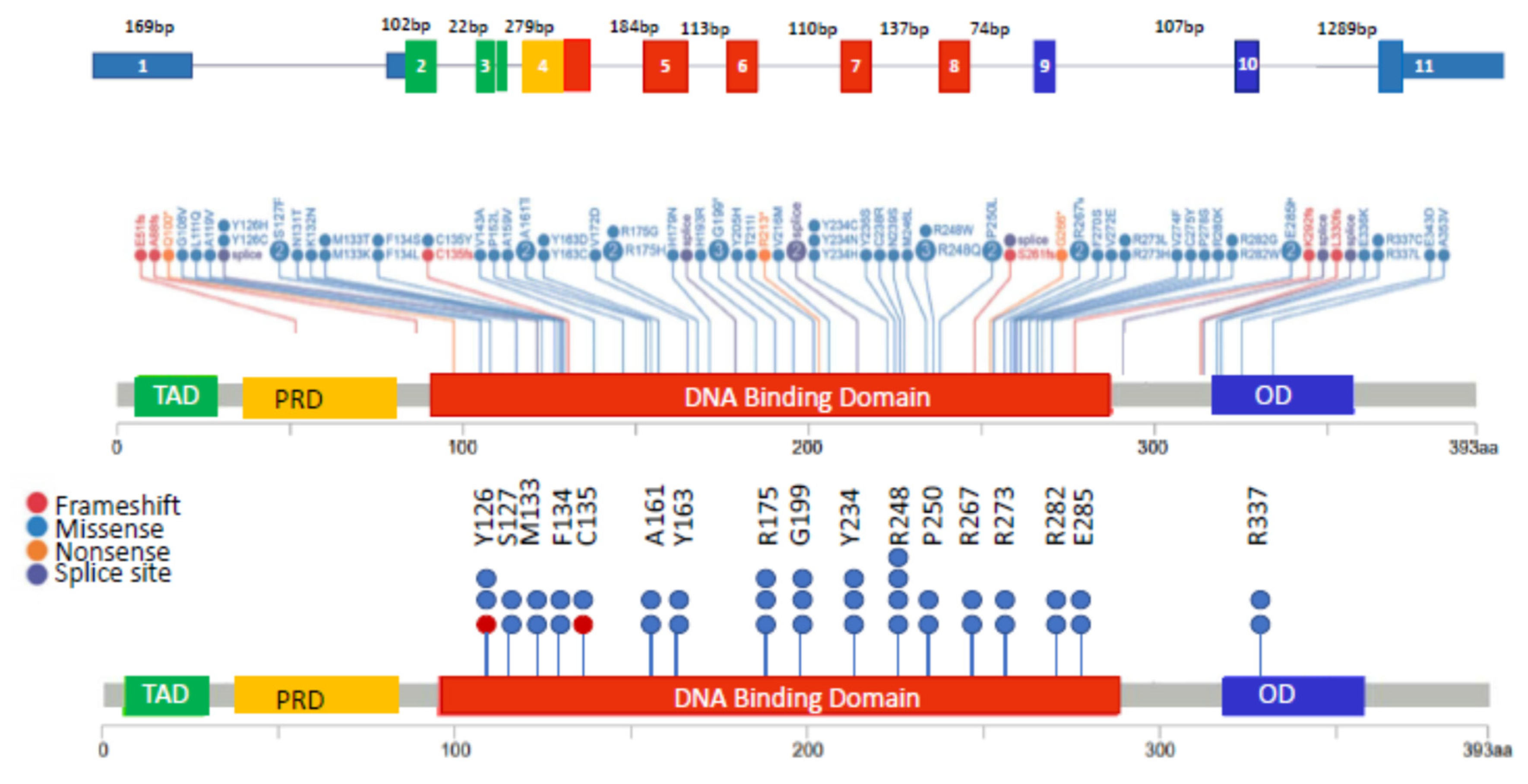
3.3. Monoallelic Mutation of TP53 in MM

In NDMM, TP53 mutations are generally present in 3–8% of patients (Figure 1) [5,46,64]. As in other tumor types, TP53 mutations in MM are spread across the entire gene, with many mutations occurring within the DNA-binding domain [5]. Figure 3 highlights the structure of the TP53 gene (top), mutations in TP53 in the MGP dataset (middle panel) as well as the sites most frequently mutated (Figure 3, bottom) in ≥2 patients. TP53 mutation was identified as a driver event in MM [65]. However, the relevance of monoallelic TP53 mutation as an independent poor prognostic marker in MM has not been established. Owen and colleagues concluded that TP53 mutations were rare events in MM (detected in 1/31 [3%] patients) and were therefore of limited prognostic value [64]. Chng and colleagues reported the prognosis in NDMM where TP53 mutations were detected in 3% (9/268) of patients and were associated with short OS compared to patients without a mutation, however, it should be noted that ~50% of this cohort of patients also had a del17p [46,66]. Another study reported that patients with TP53 mutation (n = 20) did not have a significantly different outcome compared to those without a mutation [25]. In the MGP dataset, there were 33 patients with monoallelic mutation of TP53, and after a median follow-up of 29.8 months, the OS of these patients (Figure 2, monoallelic mutation, blue line) was not significantly different than patients without a TP53 abnormality (Figure 2, remaining, red line). Longer follow-up or analysis of larger datasets are needed to clarify the prognostic value of monoallelic TP53 mutation in MM patients.
Nonetheless, TP53 mutations could be important drivers for maintaining/propagating MM clones in association with co-occurring driver gene mutations. The variant allele frequencies of the TP53 mutations are sub-clonal for the majority of the mutations observed in the 33 patients within the MGP dataset. The functional consequence of TP53 mutations in MM is yet to be experimentally determined, but in silico predictions suggest that multiple hot spot mutations may have a damaging effect. Figure 3 (bottom) and Table 3 summarizes the TP53 hot spot mutations (detected in ≥2 patients) in MM and their predicted functional impact and score from three different in silico methods: Mutation Assessor, Sorting Intolerant from Tolerant (SIFT), and Polymorphism Phenotyping V2 (PolyPhen2) [67,68,69]. The majority of TP53 hotspot mutations are predicted to be medium impact by Mutation Assessor and the same mutations are predicted as deleterious and damaging by SIFT and PolyPhen2 algorithms. Mutation Assessor classifies mutations into 4 categories based on functional impact score (FIS) to estimate the probability of phenotypic consequence of the mutation. A FIS score ≤0.8 is classified as neutral impact, FIS score of 0.8–1.9 as low impact, FIS score of 1.9–3.5 as medium impact and >3.5 as high impact. Among the hot spot mutations observed in MM, mutations at Y126, R175, R248, R273, and R282 are also common in other tumor types in the TCGA database [17]. Laboratory validation is needed to confirm the predicted functional consequence of the observed mutations in MM cells.

4. Biology of Del17p/P53 Inactivation

In normal cells, P53 is maintained at a low level by a series of regulators and is activated by a variety of stress stimuli. P53 controls a vast genetic network, complex transcriptional programs, and diverse biological responses that have been recently reviewed [70]. The most studied function of P53 is its ability to promote cell cycle arrest and apoptosis via transcription of P21 in response to DNA damage, which is deemed central to its role in tumor suppression. P53 also controls many other biological processes including metabolism, proliferation, inflammation, autophagy and epithelial to mesenchymal transition [70]. The complexity of the P53 signaling network has made interpretation of P53′s function, and consequences of its dysfunction challenging—particularly when considering the impact of cell type and mechanism of inactivation (e.g., mutation, deletion, or both). Here we review the major roles of P53 and the impact of P53 dysfunction in cancer overall and in MM specifically.

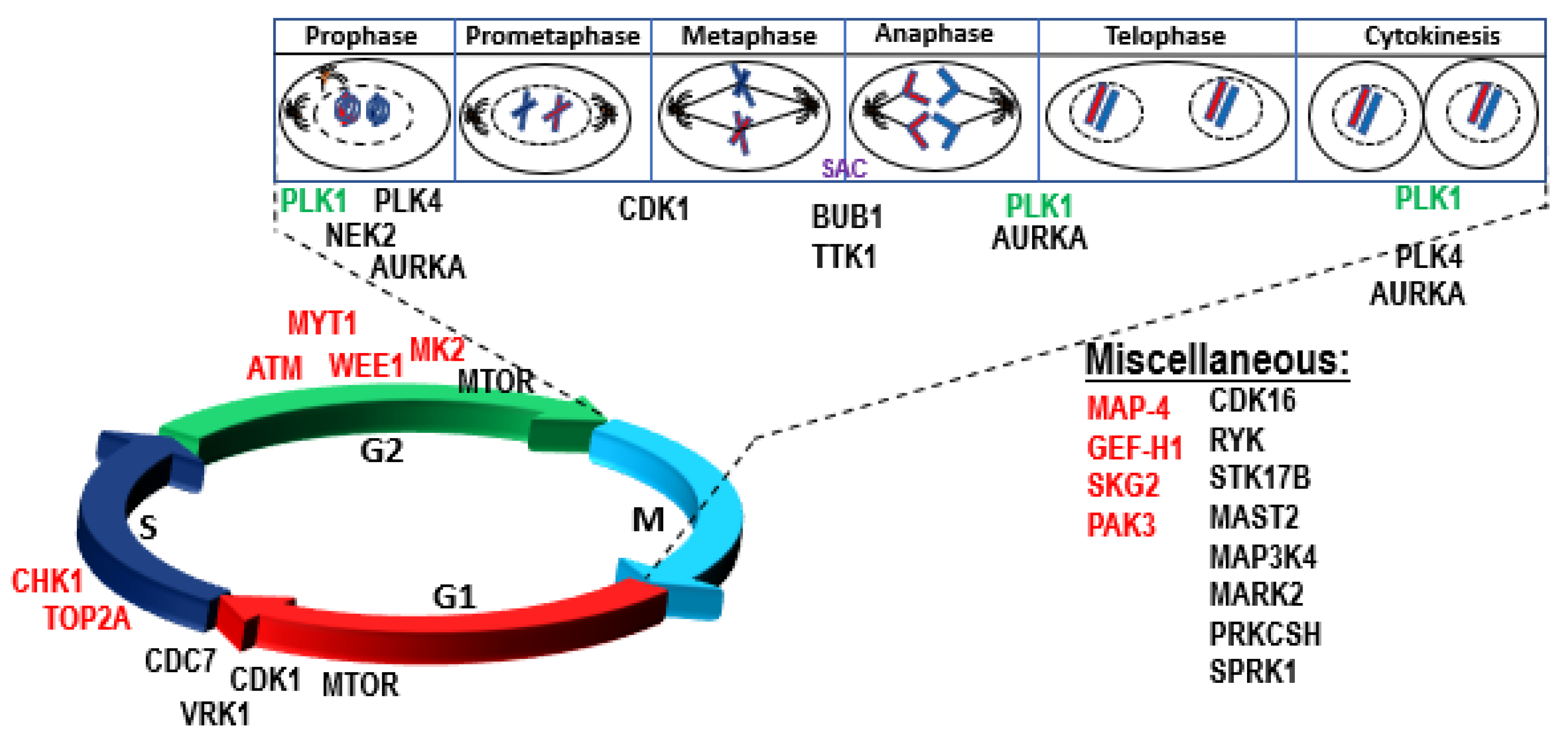
4.1. Role of P53 in Genomic Instability, DNA Repair, Aneuploidy, and Checkpoint Control

P53 maintains genome stability by inducing cell cycle arrest, senescence, or apoptosis upon DNA damage to reduce the risk of propagation of a defective genome. Genome instability is an inherent characteristic of almost all human cancers and is accompanied by dysregulation of several cellular processes, such as DNA replication, G2/M cell cycle checkpoint control, chromosomal segregation, DNA repair, and genome integrity. P53 is frequently inactivated in cancer resulting in tumors characterized by gross structural defects, chromosomal missegregation, ploidy changes, and higher prevalence of chromothripsis [12,14,49,71,72,73,74]. A pan-cancer analysis demonstrated an association between aneuploidy and mutation of TP53 [14]. One of the mechanisms by which dysregulated P53 is thought to contribute to aneuploidy is via regulation of G2/M processes and centrosome amplification [75,76,77]. Multiple studies have shown a complex link between proteins involved in cell cycle regulation, mitotic checkpoints, genomic instability, tumorigenesis, and the P53 pathway [78,79,80,81]. A tightly regulated feedback loop has been reported between P53 and mitotic kinases (eg, WEE1, PLK1, NEK2, BUB1, TTK, AURKB and PLK1) [79,82,83,84]. Phosphorylation of P53 in response to mitotic spindle damage has also been reported [85]. The intricate details of the mechanisms and regulatory signals between mitotic kinases and P53 remain poorly understood, but P53 dysfunction disrupts this critical regulation, resulting in abrogation of the G1 checkpoint and upregulation of mitotic kinases allowing cells to override the G2/M checkpoints and contribute to genome instability and tumorigenesis. A clear understanding of the biology of dysregulation of P53 dependent physiological processes in biallelic, del17p, and mutated P53 patients is critical in identifying new anti-cancer targets.

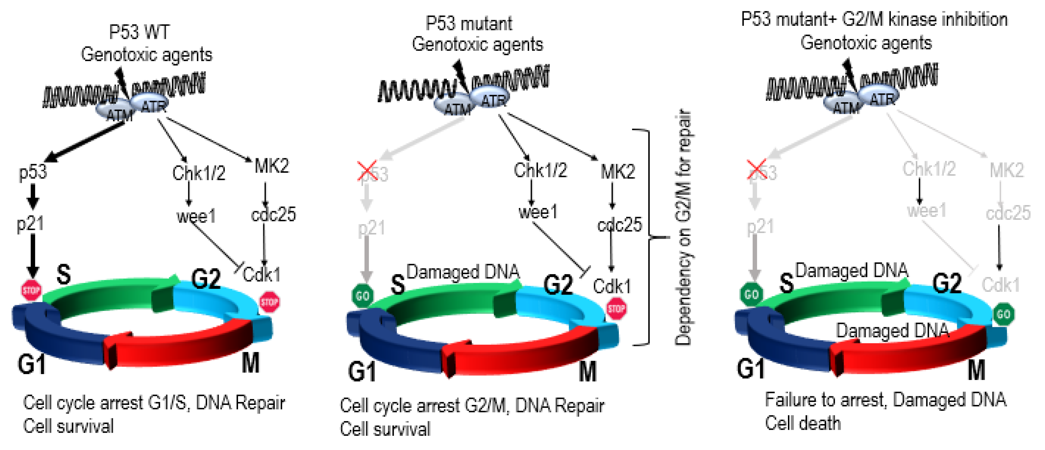
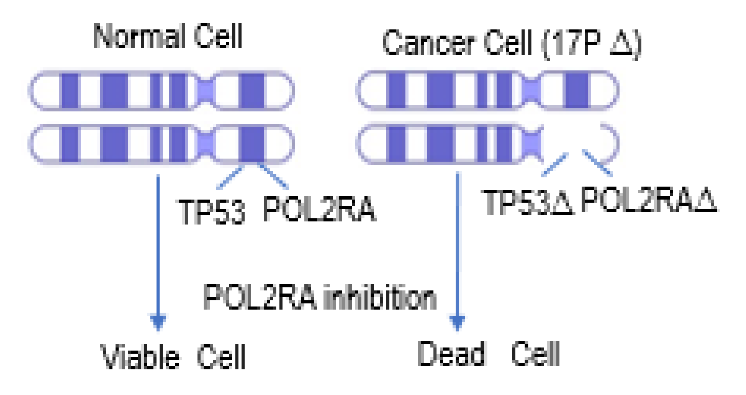
4.2. P53 Synthetic Lethality

One way to explore new targets is by identifying vulnerabilities of cancer cells with dysregulated P53 that could provide opportunities to selectively kill or inhibit the growth of P53-deficient cells versus those with wild-type P53 (Figure 4). Loss of the G1/S cell cycle checkpoint in P53-deficient cancer cells renders them entirely dependent on the G2/M checkpoints to maintain genome integrity [86]. Such dependency exposes a unique vulnerability of P53-deficient cancer cells, resulting in synthetic lethal relationship between P53 and multiple genes/pathways (Figure 5). P53-deficient cancer cells exhibit synthetic lethal interactions with ionizing radiation and genotoxic agents (e.g., cisplatin, camptothecin, doxorubicin). For example, P53-deficient cells are more sensitive to genotoxic stress when treated with inhibitors of ATR, Chk1, PLK1, and Wee1 kinases versus cells with functional P53 [87,88,89,90,91]. P53-deficient cells have also been reported to be dependent on the p38MAPK/MK2 pathway for survival following treatment with DNA-damaging agents. MK2 depletion in P53-deficient cells suppressed Cdc25A-mediated S phase arrest following cisplatin treatment and Cdc25B-mediated G2/M arrest following doxorubicin exposure, resulting in mitotic catastrophe and tumor regression in vivo [92]. Further, inhibition of ATM also exhibited synthetic lethality with topoisomerase inhibitors in a P53-deficient background [93] (Figure 4). P53 synthetic lethality has also been reported with SGK2, PAK3, CHK1, Wee1 and Myt1 in cervical cancer cell lines [94,95].
Additionally, Wang and Simon employed a computational method to predict genes with P53 synthetic lethality. Using publicly-available cell line and gene expression datasets, they identified 18 kinases with potential synthetic lethal interactions with P53, including PLK1, NEK2, BUB1, and AURKA [96]. Another study identified a similar set of potential P53 synthetic lethal genes by analyzing publicly-available data in 33 different human cancer types to identify 120 genes that were overexpressed in P53 deficient cells/tumors, including 19 genes that were common across tumor types [97]. A significant number of these putative P53 synthetic lethal genes are potentially druggable mitotic kinases (AURKA, BUB1, BUB1B, CDK1, MELK, NEK2, PLK1 and TTK), with 6 out of 8 kinases common across both studies [96,97].
The region of chromosome 17p that is commonly deleted includes several essential genes that are adjacent to TP53. For example, one of the largest subunits in the human RNA polymerase II complex (POLR2A) resides in close proximity to the TP53 gene locus (Figure 6). Concomitant deletion of POLR2A with hemizygous TP53 deletion has been reported in multiple human cancers [98,99]. Unlike TP53 that is regulated post-transcriptionally and post-translationally, expression of POLR2A is directly correlated with gene copy number. Hence, the inhibition of the POLR2A gene in cells with hemizygous del17p genomic deletion resulted in synthetic lethality and increased cell death [99]. E3 ligase Ring-Box 1 (RBX1) has also been identified as another P53 synthetic lethal partner which regulates POLR2A-mediated mRNA synthesis by K63 linked ubiquitination. Inhibition of RBX1 in P53 deleted castration-resistant prostate cancer cell lines resulted in synergistic inhibition of cell growth [98].

P53 Synthetic Lethality in MM

MM has remarkable genomic instability that leads to accumulation of aberrations resulting in tumor progression, drug resistance, and metastasis [100]. For example, increased DNA double-stranded breaks (DSB) may lead to disease progression in MM [101]. Genomic instability provides a growth advantage and may allow acquisition of drug resistance, but it may also create vulnerabilities that can be exploited by targeting synthetic lethal interactions [102]. An example of this strategy is induced sensitivity of MM cells to poly (ADP-ribose) polymerase (PARP) inhibitors following 26S proteasome inhibition. Inhibition of the proteasome in MM cells abrogates H2AX polyubiquitination and abolishes recruitment of BRCA1 and RAD51 to DSB sites and homologous-recombination (HR)-mediated DNA repair [103]. Co-treatment of MM cells with proteasome inhibitors and PARP inhibitors leads to accumulation of unrepaired DNA DSBs and cell death [103]. Similarly, mutations in DNA editing enzymes, (eg, APOBECs) have been reported in a specific MM genomic subgroup that is associated with primary/secondary translocations, poor prognosis, drug resistance, oncogenic activation, and sub-clonal diversity [104,105,106,107,108,109,110]. APOBEC mutagenesis induces the DNA damage response and can result in cell death [111,112,113]. Therefore, loss of P53 enables tolerance to APOBEC-mutagenesis-induced DNA damage and promotes cancer cell survival [113]. Together, these data suggest that targeting genes with a synthetic lethal relationship with P53 could be an effective therapeutic approach for multiple P53-deficient malignancies. Additional research is needed to identify genes with significant P53 synthetic lethal relationships in MM.

4.3. Biology of High-risk Del17p in MM

The clinical relevance of del17p is well established in MM, but the exact mechanism by which del17p promotes aggressive disease biology remains unclear. Questions remain on the role of the deletion size, clonality, and cellular signaling. Without a better understanding of the features that are driving poor clinical outcome, development of effective therapies to target those features remains challenging. An integrative multi-omics analysis identified 12 distinct disease subsets of MM and indicated that del17p and DHMM, despite being associated with poor clinical outcome, did not all cluster together in one subset [114]. Instead, patients with DHMM or del17p were distributed across several subsets, including high-risk and non-high-risk groups indicating that these features are not the only drivers of the underlying high-risk biology.
The length of the deleted region can vary from a few mega bases (MBs) to deletion of the entire short arm of chromosome 17. The TP53 gene is located in the minimally deleted region (0.25 MB) suggesting that it is a critical gene in the 17p13 region [50]. A deletion event usually involves several genes and it remains unclear how genes other than TP53 contribute to tumorigenesis. A heterozygous deletion of a 4MB region in mouse chromosome 11B3, syntenic to human 17p13.1, showed that co-deletion of TP53 along with Eif5a and Alox15b resulted in more aggressive disease [115]. Additional research is needed to improve our understanding of drivers of high-risk biology in MM patients with del17p.

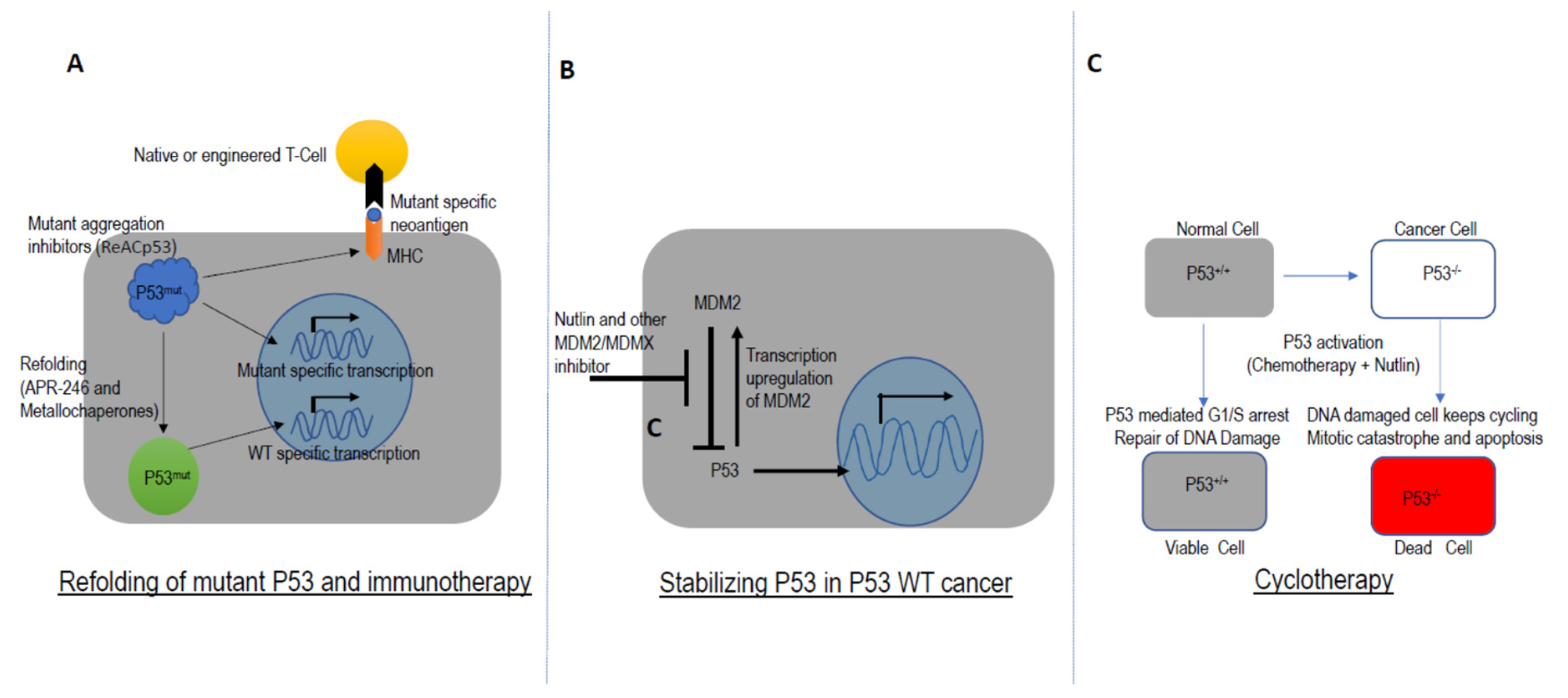
5. Targeting TP53 in Drug Development

The majority of TP53-related drug development efforts have been directed towards designing therapies to exploit cancer-specific vulnerabilities associated with dysregulation of TP53, such as del17p, TP53 mutations, TP53 promoter methylation, and MDM2 overexpression [70,116]. The therapeutic utility of synthetic lethal interaction of P53 with POL2RA is currently being examined in preclinical models. The amanitin toxin, which inhibits POL2RA is being tested as an antibody-drug conjugate (ADC). In MM, anti-B cell maturation antigen (anti-BCMA) amanitin-ADCs have shown efficacy and tolerability in preclinical models [117,118]. Additional testing is needed to determine clinical efficacy and tolerability of these agents.
Another promising approach has been the deployment of compounds which can restore the wild-type function of P53. Small molecules and peptides have been designed to stabilize P53 mutant proteins [119,120,121,122,123]. Metallochaperones have been reported to restore the function of mutant P53 by zinc incorporation [124]. Similarly, inhibition of amyloid-like structure formation in aggregation-prone mutants of P53 has shown promise as a therapeutic strategy in TP53-deficient tumors [124,125]. For example, APR-246 has been reported to reactivate mutant P53 and is currently in clinical development [119] (Figure 7A).
MDM2, and E3 ubiquitin ligase, regulates P53 activation in multiple ways including interaction with the transactivation domain of P53 and inhibition of P53 activity [126,127]. MDM2 also facilitates P53 nuclear export and can negatively regulate P53 through ubiquitin-mediated degradation by the proteasome [128,129,130,131]. In normal cells, MDM2 helps maintain low levels of P53 by inducing continuous ubiquitin-mediated degradation of P53. However, in response to cellular stress (eg, DNA damage, hypoxia, oncogenic activation), the interaction between MDM2 and P53 is disrupted, which leads to stabilization of P53. Interestingly, P53-mediated transcription also regulates MDM2. Thus, MDM2 and P53 are closely linked to each other through an autoregulatory negative feedback loop [132,133]. MDM2 is overexpressed in some plasma cell leukemia patients and several MM cell lines, resulting in inhibition of P53 activity [134]. Stabilization of P53 by inhibiting the MDM2-P53 interaction offered a novel strategy and led to development of Nutlin [116,135] (Figure 7B). Nutlin was reported as the first inhibitor of the P53-MDM2 interaction which demonstrated synergistic activity in MM with known anti-MM agents such as bortezomib, melphalan, and etoposide [135,136,137]. However, Nutlin is only effective in cells with wild-type P53, making it ineffective in del17p and mutant P53 backgrounds. Phase 1 trials with MDM2 antagonists in leukemia and liposarcoma have exhibited dose-limiting toxicities including neutropenia and thrombocytopenia [138].
The focus on MDM2 antagonism also led to the development of the concept of cyclotherapy (Figure 7C). These agents induce transient cell cycle arrest by stabilizing P53 in WT cells while P53 mutant cells continue to divide and exhibit enhanced sensitivity to chemotherapeutic agents [139]. The clinical development of cyclotherapy agents as well as preclinical efforts to discover cyclotherapeutic drug combinations are currently ongoing [140,141].
Another potential anticancer therapy in WT P53 containing tumors is based on cellular senescence, a known barrier to tumorigenesis [142]. Mouse cancer models have shown clear evidence of P53-dependent senescence in tumor suppression, and P53-induced senescence by MDM2 antagonists have shown promise as anticancer therapy [143,144,145,146,147,148].
Attempts to reintroduce wild-type P53 using gene therapy have unfortunately remained unsuccessful. Adenovirus-mediated transfer of wild-type P53 in ovarian cancer patients failed in randomized phase II/III trial [149]. It has also been shown that mere expression of wild-type P53 is not sufficient to arrest the growth of all transformed cells.
P53-specific antigenic peptides can be presented on major histocompatibility complex molecules from tumor cells overexpressing mutant P53 and can evoke an antitumor immune response. Vaccination against mutant P53 has been shown to be effective in tumor-bearing mice [150,151]. Several peptide vaccines and dendritic cell vaccines utilizing mutant P53-targeted immunotherapy are in clinical development [152,153,154,155]. A number of Phase I/II immunization trials have been conducted so far using P53 immunogens, but unfortunately none of them have shown acceptable clinical efficacy [156].

6. Conclusion and Future Perspective

There are 3 distinct segments of patients with TP53 dysregulation that have been identified in MM: monoallelic mutation, del17p, and biallelic inactivation. Based on analyses of the MGP data, the biallelic and high CCF del17p patients appear to have poorest prognosis, while the prognostic role of monoallelic mutations is less clear. Analysis of longitudinal patient samples is needed to more fully understand the timing and sequence of these aberrations as well as clinical outcomes following treatment with specific therapeutic regimens in each patient subset. Further, additional research is needed to identify co-occurring genetic interactions of P53/del17p dysregulation in MM. A reverse translational approach could identify dysregulated pathways and disease drivers in these segments from patient sample analysis. This approach could lead to discovery of new targets and eventually to new therapies to address the unmet medical need of these MM patients.

Author Contributions

Author contributions are as follows: conceptualization, E.F. and A.T.; writing—original draft preparation, E.F., K.B., V.S., F.T.; formal analysis, F.T., K.B., M.O., E.F., A.T.; writing—review and editing, E.F., K.B., V.S., F.T., M.O., A.T.; visualization, E.F., K.B., F.T.; supervision, A.T.; project administration, E.F.; funding acquisition, E.F., A.T. All authors have read and agreed to the published version of the manuscript.

Funding

This research was funded by Celgene Corporation.

Acknowledgments

The authors acknowledge continued support from MGP collaborators in academia and colleagues within Celgene as well as the Celgene MM Translational Development group.

Conflicts of Interest

The authors report employment and equity ownership in Celgene Corporation.

References

  1. The National Cancer Institute. Cancer Stat Facts: Myeloma. Available online: https://seer.cancer.gov/statfacts/html/mulmy.html (accessed on 26 May 2019).
  2. Fonseca, R.; Abouzaid, S.; Bonafede, M.; Cai, Q.; Parikh, K.; Cosler, L.; Richardson, P. Trends in overall survival and costs of multiple myeloma, 2000–2014. Leukemia 2016, 31, 1915. [Google Scholar] [CrossRef] [Green Version]
  3. Kumar, S.K.; Dispenzieri, A.; Lacy, M.Q.; Gertz, M.A.; Buadi, F.K.; Pandey, S.; Kapoor, P.; Dingli, D.; Hayman, S.R.; Leung, N.; et al. Continued improvement in survival in multiple myeloma: Changes in early mortality and outcomes in older patients. Leukemia 2014, 28, 1122–1128. [Google Scholar] [CrossRef] [PubMed] [Green Version]
  4. Palumbo, A.; Avet-Loiseau, H.; Oliva, S.; Lokhorst, H.M.; Goldschmidt, H.; Rosinol, L.; Richardson, P.; Caltagirone, S.; Lahuerta, J.J.; Facon, T.; et al. Revised International Staging System for Multiple Myeloma: A Report From International Myeloma Working Group. J. Clin. Oncol. 2015, 33, 2863–2869. [Google Scholar] [CrossRef] [PubMed]
  5. Walker, B.A.; Mavrommatis, K.; Wardell, C.P.; Ashby, T.C.; Bauer, M.; Davies, F.; Rosenthal, A.; Wang, H.; Qu, P.; Hoering, A.; et al. A high-risk, Double-Hit, group of newly diagnosed myeloma identified by genomic analysis. Leukemia 2018. [Google Scholar] [CrossRef] [PubMed]
  6. Thakurta, A.; Ortiz, M.; Blecua, P.; Towfic, F.; Corre, J.; Serbina, N.V.; Flynt, E.; Yu, Z.; Yang, Z.; Palumbo, A.; et al. High subclonal fraction of 17p deletion is associated with poor prognosis in multiple myeloma. Blood 2019, 133, 1217–1221. [Google Scholar] [CrossRef] [Green Version]
  7. Lane, D.P.; Crawford, L.V. T antigen is bound to a host protein in SV40-transformed cells. Nature 1979, 278, 261–263. [Google Scholar] [CrossRef]
  8. Linzer, D.I.; Levine, A.J. Characterization of a 54K dalton cellular SV40 tumor antigen present in SV40-transformed cells and uninfected embryonal carcinoma cells. Cell 1979, 17, 43–52. [Google Scholar] [CrossRef]
  9. Kress, M.; May, E.; Cassingena, R.; May, P. Simian virus 40-transformed cells express new species of proteins precipitable by anti-simian virus 40 tumor serum. J. Virol. 1979, 31, 472–483. [Google Scholar] [CrossRef] [Green Version]
  10. DeLeo, A.B.; Jay, G.; Appella, E.; Dubois, G.C.; Law, L.W.; Old, L.J. Detection of a transformation-related antigen in chemically induced sarcomas and other transformed cells of the mouse. Proc. Natl. Acad. Sci. USA 1979, 76, 2420–2424. [Google Scholar] [CrossRef] [Green Version]
  11. Finlay, C.A.; Hinds, P.W.; Levine, A.J. The p53 proto-oncogene can act as a suppressor of transformation. Cell 1989, 57, 1083–1093. [Google Scholar] [CrossRef]
  12. Baker, S.J.; Preisinger, A.C.; Jessup, J.M.; Paraskeva, C.; Markowitz, S.; Willson, J.K.; Hamilton, S.; Vogelstein, B. p53 gene mutations occur in combination with 17p allelic deletions as late events in colorectal tumorigenesis. Cancer Res. 1990, 50, 7717–7722. [Google Scholar] [PubMed]
  13. Malkin, D.; Li, F.P.; Strong, L.C.; Fraumeni, J.F., Jr.; Nelson, C.E.; Kim, D.H.; Kassel, J.; Gryka, M.A.; Bischoff, F.Z.; Tainsky, M.A.; et al. Germ line p53 mutations in a familial syndrome of breast cancer, sarcomas, and other neoplasms. Science 1990, 250, 1233–1238. [Google Scholar] [CrossRef] [PubMed]
  14. Ciriello, G.; Miller, M.L.; Aksoy, B.A.; Senbabaoglu, Y.; Schultz, N.; Sander, C. Emerging landscape of oncogenic signatures across human cancers. Nat. Genet. 2013, 45, 1127–1133. [Google Scholar] [CrossRef] [PubMed] [Green Version]
  15. Hollstein, M.; Sidransky, D.; Vogelstein, B.; Harris, C.C. p53 mutations in human cancers. Science 1991, 253, 49–53. [Google Scholar] [CrossRef] [Green Version]
  16. Vogelstein, B.; Lane, D.; Levine, A.J. Surfing the p53 network. Nature 2000, 408, 307–310. [Google Scholar] [CrossRef]
  17. Cancer Genome Atlas Research, N.; Weinstein, J.N.; Collisson, E.A.; Mills, G.B.; Shaw, K.R.; Ozenberger, B.A.; Ellrott, K.; Shmulevich, I.; Sander, C.; Stuart, J.M. The Cancer Genome Atlas Pan-Cancer analysis project. Nat. Genet. 2013, 45, 1113–1120. [Google Scholar] [CrossRef]
  18. Li, V.D.; Li, K.H.; Li, J.T. TP53 mutations as potential prognostic markers for specific cancers: Analysis of data from The Cancer Genome Atlas and the International Agency for Research on Cancer TP53 Database. J. Cancer Res. Clin. Oncol. 2019, 145, 625–636. [Google Scholar] [CrossRef]
  19. Yachida, S.; White, C.M.; Naito, Y.; Zhong, Y.; Brosnan, J.A.; Macgregor-Das, A.M.; Morgan, R.A.; Saunders, T.; Laheru, D.A.; Herman, J.M.; et al. Clinical significance of the genetic landscape of pancreatic cancer and implications for identification of potential long-term survivors. Clin. Cancer Res. 2012, 18, 6339–6347. [Google Scholar] [CrossRef] [Green Version]
  20. Ding, L.; Getz, G.; Wheeler, D.A.; Mardis, E.R.; McLellan, M.D.; Cibulskis, K.; Sougnez, C.; Greulich, H.; Muzny, D.M.; Morgan, M.B.; et al. Somatic mutations affect key pathways in lung adenocarcinoma. Nature 2008, 455, 1069–1075. [Google Scholar] [CrossRef]
  21. Wood, L.D.; Parsons, D.W.; Jones, S.; Lin, J.; Sjoblom, T.; Leary, R.J.; Shen, D.; Boca, S.M.; Barber, T.; Ptak, J.; et al. The genomic landscapes of human breast and colorectal cancers. Science 2007, 318, 1108–1113. [Google Scholar] [CrossRef] [Green Version]
  22. Baugh, E.H.; Ke, H.; Levine, A.J.; Bonneau, R.A.; Chan, C.S. Why are there hotspot mutations in the TP53 gene in human cancers? Cell Death Differ. 2018, 25, 154–160. [Google Scholar] [CrossRef] [PubMed]
  23. Bouaoun, L.; Sonkin, D.; Ardin, M.; Hollstein, M.; Byrnes, G.; Zavadil, J.; Olivier, M. TP53 Variations in Human Cancers: New Lessons from the IARC TP53 Database and Genomics Data. Hum. Mutat. 2016, 37, 865–876. [Google Scholar] [CrossRef] [PubMed]
  24. Leroy, B.; Fournier, J.L.; Ishioka, C.; Monti, P.; Inga, A.; Fronza, G.; Soussi, T. The TP53 website: An integrative resource centre for the TP53 mutation database and TP53 mutant analysis. Nucleic Acids Res. 2013, 41, D962–D969. [Google Scholar] [CrossRef] [PubMed]
  25. Lode, L.; Eveillard, M.; Trichet, V.; Soussi, T.; Wuilleme, S.; Richebourg, S.; Magrangeas, F.; Ifrah, N.; Campion, L.; Traulle, C.; et al. Mutations in TP53 are exclusively associated with del(17p) in multiple myeloma. Haematologica 2010, 95, 1973–1976. [Google Scholar] [CrossRef] [PubMed] [Green Version]
  26. Walker, B.A.; Leone, P.E.; Chiecchio, L.; Dickens, N.J.; Jenner, M.W.; Boyd, K.D.; Johnson, D.C.; Gonzalez, D.; Dagrada, G.P.; Protheroe, R.K.; et al. A compendium of myeloma-associated chromosomal copy number abnormalities and their prognostic value. Blood 2010, 116, E56–E65. [Google Scholar] [CrossRef]
  27. Donehower, L.A.; Soussi, T.; Korkut, A.; Liu, Y.; Schultz, A.; Cardenas, M.; Li, X.; Babur, O.; Hsu, T.K.; Lichtarge, O.; et al. Integrated Analysis of TP53 Gene and Pathway Alterations in The Cancer Genome Atlas. Cell Rep. 2019, 28, 3010. [Google Scholar] [CrossRef]
  28. Teoh, P.J.; Chung, T.H.; Sebastian, S.; Choo, S.N.; Yan, J.; Ng, S.B.; Fonseca, R.; Chng, W.J. p53 haploinsufficiency and functional abnormalities in multiple myeloma. Leukemia 2014, 28, 2066–2074. [Google Scholar] [CrossRef]
  29. Lohr, J.G.; Stojanov, P.; Carter, S.L.; Cruz-Gordillo, P.; Lawrence, M.S.; Auclair, D.; Sougnez, C.; Knoechel, B.; Gould, J.; Saksena, G.; et al. Widespread genetic heterogeneity in multiple myeloma: Implications for targeted therapy. Cancer Cell 2014, 25, 91–101. [Google Scholar] [CrossRef] [Green Version]
  30. Brosh, R.; Rotter, V. When mutants gain new powers: News from the mutant p53 field. Nat. Rev. Cancer 2009, 9, 701–713. [Google Scholar] [CrossRef]
  31. Datta, A.; Ghatak, D.; Das, S.; Banerjee, T.; Paul, A.; Butti, R.; Gorain, M.; Ghuwalewala, S.; Roychowdhury, A.; Alam, S.K.; et al. p53 gain-of-function mutations increase Cdc7-dependent replication initiation. EMBO Rep. 2017, 18, 2030–2050. [Google Scholar] [CrossRef]
  32. Barta, J.A.; McMahon, S.B. Lung-Enriched Mutations in the p53 Tumor Suppressor: A Paradigm for Tissue-Specific Gain of Oncogenic Function. Mol. Cancer Res. 2019, 17, 3–9. [Google Scholar] [CrossRef] [Green Version]
  33. Xu, J.; Qian, J.; Hu, Y.; Wang, J.; Zhou, X.; Chen, H.; Fang, J.Y. Heterogeneity of Li-Fraumeni syndrome links to unequal gain-of-function effects of p53 mutations. Sci. Rep. 2014, 4, 4223. [Google Scholar] [CrossRef] [PubMed] [Green Version]
  34. Van Oijen, M.G.; Slootweg, P.J. Gain-of-function mutations in the tumor suppressor gene p53. Clin. Cancer Res. 2000, 6, 2138–2145. [Google Scholar] [PubMed]
  35. Dittmer, D.; Pati, S.; Zambetti, G.; Chu, S.; Teresky, A.K.; Moore, M.; Finlay, C.; Levine, A.J. Gain of function mutations in p53. Nat. Genet. 1993, 4, 42–46. [Google Scholar] [CrossRef] [PubMed]
  36. Kang, H.J.; Chun, S.M.; Kim, K.R.; Sohn, I.; Sung, C.O. Clinical relevance of gain-of-function mutations of p53 in high-grade serous ovarian carcinoma. PLoS ONE 2013, 8, e72609. [Google Scholar] [CrossRef] [Green Version]
  37. Olive, K.P.; Tuveson, D.A.; Ruhe, Z.C.; Yin, B.; Willis, N.A.; Bronson, R.T.; Crowley, D.; Jacks, T. Mutant p53 gain of function in two mouse models of Li-Fraumeni syndrome. Cell 2004, 119, 847–860. [Google Scholar] [CrossRef] [PubMed] [Green Version]
  38. De Vries, A.; Flores, E.R.; Miranda, B.; Hsieh, H.M.; van Oostrom, C.T.; Sage, J.; Jacks, T. Targeted point mutations of p53 lead to dominant-negative inhibition of wild-type p53 function. Proc. Natl. Acad. Sci. USA 2002, 99, 2948–2953. [Google Scholar] [CrossRef] [Green Version]
  39. Hegi, M.E.; Klein, M.A.; Ruedi, D.; Chene, P.; Hamou, M.F.; Aguzzi, A. p53 transdominance but no gain of function in mouse brain tumor model. Cancer Res. 2000, 60, 3019–3024. [Google Scholar]
  40. Boettcher, S.; Miller, P.G.; Sharma, R.; McConkey, M.; Leventhal, M.; Krivtsov, A.V.; Giacomelli, A.O.; Wong, W.; Kim, J.; Chao, S.; et al. A dominant-negative effect drives selection of TP53 missense mutations in myeloid malignancies. Science 2019, 365, 599–604. [Google Scholar] [CrossRef]
  41. Lee, M.K.; Teoh, W.W.; Phang, B.H.; Tong, W.M.; Wang, Z.Q.; Sabapathy, K. Cell-type, dose, and mutation-type specificity dictate mutant p53 functions in vivo. Cancer Cell 2012, 22, 751–764. [Google Scholar] [CrossRef] [Green Version]
  42. Srivastava, S.; Wang, S.; Tong, Y.A.; Hao, Z.M.; Chang, E.H. Dominant negative effect of a germ-line mutant p53: A step fostering tumorigenesis. Cancer Res. 1993, 53, 4452–4455. [Google Scholar] [PubMed]
  43. Avet-Loiseau, H.; Attal, M.; Moreau, P.; Charbonnel, C.; Garban, F.; Hulin, C.; Leyvraz, S.; Michallet, M.; Yakoub-Agha, I.; Garderet, L.; et al. Genetic abnormalities and survival in multiple myeloma: The experience of the Intergroupe Francophone du Myélome. Blood 2007, 109, 3489–3495. [Google Scholar] [CrossRef] [Green Version]
  44. Thanendrarajan, S.; Tian, E.; Qu, P.; Mathur, P.; Schinke, C.; van Rhee, F.; Zangari, M.; Rasche, L.; Weinhold, N.; Alapat, D.; et al. The level of deletion 17p and bi-allelic inactivation of TP53 has a significant impact on clinical outcome in multiple myeloma. Haematologica 2017. [Google Scholar] [CrossRef] [PubMed] [Green Version]
  45. Pawlyn, C.; Davies, F.E. Towards personalized treatment in multiple myeloma based on molecular characteristics. Blood 2018. [Google Scholar] [CrossRef] [PubMed] [Green Version]
  46. Chng, W.J.; Price-Troska, T.; Gonzalez-Paz, N.; Van Wier, S.; Jacobus, S.; Blood, E.; Henderson, K.; Oken, M.; Van Ness, B.; Greipp, P.; et al. Clinical significance of TP53 mutation in myeloma. Leukemia 2007, 21, 582–584. [Google Scholar] [CrossRef] [Green Version]
  47. Shah, V.; Johnson, D.C.; Sherborne, A.L.; Ellis, S.; Aldridge, F.M.; Howard-Reeves, J.; Begum, F.; Price, A.; Kendall, J.; Chiecchio, L.; et al. Sub-clonal TP53 copy number is associated with prognosis in multiple myeloma. Blood 2018, 132, 2465–2469. [Google Scholar] [CrossRef] [Green Version]
  48. Shah, V.; Sherborne, A.L.; Walker, B.A.; Johnson, D.C.; Boyle, E.M.; Ellis, S.; Begum, D.B.; Proszek, P.Z.; Jones, J.R.; Pawlyn, C.; et al. Prediction of outcome in newly diagnosed myeloma: A meta-analysis of the molecular profiles of 1905 trial patients. Leukemia 2018, 32, 102–110. [Google Scholar] [CrossRef] [Green Version]
  49. An, G.; Li, Z.; Tai, Y.-T.; Acharya, C.; Li, Q.; Qin, X.; Yi, S.; Xu, Y.; Feng, X.; Li, C.; et al. The Impact of Clone Size on the Prognostic Value of Chromosome Aberrations by Fluorescence In Situ Hybridization in Multiple Myeloma. Clin. Cancer Res. 2015, 21, 2148–2156. [Google Scholar] [CrossRef] [Green Version]
  50. Boyd, K.D.; Ross, F.M.; Tapper, W.J.; Chiecchio, L.; Dagrada, G.; Konn, Z.J.; Gonzalez, D.; Walker, B.A.; Hockley, S.L.; Wardell, C.P.; et al. The clinical impact and molecular biology of del(17p) in multiple myeloma treated with conventional or thalidomide-based therapy. Genes Chromosomes Cancer 2011, 50, 765–774. [Google Scholar] [CrossRef]
  51. Neben, K.; Jauch, A.; Bertsch, U.; Heiss, C.; Hielscher, T.; Seckinger, A.; Mors, T.; Müller, N.Z.; Hillengass, J.; Raab, M.S.; et al. Combining chromosomal aberrations t(4;14) and del(17p13) with ISS allows a stratification of myeloma patients undergoing autologous stem cell transplantation. Haematologica 2010, haematol-2009. [Google Scholar]
  52. Lonial, S.; Dimopoulos, M.; Palumbo, A.; White, D.; Grosicki, S.; Spicka, I.; Walter-Croneck, A.; Moreau, P.; Mateos, M.V.; Magen, H.; et al. Elotuzumab Therapy for Relapsed or Refractory Multiple Myeloma. N. Engl. J. Med. 2015, 373, 621–631. [Google Scholar] [CrossRef] [PubMed] [Green Version]
  53. Schouten, J.P.; McElgunn, C.J.; Waaijer, R.; Zwijnenburg, D.; Diepvens, F.; Pals, G. Relative quantification of 40 nucleic acid sequences by multiplex ligation-dependent probe amplification. Nucleic Acids Res. 2002, 30, e57. [Google Scholar] [CrossRef] [PubMed] [Green Version]
  54. Lakshman, A.; Painuly, U.; Rajkumar, S.V.; Ketterling, R.P.; Kapoor, P.; Greipp, P.T.; Dispenzieri, A.; Gertz, M.A.; Buadi, F.K.; Lacy, M.Q.; et al. Impact of acquired del(17p) in multiple myeloma. Blood Adv. 2019, 3, 1930–1938. [Google Scholar] [CrossRef] [PubMed]
  55. Avet-Loiseau, H.; Bahlis, N.J.; Chng, W.J.; Masszi, T.; Viterbo, L.; Pour, L.; Ganly, P.; Palumbo, A.; Cavo, M.; Langer, C.; et al. Ixazomib significantly prolongs progression-free survival in high-risk relapsed/refractory myeloma patients. Blood 2017, 130, 2610–2618. [Google Scholar] [CrossRef] [Green Version]
  56. Chang, H.; Qi, C.; Yi, Q.-L.; Reece, D.; Stewart, A.K. p53 gene deletion detected by fluorescence in situ hybridization is an adverse prognostic factor for patients with multiple myeloma following autologous stem cell transplantation. Blood 2005, 105, 358–360. [Google Scholar] [CrossRef] [Green Version]
  57. Gaballa, S.; Saliba, R.M.; Srour, S.; Lu, G.; Brammer, J.E.; Shah, N.; Bashir, Q.; Patel, K.; Bock, F.; Parmar, S.; et al. Outcomes in patients with multiple myeloma with TP53 deletion after autologous hematopoietic stem cell transplant. Am. J. Hematol. 2016, 91, E442–E447. [Google Scholar] [CrossRef] [Green Version]
  58. Lakshman, A.; Painuly, U.; Rajkumar, S.V.; Ketterling, R.P.; Kapoor, P.; Greipp, P.T.; Gertz, M.A.; Buadi, F.K.; Lacy, M.Q.; Dingli, D.; et al. Natural history of multiple myeloma with de novo del(17p). Blood Cancer J. 2019, 9, 32. [Google Scholar] [CrossRef]
  59. Chin, M.; Sive, J.I.; Allen, C.; Roddie, C.; Chavda, S.J.; Smith, D.; Blombery, P.; Jones, K.; Ryland, G.L.; Popat, R.; et al. Prevalence and timing of TP53 mutations in del(17p) myeloma and effect on survival. Blood Cancer J. 2017, 7, e610. [Google Scholar] [CrossRef] [Green Version]
  60. Chang, H.; Trieu, Y.; Qi, X.; Jiang, N.N.; Xu, W.; Reece, D. Impact of cytogenetics in patients with relapsed or refractory multiple myeloma treated with bortezomib: Adverse effect of 1q21 gains. Leuk. Res. 2011, 35, 95–98. [Google Scholar] [CrossRef]
  61. Chen, M.H.; Qi, C.X.; Saha, M.N.; Chang, H. p53 nuclear expression correlates with hemizygous TP53 deletion and predicts an adverse outcome for patients with relapsed/refractory multiple myeloma treated with lenalidomide. Am. J. Clin. Pathol. 2012, 137, 208–212. [Google Scholar] [CrossRef] [Green Version]
  62. Ashby, C.; Boyle, E.; Tytarenko, R.G.; Wang, H.; Rosenthal, A.; Patel, P.; Wang, Y.; Deshpande, S.; Ortiz, M.; Flynt, E.; et al. Long-Term Follow-up Identifies Double Hit and Key Mutations as Impacting Progression Free and Overall Survival in Multiple Myeloma. Blood 2018, 132, 110. [Google Scholar] [CrossRef]
  63. Munawar, U.; Rasche, L.; Muller, N.; Vogt, C.; Da-Via, M.; Haertle, L.; Arampatzi, P.; Dietrich, S.; Roth, M.; Garitano-Trojaola, A.; et al. Hierarchy of mono- and biallelic TP53 alterations in multiple myeloma cell fitness. Blood 2019, 134, 836–840. [Google Scholar] [CrossRef] [PubMed]
  64. Owen, R.G.; Davis, S.A.; Randerson, J.; Rawstron, A.C.; Davies, F.; Child, J.A.; Jack, A.S.; Morgan, G.J. p53 gene mutations in multiple myeloma. Mol. Pathol. 1997, 50, 18–20. [Google Scholar] [CrossRef] [PubMed] [Green Version]
  65. Walker, B.A.; Mavrommatis, K.; Wardell, C.P.; Ashby, T.C.; Bauer, M.; Davies, F.E.; Rosenthal, A.; Wang, H.; Qu, P.; Hoering, A.; et al. Identification of novel mutational drivers reveals oncogene dependencies in multiple myeloma. Blood 2018. [Google Scholar] [CrossRef] [PubMed]
  66. Fonseca, R.; Blood, E.; Rue, M.; Harrington, D.; Oken, M.M.; Kyle, R.A.; Dewald, G.W.; Van Ness, B.; Van Wier, S.A.; Henderson, K.J.; et al. Clinical and biologic implications of recurrent genomic aberrations in myeloma. Blood 2003, 101, 4569–4575. [Google Scholar] [CrossRef] [PubMed] [Green Version]
  67. Adzhubei, I.; Jordan, D.M.; Sunyaev, S.R. Predicting functional effect of human missense mutations using PolyPhen-2. Curr. Protoc. Hum. Genet. 2013, 7, 20. [Google Scholar] [CrossRef] [Green Version]
  68. Reva, B.; Antipin, Y.; Sander, C. Predicting the functional impact of protein mutations: Application to cancer genomics. Nucleic Acids Res. 2011, 39, e118. [Google Scholar] [CrossRef] [Green Version]
  69. Vaser, R.; Adusumalli, S.; Leng, S.N.; Sikic, M.; Ng, P.C. SIFT missense predictions for genomes. Nat. Protoc. 2016, 11, 1–9. [Google Scholar] [CrossRef]
  70. Kastenhuber, E.R.; Lowe, S.W. Putting p53 in Context. Cell 2017, 170, 1062–1078. [Google Scholar] [CrossRef] [Green Version]
  71. Eischen, C.M.; Lozano, G. The Mdm network and its regulation of p53 activities: A rheostat of cancer risk. Hum. Mutat. 2014, 35, 728–737. [Google Scholar] [CrossRef] [Green Version]
  72. Kandoth, C.; McLellan, M.D.; Vandin, F.; Ye, K.; Niu, B.; Lu, C.; Xie, M.; Zhang, Q.; McMichael, J.F.; Wyczalkowski, M.A.; et al. Mutational landscape and significance across 12 major cancer types. Nature 2013, 502, 333–339. [Google Scholar] [CrossRef] [PubMed] [Green Version]
  73. Filipescu, D.; Naughtin, M.; Podsypanina, K.; Lejour, V.; Wilson, L.; Gurard-Levin, Z.A.; Orsi, G.A.; Simeonova, I.; Toufektchan, E.; Attardi, L.D.; et al. Essential role for centromeric factors following p53 loss and oncogenic transformation. Genes Dev. 2017, 31, 463–480. [Google Scholar] [CrossRef] [PubMed] [Green Version]
  74. Rausch, T.; Jones, D.T.; Zapatka, M.; Stutz, A.M.; Zichner, T.; Weischenfeldt, J.; Jager, N.; Remke, M.; Shih, D.; Northcott, P.A.; et al. Genome sequencing of pediatric medulloblastoma links catastrophic DNA rearrangements with TP53 mutations. Cell 2012, 148, 59–71. [Google Scholar] [CrossRef] [PubMed] [Green Version]
  75. Duensing, A.; Duensing, S. Guilt by association? p53 and the development of aneuploidy in cancer. Biochem. Biophys. Res. Commun. 2005, 331, 694–700. [Google Scholar] [CrossRef]
  76. Fukasawa, K.; Choi, T.; Kuriyama, R.; Rulong, S.; Vande Woude, G.F. Abnormal centrosome amplification in the absence of p53. Science 1996, 271, 1744–1747. [Google Scholar] [CrossRef] [PubMed] [Green Version]
  77. Vitre, B.D.; Cleveland, D.W. Centrosomes, chromosome instability (CIN) and aneuploidy. Curr. Opin. Cell Biol. 2012, 24, 809–815. [Google Scholar] [CrossRef] [Green Version]
  78. Ha, G.H.; Baek, K.H.; Kim, H.S.; Jeong, S.J.; Kim, C.M.; McKeon, F.; Lee, C.W. p53 activation in response to mitotic spindle damage requires signaling via BubR1-mediated phosphorylation. Cancer Res. 2007, 67, 7155–7164. [Google Scholar] [CrossRef] [Green Version]
  79. Oikawa, T.; Okuda, M.; Ma, Z.; Goorha, R.; Tsujimoto, H.; Inokuma, H.; Fukasawa, K. Transcriptional control of BubR1 by p53 and suppression of centrosome amplification by BubR1. Mol. Cell Biol. 2005, 25, 4046–4061. [Google Scholar] [CrossRef] [Green Version]
  80. Vitale, I.; Galluzzi, L.; Vivet, S.; Nanty, L.; Dessen, P.; Senovilla, L.; Olaussen, K.A.; Lazar, V.; Prudhomme, M.; Golsteyn, R.M.; et al. Inhibition of Chk1 kills tetraploid tumor cells through a p53-dependent pathway. PLoS ONE 2007, 2, e1337. [Google Scholar] [CrossRef]
  81. Wang, X.; Zhou, Y.X.; Qiao, W.; Tominaga, Y.; Ouchi, M.; Ouchi, T.; Deng, C.X. Overexpression of aurora kinase A in mouse mammary epithelium induces genetic instability preceding mammary tumor formation. Oncogene 2006, 25, 7148–7158. [Google Scholar] [CrossRef] [Green Version]
  82. Choi, B.K.; Dayaram, T.; Parikh, N.; Wilkins, A.D.; Nagarajan, M.; Novikov, I.B.; Bachman, B.J.; Jung, S.Y.; Haas, P.J.; Labrie, J.L.; et al. Literature-based automated discovery of tumor suppressor p53 phosphorylation and inhibition by NEK2. Proc. Natl. Acad. Sci. USA 2018, 115, 10666–10671. [Google Scholar] [CrossRef] [PubMed] [Green Version]
  83. McKenzie, L.; King, S.; Marcar, L.; Nicol, S.; Dias, S.S.; Schumm, K.; Robertson, P.; Bourdon, J.C.; Perkins, N.; Fuller-Pace, F.; et al. p53-dependent repression of polo-like kinase-1 (PLK1). Cell Cycle 2010, 9, 4200–4212. [Google Scholar] [CrossRef] [PubMed] [Green Version]
  84. Nabilsi, N.H.; Ryder, D.J.; Peraza-Penton, A.C.; Poudyal, R.; Loose, D.S.; Kladde, M.P. Local depletion of DNA methylation identifies a repressive p53 regulatory region in the NEK2 promoter. J. Biol. Chem. 2013, 288, 35940–35951. [Google Scholar] [CrossRef] [PubMed] [Green Version]
  85. Ha, G.H.; Breuer, E.K. Mitotic Kinases and p53 Signaling. Biochem. Res. Int. 2012, 2012, 195903. [Google Scholar] [CrossRef]
  86. Bartek, J.; Lukas, J. Chk1 and Chk2 kinases in checkpoint control and cancer. Cancer Cell 2003, 3, 421–429. [Google Scholar] [CrossRef] [Green Version]
  87. Sur, S.; Pagliarini, R.; Bunz, F.; Rago, C.; Diaz, L.A., Jr.; Kinzler, K.W.; Vogelstein, B.; Papadopoulos, N. A panel of isogenic human cancer cells suggests a therapeutic approach for cancers with inactivated p53. Proc. Natl. Acad. Sci. USA 2009, 106, 3964–3969. [Google Scholar] [CrossRef] [PubMed] [Green Version]
  88. Diab, A.; Kao, M.; Kehrli, K.; Kim, H.Y.; Sidorova, J.; Mendez, E. Multiple defects sensitize p53-deficient head and neck cancer cells to the WEE1 kinase inhibition. Mol. Cancer Res. 2019. [Google Scholar] [CrossRef] [Green Version]
  89. Moser, R.; Xu, C.; Kao, M.; Annis, J.; Lerma, L.A.; Schaupp, C.M.; Gurley, K.E.; Jang, I.S.; Biktasova, A.; Yarbrough, W.G.; et al. Functional kinomics identifies candidate therapeutic targets in head and neck cancer. Clin. Cancer Res. 2014, 20, 4274–4288. [Google Scholar] [CrossRef] [Green Version]
  90. Pappano, W.N.; Zhang, Q.; Tucker, L.A.; Tse, C.; Wang, J. Genetic inhibition of the atypical kinase Wee1 selectively drives apoptosis of p53 inactive tumor cells. BMC Cancer 2014, 14, 430. [Google Scholar] [CrossRef] [Green Version]
  91. Webster, P.J.; Littlejohns, A.T.; Gaunt, H.J.; Prasad, K.R.; Beech, D.J.; Burke, D.A. AZD1775 induces toxicity through double-stranded DNA breaks independently of chemotherapeutic agents in p53-mutated colorectal cancer cells. Cell Cycle 2017, 16, 2176–2182. [Google Scholar] [CrossRef]
  92. Morandell, S.; Reinhardt, H.C.; Cannell, I.G.; Kim, J.S.; Ruf, D.M.; Mitra, T.; Couvillon, A.D.; Jacks, T.; Yaffe, M.B. A reversible gene-targeting strategy identifies synthetic lethal interactions between MK2 and p53 in the DNA damage response in vivo. Cell Rep. 2013, 5, 868–877. [Google Scholar] [CrossRef] [PubMed] [Green Version]
  93. Gurpinar, E.; Vousden, K.H. Hitting cancers’ weak spots: Vulnerabilities imposed by p53 mutation. Trends Cell Biol. 2015, 25, 486–495. [Google Scholar] [CrossRef] [PubMed]
  94. Baldwin, A.; Grueneberg, D.A.; Hellner, K.; Sawyer, J.; Grace, M.; Li, W.; Harlow, E.; Munger, K. Kinase requirements in human cells: V. Synthetic lethal interactions between p53 and the protein kinases SGK2 and PAK3. Proc. Natl. Acad. Sci. USA 2010, 107, 12463–12468. [Google Scholar] [CrossRef] [Green Version]
  95. Wang, Y.; Decker, S.J.; Sebolt-Leopold, J. Knockdown of Chk1, Wee1 and Myt1 by RNA interference abrogates G2 checkpoint and induces apoptosis. Cancer Biol. Ther. 2004, 3, 305–313. [Google Scholar] [CrossRef] [PubMed] [Green Version]
  96. Wang, X.; Simon, R. Identification of potential synthetic lethal genes to p53 using a computational biology approach. BMC Med. Genom. 2013, 6, 30. [Google Scholar] [CrossRef] [Green Version]
  97. Wang, X.; Sun, Q. TP53 mutations, expression and interaction networks in human cancers. Oncotarget 2017, 8, 624–643. [Google Scholar] [CrossRef] [Green Version]
  98. Li, Y.; Liu, Y.; Xu, H.; Jiang, G.; Van der Jeught, K.; Fang, Y.; Zhou, Z.; Zhang, L.; Frieden, M.; Wang, L.; et al. Heterozygous deletion of chromosome 17p renders prostate cancer vulnerable to inhibition of RNA polymerase II. Nat. Commun. 2018, 9, 4394. [Google Scholar] [CrossRef] [Green Version]
  99. Liu, Y.; Zhang, X.; Han, C.; Wan, G.; Huang, X.; Ivan, C.; Jiang, D.; Rodriguez-Aguayo, C.; Lopez-Berestein, G.; Rao, P.H.; et al. TP53 loss creates therapeutic vulnerability in colorectal cancer. Nature 2015, 520, 697–701. [Google Scholar] [CrossRef] [Green Version]
  100. Morgan, G.J.; Walker, B.A.; Davies, F.E. The genetic architecture of multiple myeloma. Nat. Rev. Cancer 2012, 12, 335–348. [Google Scholar] [CrossRef]
  101. Herrero, A.B.; San Miguel, J.; Gutierrez, N.C. Deregulation of DNA double-strand break repair in multiple myeloma: Implications for genome stability. PLoS ONE 2015, 10, e0121581. [Google Scholar] [CrossRef]
  102. Kaelin, W.G., Jr. The concept of synthetic lethality in the context of anticancer therapy. Nat. Rev. Cancer 2005, 5, 689–698. [Google Scholar] [CrossRef] [PubMed]
  103. Neri, P.; Ren, L.; Gratton, K.; Stebner, E.; Johnson, J.; Klimowicz, A.; Duggan, P.; Tassone, P.; Mansoor, A.; Stewart, D.A.; et al. Bortezomib-induced “BRCAness” sensitizes multiple myeloma cells to PARP inhibitors. Blood 2011, 118, 6368–6379. [Google Scholar] [CrossRef] [PubMed] [Green Version]
  104. de Bruin, E.C.; McGranahan, N.; Mitter, R.; Salm, M.; Wedge, D.C.; Yates, L.; Jamal-Hanjani, M.; Shafi, S.; Murugaesu, N.; Rowan, A.J.; et al. Spatial and temporal diversity in genomic instability processes defines lung cancer evolution. Science 2014, 346, 251–256. [Google Scholar] [CrossRef] [PubMed] [Green Version]
  105. Henderson, S.; Chakravarthy, A.; Su, X.; Boshoff, C.; Fenton, T.R. APOBEC-mediated cytosine deamination links PIK3CA helical domain mutations to human papillomavirus-driven tumor development. Cell Rep. 2014, 7, 1833–1841. [Google Scholar] [CrossRef] [PubMed] [Green Version]
  106. Jamal-Hanjani, M.; Wilson, G.A.; McGranahan, N.; Birkbak, N.J.; Watkins, T.B.K.; Veeriah, S.; Shafi, S.; Johnson, D.H.; Mitter, R.; Rosenthal, R.; et al. Tracking the Evolution of Non-Small-Cell Lung Cancer. N. Engl. J. Med. 2017, 376, 2109–2121. [Google Scholar] [CrossRef] [Green Version]
  107. Law, E.K.; Sieuwerts, A.M.; LaPara, K.; Leonard, B.; Starrett, G.J.; Molan, A.M.; Temiz, N.A.; Vogel, R.I.; Meijer-van Gelder, M.E.; Sweep, F.C.; et al. The DNA cytosine deaminase APOBEC3B promotes tamoxifen resistance in ER-positive breast cancer. Sci. Adv. 2016, 2, e1601737. [Google Scholar] [CrossRef] [Green Version]
  108. McGranahan, N.; Favero, F.; de Bruin, E.C.; Birkbak, N.J.; Szallasi, Z.; Swanton, C. Clonal status of actionable driver events and the timing of mutational processes in cancer evolution. Sci. Transl. Med. 2015, 7, 283ra254. [Google Scholar] [CrossRef] [Green Version]
  109. Sieuwerts, A.M.; Willis, S.; Burns, M.B.; Look, M.P.; Meijer-Van Gelder, M.E.; Schlicker, A.; Heideman, M.R.; Jacobs, H.; Wessels, L.; Leyland-Jones, B.; et al. Elevated APOBEC3B correlates with poor outcomes for estrogen-receptor-positive breast cancers. Horm. Cancer 2014, 5, 405–413. [Google Scholar] [CrossRef] [Green Version]
  110. Walker, B.A.; Wardell, C.P.; Murison, A.; Boyle, E.M.; Begum, D.B.; Dahir, N.M.; Proszek, P.Z.; Melchor, L.; Pawlyn, C.; Kaiser, M.F.; et al. APOBEC family mutational signatures are associated with poor prognosis translocations in multiple myeloma. Nat. Commun. 2015, 6, 6997. [Google Scholar] [CrossRef]
  111. Caval, V.; Suspene, R.; Shapira, M.; Vartanian, J.P.; Wain-Hobson, S. A prevalent cancer susceptibility APOBEC3A hybrid allele bearing APOBEC3B 3’UTR enhances chromosomal DNA damage. Nat. Commun. 2014, 5, 5129. [Google Scholar] [CrossRef]
  112. Landry, S.; Narvaiza, I.; Linfesty, D.C.; Weitzman, M.D. APOBEC3A can activate the DNA damage response and cause cell-cycle arrest. EMBO Rep. 2011, 12, 444–450. [Google Scholar] [CrossRef] [Green Version]
  113. Nikkila, J.; Kumar, R.; Campbell, J.; Brandsma, I.; Pemberton, H.N.; Wallberg, F.; Nagy, K.; Scheer, I.; Vertessy, B.G.; Serebrenik, A.A.; et al. Elevated APOBEC3B expression drives a kataegic-like mutation signature and replication stress-related therapeutic vulnerabilities in p53-defective cells. Br. J. Cancer 2017, 117, 113–123. [Google Scholar] [CrossRef] [PubMed] [Green Version]
  114. Ortiz, M.; Towfic, F.; Samur, M.K.; Flynt, E.; Jang, I.S.; Wang, K.; Ashby, C.; Walker, B.A.; Trotter, M.; Morgan, G.; et al. A High-Risk Multiple Myeloma Group Identified By Integrative Multi-Omics Segmentation of Newly Diagnosed Patients. Blood 2018, 132, 3165. [Google Scholar] [CrossRef]
  115. Liu, Y.; Chen, C.; Xu, Z.; Scuoppo, C.; Rillahan, C.D.; Gao, J.; Spitzer, B.; Bosbach, B.; Kastenhuber, E.R.; Baslan, T.; et al. Deletions linked to TP53 loss drive cancer through p53-independent mechanisms. Nature 2016, 531, 471–475. [Google Scholar] [CrossRef] [PubMed] [Green Version]
  116. Cheok, C.F.; Lane, D.P. Exploiting the p53 Pathway for Therapy. Cold Spring Harb. Perspect. Med. 2017, 7. [Google Scholar] [CrossRef] [PubMed]
  117. Ko, J.; Breunig, C.; Figueroa, V.; Lehners, N.; Baumann, A.; Pálfi, A.; Müller, C.; Lutz, C.; Hechler, T.; Kulke, M.; et al. Preclinical Evaluation of Hdp-101, a Novel Anti-BCMA Antibody-Drug Conjugate, in Multiple Myeloma. Blood 2017, 130, 3070. [Google Scholar] [CrossRef]
  118. Palfi, A.; Hechler, T.; Mueller, C.; Pahl, A.; Kulke, M. Abstract 2973: CD269 - A promising target for amanitin based ADCs. Cancer Res. 2016, 76, 2973. [Google Scholar]
  119. Deneberg, S.; Cherif, H.; Lazarevic, V.; Andersson, P.O.; von Euler, M.; Juliusson, G.; Lehmann, S. An open-label phase I dose-finding study of APR-246 in hematological malignancies. Blood Cancer J. 2016, 6, e447. [Google Scholar] [CrossRef] [Green Version]
  120. Foster, B.A.; Coffey, H.A.; Morin, M.J.; Rastinejad, F. Pharmacological rescue of mutant p53 conformation and function. Science 1999, 286, 2507–2510. [Google Scholar] [CrossRef] [Green Version]
  121. Lehmann, S.; Bykov, V.J.; Ali, D.; Andren, O.; Cherif, H.; Tidefelt, U.; Uggla, B.; Yachnin, J.; Juliusson, G.; Moshfegh, A.; et al. Targeting p53 in vivo: A first-in-human study with p53-targeting compound APR-246 in refractory hematologic malignancies and prostate cancer. J. Clin. Oncol. 2012, 30, 3633–3639. [Google Scholar] [CrossRef]
  122. Tal, P.; Eizenberger, S.; Cohen, E.; Goldfinger, N.; Pietrokovski, S.; Oren, M.; Rotter, V. Cancer therapeutic approach based on conformational stabilization of mutant p53 protein by small peptides. Oncotarget 2016, 7, 11817–11837. [Google Scholar] [CrossRef] [PubMed] [Green Version]
  123. Yu, X.; Vazquez, A.; Levine, A.J.; Carpizo, D.R. Allele-specific p53 mutant reactivation. Cancer Cell 2012, 21, 614–625. [Google Scholar] [CrossRef] [PubMed] [Green Version]
  124. Blanden, A.R.; Yu, X.; Loh, S.N.; Levine, A.J.; Carpizo, D.R. Reactivating mutant p53 using small molecules as zinc metallochaperones: Awakening a sleeping giant in cancer. Drug Discov. Today 2015, 20, 1391–1397. [Google Scholar] [CrossRef] [PubMed] [Green Version]
  125. Soragni, A.; Janzen, D.M.; Johnson, L.M.; Lindgren, A.G.; Thai-Quynh Nguyen, A.; Tiourin, E.; Soriaga, A.B.; Lu, J.; Jiang, L.; Faull, K.F.; et al. A Designed Inhibitor of p53 Aggregation Rescues p53 Tumor Suppression in Ovarian Carcinomas. Cancer Cell 2016, 29, 90–103. [Google Scholar] [CrossRef] [Green Version]
  126. Momand, J.; Zambetti, G.P.; Olson, D.C.; George, D.; Levine, A.J. The mdm-2 oncogene product forms a complex with the p53 protein and inhibits p53-mediated transactivation. Cell 1992, 69, 1237–1245. [Google Scholar] [CrossRef]
  127. Chen, J.; Marechal, V.; Levine, A.J. Mapping of the p53 and mdm-2 interaction domains. Mol. Cell Biol. 1993, 13, 4107–4114. [Google Scholar] [CrossRef]
  128. Tao, W.; Levine, A.J. Nucleocytoplasmic shuttling of oncoprotein Hdm2 is required for Hdm2-mediated degradation of p53. Proc. Natl. Acad. Sci. USA 1999, 96, 3077–3080. [Google Scholar] [CrossRef] [Green Version]
  129. Honda, R.; Tanaka, H.; Yasuda, H. Oncoprotein MDM2 is a ubiquitin ligase E3 for tumor suppressor p53. FEBS Lett. 1997, 420, 25–27. [Google Scholar] [CrossRef] [Green Version]
  130. Haupt, Y.; Maya, R.; Kazaz, A.; Oren, M. Mdm2 promotes the rapid degradation of p53. Nature 1997, 387, 296–299. [Google Scholar] [CrossRef] [PubMed]
  131. Kubbutat, M.H.; Jones, S.N.; Vousden, K.H. Regulation of p53 stability by Mdm2. Nature 1997, 387, 299–303. [Google Scholar] [CrossRef] [PubMed]
  132. Wu, X.; Bayle, J.H.; Olson, D.; Levine, A.J. The p53-mdm-2 autoregulatory feedback loop. Genes Dev. 1993, 7, 1126–1132. [Google Scholar] [CrossRef] [PubMed] [Green Version]
  133. Picksley, S.M.; Lane, D.P. The p53-mdm2 autoregulatory feedback loop: A paradigm for the regulation of growth control by p53? Bioessays 1993, 15, 689–690. [Google Scholar] [CrossRef] [PubMed]
  134. Teoh, G.; Urashima, M.; Ogata, A.; Chauhan, D.; DeCaprio, J.A.; Treon, S.P.; Schlossman, R.L.; Anderson, K.C. MDM2 protein overexpression promotes proliferation and survival of multiple myeloma cells. Blood 1997, 90, 1982–1992. [Google Scholar] [CrossRef] [PubMed] [Green Version]
  135. Vassilev, L.T.; Vu, B.T.; Graves, B.; Carvajal, D.; Podlaski, F.; Filipovic, Z.; Kong, N.; Kammlott, U.; Lukacs, C.; Klein, C.; et al. In vivo activation of the p53 pathway by small-molecule antagonists of MDM2. Science 2004, 303, 844–848. [Google Scholar] [CrossRef] [PubMed] [Green Version]
  136. Saha, M.N.; Jiang, H.; Chang, H. Molecular mechanisms of nutlin-induced apoptosis in multiple myeloma: Evidence for p53-transcription-dependent and -independent pathways. Cancer Biol. Ther. 2010, 10, 567–578. [Google Scholar] [CrossRef] [Green Version]
  137. Saha, M.N.; Jiang, H.; Jayakar, J.; Reece, D.; Branch, D.R.; Chang, H. MDM2 antagonist nutlin plus proteasome inhibitor velcade combination displays a synergistic anti-myeloma activity. Cancer Biol. Ther. 2010, 9, 936–944. [Google Scholar] [CrossRef] [Green Version]
  138. Andreeff, M.; Kelly, K.R.; Yee, K.; Assouline, S.; Strair, R.; Popplewell, L.; Bowen, D.; Martinelli, G.; Drummond, M.W.; Vyas, P.; et al. Results of the Phase I Trial of RG7112, a Small-Molecule MDM2 Antagonist in Leukemia. Clin. Cancer Res. 2016, 22, 868–876. [Google Scholar] [CrossRef] [Green Version]
  139. Rao, B.; Lain, S.; Thompson, A.M. p53-Based cyclotherapy: Exploiting the ‘guardian of the genome’ to protect normal cells from cytotoxic therapy. Br. J. Cancer 2013, 109, 2954–2958. [Google Scholar] [CrossRef]
  140. Choong, M.L.; Yang, H.; Lee, M.A.; Lane, D.P. Specific activation of the p53 pathway by low dose actinomycin D: A new route to p53 based cyclotherapy. Cell Cycle 2009, 8, 2810–2818. [Google Scholar] [CrossRef] [Green Version]
  141. Van Leeuwen, I.M. Cyclotherapy: Opening a therapeutic window in cancer treatment. Oncotarget 2012, 3, 596–600. [Google Scholar] [CrossRef] [Green Version]
  142. Campisi, J. Cellular senescence as a tumor-suppressor mechanism. Trends Cell Biol. 2001, 11, S27–S31. [Google Scholar] [CrossRef] [Green Version]
  143. Beausejour, C.M.; Krtolica, A.; Galimi, F.; Narita, M.; Lowe, S.W.; Yaswen, P.; Campisi, J. Reversal of human cellular senescence: Roles of the p53 and p16 pathways. EMBO J. 2003, 22, 4212–4222. [Google Scholar] [CrossRef] [PubMed]
  144. Chen, Z.; Trotman, L.C.; Shaffer, D.; Lin, H.K.; Dotan, Z.A.; Niki, M.; Koutcher, J.A.; Scher, H.I.; Ludwig, T.; Gerald, W.; et al. Crucial role of p53-dependent cellular senescence in suppression of Pten-deficient tumorigenesis. Nature 2005, 436, 725–730. [Google Scholar] [CrossRef] [PubMed] [Green Version]
  145. Hasegawa, H.; Yamada, Y.; Iha, H.; Tsukasaki, K.; Nagai, K.; Atogami, S.; Sugahara, K.; Tsuruda, K.; Ishizaki, A.; Kamihira, S. Activation of p53 by Nutlin-3a, an antagonist of MDM2, induces apoptosis and cellular senescence in adult T-cell leukemia cells. Leukemia 2009, 23, 2090–2101. [Google Scholar] [CrossRef] [PubMed] [Green Version]
  146. Ablain, J.; Rice, K.; Soilihi, H.; de Reynies, A.; Minucci, S.; de The, H. Activation of a promyelocytic leukemia-tumor protein 53 axis underlies acute promyelocytic leukemia cure. Nat. Med. 2014, 20, 167–174. [Google Scholar] [CrossRef] [PubMed]
  147. Wiley, C.D.; Schaum, N.; Alimirah, F.; Lopez-Dominguez, J.A.; Orjalo, A.V.; Scott, G.; Desprez, P.Y.; Benz, C.; Davalos, A.R.; Campisi, J. Small-molecule MDM2 antagonists attenuate the senescence-associated secretory phenotype. Sci. Rep. 2018, 8, 2410. [Google Scholar] [CrossRef]
  148. Tonnessen-Murray, C.A.; Frey, W.D.; Rao, S.G.; Shahbandi, A.; Ungerleider, N.A.; Olayiwola, J.O.; Murray, L.B.; Vinson, B.T.; Chrisey, D.B.; Lord, C.J.; et al. Chemotherapy-induced senescent cancer cells engulf other cells to enhance their survival. J. Cell Biol. 2019, 218, 3827–3844. [Google Scholar] [CrossRef] [Green Version]
  149. Zeimet, A.G.; Marth, C. Why did p53 gene therapy fail in ovarian cancer? Lancet Oncol 2003, 4, 415–422. [Google Scholar] [CrossRef]
  150. Fedoseyeva, E.V.; Boisgerault, F.; Anosova, N.G.; Wollish, W.S.; Arlotta, P.; Jensen, P.E.; Ono, S.J.; Benichou, G. CD4+ T cell responses to self- and mutated p53 determinants during tumorigenesis in mice. J. Immunol. 2000, 164, 5641–5651. [Google Scholar] [CrossRef] [Green Version]
  151. Roth, J.; Dittmer, D.; Rea, D.; Tartaglia, J.; Paoletti, E.; Levine, A.J. p53 as a target for cancer vaccines: Recombinant canarypox virus vectors expressing p53 protect mice against lethal tumor cell challenge. Proc. Natl. Acad. Sci. USA 1996, 93, 4781–4786. [Google Scholar] [CrossRef] [Green Version]
  152. McArdle, S.E.; Rees, R.C.; Mulcahy, K.A.; Saba, J.; McIntyre, C.A.; Murray, A.K. Induction of human cytotoxic T lymphocytes that preferentially recognise tumour cells bearing a conformational p53 mutant. Cancer Immunol. Immunother. 2000, 49, 417–425. [Google Scholar] [CrossRef] [PubMed]
  153. Offringa, R.; Vierboom, M.P.; van der Burg, S.H.; Erdile, L.; Melief, C.J. p53: A potential target antigen for immunotherapy of cancer. Ann. N. Y. Acad. Sci. 2000, 910, 223–233. [Google Scholar] [CrossRef] [PubMed]
  154. Kuball, J.; Wen, S.F.; Leissner, J.; Atkins, D.; Meinhardt, P.; Quijano, E.; Engler, H.; Hutchins, B.; Maneval, D.C.; Grace, M.J.; et al. Successful adenovirus-mediated wild-type p53 gene transfer in patients with bladder cancer by intravesical vector instillation. J. Clin. Oncol. 2002, 20, 957–965. [Google Scholar] [CrossRef] [PubMed]
  155. Bertholet, S.; Iggo, R.; Corradin, G. Cytotoxic T lymphocyte responses to wild-type and mutant mouse p53 peptides. Eur. J. Immunol. 1997, 27, 798–801. [Google Scholar] [CrossRef]
  156. Vermeij, R.; Leffers, N.; van der Burg, S.H.; Melief, C.J.; Daemen, T.; Nijman, H.W. Immunological and clinical effects of vaccines targeting p53-overexpressing malignancies. J. Biomed. Biotechnol. 2011, 2011, 702146. [Google Scholar] [CrossRef]
Figure 1. Prevalence of wild-type (WT), deletion (del), mutation (mut), and biallelic inactivation of TP53 in select hematologic malignancies, DLBCL (left), AML (middle), and MM (right).
Cells 09 00287 g001
Figure 2. Kaplan-Meier survival curve for updated OS in months from the MGP dataset (n = 1061) showing patients with (a) CCF > 0.55 + mutation of TP53 (highccf.biTP53, orange line), (b) CCF > 0.55 without a mutation in TP53 (highccf.noTP53mut, purple line), (c) CCF < 0.55 (lowCCF, green line), (d) TP53 mutation without a deletion of 17p (has.TP53mut.noTP53del, blue line), and remaining patients who did not fall into any of these groups (red line). The number in each group, number of events, median OS, and lower and upper 95% lower/upper confidence intervals (LCL, UCL) are provided in the table.
Cells 09 00287 g002
Figure 3. TP53 mutation spectrum in MM: Top: Structure of TP53 gene with numbered exons, colors correspond to domains highlighted in middle and bottom panels. Middle: TP53 mutations in MM by type and location within the gene. Bottom: Hotspot mutations in TP53 in MM. Only mutation sites that were detected in ≥2 patients in the MGP dataset (n = 863) are shown. Each circle represents one patient with blue representing missense mutations, purple representing splice site mutations, red representing frameshifts, and orange representing nonsense mutations. TAD = Transactivation domain TAD (green), PRD = proline rich domain (yellow), OD = oligomerization domain (blue).
Cells 09 00287 g003
Figure 4. Mechanism of synthetic lethality of P53 with G2/M kinases: When cells are treated with genotoxic agents, they undergo a P53-mediated G1/S arrest to allow time for DNA repair, but in a P53-deficient background, the G1/S checkpoint is absent, and cells are dependent upon the G2/M checkpoint for survival.
Cells 09 00287 g004
Figure 5. Synthetic lethal interaction of P53 with cell cycle and other genes and their distribution across cell cycle. Experimentally validated or detected in functional genomic screens (red), in silico predicted (black) and both (green). PLK1 = polo-kinase 1, ATM = ataxia telangiectasia mutated, GEF-H1 = guanine nucleotide exchange factor-H1, MK2 = MAP-kinase activated protein kinase 2, SGK2 = serine/threonine kinase, CHK1 = Serine/threonine-checkpoint kinase 1, MAP4 = microtubule-associated protein 4, MYT-1 = Membrane-associated tyrosine- and threonine-specific cdc2-inhibitory kinase, Wee1 = nuclear serine/threonine kinase 1, CDK16 = cyclin-dependent kinase 16, RYK = receptor-like tyrosine kinase, MTOR = mechanistic target of rapamycin, STK17B = serine/threonine kinase 17b, PLK4 = polo-like kinase 4, MAST2 = microtubule associated serine/threonine kinase 2, MAP3K = 4 mitogen-activated protein kinase kinase kinase 4, MARK2 = MAP/microtubule affinity-regulating kinase 2, CDK1 = cyclin-dependent kinase 1, NEK2 = NIMA (never in mitosis gene a)-related kinase 2, PRKCSH = protein kinase C substrate 80K-H, AURKA = aurora kinase A, BUB1 mitotic checkpoint serine/threonine kinase, CDC7 = cell division cycle 7 homolog, SRPK1 = SRSF protein kinase 1, TTK = TTK protein kinase, VRK1 = vaccinia related kinase 1.
Cells 09 00287 g005
Figure 6. Example of P53 synthetic lethality with POLR2A (RNA polymerase II catalytic subunit), a co-deleted gene with deletion of chromosome 17p that results in vulnerability to POLR2A inhibition and synthetic lethality with α-amanitin.
Cells 09 00287 g006
Figure 7. Targeting P53 in Drug Development. (A) APR-246 and similar compounds refold mutant P53 into wild-type-like conformation. Some P53 mutants are prone to aggregation and ReACp53 inhibits this aggregation. Mutant P53 can be presented on the surface of cancer cells as a neoantigen, providing an opportunity for the development of immunotherapy. (B) In P53 WT cancer cells, nutlin and other MDM2/MDMX inhibitors promote the accumulation and activity of WT P53. (C) Cyclotherapy protects normal cells from genotoxic agents. Nutlin arrests WT P53 containing normal cells, while P53 mutant cancer cells continue cycling and die due to accumulation of DNA damage.
Cells 09 00287 g007
Table 1. Del17p in NDMM Datasets 1.
AuthorNPrevalence Del17p in Full DatasetMethodRange (Median) of % Positive CellsThreshold/CCF for High-Risk mPFSmOS
Chang [56]1059.5%
(n = 10)
FISH18–95%
(Median 53%)
NoneMedian
7.9 mo
Median
14.7 mo
Avet-Loiseau [43]53211% (n = 58)FISH32–94%
(Median 75%)
≥60% PCsmEFS
14.6 mo
Median
22.4 mo
Neben [51]28910% (n = 29)FISHNR60–70% PCs3-yr: 27%3-yr: 50%
Lode [25]9257% n = 54)FISHNR≥60% PCsNRNR
Boyd [50]85100% (n = 85)
(selected population of del17p patients)
FISHNRNone14.7 mo26.6 mo
An [49]3336.6% (n = 22)FISH25–100%
(Median 65%)
>50% PCs4 mo16 mo
Lonial [52]64632% (n = 206)FISHNR≥1 cellNRNR
Thanendrarajan [44] 77910% (>20% cutoff; n = 76)
8% (>40% cutoff; n = 62)
7% (>60%; cutoff n = 51)
4% (>80% cutoff; n = 34)
FISHInvestigated >20, >40, >60, and >80% cutoffs≥20% PCs3 yr: 61%3 yr: 67%
Shah [48]19059% (n = 175)MLPANRNRNR;
HR: 1.57
NR;
HR: 2.10
Shah [47]177710.8% (MLPA < 0.8) (n = 192)MLPAInvestigated > 0.8,
0.7–0.79,
0.55–0.69, and <0.55 cutoffs
MLPA value >0.8 (>20% PCs)NR≥0.7 to <0.8:
HR = 1.8
≥0.55 to <0.7:
HR = 3.1
<0.5: HR = 2.2
Gaballa [57]14523.4% (n = 34)FISHNRNR8 mo21 mo
Lakshman [58]310100% (n = 310)
(selected population of del17p patients)
FISH8–100%
(Median 69.5%)
Investigated ≥ 20 vs. <20%
≥30 vs. <30%
≥40 vs. <40%
≥50 vs. <50%
≥60 vs. <60%
19.2 vs. 32.5
18.8 vs. 30.8
18.3 vs. 30.8
17.8 vs. 30.3
16.8 vs. 28.3
45.3 vs. NR
45.2 vs. 89.6
45.2 vs. 89.6
44.8 vs. 58.3
38.1 vs. 58.3
Thakurta [6]605100% (n = 605)
(selected population of del17p patients)
FISH (discovery)Investigated CCF range 0.3 to 0.8>0.55 CCF14.3 mo36.1 mo
235100% (n = 235)
(selected population of del17p patients)
FISH (replication)>0.55 CCF17 mo32 mo
108100%
(selected population of del17p patients [n = 108] from n = 1273 MGP)
NGS>0.55 CCF26 mo36 mo
1 3-yr = 3-year estimates; del17p = deletion of chromosome 17p; FISH = fluorescent in situ hybridization; HR = hazard ratio; PCs = plasma cells; CCF = cancer clonal fraction; IFM = Intergroupe Francophone de Myélome; MGP = Myeloma Genome Project; MLPA = multiplex ligation-dependent probe amplification; mo = month; mPFS = median progression free survival; mEFS = median event free survival; mOS = median overall survival; ND = not determined; NGS = next generation sequencing; NR = not reported; vs. = versus; yr = year.
Table 2. Del17p in RRMM Datasets 1.
AuthorTotal NPrevalence Del17p in Full Dataset MethodRange (Median) of % Positive CellsThreshold/CCF for High-Risk mPFSmOS
Lakshman [39]228 (152 control + 76 acquired del17p)33% (n = 65)FISH9–100%
(Median 89%)
None23.0 mo
(from diagnosis)
5.4 mo (after detection of del17p)
68.2 mo
(from diagnosis)
18.1 mo (after detection of del17p)
Chin [59]18822.3% (n = 42)FISHNRNoneNR; Mixed NDMM and RRMM NR
Chang [60]8522% (n = 17)FISHNR>10%del17p+ vs. neg.
5.4 vs. 5.0 ns p = 0.60
11.5 vs. 15
ns P = 0.41
Chen [61]8815% (n = 13)
13% (n = 11)
FISH
IHC
NR
10–90% (40%)
>10%
>10%
P53/del17p+ vs. neg 2
3.4 vs. 11 mo
3.4 vs. 11 mo
12.1 vs. 28.8 mo
7.2 vs. 28.8 mo
Avet-Loiseau [55]55210% (n = 69)
Ixazomib Rd n = 36
placebo-Rd n = 33
FISHInvestigated >5%
>20%
>60%
NoneIRd vs. Rd
21.4 vs. 9.7
21.4 vs. 6.7
15.7 vs. 5.1
NR
1 del17p = deletion of chromosome 17p; FISH = fluorescent in situ hybridization; HR = hazard ratio; CCF = cancer clonal fraction; neg = negative; NDMM = newly-diagnosed multiple myeloma; IHC: Immunohistochemistry; IRd = ixazomib + lenalidomide + dexamethasone; mo = month; mOS = median overall survival; mPFS = median progression free survival; neg = negative; NR = not reported; Rd = lenalidomide + dexamethasone; RRMM = relapsed/refractory multiple myeloma; vs. = versus. 2 Presence of nuclear P53 by IHC or del17p by FISH vs. patients who were negative for P53/del17p by the same method.
Table 3. In Silico Predicted Functional Consequence of TP53 Mutations in MM 1.
MutationScore and Predicted Impact
Mutation AssessorSIFTPolyPhen2
1Y126C3.25, Medium0.00, Deleterious1.00, Probably damaging
2Y126H3.25, Medium0.00, Deleterious1.00, Probably damaging
3Y126spliceNANANA
4S127F3.29, Medium0.00, Deleterious1.00, Probably damaging
5M133K0.00, Neutral0.00, Deleterious0.12, Benign
6M133T0.00, Neutral0.00, Deleterious0.09, Benign
7F134S2.00, Medium0.00, Deleterious1.00, Probably damaging
8F134L2.64, Medium0.00, Deleterious1.00, Probably damaging
9C135Y3.08, Medium0.00, Deleterious1.00, Probably damaging
10C135FSNANANA
11A161T2.99, Medium0.00, Deleterious1.00, Probably damaging
12Y163D3.17, Medium0.00, Deleterious1.00, Probably damaging
13Y163C3.17, Medium0.00, Deleterious1.00, Probably damaging
14R175G3.28, Medium0.00, Deleterious1.00, Probably damaging
15R175H2.58, Medium0.11, Tolerated0.31, Benign
16G199V3.11, Medium0.00, Deleterious1.00, Probably damaging
17Y234C2.99, Medium0.00, Deleterious0.99, Probably damaging
18R248W3.28, Medium0.00, Deleterious1.00, Probably damaging
19R248Q2.94, Medium0.00, Deleterious1.00, Probably damaging
20P250L3.27, Medium0.00, Deleterious1.00, Probably damaging
21R267W3.22, Medium0.05, Tolerated0.73, Probably damaging
22R273L3.18, Medium0.00, Deleterious0.99, Probably damaging
23R273H2.08, Medium0.13, Tolerated0.63, Probably damaging
24R282G2.46, Medium0.03, Deleterious0.28, Benign
25E285K3.04, Medium0.13, Tolerated0.98, Probably damaging
26R337C1.56, Low0.09, Tolerated0.34, Benign
27R337L2.95, Medium0.01, Deleterious0.91, Probably damaging
1 Predicted functional consequences of mutations by three different in silico methods. Mutation Assessor classifies mutations based on functional impact score: ≤0.8 as neutral, 0.8–1.9 as low, 1.9–3.5 as medium and >3.5 as high. SIFT (Sorting Intolerant From Tolerant) classifies scores of <0.05 as deleterious. PolyPhen2 (Polymorphism Phenotyping) scores range from 0 (benign) to 1 (probably damaging).

