Next Article in Journal
Vpr and Its Cellular Interaction Partners: R We There Yet?
Previous Article in Journal
Chaperone-Mediated Autophagy in the Liver:
Good or Bad?
 
 
Article

Off-Target Deletion of Conditional Dbc1 Allele in the Foxp3YFP-Cre Mouse Line under Specific Setting

1
Department of Clinical Immunology, The Third Affiliated Hospital of Sun Yat-Sen University, Guangzhou 510630, China
2
Shanghai Key Laboratory of Bio-Energy Crops, School of Life Science, Shanghai University, Shanghai 200025, China
3
Department of Internal Medicine, Ohio State University College of Medicine and Wexner Medical Center, Columbus, OH 43210, USA
4
Department of Pathology, Immunology and Laboratory Medicine UF Health Cancer Center, Gainesville, FL 32610, USA
5
Department of Pediatrics and Microbiology-Immunology, Georgetown University Medical Center, Washington, DC 20057, USA
6
Shanghai Institute of Immunology and Department of Immunology and Microbiology, Shanghai Jiaotong University School of Medicine, Shanghai 200025, China
7
Research Service, Memphis VA Medical Center, Memphis, TN 38104, USA
8
Department of Medicine, Penn State University Hershey College of Medicine, Hershey, PA 17033, USA
*
Author to whom correspondence should be addressed.
Cells 2019, 8(11), 1309; https://doi.org/10.3390/cells8111309
Submission received: 3 September 2019 / Revised: 18 October 2019 / Accepted: 23 October 2019 / Published: 24 October 2019

Abstract

:
The Cre-LoxP conditional knockout strategy has been used extensively to study gene function in a specific cell-type. In this study, the authors tried to engineer mice in which the Dbc1 gene is conditionally knocked out in Treg cells. Unexpectedly, the conditional Dbc1 allele was completely deleted with a low frequency in some Foxp3YFP-Cre mice harboring floxed Dbc1 allele under specific settings. It was found that the germline recombination of floxed Dbc1 allele, which caused Dbc1 knock out mice, occurred in the male Foxp3YFP-Cre mice harboring floxed Dbc1 allele. Even though the authors documented that Foxp3 is expressed in the testis, the germline recombination was not caused by the germline expression of Cre, which was driven by the Foxp3 promoter. The germline recombination may be caused by the unspecific expression of Cre recombinase in the fetus, in which the floxed Dbc1 allele of some stem cells with development potential to germ cells may be recombined. Additionally, this study found that the floxed Dbc1 allele was recombined in non-T cells of some Foxp3Cre Dbc1fl mice, which need to be characterized. Our results also suggest that using male mice with a low frequency of recombined gene allele can reduce the risk of having full knock out mice.

1. Introduction

Cre–LoxP technology has been utilized extensively for conditional knockouts in yeast [1], plants [2], cultured mammalian cells [3], T cells [4], neuronal tissue [5] and so forth. Conditional knockout mice using this technology are constructed by breeding a floxed (i.e., flanked by LoxP) gene of interest with mice expressing Cre-recombinase driven by a tissue or cell-type specific promoter. Some examples include the construction of CD4 positive cell knockouts of Irf4 by breeding Cd4Cre mice with those containing floxed Irf4 sites [6]; breeding dendritic cell driven CD11cCre mice with floxed TGFRβII [7] and the construction of neuronal tissue knockouts of Rem2 by breeding Rgs9Cre with Rem2LoxP/LoxP mice [8].
Although the Cre-Lox conditional knockout method is an ideal tool for the study of a gene of interest in a specific cell type, some limitations exist [9]. One limitation is the non-specific expression of the targeted gene resulting in off-target recombination in other cell types [10]. Another limitation is the potential for germline recombination of conditional alleles due to the transient Cre expression in the germline [8,9,11,12,13,14]. Thus, a careful characterization of mice generated with this system is an absolute requirement to confirm proper control of conditional recombination.
Treg cells have been recognized to be crucial in immune tolerance and the prevention of autoimmune responses [15,16]. Foxp3YFP-Cre (or Foxp3Cre) mice were constructed by Rubtsov et al. [17] to study genes related to regulatory T (Treg) cell biology. In the Foxp3YFP-Cre mouse, the Cre gene is located after an internal ribosome entry site (IRES) sequence which is inserted just after the normal termination codon of Foxp3. This mouse was used by several groups to study the function of genes such as Uqcrsf1 [18], Id2 [19], Foxp1 [20], Maf [21] in Treg cells. However, Rubtsov et al. [17] and Franckaert et al. [22] have reported the off-target recombination in some hematopoietic lineage cells of a Foxp3YFP-Cre mouse.
The authors have found that the DBC1 is involved in regulatory T-cell function under conditions of inflammation [23]. To study the specific function of Dbc1 in regulatory T-cells, Foxp3YFP-Cre mice were crossed with floxed Dbc1 mice to gain the regulatory T-cell knockouts of Dbc1. During the breeding process, it was unexpectedly found that the floxed Dbc1 allele in some mice was totally deleted and the off-target recombination of floxed Dbc1 allele occurred in tissues other than T cells.

2. Materials and Methods

2.1. Mice

All mouse studies were carried out in strict accordance with the guidelines of the Institutional Animal Care and Use Committee at the Sun Yat-sen University. All mice were maintained under specific pathogen-free conditions. The mouse line carrying the Foxp3YFP-Cre [17], CD4Cre [24] or Pkm2fl/fl [25] allele was purchased from the International Mouse Knockout Consortium. The Dbc1 conditional knockout mice, which contain LoxP sequences flanking exons 4, 5, 6, and 7 were constructed by Shanghai Model Organisms Center, Inc. The breeding strategy is indicated in the text.

2.2. Genomic PCR

The genomic DNA (gDNA) was prepared from the toe clips or tail snips of mice. The tissues were lysed by incubation with lysis buffer [100 mM Tris HCl (pH 7.8), 5 mM EDTA, 0.2% SDS, 200 mM NaCl and 100 µg/mL proteinase K] overnight at 56 °C, followed by centrifugation at a speed of 10,000 g for five minutes to obtain supernatants with genomic DNA. The DNA was precipitated by isopropanol, washed in 70% ethanol and dissolved in deionized water. The wild type Dbc1 allele and the floxed Dbc1 allele were genotyped using the following two primers: 1 (P1, 5’-TCTCACTGTAGCGCAGCCTGAC-3’) and 2 (P2, 5’-ACCCAGCAGAGTACAACAGAAGACAC-3’). The recombined Dbc1 allele was genotyped by using primer 0 (P0, 5’-CCTGCAGCCCAATTCCGATCA-3’) and primer 2. The wild type Foxp3 allele and Foxp3-IRES-YFP-Cre allele (Foxp3YFP-Cre or Foxp3Cre) were genotyped using the following primer set: (Wt-fwd: 5’-CTATGGAAACCGGGCGATGA-3’, Wt-rev: 5’-AGTGGCAAGTGAGACGTGGG-3’, Cre-fwd: 5’-AGGATGTGAGGGACTACCTCCTGTA-3’, Cre-rev: 5’- TCCTTCACTCTGATTCTGGCAATTT-3’). The PCR product of the wild type Foxp3 allele is 630 bp, while the Foxp3-IRES-YFP-Cre allele is 346 bp. The wild type Cd4 allele and Cd4-Cre allele (Cd4Cre) were genotyped using the following primer set: (Cd4-Cre-Co: 5’-AACTTGCACAGCTCAGAATGC-3’, Cd4-Cre-Wt: 5’-ACCTGAGATTCCACCAAACTTGA-3’, Cd4-Cre-Mu: 5’-TTAGGGTGGGGCTCAGAAGG-3’) [26]. The PCR product of the wild type Cd4 allele is 560 bp, while the Cd4-Cre allele is 741 bp. The wild type Pkm2 allele and floxed Pkm2 allele were genotyped using the following primer set: (P-F: 5’- CCAAAGGATTCCCTTGGGCACAG -3’, P-R: 5’- GCTTTGTCAGAGCTTTGTCACAAATGG-3’). The PCR product of the deleted Dbc1 fragment was purified using SanPrep Column DNA Gel Extraction Kit (Sangon Biotech Co., Ltd., Shanghai, China) and sequenced by Beijing TsingKe Biotech Co., Ltd in China.

2.3. Quantitative PCR (qPCR)

To quantify the floxed or deleted Dbc1 alleles, the tissues were dissected and gDNA was purified using the Ezup Column Animal Genomic DNA Purification Kit (Sangon Biotech Co., Ltd., Shanghai, China) according to the instructions provided by the manufacturer [27]. The purified DNA was analyzed by real-time PCR analysis with SYBR Green PCR master mix (Takara Biomedical Technology (Beijing) Co., Ltd., Beijing, China). For the floxed Dbc1 allele, the primer set was Q-mgDbc1-F (5’-TACGAAGTTATTAGGTCCCTC-3’) and Q-mgDbc1-R(5’-CTTGACTTCATCCAAACCG-3’). For the deleted Dbc1 allele, the primer set was Q -mzDbc1-F (5’-CCAATTCCGATCATATTCAATAAC-3’) and Q-mzDbc1-R (5’-ATAAGCAAGGAAGGTCTGA-3’). The fragment amplified by the primer set of Q-miDbc1-F (5’-AGAATACTCCACTTTCCCTG-3’) and Q-miDbc1-R (5’-GTAGACCTCATAAGTGGAGG-3’), which targeted the intron 15 of Dbc1 served as a normalization control. The quantification of the floxed Dbc1 allele was normalized against Dbc1fl/+ mice, while the deleted Dbc1 allele was normalized against the mouse #4 (Dbc1-/+). For the Foxp3 and Cre mRNA level, the primer set qmFoxp3-F (5’-CCCATCCCCAGGAGTCTTG-3’) and qmFoxp3-R (5’-ACCATGACTAGGGGCACTGTA-3’) was used to amplify Foxp3 [28]. The primer set qmCre-F (5’-CCTTTGAACGCACTGACTTTG-3’) and qmCre-R (5’-GTCCTTCACTCTGATTCTGGC-3’) was used to amplify Cre. The fragment amplified by the primer set of qmβActin-F (5’-GGCTGTATTCCCCTCCATCG-3’) and qmβActin-R (5’-CCAGTTGGTAACAATGCCATGT-3’), which targeted Actb served as a normalization control. The amplification was quantified using the 2−ΔΔCT method. The qPCR data was plotted as the mean ± standard deviation and analyzed by two-way ANOVA. For all tests, α was set at 0.05.

2.4. Immunoblot Analysis

The tissues were dissected and the cells were lysed in RIPA buffer containing 20 mM Tris/HCl, pH 7.5, 1% Nonidet P-40, 0.5% Na-deoxycholate, 135 mM NaCl, 1 mM EDTA, 10% glycerol with 1 mM PMSF, 1 mM Na3VO4, 1 mM NaF and cocktail protease inhibitor (Sigma, St. Louis, MO, USA). The samples were analyzed by western blotting using the standard procedures [29]. The targeted protein was detected by immunoblot with anti-Foxp3 (14-7979-82; Invitrogen, Carlsbad, CA, USA), anti-GAPDH (#5174; Cell Signaling Technology, Inc., Danvers, MA, USA), and ECL western blotting detection reagents were used (Millipore, Darmstadt, Germany) and visualized using a Tanon 5500 Chemiluminescent Imaging System.

3. Results

3.1. The Conditional Dbc1 Allele Was Completely Knocked Out in Some Progeny of Foxp3Cre/Y Dbc1fl/+ × Foxp3Cre/Cre Dbc1fl/Fl Mice

It was found that Dbc1 is involved in the maintenance of Treg in an inflammatory environment [23]. To study the function of Dbc1 in a specific cell type, a transgenic mouse strain carrying a LoxP-flanked (floxed) Dbc1 allele was generated, in which the LoxP sites were placed upstream of exon 4 and downstream of exon 7 (Figure 1a). In these mice, the wild type or floxed Dbc1 allele was identified by a PCR assay using primer 1 (P1) and primer 2 (P2) (Figure 1b). The PCR product of the wild type Dbc1 allele is 384 bp, while the floxed Dbc1 allele is 488 bp. The mice harboring wild type and floxed Dbc1 alleles are called Dbc1fl/+ mice, while the mice containing homozygous floxed Dbc1 alleles are called Dbc1fl/fl mice in this study.
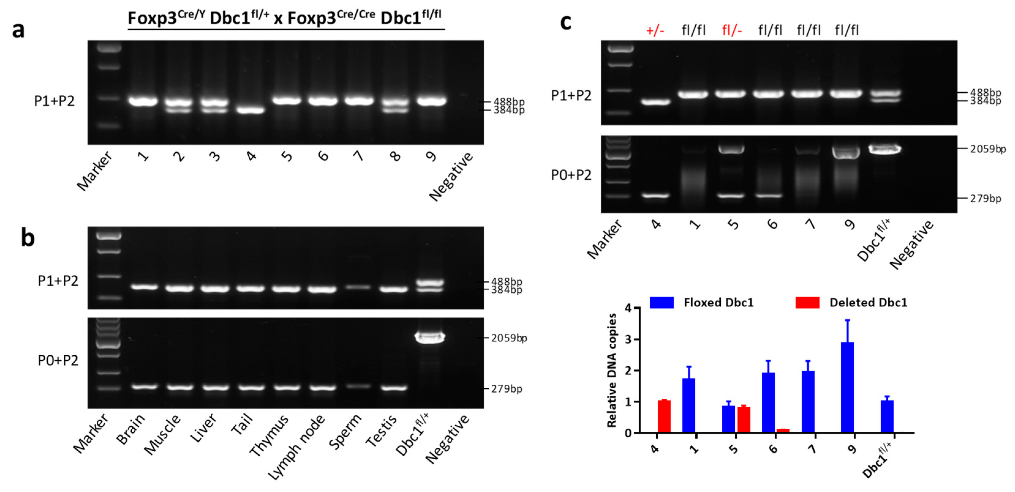
In order to knockout Dbc1 specifically in Treg cells, the conditional Dbc1 mice were crossed with Foxp3YFP-Cre mice [17], in which the Cre recombinase was under the control of Foxp3. In this study, the male mice harboring Foxp3YFP-Cre allele are designated Foxp3Cre/Y while the female mice are designated as Foxp3Cre/Cre or Foxp3Cre/+. When the female mice homozygous for floxed Dbc1 and Foxp3Cre alleles were crossed with the male mice heterozygous for floxed Dbc1 and hemizygous for Foxp3Cre (Foxp3Cre/Y Dbc1fl/+ × Foxp3Cre/Cre Dbc1fl/fl), it was found that some progeny contained no floxed Dbc1 alleles, such as the mouse #4 in Figure 2a. This phenomenon could be explained by the fact that the floxed Dbc1 allele was totally deleted, which could not be detected using primers 1 and 2. To verify this hypothesis, another primer (P0) (Figure 1) was designed to test the deleted Dbc1 allele. The results showed that the floxed Dbc1 allele of mouse #4 was deleted in the tail, muscle, brain, liver, immune organs and the reproductive system (Figure 2b). These results demonstrated that the floxed Dbc1 allele changed to deleted Dbc1 allele in mouse #4. As the deleted Dbc1 allele could be also present in the Dbc1fl/fl mice (which would not be detected using P1 and P2), the Dbc1 allele of other mice homozygous for floxed Dbc1 allele were also analyzed using P0 and P2. The results showed that two mice (#5 and #6) had a deleted Dbc1 fragment (Figure 2c). Since the deleted Dbc1 fragment may be from Treg cells, qPCR was used to verify how many floxed Dbc1 allele were recombined. The results showed that mouse #5 had an equal frequency of deleted Dbc1 fragment to mouse #4 and floxed Dbc1 fragment to Dbc1fl/+ mouse, while mouse #6 was not (Figure 2c). This suggests that one of the floxed Dbc1 allele in mouse #5 may have changed to the deleted Dbc1 allele. To further confirm the deleted Dbc1 allele, the two mice (#4 and #5) were back crossed to wild type mice (Wt). Interestingly, the results confirmed that indeed some of the offspring had deleted Dbc1 allele (Figure S1a,b).
To identify whether the deleted Dbc1 allele was derived from the parent mice, the Dbc1 alleles of the parent mice were further analyzed. It was observed that one of the mothers also contained a deleted Dbc1 allele (Figure S1c). Therefore, the deleted Dbc1 alleles in mouse #4 and #5 are derived from the mother 1 mouse. These results suggest that using the Cre/Lox conditional knockout method in the context of Foxp3 may indeed result in off-target deletion effects.
To verify the deleted Dbc1 allele, the deleted Dbc1 fragment (279 bp) was purified and sequenced. The sequence of deleted Dbc1 fragment is just the sequence of deleted Dbc1 allele, which supports that the floxed Dbc1 alleles in mouse #4 or #5 have changed to deleted Dbc1 alleles (Figure S1d).
This study also observed some mice (Figure S2, mouse #P-18 and #P-21) without the floxed Pkm2 allele in the progeny of male Foxp3Cre/Y Pkm2fl/+ mice crossed with female Foxp3Cre/Cre Pkm2fl/fl mice, in which the Pkm2fl/fl floxed mutant mice were from the Jackson Laboratory [25]. These results suggest that using the Cre/Lox conditional knockout method in the context of Foxp3 may result in some mice with gene knock out.

3.2. Germline Recombination of Floxed Dbc1 Allele Occurs in the Male Foxp3Cre/Y Dbc1fl/+ Mice

The germline recombination of floxed Dbc1 allele can result in the progeny with a deleted Dbc1 allele. To verify this and ascertain whether the germline recombination occurred in male mice or female mice, different genotypes of male or female Foxp3Cre Dbc1fl mice were used to cross with Wt mice. To rule out that the deleted Dbc1 allele in the progeny was derived from the maternal/paternal mice with deleted Dbc1 allele, the Foxp3Cre mice heterozygous for floxed Dbc1 allele were chosen to breed (Table 1).
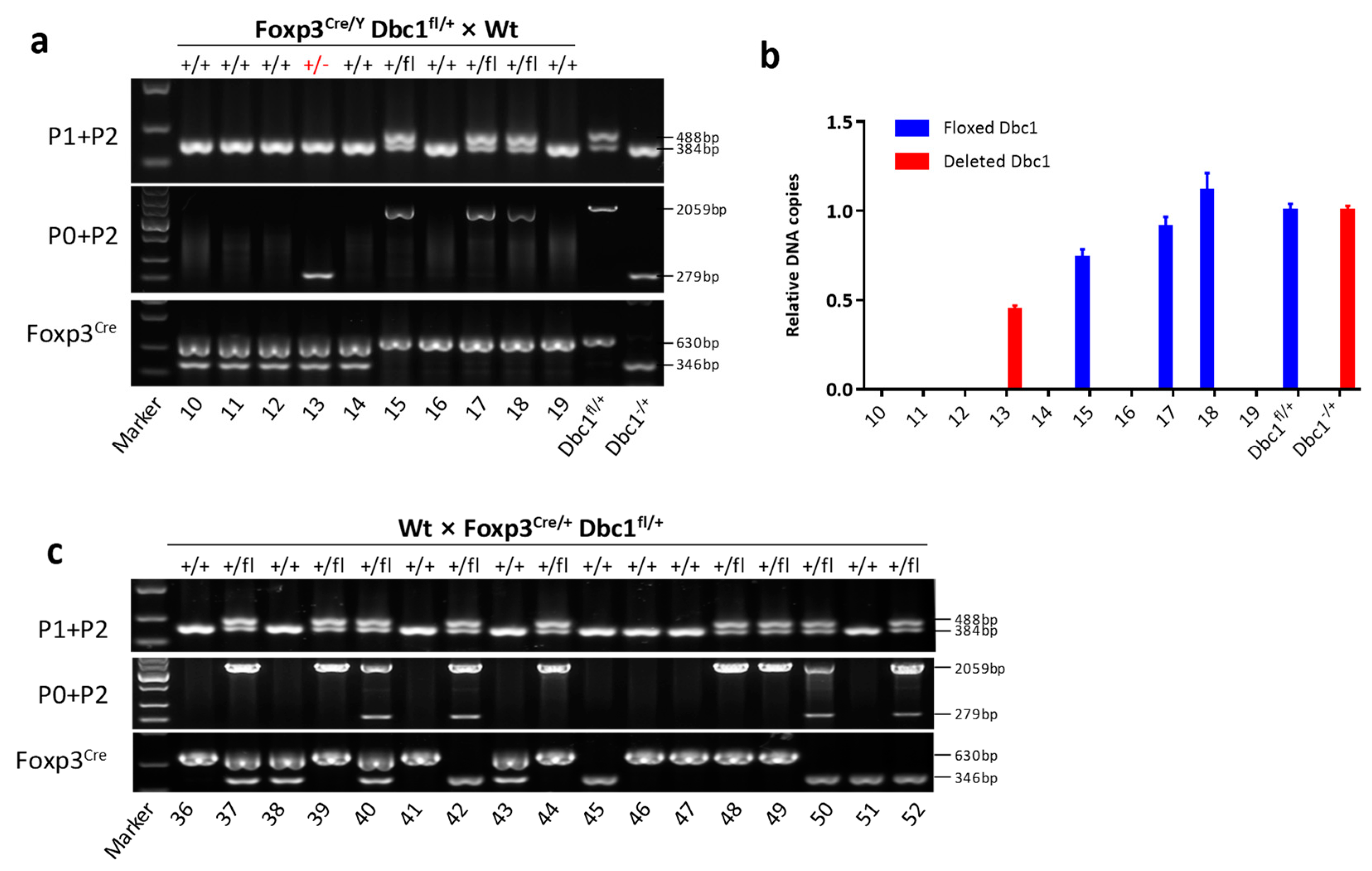
After identifying the genotypes of progeny mice using primers P1 and P2 or P0 and P2, this study found 1.28% (2 out of 156) mice (#13 and #26) only contained the deleted Dbc1 allele in the offspring of Foxp3Cre/Y Dbc1fl/+ × Wt mice (Figure 3a, Figure S3a). The results of qPCR also were consistent with mouse #13 containing deleted Dbc1 allele (Figure 3b). It was also observed that a deleted Dbc1 allele could be found in the offspring of Foxp3Cre/+ Dbc1+/+ × Foxp3Cre/Y Dbc1fl/+ mice (Figure S3b). In contrast, the genotypes of 119 mice in the offspring of Wt × Foxp3Cre/+ Dbc1fl/+ mice were identified, but no mice were obtained with the deleted Dbc1 allele (Table 1; some results are shown in Figure 3c). Considering that the frequency of the deleted Dbc1 allele might be lower when mice heterozygous for floxed Dbc1 allele were used and to rule out that the deleted Dbc1 allele in the progeny was derived from the maternal mice with deleted Dbc1 allele, the female Foxp3Cre/Cre Dbc1fl/fl mice with a lower frequency of deleted Dbc1 fragment than Cd4Cre/Cre Dbc1fl/fl mice were also used to cross Wt male mice (Table 1). By genotyping 79 mice in the progeny, not one mouse could be found with deleted Dbc1 allele (Table 1, some results shown in Figure S3c). All the results demonstrated that germline recombination of floxed Dbc1 allele could occur in the male Foxp3Cre/Y Dbc1fl/+ mice, which could result in progeny with deleted Dbc1 allele.

3.3. The Floxed Dbc1 Allele Was Recombined in non-T Cells of Some Foxp3Cre Dbc1fl Mice

In the process of genotyping, it was observed that some Foxp3Cre Dbc1fl mice showed an obvious deleted Dbc1 fragment, which was amplified by primers P0 and P2, while the others were not (Figure S3). This suggests that the recombination of floxed Dbc1 allele in these Foxp3Cre Dbc1fl mice may be various. To check that possibility, qPCR was first used to quantify the recombined Dbc1 allele in toe and tail, which were tissues used for mice genotyping. The results demonstrated that mice expressing a recombined Dbc1 fragment obviously as determined by conventional PCR exhibited a greater frequency of recombined Dbc1 allele (Figure 4a). Additionally, some mice showed a higher frequency of a recombined Dbc1 fragment than Cd4Cre/+ Dbc1fl/+ mice. This indicates that the floxed Dbc1 allele was recombined in more than T cells in these Foxp3Cre Dbc1fl mice.
To investigate whether the recombination of floxed Dbc1 allele occurred in other tissues, two male mice (#B-3 and #G-3) and two female mice (#A-2 and G-1) with a high frequency of deleted Dbc1 allele in the toe and tail were chosen to measure the extent of Dbc1 deletion in the brain, muscle, liver, thymus, lymph node, testis, ovary and sperm. As predicted, the Cd4Cre/+ Dbc1fl/+ mice showed the highest incidence of Dbc1 deletion in the thymus and lymph node, while almost no Dbc1 deletions were detected in other tissues. The Foxp3Cre Dbc1fl/+ mice expressed a detectable but lower incidence of Dbc1 deletion in the immune organs than Cd4Cre/+ Dbc1fl/+ mice. However, the Foxp3Cre Dbc1fl/+ mice with an obvious deleted Dbc1 fragment in the toe and tail showed many deleted Dbc1 fragments in the other tissues, while the mice without obvious deleted Dbc1 did not (Figure 4b). All these results suggest that the floxed Dbc1 allele is recombined in non-T cells of some Foxp3Cre Dbc1fl mice. These mice with nonspecific recombination should be ruled out when this kind of conditional knockout mice are used.

3.4. Expression of Cre Recombinase in the Fetus Results in Germline Recombination and Dbc1 Knock out Mice

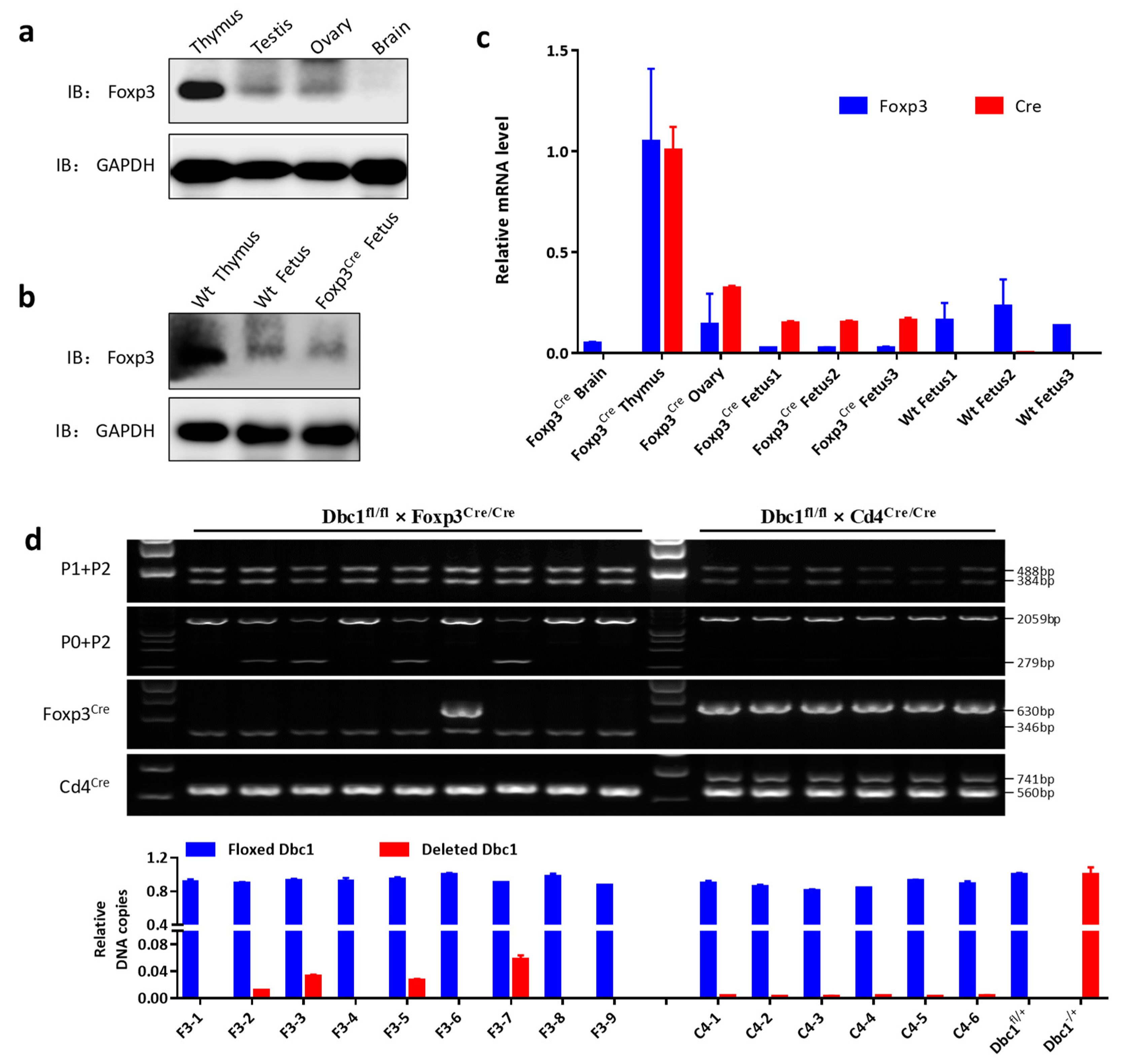
The germline recombination in Foxp3Cre Dbc1fl mice can be caused by the germline expression of the Cre recombinase driven by a Foxp3 promoter. Jasurda et al. [30] reported that even though Foxp3 protein was not detected, the Foxp3 transcript could be found in the testis. In the ENCODE database, the Foxp3 mRNA was shown to be present in the testis and ovary (Figure S5a). The Foxp3 protein in the testis and ovary were also detected using western blotting (Figure 5a). However, this study observed that the two mice (#13 and #26 in Figure 3a, Figure S3a, Figure S4 and Table 1) with deleted Dbc1 allele are the progeny of male Foxp3Cre/Y Dbc1fl/+ mice with a high frequency of the recombined Dbc1 fragment. There were no mice with deleted Dbc1 allele in the progeny of male Foxp3Cre/Y Dbc1fl mice with a low frequency of recombined Dbc1 fragment, even though the floxed Dbc1 allele is homozygous (Figure S5b,c; Table 1). This study also could not obtain any mice with deleted Dbc1 allele in the progeny of female Foxp3Cre Dbc1fl crossed with Wt male mice (Figure 3c and Figure S3c; Table 1). This means that the off-target deletion is not dependent on whether the Dbc1 allele in the breeding mouse is heterozygous or homozygous. Conversely, the off-target deletion depends on whether the breeding mice contain higher frequency of the recombined Dbc1 fragment or not. The germline recombination probably occurred at the developmental stage of the mice.
In the EMAGE database (http://www.emouseatlas.org/emap/home.html), Gray et al. [31] and Diez-Roux et al. [32] detected the Foxp3 expression in the fetus by RNA in situ hybridization. This study also detected the Foxp3 protein, Foxp3 and Cre mRNA in the fetus of Foxp3Cre mouse at embryonic day 14.5 (Figure 5b,c). In addition, by quantifying the frequency of the recombined Dbc1 fragment, it was indeed found that some fetuses of Foxp3Cre Dbc1fl/+ mice (mouse #F3-2, # F3-3, # F3-5 and # F3-7) showed an obvious higher incidence of Dbc1 deletion than Cd4Cre/+ Dbc1fl/+ mice (Figure 5d). These results suggest that the nonspecific expression of Cre in non-T cells of the fetus could activate and excise the floxed Dbc1 allele. When some cells with deleted Dbc1 allele develop to germ cells, it can result in Dbc1 knock out mice in the offspring. Thereby, to limit the risk of having full knock out mice, the male mice with a high frequency of recombined gene allele should be ruled out as breeders.
Comparing the mice genotype in the progeny of each breeding pair, it seems that the Foxp3Cre allele derived from a female breeder causes a higher frequency of the recombined Dbc1 fragment in the progeny (Figure 3c and Figure S3c, and Figure S5b,c). Even though the floxed Dbc1 allele was not inherited from the same female mice harboring the Foxp3Cre allele, the floxed Dbc1 allele still could recombine in some Foxp3Cre Dbc1fl mice (#106 and #114) (Figure S6). These support that the Foxp3Cre allele derived from a female breeder can more easily be expressed and cause off-target recombination in the offspring.
All the results show that there is a risk of high nonspecific recombination in the Foxp3Cre mice. Thus, a careful characterization of Foxp3Cre mice with the floxed gene is required.

4. Discussion

The data reported here demonstrate that Dbc1 conditional alleles can be completely deleted when mice are bred with Foxp3Cre mice. Although the frequency is not high, it remains possible that the Dbc1 allele deletion existed in the Dbc1fl/fl mice, while the genotype identification using primers flanking only one LoxP site could fail to detect this. Moreover, the deleted Dbc1 allele is inherited by the offspring. Additionally, this study observed the off-target deletion in the Foxp3Cre Pkm2fl mice. The off-target deletion may be limited to the conditional Dbc1 and Pkm2 alleles. Otherwise, the off-target deletion may also occur in Foxp3Cre mice with other conditional gene alleles. If functional inactivation of the targeted gene requires the deletion of both alleles, it will not affect the results. Nonetheless, when expression of a single copy affects the overall gene function in cells, this off-target deletion needs to be carefully characterized.
It was reported that germline recombination occurred in the Rgs9cre line, and that the germline Cre expression could cause this recombination [8]. This study found that Foxp3 was expressed in the ovary and testis, a finding which is consistent with that of Jasurda et al. [30]. However, the probable Cre expression following the Foxp3 expression in the testis or ovary of Foxp3cre mice did not cause germline recombination. Schmidt-Supprian et al. [33] reported that some floxed genes are more easily be excised than others. Therefore, the Foxp3 expression in the testis or ovary of Foxp3cre mice did not cause germline recombination which might be either as a result of Cre’s unequal sensitivity for conditional gene alleles, or as a result of Cre’s inability to cleave the LoxP site in the final haploid gametes.
In this report, the fetal Cre expression caused deletion, an interesting finding which to date has not been previously reported. In the fetal development stage, Cre driven by Foxp3 may function in some embryonic stem cells, resulting in the Dbc1 allele deletion. These stem cells differentiate into the germline, which resulted in the Dbc1 deleted offspring.
It is likely that the Cre expression in fetal cells caused the off-target recombination in the fetus of Foxp3Cre mice. However, there is a small possibility that the source Cre is from the egg, but less likely from sperm. While the Cre expressed in the egg might not function in the egg itself, it may have functional effects in the early development of the fetus. Although the source of the Cre may indeed have been from the egg, the Cre expression does not occur during the diploid stage of gametogenesis, since all mice with recombined Dbc1 alleles contain Foxp3Cre allele simultaneously. This study detected the Foxp3 expression in the ovary, but this does not mean that the Foxp3 expression is in mature eggs. The Foxp3 expression could not be identified in mature eggs, limited by the number of eggs, which might be resolved by single cell sequencing.
Even though this study found that Foxp3 drove fetal Cre function, the frequency of deleted Dbc1 was variable in the offspring. This means that different mice may contain different levels of the off-target Dbc1 recombination. Additionally, the off-target recombination possibly occurred in one or both of the two Dbc1 alleles in non-Treg cells, which results in the same type of non-Treg cells containing different levels of the Dbc1 protein. The variation of the off-target Dbc1 recombination implies that the fetal Cre expression is transient and the resultant gene recombination is random. The stochastic Foxp3 expression in the fetus shown by Gray et al. [31] and Diez-Roux et al. [32] also provides support.
When Rubtsov et al. [17] generated Foxp3Cre mice, they used a ROSA26YFP (R26Y) recombination reporter allele in Foxp3Cre mice to examine the specificity of Cre-mediated recombination. They observed that Foxp3Cre mice exhibited varying degrees (2–10%) of R26Y recombination in different hematopoietic lineage cells. Further, Franckaert et al. [22] observed the deletion of CD28 on conventional CD4+ T cells in Foxp3Cre Cd28fl/fl mice. When considered in the context of our results, the off-target recombination in different hematopoietic lineage cells could indeed be as a result of recombination in the fetus.
In this report, it was found that the off-target deletion and many somatic cells rather than Treg cells may contain the deleted floxed gene in the Foxp3Cre line. The off-target deletion can be genotyped using primers that flank the entire floxed locus of a conditional gene in combination with primers flanking a single LoxP site as suggested by Song et al. [9]. Furthermore, our results suggested that avoiding the use of male mice with a high frequency of recombined floxed allele to breed can limit the risk of having full knock out mice in the Foxp3Cre line. However, the deleted gene allele can exist in either of the homologous chromatids of the somatic cells and this cannot be detected by ordinary PCR or qPCR. Our results showed that there is no breeding scheme to limit the risk of having mice with an off-target gene recombination. Therefore, to avoid errors, the mice with a low frequency of deleted gene fragments as detected in ordinary PCR or qPCR should be chosen to do the experiment.

Supplementary Materials

The following are available online at https://www.mdpi.com/2073-4409/8/11/1309/s1, Figures S1, S2, S3, S4, S5 and S6.

Author Contributions

C.X. designed and performed the experiments, prepared the figures, and wrote the manuscript. F.Z. and J.W. contributed to the performance of the experiments. W.Z., J.A.B., B.L., and D.B. contributed critical comments. N.O. reviewed the manuscript and made significant revisions on the drafts. S.G.Z. supervised the work and wrote the manuscript.

Funding

This research was supported by grants from the NIH (R01 AR059103, R61 AR073 409, NIH Star Award to S.G.Z); the Zhujiang Innovative and Entrepreneurial Talent Team Award of Guangdong Province (2016 ZT 06S 252 to C.X); the National Natural Science Foundation of China (30972951, 81671611, 31900659 to C.X). The work was partially supported by NIH grants (CA200673, CA203834 to W.Z.) and startup grants (to W.Z.) from the UF Health Cancer Center and the Department of Pathology, Immunology and Laboratory Medicine.

Acknowledgments

We much appreciate the valuable comments that we received from other members of our laboratories.

Conflicts of Interest

The authors declare no financial or commercial conflicts of interest.

References

  1. Sauer, B. Functional expression of the cre-lox site-specific recombination system in the yeast Saccharomyces cerevisiae. Mol. Cell. Biol. 1987, 7, 2087–2096. [Google Scholar] [CrossRef] [PubMed]
  2. Hou, L.; Yau, Y.Y.; Wei, J.; Han, Z.; Dong, Z.; Ow, D.W. An open-source system for in planta gene stacking by Bxb1 and Cre recombinases. Mol. Plant 2014, 7, 1756–1765. [Google Scholar] [CrossRef] [PubMed]
  3. Sauer, B.; Henderson, N. Site-specific DNA recombination in mammalian cells by the Cre recombinase of bacteriophage P1. Proc. Natl. Acad. Sci. USA 1988, 85, 5166–5170. [Google Scholar] [CrossRef] [PubMed]
  4. Gu, H.; Marth, J.D.; Orban, P.C.; Mossmann, H.; Rajewsky, K. Deletion of a DNA polymerase beta gene segment in T cells using cell type-specific gene targeting. Science 1994, 265, 103–106. [Google Scholar] [CrossRef] [PubMed]
  5. Tsien, J.Z.; Chen, D.F.; Gerber, D.; Tom, C.; Mercer, E.H.; Anderson, D.J.; Mayford, M.; Kandel, E.R.; Tonegawa, S. Subregion- and cell type-restricted gene knockout in mouse brain. Cell 1996, 87, 1317–1326. [Google Scholar] [CrossRef]
  6. Wu, J.; Zhang, H.; Shi, X.; Xiao, X.; Fan, Y.; Minze, L.J.; Wang, J.; Ghobrial, R.M.; Xia, J.; Sciammas, R.; et al. Ablation of Transcription Factor IRF4 Promotes Transplant Acceptance by Driving Allogenic CD4(+) T Cell Dysfunction. Immunity 2017, 47, 1114–1128.e6. [Google Scholar] [CrossRef] [PubMed]
  7. Ramalingam, R.; Larmonier, C.B.; Thurston, R.D.; Midura-Kiela, M.T.; Zheng, S.G.; Ghishan, F.K.; Kiela, P.R. Dendritic cell-specific disruption of TGF-beta receptor II leads to altered regulatory T cell phenotype and spontaneous multiorgan autoimmunity. J. Immunol. 2012, 189, 3878–3893. [Google Scholar] [CrossRef]
  8. Liput, D.J. Cre-Recombinase Dependent Germline Deletion of a Conditional Allele in the Rgs9cre Mouse Line. Front. Neural. Circuits 2018, 12, 68. [Google Scholar] [CrossRef]
  9. Song, A.J.; Palmiter, R.D. Detecting and Avoiding Problems When Using the Cre-lox System. Trends Genet. 2018, 34, 333–340. [Google Scholar] [CrossRef]
  10. Hu, H.; Cavendish, J.Z.; Agmon, A. Not all that glitters is gold: Off-target recombination in the somatostatin-IRES-Cre mouse line labels a subset of fast-spiking interneurons. Front. Neural Circuits 2013, 7, 195. [Google Scholar] [CrossRef]
  11. Rempe, D.; Vangeison, G.; Hamilton, J.; Li, Y.; Jepson, M.; Federoff, H.J. Synapsin I Cre transgene expression in male mice produces germline recombination in progeny. Genesis 2006, 44, 44–49. [Google Scholar] [CrossRef] [PubMed]
  12. Kobayashi, Y.; Hensch, T.K. Germline recombination by conditional gene targeting with Parvalbumin-Cre lines. Front. Neural Circuits 2013, 7, 168. [Google Scholar] [CrossRef] [PubMed] [Green Version]
  13. Zhang, J.; Dublin, P.; Griemsmann, S.; Klein, A.; Brehm, R.; Bedner, P.; Fleischmann, B.K.; Steinhauser, C.; Theis, M. Germ-line recombination activity of the widely used hGFAP-Cre and nestin-Cre transgenes. PLoS ONE 2013, 8, e82818. [Google Scholar] [CrossRef] [PubMed]
  14. He, Y.; Sun, X.; Wang, L.; Mishina, Y.; Guan, J.L.; Liu, F. Male germline recombination of a conditional allele by the widely used Dermo1-cre (Twist2-cre) transgene. Genesis 2017, 55. [Google Scholar] [CrossRef]
  15. Horwitz, D.A.; Zheng, S.G.; Gray, J.D.; Wang, J.H.; Ohtsuka, K.; Yamagiwa, S. Regulatory T cells generated ex vivo as an approach for the therapy of autoimmune disease. Semin. Immunol. 2004, 16, 135–143. [Google Scholar] [CrossRef]
  16. Hu, G.; Liu, Z.; Zheng, C.; Zheng, S.G. Antigen-non-specific regulation centered on CD25+Foxp3+ Treg cells. Cell Mol. Immunol. 2010, 7, 414–418. [Google Scholar] [CrossRef] [Green Version]
  17. Rubtsov, Y.P.; Rasmussen, J.P.; Chi, E.Y.; Fontenot, J.; Castelli, L.; Ye, X.; Treuting, P.; Siewe, L.; Roers, A.; Henderson, W.R., Jr.; et al. Regulatory T cell-derived interleukin-10 limits inflammation at environmental interfaces. Immunity 2008, 28, 546–558. [Google Scholar] [CrossRef]
  18. Weinberg, S.E.; Singer, B.D.; Steinert, E.M.; Martinez, C.A.; Mehta, M.M.; Martinez-Reyes, I.; Gao, P.; Helmin, K.A.; Abdala-Valencia, H.; Sena, L.A.; et al. Mitochondrial complex III is essential for suppressive function of regulatory T cells. Nature 2019, 565, 495–499. [Google Scholar] [CrossRef]
  19. Hwang, S.M.; Sharma, G.; Verma, R.; Byun, S.; Rudra, D.; Im, S.H. Inflammation-induced Id2 promotes plasticity in regulatory T cells. Nat. Commun. 2018, 9, 4736. [Google Scholar] [CrossRef]
  20. Ghosh, S.; Roy-Chowdhuri, S.; Kang, K.; Im, S.H.; Rudra, D. The transcription factor Foxp1 preserves integrity of an active Foxp3 locus in extrathymic Treg cells. Nat. Commun. 2018, 9, 4473. [Google Scholar] [CrossRef]
  21. Xu, M.; Pokrovskii, M.; Ding, Y.; Yi, R.; Au, C.; Harrison, O.J.; Galan, C.; Belkaid, Y.; Bonneau, R.; Littman, D.R. c-MAF-dependent regulatory T cells mediate immunological tolerance to a gut pathobiont. Nature 2018, 554, 373–377. [Google Scholar] [CrossRef] [PubMed]
  22. Franckaert, D.; Dooley, J.; Roos, E.; Floess, S.; Huehn, J.; Luche, H.; Fehling, H.J.; Liston, A.; Linterman, M.A.; Schlenner, S.M. Promiscuous Foxp3-cre activity reveals a differential requirement for CD28 in Foxp3(+) and Foxp3(−) T cells. Immunol. Cell Biol. 2015, 93, 417–423. [Google Scholar] [CrossRef] [PubMed]
  23. Gao, Y.; Tang, J.; Chen, W.; Li, Q.; Nie, J.; Lin, F.; Wu, Q.; Chen, Z.; Gao, Z.; Fan, H.; et al. Inflammation negatively regulates FOXP3 and regulatory T-cell function via DBC1. Proc. Natl. Acad. Sci. USA 2015, 112, E3246–E3254. [Google Scholar] [CrossRef] [PubMed] [Green Version]
  24. Lee, P.P.; Fitzpatrick, D.R.; Beard, C.; Jessup, H.K.; Lehar, S.; Makar, K.W.; Perez-Melgosa, M.; Sweetser, M.T.; Schlissel, M.S.; Nguyen, S.; et al. A critical role for Dnmt1 and DNA methylation in T cell development, function, and survival. Immunity 2001, 15, 763–774. [Google Scholar] [CrossRef]
  25. Israelsen, W.J.; Dayton, T.L.; Davidson, S.M.; Fiske, B.P.; Hosios, A.M.; Bellinger, G.; Li, J.; Yu, Y.; Sasaki, M.; Horner, J.W.; et al. PKM2 isoform-specific deletion reveals a differential requirement for pyruvate kinase in tumor cells. Cell 2013, 155, 397–409. [Google Scholar] [CrossRef] [PubMed]
  26. Westendorf, K.; Durek, P.; Ayew, S.; Mashreghi, M.F.; Radbruch, A. Chromosomal localisation of the CD4cre transgene in B6. Cg-Tg(Cd4-cre)1Cwi mice. J. Immunol. Methods 2016, 436, 54–57. [Google Scholar] [CrossRef] [PubMed]
  27. Lu, L.; Lan, Q.; Li, Z.; Zhou, X.; Gu, J.; Li, Q.; Wang, J.; Chen, M.; Liu, Y.; Shen, Y.; et al. Critical role of all-trans retinoic acid in stabilizing human natural regulatory T cells under inflammatory conditions. Proc. Natl. Acad. Sci. USA 2014, 111, E3432–E3440. [Google Scholar] [CrossRef]
  28. Singh, A.K.; Khare, P.; Obaid, A.; Conlon, K.P.; Basrur, V.; DePinho, R.A.; Venuprasad, K. SUMOylation of ROR-gammat inhibits IL-17 expression and inflammation via HDAC2. Nat. Commun. 2018, 9, 4515. [Google Scholar] [CrossRef]
  29. Peng, W.X.; Zhu, S.L.; Zhang, B.Y.; Shi, Y.M.; Feng, X.X.; Liu, F.; Huang, J.L.; Zheng, S.G. Smoothened Regulates Migration of Fibroblast-Like Synoviocytes in Rheumatoid Arthritis via Activation of Rho GTPase Signaling. Front. Immunol. 2017, 8, 159. [Google Scholar] [CrossRef] [Green Version]
  30. Jasurda, J.S.; Jung, D.O.; Froeter, E.D.; Schwartz, D.B.; Hopkins, T.D.; Farris, C.L.; McGee, S.; Narayan, P.; Ellsworth, B.S. The forkhead transcription factor, FOXP3: A critical role in male fertility in mice. Biol. Reprod. 2014, 90, 4. [Google Scholar] [CrossRef]
  31. Gray, P.A.; Fu, H.; Luo, P.; Zhao, Q.; Yu, J.; Ferrari, A.; Tenzen, T.; Yuk, D.I.; Tsung, E.F.; Cai, Z.; et al. Mouse brain organization revealed through direct genome-scale TF expression analysis. Science 2004, 306, 2255–2257. [Google Scholar] [CrossRef] [PubMed]
  32. Diez-Roux, G.; Banfi, S.; Sultan, M.; Geffers, L.; Anand, S.; Rozado, D.; Magen, A.; Canidio, E.; Pagani, M.; Peluso, I.; et al. A high-resolution anatomical atlas of the transcriptome in the mouse embryo. PLoS Biol. 2011, 9, e1000582. [Google Scholar] [CrossRef] [PubMed]
  33. Schmidt-Supprian, M.; Rajewsky, K. Vagaries of conditional gene targeting. Nat. Immunol. 2007, 8, 665–668. [Google Scholar] [CrossRef] [PubMed]
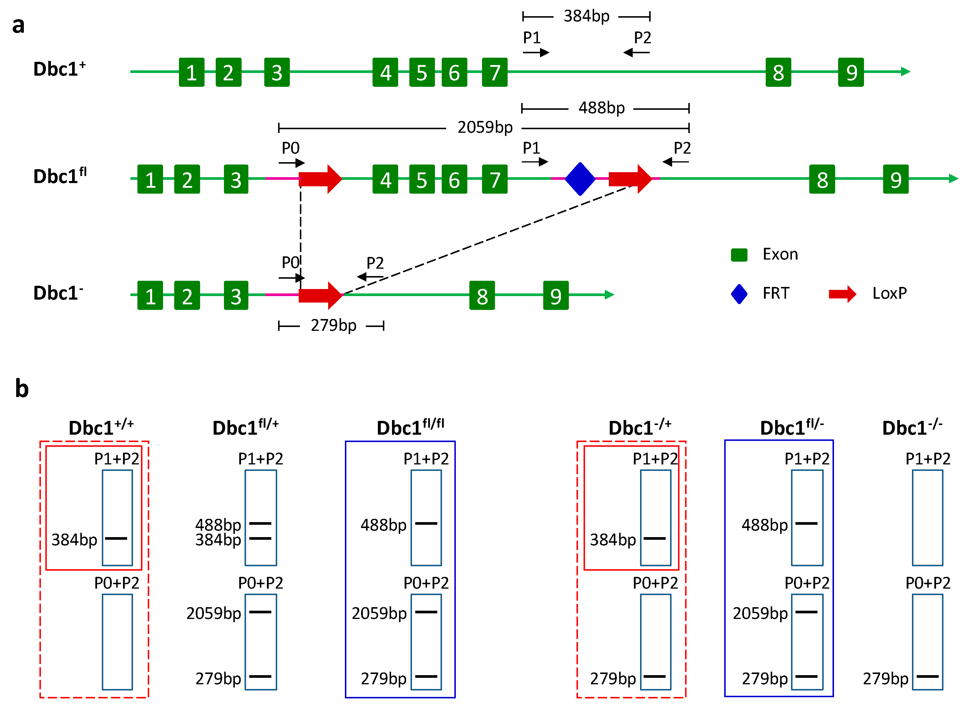
Figure 1. The generation and genotype strategy of the conditional Dbc1 allele. (a) Targeting strategy of the conditional Dbc1 allele. Shown from top to bottom (i) the Dbc1 locus with the exons, the site of the primer 1 (P1) and primer 2 (P2); (ii) the targeted locus with the site of the primer 0 (P0), LoxP and FRT sites; (iii) the locus after Cre-mediated deletion of the LoxP sites. (b) The genotype strategy of different Dbc1 alleles. The figure shows the predicted site and number of fragments of the mice with different Dbc1 alleles using the indicated primer.
Cells 08 01309 g001
Figure 2. The conditional Dbc1 allele was complete knockout in some offspring of Foxp3Cre/Y Dbc1fl/+ crossing Foxp3Cre/Cre Dbc1fl/fl mice. (a) The PCR product sizes of the offspring of Foxp3Cre/Y Dbc1fl/+ crossing Foxp3Cre/Cre Dbc1fl/fl mice using the primer indicated. (b) The PCR product sizes of the indicated tissue of mouse #4 in (a) using the primer indicated. (c) Top panel: the PCR product sizes of the Foxp3Cre Dbc1fl/fl mice using the primer indicated; bottom panel: the relative amount of the floxed Dbc1 and the recombined Dbc1 of the mice in the top panel was tested by qPCR. For qPCR, the results were normalized to the DNA sample from Dbc1fl/+ mice or mouse #4 in (a). The data were represented as the mean ± standard deviation of three independent experiments. +/−, fl/fl, and fl/− show the genotype of Dbc1 alleles.
Cells 08 01309 g002
Figure 3. The mice with deleted Dbc1 allele was present in the progeny of male Foxp3Cre/Y Dbc1fl/+ mice. (a) The PCR product sizes of the progeny of male Foxp3Cre/Y Dbc1fl/+ mice crossing Wt female mice using the primer indicated. (b) The relative amount of the floxed Dbc1 and recombined Dbc1 of the mice in (a) was tested by qPCR. For qPCR, the results were normalized to the DNA sample from Dbc1fl/+ mice or mouse #4 in Figure 2a. The data were represented as the mean ± standard deviation of three independent experiments. (c) The PCR product sizes of the progeny of female Foxp3Cre/+ Dbc1fl/+ mice crossed with Wt male mice using the primer indicated. The panel of Foxp3Cre shows the genotype of Foxp3-IRES-YFP-Cre alleles. +/−, +/+, and +/fl show the genotype of Dbc1 alleles.
Cells 08 01309 g003
Figure 4. The floxed Dbc1 allele was recombined in non-T cells of some Foxp3Cre Dbc1fl mice. (a) The relative amount of floxed Dbc1 and the recombined Dbc1 of the mice in the progeny of different breeding pairs. The results were compared to the Cd4Cre/+ Dbc1fl/+ mice. For qPCR, the results were normalized to the DNA sample from Dbc1fl/+ mice or mouse #4 in Figure 2a. Mouse #A-1, A-2, A-3, A-4 are offspring of Wt × Foxp3Cre/Y Dbc1fl/fl mice. Mouse #B-1, B-2, B-3, B-4 are offspring of Foxp3Cre/Cre Dbc1fl/fl × Wt mice. Mouse #D-1, D-2, D-3, D-4 are offspring of Wt × Foxp3Cre/Y Dbc1fl/+ mice. Mouse #E-1, E-2, E-3, E-4 are offspring of Foxp3Cre/+ Dbc1fl/+ × Wt mice. Mouse #G-1, G-2, G-3, G-4 are offspring of Foxp3Cre/+ × Dbc1fl/+ mice. The data were represented as the mean ± standard deviation of three independent experiments. The panel of Foxp3Cre shows the genotype of Foxp3-IRES-YFP-Cre alleles. (b) The relative amount of the floxed Dbc1 and recombined Dbc1 of the indicated tissue of the mice in (a) was tested by qPCR. For qPCR, the results were normalized to the DNA sample from Dbc1fl/+ mice or mouse #4 in Figure 2a. The data were represented as the mean ± standard deviation of three independent experiments. “Cd4 F” means female Cd4Cre/+ Dbc1fl/+ mouse while “Cd4 M” means male Cd4Cre/+ Dbc1fl/+ mouse. * p < 0.05; ** p < 0.01.
Cells 08 01309 g004
Figure 5. The Foxp3 was expressed in the ovary, testis and fetus. (a,b) The Foxp3 protein level in the indicated tissue was tested by western blotting. (c) Foxp3 and Cre mRNA level in the indicated tissue was tested by qPCR. The data were represented as the mean ± standard deviation of three independent experiments. (d) The PCR product sizes of the fetus at embryonic day 14.5 in the progeny of male Dbc1fl/fl mice crossed with female Foxp3Cre/Cre mice or Cd4Cre/Cre mice, using the primer indicated. The relative amount of the floxed Dbc1 and recombined Dbc1 of the fetus was tested by qPCR. For qPCR, the results were normalized to the DNA sample from the Dbc1fl/+ mice or mouse #4 in (a). The panel of Foxp3Cre shows the genotype of Foxp3-IRES-YFP-Cre alleles. The panel of Cd4Cre shows the genotype of Cd4-Cre alleles. The data were represented as the mean ± standard deviation of three independent experiments.
Cells 08 01309 g005
Table 1. Frequency of mice with deleted Dbc1 allele in the progeny of different breeding pairs.
Breeding MiceMouse No.Frequency of Recombined Dbc1 FragmentLitter SizeNumber of Dbc1−/+ Mice in Progeny
Foxp3Cre/YDbc1fl/+ × WtYN-3, 4, 5low890
Foxp3Cre/YDbc1fl/+ × WtYN-1, 2high672
Wt × Foxp3Cre/+ Dbc1fl/+VN-3, 4low550
Wt × Foxp3Cre/+ Dbc1fl/+VN-1, 2, 5, 6high640
Foxp3Cre/YDbc1fl/fl × WtYM-1, 2low750
Wt × Foxp3Cre/Cre Dbc1fl/flXM-1, 2low790
“high” means higher frequency of recombined Dbc1 fragment than Cd4Cre Dbc1fl mice tested by qPCR; “low” means lower frequency of recombined Dbc1 fragment than Cd4Cre Dbc1fl mice tested by qPCR (Figure S4).