Review

The Glucocorticoid Receptor in Cardiovascular Health and Disease

1
Department of Pediatrics, Yale University School of Medicine, New Haven, CT 06520, USA
2
Vascular Biology and Therapeutics Program, Yale University School of Medicine, New Haven, CT 06520, USA
3
Department of Pediatrics, Shengjing Hospital of China Medical University, Shenyang 110004, China
*
Author to whom correspondence should be addressed.
These authors contributed equally to this work.
Cells 2019, 8(10), 1227; https://doi.org/10.3390/cells8101227
Submission received: 28 August 2019 / Revised: 4 October 2019 / Accepted: 8 October 2019 / Published: 9 October 2019
(This article belongs to the Collection Functions of Nuclear Receptors)

Abstract

:
The glucocorticoid receptor is a member of the nuclear receptor family that controls many distinct gene networks, governing various aspects of development, metabolism, inflammation, and the stress response, as well as other key biological processes in the cardiovascular system. Recently, research in both animal models and humans has begun to unravel the profound complexity of glucocorticoid signaling and convincingly demonstrates that the glucocorticoid receptor has direct effects on the heart and vessels in vivo and in vitro. This research has contributed directly to improving therapeutic strategies in human disease. The glucocorticoid receptor is activated either by the endogenous steroid hormone cortisol or by exogenous glucocorticoids and acts within the cardiovascular system via both genomic and non-genomic pathways. Polymorphisms of the glucocorticoid receptor are also reported to influence the progress and prognosis of cardiovascular disease. In this review, we provide an update on glucocorticoid signaling and highlight the critical role of this signaling in both physiological and pathological conditions of the cardiovascular system. With increasing in-depth understanding of glucocorticoid signaling, the future is promising for the development of targeted glucocorticoid treatments and improved clinical outcomes.

1. Introduction

Cardiovascular diseases (CVDs) are disorders of the heart and blood vessels and are the leading cause of death globally. An estimated 17.9 million people died from CVDs in 2016, representing 31% of all deaths worldwide [1]. CVDs are a diverse group of conditions, which include such entities as coronary artery disease, stroke, peripheral vascular disease, rheumatic heart disease, and pulmonary embolism. These diseases may occur throughout the lifespan, however, the vast majority of all deaths due to CVD are the result of one of six conditions: ischemic heart disease, stroke, hypertensive heart disease, cardiomyopathy, rheumatic heart disease, and atrial fibrillation [2].
Common behavioral risk factors for CVDs include unhealthy dietary patterns, physical inactivity, high sodium/tobacco consumption, and alcohol misuse. In addition to these risk factors, several quantifiable physiological changes are associated with CVD, such as high blood pressure (hypertension), elevated blood glucose (diabetes mellitus), elevated cholesterol (hyperlipidemia), being overweight, and obesity. Genetic predisposition and family history of CVDs [3,4,5] are considered risk factors as well.
The pathological mechanisms underlying CVDs are extremely complicated and involve interactions between diverse bio-molecules. Glucocorticoids and their actions via the glucocorticoid receptor (GR) are highly involved in the genesis and development of CVDs. Glucocorticoids are steroid hormones that are essential for life, which are synthesized and released by the adrenal cortex, under regulation by the hypothalamic-pituitary-adrenal gland axis. Glucocorticoid release occurs in a circadian manner, as well as in response to stress, and coordinates a variety of fundamental processes including the inflammatory and immune responses, metabolic homeostasis, cell proliferation, reproduction, and cognition [6,7].
In addition to their physiological roles, glucocorticoids can influence the cardiovascular system in various pathological conditions [8,9,10]. Synthetic glucocorticoids are commonly prescribed in diverse cardiovascular disorders, including infectious conditions such as rheumatic fever and myocarditis, structural conditions such as conduction defects and cardiomyopathy, and vascular conditions such as angina and acute myocardial infarction [11]. However, due to the existence of severe side effects in many organ systems, the therapeutic benefits of (synthetic) glucocorticoids are limited. These adverse effects include diabetes, abdominal obesity, and hypertension, all of which are risk factors for CVDs. Thus, in order to enhance the safety and efficacy of glucocorticoid treatment and facilitate the development of novel glucocorticoids, it is essential to have comprehensive insight into the specific functions of glucocorticoids in the cardiovascular system. Since binding to the glucocorticoid receptor is a critical process in glucocorticoid action, this review aims to deliver an update on GR signaling and highlight the roles of GR signaling in both physiological and pathological conditions of the cardiovascular system.

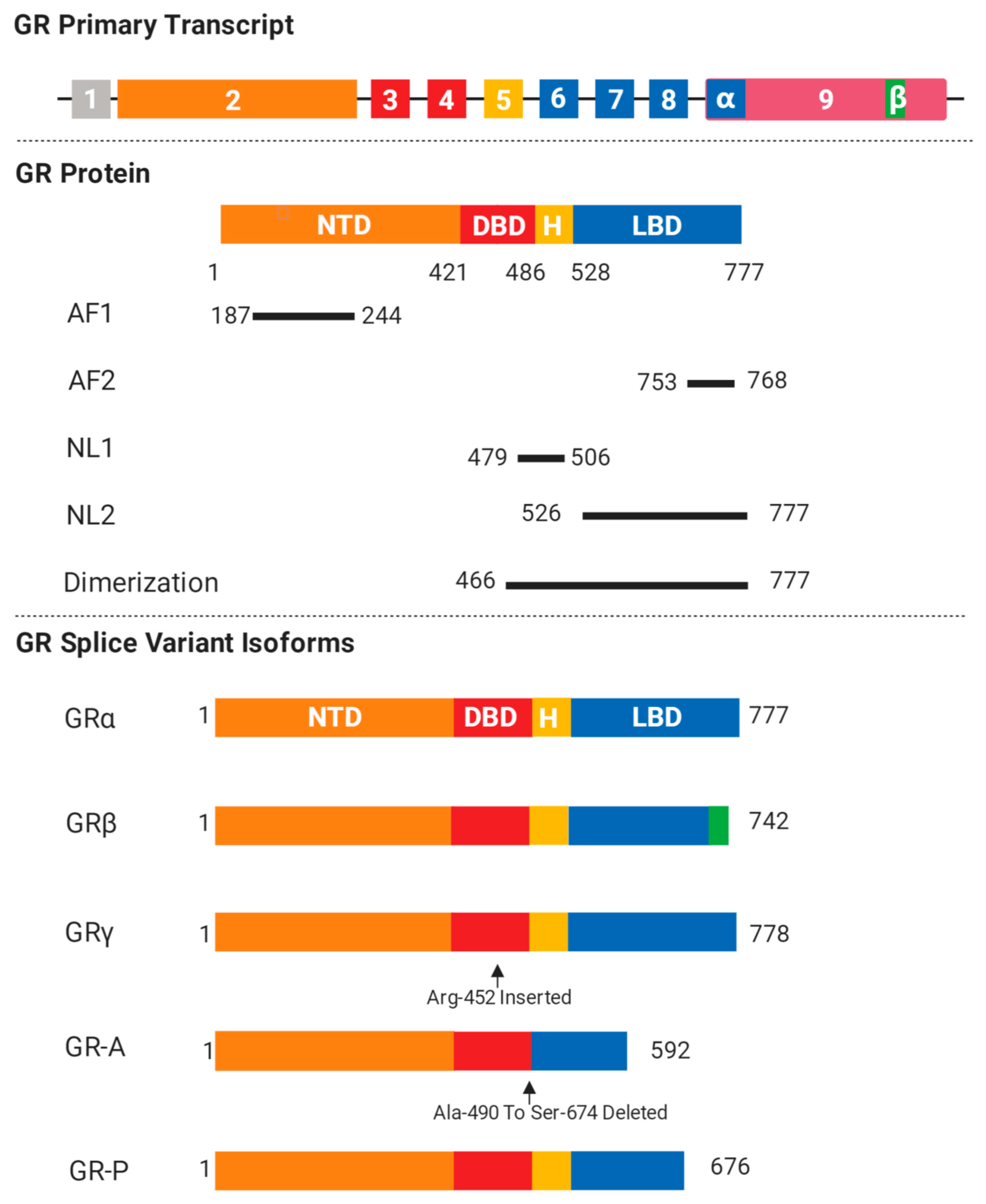
2. GR Isoforms and Structure

GR is encoded by the gene NR3C1, which is located on chromosome 5q31-32 in humans, and acts as a ligand-inducible transcription factor belonging to the nuclear receptor superfamily [12]. The molecular structure of GR includes four components: (1) an N-terminal transactivation domain (NTD), (2) a central DNA binding domain (DBD), (3) a C-terminal ligand-binding domain (LBD), and (4) a hinge region separating the DBD and the LBD [13]. The NTD is the most variable region among the nuclear receptor superfamily members and contains regulatory regions that allow binding to diverse co-regulators and components of the transcriptional machinery through activation function domain 1 (AF-1). Besides this, it is also the primary site for post-translational modifications. Unlike the NTD, the DBD is the most conserved region among the nuclear receptors and comprises two zinc finger motifs that mediate dimerization and direct binding to specific genomic sequences, known as glucocorticoid response elements (GREs). Meanwhile, the LBD is a highly structured domain, consisting of 12 α-helices and four small β-strands, which forms a hydrophobic cavity for glucocorticoid binding and also contains an activation function (AF2) [14]. Upon ligand binding, the AF2 would experience conformational changes in order to allow the LBD to interact correspondingly with either coactivators or corepressors containing LXXLL motifs. Although AF2 is obligately ligand-dependent, AF1 is constitutively active and can function in the absence of the LBD. GR AF1 has been classified as an intrinsically disordered protein (IDP) and it has been proposed that AF1 can “sample” its environment for appropriate binding partners and result in a rapidly changing molecular conformation [15].
Two nuclear localization signals, NL1 and NL2, are located at the DBD/hinge region and the LBD and are responsible for triggering translocation to the nucleus via an importin-dependent mechanism [16].
The human GR gene consists of nine exons numbered from 1 to 9 (Figure 1). Exon 1 forms the 5′-untranslated region and exon 2 encodes for the entire NTD. Exons 3 and 4 comprise the DBD and exons 5–9 comprise the LBD. GR has several receptor isoforms, for example, GRα and GRβ, which differ only in their C-termini and result from splicing differences in exon 9 [17,18] (Figure 1). The GRα isoform undergoes alternative translation initiation from conserved AUG start codons in exon 2 of the GR gene, generating eight additional isoforms of GR (GRα-A, GRα-B, GRα-C1, GRα-C2, GRα-C3, GRα-D1, GRα-D2, and GRα-D3) with progressively shorter NTDs [19]. Although they do demonstrate similar binding affinities for glucocorticoids and a similar ability to interact with GREs [19], there are some isoform-specific differences. For instance, the GR-C isoform was demonstrated to confer an increased susceptibility to apoptosis in cell culture while the GR-D isoform resulted in a relative resistance to apoptosis under the same conditions [20].
The GR primary transcript consists of nine exons and GR protein comprises NTD, DBD, “hinge region”, and LBD. The location of AF1, AF2, NL1, and NL2 are shown. Common GR isoforms like GRα, GRβ, GRγ, GR-A, and GR-P are generated after specific alternative splicing.
Unlike GRα, the GRβ splice variant does not bind glucocorticoids and resides constitutively in the nucleus of cells, acting as a natural dominant negative inhibitor of the GRα isoform on many glucocorticoid-responsive target genes [21]. Increased expression of GRβ has been associated with glucocorticoid resistance, which may be due to competition for GRE binding, competition for transcriptional co-regulators, or the formation of inactive GRα/GRβ heterodimers. The ability of GRβ to inhibit the activity of GRα suggests that high levels of GRβ may lead to glucocorticoid resistance [22]. Furthermore, GRβ may also generate eight β isoforms that are similar to the GRα [17] and is also able to exert its own independent functions, which include suppressing the transcriptional activity of the GATA3 transcription factor on its responsive IL-5 and -13 promoters by attracting histone deacetylases [23]. There is also evidence that GRβ has some GRα-independent transcriptional activity, as Kino et al. showed in HeLa cells [24].
Additional GR isoforms have been associated with glucocorticoid insensitivity [16]. GRγ exhibits about 50% of the activity of GRα for canonical glucocorticoid target genes. Recently, the level of GRγ was correlated with glucocorticoid resistance in childhood acute lymphoblastic leukemia and small cell lung carcinoma [25,26]. Two others, GR-P and GR-A, are splice variants that are missing large regions of the LBD (GR-P: missing appropriate exons 8 and 9, GR-A: missing the entire sequences of exons 5, 6, and 7) [27]. Due to these defective changes in the LBD, GR-P and GR-A do not bind glucocorticoids and both GR-P and GR-A have been determined to contribute to glucocorticoid resistance. Of note, GR-P is found in many tissues and has been shown to modulate the transcriptional activity of GRα in a cell-specific manner [17,28,29].
Specific tissues display particular relative abundances of these GR isoforms and react in individualized fashions to unique stimuli such as differentiation and aging. These differences are of crucial importance in determining the tissue-specific actions of glucocorticoids.

3. Genomic and Non-Genomic Effects of GR

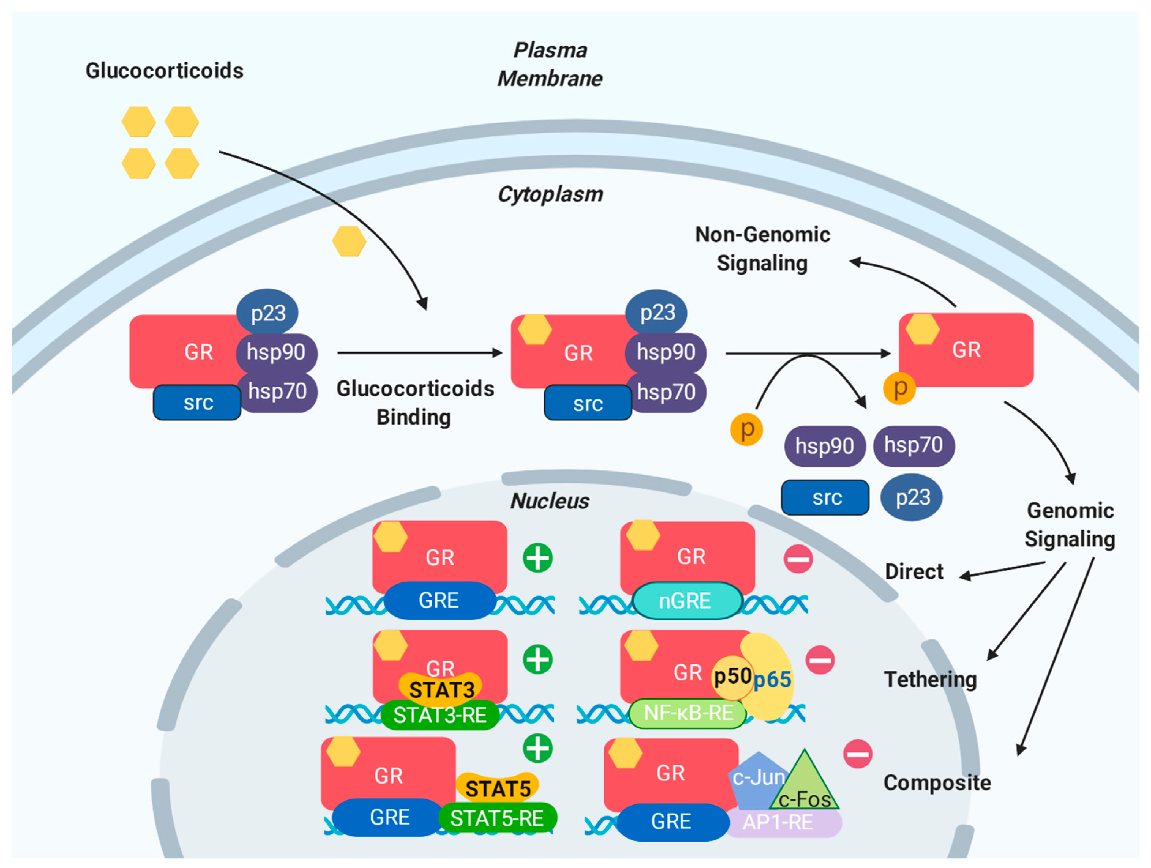
3.1. Genomic Effects of GR

Classically, the effects of glucocorticoid signaling are genomic, meaning that they are governed by GR-mediated transcription and protein synthesis. In its quiescent state, GR is located in the cytoplasm, which is bound to a chaperone complex which consists of the heat shock proteins hsp90, hsp70, and hsp56, as well as the immunophilins KBP51, FKBP52, Cyp44, and PP5 [17,30]. Upon ligand binding, the GR complex undergoes a conformational change, involving post-translation modifications such as phosphorylation and acetylation. This structural rearrangement exposes the two nuclear localization signals and GR rapidly translocates into the nucleus, where it can exert its actions through genomic (transactivation and transrepression) mechanisms [13]. Within the nucleus, GR regulates the transcription of its target genes in three main ways (Figure 2): (i) direct binding to GREs, (ii) interacting with other transcription factors, or (iii) by both direct binding to GREs and interaction with other transcription factors.
In classical “direct” GR transcriptional regulation, GR that is bound to its ligand homodimerizes in the nucleus and exerts transcriptional activity by direct binding to GREs. The classic GRE sequence, 5′-AGAACAnnnTGTTCT-3′, is a palindromic sequence that is composed of two hexamers that are separated by three base pairs. These bases may be any DNA residue, though one is typically highly conserved, while the other two are more variable [18]. GR binds to GREs as a homodimer, with each half site occupied by one receptor subunit. The three-nucleotide spacing between the two half sites is strictly required for GR to dimerize on directly regulated GREs. Meanwhile “tethering” GREs allow GR to indirectly regulate gene expression, without the receptor itself binding DNA. Although lacking a DNA binding site, tethering GREs can recruit other transcription factors that bind to GR [16]. GR can also regulate gene expression by binding to “composite” GREs. In this case, target genes contain binding sites for both GR and other transcription factors [18].
Historically, GREs have been shown to mediate the glucocorticoid-dependent induction of many genes and have been regarded exclusively as activating. More recently, data have shown that GR occupancy of canonical GREs can result in the repression of target genes as well [31].
In contrast to the regulation of classical GREs, the repression of negatively regulated target genes is usually mediated by negative GREs (nGREs). The consensus nGRE sequence, 5′-CTCC(n)0-2GGAGA-3′, is distinct from the classic GRE in sequence and function. The nGRE sequence has a variable spacer ranging from 0 to 2 nucleotides and (unlike classical GREs) is occupied by two GR monomers that do not homodimerize [32]. nGREs are abundant throughout the genome and contribute to the regulation of the hypothalamic-pituitary-adrenal (HPA) axis, inflammation, and angiogenesis.
Both excesses and deficiencies of glucocorticoids, mediated by changes in genomic GR signaling pathways as described above, can lead to pathological conditions and impair the cardiovascular stress response [33]. For instance, hypotension, hypoglycemia, and pancytopenia are regarded as signs of cortisol insufficiency; however, the provision of large doses of exogenous glucocorticoids may result in hypertension [34] and decreased total peripheral vascular resistance in both healthy patients and those in shock [9]. In addition, excess glucocorticoids also induce pathophysiological changes in the myocardium through the angiotensin II signaling pathway [35,36,37].

3.2. Non-Genomic Effects of GR

In addition to functioning through genomic mechanisms, which usually occur in hours, glucocorticoids can also exert their actions more rapidly (within minutes) via non-genomic signaling mechanisms (Figure 2). Instead of requiring nuclear GR-mediated transcription and translation, such non-genomic actions are initiated at the cell surface via either membrane-bound [38] or cytoplasmic GR [39]. During the structural change in the GR–ligand complex that follows glucocorticoid binding, GR-bound proteins including HSPs and Src are released from the multimeric GR complex [40,41,42,43] and these dislocated components from the GR complex can themselves influence cellular signaling (Figure 2). For example, GR has been reported to activate the PI3K-Akt pathway [44], which in turn can activate eNOS and has been shown to reduce myocardial infarct size [45]. Other evidence suggests that GC-GR signaling can induce rapid biological modulation in contractility, vascular reactivity, and blood pressure in a non-genomic manner in the cardiovascular system [9].

4. Glucocorticoid Metabolism in the Cardiovascular System

The availability of cellular glucocorticoid is regulated by the tissue-specific metabolic enzymes 11β-hydroxysteroid dehydrogenase 1 and 2 [46]. 11β-HSD2 converts active cortisol into inactive cortisone, while 11β-HSD1 converts cortisone to cortisol (Figure 3). Since the activity of 11β-HSD2 in the vasculature is minimal, the cardiovascular system is directly affected by circulating cortisol levels [17]. It is important to note that the expression of 11β-HSD2 can be up regulated under conditions of chronic, intermittent hypoxia and in this case, the increased 11β-HSD2 level plays an essential role in the regulation of tissue sensitivity to glucocorticoids [9]. Furthermore, circulating glucocorticoid levels can be modulated by corticosteroid binding globulin (CBG) [47], which not only accelerates the distribution of cortisol, but also participates in its release to tissues.

5. GR Polymorphisms

In addition to the genomic and non-genomic effects of GR, GR polymorphisms are also reported to influence cardiovascular disease. The capacity of GR to exert activation or repression of transcriptional regulation is affected by several polymorphisms in the GR gene that alter the amino acid sequence of the encoded receptor. Subsequently, such genetic variations can affect both the efficiency of GC therapy and disease pathology [48,49]. Table 1 describes several key pathological mutations that have been identified in the human NR3C1 gene [50,51,52,53,54,55,56,57,58,59,60,61,62,63,64,65,66,67,68,69,70,71]. For cardiovascular disease or its underlying risk factors, the most extensively studied polymorphisms are the ER22/23EK, GR-9R, N363S, and BclI polymorphisms. Generally speaking, glucocorticoid hypersensitivity has been identified when N363S and BclI polymorphisms are present in the context of visceral obesity. However, other polymorphisms, such as ER22/23EK or GR-9β, are associated with glucocorticoid resistance in the context of healthier metabolic syndrome.
For example, the ER22/23EK (rs6189 and rs6190) polymorphism, which occurs in about 3% of the population, is associated with a more favorable metabolic profile, including a lower risk of both type 2 diabetes and cardiovascular disease [50]. This effect is conferred via reduced glucocorticoid sensitivity, which is mediated by a change from GAG AGG to GAA AAG in exon 2 at position 23 within the NTD [72,73].
The N363S (rs6195) polymorphism, located in exon 2, is found at a 4% frequency in the general population. It corresponds to a change from an A to a G [74]. Unlike ER22/23EK, the presence of the N363S polymorphism enhances the transcriptional activity of GR and is associated with glucocorticoid hypersensitivity. Patients carrying this mutation have a higher body mass index [51], as well as an increased incidence of obesity [52,53], type 2 diabetes [54], and coronary artery disease [55].
The GR-9β polymorphism, which occurs in 8% of the general population, consists of an A to G substitution in the 3′ UTR end of exon 9 (GTTTA motif). This change also confers a more favorable metabolic profile and is associated with a decreased risk of obesity in women, decreased total cholesterol levels, and increased HDL cholesterol levels. In addition, a recent clinical study found that the GR-9β polymorphism is also involved in the regulation of blood pressure [56].
Finally, the BclI polymorphism is located in intron 2 with a C to G nucleotide change and is found in 25% of the general population. As mentioned above, it is associated with GC hypersensitivity [57,58]. Many studies have demonstrated that the BclI mutation is related to a wide variety of metabolic, cardiovascular, and psychiatric disorders, such as hypertension, adiposity, obesity [59,60], and bulimia nervosa [61]. Furthermore, BclI carriers also have a higher risk of post-traumatic stress disorder [62], cardiovascular disease [63], and atherosclerosis [64]. There are other known polymorphisms that influence the risk of cardiovascular disease, including D401H, A714Q, F737L, F774S, V575G, D641V, and G679S, and these are also listed in Table 1.

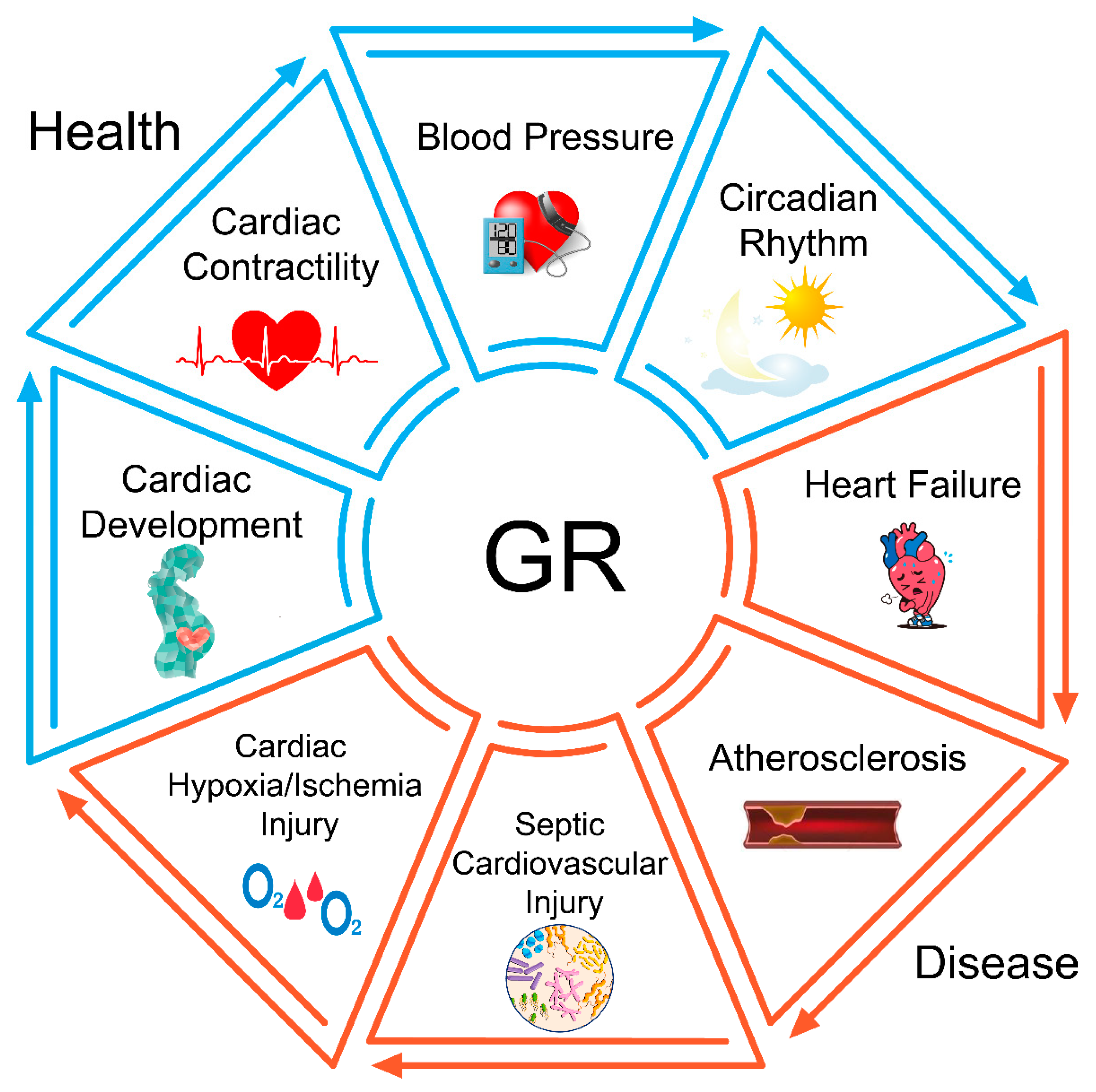
6. The Role of GR in Physiological Conditions

6.1. GR in Cardiac Development

The surge in levels of maternal glucocorticoids entering the fetal circulation that is observed during late gestation suggests an essential role for glucocorticoids in preparation for life after birth [75]. Studies in vivo have found that globally GR-deficient (GR-/-) mice are glucocorticoid resistant and all die soon after birth, which implies the critical role of GR in the development of organ systems that are adapted to postnatal survival [76]. Compared with other organs, the heart is functional relatively early in gestation and is required for fetal survival [77]. To reveal the potential role of GR during cardiac development, Oakley et al. developed a mouse model lacking GR expression in heart tissue (the cardiomyocyte-specific GR knock out) [78]. They found that although at first the mice were born in the expected Mendelian ratios and displayed a normal phenotype, they died prematurely (median survival approximately 7 months) from pathological cardiac hypertrophy that progressed to dilated cardiomyopathy and heart failure [78]. Additionally, Rog-Zielinska et al. demonstrated that application of glucocorticoid to primary mouse fetal cardiomyocytes could replicate the structural, functional, and biochemical consequences of GR activation in heart tissue [77]. This includes improving the contractility of cardiomyocytes, promoting Z disc assembly and the appearance of mature myofibrils, and increasing mitochondrial activity [77]. These effects demonstrate the potentially vital role of GR in intrauterine cardiac development.
In addition to studies in vivo, there are also several in vitro studies that demonstrate an important role for GR during heart development. In order to understand the influence of GR at the gene level, Oakley et al. constructed the GR-/- mouse and found that the expression of several key genes changed relative to wild type, including some critical genes for cardiac contractility (ryanodine receptors 2, RyR2), cardiomyocyte survival (prostaglandin D2 synthase, Ptgds), and inhibition of inflammation (lipocalin 2, Lcn 2) [78]. This suggests that there is specific activation of GR in heart tissue and reveals an obligate role for GR in maintaining normal cardiovascular function. At the molecular level, another recent study identified genes that are directly regulated by GR using next-generation sequencing. In this work, the structural maturation of fetal cardiomyocytes catalyzed by glucocorticoid treatment was shown to be critically influenced by the transcriptional coactivator, PGC-1α [79].
Similar investigations have also been performed in non-mouse animal models. For example, Kim et al. found that developing piglet hearts had smaller myocytes, with reduced binucleation, fewer apoptotic nuclei, and more proliferative nuclei than term hearts before the glucocorticoid surge [80]. This suggests that GR-related pathways could be involved in the regulation of myocyte size during cardiac development. An additional study performed by Eiby et al. showed that glucocorticoids could induce myocyte structural maturation through GR-related pathways and thus, influence heart function [81]. They isolated working hearts from piglets and stabilized them in the Langendorff mode to demonstrate the influence of GR on myocyte structure [81]. From these in vitro and in vivo studies, which are summarized in Table 2, it is evident that glucocorticoids and GR are critical for cardiac development and maturation.
Many clinical trials have suggested that sustained exposure or repeated low doses of glucocorticoids prenatally in humans are effective in improving fetal outcomes [82,83]. Studies have found that exposure to multiple courses of prenatal corticosteroid therapy resulted in a significant reduction in neonatal morbidity [83] and especially in the incidence of respiratory distress syndrome [82]. However, specific data regarding the effect of prenatal glucocorticoids on human cardiac development and maturation are still lacking. Several studies have identified the potential role of GR-related signaling in cardiac development, while a smaller number have demonstrated that excessive prenatal glucocorticoid exposure could potentially result in a delay in cardiac maturation and even cause cardiovascular problems in adult life [84,85]. As such, it is evident that the time- and concentration-dependent effects of GR-related pathways in cardiac development are still not fully understood and require further exploration in future studies.

6.2. GR in Cardiac Contractility

Cardiac contractility is regulated by changes in the concentration of intracellular calcium ([Ca2+]i) and calcium signaling has a key role in maintaining normal function in myocardial cells. Maintenance of normal heart function requires that cytoplasmic [Ca2+]i is sufficiently high in systole and low in diastole [86,87] to allow contraction and relaxation of cardiomyocytes. Under physiological conditions, cytosolic calcium binds to troponin, resulting in sliding of the thick and thin filaments. This causes cell shortening, the development of pressure within the ventricle, and, finally, the ejection of blood [88]. Calcium channels may be divided into three groups: L-type, T-type, and N-type channels. In heart tissue, the L-type calcium channel is the main type in the adult and plays an important role in the process of myocardial contraction. The opening of L-type calcium channels generates depolarization and the entry of a small number of calcium ions, which contributes to a significant increase in [Ca2+]i in the dyadic space (the region bounded by the t-tubule and sarcoplasmic reticulum (SR)). This small increase in [Ca2+]i then causes the SR calcium release channels to open, releasing a much larger amount of calcium from the SR in a process of positive feedback. For relaxation to occur, calcium is removed from the cytoplasm through RyR closure, then calcium is re-sequestered in the SR, reducing the free cytosolic [Ca2+]i.
Recently, several studies have found that GR may have essential functions during cardiac contractility both in vivo and in vitro. For example, Oakley et al. generated mice with cardiomyocyte-specific deletion of GR (cardio GR knockout (KO)) and found that these mice displayed different phenotypes, including heart dysfunction, compared with wild type mice [89], thus demonstrating that insufficient GR signaling is pathogenic in cardiomyocytes. To investigate these mechanisms in more depth, Oakley et al. used a microarray technique to examine the signaling pathways that are associated with the dysregulated genes and found that “Cardiac β-Adrenergic Signaling” was strongly associated with the dysregulated genes in the cardio GR KO heart [89]. Notably, the canonical signaling pathways associated with cardiac contractility through calcium channels, suggesting that GR could also affect calcium signaling in cardiomyocytes. Furthermore, Oakley et al. also examined the mRNA expression three key calcium regulators: (1) voltage-gated L-type calcium channel (LTCC), (2) sarcoplasmic/endoplasmic reticulum calcium adenosine triphosphatase 2 (SERCA2), and (3) sodium/calcium exchanger 1 (NCX1), as well as the RyR2 gene [89]. The expression of these four genes was significantly decreased in cardio GR KO mice relative to wild type, suggesting that calcium handling proteins in the β-adrenergic signaling pathway exhibited genotype-specific differences in expression. Further experiments by Cruz-Topete et al. found that, consistent with the differences in phenotype, hearts from cardio GR KO males displayed a more pronounced dysregulation in the expression of genes involved in heart rate regulation, calcium handling, and cardiomyocyte structure and viability [90]. Notably, the RyR2 gene was among the dysregulated genes in male knockout hearts implicated in calcium handling through RNA-seq analyses [90]. These data suggest that the GR regulates cardiac contractility though L-type calcium channels.
However, not limited to L-type calcium channels, studies in vitro have found that GR may also regulate the inner-heart T-type calcium channels, which are expressed in healthy ventricular myocytes during the fetal and perinatal period, but re-express during several pathological conditions in adults. A recent study showed that dexamethasone induced the up regulation of CaV3.2 mRNA in neonatal rat ventricular myocytes, which was mediated by the activation and nuclear translocation of GR [91], further adding to the complex role of GR in the process of regulating cardiac contractility.

6.3. GR in Blood Pressure Regulation

High blood pressure is an independent risk factor for many cardiovascular events. Blood pressure is determined by the product of cardiac output and systemic vascular resistance and is regulated by baroreceptors. Glucocorticoids have been confirmed as vital hormones in the regulation of blood pressure (BP) [92] and there is strong evidence that GR is present in both vascular smooth muscle (VSM) [93,94,95] and vascular endothelial cells [94,96,97]. In a previous study by our group, we created a VSM-specific GR KO mouse model. Control and KO mice had similar baseline BP; however, when provided with exogenous dexamethasone, KO mice demonstrated significantly attenuated acute and chronic hypertensive responses. These data suggest that VSM GR is a critical mediator of the hypertensive response in vivo [98]. Additionally, we investigated the association between BP and GR expressed in vascular endothelial cells by developing tissue-specific KO of GR in the vascular endothelium. We found that the KO mice had slightly elevated baseline BP and were relatively resistant to dexamethasone-induced hypertension compared with wild type mice, highlighting the importance of GR in vascular endothelial cells in the adjustment of BP in vivo [99].
In vitro studies also support an important role for GR in the regulation of BP. Investigations in smooth muscle cell culture found that glucocorticoid could increase the expression of angiotensin II type I receptors, resulting in a change in blood pressure [100,101]. Additionally, other studies indicate that GR, as well as the mineralocorticoid receptor (MR), mediates the effect of glucocorticoids on the influx of Na+ and/or Ca2+ into vascular smooth muscle, altering contractility and, therefore, influencing blood pressure [94,102].
Besides its effects on smooth muscle cells, GR may also regulate blood pressure via alternative pathways in vascular endothelial cells. For instance, through GR-related signaling, glucocorticoids can influence the nitric oxide synthase pathway by decreasing the expression of guanosine triphosphate cyclohydrolase 1 (GTPCH1) mRNA, which is the rate-limiting enzyme in the production of tetrahydrobiopterin (BH4), [103], and in this way, further influence the response of vascular cells to blood pressure in vitro. GR may thus participate in the regulation of blood pressure through several different pathways in different vascular cell types.

6.4. GR in the Circadian Rhythm

Living organisms, including humans, have evolved and maintain the highly conserved circadian clock system to adjust the body’s activity to circadian changes in the environment [104,105]. Clock, the “circadian locomotor output cycle kaput”, and its heterodimer partner “brain-muscle-arntlike protein 1” (Bmal1) play an essential role in the establishment of the circadian rhythm and function as internal circadian timekeepers [105]. A previous study found that circulating hormones acting through GR were involved in the maintenance of peripheral circadian clocks. Dysregulation of these clocks is a known risk factor for myocardial infarction [106], suggesting that GR could participate in the regulation of circadian rhythm and thus influence cardiovascular disease. An in vivo study by our group found that in contrast to control mice, mice with tissue specific KO of vascular endothelial GR were able to recover a significant portion of their normal circadian BP rhythm following dexamethasone administration, suggesting that GCs may act partly through the vascular endothelial GR to disrupt the BP rhythm, which is normally maintained by this peripheral circadian clock [99].
The molecular mechanisms that form peripheral circadian clocks are beginning to be understood. Nader et al. showed that the transcription factor clock, a master regulator of circadian rhythm, acetylates GR and represses the transcriptional activity of several glucocorticoid-responsive genes [107]. GR also suppresses the expression of Rev-erbα (Nr1d1), which is also an important component of circadian regulation [108]. These findings provide new insights into the role of GR in the regulation of the circadian rhythm.

7. The Role of GR in Cardiovascular Diseases

7.1. GR in Heart Failure

Heart failure has become one of the leading causes of death worldwide. It is well known that glucocorticoids can exert both positive and negative effects on the heart. However, the direct role that GR signaling plays in cardiomyocytes is poorly understood. The first study to demonstrate the role of cardiac GR signaling in vivo indicated that GR could have a direct effect on the heart [109]. Overexpression of human GR (hGR) in cardiomyocytes at three times the level of endogenous GR did not lead to a major alteration in cardiac function, but could generate bradycardia and atrioventricular block [109]. This may have been caused by reduced Na+ and K+ currents and increased L-type calcium currents, calcium transient amplitudes, and SR calcium content, all of which were induced by hGR overexpression [109]. Additionally, several studies in mice with cardiomyocyte-specific deletion of GR and in mice with specific deletion of GR in both vascular smooth muscle cells and cardiomyocytes have demonstrated that reduced GR could lead to pathological consequences in the adult heart [78,110]. These two models found cardiac hypertrophy in adult GR KO heart and up regulation of myosin heavy chain-β, a marker of pathological cardiac remodeling. Mice with cardiomyocyte-specific deletion of GR appeared normal in early life, but developed left ventricular systolic dysfunction at three months of age and died early due to congestive heart failure [78]. Several key genes showed obvious changes, including a decrease in RyR2 and increases in some genes that are associated with inflammation [78].
A few more recent studies also draw similar conclusions. For example, the cardio GR KO mouse model developed by Oakley et al. spontaneously developed cardiac hypertrophy and left ventricular systolic dysfunction, then died prematurely from heart failure [89]. At a molecular level, this model showed reductions in RyR2, Krüppel-like factor 15 (Klf15), and Ptgds. Since this decreased expression occurred before evidence of functional impairment, these proteins are hypothesized to be proximal mediators of heart failure [89]. Krüppel-like factors are zinc finger DNA-binding transcription factors [111] and have been implicated in the regulation of myriad cellular processes [112], including cardiac biology [113,114]. Klf13 and Klf15, in particular, have both been suggested to influence heart function [113,115]. Work by Cruz-Topete et al. showed that Klf13 mRNA and protein levels were significantly decreased in the hearts of cardiomyocyte GR KO mice, suggesting that they are potentially regulated by GR in the heart [116]. Based on next-generation sequencing, Cruz-Topete et al. also revealed that deleting GR in male mouse hearts leads to a profound dysregulation in the expression of genes involved in calcium handling that are implicated in the progression of heart failure [90].

7.2. GR in Atherosclerosis

Atherosclerosis is a chronic metabolic disorder resulting from a complex interplay between genes and the environment, however the underlying mechanism remains unclear [117]. Previous studies showed that glucocorticoids could have an effect on regulating lipid metabolism and thus, influence the process of atherosclerosis. This process may be mediated by enzymes 11β-HSD1 and 11β-HSD2. Both isoforms take part in the regulation of lipid metabolism—for example, studies have found that inhibition of 11β-HSD1 slowed atherosclerosis [118,119], while loss of 11β-HSD2 could lead to striking atherogenesis [120]. However, the function of 11β-HSD1 and 11β-HSD2 may be partly attributed to their suppression of GC-mediated MR activation. It is less understood how GR-mediated pathways contribute to atherosclerosis.
To this end, we developed a mouse model with specific GR-/- in vascular endothelial cells. When these animals were bred onto an ApoE (-/-) background, they developed a more severe atherosclerotic phenotype compared to ApoE (-/-) controls. These data support an important role of endogenous corticosterone via endothelial GR in reducing vascular inflammation [121]. However, another study performed by Yang et al. found that there was no impact of elevated cholesterol on the expression of GR or on the hypothalamic-pituitary-adrenal axis, measured by a DEX suppression test [122], suggesting the existence of crosstalk between GR and some other signaling pathways. The exact regulatory mechanisms underpinning the relationship between GR and atherosclerosis still need to be studied further.

7.3. GR in Sepsis-Induced Cardiovascular Injury

Sepsis is a syndrome of physiological, pathological, and biochemical abnormalities that is caused by an altered systemic host response to infection [123,124,125]. During sepsis, the cardiovascular system may be the target of infection and there may also be injury to vascular endothelial cells, impaired myocardial contractility, and a reduced cardiac ejection fraction [126]. Although it is still controversial whether patients with sepsis should be administered glucocorticoids clinically, GR has been proven to play an important role in sepsis. One of our previous studies found that mice lacking endothelial GR showed significantly increased mortality, more hemodynamic instability, and higher levels of inflammatory cytokines compared with control mice [127]. The presence of endothelial GR is also required for DEX to rescue animals from LPS-induced sepsis in vivo [128], suggesting that GR is a critical regulator during sepsis. An in vitro study by Dschietzig et al. found that relaxin, a peptide belonging to the insulin superfamily, could improve TNF-α-induced endothelial dysfunction, a process which might also be mediated by GR signaling [129]. At a molecular level, relaxin improved endothelial injury by promoting eNOS activity, suppressing endothelin-1 and arginase-II expression, and up-regulating SOD1 via GR and GR-c/EBP-β pathways [129]. This molecular evidence again shows that GR participates in regulatory processes in endothelial cells under inflammatory conditions.
In addition to its vascular effects, GR also plays a critical role in sepsis-induced myocardial dysfunction. For example, Zhang et al. showed that inhibition of GR signaling pathways with the GR antagonist mifepristone increased the levels of basal and LPS-induced proinflammatory cytokines in a rat model [130]. Further cardiac function studies demonstrated that the blockade of GR-related signaling pathways aggravated inflammation-induced cardiac dysfunction. Moreover, in cecal ligation and puncture mouse models, the other major type of in vivo sepsis model, Abraham et al. found that sepsis could alter the expression of GR-α and GR-βisoforms in heart tissue: sepsis decreased GR-α but increased GR-β protein abundance in the heart [131]. These changes might explain the diminished glucocorticoid responsiveness observed in sepsis. However, further studies are still required to explore the role of GR in sepsis-induced cardiovascular injury.

7.4. GR in Cardiac Hypoxia/Ischemia Injury

Hypoxia is one of the most important and clinically relevant physiological stresses and results in increased cardiac vulnerability to ischemic injury. Until now, there have been few studies reporting that GR participates in the process of hypoxia in heart tissue. The majority of studies investigating the role of GR in hypoxia conditions have employed in vitro models. For example, a study performed by Xue et al. found that hypoxia could decrease GR expression in hearts from fetal, 3-week-old, and 3-month-old rats, resulting in decreased GR binding to GREs at the AT2R promoter, which was then followed by improved post-ischemia recovery of left ventricular function and avoidance of the fetal hypoxia-induced cardiac ischemic vulnerability [132]. Hypoxia resulting in a long-term change in GR gene expression in the heart in rat models might be caused by hypermethylation of the GR promoter, which results in a decrease in the binding of transcription factors [133]. Additionally, a recent study performed by Martinez et al. found that microRNA (miRNA) could participate in the regulation of GR in response to hypoxia [134]. They reported that hypoxia induces HIF-1α-dependent miR-210 production and mediates GR suppression in H9c2 rat heart cell line [134]. However, the injury caused by hypoxia may be averted. A recent study performed by Zuo et al. showed that an acid polysaccharide fraction of ginseng (AP1) was the most effective fraction at protecting cardiomyocytes from hypoxia [135]. AP1 exerted a protective effect by increasing the expression of both GR and the estrogen receptor, which in turn mediated the activation of the RISK pathway and eNOS-dependent mechanisms to resist reperfusion injury [136]. Although these in vitro studies provide evidence that GR is involved in hypoxia in heart tissues, there is still a need for more studies that explore the role of GR in heart hypoxia in vivo.
In myocardial infarction, GR has also been shown to both mediate pathological processes and act as a determinant of the disease state. For instance, Xu et al. illustrated that DEX could induce the expression of the Bcl-xL gene in mice and have a potentially protective effect, which could be blocked by the GR antagonist mifepristone, suggesting an important role for GR in myocardial infarction [136]. An additional study performed by Xue et al. drew a similar conclusion [137]. They found that the protective effect of DEX was due to its ability to increase GR binding to GC response elements at AT1aR and AT2R promoters, resulting simultaneously in a significant increase in the expression of AT1R and a decrease in AT2R in the heart [137]. However, further clinical studies are required to confirm these conclusions in humans.

8. Conclusions

Tissue-specific signaling via the glucocorticoid receptor has profound implications for cardiovascular health and disease. GR mediates a variety of diverse physiological effects in different tissues, using both genomic and non-genomic signaling mechanisms. The diversity of responses observed in different tissues is possible in part due to the presence of tissue-specific GR isoforms, generated through alternative splicing, alternative translation initiation, and post-translational modifications. Physiological changes generated by GR can include alterations in myocardial contractility, vascular tone, endothelial permeability, and arterial blood pressure. GR has also been shown to be intimately involved in the pathogenesis of many common cardiovascular diseases, including heart failure, atherosclerosis, sepsis, and myocardial ischemia (Figure 4). For example, in animal models, both reduction and overexpression of cardiomyocyte GR can disrupt cardiac contractility, while loss of the endothelial GR has been shown to worsen the development of atherosclerotic plaques and reduce hemodynamic stability in septic shock. The use of GR knockdown cell lines and cell-type-specific knockout mice continue to be important experimental approaches in understanding the molecular mechanisms that underlie these tissue-specific effects.
Glucocorticoids are commonly prescribed drugs in various conditions including autoimmune disease and transplant medicine, however their therapeutic utility is often limited by off-target adverse effects. Improved understanding of GR in cardiovascular biology may allow better prediction and prevention of many side effects of glucocorticoid treatment. Simultaneously, understanding the role of endogenous glucocorticoids in the pathogenesis of cardiovascular diseases may permit the development of novel therapeutic strategies that exploit these signaling pathways.

Future Directions

  • Targeting endothelial GR-linked pathways in septic shock or atherosclerosis.
  • Modulating cardiomyocyte GR-linked pathways in heart failure or myocardial infarction
  • Investigating GR regulation of peripheral circadian rhythms.
  • Controlling tissue microenvironments by 11-β-HSDs.
  • Exploring cardiovascular risk reduction models using GR biology.
Future research into the role of GR signaling in cardiovascular disease thus has the potential to provide both scientific insights and clinical practice improvement.

Funding

J.G. is supported by the National Institutes of Health, Grant HL131952.

Conflicts of Interest

The authors have no conflicts of interest.

References

  1. World Health Organization. Available online: https://www.who.int/news-room/fact-sheets/detail/cardiovascular-diseases-(cvds) (accessed on 8 October 2019).
  2. Joseph, P.; Leong, D.; McKee, M.; Anand, S.S.; Schwalm, J.D.; Teo, K.; Mente, A.; Yusuf, S. Reducing the global burden of cardiovascular disease, part 1: The epidemiology and risk factors. Circ. Res. 2017, 121, 677–694. [Google Scholar] [CrossRef] [PubMed]
  3. Walker, L.E.; Poltavskiy, E.; Janak, J.C.; Beyer, C.A.; Stewart, I.J.; Howard, J.T. Us military service and racial/ethnic differences in cardiovascular disease: An analysis of the 2011–2016 behavioral risk factor surveillance system. Ethn. Dis. 2019, 29, 451–462. [Google Scholar] [CrossRef] [PubMed]
  4. Maharani, A.; Sujarwoto; Praveen, D.; Oceady, D.; Tampubolon, G.; Patel, A. Cardiovascular disease risk factor prevalence and estimated 10-year cardiovascular risk scores in indonesia: The smarthealth extend study. PLoS ONE 2019, 14, e0215219. [Google Scholar] [CrossRef] [PubMed]
  5. Kashani, M.; Eliasson, A.; Vernalis, M.; Costa, L.; Terhaar, M. Improving assessment of cardiovascular disease risk by using family history: An integrative literature review. J. Cardiovasc. Nurs. 2013, 28, 18–27. [Google Scholar] [CrossRef] [PubMed]
  6. Whirledge, S.; DeFranco, D.B. Glucocorticoid signaling in health and disease: Insights from tissue-specific gr knockout mice. Endocrinology 2018, 159, 46–64. [Google Scholar] [CrossRef] [PubMed]
  7. Kumar, R.; Thompson, E.B. Gene regulation by the glucocorticoid receptor: Structure:Function relationship. J. Steroid Biochem. Mol. Biol. 2005, 94, 383–394. [Google Scholar] [CrossRef] [PubMed]
  8. Gomez-Sanchez, C.E.; Gomez-Sanchez, E.P. Editorial: Cardiac steroidogenesis--new sites of synthesis, or much ado about nothing? J. Clin. Endocrinol. Metab. 2001, 86, 5118–5120. [Google Scholar]
  9. Lee, S.R.; Kim, H.K.; Youm, J.B.; Dizon, L.A.; Song, I.S.; Jeong, S.H.; Seo, D.Y.; Ko, K.S.; Rhee, B.D.; Kim, N.; et al. Non-genomic effect of glucocorticoids on cardiovascular system. Pflug. Arch. 2012, 464, 549–559. [Google Scholar] [CrossRef]
  10. Taves, M.D.; Gomez-Sanchez, C.E.; Soma, K.K. Extra-adrenal glucocorticoids and mineralocorticoids: Evidence for local synthesis, regulation, and function. Am. J. Physiol. Endocrinol. Metab. 2011, 301, 11–24. [Google Scholar] [CrossRef]
  11. Nussinovitch, U.; de Carvalho, J.F.; Pereira, R.M.; Shoenfeld, Y. Glucocorticoids and the cardiovascular system: State of the art. Curr. Pharm. Des. 2010, 16, 3574–3585. [Google Scholar] [CrossRef]
  12. Zhou, J.; Cidlowski, J.A. The human glucocorticoid receptor: One gene, multiple proteins and diverse responses. Steroids 2005, 70, 407–417. [Google Scholar] [CrossRef] [PubMed] [Green Version]
  13. Vandevyver, S.; Dejager, L.; Libert, C. Comprehensive overview of the structure and regulation of the glucocorticoid receptor. Endocr. Rev. 2014, 35, 671–693. [Google Scholar] [CrossRef]
  14. Bledsoe, R.K.; Montana, V.G.; Stanley, T.B.; Delves, C.J.; Apolito, C.J.; McKee, D.D.; Consler, T.G.; Parks, D.J.; Stewart, E.L.; Willson, T.M.; et al. Crystal structure of the glucocorticoid receptor ligand binding domain reveals a novel mode of receptor dimerization and coactivator recognition. Cell 2002, 110, 93–105. [Google Scholar] [CrossRef]
  15. Lavery, D.N.; McEwan, I.J. Structure and function of steroid receptor af1 transactivation domains: Induction of active conformations. Biochem. J. 2005, 391, 449–464. [Google Scholar] [CrossRef] [PubMed]
  16. Oakley, R.H.; Cidlowski, J.A. Cellular processing of the glucocorticoid receptor gene and protein: New mechanisms for generating tissue-specific actions of glucocorticoids. J. Biol. Chem. 2011, 286, 3177–3184. [Google Scholar] [CrossRef] [PubMed]
  17. Oakley, R.H.; Cidlowski, J.A. The biology of the glucocorticoid receptor: New signaling mechanisms in health and disease. J. Allergy Clin. Immunol. 2013, 132, 1033–1044. [Google Scholar] [CrossRef] [PubMed] [Green Version]
  18. Kadmiel, M.; Cidlowski, J.A. Glucocorticoid receptor signaling in health and disease. Trends Pharmacol. Sci. 2013, 34, 518–530. [Google Scholar] [CrossRef] [PubMed] [Green Version]
  19. Lu, N.Z.; Cidlowski, J.A. Translational regulatory mechanisms generate n-terminal glucocorticoid receptor isoforms with unique transcriptional target genes. Mol. Cell. 2005, 18, 331–342. [Google Scholar] [CrossRef] [PubMed]
  20. Lu, N.Z.; Collins, J.B.; Grissom, S.F.; Cidlowski, J.A. Selective regulation of bone cell apoptosis by translational isoforms of the glucocorticoid receptor. Mol. Cell. Biol. 2007, 27, 7143–7160. [Google Scholar] [CrossRef]
  21. Kino, T.; Su, Y.A.; Chrousos, G.P. Human glucocorticoid receptor isoform beta: Recent understanding of its potential implications in physiology and pathophysiology. Cell. Mol. Life Sci. 2009, 66, 3435–3448. [Google Scholar] [CrossRef]
  22. Lewis-Tuffin, L.J.; Cidlowski, J.A. The physiology of human glucocorticoid receptor beta (hgrbeta) and glucocorticoid resistance. Ann. N. Y. Acad. Sci. 2006, 1069, 1–9. [Google Scholar] [CrossRef] [PubMed]
  23. Kelly, A.; Bowen, H.; Jee, Y.K.; Mahfiche, N.; Soh, C.; Lee, T.; Hawrylowicz, C.; Lavender, P. The glucocorticoid receptor beta isoform can mediate transcriptional repression by recruiting histone deacetylases. J. Allergy Clin. Immunol. 2008, 121, 203–208. [Google Scholar] [CrossRef] [PubMed]
  24. Kino, T.; Manoli, I.; Kelkar, S.; Wang, Y.; Su, Y.A.; Chrousos, G.P. Glucocorticoid receptor (gr) beta has intrinsic, gralpha-independent transcriptional activity. Biochem. Biophys. Res. Commun. 2009, 381, 671–675. [Google Scholar] [CrossRef] [PubMed]
  25. Beger, C.; Gerdes, K.; Lauten, M.; Tissing, W.J.; Fernandez-Munoz, I.; Schrappe, M.; Welte, K. Expression and structural analysis of glucocorticoid receptor isoform gamma in human leukaemia cells using an isoform-specific real-time polymerase chain reaction approach. Br. J. Haematol. 2003, 122, 245–252. [Google Scholar] [CrossRef] [PubMed]
  26. Ray, D.W.; Davis, J.R.; White, A.; Clark, A.J. Glucocorticoid receptor structure and function in glucocorticoid-resistant small cell lung carcinoma cells. Cancer Res. 1996, 56, 3276–3280. [Google Scholar] [PubMed]
  27. Moalli, P.A.; Pillay, S.; Krett, N.L.; Rosen, S.T. Alternatively spliced glucocorticoid receptor messenger rnas in glucocorticoid-resistant human multiple myeloma cells. Cancer Res. 1993, 53, 3877–3879. [Google Scholar]
  28. de Lange, P.; Segeren, C.M.; Koper, J.W.; Wiemer, E.; Sonneveld, P.; Brinkmann, A.O.; White, A.; Brogan, I.J.; de Jong, F.H.; Lamberts, S.W. Expression in hematological malignancies of a glucocorticoid receptor splice variant that augments glucocorticoid receptor-mediated effects in transfected cells. Cancer Res. 2001, 61, 3937–3941. [Google Scholar]
  29. Gaitan, D.; DeBold, C.R.; Turney, M.K.; Zhou, P.; Orth, D.N.; Kovacs, W.J. Glucocorticoid receptor structure and function in an adrenocorticotropin-secreting small cell lung cancer. Mol. Endocrinol. 1995, 9, 1193–1201. [Google Scholar]
  30. Grad, I.; Picard, D. The glucocorticoid responses are shaped by molecular chaperones. Mol. Cell Endocrinol. 2007, 275, 2–12. [Google Scholar] [CrossRef]
  31. Uhlenhaut, N.H.; Barish, G.D.; Yu, R.T.; Downes, M.; Karunasiri, M.; Liddle, C.; Schwalie, P.; Hubner, N.; Evans, R.M. Insights into negative regulation by the glucocorticoid receptor from genome-wide profiling of inflammatory cistromes. Mol. Cell. 2013, 49, 158–171. [Google Scholar] [CrossRef]
  32. Hudson, W.H.; Youn, C.; Ortlund, E.A. The structural basis of direct glucocorticoid-mediated transrepression. Nat. Struct. Mol. Biol. 2013, 20, 53–58. [Google Scholar] [CrossRef] [PubMed]
  33. Sapolsky, R.M.; Romero, L.M.; Munck, A.U. How do glucocorticoids influence stress responses? Integrating permissive, suppressive, stimulatory, and preparative actions. Endocr. Rev. 2000, 21, 55–89. [Google Scholar] [PubMed]
  34. Whitworth, J.A. Studies on the mechanisms of glucocorticoid hypertension in humans. Blood Press 1994, 3, 24–32. [Google Scholar] [CrossRef] [PubMed]
  35. Batenburg, W.W.; Jansen, P.M.; van den Bogaerdt, A.J.; AH, J.D. Angiotensin ii-aldosterone interaction in human coronary microarteries involves gpr30, egfr, and endothelial no synthase. Cardiovasc. Res. 2012, 94, 136–143. [Google Scholar] [CrossRef] [PubMed]
  36. Shaltout, H.A.; Rose, J.C.; Figueroa, J.P.; Chappell, M.C.; Diz, D.I.; Averill, D.B. Acute at(1)-receptor blockade reverses the hemodynamic and baroreflex impairment in adult sheep exposed to antenatal betamethasone. Am. J. Physiol. Heart Circ. Physiol. 2010, 299, 541–547. [Google Scholar] [CrossRef] [PubMed]
  37. Matsubara, H. Pathophysiological role of angiotensin ii type 2 receptor in cardiovascular and renal diseases. Circ. Res. 1998, 83, 1182–1191. [Google Scholar] [CrossRef]
  38. Talaber, G.; Boldizsar, F.; Bartis, D.; Palinkas, L.; Szabo, M.; Berta, G.; Setalo, G., Jr.; Nemeth, P.; Berki, T. Mitochondrial translocation of the glucocorticoid receptor in double-positive thymocytes correlates with their sensitivity to glucocorticoid-induced apoptosis. Int. Immunol. 2009, 21, 1269–1276. [Google Scholar] [CrossRef] [Green Version]
  39. Stahn, C.; Buttgereit, F. Genomic and nongenomic effects of glucocorticoids. Nat. Clin. Pract. Rheumatol. 2008, 4, 525–533. [Google Scholar] [CrossRef]
  40. Buttgereit, F.; Straub, R.H.; Wehling, M.; Burmester, G.R. Glucocorticoids in the treatment of rheumatic diseases: An update on the mechanisms of action. Arthritis. Rheum. 2004, 50, 3408–3417. [Google Scholar] [CrossRef]
  41. Hedman, E.; Widen, C.; Asadi, A.; Dinnetz, I.; Schroder, W.P.; Gustafsson, J.A.; Wikstrom, A.C. Proteomic identification of glucocorticoid receptor interacting proteins. Proteomics 2006, 6, 3114–3126. [Google Scholar] [CrossRef]
  42. McMaster, A.; Ray, D.W. Modelling the glucocorticoid receptor and producing therapeutic agents with anti-inflammatory effects but reduced side-effects. Exp. Physiol. 2007, 92, 299–309. [Google Scholar] [CrossRef] [PubMed] [Green Version]
  43. Zuo, Z.; Urban, G.; Scammell, J.G.; Dean, N.M.; McLean, T.K.; Aragon, I.; Honkanen, R.E. Ser/thr protein phosphatase type 5 (pp5) is a negative regulator of glucocorticoid receptor-mediated growth arrest. Biochemistry 1999, 38, 8849–8857. [Google Scholar] [CrossRef] [PubMed]
  44. Kfir-Erenfeld, S.; Sionov, R.V.; Spokoini, R.; Cohen, O.; Yefenof, E. Protein kinase networks regulating glucocorticoid-induced apoptosis of hematopoietic cancer cells: Fundamental aspects and practical considerations. Leuk. Lymphoma 2010, 51, 1968–2005. [Google Scholar] [CrossRef] [PubMed]
  45. Hafezi-Moghadam, A.; Simoncini, T.; Yang, Z.; Limbourg, F.P.; Plumier, J.C.; Rebsamen, M.C.; Hsieh, C.M.; Chui, D.S.; Thomas, K.L.; Prorock, A.J.; et al. Acute cardiovascular protective effects of corticosteroids are mediated by non-transcriptional activation of endothelial nitric oxide synthase. Nat. Med. 2002, 8, 473–479. [Google Scholar] [CrossRef] [PubMed]
  46. Ramamoorthy, S.; Cidlowski, J.A. Corticosteroids: Mechanisms of action in health and disease. Rheum. Dis. Clin. North. Am. 2016, 42, 15–31. [Google Scholar] [CrossRef] [PubMed]
  47. Lewis, J.G.; Bagley, C.J.; Elder, P.A.; Bachmann, A.W.; Torpy, D.J. Plasma free cortisol fraction reflects levels of functioning corticosteroid-binding globulin. Clin. Chim. Acta 2005, 359, 189–194. [Google Scholar] [CrossRef] [PubMed]
  48. Gross, K.L.; Cidlowski, J.A. Tissue-specific glucocorticoid action: A family affair. Trends Endocrinol. Metab. 2008, 19, 331–339. [Google Scholar] [CrossRef] [PubMed]
  49. Kino, T. Single nucleotide variations of the human gr gene manifested as pathologic mutations or polymorphisms. Endocrinology 2018, 159, 2506–2519. [Google Scholar] [CrossRef] [PubMed]
  50. van Rossum, E.F.; Koper, J.W.; Huizenga, N.A.; Uitterlinden, A.G.; Janssen, J.A.; Brinkmann, A.O.; Grobbee, D.E.; de Jong, F.H.; van Duyn, C.M.; Pols, H.A.; et al. A polymorphism in the glucocorticoid receptor gene, which decreases sensitivity to glucocorticoids in vivo, is associated with low insulin and cholesterol levels. Diabetes 2002, 51, 3128–3134. [Google Scholar] [CrossRef] [PubMed]
  51. Di Blasio, A.M.; van Rossum, E.F.; Maestrini, S.; Berselli, M.E.; Tagliaferri, M.; Podesta, F.; Koper, J.W.; Liuzzi, A.; Lamberts, S.W. The relation between two polymorphisms in the glucocorticoid receptor gene and body mass index, blood pressure and cholesterol in obese patients. Clin. Endocrinol. 2003, 59, 68–74. [Google Scholar] [CrossRef]
  52. Cellini, E.; Castellini, G.; Ricca, V.; Bagnoli, S.; Tedde, A.; Rotella, C.M.; Faravelli, C.; Sorbi, S.; Nacmias, B. Glucocorticoid receptor gene polymorphisms in italian patients with eating disorders and obesity. Psychiatr. Genet. 2010, 20, 282–288. [Google Scholar] [CrossRef] [PubMed]
  53. Lin, R.C.; Wang, X.L.; Dalziel, B.; Caterson, I.D.; Morris, B.J. Association of obesity, but not diabetes or hypertension, with glucocorticoid receptor n363s variant. Obes. Res. 2003, 11, 802–808. [Google Scholar] [CrossRef] [PubMed]
  54. Roussel, R.; Reis, A.F.; Dubois-Laforgue, D.; Bellanne-Chantelot, C.; Timsit, J.; Velho, G. The n363s polymorphism in the glucocorticoid receptor gene is associated with overweight in subjects with type 2 diabetes mellitus. Clin. Endocrinol. Oxf. 2003, 59, 237–241. [Google Scholar] [CrossRef] [PubMed]
  55. Lin, R.C.; Wang, X.L.; Morris, B.J. Association of coronary artery disease with glucocorticoid receptor n363s variant. Hypertension 2003, 41, 404–407. [Google Scholar] [CrossRef] [PubMed]
  56. Geelhoed, J.J.; van Duijn, C.; van Osch-Gevers, L.; Steegers, E.A.; Hofman, A.; Helbing, W.A.; Jaddoe, V.W. Glucocorticoid receptor-9beta polymorphism is associated with systolic blood pressure and heart growth during early childhood. The generation r study. Early Hum. Dev. 2011, 87, 97–102. [Google Scholar] [CrossRef] [PubMed]
  57. Cuzzoni, E.; De Iudicibus, S.; Bartoli, F.; Ventura, A.; Decorti, G. Association between bcli polymorphism in the nr3c1 gene and in vitro individual variations in lymphocyte responses to methylprednisolone. Br. J. Clin. Pharmacol. 2012, 73, 651–655. [Google Scholar] [CrossRef]
  58. van Rossum, E.F.; Koper, J.W.; van den Beld, A.W.; Uitterlinden, A.G.; Arp, P.; Ester, W.; Janssen, J.A.; Brinkmann, A.O.; de Jong, F.H.; Grobbee, D.E.; et al. Identification of the bcli polymorphism in the glucocorticoid receptor gene: Association with sensitivity to glucocorticoids in vivo and body mass index. Clin. Endocrinol. Oxf. 2003, 59, 585–592. [Google Scholar] [CrossRef]
  59. Giordano, R.; Marzotti, S.; Berardelli, R.; Karamouzis, I.; Brozzetti, A.; D’Angelo, V.; Mengozzi, G.; Mandrile, G.; Giachino, D.; Migliaretti, G.; et al. Bcli polymorphism of the glucocorticoid receptor gene is associated with increased obesity, impaired glucose metabolism and dyslipidaemia in patients with addison’s disease. Clin. Endocrinol. Oxf. 2012, 77, 863–870. [Google Scholar] [CrossRef]
  60. Rosmond, R.; Chagnon, Y.C.; Holm, G.; Chagnon, M.; Perusse, L.; Lindell, K.; Carlsson, B.; Bouchard, C.; Bjorntorp, P. A glucocorticoid receptor gene marker is associated with abdominal obesity, leptin, and dysregulation of the hypothalamic-pituitary-adrenal axis. Obes. Res. 2000, 8, 211–218. [Google Scholar] [CrossRef]
  61. Steiger, H.; Gauvin, L.; Joober, R.; Israel, M.; Badawi, G.; Groleau, P.; Bruce, K.R.; Yin Kin, N.M.; Sycz, L.; Ouelette, A.S. Interaction of the bcii glucocorticoid receptor polymorphism and childhood abuse in bulimia nervosa (bn): Relationship to bn and to associated trait manifestations. J. Psychiatr. Res. 2012, 46, 152–158. [Google Scholar] [CrossRef]
  62. Hauer, D.; Weis, F.; Papassotiropoulos, A.; Schmoeckel, M.; Beiras-Fernandez, A.; Lieke, J.; Kaufmann, I.; Kirchhoff, F.; Vogeser, M.; Roozendaal, B.; et al. Relationship of a common polymorphism of the glucocorticoid receptor gene to traumatic memories and posttraumatic stress disorder in patients after intensive care therapy. Crit. Care Med. 2011, 39, 643–650. [Google Scholar] [CrossRef] [PubMed]
  63. Koeijvoets, K.C.; van der Net, J.B.; van Rossum, E.F.; Steyerberg, E.W.; Defesche, J.C.; Kastelein, J.J.; Lamberts, S.W.; Sijbrands, E.J. Two common haplotypes of the glucocorticoid receptor gene are associated with increased susceptibility to cardiovascular disease in men with familial hypercholesterolemia. J. Clin. Endocrinol. Metab. 2008, 93, 4902–4908. [Google Scholar] [CrossRef] [PubMed]
  64. Ukkola, O.; Rosmond, R.; Tremblay, A.; Bouchard, C. Glucocorticoid receptor bcl i variant is associated with an increased atherogenic profile in response to long-term overfeeding. Atherosclerosis 2001, 157, 221–224. [Google Scholar] [CrossRef]
  65. Charmandari, E.; Ichijo, T.; Jubiz, W.; Baid, S.; Zachman, K.; Chrousos, G.P.; Kino, T. A novel point mutation in the amino terminal domain of the human glucocorticoid receptor (hgr) gene enhancing hgr-mediated gene expression. J. Clin. Endocrinol. Metab. 2008, 93, 4963–4968. [Google Scholar] [CrossRef] [PubMed]
  66. Nader, N.; Bachrach, B.E.; Hurt, D.E.; Gajula, S.; Pittman, A.; Lescher, R.; Kino, T. A novel point mutation in helix 10 of the human glucocorticoid receptor causes generalized glucocorticoid resistance by disrupting the structure of the ligand-binding domain. J. Clin. Endocrinol. Metab. 2010, 95, 2281–2285. [Google Scholar] [CrossRef]
  67. Charmandari, E.; Kino, T.; Ichijo, T.; Jubiz, W.; Mejia, L.; Zachman, K.; Chrousos, G.P. A novel point mutation in helix 11 of the ligand-binding domain of the human glucocorticoid receptor gene causing generalized glucocorticoid resistance. J. Clin. Endocrinol. Metab. 2007, 92, 3986–3990. [Google Scholar] [CrossRef] [PubMed]
  68. McMahon, S.K.; Pretorius, C.J.; Ungerer, J.P.; Salmon, N.J.; Conwell, L.S.; Pearen, M.A.; Batch, J.A. Neonatal complete generalized glucocorticoid resistance and growth hormone deficiency caused by a novel homozygous mutation in helix 12 of the ligand binding domain of the glucocorticoid receptor gene (nr3c1). J. Clin. Endocrinol. Metab. 2010, 95, 297–302. [Google Scholar] [CrossRef] [PubMed]
  69. Nicolaides, N.C.; Roberts, M.L.; Kino, T.; Braatvedt, G.; Hurt, D.E.; Katsantoni, E.; Sertedaki, A.; Chrousos, G.P.; Charmandari, E. A novel point mutation of the human glucocorticoid receptor gene causes primary generalized glucocorticoid resistance through impaired interaction with the lxxll motif of the p160 coactivators: Dissociation of the transactivating and transreppressive activities. J. Clin. Endocrinol. Metab. 2014, 99, 902–907. [Google Scholar]
  70. Hurley, D.M.; Accili, D.; Stratakis, C.A.; Karl, M.; Vamvakopoulos, N.; Rorer, E.; Constantine, K.; Taylor, S.I.; Chrousos, G.P. Point mutation causing a single amino acid substitution in the hormone binding domain of the glucocorticoid receptor in familial glucocorticoid resistance. J. Clin. Investig. 1991, 87, 680–686. [Google Scholar] [CrossRef]
  71. Raef, H.; Baitei, E.Y.; Zou, M.; Shi, Y. Genotype-phenotype correlation in a family with primary cortisol resistance: Possible modulating effect of the er22/23ek polymorphism. Eur. J. Endocrinol. 2008, 158, 577–582. [Google Scholar] [CrossRef]
  72. Bertalan, R.; Patocs, A.; Boyle, B.; Rigo, J.; Racz, K. The protective effect of the er22/23ek polymorphism against an excessive weight gain during pregnancy. Gynecol. Endocrinol. 2009, 25, 379–382. [Google Scholar] [CrossRef] [PubMed]
  73. van Rossum, E.F.; Lamberts, S.W. Polymorphisms in the glucocorticoid receptor gene and their associations with metabolic parameters and body composition. Recent Prog. Horm. Res. 2004, 59, 333–357. [Google Scholar] [CrossRef] [PubMed]
  74. Marti, A.; Ochoa, M.C.; Sanchez-Villegas, A.; Martinez, J.A.; Martinez-Gonzalez, M.A.; Hebebrand, J.; Hinney, A.; Vedder, H. Meta-analysis on the effect of the n363s polymorphism of the glucocorticoid receptor gene (grl) on human obesity. BMC Med. Genet. 2006, 7, 50. [Google Scholar] [CrossRef] [PubMed]
  75. Fowden, A.L.; Li, J.; Forhead, A.J. Glucocorticoids and the preparation for life after birth: Are there long-term consequences of the life insurance? Proc. Nutr. Soc. 1998, 57, 113–122. [Google Scholar] [CrossRef] [PubMed]
  76. Cole, T.J.; Blendy, J.A.; Monaghan, A.P.; Krieglstein, K.; Schmid, W.; Aguzzi, A.; Fantuzzi, G.; Hummler, E.; Unsicker, K.; Schutz, G. Targeted disruption of the glucocorticoid receptor gene blocks adrenergic chromaffin cell development and severely retards lung maturation. Genes Dev. 1995, 9, 1608–1621. [Google Scholar] [CrossRef] [PubMed]
  77. Rog-Zielinska, E.A.; Thomson, A.; Kenyon, C.J.; Brownstein, D.G.; Moran, C.M.; Szumska, D.; Michailidou, Z.; Richardson, J.; Owen, E.; Watt, A.; et al. Glucocorticoid receptor is required for foetal heart maturation. Hum. Mol. Genet. 2013, 22, 3269–3282. [Google Scholar] [CrossRef] [Green Version]
  78. Oakley, R.H.; Ren, R.; Cruz-Topete, D.; Bird, G.S.; Myers, P.H.; Boyle, M.C.; Schneider, M.D.; Willis, M.S.; Cidlowski, J.A. Essential role of stress hormone signaling in cardiomyocytes for the prevention of heart disease. Proc. Natl. Acad. Sci. USA 2013, 110, 17035–17040. [Google Scholar] [CrossRef] [Green Version]
  79. Rog-Zielinska, E.A.; Craig, M.A.; Manning, J.R.; Richardson, R.V.; Gowans, G.J.; Dunbar, D.R.; Gharbi, K.; Kenyon, C.J.; Holmes, M.C.; Hardie, D.G.; et al. Glucocorticoids promote structural and functional maturation of foetal cardiomyocytes: A role for pgc-1alpha. Cell. Death. Differ. 2015, 22, 1106–1116. [Google Scholar] [CrossRef]
  80. Kim, M.Y.; Eiby, Y.A.; Lumbers, E.R.; Wright, L.L.; Gibson, K.J.; Barnett, A.C.; Lingwood, B.E. Effects of glucocorticoid exposure on growth and structural maturation of the heart of the preterm piglet. PLoS ONE 2014, 9, e93407. [Google Scholar] [CrossRef]
  81. Eiby, Y.A.; Lumbers, E.R.; Headrick, J.P.; Lingwood, B.E. Left ventricular output and aortic blood flow in response to changes in preload and afterload in the preterm piglet heart. Am. J. Physiol. Regul. Integr. Comp. Physiol. 2012, 303, 769–777. [Google Scholar] [CrossRef]
  82. Abbasi, S.; Hirsch, D.; Davis, J.; Tolosa, J.; Stouffer, N.; Debbs, R.; Gerdes, J.S. Effect of single versus multiple courses of antenatal corticosteroids on maternal and neonatal outcome. Am. J. Obstet. Gynecol. 2000, 182, 1243–1249. [Google Scholar] [CrossRef] [PubMed]
  83. Crowther, C.A.; Haslam, R.R.; Hiller, J.E.; Doyle, L.W.; Robinson, J.S.; Australasian Collaborative Trial of Repeat Doses of Steroids Study Group. Neonatal respiratory distress syndrome after repeat exposure to antenatal corticosteroids: A randomised controlled trial. Lancet 2006, 367, 1913–1919. [Google Scholar] [CrossRef]
  84. Gay, M.S.; Li, Y.; Xiong, F.; Lin, T.; Zhang, L. Dexamethasone treatment of newborn rats decreases cardiomyocyte endowment in the developing heart through epigenetic modifications. PLoS ONE 2015, 10, e0125033. [Google Scholar] [CrossRef] [PubMed]
  85. Xiong, F.; Lin, T.; Song, M.; Ma, Q.; Martinez, S.R.; Lv, J.; MataGreenwood, E.; Xiao, D.; Xu, Z.; Zhang, L. Antenatal hypoxia induces epigenetic repression of glucocorticoid receptor and promotes ischemic-sensitive phenotype in the developing heart. J. Mol. Cell. Cardiol. 2016, 91, 160–171. [Google Scholar] [CrossRef] [PubMed] [Green Version]
  86. Tymianski, M.; Tator, C.H. Normal and abnormal calcium homeostasis in neurons: A basis for the pathophysiology of traumatic and ischemic central nervous system injury. Neurosurgery 1996, 38, 1176–1195. [Google Scholar] [PubMed]
  87. Bers, D. Sources and Sinks of Extracellular Calcium. In Excitation-Contraction Coupling and Cardiac Contractile Force, 2nd ed.; Springer: Dordrecht, The Netherlands, 2001; Volume 237, pp. 39–62. [Google Scholar]
  88. Eisner, D.A.; Caldwell, J.L.; Kistamas, K.; Trafford, A.W. Calcium and excitation-contraction coupling in the heart. Circ. Res. 2017, 121, 181–195. [Google Scholar] [CrossRef] [PubMed]
  89. Oakley, R.H.; Cruz-Topete, D.; He, B.; Foley, J.F.; Myers, P.H.; Xu, X.; Gomez-Sanchez, C.E.; Chambon, P.; Willis, M.S.; Cidlowski, J.A. Cardiomyocyte glucocorticoid and mineralocorticoid receptors directly and antagonistically regulate heart disease in mice. Sci. Signal. 2019, 12, 577. [Google Scholar] [CrossRef] [PubMed]
  90. Cruz-Topete, D.; Oakley, R.H.; Carroll, N.G.; He, B.; Myers, P.H.; Xu, X.; Watts, M.N.; Trosclair, K.; Glasscock, E.; Dominic, P.; et al. Deletion of the cardiomyocyte glucocorticoid receptor leads to sexually dimorphic changes in cardiac gene expression and progression to heart failure. J. Am. Heart Assoc. 2019, 8, e011012. [Google Scholar] [CrossRef]
  91. Falcon, D.; Gonzalez-Montelongo, R.; Sanchez de Rojas-de Pedro, E.; Ordonez, A.; Urena, J.; Castellano, A. Dexamethasone-induced upregulation of cav3.2 t-type ca(2+) channels in rat cardiac myocytes. J. Steroid. Biochem. Mol. Biol. 2018, 178, 193–202. [Google Scholar] [CrossRef]
  92. Goodwin, J.E.; Geller, D.S. Glucocorticoid-induced hypertension. Pediatr. Nephrol. 2012, 27, 1059–1066. [Google Scholar] [CrossRef]
  93. Provencher, P.H.; Saltis, J.; Funder, J.W. Glucocorticoids but not mineralocorticoids modulate endothelin-1 and angiotensin ii binding in shr vascular smooth muscle cells. J. Steroid. Biochem. Mol. Biol. 1995, 52, 219–225. [Google Scholar] [CrossRef]
  94. Kornel, L.; Nelson, W.A.; Manisundaram, B.; Chigurupati, R.; Hayashi, T. Mechanism of the effects of glucocorticoids and mineralocorticoids on vascular smooth muscle contractility. Steroids 1993, 58, 580–587. [Google Scholar] [CrossRef]
  95. Tsugita, M.; Iwasaki, Y.; Nishiyama, M.; Taguchi, T.; Shinahara, M.; Taniguchi, Y.; Kambayashi, M.; Terada, Y.; Hashimoto, K. Differential regulation of 11beta-hydroxysteroid dehydrogenase type-1 and -2 gene transcription by proinflammatory cytokines in vascular smooth muscle cells. Life Sci. 2008, 83, 426–432. [Google Scholar] [CrossRef] [PubMed]
  96. Wallerath, T.; Witte, K.; Schafer, S.C.; Schwarz, P.M.; Prellwitz, W.; Wohlfart, P.; Kleinert, H.; Lehr, H.A.; Lemmer, B.; Forstermann, U. Down-regulation of the expression of endothelial no synthase is likely to contribute to glucocorticoid-mediated hypertension. Proc. Natl. Acad. Sci. USA 1999, 96, 13357–13362. [Google Scholar] [CrossRef] [PubMed]
  97. Ray, K.P.; Searle, N. Glucocorticoid inhibition of cytokine-induced e-selectin promoter activation. Biochem. Soc. Trans. 1997, 25, 189. [Google Scholar] [CrossRef] [PubMed]
  98. Goodwin, J.E.; Zhang, J.; Geller, D.S. A critical role for vascular smooth muscle in acute glucocorticoid-induced hypertension. J. Am. Soc. Nephrol. 2008, 19, 1291–1299. [Google Scholar] [CrossRef] [PubMed]
  99. Goodwin, J.E.; Zhang, J.; Gonzalez, D.; Albinsson, S.; Geller, D.S. Knockout of the vascular endothelial glucocorticoid receptor abrogates dexamethasone-induced hypertension. J. Hypertens. 2011, 29, 1347–1356. [Google Scholar] [CrossRef] [Green Version]
  100. Yang, S.; Zhang, L. Glucocorticoids and vascular reactivity. Curr. Vasc. Pharmacol. 2004, 2, 1–12. [Google Scholar] [CrossRef]
  101. Sato, A.; Suzuki, H.; Nakazato, Y.; Shibata, H.; Inagami, T.; Saruta, T. Increased expression of vascular angiotensin ii type 1a receptor gene in glucocorticoid-induced hypertension. J. Hypertens. 1994, 12, 511–516. [Google Scholar] [CrossRef]
  102. Kornel, L.; Prancan, A.V.; Kanamarlapudi, N.; Hynes, J.; Kuzianik, E. Study on the mechanisms of glucocorticoid-induced hypertension: Glucocorticoids increase transmembrane ca2+ influx in vascular smooth muscle in vivo. Endocr. Res. 1995, 21, 203–210. [Google Scholar] [CrossRef]
  103. Mitchell, B.M.; Dorrance, A.M.; Mack, E.A.; Webb, R.C. Glucocorticoids decrease gtp cyclohydrolase and tetrahydrobiopterin-dependent vasorelaxation through glucocorticoid receptors. J. Cardiovasc. Pharmacol. 2004, 43, 8–13. [Google Scholar] [CrossRef] [PubMed]
  104. Cermakian, N.; Sassone-Corsi, P. Multilevel regulation of the circadian clock. Nat. Rev. Mol. Cell. Biol. 2000, 1, 59–67. [Google Scholar] [CrossRef] [PubMed]
  105. Takahashi, J.S.; Hong, H.K.; Ko, C.H.; McDearmon, E.L. The genetics of mammalian circadian order and disorder: Implications for physiology and disease. Nat. Rev. Genet. 2008, 9, 764–775. [Google Scholar] [CrossRef] [PubMed]
  106. Curtis, A.M.; Cheng, Y.; Kapoor, S.; Reilly, D.; Price, T.S.; Fitzgerald, G.A. Circadian variation of blood pressure and the vascular response to asynchronous stress. Proc. Natl. Acad. Sci. USA 2007, 104, 3450–3455. [Google Scholar] [CrossRef] [PubMed] [Green Version]
  107. Nader, N.; Chrousos, G.P.; Kino, T. Circadian rhythm transcription factor clock regulates the transcriptional activity of the glucocorticoid receptor by acetylating its hinge region lysine cluster: Potential physiological implications. FASEB J. 2009, 23, 1572–1583. [Google Scholar] [CrossRef]
  108. Murayama, Y.; Yahagi, N.; Takeuchi, Y.; Aita, Y.; Mehrazad Saber, Z.; Wada, N.; Li, E.; Piao, X.; Sawada, Y.; Shikama, A.; et al. Glucocorticoid receptor suppresses gene expression of rev-erbalpha (nr1d1) through interaction with the clock complex. FEBS Lett. 2019, 593, 423–432. [Google Scholar] [CrossRef]
  109. Sainte-Marie, Y.; Nguyen Dinh Cat, A.; Perrier, R.; Mangin, L.; Soukaseum, C.; Peuchmaur, M.; Tronche, F.; Farman, N.; Escoubet, B.; Benitah, J.P.; et al. Conditional glucocorticoid receptor expression in the heart induces atrio-ventricular block. FASEB J. 2007, 21, 3133–3141. [Google Scholar] [CrossRef]
  110. Richardson, R.V.; Rog-Zielinska, E.A.; Thomson, A.J.W.; Moran, C.M.; Kenyon, C.J.; Gray, G.A.; Chapman, K.E. Pathological cardiac remodeling caused by cardiomyocyte/vascular smooth muscle glucocorticoid receptor deficiency. Cardiovasc. Res. 2014, 1031, 362. [Google Scholar]
  111. Zuo, P.; Stanojevic, D.; Colgan, J.; Han, K.; Levine, M.; Manley, J.L. Activation and repression of transcription by the gap proteins hunchback and kruppel in cultured drosophila cells. Genes Dev. 1991, 5, 254–264. [Google Scholar] [CrossRef]
  112. McConnell, B.B.; Yang, V.W. Mammalian kruppel-like factors in health and diseases. Physiol. Rev. 2010, 90, 1337–1381. [Google Scholar] [CrossRef]
  113. Fisch, S.; Gray, S.; Heymans, S.; Haldar, S.M.; Wang, B.; Pfister, O.; Cui, L.; Kumar, A.; Lin, Z.; Sen-Banerjee, S.; et al. Kruppel-like factor 15 is a regulator of cardiomyocyte hypertrophy. Proc. Natl. Acad. Sci. USA 2007, 104, 7074–7079. [Google Scholar] [CrossRef] [PubMed] [Green Version]
  114. Prosdocimo, D.A.; Anand, P.; Liao, X.; Zhu, H.; Shelkay, S.; Artero-Calderon, P.; Zhang, L.; Kirsh, J.; Moore, D.; Rosca, M.G.; et al. Kruppel-like factor 15 is a critical regulator of cardiac lipid metabolism. J. Biol. Chem. 2014, 289, 5914–5924. [Google Scholar] [CrossRef] [PubMed]
  115. Lavallee, G.; Andelfinger, G.; Nadeau, M.; Lefebvre, C.; Nemer, G.; Horb, M.E.; Nemer, M. The kruppel-like transcription factor klf13 is a novel regulator of heart development. EMBO J. 2006, 25, 5201–5213. [Google Scholar] [CrossRef] [PubMed]
  116. Cruz-Topete, D.; He, B.; Xu, X.; Cidlowski, J.A. Kruppel-like factor 13 is a major mediator of glucocorticoid receptor signaling in cardiomyocytes and protects these cells from DNA damage and death. J. Biol. Chem. 2016, 291, 19374–19386. [Google Scholar] [CrossRef] [PubMed]
  117. Singh, R.B.; Mengi, S.A.; Xu, Y.J.; Arneja, A.S.; Dhalla, N.S. Pathogenesis of atherosclerosis: A multifactorial process. Exp. Clin. Cardiol. 2002, 7, 40–53. [Google Scholar] [PubMed]
  118. Luo, M.J.; Thieringer, R.; Springer, M.S.; Wright, S.D.; Hermanowski-Vosatka, A.; Plump, A.; Balkovec, J.M.; Cheng, K.; Ding, G.J.; Kawka, D.W.; et al. 11beta-hsd1 inhibition reduces atherosclerosis in mice by altering proinflammatory gene expression in the vasculature. Physiol. Genom. 2013, 45, 47–57. [Google Scholar] [CrossRef]
  119. Hermanowski-Vosatka, A.; Balkovec, J.M.; Cheng, K.; Chen, H.Y.; Hernandez, M.; Koo, G.C.; Le Grand, C.B.; Li, Z.; Metzger, J.M.; Mundt, S.S.; et al. 11beta-hsd1 inhibition ameliorates metabolic syndrome and prevents progression of atherosclerosis in mice. J. Exp. Med. 2005, 202, 517–527. [Google Scholar] [CrossRef]
  120. Deuchar, G.A.; McLean, D.; Hadoke, P.W.F.; Brownstein, D.G.; Webb, D.J.; Mullins, J.J.; Chapman, K.; Seckl, J.R.; Kotelevtsev, Y.V. 11beta-hydroxysteroid dehydrogenase type 2 deficiency accelerates atherogenesis and causes proinflammatory changes in the endothelium in apoe-/- mice. Endocrinology 2011, 152, 236–246. [Google Scholar] [CrossRef]
  121. Goodwin, J.E.; Zhang, X.; Rotllan, N.; Feng, Y.; Zhou, H.; Fernandez-Hernando, C.; Yu, J.; Sessa, W.C. Endothelial glucocorticoid receptor suppresses atherogenesis--brief report. Arterioscler. Thromb. Vasc. Biol. 2015, 35, 779–782. [Google Scholar] [CrossRef]
  122. Yang, N.; Caratti, G.; Ince, L.M.; Poolman, T.M.; Trebble, P.J.; Holt, C.M.; Ray, D.W.; Matthews, L.C. Serum cholesterol selectively regulates glucocorticoid sensitivity through activation of jnk. J. Endocrinol. 2014, 223, 155–166. [Google Scholar] [CrossRef]
  123. Zhang, T.N.; Yang, N.; Goodwin, J.E.; Mahrer, K.; Li, D.; Xia, J.; Wen, R.; Zhou, H.; Zhang, T.; Song, W.L.; et al. Characterization of circular rna and microrna profiles in septic myocardial depression: A lipopolysaccharide-induced rat septic shock model. Inflammation 2019, 22, 1–13. [Google Scholar] [CrossRef] [PubMed]
  124. Zhang, T.N.; Li, D.; Xia, J.; Wu, Q.J.; Wen, R.; Yang, N.; Liu, C.F. Non-coding rna: A potential biomarker and therapeutic target for sepsis. Oncotarget 2017, 8, 91765–91778. [Google Scholar] [CrossRef] [PubMed]
  125. Singer, M.; Deutschman, C.S.; Seymour, C.W.; Shankar-Hari, M.; Annane, D.; Bauer, M.; Bellomo, R.; Bernard, G.R.; Chiche, J.D.; Coopersmith, C.M.; et al. The third international consensus definitions for sepsis and septic shock (sepsis-3). JAMA 2016, 315, 801–810. [Google Scholar] [CrossRef] [PubMed]
  126. Yang, N.; Shi, X.L.; Zhang, B.L.; Rong, J.; Zhang, T.N.; Xu, W.; Liu, C.F. The trend of beta3-adrenergic receptor in the development of septic myocardial depression: A lipopolysaccharide-induced rat septic shock model. Cardiology 2018, 139, 234–244. [Google Scholar] [CrossRef] [PubMed]
  127. Goodwin, J.E.; Feng, Y.; Velazquez, H.; Sessa, W.C. Endothelial glucocorticoid receptor is required for protection against sepsis. Proc. Natl. Acad Sci. USA 2013, 110, 306–311. [Google Scholar] [CrossRef] [PubMed]
  128. Goodwin, J.E.; Feng, Y.; Velazquez, H.; Zhou, H.; Sessa, W.C. Loss of the endothelial glucocorticoid receptor prevents the therapeutic protection afforded by dexamethasone after lps. PLoS ONE 2014, 9, e108126. [Google Scholar] [CrossRef] [PubMed]
  129. Dschietzig, T.; Brecht, A.; Bartsch, C.; Baumann, G.; Stangl, K.; Alexiou, K. Relaxin improves tnf-alpha-induced endothelial dysfunction: The role of glucocorticoid receptor and phosphatidylinositol 3-kinase signalling. Cardiovasc. Res. 2012, 95, 97–107. [Google Scholar] [CrossRef] [PubMed]
  130. Zhang, H.N.; He, Y.H.; Zhang, G.S.; Luo, M.S.; Huang, Y.; Wu, X.Q.; Liu, S.M.; Luo, J.D.; Chen, M.S. Endogenous glucocorticoids inhibit myocardial inflammation induced by lipopolysaccharide: Involvement of regulation of histone deacetylation. J. Cardiovasc. Pharmacol. 2012, 60, 33–41. [Google Scholar] [CrossRef]
  131. Abraham, M.N.; Jimenez, D.M.; Fernandes, T.D.; Deutschman, C.S. Cecal ligation and puncture alters glucocorticoid receptor expression. Crit. Care Med. 2018, 46, 797–804. [Google Scholar] [CrossRef]
  132. Xue, Q.; Dasgupta, C.; Chen, M.; Zhang, L. Foetal hypoxia increases cardiac at(2)r expression and subsequent vulnerability to adult ischaemic injury. Cardiovasc. Res. 2011, 89, 300–308. [Google Scholar] [CrossRef]
  133. Lv, J.; Ma, Q.; Dasgupta, C.; Xu, Z.; Zhang, L. Antenatal hypoxia and programming of glucocorticoid receptor expression in the adult rat heart. Front Physiol. 2019, 10, 323. [Google Scholar] [CrossRef] [PubMed]
  134. Martinez, S.R.; Ma, Q.; Dasgupta, C.; Meng, X.; Zhang, L. Microrna-210 suppresses glucocorticoid receptor expression in response to hypoxia in fetal rat cardiomyocytes. Oncotarget 2017, 8, 80249–80264. [Google Scholar] [CrossRef] [PubMed]
  135. Zuo, Y.H.; Han, Q.B.; Dong, G.T.; Yue, R.Q.; Ren, X.C.; Liu, J.X.; Liu, L.; Luo, P.; Zhou, H. Panax ginseng polysaccharide protected h9c2 cardiomyocyte from hypoxia/reoxygenation injury through regulating mitochondrial metabolism and risk pathway. Front Physiol. 2018, 9, 699. [Google Scholar] [CrossRef] [PubMed]
  136. Xu, B.; Strom, J.; Chen, Q.M. Dexamethasone induces transcriptional activation of bcl-xl gene and inhibits cardiac injury by myocardial ischemia. Eur. J. Pharmacol. 2011, 668, 194–200. [Google Scholar] [CrossRef] [PubMed]
  137. Xue, Q.; Patterson, A.J.; Xiao, D.; Zhang, L. Glucocorticoid modulates angiotensin ii receptor expression patterns and protects the heart from ischemia and reperfusion injury. PLoS ONE 2014, 9, e106827. [Google Scholar] [CrossRef] [PubMed]
Figure 1. Glucocorticoid receptor (GR) isoforms and structure.
Cells 08 01227 g001
Figure 2. GR signaling pathway. Glucocorticoids bind to GR via both genomic and non-genomic signaling pathways. Once inside the nucleus, GR can function in three ways, all of which can activate or repress gene expression.
Cells 08 01227 g002
Figure 3. Metabolism of endogenous steroid. 11β-HSD1 and 11β-HSD2 regulate the conversion between cortisol and cortisone in vivo.
Cells 08 01227 g003
Figure 4. GR influences a variety of physiological processes in states of health and disease.
Cells 08 01227 g004
Table 1. Polymorphisms in the NR3C1 gene related to cardiovascular diseases (CVDs).
PolymorphismGlucocorticoid SensitivityInvolved Risk Factors/DiseaseReferences
ER22/23EKDecreasedLower risk of type 2 diabetes mellitus and cardiovascular disease[50]
N363SIncreasedObesity, type 2 diabetes, coronary artery disease[54,55,56,57,58]
GR-9βDecreasedDecreased total cholesterol levels and increased HDL cholesterol levels, regulation of human blood pressure[59]
BclIIncreasedHypertension, adiposity, obesity, atherosclerosis[60,61,62,63,64,65,66,67]
D401HIncreasedHypertension, diabetes, accumulation of visceral fat[68]
A714QDecreasedHypoglycemia, hypertension[69]
F737LDecreasedHypertension, hypokalemia[70]
F774SDecreasedHypoglycemia, hypertension[71]
V575GDecreasedHypertension, hypokalemia[72]
D641VDecreasedHypertension, hypokalemic alkalosis[73]
G679SDecreasedHypertension, fatigue[74]
Table 2. The summary of GR in cardiac development.
Animal ModelStudy TypeGR Knock Out ConditionOutcomesReference
Mouse In vivoGlobalMice died soon after birth because of organ dysfunction.[76]
Mouse In vivoHeart-specificMice died prematurely from pathological cardiac hypertrophy.[78]
MouseIn vitroHeart-specificExpression of several key genes regarding cardiac contractility, cardiomyocyte survival, and inflammation changed.[78]
PigletIn vitroN/AGR-related pathways that participated in the regulation of myocyte size.[80]
PigletIn vitroN/AGR-related pathways were related with myocyte structural maturation.[81]
N/A: not available.

