Article

Sex Differences in B2 SINE RNA Expression and Their Role in Hippocampal Development

1
Department of Psychology, University of Massachusetts Boston, Boston, MA 02125, USA
2
Department of Psychiatry, McLean Hospital—Harvard Medical School, Belmont, MA 02478, USA
3
Department of Psychology, University of Texas at Austin, Austin, TX 78712, USA
*
Author to whom correspondence should be addressed.
Cells 2026, 15(9), 816; https://doi.org/10.3390/cells15090816
Submission received: 29 January 2026 / Revised: 18 April 2026 / Accepted: 22 April 2026 / Published: 30 April 2026

Highlights

What are the main findings?
  • B2 SINE RNA is expressed more in male rat hippocampus compared to female.
  • Knocking down B2 SINE RNA results in a reduction in dendritic complexity in male but not female primary hippocampal neurons.
What are the implications of the main findings?
  • B2 SINE RNA plays an important role in the development of hippocampal neurons differentially across sexes.
  • First to demonstrate a role of B2 SINE RNA in the development of neuronal dendritic structure.

Abstract

Once dismissed as “junk”, transposable elements (TEs) have recently gained recognition for their regulatory roles, notably in the brain and during development. The brain is hormone-responsive and the hippocampus in particular is sensitive to circulating gonadal hormones. While transcriptionally active, TE function remains poorly understood, especially in the brain. We and other researchers have shown that one particular TE RNA, B2 SINE ncRNA, is a regulator in the rodent hippocampus, especially after a psychologically stressful event like acute restraint stress. However, it is unknown if B2 SINE ncRNA is necessary for the proper development of hippocampal neurons, and, furthermore, if there are sex differences in this development. This work investigates the differences in the expression of B2 SINE RNA across sexes and its role in the development of primary hippocampal neurons. We utilized pooled locked nucleic acid (LNA) GapmeRs to knock down the expression of B2 SINE RNA, and we treated primary hippocampal neurons with dihydrotestosterone (DHT) to test if there is a difference in dendritic complexity. We used Sholl analysis to quantify branching, number of tips, and Sholl mean. We found a sex difference in both B2 SINE, higher in males compared to females, and ß-actin, lower in males compared to females. Additionally, knocking down B2 SINE RNA results in a reduction in dendritic complexity in male but not in female neurons. Taken together, this work suggests that B2 SINE RNA is expressed differentially and that it plays an important role in the proper development of hippocampal neurons in a sex-dependent manner. Our findings support the identification of a sex-specific biomarker that may enable individualized treatment of conditions influenced by sex. This is the first evidence of the role B2 SINE RNA may play in the regulation of the development of neuronal dendritic structure and the first to show differential regulation by sex.

1. Introduction

In most mammals, androgens are synthesized in the brain, gonads, and adrenal glands in both sexes and impart physiologically important consequences on the structure and function of the central nervous system. These consequences may lead to the development of neurodevelopmental and other neurological disorders like autism spectrum disorder, ADHD, Alzheimer’s disease, and schizophrenia, which all have different incidences across sexes [1,2]. In terms of hippocampal synaptic plasticity, there are a number of sex differences already discovered. For example, after a stressful stimulus, males show a higher dendritic spine density in the CA1 region of the hippocampus compared to females [3]. Much of the literature has focused on spine numbers, but with fewer studies examining the development of overall dendritic complexity [4,5,6]. Receptors for androgens (AR) are present at high levels in the nuclei of hippocampal neurons, as well as in extranuclear sites like the plasma membrane, mitochondria, and synaptic vesicles [7,8,9,10,11]. The presence of AR in the hippocampus suggests that the actions of androgens play a role in the function of the hippocampus itself in adulthood [12]. However, little attention has been paid to the developmental role of androgens in organizing the fine structure of hippocampal neurons.
The classic method of action for activated AR is to form a dimer and translocate to the nucleus, where it acts as a transcription factor, promoting or suppressing the transcription of androgen-responsive genes. In this way, circulating androgens can interact with the genome and confer changes to the cell. Studies have shown that steroids are synthesized in the developing hippocampus and throughout life [13]. Furthermore, these synthesized hormones in the hippocampus can contribute to the modulation of memory processes, especially if the levels of these hormones are altered during the development [14,15,16,17]. While studies are increasingly focused on how sex hormones can alter synaptic plasticity, specifically at the level of synapses and dendritic spines, there are currently no studies exploring sex differences that may change the dendritic arborization of primary hippocampal neurons after transient treatment with dihydrotestosterone (DHT).
Transposable elements (TEs) make up the largest fraction of the mammalian genome, estimated to occupy anywhere from just over half to two-thirds of a given genome [18,19,20]. Historically viewed as “junk” DNA, the ENCODE project found that nearly 80% of the human genome was transcriptionally active in at least one cell type and that much of this was TE-derived RNA [21]. TE expression is tightly regulated, tissue-specific, and in some contexts necessary for host biological function [22]. Given their genomic occupancy and activity in eukaryotes, TEs represent a largely unexplored class of regulatory elements. Murine SINEs are short (<300 bp) repetitive elements, with the subclass B2 being the most common in rodents. These elements are transcribed into non-coding RNA by RNA Polymerase III. The most well-known event by which B2 SINEs are transcribed into RNA is during cellular heat shock. In response to heat shock, B2 SINE RNA becomes a master regulator of the cellular response. The heat shock response involves the rapid coordination of cellular machinery, simultaneously upregulating heat shock proteins while downregulating other processes, including the expression of RNA Pol II-dependent HKG expression for example ß-actin [23]. RNA Pol III-dependent expression of HKGs, for example 7SK, remains unchanged during heat shock. B2 SINE is a crucial part of this response, with abundance increasing as much as 40-fold from baseline levels [24]. The accumulation of B2 SINE transcripts selectively downregulates RNA Pol II-dependent transcription by disrupting Pol II association with the promoter [25,26]. When B2 SINE induction is inhibited, the heat shock response becomes dysregulated and the expression of these HKGs remains “on” [23]. In order to circumvent the potential confound of B2 SINE regulation of RNA Pol-II HKGs, we chose to use two different RNA Pol III-dependent controls, 7SK and 5S, in parallel for normalizing both B2 SINE and ß-actin expression levels in the adult rat hippocampus. We have previously shown that B2 SINE is also a psychologically stress-sensitive transposable element that transiently generates a non-coding RNA under psychologically stressful conditions, such as acute restraint stress or developmental stress, such as maternal immune activation (MIA) [27,28]. Under basal conditions, these elements are epigenetically silenced in rat hippocampus, but under acute restraint stress, this particular element is released from epigenetic silencing and allowed to transcribe into RNA [29,30]. B2 RNA binds to glucocorticoid receptor to alter its transcriptional regulation, something that has been shown both in vivo and in vitro [29,31]. Given the structural similarity of the androgen and glucocorticoid receptors, we hypothesized that B2 may interact with the androgen system as well. The in silico analysis predicted a similar interaction between B2 RNA and AR as it had for GR (Supplemental Figure S3) [32,33,34,35]. Other studies have shown that B2 RNAs undergo increased processing in the hippocampi of rodents with amyloid pathology [36]. The study that follows is the first to knock down B2 SINE RNA through an siRNA GapmeR pool in primary hippocampal neurons to assess the contribution of B2 RNA to sex differences in dendritic arborization in the presence and absence of the androgen DHT.

2. Materials and Methods

2.1. Ethics Statement

The animals were maintained in accordance with the guidelines of the University of Massachusetts and Randolph Macon College Institutional Animal Care and Use committees. All animals were fed standard chow ad libitum and kept on a 12:12 light cycle in standard polycarbonate laboratory cages (48 cm × 26 cm × 21 cm).

2.2. Animals

Thirty-two male and female Long Evans rats were reared in-house from eight different mothers. The rats were weaned at post-natal day (PND) 21. On PND 60, 18 females and 14 males were anesthetized with 1 mL of halothane and upon determination that the animal was nonresponsive, they were sacrificed via rapid decapitation. The brains were dissected and hippocampi extracted prior to being flash frozen. For primary hippocampal culture, post-natal Sprague Dawley rats at day 0 of age were used.

2.3. Primary Hippocampal Culture

Post-natal Sprague Dawley rats at day 0 of age were used to generate primary hippocampal culture as previously described with minor modifications [37,38]. The rats were sexed by measuring the anogenital distance. The hippocampi were aseptically dissected bilaterally from pups and placed into HBSS (Hanks Balanced Salt Serum, HEPES, and Antibiotic-Antimycotic). The hippocampi from males and females were placed in their corresponding tubes based on sex. The combined hippocampi, separated by sex, were then dissociated with trypsin and DNase I, and then they were triturated. Cell viability was determined using the trypan blue exclusion method. Dissociated cells were then added at a concentration of 4 × 10⁵ onto etched poly-L coverslips in 6-well plates for 2 to 4 h in plating medium (minimum essential medium, charcoal-stripped fetal bovine serum, glucose, and pyruvic acid) in a humidified 37 °C incubator with 5% CO2. For each culture preparation, four animals of each sex were utilized. Three separate culture preparations were utilized. Once the cells were viable and attached, plating media was aspirated and Neurobasal+ (Neurobasal media, L-Glutamine, Antibiotic-Antimycotic, B-27 supplement) was added to wells containing coverslips. Every 2–3 days, 50% of the media was replaced with fresh media until day in vitro (DIV) 14. For the DHT experiment, on DIV 14, 100 nM dihydrotestosterone (DHT) or vehicle (ethanol) was added to cultures for 48 h. On DIV 16, immunocytochemistry staining was started for the analysis of dendritic arborization. For the knockdown experiment, GapmeRs were added on DIV 14 for 5 h, and DHT was added for 1 h, after which immunocytochemistry staining was started.

2.4. Immunocytochemistry

Immunocytochemistry was performed either 1 h after DHT or 48 h after treatment. The cells on coverslips were fixed with 4% formaldehyde and subsequently blocked in phosphate-buffered saline (PBS) supplemented with 5% normal goat serum and 0.3% Triton X-100. Hippocampal neurons were stained with a primary polyclonal antibody against MAP2 (4542S) (Cell Signaling, Danvers, MA, USA) (1:1600). Primary antibody was detected with goat antirabbit-IgG AlexaFluor 555 (Cell Signaling) (1:1000). The nuclei were counterstained and coverslips were mounted with ProLong gold antifade mounting medium with DAPI (Cell Signaling). The images were captured on a Leica DM5500 B confocal microscope (Leica, Wetzlar, Germany) equipped with a K3M camera (Leica) with a 20× objective. All images were taken randomly near the center of the coverslip. Between N = 25 and 50 neurons were analyzed for each group. The images were captured and transferred for neuronal tracing.

2.5. Neuron Tracing

The tracing of primary hippocampal neurons was semi-automated. The 12-bit images of primary hippocampal culture neurons were traced using Simple Neurite Tracer (SNT, v4.2.1) [39], which is a part of the Neuroanatomy plugin for Fiji [40]. The tracing was performed blind to the group membership of individual neurons.

2.6. Branch Number and Sholl Analysis

Sholl, branch number, and branch tip analyses were performed using SNT with default settings. A linear plot containing the calculated best-fit polynomial and a detailed table were generated and saved, and then merged and compared using a script from SNT. The mean, standard deviation, and number were then brought over to Prism for statistical analyses.

2.7. LNA GapmeRs

To knock down the expression of B2 SINE RNA, we used a pool of locked nucleic acids (LNAs) GapmeRs adapted from [36,41] with minor modifications. anti-sense oligonucleotides (ASO) were utilized to bind to B2 SINE RNAs to elicit RNase-H-mediated degradation. The ASOs were synthesized into a pool of four unique sequences. Each sequence was synthesized as single-stranded DNA bases, each flanked with three 2-O-methyl modified RNA bases linked by a phosphorothioate backbone (Integrated DNA Technologies). The sequence of scrambled B2 GapmeR is synthesized as single-stranded 2-O-methyl modified RNA bases linked by a phosphorothioate backbone. The sequences of the GapmeRs are listed in Supplementary Materials.

2.8. Transfection of LNA GapmeRs

LNAs were provided as dried down flakes and subsequently reconstituted with ultrapure nuclease-free water (ThermoFisher, Waltham, MA, USA). On the day of LNA transfections, DIV 14, a pool was made by mixing 10μM of each LNA GapmeR into one tube. The pool was then combined with the RNAiMAX reagent (Lipofectamine, ThermoFisher) and Neurobasal+ media, and it was incubated for 5 min at room temperature. LNAs were then added dropwise to the cells to a final amount of 25 pmol per cover slip. The control cells received a scrambled version instead. A total of 10 nM DHT was added 5 h after transfection for 1 h. Successful transfection was verified by the detection of B2 SINE RNA via RT-qPCR.

2.9. RNA Extraction and cDNA Preparation

RNA was extracted using RNeasy Lipid Tissue Mini kit according to the manufacturer’s protocol (Qiagen, Germantown, MD, USA). RNA was then analyzed via Nanodrop2000 for purity and concentration. Residual gDNA was removed, and then, cDNA was prepared using random hexamers using the QuantiTect Reverse Transcription kit according to the manufacturer’s protocol (Qiagen).

2.10. RT-qPCR

Sybr green master mix or TaqMan master mix were used to determine relative expression via the deltaCT method. Primers targeting the consensus sequences for Rattus norvegicus B2 SINE and ß-actin were used as previously described [29]. Rattus norvegicus, 7SK snRNA and 5S rRNA, were built against the refSeq RNA sequence using IDT’s primer design tool and verified for specificity using Primer BLAST (Primer3, v 2.5.0). The sequences are listed in Supplementary Materials.

2.11. Statistical Analysis

All statistical analysis were done using R (v4.2.1) or GraphPad Prism (v10). A Student’s t-test was used to assess the differences in B2 and ß-actin expression between the sexes. Pearson’s correlation test was used to determine correlations between B2 and ß-actin expression. The “factoextra” and “cluster” R packages were used for the PCA. The “MASS” and “klaR” R packages were used for the linear discrimination analysis (LDA, sampling with replacement, 60 percent of the data set for training) [42,43]. Statistical significance for dendritic analysis after the DHT treatment was determined through a two-way ANOVA with Tukey’s correction for multiple comparisons. Statistical significance for dendritic analysis after RNA knockdown was determined through three-way ANOVA with Tukey’s correction for multiple comparisons. All data are represented as the mean ± standard error of the mean, except for the Sholl mean line chart, where data is represented as the mean ± standard deviation. Statistical significance threshold was set at p < 0.05.

3. Results

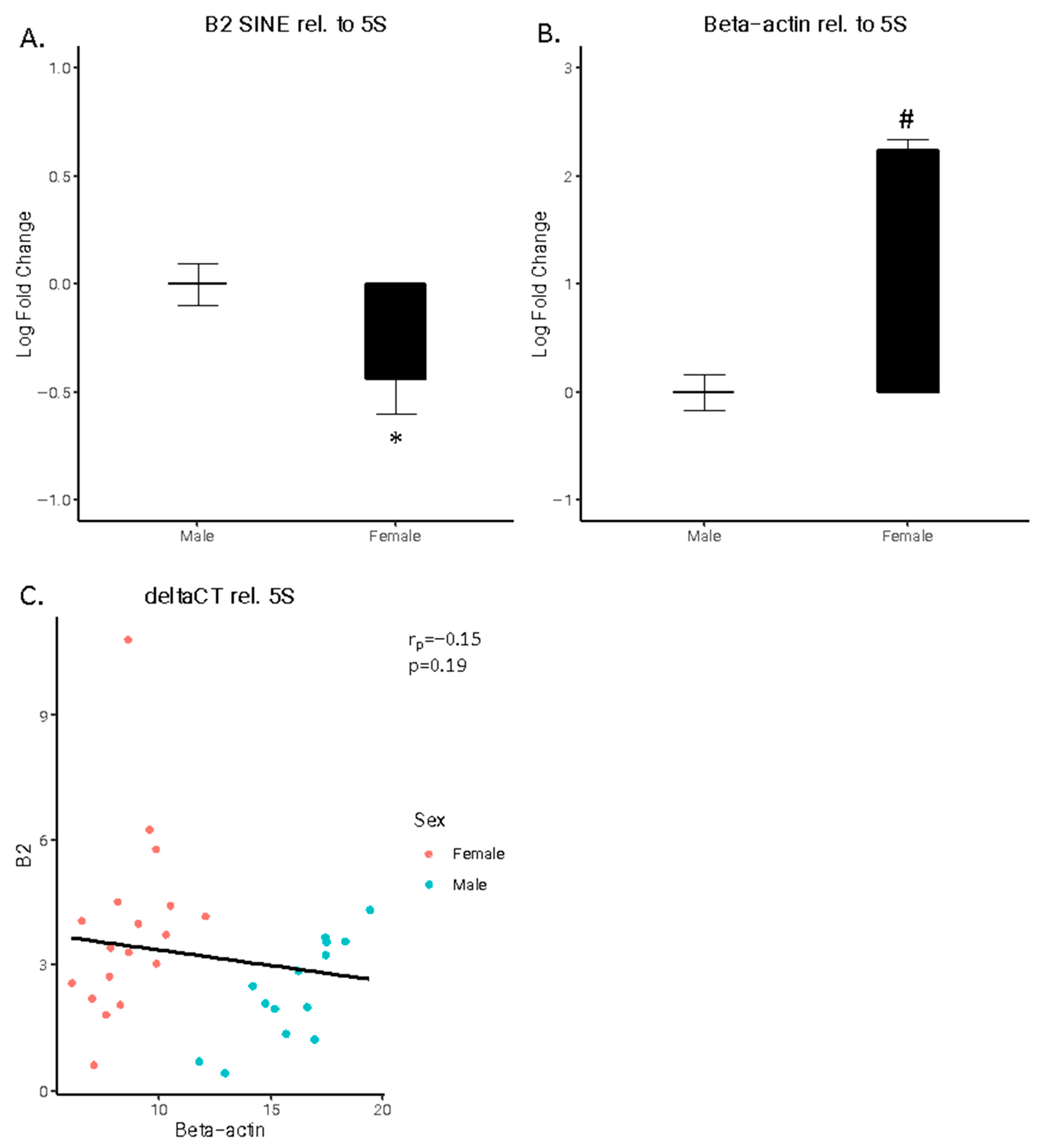
B2 SINE RNA is an allosteric regulator of pol II-dependent HKG expression. Therefore, we measured the expression of two pol III-dependent HKGs, 5S and 7SK, in tandem with both B2 SINE and ß-actin in the adult rat hippocampus. A significant difference was observed for both B2 SINE (t(30) = 2.2358, p < 0.05) and ß-actin (t(30) = 11.504, p < 1 × 10⁻¹²), normalized to 5 s. B2 SINE expression was higher in males compared to females (Figure 1A), whereas ß-actin expression was lower in males compared to females (Figure 1B). A weak, not statistically significant negative correlation (r(30) = 0.86803, p = 0.1961), was observed between B2 SINE and ß-actin deltaCT values (Figure 1C)
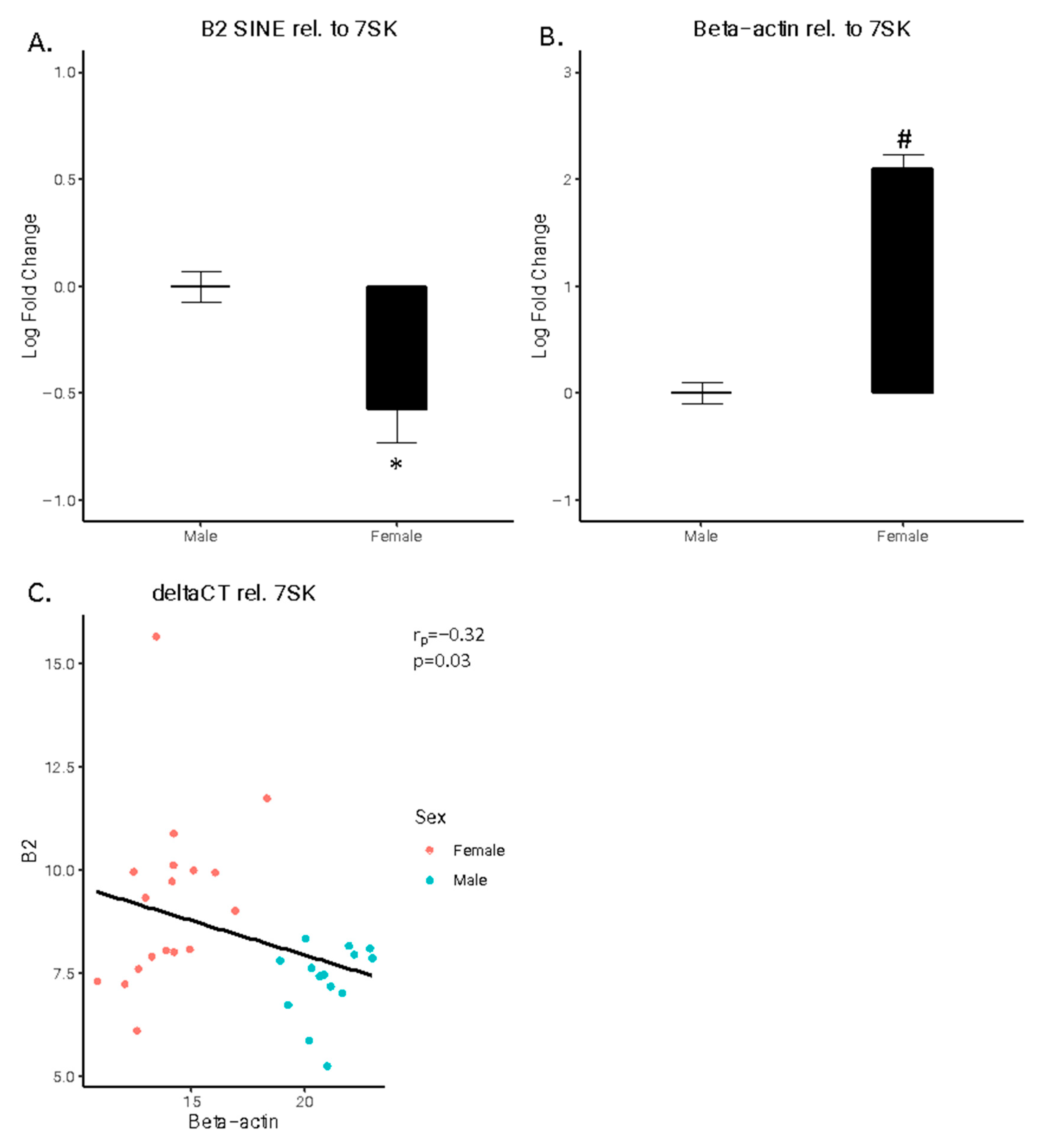
A significant difference was also observed for both B2 SINE and ß-actin normalized to 7SK. Once again, B2 SINE expression was higher in males compared to females (Figure 2A; t(30) = 3.1148, p < 0.01), whereas ß-actin expression was lower in males compared to females (Figure 2B; t(30) = 12.37, p < 1 × 10⁻¹³). A significant negative correlation was observed between B2 SINE and ß-actin deltaCT values (Figure 2C; r(30) = 1.91, p < 0.05).
We tested whether a subset of B2 SINE and ß-actin expression could be used to generate a model to successfully predict sex. the principal component analysis classified the data into two distinct groups regardless of HKG used for normalization (Figure S1A,B). Using linear discriminant analysis, we created a model using a 0.60 subset of the data for training. The generated models predicted the categorical variable, sex, without error, regardless of HKG used for normalization (Figure S1C,D). Similar results were obtained using LDA without replacement and with k-means clustering.

3.1. Higher Dendritic Complexity in Female Primary Hippocampal Neurons Compared to Male Neurons

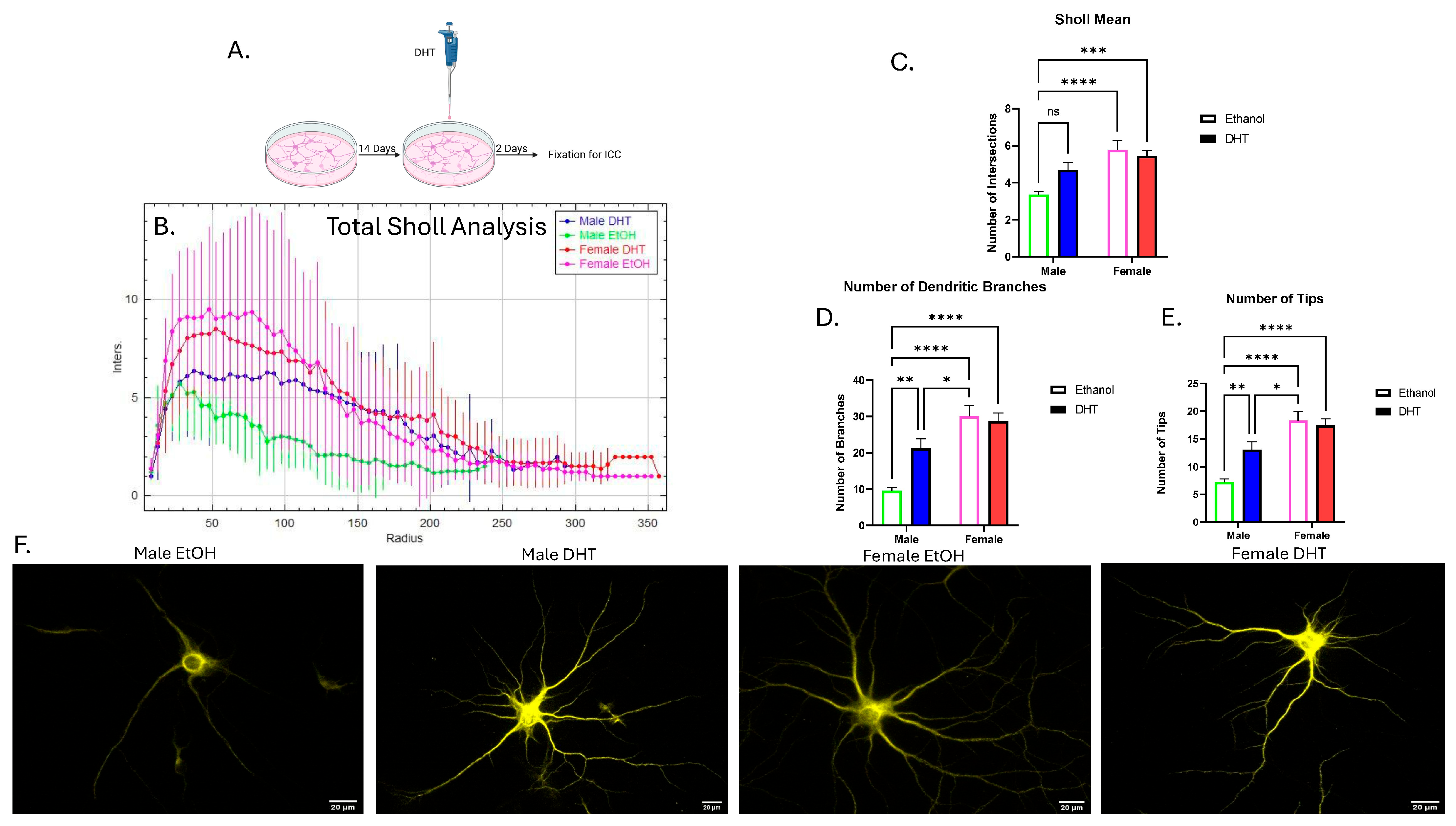
To investigate the role that DHT plays in dendritic arborization over the long term, DHT was added to neurons for 2 days, and dendritic arborization was assessed. Neurons were stained with microtubule-associated protein 2 (MAP2), a marker that shows up in dendrites and somas but not axons.
For Sholl mean, after a two-way ANOVA, there was a significant main effect of sex (η2 = 0.1449, p < 0.0001) and an interaction effect (η2 = 0.04037, p < 0.05). After multiple comparisons, there was a difference between the male neurons that received ethanol and both groups of female neurons (Figure 3B,C). However, there was no difference between male neurons treated with ethanol and males treated with DHT.
For the mean number of dendritic branches, a two-way ANOVA revealed a significant main effect of sex (η2 = 0.2357, p < 0.0001) and treatment (η2 = 0.03259, p < 0.05), as well as an interaction effect (η2 = 0.05214, p < 0.01). After multiple comparisons, there was a difference between male neurons that received ethanol and male neurons that received DHT, female neurons that received ethanol, and female neurons that received DHT. Finally, there was a difference between male neurons that received DHT and female neurons that received ethanol (Figure 3D).
For the mean number of tips, after a two-way ANOVA, there was a significant main effect of sex (η2 = 0.2509, p < 0.0001), as well as an interaction effect (η2 = 0.04835, p < 0.01). After multiple comparisons, there was a difference between male neurons treated with ethanol and male neurons treated with DHT. Additionally, there was a difference between male neurons treated with ethanol and both groups of female neurons. Finally, there was a difference between male neurons treated with DHT and female neurons treated with ethanol (Figure 3E). Representative images of one neuron from each group are presented in Figure 3F.

3.2. Knocking Down B2 SINE RNA Reduces Dendritic Complexity in Male Primary Hippocampal Neurons but Not in Females

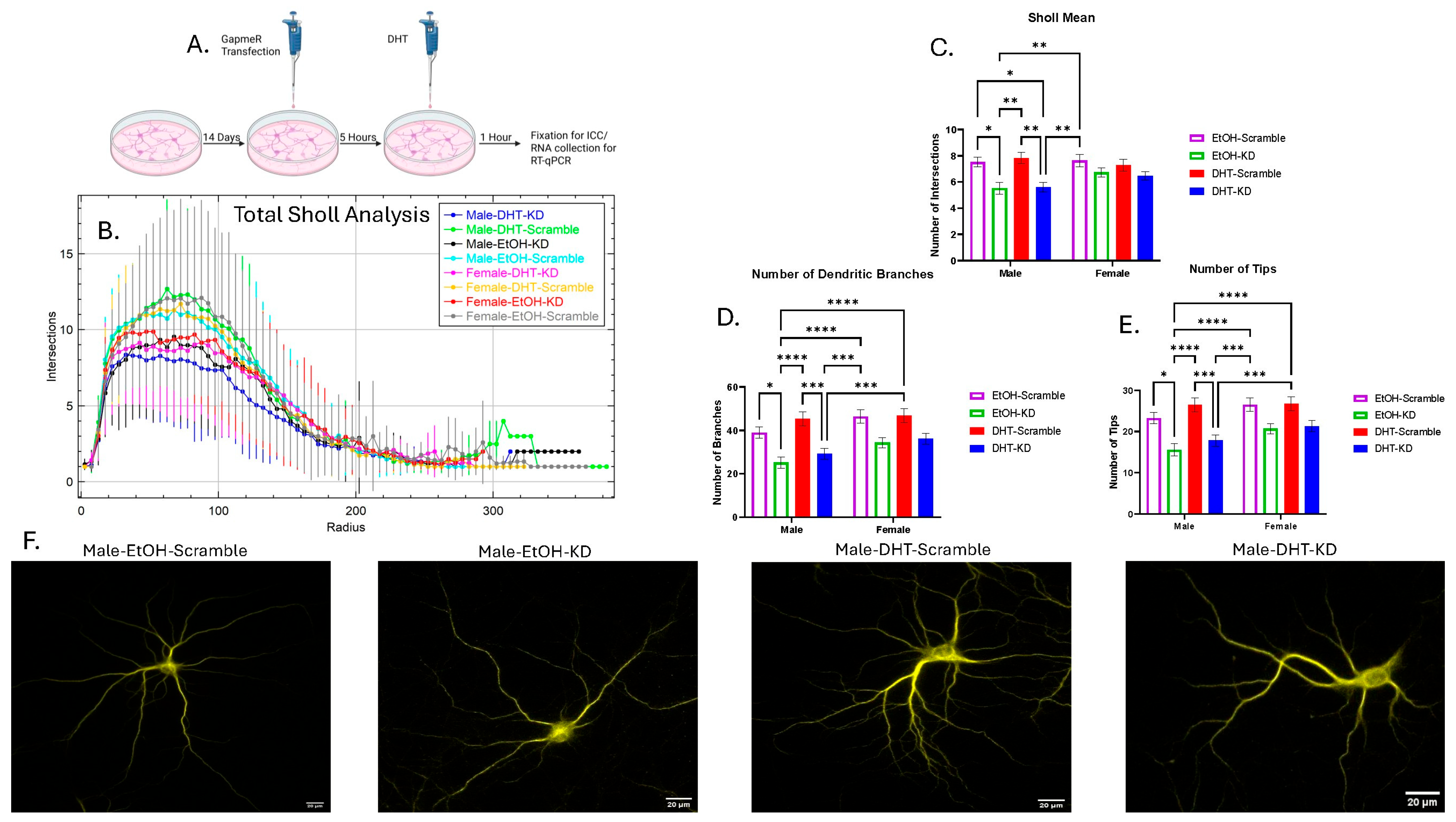
We utilized pooled B2 GapmeRs to knock down (KD) the expression of B2 SINE RNA in primary hippocampal neurons. We then treated these neurons with DHT or ethanol and measured the dendritic complexity through Sholl analysis.
For the Sholl mean, after a three-way ANOVA, there was a significant main effect of knockdown (η2 = 0.07356, p < 0.0001) and an interaction effect between sex and knockdown (η2 = 0.01290, p < 0.05). After multiple comparisons, there was a difference between Male-DHT-KD and Male-DHT-Scramble, Male-DHT-KD and Male-EtOH-Scramble, Male-DHT-KD and Female-EtOH-Scramble, Male-DHT-Scramble and Male-EtOH-KD, Male-EtOH-KD and Male-EtOH-Scramble, and Male-EtOH-KD and Female-EtOH-Scramble (Figure 4B,C).
For the number of branches, after a three-way ANOVA, there was a significant main effect of knockdown (η2 = 0.1104, p < 0.0001) and a simple main effect of sex (η2 = 0.02507, p < 0.01). After multiple comparisons, there was a difference between Male-DHT-KD and Male-DHT-Scramble, Male-DHT-KD and Female-DHT-Scramble, Male-DHT-KD and Female-EtOH-Scramble, Male-DHT-Scramble and Male-EtOH-KD, Male-EtOH-KD and Male-EtOH-Scramble, Male-EtOH-KD and Female-DHT-Scramble, and Male-EtOH-KD and Female-EtOH-Scramble (Figure 4D).
For the number of tips, after a three-way ANOVA, there was a significant main effect of knockdown (η2 = 0.1079, p < 0.0001) and a simple main effect of sex (η2 = 0.02098, p < 0.01). After multiple comparisons, there was a difference between Male-DHT-KD and Male-DHT-Scramble, Male-DHT-KD and Female-DHT-Scramble, Male-DHT-KD and Female-EtOH-Scramble, Male-DHT-Scramble and Male-EtOH-KD, Male-EtOH-KD and Male-EtOH-Scramble, Male-EtOH-KD and Female-DHT-Scramble, and Male-EtOH-KD and Female-EtOH-Scramble (Figure 4E). Representative images of one neuron from the male groups are presented in Figure 4F.
Altogether, this data suggests that B2 SINE RNA plays a positive role in dendritic complexity in males’ primary hippocampal neurons but not in females.

4. Discussion

We have found that B2 SINE RNA is a novel regulator of dendritic complexity of male neurons but not female neurons in primary hippocampal culture. Our results point to a B2 male-specific effect that may be independent of the acute effects of androgen treatment. Though it remains to be seen if there are organizational effects of androgens not captured by our experimental design, we believe that this is the first instance of a transposable element’s RNA being implicated in the development of the cytoarchitecture of neurons in a sex-dependent manner.
B2 SINEs generate ncRNA via RNA polymerase III and have a high copy number in the mouse genome [44,45]. Typically, these elements are repressed in cells, but in times of stress, they can be released from silencing and they can be upregulated [29,30,31,44,45,46]. There are remarkably few studies examining B2 SINE RNA in the nervous system, specifically within individual neurons. The studies that do exist focus on a stress effect [47,48]. Very recently, it was determined that B2 SINEs act as intrinsic axon growth regulators [49]. This study showed that B2 SINE ncRNA regulates neuronal growth specifically in nerve injury regeneration. These studies suggest that B2 SINE’s ncRNAs might have diverse biological roles in neurons separate from the stress effects observed. Since B2 SINEs have a recently demonstrated role in axonal repair and regrowth, we hypothesize that they may also regulate sex-specific aspects of neuron structure, though further work is needed to fully elucidate this activity. Our study suggests that B2 SINE RNA supports dendritic complexity in the developing hippocampus but only in male neurons.
Sex differences in the rodent brain are believed to occur due to the actions of gonadal hormones and their respective receptors, in large part. However, what is unclear are the mechanisms under which males’ and females’ hippocampi are differentially developed. It is believed that sexual differentiation of the rodent hippocampus starts early in life, with the organizational effects of circulating androgens and estrogens beginning in utero (see [50]) for a review). Here, we see a sex effect of dendritic arborization in part because of androgens and not estradiol. DHT is a non-aromatizable androgen, and this hormone acts through AR. Previously, it was shown that the DHT treatment contributes to a sex difference in hippocampal morphology [14,15], suggesting that sex differentiation of the rodent hippocampus begins early in life, with the organizational effects of DHT and AR. As the cells used in our study were collected after androgen secretion had begun in the embryo, it is possible that the effects we observed occurred due to earlier organizational effects of androgen rather than due to the acute DHT treatments we performed here. Another possibility is that B2 is under less epigenetic control in males than in females, leading to higher expression at all the developmental time points we have thus far observed. Taken together, this suggests that male hippocampal neurons are using higher amounts of B2 SINE ncRNA for the proper development of dendritic complexity, while females do not appear to require ncRNA for normal dendritic structure to emerge.
In humans and other primates, ALU SINE RNA is the closest functionally related ncRNA to B2 SINEs [51]. B2 and ALU SINEs have evolved convergently to the same function in rodent and primate genomes, respectively [52,53]. Recent research has begun to show relationships between SINE RNAs and brain disorders such as Alzheimer’s and autism. In the case of Alzheimer’s pathology, where increased processing of B2 and ALU SINEs showed transcriptome deregulation and gene expression changes in amyloid beta neuropathology in rodent and human Alzheimer’s brains, respectively [36,54]. Altogether, our study combined with others in the literature shows that B2 SINE ncRNA is contributing to the development of the cytoarchitecture of neurons in the brain. Our study expands on this by showing a sex effect on the development of neurons in the hippocampus. However, these connections remain speculative and more research would be needed to elucidate the link between the findings reported here and Alzheimer’s and other human brain disorders.
We identified a novel sex difference in brain retrotransposon RNA expression. To our knowledge, only one previous study has identified a sex difference in retrotransposon RNA expression in the hippocampus [55]. Furthermore, we have shown that the hippocampal expression of B2 RNA and ß-actin can be used to predict sex, indicating that this difference is robust. In light of the role of B2 SINE RNA during the cellular heat shock response, we speculate that this retrotransposon regulates the expression of ß-actin, which is known to vary between sexes in a variety of tissues, including the brain [25,56,57]. In this same vein, Alu RNA has shown to be critical to the heat shock response in primates [58]. Alu RNA abundance has also been linked to retinal degradation in cellular models of macular degeneration, and its expression is correlated with Tau pathology in the fly brain, illustrating the potential for dysregulation of TEs to lead to neuronal pathologies [59,60,61,62]. Future studies looking into these contexts may reveal sex differences in Alu RNA expression, potentially conferring resistance or susceptibility to pathological phenotype. Yet, the mechanisms by which B2 SINE RNA may be differentially regulated in the brain by sex are of potentially profound interest. Our previous observation that B2 SINE RNA is regulated in the brain by maternal immune activation, also in a sex specific manner, is salient in this context. MIA, like male sex, is a significant risk factor for ASD, which suggests that these ncRNAs may be an important target for future research regarding the etiology of ASD and other neurodevelopmental disorders [27,28]. These data provide a scaffold for future studies for determining such potential effects in rodent and human neurons, as well as further studies for establishing a clearer mechanistic relation between B2 and the machinery of sex determination and differentiation.

Supplementary Materials

The following supporting information can be downloaded at: https://www.mdpi.com/article/10.3390/cells15090816/s1, Figure S1: B2 SINE and beta-actin expression predictive models. (A) Principal component analysis plot using expression values normalized to 5S and (B) to 7SK PCA successfully segregates data into two clusters. (C) Partition plot from linear discriminant analysis normalized to 5S and (D) to 7SK. The models created from a subset of the data correctly predicts sex. Figure S2: LNA GapmeR pool targets and knocks down B2 SINE RNA in primary hippocampal cells. Expression of B2 SINE RNA after either GapmeR pool or scrambled vector transfection. Data is represented as mean fold change over 7sk RNA ± SEM (n = 3/group) (** p < 0.01). In order to assess if the pooled B2 GapmeRs successfully knocked down B2 SINE RNA in primary hippocampal neurons after 6 h, we transfected a subset of neurons for 6 h, collected RNA, and tested for presence by RT-qPCR. We found a significant difference between the neurons transfected with the GapmeR pool and neurons transfected with a scrambled version. This shows that B2 SINE RNA is depleted after transfection with GapmeRs for 6 h. Figure S3: The secondary structure of B2 SINE RNA generated with RNAfold. B2 SINE RNA contains a putative ARE identified with motif analysis using FIMO from the MEME suite.

Author Contributions

T.A.R., E.T.O., E.K.P., A.A.B. and H.E.L. acquired data. T.A.R., E.T.O., A.A.B., G.G. and H.E.L. analyzed and interpreted the data. T.A.R., E.K.P., A.A.B., H.E.L., S.L.Z. and R.G.H. designed the experiments. T.A.R. and A.A.B. wrote the manuscript, and all authors contributed to manuscript revision. All authors have read and agreed to the published version of the manuscript.

Funding

Funding for this work was provided by the University of Massachusetts Boston through the College of Liberal Arts Dean’s office and the Office of Sponsored Research Projects for Dr. Richard Hunter.

Institutional Review Board Statement

The animal study protocol was approved by the IACUC of University of Massachusetts Boston (#35, 25 January 2021).

Informed Consent Statement

Not applicable.

Data Availability Statement

The data analyzed during the current study are available from the corresponding author on reasonable request. Supplementary information is available for this paper. Correspondence and requests for materials should be addressed to Troy A. Richter, PhD.

Acknowledgments

We wish to thank Kelly Lambert and Molly Kent for providing brain tissue for this study.

Conflicts of Interest

The authors declare no conflicts of interest.

References

  1. Maenner, M.J. Prevalence and Characteristics of Autism Spectrum Disorder Among Children Aged 8 Years—Autism and Developmental Disabilities Monitoring Network, 11 Sites, United States, 2020. MMWR Surveill. Summ. 2023, 72, 1–14. [Google Scholar] [CrossRef] [Scilit] [PubMed]
  2. Shaw, K.A.; Williams, S.; E Patrick, M.; Valencia-Prado, M.; Durkin, M.S.; Howerton, E.M.; Ladd-Acosta, C.M.; Pas, E.T.; Bakian, A.V.; Bartholomew, P.; et al. Prevalence and Early Identification of Autism Spectrum Disorder Among Children Aged 4 and 8 Years—Autism and Developmental Disabilities Monitoring Network, 16 Sites, United States, 2022. MMWR Surveill. Summ. 2025, 74, 1–22. [Google Scholar] [CrossRef] [Scilit]
  3. Shors, T.J.; Chua, C.; Falduto, J. Sex Differences and Opposite Effects of Stress on Dendritic Spine Density in the Male Versus Female Hippocampus. J. Neurosci. 2001, 21, 6292–6297. [Google Scholar] [CrossRef] [Scilit]
  4. Hatanaka, Y.; Hojo, Y.; Mukai, H.; Murakami, G.; Komatsuzaki, Y.; Kim, J.; Ikeda, M.; Hiragushi, A.; Kimoto, T.; Kawato, S. Rapid increase of spines by dihydrotestosterone and testosterone in hippocampal neurons: Dependence on synaptic androgen receptor and kinase networks. Brain Res. 2015, 1621, 121–132. [Google Scholar] [CrossRef] [Scilit]
  5. Kuwahara, N.; Nicholson, K.; Isaacs, L.; MacLusky, N.J. Androgen Effects on Neural Plasticity. Androg. Clin. Res. Ther. 2021, 2, 216–230. [Google Scholar] [CrossRef] [Scilit] [PubMed]
  6. Parducz, A.; Garcia-Segura, L.M. Sexual differences in the synaptic connectivity in the rat dentate gyrus. Neurosci. Lett. 1993, 161, 53–56. [Google Scholar] [CrossRef] [Scilit]
  7. Atwi, S.; McMahon, D.; Scharfman, H.; MacLusky, N.J. Androgen Modulation of Hippocampal Structure and Function. Neuroscientist 2016, 22, 46–60. [Google Scholar] [CrossRef] [Scilit]
  8. Kerr, J.E.; Allore, R.J.; Beck, S.G.; Handa, R.J. Distribution and hormonal regulation of androgen receptor (AR) and AR messenger ribonucleic acid in the rat hippocampus. Endocrinology 1995, 136, 3213–3221. [Google Scholar] [CrossRef] [Scilit] [PubMed]
  9. Sar, M.; Lubahn, D.B.; French, F.S.; Wilson, E.M. Immunohistochemical Localization of the Androgen Receptor in Rat and Human Tissues. Endocrinology 1990, 127, 3180–3186. [Google Scholar] [CrossRef] [Scilit] [PubMed]
  10. Shughrue, P.J.; Merchenthaler, I. Evidence for novel estrogen binding sites in the rat hippocampus. Neuroscience 2000, 99, 605–612. [Google Scholar] [CrossRef] [Scilit]
  11. Tabori, N.E.; Stewart, L.; Znamensky, V.; Romeo, R.; Alves, S.; McEwen, B.; Milner, T. Ultrastructural evidence that androgen receptors are located at extranuclear sites in the rat hippocampal formation. Neuroscience 2005, 130, 151–163. [Google Scholar] [CrossRef] [Scilit]
  12. Celec, P.; OstatnÃ-ková, D.; Hodosy, J. On the effects of testosterone on brain behavioral functions. Front. Neurosci. 2015, 9, 12. [Google Scholar] [CrossRef] [Scilit]
  13. Konkle, A.T.M.; McCarthy, M.M. Developmental Time Course of Estradiol, Testosterone, and Dihydrotestosterone Levels in Discrete Regions of Male and Female Rat Brain. Endocrinology 2011, 152, 223–235. [Google Scholar] [CrossRef] [Scilit] [PubMed]
  14. Isgor, C.; Sengelaub, D.R. Prenatal gonadal steroids affect adult spatial behavior, CA1 and CA3 pyramidal cell morphology in rats. Horm. Behav. 1998, 34, 183–198. [Google Scholar] [CrossRef] [Scilit] [PubMed]
  15. Isgor, C.; Sengelaub, D.R. Effects of neonatal gonadal steroids on adult CA3 pyramidal neuron dendritic morphology and spatial memory in rats. J. Neurobiol. 2003, 55, 179–190. [Google Scholar] [CrossRef] [Scilit]
  16. Kight, K.E.; McCarthy, M.M. Androgens and the developing hippocampus. Biol. Sex Differ. 2020, 11, 30. [Google Scholar] [CrossRef] [Scilit]
  17. Williams, C.L.; Barnett, A.M.; Meck, W.H. Organizational effects of early gonadal secretions on sexual differentiation in spatial memory. Behav. Neurosci. 1990, 104, 84–97. [Google Scholar] [CrossRef] [PubMed]
  18. de Koning, A.P.J.; Gu, W.; Castoe, T.A.; Batzer, M.A.; Pollock, D.D. Repetitive elements may comprise over two-thirds of the human genome. PLoS Genet. 2011, 7, e1002384. [Google Scholar] [CrossRef] [Scilit]
  19. Lander, E.S.; Linton, L.M.; Birren, B.; Nusbaum, C. Initial sequencing and analysis of the human genome. Nature 2001, 409, 860–921. [Google Scholar] [CrossRef] [Scilit]
  20. Mouse Genome Sequencing Consortium. Initial Sequencing and Comparative Analysis of the Mouse Genome. Nature 2002, 420, 520–562. [Google Scholar] [CrossRef] [Scilit]
  21. ENCODE Project Consortium. An integrated encyclopedia of DNA elements in the human genome. Nature 2012, 489, 57–74. [Google Scholar] [CrossRef] [Scilit]
  22. Faulkner, G.J.; Kimura, Y.; Daub, C.O.; Wani, S.; Plessy, C.; Irvine, K.M.; Schroder, K.; Cloonan, N.; Steptoe, A.L.; Lassmann, T.; et al. The regulated retrotransposon transcriptome of mammalian cells. Nat. Genet. 2009, 41, 563–571. [Google Scholar] [CrossRef] [Scilit]
  23. Allen, T.A.; Von Kaenel, S.; Goodrich, J.A.; Kugel, J.F. The SINE-encoded mouse B2 RNA represses mRNA transcription in response to heat shock. Nat. Struct. Mol. Biol. 2004, 11, 816–821. [Google Scholar] [CrossRef] [Scilit]
  24. Fornace, A.J.; Mitchell, J.B. Induction of B2 RNA polymerase III transcription by heat shock: Enrichment for heat shock induced sequences in rodent cells by hybridization subtraction. Nucleic Acids Res. 1986, 14, 5793–5811. [Google Scholar] [CrossRef] [Scilit]
  25. Espinoza, C.A.; Allen, T.A.; Hieb, A.R.; Kugel, J.F.; Goodrich, J.A. B2 RNA binds directly to RNA polymerase II to repress transcript synthesis. Nat. Struct. Mol. Biol. 2004, 11, 822–829. [Google Scholar] [CrossRef] [Scilit]
  26. Espinoza, C.A.; Goodrich, J.A.; Kugel, J.F. Characterization of the structure, function, and mechanism of B2 RNA, an ncRNA repressor of RNA polymerase II transcription. RNA 2007, 13, 583–596. [Google Scholar] [CrossRef] [Scilit]
  27. Richter, T.A.; Aiken, A.A.; Puracchio, M.J.; Maganga-Bakita, I.; Hunter, R.G. Maternal Immune Activation and Enriched Environments Impact B2 SINE Expression in Stress Sensitive Brain Regions of Rodent Offspring. Genes 2023, 14, 858. [Google Scholar] [CrossRef] [Scilit] [PubMed]
  28. DeRosa, H.; Smith, A.; Geist, L.; Cheng, A.; Hunter, R.G.; Kentner, A.C. Maternal immune activation alters placental histone-3 lysine-9 tri-methylation, offspring sensorimotor processing, and hypothalamic transposable element expression in a sex-specific manner. Neurobiol. Stress 2023, 24, 100538. [Google Scholar] [CrossRef] [Scilit] [PubMed]
  29. Bartlett, A.A.; DeRosa, H.; Clark, M.; Lapp, H.; Guffanti, G.; Hunter, R. Corticosterone dynamically regulates retrotransposable element expression in the rat hippocampus and C6 cells. Neurobiol. Stress 2021, 15, 100397. [Google Scholar] [CrossRef] [Scilit]
  30. Hunter, R.G.; Murakami, G.; Dewell, S.; Seligsohn, M.; Baker, M.E.R.; Datson, N.A.; McEwen, B.S.; Pfaff, D.W. Acute stress and hippocampal histone H3 lysine 9 trimethylation, a retrotransposon silencing response. Proc. Natl. Acad. Sci. USA 2012, 109, 17657–17662. [Google Scholar] [CrossRef] [Scilit] [PubMed]
  31. Bartlett, A.A.; Guffanti, G.; Hunter, R.G. B2 SINE RNA as a novel regulator of glucocorticoid receptor transcriptional activity. Neurobiol. Stress 2023, 23, 100522. [Google Scholar] [CrossRef] [Scilit]
  32. Jurka, J.; Kapitonov, V.V.; Pavlicek, A.; Klonowski, P.; Kohany, O.; Walichiewicz, J. Repbase Update, a Database of Eukaryotic Repetitive Elements. Cytogenet. Genome Res. 2005, 110, 462–467. [Google Scholar] [CrossRef] [Scilit]
  33. Bailey, T.L.; Johnson, J.; Grant, C.E.; Noble, W.S. The MEME Suite. Nucleic Acids Res. 2015, 43, W39–W49. [Google Scholar] [CrossRef] [Scilit] [PubMed]
  34. Gupta, S.; Stamatoyannopoulos, J.A.; Bailey, T.L.; Noble, W.S. Quantifying Similarity between Motifs. Genome Biol. 2007, 8, R24. [Google Scholar] [CrossRef] [Scilit]
  35. Grant, C.E.; Bailey, T.L.; Noble, W.S. FIMO: Scanning for Occurrences of a given Motif. Bioinformatics 2011, 27, 1017–1018. [Google Scholar] [CrossRef] [Scilit]
  36. Cheng, Y.; Saville, L.; Gollen, B.; Isaac, C.; Belay, A.; Mehla, J.; Patel, K.; Thakor, N.; Mohajerani, M.H.; Zovoilis, A. Increased processing of SINE B2 ncRNAs unveils a novel type of transcriptome deregulation in amyloid beta neuropathology. eLife 2020, 9, e61265. [Google Scholar] [CrossRef] [Scilit] [PubMed]
  37. Nunez, J. Primary Culture of Hippocampal Neurons from P0 Newborn Rats. J. Vis. Exp. 2008, e895. [Google Scholar] [CrossRef] [Scilit]
  38. Zup, S.L.; Edwards, N.S.; McCarthy, M.M. Sex and age dependent effects of androgens on glutamate-induced cell death and intracellular calcium regulation in the developing hippocampus. Neuroscience 2014, 281, 77–87. [Google Scholar] [CrossRef] [Scilit]
  39. Arshadi, C.; Günther, U.; Eddison, M.; Harrington, K.I.S.; Ferreira, T.A. SNT: A unifying toolbox for quantification of neuronal anatomy. Nat. Methods 2021, 18, 374–377. [Google Scholar] [CrossRef] [Scilit]
  40. Schindelin, J.; Arganda-Carreras, I.; Frise, E.; Kaynig, V.; Longair, M.; Pietzsch, T.; Preibisch, S.; Rueden, C.; Saalfeld, S.; Schmid, B.; et al. Fiji: An open-source platform for biological-image analysis. Nat. Methods 2012, 9, 676–682. [Google Scholar] [CrossRef] [Scilit] [PubMed]
  41. Do, D.V.; Strauss, B.; Cukuroglu, E.; Macaulay, I.; Wee, K.B.; Hu, T.X.; Igor, R.D.L.M.; Lee, C.; Harrison, A.; Butler, R.; et al. SRSF3 maintains transcriptome integrity in oocytes by regulation of alternative splicing and transposable elements. Cell Discov. 2018, 4, 33. [Google Scholar] [CrossRef] [Scilit]
  42. Weihs, C.; Ligges, U.; Luebke, K.; Raabe, N. klaR Analyzing German Business Cycles. In Data Analysis and Decision Support; Baier, D., Decker, R., Schmidt-Thieme, L., Eds.; Springer: Berlin/Heidelberg, Germany, 2005; pp. 335–343. [Google Scholar] [CrossRef] [Scilit]
  43. Venables, W.N.; Ripley, B.D. Modern Applied Statistics with S.; Springer: New York, NY, USA, 2002. [Google Scholar] [CrossRef] [Scilit]
  44. Ponicsan, S.L.; Kugel, J.F.; Goodrich, J.A. Genomic gems: SINE RNAs regulate mRNA production. Curr. Opin. Genet. Dev. 2010, 20, 149–155. [Google Scholar] [CrossRef] [Scilit]
  45. Walters, R.D.; Kugel, J.F.; Goodrich, J.A. InvAluable junk: The cellular impact and function of Alu and B2 RNAs. IUBMB Life 2009, 61, 831–837. [Google Scholar] [CrossRef] [Scilit]
  46. Zovoilis, A.; Cifuentes-Rojas, C.; Chu, H.-P.; Hernandez, A.J.; Lee, J.T. Destabilization of B2 RNA by EZH2 activates the stress response. Cell 2016, 167, 1788–1802.e13. [Google Scholar] [CrossRef] [Scilit] [PubMed]
  47. Middleton, S.A.; Eberwine, J.; Kim, J. Comprehensive catalog of dendritically localized mRNA isoforms from sub-cellular sequencing of single mouse neurons. BMC Biol. 2019, 17, 5. [Google Scholar] [CrossRef] [Scilit]
  48. Policarpi, C.; Crepaldi, L.; Brookes, E.; Nitarska, J.; French, S.M.; Coatti, A.; Riccio, A. Enhancer SINEs Link Pol III to Pol II Transcription in Neurons. Cell Rep. 2017, 21, 2879–2894. [Google Scholar] [CrossRef] [Scilit] [PubMed]
  49. Zahavi, E.E.; Koppel, I.; Kawaguchi, R.; Oses-Prieto, J.A.; Briner, A.; Monavarfeshani, A.; Costa, I.D.; van Niekerk, E.; Lee, J.; Matoo, S.; et al. Repeat-element RNAs integrate a neuronal growth circuit. Cell 2025, 188, 4350–4365.e22. [Google Scholar] [CrossRef] [Scilit] [PubMed]
  50. Premachandran, H.; Zhao, M.; Arruda-Carvalho, M. Sex Differences in the Development of the Rodent Corticolimbic System. Front. Neurosci. 2020, 14, 583477. [Google Scholar] [CrossRef] [Scilit]
  51. Hernandez, A.J.; Zovoilis, A.; Cifuentes-Rojas, C.; Han, L.; Bujisic, B.; Lee, J.T. B2 and ALU retrotransposons are self-cleaving ribozymes whose activity is enhanced by EZH2. Proc. Natl. Acad. Sci. USA 2020, 117, 415–425. [Google Scholar] [CrossRef] [Scilit]
  52. Lucas, B.A.; Lavi, E.; Shiue, L.; Cho, H.; Katzman, S.; Miyoshi, K.; Siomi, M.C.; Carmel, L.; Ares, M., Jr.; Maquat, L.E. Evidence for convergent evolution of SINE-directed Staufen-mediated mRNA decay. Proc. Natl. Acad. Sci. USA 2018, 115, 968–973. [Google Scholar] [CrossRef] [Scilit]
  53. Maquat, L.E. Short interspersed nuclear element (SINE)-mediated post-transcriptional effects on human and mouse gene expression: SINE-UP for active duty. Philos. Trans. R. Soc. B Biol. Sci. 2020, 375, 20190344. [Google Scholar] [CrossRef] [Scilit]
  54. Cheng, Y.; Saville, L.; Gollen, B.; Veronesi, A.A.; Mohajerani, M.; Joseph, J.T.; Zovoilis, A. Increased Alu RNA processing in Alzheimer brains is linked to gene expression changes. EMBO Rep. 2021, 22, e52255. [Google Scholar] [CrossRef] [Scilit]
  55. Cuarenta, A.; Kigar, S.L.; Henion, I.C.; Karls, K.E.; Chang, L.; Bakshi, V.P.; Auger, A.P. Early life stress increases Line1 within the developing brain in a sex-dependent manner. Brain Res. 2020, 1748, 147123. [Google Scholar] [CrossRef] [Scilit] [PubMed]
  56. Derks, N.M.; Müller, M.; Gaszner, B.; Tilburg-Ouwens, D.; Roubos, E.; Kozicz, L. Housekeeping genes revisited: Different expressions depending on gender, brain area and stressor. Neuroscience 2008, 156, 305–309. [Google Scholar] [CrossRef] [Scilit]
  57. Tanic, N.; Perovic, M.; Mladenovic, A.; Ruzdijic, S.; Kanazir, S. Effects of aging, dietary restriction and glucocorticoid treatment on housekeeping gene expression in rat cortex and hippocampus—Evaluation by real time RT-PCR. J. Mol. Neurosci. 2007, 32, 38–46. [Google Scholar] [CrossRef] [Scilit]
  58. Yakovchuk, P.; Goodrich, J.A.; Kugel, J.F. B2 RNA and Alu RNA repress transcription by disrupting contacts between RNA polymerase II and promoter DNA within assembled complexes. Proc. Natl. Acad. Sci. USA 2009, 106, 5569–5574. [Google Scholar] [CrossRef] [Scilit]
  59. Kumar, M.S.; Pester, R.E.; Chen, C.Y.; Lane, K.; Chin, C.; Lu, J.; Kirsch, D.G.; Golub, T.R.; Jacks, T. Dicer1 functions as a haploinsufficient tumor suppressor. Genes Dev. 2009, 23, 2700–2704. [Google Scholar] [CrossRef] [Scilit] [PubMed]
  60. Kaneko, H.; Dridi, S.; Tarallo, V.; Gelfand, B.D.; Fowler, B.J.; Gil Cho, W.; Kleinman, M.E.; Ponicsan, S.L.; Hauswirth, W.W.; Chiodo, V.A.; et al. DICER1 deficit induces Alu RNA toxicity in age-related macular degeneration. Nature 2011, 471, 325. [Google Scholar] [CrossRef] [Scilit]
  61. Tarallo, V.; Hirano, Y.; Gelfand, B.D.; Dridi, S.; Kerur, N.; Kim, Y.; Gil Cho, W.; Kaneko, H.; Fowler, B.J.; Bogdanovich, S.; et al. DICER1 loss and Alu RNA induce age-related macular degeneration via the NLRP3 inflammasome and MyD88. Cell 2012, 149, 847–859. [Google Scholar] [CrossRef] [Scilit] [PubMed]
  62. Guo, C.; Jeong, H.-H.; Hsieh, Y.-C.; Klein, H.-U.; Bennett, D.A.; De Jager, P.L.; Liu, Z.; Shulman, J.M. Tau Activates Transposable Elements in Alzheimer’s Disease. Cell Rep. 2018, 23, 2874–2880. [Google Scholar] [CrossRef] [Scilit]
Figure 1. B2 SINE and beta-actin expression relative to 5S. (A) B2 SINE expression and (B) beta-actin expression. Log fold changes are shown for B2 SINE and beta-actin expression relative to 5S. (C) B2 SINE vs. beta-actin expression. The deltaCT values for B2 SINE and beta-actin are shown relative to 5S. Males are represented by blue circles and females by red circles. The 95% confidence interval for predictions from a linear model is noted in gray. (* p < 0.05, # p < 10−10).
Cells 15 00816 g001
Figure 2. B2 SINE and beta-actin expression relative to 7SK. (A) B2 SINE expression and (B) beta-actin expression. Log fold changes are shown for B2 SINE and beta-actin expression relative to 7SK. (C) B2 SINE vs. beta-actin expression. The deltaCT values for B2 SINE and beta-actin are shown relative to 7SK. Males are represented by blue and females by red. The 95% confidence interval for predictions from a linear model is noted in gray. (* p < 0.05, # p < 10−10).
Cells 15 00816 g002
Figure 3. Sholl analysis of treated male and female primary hippocampal neurons. (A) Experimental paradigm for primary hippocampal culture. (B) Total Sholl analysis for all four groups. The data is represented as the number of intersections (Inters.) for each concentric circle radius (Radius) ± SD (male DHT, n = 27; male EtOH, n = 26; female DHT, n = 27; and female EtOH, n = 26). (C) Sholl mean, (D) Number of dendritic branches. (E) Number of tips is shown. The data is represented as mean ± SEM (ns: not significant, * p < 0.05, ** p < 0.01, *** p < 0.001, **** p < 0.0001). (F) Representative images of MAP2 stained neurons.
Cells 15 00816 g003
Figure 4. Sholl analysis of treated and knocked down male and female primary hippocampal neurons. (A) Experimental paradigm for primary hippocampal culture knockdown. (B) Total Sholl analysis for all eight groups. The data is represented as the number of intersections (Inters.) for each concentric circles radius (Radius) ± SD (Male-DHT-KD, n = 50; Male-DHT-Scramble, n = 45; Male-EtOH-KD, n = 39; Male-EtOH-Scramble, n = 42; Female-DHT-KD, n = 51; Female-DHT-Scramble, n = 41; Female-EtOH-KD, n = 37; Female-EtOH-Scramble, n = 42). (C) Sholl Mean. (D) Number of dendritic branches. (E) Number of tips is shown. The data is represented as mean ± SEM (* p < 0.05, ** p < 0.01, *** p < 0.001, **** p < 0.0001). (F) Representative images of male MAP2-stained neurons.
Cells 15 00816 g004
Disclaimer/Publisher’s Note: The statements, opinions and data contained in all publications are solely those of the individual author(s) and contributor(s) and not of MDPI and/or the editor(s). MDPI and/or the editor(s) disclaim responsibility for any injury to people or property resulting from any ideas, methods, instructions or products referred to in the content.