Ultrastructural Signs of High Functional Activity of Neuromuscular Synapses in Aging Rats After Photobiomodulation

 , , , , and
Highlights
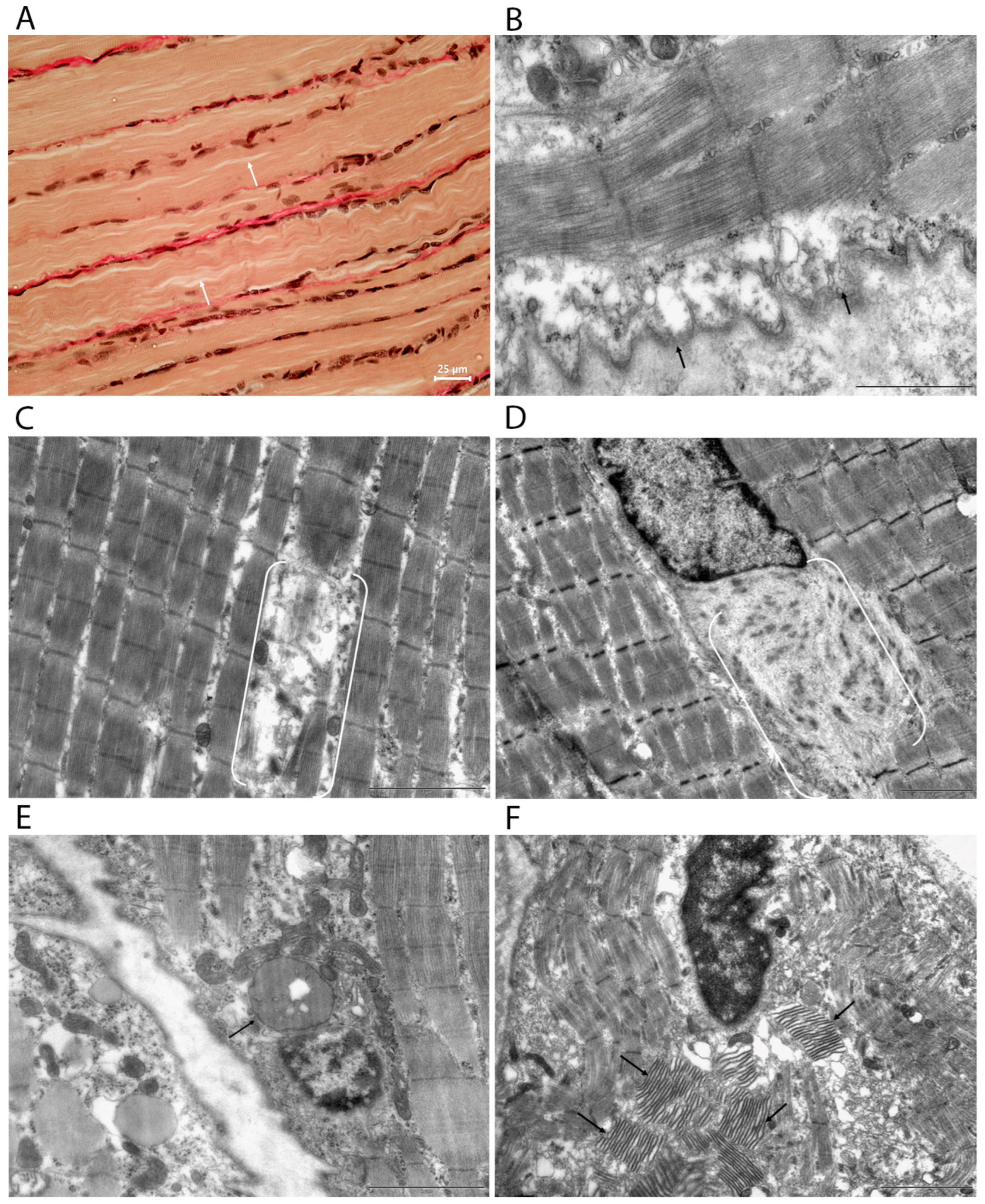
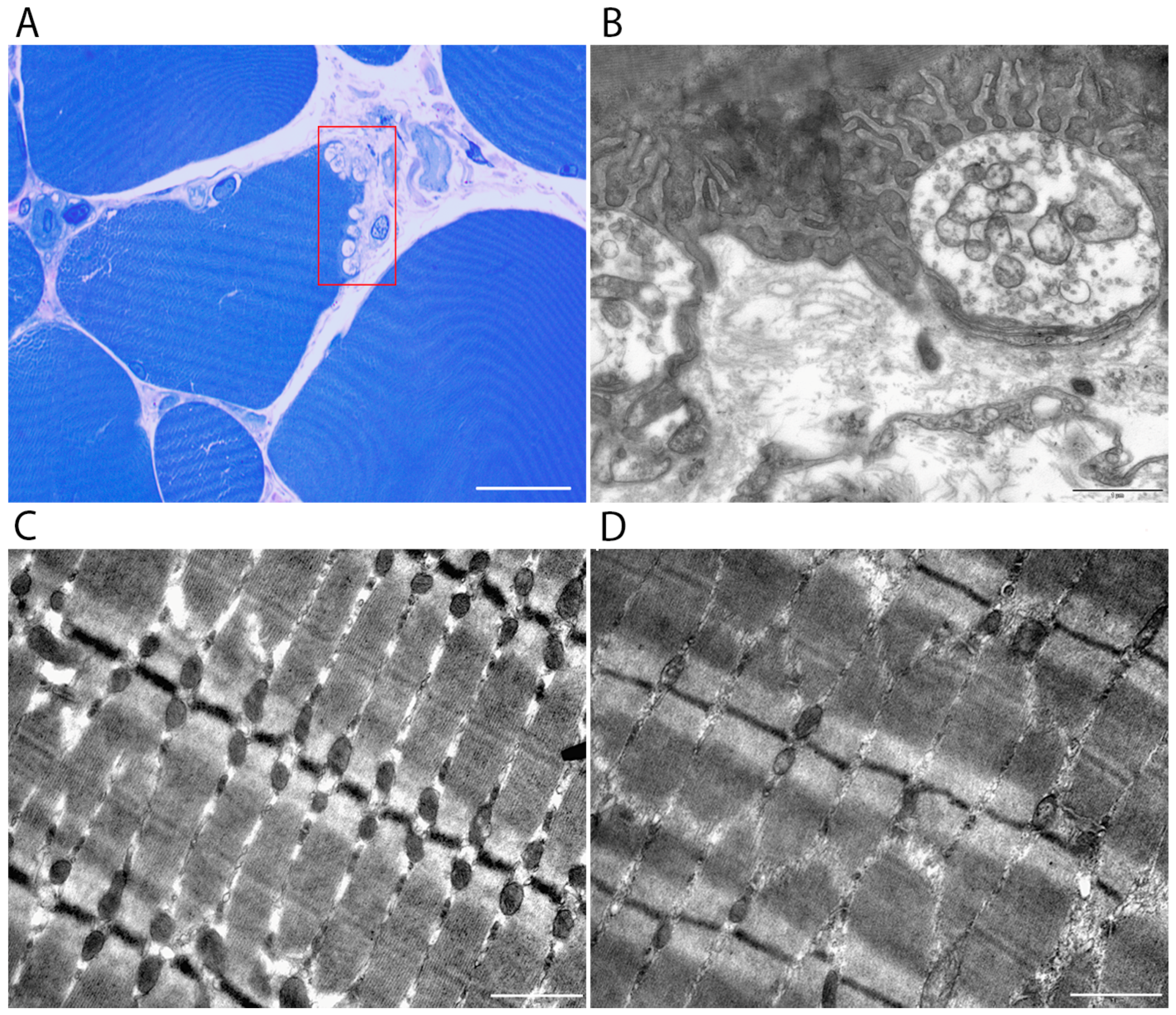
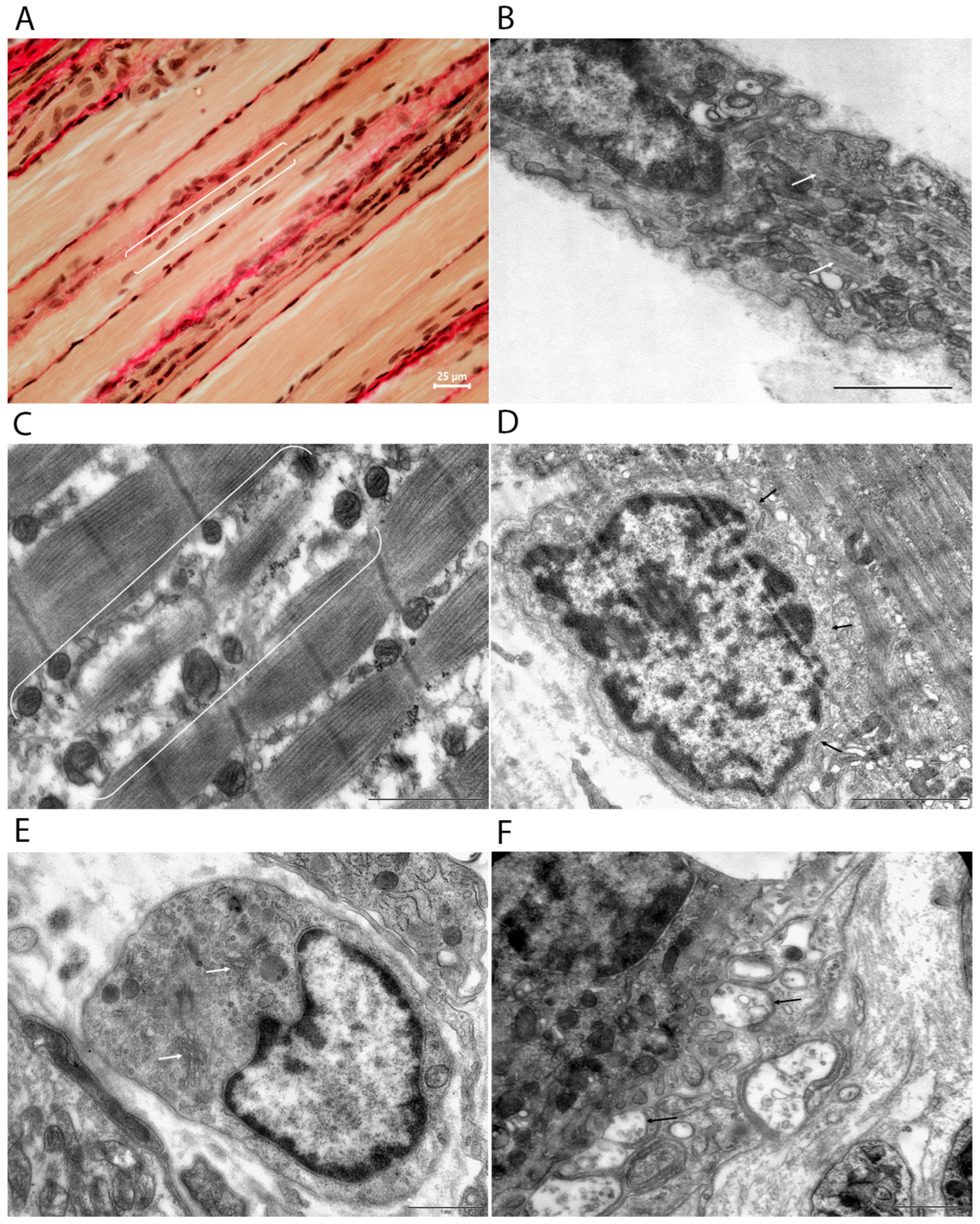
- Light and transmission electron microscopy revealed characteristic age-related changes in muscle fibers and neuromuscular synapses.
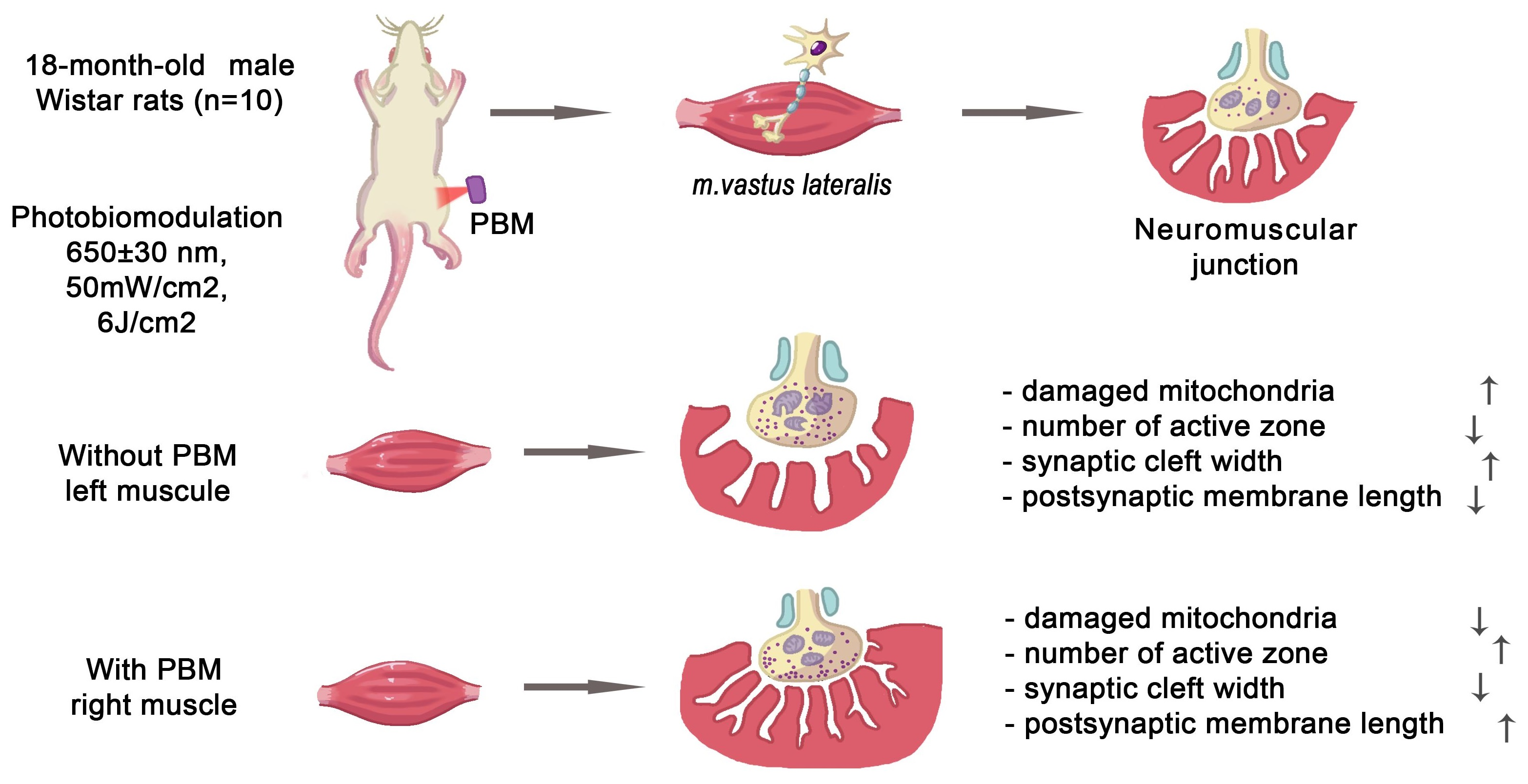
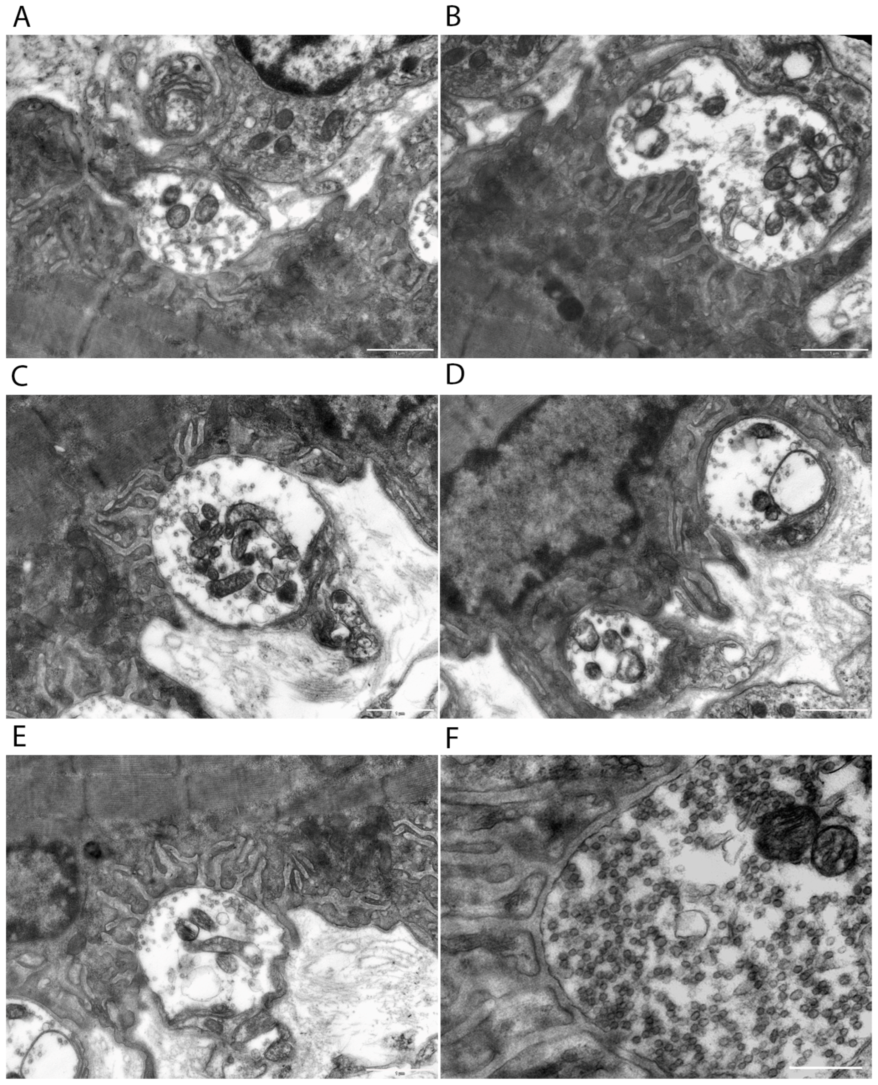
- Photobiomodulation causes noticeable ultrastructural reorganization of neuromuscular junctions in aged rats, including an increase in the number of active zones, elongation of the postsynaptic membrane, narrowing of the synaptic cleft, and mitochondrial hyperplasia.
- Photobiomodulation promotes compensatory structural reorganization of neuromuscular junctions and enhances their functional activity.
- These results highlight the therapeutic potential of photobiomodulation for alleviating age-related sarcopenia and muscle weakness.
Abstract
1. Introduction
2. Materials and Methods
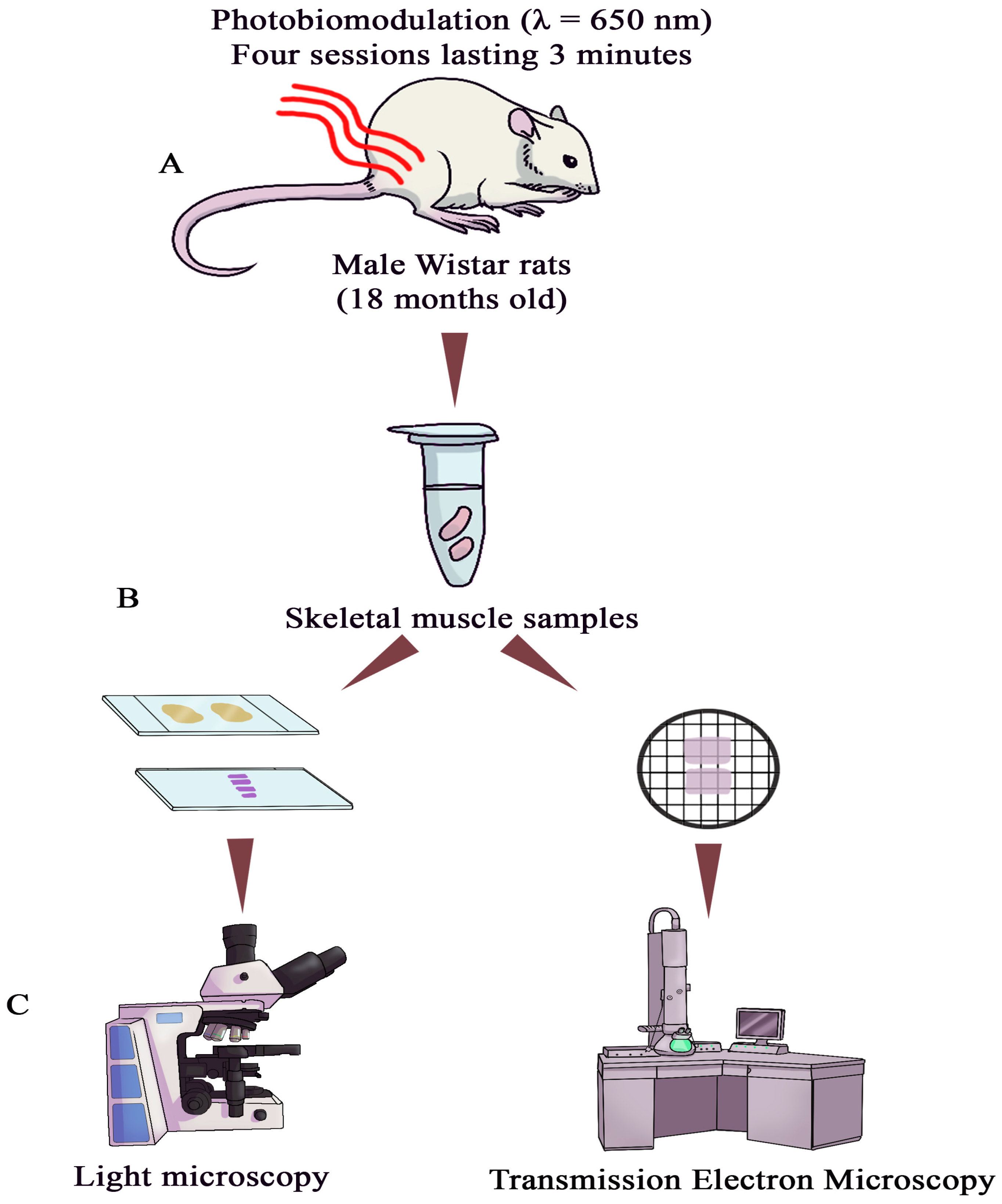
2.1. Photobiomodulation Mode
2.2. Light Microscopy
2.3. Transmission Electron Microscopy (TEM)
2.4. Morphometric Analysis of Nerve Terminal
- The readily releasable pool (RRP) is a small group of vesicles located directly adjacent to the presynaptic membrane in the active zone of the synapse. The vesicles in this pool are “loaded” and ready for immediate exocytosis upon the arrival of an action potential. They participate in rapid synaptic transmission, with release occurring within milliseconds after depolarization. After release, they are quickly replenished from other pools.
- The reserve pool (RP) consists of a large number of vesicles located in the cytoplasm of the presynaptic terminal, away from the active zone. These vesicles do not participate in immediate transmission but serve as a reserve during prolonged or intense stimulation. During sustained activity, vesicles from the reserve pool are mobilized and transported to the active zone via cytoskeletal elements. Their mobilization is regulated by calcium and several proteins, such as synapsin.
- The recycling pool (RPc) is intermediate in both function and localization between the RRP and RP. Vesicles in this pool are situated closer to the active zone than those in the reserve pool but are not as tightly associated with it as in the RRP. They participate in maintaining synaptic transmission at moderate stimulation frequencies and continuously replenish the RRP through endocytosis and vesicle reformation after exocytosis. This pool ensures the stability of synaptic transmission during repeated action potentials.
2.5. Statistical Analysis
3. Results
3.1. Morphological and Ultrastructural Characteristics of Skeletal Muscle in Aging Rats Before and After PBM
3.2. Morphometric Analysis of NMS
4. Discussion
5. Conclusions
Author Contributions
Funding
Institutional Review Board Statement
Informed Consent Statement
Data Availability Statement
Acknowledgments
Conflicts of Interest
Abbreviations
| ACh | Acetylcholine |
| AM | Cross-section area of mitochondrion |
| APT | Presynaptic terminal area |
| DMAX | Maximum presynaptic terminal diameter |
| DMIN | Minimum presynaptic terminal diameter |
| LPRE | Length of the presynaptic membrane |
| LPOST | Length of the postsynaptic membrane |
| NAZ | Number of active zones |
| NMS | Neuromuscular synapse |
| PBM | Photobiomodulation |
| PPT | Presynaptic terminal perimeter |
| RRP | Readily releasable pool (of synaptic vesicles) |
| RP | Reserve pool (of synaptic vesicles) |
| RPc | Recycling pool (of synaptic vesicles) |
| TEM | Transmission electron microscopy |
| WSC | Width of the synaptic cleft |
Appendix A

| Parameter | Symbol | Control | PBM |
|---|---|---|---|
| n | n | ||
| Presynaptic terminal area (µm2) | APT | 20 | 20 |
| Presynaptic terminal perimeter (µm) | PPT | 20 | 20 |
| Maximum presynaptic terminal diameter (µm) | DMAX | 20 | 20 |
| Minimum presynaptic terminal diameter (µm) | DMIN | 20 | 20 |
| Presynaptic membrane length (µm) | LPRE | 20 | 20 |
| Postsynaptic membrane length (µm) | LPOST | 20 | 20 |
| Synaptic cleft width (nm) | WSC | 340 | 340 |
| Number of active zone (number) | NAZ | 224 | 261 |
| Cross-section area of mitochondrion (µm2) | AM | 140 | 136 |
| Total number of mitochondria (number) | NM | 140 | 136 |
| Mitochondrial number with inner membrane defects (number) | NIMD | 49 | 62 |
| Mitochondrial number with outer membrane defects (number) | NOMD | 41 | 41 |
| Number of Readily Releasable Vesicles (number) | NRRP | 307 | 210 |
| Number of Recycling Vesicles (number) | NRPC | 986 | 575 |
| Number of Reserve Vesicles (number) | NRV | 1657 | 1189 |
| Morphometric Parameters | Control | PBM | Δ% | ||
|---|---|---|---|---|---|
| Presynaptic terminal area, µm2 | 4.24 | [2.59–5.51] | 3.73 | [2.41–5.04] | −12.0 |
| Presynaptic terminal perimeter (PPT), µm | 8.20 | [6.42–10.22] | 8.08 | [6.35–8.61] | −1.5 |
| Maximum presynaptic terminal diameter (DMAX), µm | 3.00 | [2.17–3.66] | 2.65 | [1.96–3.11] | −11.7 |
| Minimum presynaptic terminal diameter (DMIN), µm | 1.57 | [1.37–1.81] | 1.79 | [1.47–2.12] | +14.0 |
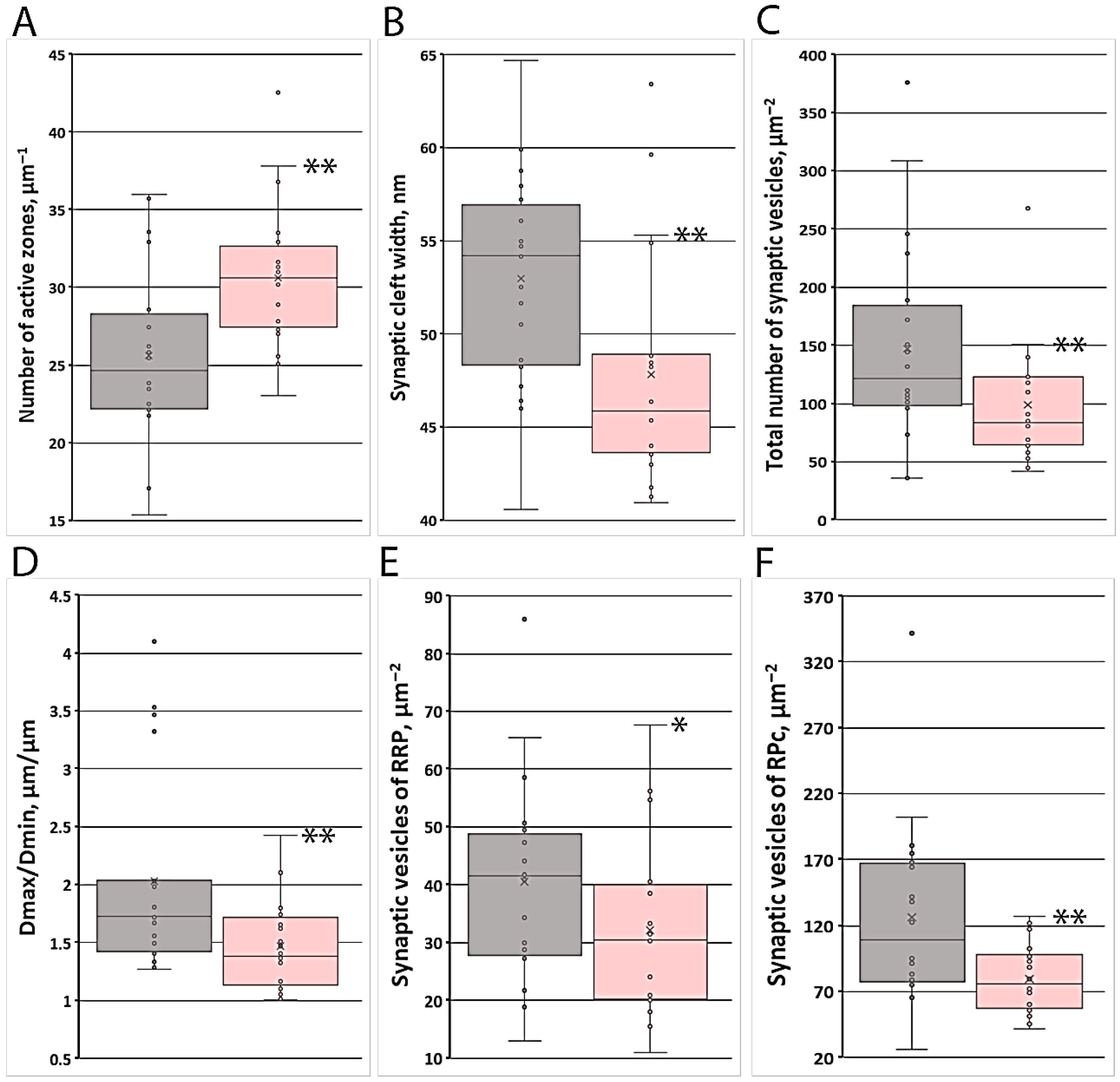
| DMAX/DMIN, µm/µm | 1.73 | [1.47–2.04] | 1.39 | [1.15–1.68] ** | −17.6 |
| Presynaptic membrane length (LPRE), µm | 4.12 | [3.77–5.30] | 4.13 | [3.47–5.25] | +0.2 |
| Postsynaptic membrane length (LPOST), µm | 18.14 | [12.53–23.52] | 21.79 | [12.93–26.71] | +20.1 |
| PPT/LPOST, µm/µm | 0.54 | [0.32–0.69] | 0.38 | [0.34–0.45] | −29.6 |
| LPRE/LPOST, µm/µm | 0.26 | [0.18–0.38] | 0.22 | [0.20–0.26] | −15.4 |
| Number of active zones per unit LPRE, µm−1 | 24.7 | [22.2–27.7] | 30.6 | [27.7–32.0] ** | +23.9 |
| Synaptic cleft width, nm | 54.2 | [48.5–56.4] | 45.9 | [43.9–48.9] ** | −15.3 |
| Axoplasm volume fraction, % | 80.4 | [72.1–85.3] | 79.5 | [73.5–83.4] | −1.1 |
| Mitochondrial volume fraction, % | 12.8 | [7.2–23.7] | 15.3 | [12.1–22.0] | +19.5 |
| Synaptic vesicle volume fraction, % | 6.4 | [4.2–7.0] | 4.2 | [3.6–4.9] * | −34.4 |
| Morphometric Parameters | Control | PBM | Δ% | ||
|---|---|---|---|---|---|
| Cross-section area, µm2 | 0.095 | [0.082–0.116] | 0.093 | [0.084–0.128] | −2.1 |
| Total number, µm−2 | 13.5 | [10.5–22.4] | 15.5 | [13.2–19.8] | +14.8 |
| Number with inner membrane defects, µm−2 | 6.5 | [2.0–7.7] | 7.7 | [4.7–11.0] | +18.5 |
| Number with outer membrane defects, µm−2 | 4.1 | [3.1–7.1] | 4.9 | [4.0–6.5] | +19.5 |
| Fraction with inner membrane defects, % | 41.4 | [19.2–50.0] | 36.4 | [33.3–58.2] | −12.1 |
| Fraction with outer membrane defects, % | 30.9 | [20.0–50.0] | 33.3 | [25.8–44.6] | +7.8 |
| Morphometric Parameters | Control | PBM | Δ% | ||
|---|---|---|---|---|---|
| Number of synaptic vesicles per unit area (µm−2) | |||||
| Total number | 375.5 | [246.2–411.2] | 244.4 | [210.9–289.5] * | −34.9 |
| RRP | 41.6 | [28.5–47.8] | 30.5 | [20.6–39.1] * | −26.7 |
| RPC | 109.3 | [78.3–165.1] | 76.0 | [59.2–97.1] ** | −30.5 |
| RP | 181.7 | [124.9–240.9] | 130.2 | [110.0–155.2] | −28.3 |
| Number of synaptic vesicles per unit presynaptic membrane length (µm−1) | |||||
| RRP | 33.1 | [25.3–39.6] | 27.2 | [21.9–29.7] ** | −17.8 |
| RPC | 9.1 | [6.7–12.1] | 5.2 | [3.9–7.3] ** | −42.9 |
| RP | 102.7 | [76.4–139.2] | 66.4 | [50.9–79.1] ** | −35.3 |
| Number of synaptic vesicles per unit postsynaptic membrane length (µm−1) | |||||
| RRP | 28.5 | [18.0–39.3] | 13.9 | [11.1–17.9] ** | −51.2 |
| RPC | 146.9 | [95.0–237.1] | 117.2 | [77.5–156.8] | −20.2 |
| RP | 47.3 | [23.1–75.1] | 24.3 | [18.3–35.6] * | −48.6 |
| Fraction of Pool (%) | |||||
| RRP | 11.8 | [7.0–16.9] | 12.0 | [6.5–17.0] | +1.7 |
| RPC | 32.4 | [29.9–40.6] | 30.3 | [26.7–35.3] | −6.5 |
| RP | 50.0 | [45.9–63.0] | 57.7 | [48.0–60.2] | +15.4 |
References
- Kubat, G.B.; Bouhamida, E.; Ulger, O.; Turkel, I.; Pedriali, G.; Ramaccini, D.; Ekinci, O.; Ozerklig, B.; Atalay, O.; Patergnani, S.; et al. Mitochondrial dysfunction and skeletal muscle atrophy: Causes, mechanisms, and treatment strategies. Mitochondrion 2023, 72, 33–58. [Google Scholar] [CrossRef] [Scilit]
- Frontera, W.R.; Ochala, J. Skeletal muscle: A brief review of structure and function. Calcif. Tissue Int. 2015, 96, 183–195. [Google Scholar] [CrossRef] [Scilit]
- Deschenes, M.R.; Flannery, R.; Hawbaker, A.; Patek, L.; Mifsud, M. Adaptive Remodeling of the Neuromuscular Junction with Aging. Cells 2022, 11, 1150. [Google Scholar] [CrossRef] [Scilit]
- Sweeney, H.L.; Hammers, D.W. Muscle Contraction. Cold Spring Harb. Perspect. Biol. 2018, 10, a023200. [Google Scholar] [CrossRef] [Scilit]
- Rudolf, R.; Khan, M.M.; Witzemann, V. Motor Endplate-Anatomical, Functional, and Molecular Concepts in the Historical Perspective. Cells 2019, 8, 387. [Google Scholar] [CrossRef] [Scilit]
- Sayed, R.K.; de Leonardis, E.C.; Guerrero-Martínez, J.A.; Rahim, I.; Mokhtar, D.M.; Saleh, A.M.; Abdalla, K.E.; Pozo, M.J.; Escames, G.; López, L.C.; et al. Identification of morphological markers of sarcopenia at early stage of aging in skeletal muscle of mice. Exp. Gerontol. 2016, 83, 22–30. [Google Scholar] [CrossRef] [Scilit]
- Paez, H.G.; Pitzer, C.R.; Alway, S.E. Age-Related Dysfunction in Proteostasis and Cellular Quality Control in the Development of Sarcopenia. Cells 2023, 12, 249. [Google Scholar] [CrossRef] [Scilit] [PubMed]
- Stanga, S.; Boido, M.; Kienlen-Campard, P. How to Build and to Protect the Neuromuscular Junction: The Role of the Glial Cell Line-Derived Neurotrophic Factor. Int. J. Mol. Sci. 2020, 22, 136. [Google Scholar] [CrossRef] [Scilit]
- Krajewska-Węglewicz, L.; Felczak, P.; Dorobek, M. Effects of Aging on Orbicularis Oculi Muscle Strength and Ultrastructure in Dermatochalasis: A Pilot Study. J. Clin. Med. 2025, 14, 162. [Google Scholar] [CrossRef] [Scilit]
- Trovato, F.M.; Imbesi, R.; Conway, N.; Castrogiovanni, P. Morphological and Functional Aspects of Human Skeletal Muscle. J. Funct. Morphol. Kinesiol. 2016, 1, 289–302. [Google Scholar] [CrossRef] [Scilit]
- de Vasconcelos, F.T.G.R.; Souza, B.W.; Souza, L.S.; Vainzof, M. Tubular Aggregates as a Marker of Aging in Skeletal Muscle. Methods Mol. Biol. 2025, 2960, 19–26. [Google Scholar] [CrossRef] [Scilit]
- Grigorieva, I.I.; Raskina, T.A.; Letaeva, M.V.; Malyshenko, O.S.; Averkieva, Y.V.; Masenko, V.L.; Kokov, A.N. Sarcopenia: Pathogenesis and diagnosis. Fundam. Clin. Med. 2019, 4, 105–116. (In Russian) [Google Scholar] [CrossRef] [Scilit]
- Quirk, B.J.; Whelan, H.T. What Lies at the Heart of Photobiomodulation: Light, Cytochrome C Oxidase, and Nitric Oxide-Review of the Evidence. Photobiomodulation Photomed. Laser Surg. 2020, 389, 527–530. [Google Scholar] [CrossRef] [Scilit]
- Kumar, P.; Umakanth, S.; N, G. Photobiomodulation therapy as an adjunct to resistance exercises on muscle metrics, functional balance, functional capacity, and physical performance among older adults: A systematic scoping review. Lasers Med. Sci. 2024, 39, 232. [Google Scholar] [CrossRef] [Scilit] [PubMed]
- Liu, H.; Cheema, U.; Player, D.J. Photobiomodulation therapy (PBMT) in skeletal muscle regeneration: A comprehensive review of mechanisms, clinical applications, and future directions. Photodiagnosis Photodyn. Ther. 2025, 53, 104634. [Google Scholar] [CrossRef] [Scilit]
- Dobrowolny, G.; Barbiera, A.; Sica, G.; Scicchitano, B.M. Age-Related Alterations at Neuromuscular Junction: Role of Oxidative Stress and Epigenetic Modifications. Cells 2021, 10, 1307. [Google Scholar] [CrossRef] [Scilit]
- Monich, V.A.; Bavrina, A.P.; Malinovskaya, S.L. Modification in oxidative processes in muscle tissues exposed to laser- and light-emitting diode radiation. Lasers Med. Sci. 2018, 33, 159–164. [Google Scholar] [CrossRef] [Scilit]
- Bavrina, A.P.; Shchelchkova, N.A.; Vasyagina, T.I.; Pchelin, P.V.; Lapshin, R.D.; Belousova, I.I.; Nefedova, D.A. Ultrastructure of Myofascial Trigger Points of Skeletal Muscles of Rats after Photobiostimulation with Low-Intensity Red Light. Bull. Exp. Biol. Med. 2023, 175, 711–713. [Google Scholar] [CrossRef] [Scilit]
- Humphrey, C.D.; Pittman, F.E. A simple methylene blue-azure II-basic fuchsin stain for epoxy-embedded tissue sections. Stain. Technol. 1974, 49, 9–14. [Google Scholar] [CrossRef] [Scilit] [PubMed]
- Venable, J.H.; Coggeshall, R.A. A simplified lead citrate stain for use in electron microscopy. J. Cell Biol. 1965, 25, 407–408. [Google Scholar] [CrossRef] [Scilit] [PubMed]
- Lam, J.; Katti, P.; Biete, M.; Mungai, M.; AshShareef, S.; Neikirk, K.; Garza Lopez, E.; Vue, Z.; Christensen, T.A.; Beasley, H.K.; et al. A Universal Approach to Analyzing Transmission Electron Microscopy with ImageJ. Cells 2021, 10, 2177. [Google Scholar] [CrossRef] [Scilit]
- Avtandilov, G.G. Medical Morphometry: A Guide; Medicine: Moscow, Russia, 1990; 384p. (In Russian) [Google Scholar]
- Denker, A.; Rizzoli, S.O. Synaptic vesicle pools: An update. Front. Synaptic Neurosci. 2010, 2, 135. [Google Scholar] [CrossRef] [Scilit]
- Guarnieri, F.C. How Do Synaptic Vesicles “Know” Which Pool They Belong to? J. Neurosci. 2017, 37, 2276–2278. [Google Scholar] [CrossRef] [Scilit]
- Petrov, A.M.; Giniatullin, A.R.; Zefirov, A.L. The role of the cAMP signaling cascade in the circulation of synaptic vesicles in the motor nerve endings of frogs. Neurochemistry 2008, 25, 202–210. (In Russian) [Google Scholar]
- Anokhin, P.K. Key Issues in the Theory of Functional Systems; Nauka: Moscow, Russia, 1980; 196p. (In Russian) [Google Scholar]
- Rudolf, R.; Khan, M.M.; Labeit, S.; Deschenes, M.R. Degeneration of neuromuscular junction in age and dystrophy. Front. Aging Neurosci. 2014, 6, 99. [Google Scholar] [CrossRef] [Scilit]
- Hunter, S.K.; Pereira, H.M.; Keenan, K.G. The aging neuromuscular system and motor performance. J. Appl. Physiol. 2016, 121, 982–995. [Google Scholar] [CrossRef] [Scilit]
- Deschenes, M.R.; Stock, M.S. Neuromuscular adaptations to exercise and aging. Exp. Gerontol. 2022, 160, 111712. [Google Scholar] [CrossRef] [Scilit]
- Andreo, L.; Ribeiro, B.G.; Alves, A.N.; Martinelli, A.S.A.; Soldera, C.B.; Horliana, A.C.R.T.; Bussadori, S.K.; Fernandes, K.P.S.; Mesquita-Ferrari, R.A. Effects of Photobiomodulation with Low-level Laser Therapy on Muscle Repair Following a Peripheral Nerve Injury in Wistar Rats. Photochem. Photobiol. 2020, 96, 1124–1132. [Google Scholar] [CrossRef] [Scilit]
- Sasso, L.L.; de Souza, L.G.; Girasol, C.E.; Marcolino, A.M.; de Jesus Guirro, R.R.; Barbosa, R.I. Photobiomodulation in Sciatic Nerve Crush Injuries in Rodents: A Systematic Review of the Literature and Perspectives for Clinical Treatment. J. Lasers Med. Sci. 2020, 11, 332–344. [Google Scholar] [CrossRef] [Scilit] [PubMed]
- Sridharan, K.; Waheed, T.O.; Staehlke, S.; Riess, A.; Mand, M.; Meyer, J.; Seitz, H.; Peters, K.; Hahn, O. Light Exposure as a Tool to Enhance the Regenerative Potential of Adipose-Derived Mesenchymal Stem/Stromal Cells. Cells 2025, 14, 1143. [Google Scholar] [CrossRef] [Scilit]
- Weiss, V.B.; Vangeli, I.M.; Eldarov, C.M.; Bakeeva, L.E. Ultrastructure of Mitochondria in Skeletal Muscle Weakened by Sarcopenia during Aging. Biochem. Suppl. Ser. B Biomed. Chem. 2024, 18, 132–143. [Google Scholar] [CrossRef] [Scilit]
- Ferraresi, C.; Hamblin, M.R.; Parizotto, N.A. Low-level laser (light) therapy (LLLT) on muscle tissue: Performance, fatigue and repair benefited by the power of light. Photonics Lasers Med. 2012, 1, 267–286. [Google Scholar] [CrossRef] [Scilit]
- Hamblin, M.R. Mechanisms and Mitochondrial Redox Signaling in Photobiomodulation. Photochem. Photobiol. 2018, 94, 199–212. [Google Scholar] [CrossRef] [Scilit]
- Ferraresi, C.; Kaippert, B.; Avci, P.; Huang, Y.Y.; de Sousa, M.V.; Bagnato, V.S.; Parizotto, N.A.; Hamblin, M.R. Low-level laser (light) therapy increases mitochondrial membrane potential and ATP synthesis in C2C12 myotubes with a peak response at 3–6 h. Photochem. Photobiol. 2015, 91, 411–416. [Google Scholar] [CrossRef] [Scilit]
- Salehpour, F.; Ahmadian, N.; Rasta, S.H.; Farhoudi, M.; Karimi, P.; Sadigh-Eteghad, S. Transcranial low-level laser therapy improves brain mitochondrial function and cognitive impairment in D-galactose-induced aging mice. Neurobiol. Aging 2017, 58, 140–150. [Google Scholar] [CrossRef] [Scilit]
- Cardoso, F.D.S.; Gonzalez-Lima, F.; Coimbra, N.C. Mitochondrial Photobiomodulation as a Neurotherapeutic Strategy for Epilepsy. Front. Neurol. 2022, 13, 873496. [Google Scholar] [CrossRef] [Scilit]
- Li, X.; Wang, X.K.; Zhu, Z.J.; Liang, Z.W.; Li, P.H.; Ma, Y.G.; Ding, T.; Li, K.; Zuo, X.S.; Ju, C.; et al. Photobiomodulation provides neuroprotection through regulating mitochondrial fission imbalance in the subacute phase of spinal cord injury. Neural Regen. Res. 2023, 18, 2005–2010. [Google Scholar] [CrossRef] [Scilit] [PubMed]
- de Castro, B.M.; De Jaeger, X.; Martins-Silva, C.; Lima, R.D.; Amaral, E.; Menezes, C.; Lima, P.; Neves, C.M.; Pires, R.G.; Gould, T.W.; et al. The vesicular acetylcholine transporter is required for neuromuscular development and function. Mol. Cell. Biol. 2009, 29, 5238–5250. [Google Scholar] [CrossRef] [Scilit]
- Kuromi, H.; Kidokoro, Y. Two synaptic vesicle pools, vesicle recruitment and replenishment of pools at the Drosophila neuromuscular junction. J. Neurocytol. 2003, 32, 551–565. [Google Scholar] [CrossRef] [Scilit] [PubMed]
- Ruiter, M.; Houy, S.; Engholm-Keller, K.; Graham, M.E.; Sørensen, J.B. SNAP-25 phosphorylation at Ser187 is not involved in Ca2+ or phorbolester-dependent potentiation of synaptic release. Mol. Cell Neurosci. 2020, 102, 103452. [Google Scholar] [CrossRef] [Scilit] [PubMed]
- Rowley, K.L.; Mantilla, C.B.; Ermilov, L.G.; Sieck, G.C. Synaptic vesicle distribution and release at rat diaphragm neuromuscular junctions. J. Neurophysiol. 2007, 98, 478–487. [Google Scholar] [CrossRef] [Scilit]
- Kong, L.; Wang, X.; Choe, D.W.; Polley, M.; Burnett, B.G.; Bosch-Marcé, M.; Griffin, J.W.; Rich, M.M.; Sumner, C.J. Impaired synaptic vesicle release and immaturity of neuromuscular junctions in spinal muscular atrophy mice. J. Neurosci. 2009, 29, 842–851. [Google Scholar] [CrossRef] [Scilit]
- Rodriguez Gotor, J.J.; Mahfooz, K.; Perez-Otano, I.; Wesseling, J.F. Parallel processing of quickly and slowly mobilized reserve vesicles in hippocampal synapses. eLife 2024, 12, RP88212. [Google Scholar] [CrossRef] [Scilit] [PubMed]
- Zhang, Z.; Zhang, Z.; Liu, P.; Xue, X.; Zhang, C.; Peng, L.; Shen, W.; Yang, S.; Wang, F. The Role of Photobiomodulation to Modulate Ion Channels in the Nervous System: A Systematic Review. Cell Mol. Neurobiol. 2024, 44, 79. [Google Scholar] [CrossRef] [Scilit] [PubMed]
- Hong, N.; Kim, H.J.; Kang, K.; Park, J.O.; Mun, S.; Kim, H.G.; Kang, B.H.; Chung, P.S.; Lee, M.Y.; Ahn, J.C. Photobiomodulation improves the synapses and cognitive function and ameliorates epileptic seizure by inhibiting downregulation of Nlgn3. Cell Biosci. 2023, 13, 8. [Google Scholar] [CrossRef] [Scilit] [PubMed]
- Chu, X.L.; Zhao, X.X.; Liu, S.Y.; Li, Y.J.; Ding, N.; Liu, M.Q.; Li, Q.W.; Li, Q. Research progress in different physical therapies for treating peripheral nerve injuries. Front. Neurol. 2025, 16, 1508604. [Google Scholar] [CrossRef] [Scilit]
- Lopes-Martins, R.Á.B.; Gonzalez-Lima, F.; Gomes da Silva, S.; Leonardo, P.S.; Soncino, C.; Pacheco, R.F.; Oliveira, C.L.d.O.e.; Cardoso, F.D.S. Photobiomodulation as a Hypothetical Strategy to Reverse Botulinum Toxin Effects: Exploring the Neuroregenerative Mechanisms and Translational Potential. Life 2025, 15, 1206. [Google Scholar] [CrossRef] [Scilit]
- Tao-Cheng, J.H.; Moreira, S.L.; Winters, C.A.; Reese, T.S.; Dosemeci, A. Modification of the synaptic cleft under excitatory conditions. Front. Synaptic Neurosci. 2023, 15, 1239098. [Google Scholar] [CrossRef] [Scilit]
- Kwon, Y.N.; Yoon, S.S. Sarcopenia: Neurological Point of View. J. Bone Metab. 2017, 24, 83–89. [Google Scholar] [CrossRef] [Scilit]
- Babakova, L.L.; Pozdnyakov, O.M. Electron microscopic study of skeletal muscle in the Lambert–Eaton myasthenic syndrome. Neuromuscul. Dis. 2013, 4, 19–23. (In Russian) [Google Scholar]
- Badawi, Y.; Nishimune, H. Presynaptic active zones of mammalian neuromuscular junctions: Nanoarchitecture and selective impairments in aging. Neurosci. Res. 2018, 127, 78–88. [Google Scholar] [CrossRef] [Scilit]
- Li, B.; Wang, X. Photobiomodulation enhances facial nerve regeneration via activation of PI3K/Akt signaling pathway-mediated antioxidant response. Lasers Med. Sci. 2022, 37, 993–1006. [Google Scholar] [CrossRef] [Scilit]
- Selestin Raja, I.; Kim, C.; Oh, N.; Park, J.H.; Hong, S.W.; Kang, M.S.; Mao, C.; Han, D.W. Tailoring photobiomodulation to enhance tissue regeneration. Biomaterials 2024, 309, 122623. [Google Scholar] [CrossRef] [Scilit]
- Takhaviev, R.V.; Bryukhin, G.V.; Golovneva, E.S. Effect of Infrared and Green Photobiomodulation on the Number of MyoD-Positive Cells in the Connective Tissue of the Regenerating Skeletal Muscle Injury Site. Morphology 2025, 163, 29–38. [Google Scholar] [CrossRef] [Scilit]
- Gallyamutdinov, R.V.; Astakhova, L.V.; Golovneva, E.S.; Serysheva, O.U. Effects of infrared laser radiation on qualitative and quantitative indices of a regenerating skeletal muscle in the age aspect. Lazern. Med. 2020, 24, 90–94. (In Russian) [Google Scholar] [CrossRef] [Scilit]
- Protasi, F.; Serano, M.; Brasile, A.; Pietrangelo, L. Exercise Protects Skeletal Muscle Fibers from Age-Related Dysfunctional Remodeling of Mitochondrial Network and Sarcotubular System. Cells 2026, 15, 248. [Google Scholar] [CrossRef] [Scilit] [PubMed]
- Pchelin, P.; Shkarupa, D.; Smetanina, N.; Grigorieva, T.; Lapshin, R.; Schelchkova, N.; Machneva, T.; Bavrina, A. Red light photobiomodulation rescues murine brain mitochondrial respiration after acute hypobaric hypoxia. J. Photochem. Photobiol. B 2023, 239, 112643. [Google Scholar] [CrossRef] [Scilit] [PubMed]


Disclaimer/Publisher’s Note: The statements, opinions and data contained in all publications are solely those of the individual author(s) and contributor(s) and not of MDPI and/or the editor(s). MDPI and/or the editor(s) disclaim responsibility for any injury to people or property resulting from any ideas, methods, instructions or products referred to in the content. |
© 2026 by the authors. Licensee MDPI, Basel, Switzerland. This article is an open access article distributed under the terms and conditions of the Creative Commons Attribution (CC BY) license.
Share and Cite
Vasyagina, T.; Nefedova, D.; Seliverstov, A.; Shchelchkova, N.; Bugrova, M.; Bavrina, A. Ultrastructural Signs of High Functional Activity of Neuromuscular Synapses in Aging Rats After Photobiomodulation. Cells 2026, 15, 710. https://doi.org/10.3390/cells15080710
Vasyagina T, Nefedova D, Seliverstov A, Shchelchkova N, Bugrova M, Bavrina A. Ultrastructural Signs of High Functional Activity of Neuromuscular Synapses in Aging Rats After Photobiomodulation. Cells. 2026; 15(8):710. https://doi.org/10.3390/cells15080710
Chicago/Turabian StyleVasyagina, Tatyana, Daria Nefedova, Andrey Seliverstov, Natalya Shchelchkova, Marina Bugrova, and Anna Bavrina. 2026. "Ultrastructural Signs of High Functional Activity of Neuromuscular Synapses in Aging Rats After Photobiomodulation" Cells 15, no. 8: 710. https://doi.org/10.3390/cells15080710
APA StyleVasyagina, T., Nefedova, D., Seliverstov, A., Shchelchkova, N., Bugrova, M., & Bavrina, A. (2026). Ultrastructural Signs of High Functional Activity of Neuromuscular Synapses in Aging Rats After Photobiomodulation. Cells, 15(8), 710. https://doi.org/10.3390/cells15080710