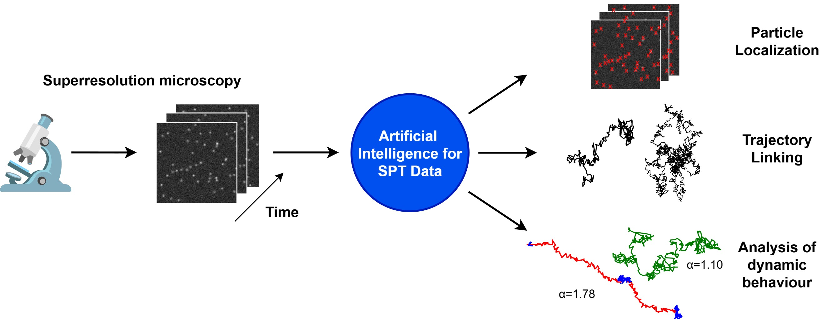
Machine Learning in Single-Molecule Tracking Analysis of Superresolution Optical Microscopy Data


Highlights
- Motion of molecules in live cells can be studied via single-molecule tracking (SMT).
- SMT beyond the diffraction limit requires optical superresolution imaging.
- Machine learning facilitates and improves the analysis of SMTs.
- Molecular motion provides functionally important information on the dynamics of molecular constituents of the cell.
Abstract
1. Introduction
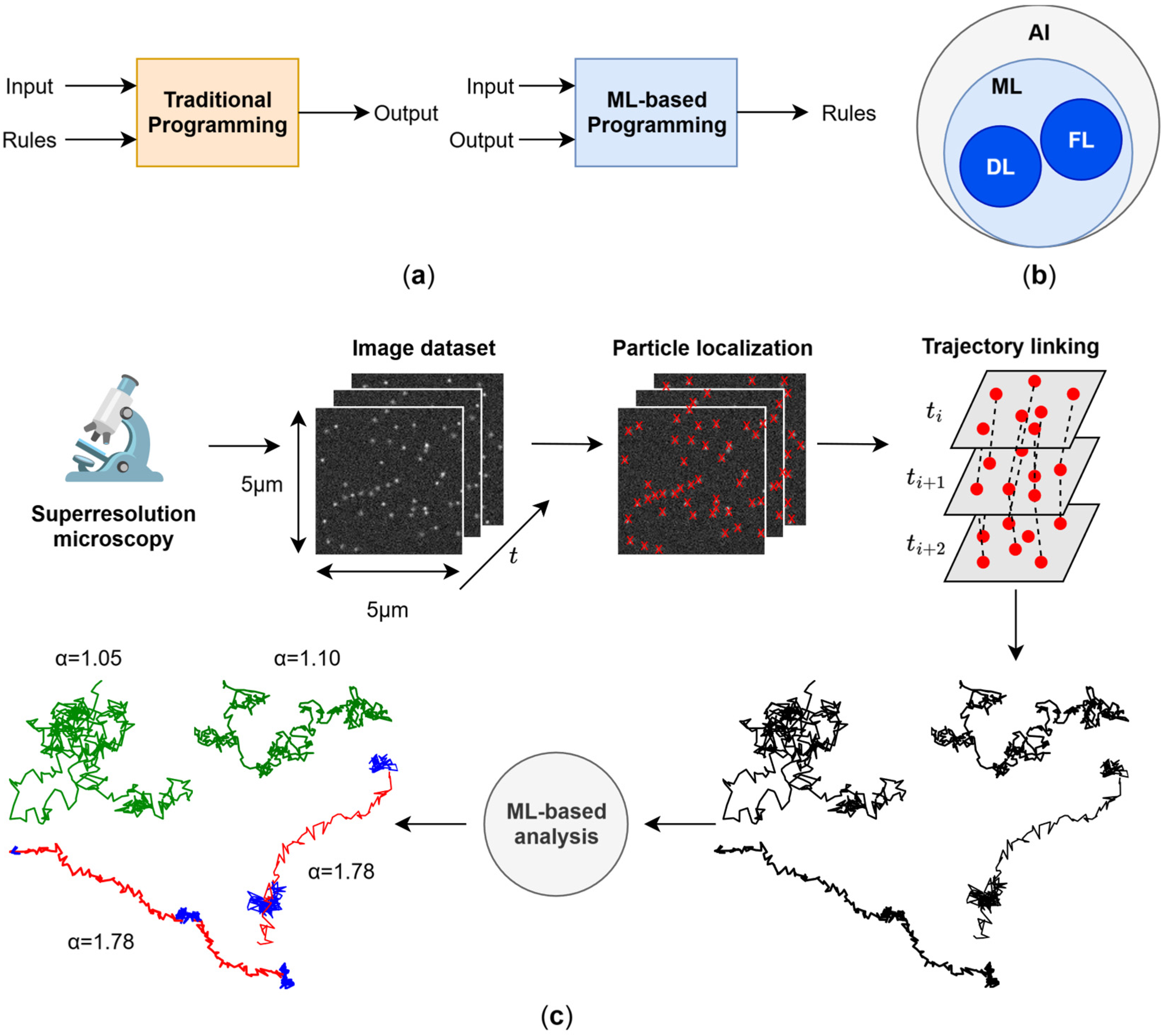
2. Single-Molecule Localisation and Trajectory Linking
2.1. Single-Molecule Localisation in Dynamic Samples
2.2. Trajectory Linking via DL
3. Single-Molecule Trajectory Characterisation

| Method | Input | Strengths | Limitations |
|---|---|---|---|
| Mean Squared Displacement (MSD) | Trajectory positions | Simple and interpretable | Strongly length-dependent and sensitive to noise |
| Extracts D and α | Biassed α estimation | ||
| Hidden Markov Models (HMM) | Trajectory positions or displacements | Probabilistic state classification | Model-dependent and limited flexibility |
| Detects transitions | Surpassed by ML in performance | ||
| Decision trees | Engineered features | Interpretable inputs | Model-dependent |
| Good generalisation | Difficult feature selection | ||
| No GPU required | Raw coordinates unsuitable | ||
| FL-based neural networks | Engineered features | Stable across variable lengths | Difficult feature selection |
| Computationally efficient | May underperform DL-based neural networks | ||
| Recurrent Neural Networks (RNN, LSTM, and Bi-LSTM) | Trajectory positions or displacements | Sequential nature of trajectories is considered | Slow training |
| Handle uneven sampling | Length-dependent | ||
| Strong regression performance | May underperform TCNs | ||
| Temporal Convolutional Networks (TCNs) | Trajectory positions or displacements | Faster training than LSTMs | Length-dependent |
| Strong β/H prediction | Length-specific models are required | ||
| Trajectory-to-image representation (GAF) | GAF representations of trajectories | Strong classification accuracy | Computational expense |
| Longer training | |||
| Length-dependent | |||
| RNN + CNN Hybrid | Trajectory positions | Combined spatial feature extraction and temporal modelling | High computational cost |
| Inefficient training due to LSTM component | |||
| Extreme Learning Machine | Engineered features | Very fast training | Shallow model |
| Suitable for initial screening | Limited precision and generalisation | ||
| Autoencoders | Trajectory positions | Unsupervised method | Limited interpretability |
| Anomaly detection | |||
| Latent representation learning | |||
| Transformers | Trajectory positions | Capture long-range dependencies | Computationally demanding |
| Strong generalisation | |||
| Fingerprint analysis | Engineered features | Unsupervised discrimination of experimental conditions | Requires supervised methods |
| Feature ranking | Depends on feature engineering | ||
| Graph Neural Networks (GNN) | Graph representation of localisations | Avoid trajectory linking | Graph construction critical |
| Performance sensitive to transformation design | |||
| Sliding-window approaches | Overlapping sub-trajectories | Local prediction | High inference time |
| No need for heterogeneous simulated trajectories | Window-size tradeoff between resolution and accuracy | ||
| Unstable for short segments | |||
| Sequence-To-Sequence | Trajectory positions | Pointwise prediction | High model complexity |
| No sub-trajectory splitting | Length-dependent | ||
| Long training time | |||
| Change-point-based methods | Trajectory positions | Faster than sliding-window approaches | Class imbalance between positions with and without a change-point |
| Explicit CP detection | Model-dependent | ||
| Segment-wise inference | |||
| Support Vector Machines (SVM) | Engineered features | Strong segmentation performance | Requires feature engineering |
| Robust classification | Less scalable than DL | ||
| Bayesian Deep Learning | Trajectory positions | Provide uncertainty estimation | High computational cost |
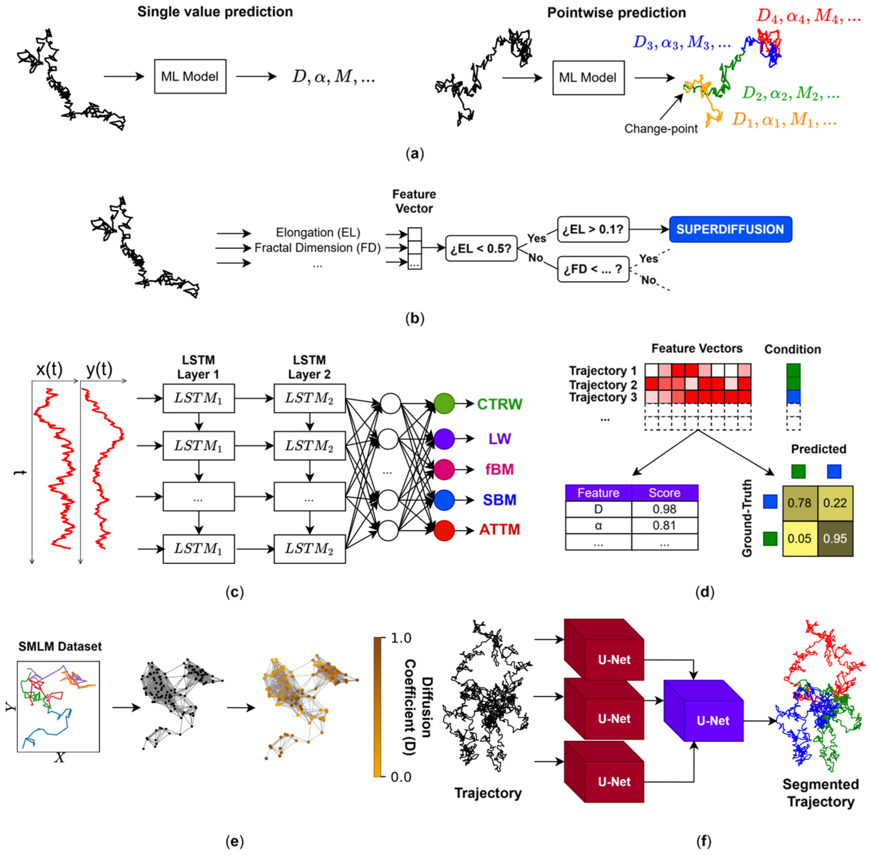
3.1. Andi Challenge: Single-Value Characterisation
3.1.1. Predictions for Qualitative Analysis
3.1.2. Prediction for Quantitative Analysis
3.1.3. Aftermaths of the First Andi Challenge
3.1.4. FL or DL?
3.2. Andi Challenge: Dynamic Pointwise Prediction of Dynamics
3.2.1. Sliding-Window Approaches
3.2.2. Sequence-to-Sequence Approaches
3.2.3. Change-Point (CP)-Based Pointwise Prediction
4. Diffusion Mapping
5. Conclusions and Prospects
Author Contributions
Funding
Institutional Review Board Statement
Informed Consent Statement
Data Availability Statement
Acknowledgments
Conflicts of Interest
Abbreviations
| ML | Machine Learning |
| FL | Feature-based learning |
| AI | Artificial Intelligence |
| HT | Hypothesis Tree |
| DL | Deep Learning |
| CNN | Convolutional Neural Network |
| SMLM | Single-Molecule Localization Microscopy |
| STED | Stimulated Emission Depletion |
| PALM | Photo-Activated Localization Microscopy |
| STORM | Stochastic Optical Reconstruction Microscopy |
| PSF | Point-Spead Function |
| ROI | Region of Interest |
| RMSE | Root Mean Square |
| SNR | Signal-to-Noise Ratio |
| LSTM | Long Short-Term Memory |
| RNN | Recurrent Neural Network |
| GNN | Graph Neural Network |
| SMT | Single-Molecule Tracking |
| SPT | Single-Particle Tracking |
| MSD | Mean Squared Displacement |
| HMM | Hidden Markov Model |
| TCN | Temporal Convolutional Network |
| RF | Random Forest |
| GBM | Gradient Boosting Machine |
| XGB | Extreme Gradient Boosting |
| Bi-LSTM | Bidirectional LSTM |
| AE | Autoencoder |
| VAE | Variational Autoencoder |
| CP | Change-Point |
| TIRF | Total Internal Reflection Fluorescence |
| SVM | Support Vector Machine |
| cGAN | Conditional Generative Adversarial Network |
| MLE | Maximum Likelihood Estimation |
| MINFLUX | MINimal Fluorescence emission FLUXes |
| YOLO | You Only Look Once |
| GPU | Graphics Processing Unit |
| CPU | Central Processing Unit |
| GFLOPs | Giga Floating Point Operations |
| MLP | Multi-Layer Perceptron |
References
- Turing, A.M.I. Computing machinery and intelligence. Mind 1950, LIX, 433–460. [Google Scholar] [CrossRef] [Scilit]
- Sheikh, H.; Prins, C.; Schrijvers, E. (Eds.) Artificial Intelligence: Definition and Background. In Mission AI: The New System Technology; Springer International Publishing: Cham, Switzerland, 2023; pp. 15–41. [Google Scholar]
- James, G.; Witten, D.; Hastie, T.; Tibshirani, R.; Taylor, J. An Introduction to Statistical Learning, 2nd ed.; Springer International Publishing: New York, NY, USA, 2021; p. 60. [Google Scholar]
- França, R.P.; Borges Monteiro, A.C.; Arthur, R.; Iano, Y. Chapter 3—An overview of deep learning in big data, image, and signal processing in the modern digital age. In Trends in Deep Learning Methodologies; Piuri, V., Raj, S., Genovese, A., Srivastava, R., Eds.; Academic Press: Cambridge, MA, USA, 2021; pp. 63–87. [Google Scholar]
- LeCun, Y.; Bengio, Y.; Hinton, G.E. Deep learning. Nature 2015, 521, 436–444. [Google Scholar] [CrossRef] [Scilit]
- Goodfellow, I.; Bengio, Y.; Courville, A. Deep Learning; The MIT Press: Cambridge, MA, USA, 2016; p. 800. [Google Scholar]
- Jumper, J.M.; Evans, R.; Pritzel, A.; Green, T.; Figurnov, M.; Ronneberger, O.; Tunyasuvunakool, K.; Bates, R.; Žídek, A.; Potapenko, A.; et al. Highly accurate protein structure prediction with AlphaFold. Nature 2021, 596, 583–589. [Google Scholar] [CrossRef] [Scilit]
- Abramson, J.; Adler, J.; Dunger, J.; Evans, R.; Green, T.; Pritzel, A.; Ronneberger, O.; Willmore, L.; Ballard, A.J.; Bambrick, J.; et al. Accurate structure prediction of biomolecular interactions with AlphaFold 3. Nature 2024, 630, 493–500. [Google Scholar] [CrossRef] [Scilit]
- Hell, S.W.; Sahl, S.J.; Bates, M.; Zhuang, X.; Rainer, H.; Martin, J.B.; Joerg, B.; Gleb, S.; Harald, H.; Philip, T.; et al. The 2015 super-resolution microscopy roadmap. J. Phys. D Appl. Phys. 2015, 48, 443001. [Google Scholar] [CrossRef] [Scilit]
- Gustafsson, M.G. Surpassing the lateral resolution limit by a factor of two using structured illumination microscopy. J. Microsc. 2000, 198, 82–87. [Google Scholar] [CrossRef] [Scilit]
- Gustafsson, M.G. Nonlinear structured-illumination microscopy: Wide-field fluorescence imaging with theoretically unlimited resolution. Proc. Natl. Acad. Sci. USA 2005, 102, 13081–13086. [Google Scholar] [CrossRef] [Scilit]
- Lelek, M.; Gyparaki, M.T.; Beliu, G.; Schueder, F.; Griffié, J.; Manley, S.; Jungmann, R.; Sauer, M.; Lakadamyali, M.; Zimmer, C. Single-molecule localization microscopy. Nat. Rev. Methods Primers 2021, 1, 39. [Google Scholar] [CrossRef] [Scilit]
- Hugelier, S.; Colosi, P.L.; Lakadamyali, M. Quantitative Single-Molecule Localization Microscopy. Annu. Rev. Biophys. 2023, 52, 139–160. [Google Scholar] [CrossRef] [Scilit] [PubMed]
- Steves, M.A.; He, C.; Xu, K. Single-Molecule Spectroscopy and Super-Resolution Mapping of Physicochemical Parameters in Living Cells. Annu. Rev. Phys. Chem. 2024, 75, 163–183. [Google Scholar] [CrossRef] [Scilit] [PubMed]
- Betzig, E.; Patterson, G.H.; Sougrat, R.; Lindwasser, O.W.; Olenych, S.; Bonifacino, J.S.; Davidson, M.W.; Lippincott-Schwartz, J.; Hess, H.F. Imaging intracellular fluorescent proteins at nanometer resolution. Science 2006, 313, 1642–1645. [Google Scholar] [CrossRef] [Scilit]
- Jungmann, R. Multiplexed 3D cellular super-resolution imaging with DNA-PAINT and Exchange-PAINT. Nat. Methods 2014, 11, 313–318. [Google Scholar] [CrossRef] [Scilit] [PubMed]
- Schnitzbauer, J.; Strauss, M.T.; Schlichthaerle, T.; Schueder, F.; Jungmann, R. Super-resolution microscopy with DNA-PAINT. Nat. Protoc. 2017, 12, 1198–1228. [Google Scholar] [CrossRef] [Scilit] [PubMed]
- Hofmann, M.; Eggeling, C.; Jakobs, S.; Hell, S.W. Breaking the diffraction barrier in fluorescence microscopy at low light intensities by using reversibly photoswitchable proteins. Proc. Natl. Acad. Sci. USA 2005, 102, 17565–17569. [Google Scholar] [CrossRef] [Scilit]
- Schwentker, M.A.; Bock, H.; Hofmann, M.; Jakobs, S.; Bewersdorf, J.; Eggeling, C.; Hell, S.W. Wide-field subdiffraction RESOLFT microscopy using fluorescent protein photoswitching. Microsc. Res. Tech. 2007, 70, 269–280. [Google Scholar] [CrossRef] [Scilit] [PubMed]
- Balzarotti, F.; Eilers, Y.; Gwosch, K.C.; Gynnå, A.H.; Westphal, V.; Stefani, F.D.; Elf, J.; Hell, S.W. Nanometer resolution imaging and tracking of fluorescent molecules with minimal photon fluxes. Science 2017, 355, 606–612. [Google Scholar] [CrossRef] [Scilit]
- Muñoz-Gil, G.; Volpe, G.; Garcia-March, M.A.; Aghion, E.; Argun, A.; Hong, C.B.; Bland, T.; Bo, S.; Conejero, J.A.; Firbas, N.; et al. Objective comparison of methods to decode anomalous diffusion. Nat. Commun. 2021, 12, 6253. [Google Scholar] [CrossRef] [Scilit]
- Manzo, C.; Garcia-Parajo, M.F. A review of progress in single particle tracking: From methods to biophysical insights. Rep. Prog. Phys. 2015, 78, 124601. [Google Scholar] [CrossRef] [Scilit]
- Krapf, D. Mechanisms underlying anomalous diffusion in the plasma membrane. Curr. Top. Membr. 2015, 75, 167–207. [Google Scholar] [CrossRef] [Scilit]
- Pennacchietti, F.; Gould, T.J.; Hess, S.T. The Role of Probe Photophysics in Localization-Based Superresolution Microscopy. Biophys. J. 2017, 113, 2037–2054. [Google Scholar] [CrossRef] [Scilit]
- Radmacher, N.; Chizhik, A.I.; Nevskyi, O.; Gallea, J.I.; Gregor, I.; Enderlein, J. Molecular Level Super-Resolution Fluorescence Imaging. Annu. Rev. Biophys. 2025, 54, 163–184. [Google Scholar] [CrossRef] [Scilit]
- Huang, B.; Wang, W.; Bates, M.; Zhuang, X. Three-dimensional super-resolution imaging by stochastic optical reconstruction microscopy. Science 2008, 319, 810–813. [Google Scholar] [CrossRef] [Scilit] [PubMed]
- Rust, M.J.; Bates, M.; Zhuang, X. Sub-diffraction-limit imaging by stochastic optical reconstruction microscopy (STORM). Nat. Methods 2006, 3, 793–795. [Google Scholar] [CrossRef] [Scilit] [PubMed]
- Pennacchietti, F.; Serebrovskaya, E.O.; Faro, A.R.; Shemyakina, I.I.; Bozhanova, N.G.; Kotlobay, A.A.; Gurskaya, N.G.; Bodén, A.; Dreier, J.; Chudakov, D.M.; et al. Fast reversibly photoswitching red fluorescent proteins for live-cell RESOLFT nanoscopy. Nat. Methods 2018, 15, 601–604. [Google Scholar] [CrossRef] [Scilit]
- Schueder, F.; Jungmann, R. In Situ Imaging of Proteins Using DNA-PAINT Super-Resolution Microscopy. Methods Mol. Biol. 2024, 2800, 103–113. [Google Scholar] [CrossRef] [Scilit]
- Aristova, D.; Kylies, D.; Del Rosario, M.; Heil, H.S.; Schwerk, M.; Kuehl, M.; Wong, M.N.; Henriques, R.; Puelles, V.G. Nanoscale imaging of biological systems via expansion and super-resolution microscopy. Appl. Phys. Rev. 2025, 12, 021311. [Google Scholar] [CrossRef] [Scilit]
- Wassie, A.T.; Zhao, Y.; Boyden, E.S. Expansion microscopy: Principles and uses in biological research. Nat. Methods 2019, 16, 33–41. [Google Scholar] [CrossRef] [Scilit]
- Wang, S.; Shin, T.W.; Yoder, H.B.; McMillan, R.B.; Su, H.; Liu, Y.; Zhang, C.; Leung, K.S.; Yin, P.; Kiessling, L.L.; et al. Single-shot 20-fold expansion microscopy. Nat. Methods 2024, 21, 2128–2134. [Google Scholar] [CrossRef] [Scilit]
- Small, A.R.; Parthasarathy, R. Superresolution Localization Methods. Annu. Rev. Phys. Chem. 2014, 65, 107–125. [Google Scholar] [CrossRef] [Scilit]
- Lambert, T.J.; Waters, J.C. Navigating challenges in the application of superresolution microscopy. J. Cell Biol. 2017, 216, 53–63. [Google Scholar] [CrossRef] [Scilit]
- Nelson, A.J.; Hess, S.T. Localization microscopy: Mapping cellular dynamics with single molecules. J. Microsc. 2014, 254, 12115. [Google Scholar] [CrossRef] [Scilit]
- Sahl, S.J.; Hell, S.W.; Jakobs, S. Fluorescence nanoscopy in cell biology. Nat. Rev. Mol. Cell Biol. 2017, 18, 685–701. [Google Scholar] [CrossRef] [Scilit]
- Fazel, M.; Wester, M.J. Analysis of super-resolution single molecule localization microscopy data: A tutorial. AIP Adv. 2022, 12, 010701. [Google Scholar] [CrossRef] [Scilit]
- Liu, Z.; Jin, L.; Chen, J.; Fang, Q.; Ablameyko, S.; Yin, Z.; Xu, Y. A survey on applications of deep learning in microscopy image analysis. Comput. Biol. Med. 2021, 134, 104523. [Google Scholar] [CrossRef] [Scilit] [PubMed]
- Zhang, B.; Sun, X.; Mai, J.; Wang, W. Deep learning-enhanced fluorescence microscopy via confocal physical imaging model. Opt. Express 2023, 31, 19048–19064. [Google Scholar] [CrossRef] [Scilit] [PubMed]
- Abdehkakha, A.; Madani, S.M.N.; Snoeyink, C. Enhancing single-molecule localization microscopy with deep learning. Appl. Opt. 2025, 64, A19–A30. [Google Scholar] [CrossRef] [Scilit]
- Kang, J.H.; Paci, G.; Mikhaleva, S.; Koehler, C.; Shymanska, N.V.; Ventura Santos, C.; Spitz, D.; Lemke, E.A.; Nikic, I.; Estrada Girona, G. Debugging Eukaryotic Genetic Code Expansion for Site-Specific Click-PAINT Super-Resolution Microscopy. Angew. Chem. Int. Ed. Engl. 2016, 55, 16172–16176. [Google Scholar] [CrossRef] [Scilit]
- Gómez-García, P.A.; Garbacik, E.T.; Otterstrom, J.J.; Garcia-Parajo, M.F.; Lakadamyali, M. Excitation-multiplexed multicolor superresolution imaging with fm-STORM and fm-DNA-PAINT. Proc. Natl. Acad. Sci. USA 2018, 115, 12991–12996. [Google Scholar] [CrossRef] [Scilit]
- Ostersehlt, L.M.; Jans, D.C.; Wittek, A.; Keller-Findeisen, J.; Inamdar, K.; Sahl, S.J.; Hell, S.W.; Jakobs, S. DNA-PAINT MINFLUX nanoscopy. Nat. Methods 2022, 19, 1072–1075. [Google Scholar] [CrossRef] [Scilit]
- Zähringer, J.; Cole, F.; Bohlen, J.; Steiner, F.; Kamińska, I.; Tinnefeld, P. Combining pMINFLUX, graphene energy transfer and DNA-PAINT for nanometer precise 3D super-resolution microscopy. Light Sci. Appl. 2023, 12, 70. [Google Scholar] [CrossRef] [Scilit]
- Srambickal, C.V.; Esmaeeli, H.; Piguet, J.; Reinkensmeier, L.; Siegmund, R.; Bates, M.; Egner, A.; Widengren, J. Near-infrared MINFLUX imaging enabled by suppression of fluorophore blinking. bioRxiv 2024, 8, 609859. [Google Scholar] [CrossRef] [Scilit] [PubMed]
- Zdańkowski, P.; Lopez, L.F.; Acuna, G.P.; Stefani, F.D. Nanometer Resolution Imaging and Tracking of Single Fluorophores by Sequential Structured Illumination. ACS Photonics 2022, 9, 3777–3785. [Google Scholar] [CrossRef] [Scilit]
- Bates, M.; Jones, S.A.; Zhuang, X. Stochastic optical reconstruction microscopy (STORM): A method for superresolution fluorescence imaging. Cold Spring Harb. Protoc. 2013, 2013, 498–520. [Google Scholar] [CrossRef] [Scilit]
- Ovesný, M.; Křížek, P.; Borkovec, J.; Svindrych, Z.; Hagen, G.M. ThunderSTORM: A comprehensive ImageJ plug-in for PALM and STORM data analysis and super-resolution imaging. Bioinformatics 2014, 30, 2389–2390. [Google Scholar] [CrossRef] [Scilit]
- Helgadottir, S.; Argun, A.; Volpe, G. Digital video microscopy enhanced by deep learning. Optica 2019, 6, 506–513. [Google Scholar] [CrossRef] [Scilit]
- Jaqaman, K.; Loerke, D.; Mettlen, M.; Kuwata, H.; Grinstein, S.; Schmid, S.L.; Danuser, G. Robust single-particle tracking in live-cell time-lapse sequences. Nat. Methods 2008, 5, 695–702. [Google Scholar] [CrossRef] [Scilit]
- Jaqaman, K.; Danuser, G. Computational image analysis of cellular dynamics: A case study based on particle tracking. Cold Spring Harb. Protoc. 2009, 2009. [Google Scholar] [CrossRef] [Scilit]
- Sgouralis, I.; Xu, L.W.Q.; Jalihal, A.P.; Kilic, Z.; Walter, N.G.; Pressé, S. BNP-Track: A framework for superresolved tracking. Nat. Methods 2024, 21, 1716–1724. [Google Scholar] [CrossRef] [Scilit]
- Pineda, J.; Midtvedt, B.; Bachimanchi, H.; Noé, S.; Midtvedt, D.; Volpe, G.; Manzo, C. Geometric deep learning reveals the spatiotemporal features of microscopic motion. Nat. Mach. Intell. 2023, 5, 71–82. [Google Scholar] [CrossRef] [Scilit]
- Zhang, Y.; Yang, G. A Motion Transformer for Single Particle Tracking in Fluorescence Microscopy Images. In Proceedings of the Medical Image Computing and Computer Assisted Intervention—MICCAI 2023, Vancouver, BC, Canada, 8–12 October 2023; Springer: Cham, Switzerland, 2023; pp. 503–513. [Google Scholar]
- Bi, C.; Scrudders, K.L.; Zheng, Y.; Gao, H.-C.; Fang, L.; Li, Y.; Low-Nam, S.T.; Huang, F. BPS2025—SPTNet: A deep-learning-based framework for end-to-end single-particle tracking and motion dynamics analysis. Biophys. J. 2025, 124, 486a. [Google Scholar] [CrossRef] [Scilit]
- Zhang, Y.; Zhu, J.; Du, Y.; Xie, H.; He, Y. Large vision model supported tracking of high-density particles based on optical flow learning. Cell Rep. Phys. Sci. 2025, 6, 102904. [Google Scholar] [CrossRef] [Scilit]
- Midtvedt, B.; Helgadottir, S.; Argun, A.; Pineda, J.; Midtvedt, D.; Volpe, G. Quantitative digital microscopy with deep learning. Appl. Phys. Rev. 2021, 8, 34891. [Google Scholar] [CrossRef] [Scilit]
- Ronneberger, O.; Fischer, P.; Brox, T. U-Net: Convolutional Networks for Biomedical Image Segmentation. In Proceedings of the Medical Image Computing and Computer-Assisted Intervention—MICCAI 2015, Munich, Germany, 5–9 October 2015; Springer: Cham, Switzerland, 2015; pp. 234–241. [Google Scholar]
- Speiser, A.; Müller, L.-R.; Hoess, P.; Matti, U.; Obara, C.J.; Legant, W.R.; Kreshuk, A.; Macke, J.H.; Ries, J.; Turaga, S.C. Deep learning enables fast and dense single-molecule localization with high accuracy. Nat. Methods 2021, 18, 1082–1090. [Google Scholar] [CrossRef] [Scilit] [PubMed]
- Nehme, E.; Weiss, L.E.; Michaeli, T.; Shechtman, Y. Deep-STORM: Super-resolution single-molecule microscopy by deep learning. Optica 2018, 5, 458–464. [Google Scholar] [CrossRef] [Scilit]
- Nehme, E.; Freedman, D.; Gordon, R.; Ferdman, B.; Weiss, L.E.; Alalouf, O.; Naor, T.; Orange, R.; Michaeli, T.; Shechtman, Y. DeepSTORM3D: Dense 3D localization microscopy and PSF design by deep learning. Nat. Methods 2020, 17, 734–740. [Google Scholar] [CrossRef] [Scilit]
- Deng, F.; Chen, T.; Qiao, Y.; Ding, J.; Zhang, Y.; Qiu, L.; Zhao, W.; Liu, J.; Xu, F. Enhancing ultra-high density single-molecule localization with deep spatiotemporal networks. Biomed. Opt. Express 2025, 16, 1773–1791. [Google Scholar] [CrossRef] [Scilit] [PubMed]
- Hochreiter, S.; Schmidhuber, J. Long Short-Term Memory. Neural Comput. 1997, 9, 1735–1780. [Google Scholar] [CrossRef] [Scilit]
- Shi, X.; Chen, Z.; Wang, H.; Yeung, D.-Y.; Wong, W.-K.; Woo, W.-C. Convolutional LSTM Network: A machine learning approach for precipitation nowcasting. In Proceedings of the 29th International Conference on Neural Information Processing Systems, Montreal, Canada, 7–12 December 2015; Curran Associates, Inc.: Red Hook, NY, USA, 2015; Volume 1, pp. 802–810. [Google Scholar]
- Song, Q.; Kuang, W.; Li, X.; Wang, Z.; Zhang, Y.; Huang, Z.-L. Dual deep learning network enables data-efficient two-color single-molecule localization microscopy with colorimetry camera. Opt. Express 2025, 33, 40040–40054. [Google Scholar] [CrossRef] [Scilit]
- Fei, Y.; Fu, S.; Shi, W.; Fang, K.; Wang, R.; Zhang, T.; Li, Y. Scalable and lightweight deep learning for efficient high accuracy single-molecule localization microscopy. Nat. Commun. 2025, 16, 7217. [Google Scholar] [CrossRef] [Scilit]
- Vaswani, A.; Shazeer, N.; Parmar, N.; Uszkoreit, J.; Jones, L.; Gomez, A.N.; Kaiser, Ł.; Polosukhin, I. Attention is all you need. In Proceedings of the 31st International Conference on Neural Information Processing Systems, Long Beach, CA, USA, 4–9 December 2017; Curran Associates, Inc.: Red Hook, NY, USA, 2017; pp. 6000–6010. [Google Scholar]
- Battaglia, P.W.; Hamrick, J.B.; Bapst, V.; Sanchez-Gonzalez, A.; Zambaldi, V.; Malinowski, M.; Tacchetti, A.; Raposo, D.; Santoro, A.; Faulkner, R.; et al. Relational inductive biases, deep learning, and graph networks. arXiv 2018, arXiv:1806.01261. [Google Scholar] [CrossRef] [Scilit]
- Saavedra, L.A.; Barrantes, F.J. Machine Learning Empowers Static and Dynamics Clustering Characterization of Transmembrane Receptors. In Proceedings of the Building Bridges in Computational Biophysics (BBCB) v3.0, Virtual, 16 October 2024; Available online: https://www.biophysics.org/upcoming-networking-events/building-bridges-in-computational-biophysics-v30-bbcbv30 (accessed on 5 March 2026).
- Saavedra, L.A.; Mosqueira, A.; Barrantes, F.J. A supervised graph-based deep learning algorithm to detect and quantify clustered particles. Nanoscale 2024, 16, 15308–15318. [Google Scholar] [CrossRef] [Scilit]
- Berg, S.; Kutra, D.; Kroeger, T.; Straehle, C.N.; Kausler, B.X.; Haubold, C.; Schiegg, M.; Ales, J.; Beier, T.; Rudy, M.; et al. ilastik: Interactive machine learning for (bio)image analysis. Nat. Methods 2019, 16, 1226–1232. [Google Scholar] [CrossRef] [Scilit]
- Sommer, C.; Straehle, C.; Köthe, U.; Hamprecht, F.A. Ilastik: Interactive learning and segmentation toolkit. In Proceedings of the 2011 IEEE International Symposium on Biomedical Imaging: From Nano to Macro, Chicago, IL, USA, 30 March–2 April 2011; IEEE: Park Avenue, NY, USA, 2011; pp. 230–233. [Google Scholar]
- Qian, H.; Sheetz, M.P.; Elson, E.L. Single particle tracking. Analysis of diffusion and flow in two-dimensional systems. Biophys. J. 1991, 60, 910–921. [Google Scholar] [CrossRef] [Scilit] [PubMed]
- Riahi, M.K.; Qattan, I.A.; Hassan, J.; Homouz, D. Identifying short- and long-time modes of the mean-square displacement: An improved nonlinear fitting approach. AIP Adv. 2019, 9, 055112. [Google Scholar] [CrossRef] [Scilit]
- Saxton, M.J.; Jacobson, K. Single-particle tracking: Applications to membrane dynamics. Annu. Rev. Biophys. Biomol. Struct. 1997, 26, 373–399. [Google Scholar] [CrossRef] [Scilit] [PubMed]
- Slator, P.J.; Burroughs, N.J. A Hidden Markov Model for Detecting Confinement in Single-Particle Tracking Trajectories. Biophys. J. 2018, 115, 1741–1754. [Google Scholar] [CrossRef] [Scilit]
- Slator, P.J.; Cairo, C.W.; Burroughs, N.J. Detection of Diffusion Heterogeneity in Single Particle Tracking Trajectories Using a Hidden Markov Model with Measurement Noise Propagation. PLoS ONE 2015, 10, e0140759. [Google Scholar] [CrossRef] [Scilit]
- Han, D.; Korabel, N.; Chen, R.; Johnston, M.; Gavrilova, A.; Allan, V.J.; Fedotov, S.; Waigh, T.A. Deciphering anomalous heterogeneous intracellular transport with neural networks. eLife 2020, 9, e52224. [Google Scholar] [CrossRef] [Scilit]
- Manzo, C.; Muñoz-Gil, G.; Volpe, G.; Garcia-March, M.A.; Lewenstein, M.; Metzler, R. Preface: Characterisation of physical processes from anomalous diffusion data. J. Phys. A Math. Theor. 2023, 56, 010401. [Google Scholar] [CrossRef] [Scilit]
- Muñoz-Gil, G.; Bachimanchi, H.; Pineda, J.; Midtvedt, B.; Fernández-Fernández, G.; Requena, B.; Ahsini, Y.; Asghar, S.; Bae, J.; Barrantes, F.J.; et al. Quantitative evaluation of methods to analyze motion changes in single-particle experiments. Nat. Commun. 2025, 16, 6749. [Google Scholar] [CrossRef] [Scilit]
- Mandelbrot, B.B.; Van Ness, J.W. Fractional Brownian Motions, Fractional Noises and Applications. SIAM Rev. 1968, 10, 422–437. [Google Scholar] [CrossRef] [Scilit]
- Klafter, J.; Sokolov, I.M. Anomalous diffusion spreads its wings. Phys. World 2005, 18, 29. [Google Scholar] [CrossRef] [Scilit]
- Lim, S.C.; Muniandy, S.V. Self-similar Gaussian processes for modeling anomalous diffusion. Phys. Rev. E Stat. Nonlin. Soft Matter Phys. 2002, 66, 021114. [Google Scholar] [CrossRef] [Scilit]
- Manzo, C.; Torreno-Pina, J.A.; Massignan, P.; Lapeyre, G.J.; Lewenstein, M.; Garcia Parajo, M.F. Weak Ergodicity Breaking of Receptor Motion in Living Cells Stemming from Random Diffusivity. Phys. Rev. X 2015, 5, 011021. [Google Scholar] [CrossRef] [Scilit]
- Lines, J.; Taylor, S.; Bagnall, A. Time Series Classification with HIVE-COTE: The Hierarchical Vote Collective of Transformation-Based Ensembles. ACM Trans. Knowl. Discov. Data 2018, 12, 52. [Google Scholar] [CrossRef] [Scilit]
- Türkcan, S.; Masson, J.-B. Bayesian decision tree for the classification of the mode of motion in single-molecule trajectories. PLoS ONE 2013, 8, e82799. [Google Scholar] [CrossRef] [Scilit]
- Jin, S.; Haggie, P.M.; Verkman, A.S. Single-particle tracking of membrane protein diffusion in a potential: Simulation, detection, and application to confined diffusion of CFTR Cl− channels. Biophys. J. 2007, 93, 1079–1088. [Google Scholar] [CrossRef] [Scilit] [PubMed]
- Maris, J.J.E.; Rabouw, F.T.; Weckhuysen, B.M.; Meirer, F. Classification-based motion analysis of single-molecule trajectories using DiffusionLab. Sci. Rep. 2022, 12, 9595. [Google Scholar] [CrossRef] [Scilit] [PubMed]
- Wagner, T.; Kroll, A.; Wiemann, M.; Lipinski, H.-G. Classification of nanoparticle diffusion processes in vital cells by a multifeature random forests approach: Application to simulated data, darkfield, and confocal laser scanning microscopy. In Proceedings of the Biophotonics: Photonic Solutions for Better Health Care V, Brussels, Belgium, 4–7 April 2016; SPIE: Bellingham, WA, USA, 2016; p. 988722. [Google Scholar]
- Wagner, T.; Kroll, A.; Haramagatti, C.R.; Lipinski, H.-G.; Wiemann, M. Classification and Segmentation of Nanoparticle Diffusion Trajectories in Cellular Micro Environments. PLoS ONE 2017, 12, e0170165. [Google Scholar] [CrossRef] [Scilit] [PubMed]
- Chen, T.; Guestrin, C. XGBoost: A Scalable Tree Boosting System. In Proceedings of the 22nd ACM SIGKDD International Conference on Knowledge Discovery and Data Mining, San Francisco, CA, USA, 13–17 August 2016; ACM: New York, NY, USA, 2016; pp. 785–794. [Google Scholar]
- Kowalek, P.; Loch-Olszewska, H.; Łaszczuk, Ł.; Opała, J.; Szwabiński, J. Boosting the performance of anomalous diffusion classifiers with the proper choice of features. J. Phys. A Math. Theor. 2022, 55, 244005. [Google Scholar] [CrossRef] [Scilit]
- Loch-Olszewska, H.; Szwabiński, J. Impact of Feature Choice on Machine Learning Classification of Fractional Anomalous Diffusion. Entropy 2020, 22, 1436. [Google Scholar] [CrossRef] [Scilit] [PubMed]
- Gentili, A.; Volpe, G. Characterization of anomalous diffusion classical statistics powered by deep learning (CONDOR). J. Phys. A Math. Theor. 2021, 54, 314003. [Google Scholar] [CrossRef] [Scilit]
- Arts, M.; Smal, I.; Paul, M.W.; Wyman, C.; Meijering, E. Particle Mobility Analysis Using Deep Learning and the Moment Scaling Spectrum. Sci. Rep. 2019, 9, 17160. [Google Scholar] [CrossRef] [Scilit]
- Reina, F.; Saavedra, L.A.; Eggeling, C.; Barrantes, F.J. Concurrent diffusion of nicotinic acetylcholine receptors and fluorescent cholesterol disclosed by two-colour sub-millisecond MINFLUX-based single-molecule tracking. Nat. Commun. 2025, 16, 6336. [Google Scholar] [CrossRef] [Scilit]
- Schuster, M.; Paliwal, K.K. Bidirectional recurrent neural networks. IEEE Trans. Signal Process. 1997, 45, 2673–2681. [Google Scholar] [CrossRef] [Scilit]
- Baldi, P.; Brunak, S.; Frasconi, P.; Soda, G.; Pollastri, G. Exploiting the past and the future in protein secondary structure prediction. Bioinformatics 1999, 15, 937–946. [Google Scholar] [CrossRef] [Scilit] [PubMed]
- Mangalam, M.; Metzler, R.; Kelty-Stephen, D.G. Ergodic characterization of nonergodic anomalous diffusion processes. Phys. Rev. Res. 2023, 5, 023144. [Google Scholar] [CrossRef] [Scilit]
- Seckler, H.; Metzler, R.; Kelty-Stephen, D.G.; Mangalam, M. Multifractal spectral features enhance classification of anomalous diffusion. Phys. Rev. E 2024, 109, 044133. [Google Scholar] [CrossRef] [Scilit]
- Argun, A.; Volpe, G.; Bo, S. Classification, inference and segmentation of anomalous diffusion with recurrent neural networks. J. Phys. A Math. Theor. 2021, 54, 294003. [Google Scholar] [CrossRef] [Scilit]
- Li, D.; Yao, Q.; Huang, Z. WaveNet-based deep neural networks for the characterization of anomalous diffusion (WADNet). J. Phys. A Math. Theor. 2021, 54, 404003. [Google Scholar] [CrossRef] [Scilit]
- Granik, N.; Weiss, L.E.; Nehme, E.; Levin, M.; Chein, M.; Perlson, E.; Roichman, Y.; Shechtman, Y. Single-Particle Diffusion Characterization by Deep Learning. Biophys. J. 2019, 117, 185–192. [Google Scholar] [CrossRef] [Scilit]
- Garibo-i-Orts, Ò.; Baeza-Bosca, A.; Garcia-March, M.A.; Conejero, J.A. Efficient recurrent neural network methods for anomalously diffusing single particle short and noisy trajectories. J. Phys. A Math. Theor. 2021, 54, 504002. [Google Scholar] [CrossRef] [Scilit]
- Bo, S.; Schmidt, F.; Eichhorn, R.; Volpe, G. Measurement of anomalous diffusion using recurrent neural networks. Phys. Rev. E 2019, 100, 010102. [Google Scholar] [CrossRef] [Scilit] [PubMed]
- van den Oord, A.; Dieleman, S.; Zen, H.; Simonyan, K.; Vinyals, O.; Graves, A.; Kalchbrenner, N.; Senior, A.; Kavukcuoglu, K. WaveNet: A Generative Model for Raw Audio. arXiv 2016, arXiv:1609.03499. [Google Scholar] [CrossRef] [Scilit]
- Saavedra, L.A.; Barrantes, F.J. Temporal convolutional networks work as general feature extractors for single-particle diffusion analysis. J. Phys. Photonics 2025, 7, 025017. [Google Scholar] [CrossRef] [Scilit]
- Bai, S.; Kolter, J.Z.; Koltun, V. An Empirical Evaluation of Generic Convolutional and Recurrent Networks for Sequence Modeling. arXiv 2018, arXiv:1803.01271. [Google Scholar] [CrossRef] [Scilit]
- Buena-Maizón, H.; Barrantes, F.J. A deep learning-based approach to model anomalous diffusion of membrane proteins: The case of the nicotinic acetylcholine receptor. Brief. Bioinform. 2022, 23, bbab435. [Google Scholar] [CrossRef] [Scilit]
- Manzo, C. Extreme learning machine for the characterization of anomalous diffusion from single trajectories (AnDi-ELM). J. Phys. A Math. Theor. 2021, 54, 334002. [Google Scholar] [CrossRef] [Scilit]
- Huang, G.-B.; Zhu, Q.-Y.; Siew, C.-K. Extreme learning machine: Theory and applications. Neurocomputing 2006, 70, 489–501. [Google Scholar] [CrossRef] [Scilit]
- Gajowczyk, M.; Szwabiński, J. Detection of Anomalous Diffusion with Deep Residual Networks. Entropy 2021, 23, 649. [Google Scholar] [CrossRef] [Scilit]
- Garibo-i-Orts, Ò.; Firbas, N.; Sebastiá, L.; Conejero, J.A. Gramian angular fields for leveraging pretrained computer vision models with anomalous diffusion trajectories. Phys. Rev. E 2023, 107, 034138. [Google Scholar] [CrossRef] [Scilit]
- Muñoz-Gil, G.; Guigo i Corominas, G.; Lewenstein, M. Unsupervised learning of anomalous diffusion data: An anomaly detection approach. J. Phys. A Math. Theor. 2021, 54, 504001. [Google Scholar] [CrossRef] [Scilit]
- Hinton, G.E.; Salakhutdinov, R.R. Reducing the Dimensionality of Data with Neural Networks. Science 2006, 313, 504–507. [Google Scholar] [CrossRef] [Scilit]
- Higgins, I.; Matthey, L.; Pal, A.; Burgess, C.; Glorot, X.; Botvinick, M.; Mohamed, S.; Lerchner, A. beta-VAE: Learning Basic Visual Concepts with a Constrained Variational Framework. In Proceedings of the International Conference on Learning Representations, Toulon, France, 24–26 April 2017. [Google Scholar]
- Berahmand, K.; Daneshfar, F.; Salehi, E.S.; Li, Y.; Xu, Y. Autoencoders and their applications in machine learning: A survey. Artif. Intell. Rev. 2024, 57, 28. [Google Scholar] [CrossRef] [Scilit]
- Fernández-Fernández, G.; Manzo, C.; Lewenstein, M.; Dauphin, A.; Munoz-Gil, G. Learning minimal representations of stochastic processes with variational autoencoders. Phys. Rev. E 2024, 110, L012102. [Google Scholar] [CrossRef] [Scilit] [PubMed]
- Pinholt, H.D.; Bohr, S.S.-R.; Iversen, J.F.; Boomsma, W.; Hatzakis, N.S. Single-particle diffusional fingerprinting: A machine-learning framework for quantitative analysis of heterogeneous diffusion. Proc. Natl. Acad. Sci. USA 2021, 118, e2104624118. [Google Scholar] [CrossRef] [Scilit] [PubMed]
- Kæstel-Hansen, J.; de Sautu, M.; Saminathan, A.; Scanavachi, G.; Bango Da Cunha Correia, R.F.; Juma Nielsen, A.; Bleshøy, S.V.; Tsolakidis, K.; Boomsma, W.; Kirchhausen, T.; et al. Deep learning-assisted analysis of single-particle tracking for automated correlation between diffusion and function. Nat. Methods 2025, 22, 1091–1100. [Google Scholar] [CrossRef] [Scilit] [PubMed]
- Firbas, N.; Garibo-i-Orts, Ò.; Garcia-March, M.A.; Conejero, J.A. Characterization of anomalous diffusion through convolutional transformers. J. Phys. A Math. Theor. 2023, 56, 014001. [Google Scholar] [CrossRef] [Scilit]
- Seckler, H.; Metzler, R. Bayesian deep learning for error estimation in the analysis of anomalous diffusion. Nat. Commun. 2022, 13, 6717. [Google Scholar] [CrossRef] [Scilit]
- Seckler, H.; Metzler, R. Change-point detection in anomalous-diffusion trajectories utilising machine-learning-based uncertainty estimates. J. Phys. Photonics 2024, 6, 045025. [Google Scholar] [CrossRef] [Scilit]
- Pudjihartono, N.; Fadason, T.; Kempa-Liehr, A.W.; O’Sullivan, J.M. A Review of Feature Selection Methods for Machine Learning-Based Disease Risk Prediction. Front. Bioinform. 2022, 2, 927312. [Google Scholar] [CrossRef] [Scilit]
- Muñoz-Gil, G.; Garcia-March, M.A.; Manzo, C.; Martín-Guerrero, J.D.; Lewenstein, M. Single trajectory characterization via machine learning. New J. Phys. 2020, 22, 013010. [Google Scholar] [CrossRef] [Scilit]
- Kowalek, P.; Loch-Olszewska, H.; Szwabiński, J. Classification of diffusion modes in single-particle tracking data: Feature-based versus deep-learning approach. Phys. Rev. E 2019, 100, 032410. [Google Scholar] [CrossRef] [Scilit]
- Michalet, X. Mean square displacement analysis of single-particle trajectories with localization error: Brownian motion in an isotropic medium. Phys. Rev. E 2010, 82, 041914. [Google Scholar] [CrossRef] [Scilit] [PubMed]
- Fish, K.N. Total Internal Reflection Fluorescence (TIRF) Microscopy. Curr. Protoc. 2022, 2, e517. [Google Scholar] [CrossRef] [Scilit]
- Dosset, P.; Rassam, P.; Fernandez, L.; Espenel, C.; Rubinstein, E.; Margeat, E.; Milhiet, P.-E. Automatic detection of diffusion modes within biological membranes using back-propagation neural network. BMC Bioinform. 2016, 17, 197. [Google Scholar] [CrossRef] [Scilit]
- Noble, W.S. What is a support vector machine? Nat. Biotechnol. 2006, 24, 1565–1567. [Google Scholar] [CrossRef] [Scilit] [PubMed]
- Helmuth, J.A.; Burckhardt, C.J.; Koumoutsakos, P.; Greber, U.F.; Sbalzarini, I.F. A novel supervised trajectory segmentation algorithm identifies distinct types of human adenovirus motion in host cells. J. Struct. Biol. 2007, 159, 347–358. [Google Scholar] [CrossRef] [Scilit] [PubMed]
- Boucetta, L.N.; Amrane, Y.; Arezki, S. Comparative Analysis of LSTM, GRU, and MLP Neural Networks for Short-Term Solar Power Forecasting. In Proceedings of the 2023 International Conference on Electrical Engineering and Advanced Technology (ICEEAT), Batna, Algeria, 5–7 November 2023; IEEE: Park Avenue, NY, USA, 2023; pp. 1–6. [Google Scholar]
- Qu, X.; Hu, Y.; Cai, W.; Xu, Y.; Ke, H.; Zhu, G.; Huang, Z. Semantic segmentation of anomalous diffusion using deep convolutional networks. Phys. Rev. Res. 2024, 6, 013054. [Google Scholar] [CrossRef] [Scilit]
- Lanza, A.; Qu, X.; Bo, S. Recurrent neural network analysis of single trajectories switching between anomalous diffusion states. J. Phys. Photonics 2025, 7, 045001. [Google Scholar] [CrossRef] [Scilit]
- Haidari, R.; Kapanidis, A.N. Pointwise prediction of protein diffusive properties using machine learning. J. Phys. Photonics 2025, 7, 035025. [Google Scholar] [CrossRef] [Scilit]
- Requena, B.; Masó-Orriols, S.; Bertran, J.; Lewenstein, M.; Manzo, C.; Muñoz-Gil, G. Inferring pointwise diffusion properties of single trajectories with deep learning. Biophys. J. 2023, 122, 4360–4369. [Google Scholar] [CrossRef] [Scilit]
- Killick, R.; Fearnhead, P.; Eckley, I.A. Optimal Detection of Changepoints with a Linear Computational Cost. J. Am. Stat. Assoc. 2012, 107, 1590–1598. [Google Scholar] [CrossRef] [Scilit]
- Truong, C.; Oudre, L.; Vayatis, N. Selective review of offline change point detection methods. Signal Process. 2020, 167, 107299. [Google Scholar] [CrossRef] [Scilit]
- Zhang, Y.; Ge, F.; Lin, X.; Xue, J.; Song, Y.; Xie, H.; He, Y. Extract latent features of single-particle trajectories with historical experience learning. Biophys. J. 2023, 122, 4451–4466. [Google Scholar] [CrossRef] [Scilit] [PubMed]
- Aminikhanghahi, S.; Cook, D.J. A survey of methods for time series change point detection. Knowl. Inf. Syst. 2017, 51, 339–367. [Google Scholar] [CrossRef] [Scilit]
- Lin, T.-Y.; Goyal, P.; Girshick, R.; He, K.; Dollár, P. Focal Loss for Dense Object Detection. IEEE Trans. Pattern Anal. Mach. Intell. 2020, 42, 318–327. [Google Scholar] [CrossRef] [Scilit] [PubMed]
- Esposito, C.; Landrum, G.A.; Schneider, N.; Stiefl, N.; Riniker, S. GHOST: Adjusting the Decision Threshold to Handle Imbalanced Data in Machine Learning. J. Chem. Inf. Model. 2021, 61, 2623–2640. [Google Scholar] [CrossRef] [Scilit] [PubMed]
- Malinowski, J.; Kostrzewa, M.; Balcerek, M.; Tomczuk, W.; Szwabiński, J. CINNAMON: A hybrid approach to change point detection and parameter estimation in single-particle tracking data. J. Phys. Photonics 2025, 7, 035008. [Google Scholar] [CrossRef] [Scilit]
- Martinez, Q.; Chen, C.; Xia, J.; Bahai, H. Sequence-to-Sequence Change-Point Detection in Single-Particle Trajectories via Recurrent Neural Network for Measuring Self-Diffusion. Transp. Porous Media 2023, 147, 679–701. [Google Scholar] [CrossRef] [Scilit]
- Park, H.H.; Wang, B.; Moon, S.; Jepson, T.; Xu, K. Machine-learning-powered extraction of molecular diffusivity from single-molecule images for super-resolution mapping. Commun. Biol. 2023, 6, 336. [Google Scholar] [CrossRef] [Scilit]
- Choi, A.A.; Park, H.H.; Chen, K.; Yan, R.; Li, W.; Xu, K. Displacement Statistics of Unhindered Single Molecules Show no Enhanced Diffusion in Enzymatic Reactions. J. Am. Chem. Soc. 2022, 144, 4839–4844. [Google Scholar] [CrossRef] [Scilit]
- Rasmussen, C.E.; Williams, C.K.I. Gaussian Processes for Machine Learning; The MIT Press: Cambridge, MA, USA, 2005. [Google Scholar]
- Kumar, V.; Bryan, J.S., IV; Rojewski, A.; Manzo, C.; Pressé, S. DiffMAP-GP: Continuous 2D Diffusion Maps from Particle Trajectories without Data Binning using Gaussian Processes. Biophys. Rep. 2025, 5, 100194. [Google Scholar] [CrossRef] [Scilit]
- Eichenberger, B.T.; Zhan, Y.; Rempfler, M.; Giorgetti, L.; Chao, J.A. deepBlink: Threshold-independent detection and localization of diffraction-limited spots. Nucleic Acids Res. 2021, 49, 7292–7297. [Google Scholar] [CrossRef] [Scilit] [PubMed]
- Redmon, J.; Divvala, S.; Girshick, R.; Farhadi, A. You Only Look Once: Unified, Real-Time Object Detection. In Proceedings of the 2016 IEEE Conference on Computer Vision and Pattern Recognition (CVPR), Las Vegas, NV, USA, 27–30 June 2016; IEEE: New York, NY, USA, 2016; pp. 779–788. [Google Scholar]
- Liu, S.; Hoess, P.; Ries, J. Super-Resolution Microscopy for Structural Cell Biology. Annu. Rev. Biophys. 2022, 51, 301–326. [Google Scholar] [CrossRef] [Scilit] [PubMed]
- Vogler, B.T.L.; Reina, F.; Eggeling, C. Blob-B-Gone: A lightweight framework for removing blob artifacts from 2D/3D MINFLUX single-particle tracking data. Front. Bioinform. 2023, 3, 1268899. [Google Scholar] [CrossRef] [Scilit] [PubMed]
- Golan, Y.; Sherman, E. Resolving mixed mechanisms of protein subdiffusion at the T cell plasma membrane. Nat. Commun. 2017, 8, 15851. [Google Scholar] [CrossRef] [Scilit]
- Wang, H.; Rivenson, Y.; Jin, Y.; Wei, Z.; Gao, R.; Gunaydin, H.; Bentolila, L.A.; Kural, C.; Ozcan, A. Deep learning enables cross-modality super-resolution in fluorescence microscopy. Nat. Methods 2019, 16, 103–110. [Google Scholar] [CrossRef] [Scilit]
- Qiao, C.; Li, D.; Guo, Y.; Liu, C.; Jiang, T.; Dai, Q.; Li, D. Evaluation and development of deep neural networks for image super-resolution in optical microscopy. Nat. Methods 2021, 18, 194–202. [Google Scholar] [CrossRef] [Scilit]
- Huang, B.; Li, J.; Yao, B.; Yang, Z.; Lam, E.Y.; Zhang, J.; Yan, W.; Qu, J. Enhancing image resolution of confocal fluorescence microscopy with deep learning. PhotoniX 2023, 4, 2. [Google Scholar] [CrossRef] [Scilit]
- Wang, X.; Yu, K.; Wu, S.; Gu, J.; Liu, Y.; Dong, C.; Qiao, Y.; Loy, C.C. ESRGAN: Enhanced Super-Resolution Generative Adversarial Networks. In Proceedings of the Computer Vision—ECCV 2018 Workshops, Munich, Germany, 8–14 September 2018; Proceedings, Part V; Springer: Berlin, Heidelberg, 2018; pp. 63–79. [Google Scholar]
- Lossano, S.; Capaccioli, S.; Cella Zanacchi, F.; Da Pozzo, E.; Del Debbio, F.; Evelina Fantacci, M.; Lizzi, F.; Magrassi, R.; Noferi, B.; Pisignano, D.; et al. Generative super-resolution AI accelerates nanoscale analysis of cells. Mach. Learn. Sci. Technol. 2025, 6, 025001. [Google Scholar] [CrossRef] [Scilit]
- Brophy, E.; Wang, Z.; She, Q.; Ward, T. Generative Adversarial Networks in Time Series: A Systematic Literature Review. ACM Comput. Surv. 2023, 55, 199. [Google Scholar] [CrossRef] [Scilit]
- Ye, K.; Wang, Z.; Chen, P.; Piao, Y.; Zhang, K.; Wang, S.; Jiang, X.; Cui, X. A novel GAN-based regression model for predicting frying oil deterioration. Sci. Rep. 2022, 12, 10424. [Google Scholar] [CrossRef] [Scilit] [PubMed]
- Marvin, G.; Hellen, N.; Jjingo, D.; Nakatumba-Nabende, J. Prompt Engineering in Large Language Models; Springer: Singapore, 2024; pp. 387–402. [Google Scholar]
- Tan, M.; Merrill, M.A.; Gupta, V.; Althoff, T.; Hartvigsen, T. Are language models actually useful for time series forecasting? In Proceedings of the 38th International Conference on Neural Information Processing Systems, Vancouver, BC, Canada, 30 November–7 December 2025; MIT: Cambridge, MA, USA, 2025; p. 1922. [Google Scholar]
- Jiang, Y.; Pan, Z.; Zhang, X.; Garg, S.; Schneider, A.; Nevmyvaka, Y.; Song, D. Empowering time series analysis with large language models: A survey. In Proceedings of the Thirty-Third International Joint Conference on Artificial Intelligence, Jeju, Republic of Korea, 3–9 August 2024; IJCAI: Red Hook, NY, USA, 2024; p. 895. [Google Scholar]

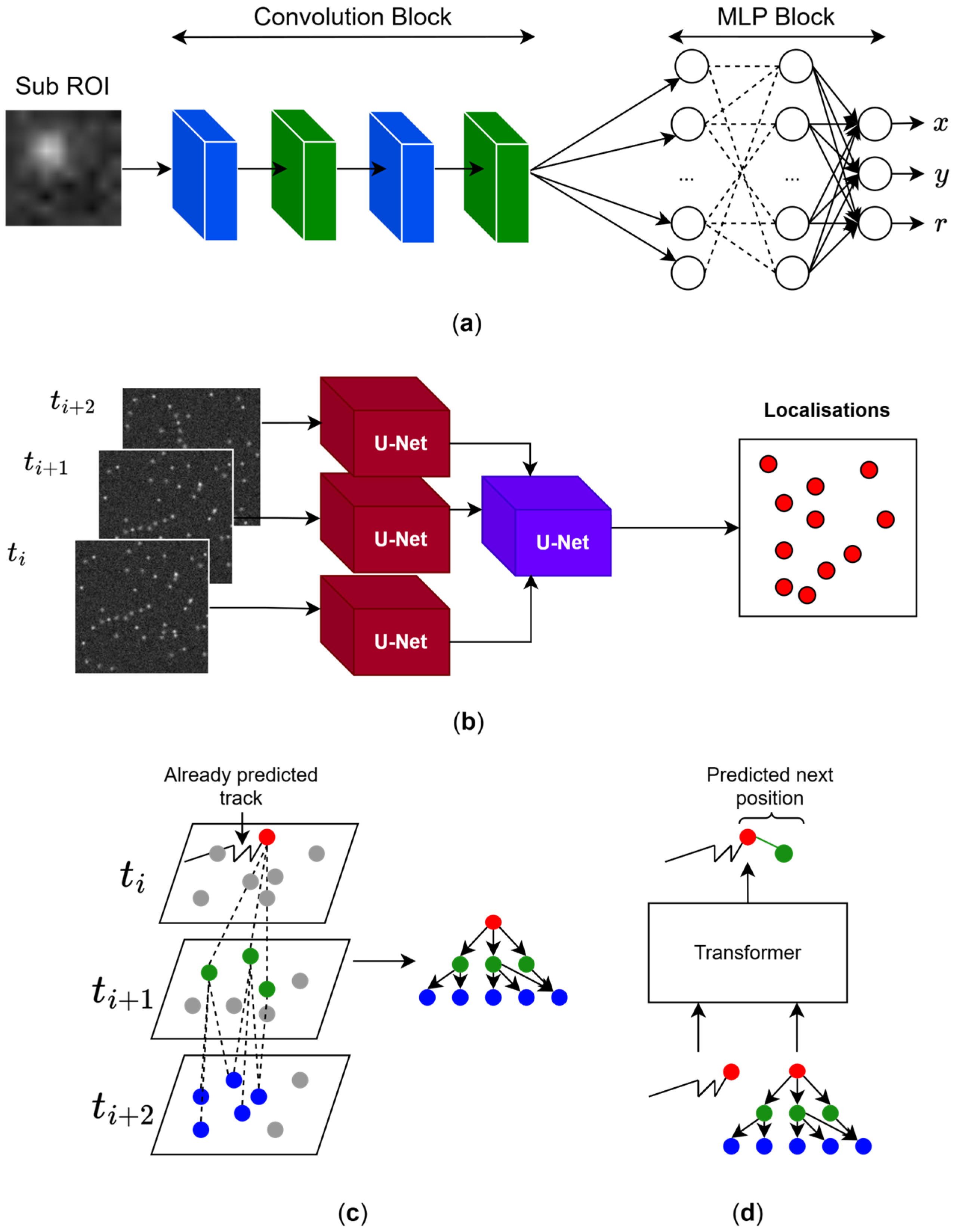
| Method | Input | Strengths | Limitations |
|---|---|---|---|
| PSF Gaussian fitting | Sub-regions of interest (ROI) | Accurate when assumptions hold | Sensitive to model assumptions |
| Suitable for sparse emitters | Prone to user bias | ||
| Not suitable at high molecule density | |||
| Does not perform trajectory linking | |||
| CNN-based localization [49] | Sub-regions of interest (ROIs) | Resistant to noise (RMSE ≈ 1 pixel at SNR = 1) | Does not account for blinking across frames |
| Independent of emitter density | Limited multi-molecule handling | ||
| Does not perform trajectory linking | |||
| Linear Assignment Problem (LAP) [50,51] | Localization coordinates | Robust to high-density conditions | Does not reach theoretical optimum |
| Considers movement heterogeneity, gap closing, and merging and splitting of tracks | Cost functions must be specifically adjusted for the tracking purpose | ||
| Bayesian nonparametric track (BNP-Track) [52] | Consecutive frames | Computational cost scales linearly with number of frames, pixels, and total emitters | Slow inference in standard desktop computers |
| Free from manual tuning | |||
| GNN-based trajectory extraction [53] | Graph representation of SMLM localizations | Can extract dynamics without explicit trajectories | Graph size can become computationally impossible |
| Flexible graph modelling | Requires careful graph building criteria | ||
| Transformer-based network (MOTT) [54] | Hypothesis tree from localisations | Models long/short-term dependencies via attention | Limited to linking |
| Iterative prediction | High memory usage | ||
| End-To-End DL Network (SPTNet) [55] | Consecutive frames | Detects localization, does trajectory linking and predicts dynamical parameters (e.g., diffusion coefficient) | Requires large synthetic datasets |
| Optical flow-based DL (VFINN) [56] | Consecutive frames | Does not require ground-truth synthetic data | May fail for rapid diffusion (large inter-frame displacements) |
| Elegant optical flow formulation | Limited temporal context |
Disclaimer/Publisher’s Note: The statements, opinions and data contained in all publications are solely those of the individual author(s) and contributor(s) and not of MDPI and/or the editor(s). MDPI and/or the editor(s) disclaim responsibility for any injury to people or property resulting from any ideas, methods, instructions or products referred to in the content. |
© 2026 by the authors. Licensee MDPI, Basel, Switzerland. This article is an open access article distributed under the terms and conditions of the Creative Commons Attribution (CC BY) license.
Share and Cite
Saavedra, L.A.; Barrantes, F.J. Machine Learning in Single-Molecule Tracking Analysis of Superresolution Optical Microscopy Data. Cells 2026, 15, 686. https://doi.org/10.3390/cells15080686
Saavedra LA, Barrantes FJ. Machine Learning in Single-Molecule Tracking Analysis of Superresolution Optical Microscopy Data. Cells. 2026; 15(8):686. https://doi.org/10.3390/cells15080686
Chicago/Turabian StyleSaavedra, Lucas A., and Francisco J. Barrantes. 2026. "Machine Learning in Single-Molecule Tracking Analysis of Superresolution Optical Microscopy Data" Cells 15, no. 8: 686. https://doi.org/10.3390/cells15080686
APA StyleSaavedra, L. A., & Barrantes, F. J. (2026). Machine Learning in Single-Molecule Tracking Analysis of Superresolution Optical Microscopy Data. Cells, 15(8), 686. https://doi.org/10.3390/cells15080686