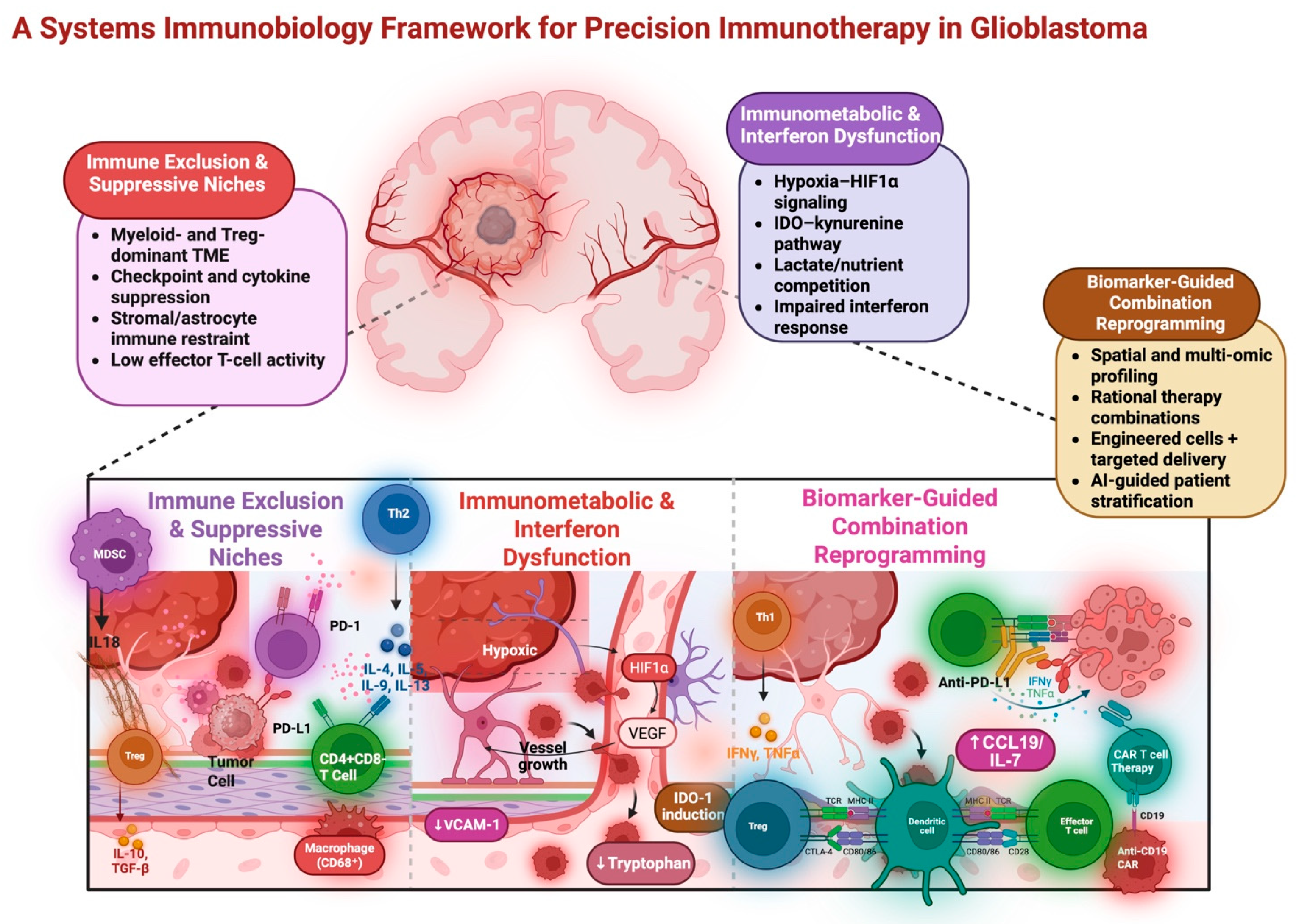
Precision Immunotherapeutics for Glioblastoma: Current Approaches and Emerging Strategies in 2026

 , , , , , , ,
Abstract
1. Introduction
2. The Unique Immunobiology of GBM
2.1. Immunologically Cold Tumor Hallmarks
2.2. Tumor Microenvironment (TME) Barriers
2.3. Physical Barriers
2.4. Emergent Concepts
3. Clinical Landscape of Immunotherapy in GBM
3.1. Immune Checkpoint Inhibitors (ICIs)
3.2. Cancer Vaccines
3.3. Oncolytic Viruses
3.4. Cell-Based Therapies
3.5. Immunometabolic Crosstalk
3.6. METRNL as a Reversible Immunometabolic Checkpoint in GBM
4. Practical Opportunity: Remodeling the TME Through the Restoration of Oxidative Fitness and Alleviation of Hypoxia
Spatial Biology
5. Emerging Translational Insights
5.1. Next-Generation and Novel Immunotherapeutic Strategies
5.2. Combinatorial Approaches
5.3. Engineering Solutions
5.4. Metabolic Immunotherapy
6. Frontiers in Delivery
7. Future Directions and Opportunities
Author Contributions
Funding
Institutional Review Board Statement
Informed Consent Statement
Data Availability Statement
Conflicts of Interest
References
- Price, M.; Ballard, C.A.P.; Benedetti, J.R.; Kruchko, C.; Barnholtz-Sloan, J.S.; Ostrom, Q.T. CBTRUS Statistical Report: Primary Brain and Other Central Nervous System Tumors Diagnosed in the United States in 2018–2022. Neuro-Oncology 2025, 27, iv1–iv66. [Google Scholar] [CrossRef]
- Giovagnoli, A.R.; Meneses, R.F.; Silvani, A.; Milanesi, I.; Fariselli, L.; Salmaggi, A.; Boiardi, A. Quality of life and brain tumors: What beyond the clinical burden? J. Neurol. 2014, 261, 894–904. [Google Scholar] [CrossRef] [PubMed]
- Witthayanuwat, S.; Pesee, M.; Supaadirek, C.; Supakalin, N.; Thamronganantasakul, K.; Krusun, S. Survival Analysis of Glioblastoma Multiforme. Asian Pac. J. Cancer Prev. 2018, 19, 2613–2617. [Google Scholar] [CrossRef] [PubMed]
- Goenka, A.; Tiek, D.; Song, X.; Huang, T.; Hu, B.; Cheng, S.-Y. The Many Facets of Therapy Resistance and Tumor Recurrence in Glioblastoma. Cells 2021, 10, 484. [Google Scholar] [CrossRef] [PubMed]
- Bianconi, A.; Palmieri, G.; Aruta, G.; Monticelli, M.; Zeppa, P.; Tartara, F.; Melcarne, A.; Garbossa, D.; Cofano, F. Updates in Glioblastoma Immunotherapy: An Overview of the Current Clinical and Translational Scenario. Biomedicines 2023, 11, 1520. [Google Scholar] [CrossRef]
- Ye, F.; Dewanjee, S.; Li, Y.; Jha, N.K.; Chen, Z.-S.; Kumar, A.; Vishakha; Behl, T.; Jha, S.K.; Tang, H. Advancements in clinical aspects of targeted therapy and immunotherapy in breast cancer. Mol. Cancer 2023, 22, 105. [Google Scholar] [CrossRef]
- Zulfiqar, B.; Farooq, A.; Kanwal, S.; Asghar, K. Immunotherapy and targeted therapy for lung cancer: Current status and future perspectives. Front. Pharmacol. 2022, 13, 1035171. [Google Scholar] [CrossRef]
- Ernst, M.; Giubellino, A. The Current State of Treatment and Future Directions in Cutaneous Malignant Melanoma. Biomedicines 2022, 10, 822. [Google Scholar] [CrossRef]
- Durga, Y.; Pusapati, R. Drug resistance in glioblastoma: Challenges, mechanisms and therapeutic strategies (Review). Mol. Clin. Oncol. 2025, 24, 4. [Google Scholar] [CrossRef]
- Koh, L.; Novera, W.; Lim, S.W.; Chong, Y.K.; Pang, Q.Y.; Low, D.; Ang, B.T.; Tang, C. Integrative multi-omics approach to targeted therapy for glioblastoma. Pharmacol. Res. 2022, 182, 106308. [Google Scholar] [CrossRef]
- Prager, B.C.; Bhargava, S.; Mahadev, V.; Hubert, C.G.; Rich, J.N. Glioblastoma Stem Cells: Driving Resilience through Chaos. Trends Cancer 2020, 6, 223–235. [Google Scholar] [CrossRef] [PubMed]
- Pouyan, A.; Ghorbanlo, M.; Eslami, M.; Jahanshahi, M.; Ziaei, E.; Salami, A.; Mokhtari, K.; Shahpasand, K.; Farahani, N.; Meybodi, T.E.; et al. Glioblastoma multiforme: Insights into pathogenesis, key signaling pathways, and therapeutic strategies. Mol. Cancer 2025, 24, 58. [Google Scholar] [CrossRef] [PubMed]
- Wu, B.; Zhang, B.; Li, B.; Wu, H.; Jiang, M. Cold and hot tumors: From molecular mechanisms to targeted therapy. Signal Transduct. Target. Ther. 2024, 9, 274. [Google Scholar] [CrossRef]
- Hodges, T.R.; Ott, M.; Xiu, J.; Gatalica, Z.; Swensen, J.; Zhou, S.; Huse, J.T.; De Groot, J.; Li, S.; Overwijk, W.W.; et al. Mutational burden, immune checkpoint expression, and mismatch repair in glioma: Implications for immune checkpoint immunotherapy. Neuro-Oncology 2017, 19, 1047–1057. [Google Scholar] [CrossRef] [PubMed]
- Yu, G.; Pang, Y.; Merchant, M.; Kesserwan, C.; Gangalapudi, V.; Abdelmaksoud, A.; Ranjan, A.; Kim, O.; Wei, J.S.; Chou, H.-C.; et al. Tumor Mutation Burden, Expressed Neoantigens and the Immune Microenvironment in Diffuse Gliomas. Cancers 2021, 13, 6092. [Google Scholar] [CrossRef]
- Touat, M.; Li, Y.Y.; Boynton, A.N.; Spurr, L.F.; Iorgulescu, J.B.; Bohrson, C.L.; Cortes-Ciriano, I.; Birzu, C.; Geduldig, J.E.; Pelton, K.; et al. Mechanisms and therapeutic implications of hypermutation in gliomas. Nature 2020, 580, 517–523. [Google Scholar] [CrossRef]
- Boussemart, L.; Johnson, A.; Schrock, A.B.; Pal, S.K.; Frampton, G.M.; Fabrizio, D.; Chalmers, Z.; Lotem, M.; Gibney, G.; Russell, J.; et al. Tumor mutational burden and response to programmed cell death protein 1 inhibitors in a case series of patients with metastatic desmoplastic melanoma. J. Am. Acad. Dermatol. 2019, 80, 1780–1782. [Google Scholar] [CrossRef]
- Chalmers, Z.R.; Connelly, C.F.; Fabrizio, D.; Gay, L.; Ali, S.M.; Ennis, R.; Schrock, A.; Campbell, B.; Shlien, A.; Chmielecki, J.; et al. Analysis of 100,000 human cancer genomes reveals the landscape of tumor mutational burden. Genome Med. 2017, 9, 34. [Google Scholar] [CrossRef]
- Stein, M.K.; Pandey, M.; Xiu, J.; Tae, H.; Swensen, J.; Mittal, S.; Brenner, A.J.; Korn, W.M.; Heimberger, A.B.; Martin, M.G. Tumor Mutational Burden Is Site Specific in Non–Small-Cell Lung Cancer and Is Highest in Lung Adenocarcinoma Brain Metastases. JCO Precis. Oncol. 2019, 3, 1–13. [Google Scholar] [CrossRef]
- Ricciuti, B.; Wang, X.; Alessi, J.V.; Rizvi, H.; Mahadevan, N.R.; Li, Y.Y.; Polio, A.; Lindsay, J.; Umeton, R.; Sinha, R.; et al. Association of High Tumor Mutation Burden in Non–Small Cell Lung Cancers With Increased Immune Infiltration and Improved Clinical Outcomes of PD-L1 Blockade Across PD-L1 Expression Levels. JAMA Oncol. 2022, 8, 1160. [Google Scholar] [CrossRef]
- Schrock, A.; Sharma, N.; Peled, N.; Bufill, J.; Srkalovic, G.; Spigel, D.; Fabrizio, D.; Frampton, G.; Connelly, C.; Lipka, M.B.; et al. MA14.01 Updated Dataset Assessing Tumor Mutation Burden (TMB) as a Biomarker for Response to PD-1/PD-L1 Targeted Therapies in Lung Cancer (LC). J. Thorac. Oncol. 2017, 12, S422. [Google Scholar] [CrossRef]
- Rooney, M.S.; Shukla, S.A.; Wu, C.J.; Getz, G.; Hacohen, N. Molecular and Genetic Properties of Tumors Associated with Local Immune Cytolytic Activity. Cell 2015, 160, 48–61. [Google Scholar] [CrossRef] [PubMed]
- Sun, S.; Liu, L.; Zhang, J.; Sun, L.; Shu, W.; Yang, Z.; Yao, H.; Zhang, Z. The role of neoantigens and tumor mutational burden in cancer immunotherapy: Advances, mechanisms, and perspectives. J Hematol Oncol 2025, 18, 84. [Google Scholar] [CrossRef]
- Nejo, T.; Matsushita, H.; Karasaki, T.; Nomura, M.; Saito, K.; Tanaka, S.; Takayanagi, S.; Hana, T.; Takahashi, S.; Kitagawa, Y.; et al. Reduced Neoantigen Expression Revealed by Longitudinal Multiomics as a Possible Immune Evasion Mechanism in Glioma. Cancer Immunol. Res. 2019, 7, 1148–1161. [Google Scholar] [CrossRef] [PubMed]
- Dhatchinamoorthy, K.; Colbert, J.D.; Rock, K.L. Cancer Immune Evasion Through Loss of MHC Class I Antigen Presentation. Front. Immunol. 2021, 12, 636568. [Google Scholar] [CrossRef]
- Zagzag, D.; Salnikow, K.; Chiriboga, L.; Yee, H.; Lan, L.; Ali, M.A.; Garcia, R.; Demaria, S.; Newcomb, E.W. Downregulation of major histocompatibility complex antigens in invading glioma cells: Stealth invasion of the brain. Lab. Investig. 2005, 85, 328–341. [Google Scholar] [CrossRef]
- Taylor, B.C.; Balko, J.M. Mechanisms of MHC-I Downregulation and Role in Immunotherapy Response. Front. Immunol. 2022, 13, 844866. [Google Scholar] [CrossRef]
- Zhang, Y.; Yang, C.; Xu, Y.; Jiang, X.; Shi, J.; Li, B.; Yu, D. The MHC-I-dependent neoantigen presentation pathway predicts response rate to PD-1/PD-L1 blockade. Biomol. Biomed. 2024, 25, 1314–1321. [Google Scholar] [CrossRef]
- Jackson, C.M.; Choi, J.; Lim, M. Mechanisms of immunotherapy resistance: Lessons from glioblastoma. Nat. Immunol. 2019, 20, 1100–1109. [Google Scholar] [CrossRef]
- Abikenari, M.A.; Enayati, I.; Fountain, D.M.; Leite, M.I. Navigating glioblastoma therapy: A narrative review of emerging immunotherapeutics and small-molecule inhibitors. Microbes Immun. 2024, 2, 132. [Google Scholar] [CrossRef]
- Liu, F.; Huang, J.; Liu, X.; Cheng, Q.; Luo, C.; Liu, Z. CTLA-4 correlates with immune and clinical characteristics of glioma. Cancer Cell Int. 2020, 20, 7. [Google Scholar] [CrossRef]
- Liu, J.; Tian, X.; Wang, Y.; Kang, X.; Song, W. Soluble cytotoxic T-lymphocyte–associated antigen 4 (sCTLA-4) as a potential biomarker for diagnosis and evaluation of the prognosis in Glioma. BMC Immunol. 2021, 22, 33. [Google Scholar] [CrossRef]
- Fathi, M.; Razavi, S.-M.; Sojoodi, M.; Ahmadi, A.; Ebrahimi, F.; Namdar, A.; Hojjat-Farsangi, M.; Gholamin, S.; Jadidi-Niaragh, F. Targeting the CTLA-4/B7 axes in glioblastoma: Preclinical evidence and clinical interventions. Expert Opin. Ther. Targets 2022, 26, 949–961. [Google Scholar] [CrossRef]
- Heiland, D.H.; Haaker, G.; Delev, D.; Mercas, B.; Masalha, W.; Heynckes, S.; Gäbelein, A.; Pfeifer, D.; Carro, M.S.; Weyerbrock, A.; et al. Comprehensive analysis of PD-L1 expression in glioblastoma multiforme. Oncotarget 2017, 8, 42214–42225. [Google Scholar] [CrossRef]
- Berghoff, A.S.; Kiesel, B.; Widhalm, G.; Rajky, O.; Ricken, G.; Wöhrer, A.; Dieckmann, K.; Filipits, M.; Brandstetter, A.; Weller, M.; et al. Programmed death ligand 1 expression and tumor-infiltrating lymphocytes in glioblastoma. Neuro-Oncology 2015, 17, 1064–1075. [Google Scholar] [CrossRef] [PubMed]
- Broggi, G.; Angelico, G.; Farina, J.; Tinnirello, G.; Barresi, V.; Zanelli, M.; Palicelli, A.; Certo, F.; Barbagallo, G.; Magro, G.; et al. Tumor-associated microenvironment, PD-L1 expression and their relationship with immunotherapy in glioblastoma, IDH-wild type: A comprehensive review with emphasis on the implications for neuropathologists. Pathol.-Res. Pract. 2024, 254, 155144. [Google Scholar] [CrossRef] [PubMed]
- Gou, Q.; Dong, C.; Xu, H.; Khan, B.; Jin, J.; Liu, Q.; Shi, J.; Hou, Y. PD-L1 degradation pathway and immunotherapy for cancer. Cell Death Dis. 2020, 11, 955. [Google Scholar] [CrossRef] [PubMed]
- Wing, K.; Yamaguchi, T.; Sakaguchi, S. Cell-autonomous and -non-autonomous roles of CTLA-4 in immune regulation. Trends Immunol. 2011, 32, 428–433. [Google Scholar] [CrossRef]
- Tekguc, M.; Wing, J.B.; Osaki, M.; Long, J.; Sakaguchi, S. Treg-expressed CTLA-4 depletes CD80/CD86 by trogocytosis, releasing free PD-L1 on antigen-presenting cells. Proc. Natl. Acad. Sci. USA 2021, 118, e2023739118. [Google Scholar] [CrossRef]
- Tai, X.; Van Laethem, F.; Pobezinsky, L.; Guinter, T.; Sharrow, S.O.; Adams, A.; Granger, L.; Kruhlak, M.; Lindsten, T.; Thompson, C.B.; et al. Basis of CTLA-4 function in regulatory and conventional CD4+ T cells. Blood 2012, 119, 5155–5163. [Google Scholar] [CrossRef]
- Qureshi, O.S.; Zheng, Y.; Nakamura, K.; Attridge, K.; Manzotti, C.; Schmidt, E.M.; Baker, J.; Jeffery, L.E.; Kaur, S.; Briggs, Z.; et al. Trans-Endocytosis of CD80 and CD86: A Molecular Basis for the Cell-Extrinsic Function of CTLA-4. Science 2011, 332, 600–603. [Google Scholar] [CrossRef]
- Mohme, M.; Schliffke, S.; Maire, C.L.; Rünger, A.; Glau, L.; Mende, K.C.; Matschke, J.; Gehbauer, C.; Akyüz, N.; Zapf, S.; et al. Immunophenotyping of Newly Diagnosed and Recurrent Glioblastoma Defines Distinct Immune Exhaustion Profiles in Peripheral and Tumor-infiltrating Lymphocytes. Clin. Cancer Res. 2018, 24, 4187–4200. [Google Scholar] [CrossRef]
- Park, J.; Kwon, M.; Kim, K.H.; Chang, J.H.; Shin, E.-C. Expression of immune checkpoint molecules on tumor infiltrating lymphocytes in glioblastoma multiforme. J. Immunol. 2017, 198, 196.3. [Google Scholar] [CrossRef]
- Woroniecka, K.; Chongsathidkiet, P.; Rhodin, K.; Kemeny, H.; Dechant, C.; Farber, S.H.; Elsamadicy, A.A.; Cui, X.; Koyama, S.; Jackson, C.; et al. T-Cell Exhaustion Signatures Vary with Tumor Type and Are Severe in Glioblastoma. Clin. Cancer Res. 2018, 24, 4175–4186. [Google Scholar] [CrossRef] [PubMed]
- Luning, R.; French, P.J.; Vanbilloen, W.J.F.; Dobber, L.; Van Hijfte, L.; Hoogendijk, R.; Van Den Bent, M.J.; Debets, R.; Geurts, M. T cell immunity in glioma and potential implications for immunotherapy: A systematic review. Neuro-Oncology 2025, 28, noaf236. [Google Scholar] [CrossRef] [PubMed]
- Musatova, O.; Kumar, V.; Vinogradov, K.; Rubtsov, Y. Immune checkpoints in immune response to glioma: Two sides of the same coin. Front. Immunol. 2025, 16, 1639521. [Google Scholar] [CrossRef] [PubMed]
- Yee, P.P.; Wei, Y.; Kim, S.Y.; Lu, T.; Chih, S.Y.; Lawson, C.; Tang, M.; Liu, Z.; Anderson, B.; Specht, C.S.; et al. Neutrophil-Induced Ferroptosis Promotes Tumor Necrosis in Glioblastoma Progression. Nat. Commun. 2020, 11, 5424. [Google Scholar] [CrossRef]
- Abikenari, M.; Choi, J.; Medikonda, R.; Kim, L.; Verma, R.; Tran, A.; Annagiri, S.; Lim, M. TMIC-43. Tumor-Associated Neutrophils: A Double-Edged Sword in Glioblastoma Progression. Neuro-Oncology 2025, 27, v453. [Google Scholar] [CrossRef]
- Cao, P.; Sun, Z.; Zhang, F.; Zhang, J.; Zheng, X.; Yu, B.; Zhao, Y.; Wang, W.; Wang, W. TGF-β Enhances Immunosuppression of Myeloid-Derived Suppressor Cells to Induce Transplant Immune Tolerance Through Affecting Arg-1 Expression. Front. Immunol. 2022, 13, 919674. [Google Scholar] [CrossRef]
- Wang, S.; Wang, J.; Chen, Z.; Luo, J.; Guo, W.; Sun, L.; Lin, L. Targeting M2-like tumor-associated macrophages is a potential therapeutic approach to overcome antitumor drug resistance. npj Precis. Oncol. 2024, 8, 31. [Google Scholar] [CrossRef]
- Arlauckas, S.P.; Garren, S.B.; Garris, C.S.; Kohler, R.H.; Oh, J.; Pittet, M.J.; Weissleder, R. Arg1 expression defines immunosuppressive subsets of tumor-associated macrophages. Theranostics 2018, 8, 5842–5854. [Google Scholar] [CrossRef]
- Zhang, M.; Qin, H.; Wu, Y.; Gao, Q. Complex role of neutrophils in the tumor microenvironment: An avenue for novel immunotherapies. Cancer Biol. Med. 2024, 21, 20240192. [Google Scholar] [CrossRef] [PubMed]
- Singel, K.L.; Segal, B.H. Neutrophils in the tumor microenvironment: Trying to heal the wound that cannot heal. Immunol. Rev. 2016, 273, 329–343. [Google Scholar] [CrossRef]
- Youn, J.-I.; Nagaraj, S.; Collazo, M.; Gabrilovich, D.I. Subsets of Myeloid-Derived Suppressor Cells in Tumor-Bearing Mice. J. Immunol. 2008, 181, 5791–5802. [Google Scholar] [CrossRef]
- Raber, P.L.; Thevenot, P.; Sierra, R.; Wyczechowska, D.; Halle, D.; Ramirez, M.E.; Ochoa, A.C.; Fletcher, M.; Velasco, C.; Wilk, A.; et al. Subpopulations of myeloid-derived suppressor cells impair T cell responses through independent nitric oxide-related pathways. Int. J. Cancer 2014, 134, 2853–2864. [Google Scholar] [CrossRef]
- Yang, Y.; Li, C.; Liu, T.; Dai, X.; Bazhin, A.V. Myeloid-Derived Suppressor Cells in Tumors: From Mechanisms to Antigen Specificity and Microenvironmental Regulation. Front. Immunol. 2020, 11, 1371. [Google Scholar] [CrossRef]
- Kusmartsev, S.; Gabrilovich, D.I. STAT1 Signaling Regulates Tumor-Associated Macrophage-Mediated T Cell Deletion. J. Immunol. 2005, 174, 4880–4891. [Google Scholar] [CrossRef]
- Huang, X.; Nepovimova, E.; Adam, V.; Sivak, L.; Heger, Z.; Valko, M.; Wu, Q.; Kuca, K. Neutrophils in Cancer immunotherapy: Friends or foes? Mol. Cancer 2024, 23, 107. [Google Scholar] [CrossRef] [PubMed]
- Corzo, C.A.; Cotter, M.J.; Cheng, P.; Cheng, F.; Kusmartsev, S.; Sotomayor, E.; Padhya, T.; McCaffrey, T.V.; McCaffrey, J.C.; Gabrilovich, D.I. Mechanism Regulating Reactive Oxygen Species in Tumor-Induced Myeloid-Derived Suppressor Cells. J. Immunol. 2009, 182, 5693–5701. [Google Scholar] [CrossRef] [PubMed]
- Kusmartsev, S.; Nefedova, Y.; Yoder, D.; Gabrilovich, D.I. Antigen-Specific Inhibition of CD8+ T Cell Response by Immature Myeloid Cells in Cancer Is Mediated by Reactive Oxygen Species. J. Immunol. 2004, 172, 989–999. [Google Scholar] [CrossRef]
- Jayaraman, P.; Parikh, F.; Lopez-Rivera, E.; Hailemichael, Y.; Clark, A.; Ma, G.; Cannan, D.; Ramacher, M.; Kato, M.; Overwijk, W.W.; et al. Tumor-Expressed Inducible Nitric Oxide Synthase Controls Induction of Functional Myeloid-Derived Suppressor Cells through Modulation of Vascular Endothelial Growth Factor Release. J. Immunol. 2012, 188, 5365–5376. [Google Scholar] [CrossRef]
- Huang, J.; Zhao, Y.; Zhao, K.; Yin, K.; Wang, S. Function of reactive oxygen species in myeloid-derived suppressor cells. Front. Immunol. 2023, 14, 1226443. [Google Scholar] [CrossRef] [PubMed]
- Ibrahim, A.; Mohamady Farouk Abdalsalam, N.; Liang, Z.; Kashaf Tariq, H.; Li, R.; Afolabi, L.O.; Rabiu, L.; Chen, X.; Xu, S.; Xu, Z.; et al. MDSC checkpoint blockade therapy: A new breakthrough point overcoming immunosuppression in cancer immunotherapy. Cancer Gene Ther. 2025, 32, 371–392. [Google Scholar] [CrossRef]
- Chen, Y.; Tan, W.; Wang, C. Tumor-associated macrophage-derived cytokines enhance cancer stem-like characteristics through epithelial–mesenchymal transition. OncoTargets Ther. 2018, 11, 3817–3826. [Google Scholar] [CrossRef] [PubMed]
- Cai, J.; Zhang, W.; Yang, P.; Wang, Y.; Li, M.; Zhang, C.; Wang, Z.; Hu, H.; Liu, Y.; Li, Q.; et al. Identification of a 6-Cytokine Prognostic Signature in Patients with Primary Glioblastoma Harboring M2 Microglia/Macrophage Phenotype Relevance. PLoS ONE 2015, 10, e0126022. [Google Scholar] [CrossRef] [PubMed]
- Andersson, E.A.; Mallard, C.; Ek, C.J. Circulating tight-junction proteins are potential biomarkers for blood–brain barrier function in a model of neonatal hypoxic/ischemic brain injury. Fluids Barriers CNS 2021, 18, 7. [Google Scholar] [CrossRef]
- Abikenari, M.; Sjoholm, M.A.; Liu, J.; Nageeb, G.; Ha, J.H.; Wu, J.; Ren, A.; Sayadi, J.; Lim, J.; Cho, K.B.; et al. Molecular and biophysical remodeling of the blood–brain barrier in glioblastoma: Mechanistic drivers of tumor–neurovascular crosstalk. Front. Phys. 2025, 13, 1723329. [Google Scholar] [CrossRef]
- Vargas, L.O.; Himic, V.; Otaner, F.; Abikenari, M.; Chandar, J.; Govindarajan, V.; Kreatsoulas, D.; Jahangiri, A.; Komotar, R.J.; Ivan, M.E.; et al. Modulating the glioma microenvironment with laser interstitial thermal therapy: Mechanisms and therapeutic implications. J. Neuro-Oncol. 2025, 176, 99. [Google Scholar] [CrossRef]
- Mokarram, N.; Case, A.; Hossainy, N.N.; Lyon, J.G.; MacDonald, T.J.; Bellamkonda, R. Device-assisted strategies for drug delivery across the blood-brain barrier to treat glioblastoma. Commun. Mater. 2025, 6, 5. [Google Scholar] [CrossRef]
- Arvanitis, C.D.; Ferraro, G.B.; Jain, R.K. The blood–brain barrier and blood–tumour barrier in brain tumours and metastases. Nat. Rev. Cancer 2020, 20, 26–41. [Google Scholar] [CrossRef]
- Van Tellingen, O.; Yetkin-Arik, B.; De Gooijer, M.C.; Wesseling, P.; Wurdinger, T.; De Vries, H.E. Overcoming the blood–brain tumor barrier for effective glioblastoma treatment. Drug Resist. Updat. 2015, 19, 1–12. [Google Scholar] [CrossRef] [PubMed]
- Markwell, S.M.; Ross, J.L.; Olson, C.L.; Brat, D.J. Necrotic reshaping of the glioma microenvironment drives disease progression. Acta Neuropathol. 2022, 143, 291–310. [Google Scholar] [CrossRef]
- Shi, T.; Zhu, J.; Zhang, X.; Mao, X. The Role of Hypoxia and Cancer Stem Cells in Development of Glioblastoma. Cancers 2023, 15, 2613. [Google Scholar] [CrossRef]
- Colegio, O.R.; Chu, N.-Q.; Szabo, A.L.; Chu, T.; Rhebergen, A.M.; Jairam, V.; Cyrus, N.; Brokowski, C.E.; Eisenbarth, S.C.; Phillips, G.M.; et al. Functional polarization of tumour-associated macrophages by tumour-derived lactic acid. Nature 2014, 513, 559–563. [Google Scholar] [CrossRef]
- Tao, H.; Zhong, X.; Zeng, A.; Song, L. Unveiling the veil of lactate in tumor-associated macrophages: A successful strategy for immunometabolic therapy. Front. Immunol. 2023, 14, 1208870. [Google Scholar] [CrossRef] [PubMed]
- Zhou, H.-C.; Yu, W.-W.; Yan, X.-Y.; Liang, X.-Q.; Ma, X.-F.; Long, J.-P.; Du, X.-Y.; Mao, H.-Y.; Liu, H.-B. Lactate-driven macrophage polarization in the inflammatory microenvironment alleviates intestinal inflammation. Front. Immunol. 2022, 13, 1013686. [Google Scholar] [CrossRef] [PubMed]
- Zhou, H.; Yan, X.-Y.; Yu, W.; Liang, X.; Du, X.; Liu, Z.; Long, J.; Zhao, G.; Liu, H. Lactic acid in macrophage polarization: The significant role in inflammation and cancer. Int. Rev. Immunol. 2022, 41, 4–18. [Google Scholar] [CrossRef]
- Gu, X.-Y.; Yang, J.-L.; Lai, R.; Zhou, Z.-J.; Tang, D.; Hu, L.; Zhao, L.-J. Impact of lactate on immune cell function in the tumor microenvironment: Mechanisms and therapeutic perspectives. Front. Immunol. 2025, 16, 1563303. [Google Scholar] [CrossRef]
- Medikonda, R.; Abikenari, M.; Schonfeld, E.; Lim, M. The Metabolic Orchestration of Immune Evasion in Glioblastoma: From Molecular Perspectives to Therapeutic Vulnerabilities. Cancers 2025, 17, 1881. [Google Scholar] [CrossRef]
- Obara-Michlewska, M. The tryptophan metabolism, kynurenine pathway and oxidative stress—Implications for glioma pathobiology. Neurochem. Int. 2022, 158, 105363. [Google Scholar] [CrossRef]
- Krupa, M.M.; Pienkowski, T.; Tankiewicz-Kwedlo, A.; Lyson, T. Targeting the kynurenine pathway in gliomas: Insights into pathogenesis, therapeutic targets, and clinical advances. Biochim. Biophys. Acta BBA-Rev. Cancer 2025, 1880, 189343. [Google Scholar] [CrossRef] [PubMed]
- McFaline-Figueroa, J.R.; Sun, L.; Youssef, G.C.; Huang, R.; Li, G.; Kim, J.; Lee, E.Q.; Nayak, L.; Chukwueke, U.; Beroukhim, R.; et al. Neoadjuvant Anti-PD1 Immunotherapy for Surgically Accessible Recurrent Glioblastoma: Clinical and Molecular Outcomes of a Stage 2 Single-Arm Expansion Cohort. Nat. Commun. 2024, 15, 10757. [Google Scholar] [CrossRef]
- Aird, R.; Wills, J.; Roby, K.F.; Bénézech, C.; Stimson, R.H.; Wabitsch, M.; Pollard, J.W.; Finch, A.; Michailidou, Z. Hypoxia-driven metabolic reprogramming of adipocytes fuels cancer cell proliferation. Front. Endocrinol. 2022, 13, 989523. [Google Scholar] [CrossRef]
- Turley, S.J.; Cremasco, V.; Astarita, J.L. Immunological hallmarks of stromal cells in the tumour microenvironment. Nat. Rev. Immunol. 2015, 15, 669–682. [Google Scholar] [CrossRef]
- Ziani, L.; Chouaib, S.; Thiery, J. Alteration of the Antitumor Immune Response by Cancer-Associated Fibroblasts. Front. Immunol. 2018, 9, 414. [Google Scholar] [CrossRef]
- Kennel, K.B.; Bozlar, M.; De Valk, A.F.; Greten, F.R. Cancer-Associated Fibroblasts in Inflammation and Antitumor Immunity. Clin. Cancer Res. 2023, 29, 1009–1016. [Google Scholar] [CrossRef]
- Cheng, B.; Yu, Q.; Wang, W. Intimate communications within the tumor microenvironment: Stromal factors function as an orchestra. J. Biomed. Sci. 2023, 30, 1. [Google Scholar] [CrossRef]
- Mao, X.; Xu, J.; Wang, W.; Liang, C.; Hua, J.; Liu, J.; Zhang, B.; Meng, Q.; Yu, X.; Shi, S. Crosstalk between cancer-associated fibroblasts and immune cells in the tumor microenvironment: New findings and future perspectives. Mol. Cancer 2021, 20, 131. [Google Scholar] [CrossRef] [PubMed]
- Faust Akl, C.; Andersen, B.M.; Li, Z.; Giovannoni, F.; Diebold, M.; Sanmarco, L.M.; Kilian, M.; Fehrenbacher, L.; Pernin, F.; Rone, J.M.; et al. Glioblastoma-instructed astrocytes suppress tumour-specific T cell immunity. Nature 2025, 643, 219–229. [Google Scholar] [CrossRef]
- Wang, Y.-Y.; Jin, W.-L. Beyond Passive Bystander: Glioblastoma-Educated Astrocyte Suppressing T Cell. Front. Biosci. 2025, 30, 44810. [Google Scholar] [CrossRef] [PubMed]
- Xiang, X.; Niu, Y.-R.; Wang, Z.-H.; Ye, L.-L.; Peng, W.-B.; Zhou, Q. Cancer-associated fibroblasts: Vital suppressors of the immune response in the tumor microenvironment. Cytokine Growth Factor Rev. 2022, 67, 35–48. [Google Scholar] [CrossRef] [PubMed]
- Luo, J.; Xiang, X.; Gong, G.; Jiang, L. Cancer-associated fibroblast-mediated immune evasion: Molecular mechanisms of stromal-immune crosstalk in the tumor microenvironment. Front. Immunol. 2025, 16, 1617662. [Google Scholar] [CrossRef]
- Pawar, J.S.; Salam, M.A.; Dipto, M.U.; Al-Amin, M.Y.; Salam, M.T.; Sengupta, S.; Kumari, S.; Gujjari, L.; Yadagiri, G. Cancer-Associated Fibroblasts: Immunosuppressive Crosstalk with Tumor-Infiltrating Immune Cells and Implications for Therapeutic Resistance. Cancers 2025, 17, 2484. [Google Scholar] [CrossRef]
- Du, Y.; Long, X.; Li, X.; Guan, F.; Gao, W.; Deng, K.; Wang, S.; Lin, X.; Huang, M.; She, X.; et al. Spatial-reprogramming derived GPNMB+ macrophages interact with COL6A3+ fibroblasts to enhance vascular fibrosis in glioblastoma. Genome Med. 2025, 17, 136. [Google Scholar] [CrossRef]
- Galbo, P.M.; Madsen, A.T.; Liu, Y.; Peng, M.; Wei, Y.; Ciesielski, M.J.; Fenstermaker, R.A.; Graff, S.; Montagna, C.; Segall, J.E.; et al. Functional Contribution and Clinical Implication of Cancer-Associated Fibroblasts in Glioblastoma. Clin. Cancer Res. 2024, 30, 865–876. [Google Scholar] [CrossRef]
- Naoi, Y.; Inukai, Y.; Izumikawa, T.; Nagasaki, J.; Ishino, T.; Ueda, Y.; Thu, Y.M.; Fujiwara, M.; Baba, T.; Makimoto, G.; et al. Close Spatial Interactions between Cancer Cells and Cancer-Associated Fibroblasts Suppress Antitumor Immunity. Cancer Immunol. Res. 2025, 13, 1471–1484. [Google Scholar] [CrossRef] [PubMed]
- Henrik Heiland, D.; Ravi, V.M.; Behringer, S.P.; Frenking, J.H.; Wurm, J.; Joseph, K.; Garrelfs, N.W.C.; Strähle, J.; Heynckes, S.; Grauvogel, J.; et al. Tumor-associated reactive astrocytes aid the evolution of immunosuppressive environment in glioblastoma. Nat. Commun. 2019, 10, 2541. [Google Scholar] [CrossRef] [PubMed]
- Perelroizen, R.; Philosof, B.; Budick-Harmelin, N.; Chernobylsky, T.; Ron, A.; Katzir, R.; Shimon, D.; Tessler, A.; Adir, O.; Gaoni-Yogev, A.; et al. Astrocyte immunometabolic regulation of the tumour microenvironment drives glioblastoma pathogenicity. Brain 2022, 145, 3288–3307. [Google Scholar] [CrossRef]
- Ghosh, M.; Pilanc-Kudlek, P.; Baluszek, S.; Jacek, K.; Poleszak, K.; Szadkowska, P.; Lenkiewicz, A.M.; Gielniewski, B.; Ellert-Miklaszewska, A.; Castro, M.G.; et al. Identification and characterization of tumor-associated astrocyte subpopulations and their interactions with the tumor microenvironment in experimental glioblastomas. PLoS Biol. 2025, 23, e3002893. [Google Scholar] [CrossRef]
- Andersen, B.M.; Faust Akl, C.; Wheeler, M.A.; Li, Z.; Diebold, M.; Kilian, M.; Rone, J.M.; Misra, A.; Kenison, J.E.; Lee, J.-H.; et al. Barcoded viral tracing identifies immunosuppressive astrocyte–glioma interactions. Nature 2025, 644, 1097–1106. [Google Scholar] [CrossRef]
- Andersen, B.M.; Faust Akl, C.; Wheeler, M.A.; Chiocca, E.A.; Reardon, D.A.; Quintana, F.J. Glial and myeloid heterogeneity in the brain tumour microenvironment. Nat. Rev. Cancer 2021, 21, 786–802. [Google Scholar] [CrossRef]
- Reardon, D.A.; Brandes, A.A.; Omuro, A.; Mulholland, P.; Lim, M.; Wick, A.; Baehring, J.; Ahluwalia, M.S.; Roth, P.; Bähr, O.; et al. Effect of Nivolumab vs Bevacizumab in Patients With Recurrent Glioblastoma. JAMA Oncol. 2020, 6, 1003–1010. [Google Scholar] [CrossRef] [PubMed]
- Nayak, L.; Standifer, N.; Dietrich, J.; Clarke, J.L.; Dunn, G.P.; Lim, M.; Cloughesy, T.; Gan, H.K.; Flagg, E.; George, E.; et al. Circulating Immune Cell and Outcome Analysis from the Phase II Study of PD-L1 Blockade with Durvalumab for Newly Diagnosed and Recurrent Glioblastoma. Clin. Cancer Res. Off. J. Am. Assoc. Cancer Res. 2022, 28, 2567–2578. [Google Scholar] [CrossRef] [PubMed]
- Weathers, S.-P.; Li, X.; Zhu, H.; Damania, A.V.; Knafl, M.; McKinley, B.; Lin, H.; Harrison, R.A.; Majd, N.K.; O’Brien, B.J.; et al. Improved overall survival in an anti-PD-L1 treated cohort of newly diagnosed glioblastoma patients is associated with distinct immune, mutation, and gut microbiome features: A single arm prospective phase I/II trial. Nat. Commun. 2025, 16, 3950. [Google Scholar] [CrossRef] [PubMed]
- Long, G.V.; Shklovskaya, E.; Satgunaseelan, L.; Mao, Y.; da Silva, I.P.; Perry, K.A.; Diefenbach, R.J.; Gide, T.N.; Shivalingam, B.; Buckland, M.E.; et al. Neoadjuvant triplet immune checkpoint blockade in newly diagnosed glioblastoma. Nat. Med. 2025, 31, 1557–1566. [Google Scholar] [CrossRef]
- Xiong, Z.; Raphael, I.; Olin, M.; Okada, H.; Li, X.; Kohanbash, G. Glioblastoma vaccines: Past, present, and opportunities. EBioMedicine 2024, 100, 104963. [Google Scholar] [CrossRef]
- Zheng, Y.; Ma, X.; Feng, S.; Zhu, H.; Chen, X.; Yu, X.; Shu, K.; Zhang, S. Dendritic cell vaccine of gliomas: Challenges from bench to bed. Front. Immunol. 2023, 14, 1259562. [Google Scholar] [CrossRef]
- Liau, L.M.; Ashkan, K.; Brem, S.; Campian, J.L.; Trusheim, J.E.; Iwamoto, F.M.; Tran, D.D.; Ansstas, G.; Cobbs, C.S.; Heth, J.A.; et al. Association of Autologous Tumor Lysate-Loaded Dendritic Cell Vaccination With Extension of Survival Among Patients With Newly Diagnosed and Recurrent Glioblastoma: A Phase 3 Prospective Externally Controlled Cohort Trial. JAMA Oncol. 2023, 9, 112–121. [Google Scholar] [CrossRef] [PubMed]
- Weller, M.; Butowski, N.; Tran, D.D.; Recht, L.D.; Lim, M.; Hirte, H.; Ashby, L.; Mechtler, L.; Goldlust, S.A.; Iwamoto, F.; et al. Rindopepimut with temozolomide for patients with newly diagnosed, EGFRvIII-expressing glioblastoma (ACT IV): A randomised, double-blind, international phase 3 trial. Lancet Oncol. 2017, 18, 1373–1385. [Google Scholar] [CrossRef]
- Keskin, D.B.; Anandappa, A.J.; Sun, J.; Tirosh, I.; Mathewson, N.D.; Li, S.; Oliveira, G.; Giobbie-Hurder, A.; Felt, K.; Gjini, E.; et al. Neoantigen vaccine generates intratumoral T cell responses in phase Ib glioblastoma trial. Nature 2019, 565, 234–239. [Google Scholar] [CrossRef]
- Hilf, N.; Kuttruff-Coqui, S.; Frenzel, K.; Bukur, V.; Stevanović, S.; Gouttefangeas, C.; Platten, M.; Tabatabai, G.; Dutoit, V.; van der Burg, S.H.; et al. Actively personalized vaccination trial for newly diagnosed glioblastoma. Nature 2019, 565, 240–245. [Google Scholar] [CrossRef]
- Coates, K.; Nibbs, R.J.; Fraser, A.R. Going viral: Targeting glioblastoma using oncolytic viruses. Immunother. Adv. 2025, 5, ltaf024. [Google Scholar] [CrossRef]
- van Putten, E.H.P.; Kleijn, A.; van Beusechem, V.W.; Noske, D.; Lamers, C.H.J.; de Goede, A.L.; Idema, S.; Hoefnagel, D.; Kloezeman, J.J.; Fueyo, J.; et al. Convection Enhanced Delivery of the Oncolytic Adenovirus Delta24-RGD in Patients with Recurrent GBM: A Phase I Clinical Trial Including Correlative Studies. Clin. Cancer Res. 2022, 28, 1572–1585. [Google Scholar] [CrossRef]
- Nizamutdinov, D.; Sentmanat, A.; Tong, J.; Qi, X.; Pan, Y.; Qi, D.; Wu, E.; Huang, J.H. The Emerging Role of Oncolytic Virotherapy in Glioblastoma Management. Cancers 2025, 17, 3465. [Google Scholar] [CrossRef] [PubMed]
- Brown, M.C.; Gromeier, M. Cytotoxic and Immunogenic Mechanisms of Recombinant Oncolytic Poliovirus. Curr. Opin. Virol. 2015, 13, 81–85. [Google Scholar] [CrossRef] [PubMed]
- Desjardins, A.; Gromeier, M.; Herndon, J.E.; Beaubier, N.; Bolognesi, D.P.; Friedman, A.H.; Friedman, H.S.; McSherry, F.; Muscat, A.M.; Nair, S.; et al. Recurrent Glioblastoma Treated with Recombinant Poliovirus. N. Engl. J. Med. 2018, 379, 150–161. [Google Scholar] [CrossRef] [PubMed]
- Vazifehmand, R.; Ali, D.S.; Othman, Z.; Chau, D.-M.; Stanslas, J.; Shafa, M.; Sekawi, Z. The evaluation expression of non-coding RNAs in response to HSV-G47∆ oncolytic virus infection in glioblastoma multiforme cancer stem cells. J. NeuroVirology 2022, 28, 566–582. [Google Scholar] [CrossRef]
- Todo, T.; Ito, H.; Ino, Y.; Ohtsu, H.; Ota, Y.; Shibahara, J.; Tanaka, M. Intratumoral oncolytic herpes virus G47∆ for residual or recurrent glioblastoma: A phase 2 trial. Nat. Med. 2022, 28, 1630–1639. [Google Scholar] [CrossRef]
- Choi, B.D.; Gerstner, E.R.; Frigault, M.J.; Leick, M.B.; Mount, C.W.; Balaj, L.; Nikiforow, S.; Carter, B.S.; Curry, W.T.; Gallagher, K.; et al. Intraventricular CARv3-TEAM-E T Cells in Recurrent Glioblastoma. N. Engl. J. Med. 2024, 390, 1290–1298. [Google Scholar] [CrossRef]
- Medikonda, R.; Abikenari, M.A.; Schonfeld, E.; Lim, M. Top advances of the year: The status of chimeric antigen receptor T cells in neuro-oncology. Cancer 2025, 131, e35935. [Google Scholar] [CrossRef]
- Walton, C.M.; Bell, M.; O’Neil, R.; Sahin, O.; Choi, B.D.; Fecci, P.E.; Strickland, B.A. Chimeric antigen receptor (CAR) T-cell therapy for glioblastoma (GBM): Current clinical insights, challenges, and future directions. J. Immunother. Cancer 2025, 13, e012308. [Google Scholar] [CrossRef] [PubMed]
- Abikenari, M.; Liu, J.; Ha, J.H.; Annagiri, S.; Himic, V.; Medikonda, R.; Kim, L.; Choi, J.; Lim, M. Emerging trends in cell-based therapies: Contemporary advances and ethical considerations in translational neurosurgical oncology. J. Neuro-Oncol. 2025, 175, 1–20. [Google Scholar] [CrossRef] [PubMed]
- Begley, S.L.; O’Rourke, D.M.; Binder, Z.A. CAR T cell therapy for glioblastoma: A review of the first decade of clinical trials. Mol. Ther. J. Am. Soc. Gene Ther. 2025, 33, 2454–2461. [Google Scholar] [CrossRef]
- O’Rourke, D.M.; Nasrallah, M.P.; Desai, A.; Melenhorst, J.J.; Mansfield, K.; Morrissette, J.J.D.; Martinez-Lage, M.; Brem, S.; Maloney, E.; Shen, A.; et al. A single dose of peripherally infused EGFRvIII-directed CAR T cells mediates antigen loss and induces adaptive resistance in patients with recurrent glioblastoma. Sci. Transl. Med. 2017, 9, eaaa0984. [Google Scholar] [CrossRef]
- Li, X.; Zhao, L.; Li, W.; Gao, P.; Zhang, N. HER2-targeting CAR-T cells show highly efficient anti-tumor activity against glioblastoma both in vitro and in vivo. Genes Immun. 2024, 25, 201–208. [Google Scholar] [CrossRef] [PubMed]
- Park, D.H.; Bhojnagarwala, P.S.; Liaw, K.; Bordoloi, D.; Tursi, N.J.; Zhao, S.; Binder, Z.A.; O’Rourke, D.; Weiner, D.B. Novel tri-specific T-cell engager targeting IL-13Rα2 and EGFRvIII provides long-term survival in heterogeneous GBM challenge and promotes antitumor cytotoxicity with patient immune cells. J. Immunother. Cancer 2024, 12, e009604. [Google Scholar] [CrossRef]
- Chih, Y.-C.; Dietsch, A.C.; Koopmann, P.; Ma, X.; Agardy, D.A.; Zhao, B.; De Roia, A.; Kourtesakis, A.; Kilian, M.; Krämer, C.; et al. Vaccine-induced T cell receptor T cell therapy targeting a glioblastoma stemness antigen. Nat. Commun. 2025, 16, 1262. [Google Scholar] [CrossRef]
- Mahase, S.; Rattenni, R.N.; Wesseling, P.; Leenders, W.; Baldotto, C.; Jain, R.; Zagzag, D. Hypoxia-Mediated Mechanisms Associated with Antiangiogenic Treatment Resistance in Glioblastomas. Am. J. Pathol. 2017, 187, 940–953. [Google Scholar] [CrossRef]
- Huang, D.; Li, C.; Zhang, H. Hypoxia and cancer cell metabolism. Acta Biochim. Biophys. Sin. 2014, 46, 214–219. [Google Scholar] [CrossRef]
- Ho, K.-H.; Hsu, S.-Y.; Chen, P.-H.; Cheng, C.-H.; Liu, A.-J.; Chien, M.-H.; Chen, K.-C. Hypoxia enhances IL-8 signaling through inhibiting miR-128-3p expression in glioblastomas. Biochim. Biophys. Acta BBA-Mol. Cell Res. 2025, 1872, 119885. [Google Scholar] [CrossRef]
- Cvitković, J.; Tan, W.-L.; Jiang, T.; Zhao, Z. Glioma tumor microenvironment and immunotherapy: Past, present, and future. Biomark. Res. 2025, 13, 150. [Google Scholar] [CrossRef] [PubMed]
- Zhang, G.; Tao, X.; Ji, B.; Gong, J. Hypoxia-Driven M2-Polarized Macrophages Facilitate Cancer Aggressiveness and Temozolomide Resistance in Glioblastoma. Oxidative Med. Cell. Longev. 2022, 2022, 1614336. [Google Scholar] [CrossRef] [PubMed]
- Jung, M.-Y.; Aibaidula, A.; Brown, D.A.; Himes, B.T.; Cumba Garcia, L.M.; Parney, I.F. Superinduction of immunosuppressive glioblastoma extracellular vesicles by IFN-γ through PD-L1 and IDO1. Neuro-Oncol. Adv. 2022, 4, vdac017. [Google Scholar] [CrossRef]
- Wainwright, D.A.; Balyasnikova, I.V.; Chang, A.L.; Ahmed, A.U.; Moon, K.-S.; Auffinger, B.; Tobias, A.L.; Han, Y.; Lesniak, M.S. IDO Expression in Brain Tumors Increases the Recruitment of Regulatory T Cells and Negatively Impacts Survival. Clin. Cancer Res. 2012, 18, 6110–6121. [Google Scholar] [CrossRef]
- Munn, D.H.; Shafizadeh, E.; Attwood, J.T.; Bondarev, I.; Pashine, A.; Mellor, A.L. Inhibition of T Cell Proliferation by Macrophage Tryptophan Catabolism. J. Exp. Med. 1999, 189, 1363–1372. [Google Scholar] [CrossRef]
- Moon, Y.W.; Hajjar, J.; Hwu, P.; Naing, A. Targeting the indoleamine 2,3-dioxygenase pathway in cancer. J. Immunother. Cancer 2015, 3, 51. [Google Scholar] [CrossRef]
- Jackson, C.M.; Pant, A.; Dinalankara, W.; Choi, J.; Jain, A.; Nitta, R.; Yazigi, E.; Saleh, L.; Zhao, L.; Nirschl, T.R.; et al. The cytokine Meteorin-like inhibits anti-tumor CD8+ T cell responses by disrupting mitochondrial function. Immunity 2024, 57, 1864–1877.e9. [Google Scholar] [CrossRef] [PubMed]
- Pant, A.; Jain, A.; Chen, Y.; Patel, K.; Saleh, L.; Tzeng, S.; Nitta, R.T.; Zhao, L.; Wu, C.Y.-J.; Bederson, M.; et al. The CCR6-CCL20 Axis Promotes Regulatory T-cell Glycolysis and Immunosuppression in Tumors. Cancer Immunol. Res. 2024, 12, 1542–1558. [Google Scholar] [CrossRef]
- Kim, A.-R.; Choi, S.J.; Park, J.; Kwon, M.; Chowdhury, T.; Yu, H.J.; Kim, S.; Kang, H.; Kim, K.-M.; Park, S.-H.; et al. Spatial immune heterogeneity of hypoxia-induced exhausted features in high-grade glioma. OncoImmunology 2022, 11, 2026019. [Google Scholar] [CrossRef]
- Liu, M.; Ji, Z.; Jain, V.; Smith, V.L.; Hocke, E.; Patel, A.P.; McLendon, R.E.; Ashley, D.M.; Gregory, S.G.; López, G.Y. Spatial transcriptomics reveals segregation of tumor cell states in glioblastoma and marked immunosuppression within the perinecrotic niche. Acta Neuropathol. Commun. 2024, 12, 64. [Google Scholar] [CrossRef]
- Zheng, Y.; Carrillo-Perez, F.; Pizurica, M.; Heiland, D.H.; Gevaert, O. Spatial cellular architecture predicts prognosis in glioblastoma. Nat. Commun. 2023, 14, 4122. [Google Scholar] [CrossRef]
- Jain, S.; Rick, J.W.; Joshi, R.S.; Beniwal, A.; Spatz, J.; Gill, S.; Chang, A.C.-C.; Choudhary, N.; Nguyen, A.T.; Sudhir, S.; et al. Single-cell RNA sequencing and spatial transcriptomics reveal cancer-associated fibroblasts in glioblastoma with protumoral effects. J. Clin. Investig. 2023, 133, e147087. [Google Scholar] [CrossRef]
- Zarodniuk, M.; Steele, A.; Lu, X.; Li, J.; Datta, M. CNS tumor stroma transcriptomics identify perivascular fibroblasts as predictors of immunotherapy resistance in glioblastoma patients. Npj Genom. Med. 2023, 8, 35. [Google Scholar] [CrossRef] [PubMed]
- Clavreul, A.; Menei, P. Mesenchymal Stromal-Like Cells in the Glioma Microenvironment: What Are These Cells? Cancers 2020, 12, 2628. [Google Scholar] [CrossRef] [PubMed]
- Zhang, J.; Chen, Y.; Xu, H. Glioblastoma induces CAF-like astrocyte activation via the AKT/mTOR–SERPINH1/COL5A1 axis. Biomol. Biomed. 2025, 25, 2737–2754. [Google Scholar] [CrossRef]
- Hosaka, K.; Yang, Y.; Seki, T.; Fischer, C.; Dubey, O.; Fredlund, E.; Hartman, J.; Religa, P.; Morikawa, H.; Ishii, Y.; et al. Pericyte–fibroblast transition promotes tumor growth and metastasis. Proc. Natl. Acad. Sci. USA 2016, 113, E5618–E5627. [Google Scholar] [CrossRef]
- Guo, T.; Xu, J. Cancer-associated fibroblasts: A versatile mediator in tumor progression, metastasis, and targeted therapy. Cancer Metastasis Rev. 2024, 43, 1095–1116. [Google Scholar] [CrossRef]
- Vymola, P.; Garcia-Borja, E.; Cervenka, J.; Balaziova, E.; Vymolova, B.; Veprkova, J.; Vodicka, P.; Skalnikova, H.; Tomas, R.; Netuka, D.; et al. Fibrillar extracellular matrix produced by pericyte-like cells facilitates glioma cell dissemination. Brain Pathol. 2024, 34, e13265. [Google Scholar] [CrossRef] [PubMed]
- Vymola, P.; Garcia-Borja, E.; Cervenka, J.; Balaziova, E.; Vymolova, B.; Vodicka, P.; Busek, P.; Sedo, A. P12.06.A pericyte-like cells expressing fibroblast activation protein are main producers of fibrillar extracellular matrix in glioblastoma. Neuro-Oncology 2023, 25, ii97–ii98. [Google Scholar] [CrossRef]
- Barrett, R.L.; Puré, E. Cancer-associated fibroblasts and their influence on tumor immunity and immunotherapy. eLife 2020, 9, e57243. [Google Scholar] [CrossRef]
- Abikenari, M.; Choi, J.; Kim, L.H.; Verma, R.; Tran, A.; Annagiri, S.; Lim, M. 1182 Therapeutic Role of Tumor-Associated Neutrophils as Inducers of Anti-Tumoral Activity Within the Glioblastoma Tumor Microenvironment. Neurosurgery 2026, 72, 230. [Google Scholar]
- Abikenari, M.; Nageeb, G.; Ha, J.H.; Sjoholm, M.A.; Liu, J.; Bergsneider, B.; Valenzuela, J.; Poe, J.; Bog Cho, K.; Verma, R.; et al. Nexus of IDO1/Kynurenine Pathway to T-Cell Exhaustion: Hypoxia-Induced Tryptophan Metabolism in Glioblastoma. Metabolites 2026, 16, 185. [Google Scholar] [CrossRef]
- Frontiers|CART Immunotherapy: Development, Success, and Translation to Malignant Gliomas and Other Solid Tumors. Available online: https://www.frontiersin.org/journals/oncology/articles/10.3389/fonc.2018.00453/full?source=content_type:react|first_level_url:article|section:main_content|button:body_link (accessed on 15 March 2026).
- Singh, S.; Dey, D.; Barik, D.; Mohapatra, I.; Kim, S.; Sharma, M.; Prasad, S.; Wang, P.; Singh, A.; Singh, G. Glioblastoma at the Crossroads: Current Understanding and Future Therapeutic Horizons. Signal Transduct. Target. Therapy 2025, 10, 213. [Google Scholar] [CrossRef]
- Pennisi, G.; Valeri, F.; Burattini, B.; Bruzzaniti, P.; Sturiale, C.L.; Talacchi, A.; Papacci, F.; Olivi, A.; Della Pepa, G.M. Targeting Macrophages in Glioblastoma: Current Therapies and Future Directions. Cancers 2025, 17, 2687. [Google Scholar] [CrossRef] [PubMed]
- Liu, J.; Abikenari, M.; Annagiri, S.; Ha, J.H.; Nageeb, G.; Sjoholm, M.A.; Velazhahan, V.; Medikonda, R.; Choi, J.; Li, G.; et al. Endogenous immune recruitment in glioblastoma CAR T therapy: Cytokine, myeloid, and chemokine circuitry. J. Neuro-Oncol. 2026, 177, 50. [Google Scholar] [CrossRef]
- Abikenari, M.; Choi, J.; Enayati, I.; Tucker, A.; Bhatnagar, K.; Chen, Y.; Himic, V.; Liu, J.; Nageeb, G.; Poe, J.; et al. When Central Tolerance Fails: Thymic Malignancies at the Intersection of Cancer Immunity and Autoimmunity. Cancers 2026, 18, 747. [Google Scholar] [CrossRef] [PubMed]
- De Leo, A.; Ugolini, A.; Veglia, F. Myeloid Cells in Glioblastoma Microenvironment. Cells 2021, 10, 18. [Google Scholar] [CrossRef]
- Strickler, J.H.; Hanks, B.A.; Khasraw, M. Tumor Mutational Burden as a Predictor of Immunotherapy Response: Is More Always Better? Clin. Cancer Res. 2021, 27, 1236–1241. [Google Scholar] [CrossRef] [PubMed]
- Bausart, M.; Préat, V.; Malfanti, A. Immunotherapy for glioblastoma: The promise of combination strategies. J. Exp. Clin. Cancer Res. 2022, 41, 35. [Google Scholar] [CrossRef]
- Pombo Antunes, A.R.; Scheyltjens, I.; Duerinck, J.; Neyns, B.; Movahedi, K.; Van Ginderachter, J.A. Understanding the glioblastoma immune microenvironment as basis for the development of new immunotherapeutic strategies. eLife 2020, 9, e52176. [Google Scholar] [CrossRef]
- Sharma, P.; Aaroe, A.; Liang, J.; Puduvalli, V.K. Tumor microenvironment in glioblastoma: Current and emerging concepts. Neuro-Oncol. Adv. 2023, 5, vdad009. [Google Scholar] [CrossRef]
- Reardon, D.A.; Brem, S.; Desai, A.S.; Bagley, S.J.; Kurz, S.C.; De La Fuente, M.I.; Nagpal, S.; Welch, M.R.; Hormigo, A.; Forsyth, P.A.J.; et al. Intramuscular (IM) INO-5401 + INO-9012 with electroporation (EP) in combination with cemiplimab (REGN2810) in newly diagnosed glioblastoma. J. Clin. Oncol. 2022, 40, 2004. [Google Scholar] [CrossRef]
- Yao, Y.; Huang, X.; Hu, L.; Chen, D.; Li, C.; Chen, L.; Zhou, N.; Hoon, D.S.B.; Ji, C.; Wang, Y.; et al. Phase IIb randomized, blinded, controlled trial evaluating neoadjuvant PD-1 blockade combined with A2B5+ glioma stem-like cell lysate-loaded DC vaccine in recurrent glioblastoma (IDH1/2 -). J. Clin. Oncol. 2024, 42, 2059. [Google Scholar] [CrossRef]
- Grippin, A.J.; Marconi, C.; Copling, S.; Li, N.; Braun, C.; Woody, C.; Young, E.; Gupta, P.; Wang, M.; Wu, A.; et al. SARS-CoV-2 mRNA vaccines sensitize tumours to immune checkpoint blockade. Nature 2025, 647, 488–497. [Google Scholar] [CrossRef]
- Chakraborty, M.; Abrams, S.I.; Coleman, C.N.; Camphausen, K.; Schlom, J.; Hodge, J.W. External Beam Radiation of Tumors Alters Phenotype of Tumor Cells to Render Them Susceptible to Vaccine-Mediated T-Cell Killing. Cancer Res. 2004, 64, 4328–4337. [Google Scholar] [CrossRef]
- Sharabi, A.B.; Lim, M.; DeWeese, T.L.; Drake, C.G. Radiation and checkpoint blockade immunotherapy: Radiosensitisation and potential mechanisms of synergy. Lancet Oncol. 2015, 16, e498–e509. [Google Scholar] [CrossRef]
- Lynch, C.; Pitroda, S.P.; Weichselbaum, R.R. Radiotherapy, immunity, and immune checkpoint inhibitors. Lancet Oncol. 2024, 25, e352–e362. [Google Scholar] [CrossRef] [PubMed]
- Formenti, S.C.; Demaria, S. Systemic effects of local radiotherapy. Lancet Oncol. 2009, 10, 718–726. [Google Scholar] [CrossRef] [PubMed]
- Kouam, P.N.; Rezniczek, G.A.; Adamietz, I.A.; Bühler, H. Ionizing radiation increases the endothelial permeability and the transendothelial migration of tumor cells through ADAM10-activation and subsequent degradation of VE-cadherin. BMC Cancer 2019, 19, 958. [Google Scholar] [CrossRef]
- Yock, A.D.; Brown, J.A.; Eley, J.; Lwin, C.; Liu, D.; Wikswo, J.P. An organ-on-a-chip investigation into dose-dependent, temporal dynamic effects of radiosurgery on the blood–brain barrier and its immunological response. Brain Res. 2026, 1872, 150092. [Google Scholar] [CrossRef] [PubMed]
- Cocito, C.; Branchtein, M.; Zhou, X.K.; Gongora, T.; Dahmane, N.; Greenfield, J.P. Single-dose radiotherapy is more effective than fractionation when combined with anti-PD-1 immunotherapy in glioblastoma. Sci. Rep. 2025, 15, 22910. [Google Scholar] [CrossRef] [PubMed]
- Mathios, D.; Kim, J.E.; Mangraviti, A.; Phallen, J.; Park, C.-K.; Jackson, C.M.; Garzon-Muvdi, T.; Kim, E.; Theodros, D.; Polanczyk, M.; et al. Anti–PD-1 antitumor immunity is enhanced by local and abrogated by systemic chemotherapy in GBM. Sci. Transl. Med. 2016, 8, 370ra180. [Google Scholar] [CrossRef] [PubMed]
- Stepanenko, A.A.; Sosnovtseva, A.O.; Valikhov, M.P.; Chernysheva, A.A.; Abramova, O.V.; Naumenko, V.A.; Chekhonin, V.P. The need for paradigm shift: Prognostic significance and implications of standard therapy-related systemic immunosuppression in glioblastoma for immunotherapy and oncolytic virotherapy. Front. Immunol. 2024, 15, 1326757. [Google Scholar] [CrossRef]
- Hampe, L.; Daumoine, S.; Limagne, E.; Roussot, N.; Borsotti, F.; Vincent, J.; Ilie, S.; Truntzer, C.; Ghiringhelli, F.; Thibaudin, M. Effect of radiochemotherapy on peripheral immune response in glioblastoma. Cancer Immunol. Immunother. 2024, 73, 133. [Google Scholar] [CrossRef]
- Ladomersky, E.; Zhai, L.; Lenzen, A.; Lauing, K.L.; Qian, J.; Scholtens, D.M.; Gritsina, G.; Sun, X.; Liu, Y.; Yu, F.; et al. IDO1 Inhibition Synergizes with Radiation and PD-1 Blockade to Durably Increase Survival Against Advanced Glioblastoma. Clin. Cancer Res. 2018, 24, 2559–2573. [Google Scholar] [CrossRef]
- Ponterio, E.; Haas, T.L.; De Maria, R. Oncolytic virus and CAR-T cell therapy in solid tumors. Front. Immunol. 2024, 15, 1455163. [Google Scholar] [CrossRef] [PubMed]
- Tonne, J.M.; Budzik, K.; Carrasco, T.F.; Ebbert, L.; Thompson, J.; Nace, R.; Kendall, B.; Diaz, R.M.; Russell, S.J.; Vile, R.G. Smoldering oncolysis by foamy virus carrying CD19 as a CAR target escapes CAR T detection by genomic modification. Mol. Ther. Oncol. 2024, 32, 200852. [Google Scholar] [CrossRef]
- Evgin, L.; Huff, A.L.; Wongthida, P.; Thompson, J.; Kottke, T.; Tonne, J.; Schuelke, M.; Ayasoufi, K.; Driscoll, C.B.; Shim, K.G.; et al. Oncolytic virus-derived type I interferon restricts CAR T cell therapy. Nat. Commun. 2020, 11, 3187. [Google Scholar] [CrossRef]
- Morello, A.; Bianconi, A.; Rizzo, F.; Bellomo, J.; Meyer, A.C.; Garbossa, D.; Regli, L.; Cofano, F. Laser Interstitial Thermotherapy (LITT) in Recurrent Glioblastoma: What Window of Opportunity for This Treatment? Technol. Cancer Res. Treat. 2024, 23, 15330338241249026. [Google Scholar] [CrossRef]
- Kamath, A.A.; Friedman, D.D.; Akbari, S.H.A.; Kim, A.H.; Tao, Y.; Luo, J.; Leuthardt, E.C. Glioblastoma Treated With Magnetic Resonance Imaging-Guided Laser Interstitial Thermal Therapy: Safety, Efficacy, and Outcomes. Neurosurgery 2019, 84, 836–843. [Google Scholar] [CrossRef]
- Mohammadi, A.M.; Hawasli, A.H.; Rodriguez, A.; Schroeder, J.L.; Laxton, A.W.; Elson, P.; Tatter, S.B.; Barnett, G.H.; Leuthardt, E.C. The role of laser interstitial thermal therapy in enhancing progression-free survival of difficult-to-access high-grade gliomas: A multicenter study. Cancer Med. 2014, 3, 971–979. [Google Scholar] [CrossRef] [PubMed]
- Mortezaei, A.; Al-Saidi, N.; Taghlabi, K.M.; Hajikarimloo, B.; Dib, C.; Hussein, A.; Abdelsalam, A.; Nemer, P.; Shepard, M.J.; Sheehan, J.P.; et al. Laser interstitial thermal therapy for high-grade glioma: A systematic review, meta-analysis, and meta-regression. Neurosurg. Focus 2025, 59, E10. [Google Scholar] [CrossRef]
- Campian, J.; Butt, O.; Ghinaseddin, A.; Rahman, M.; Chheda, M.; Johanns, T.; Ansstas, G.; Huang, J.; Liu, J.; Talcott, G.; et al. Ctim-26. phase I/II study of the combination of pembrolizumab (Mk-3475) and laser interstitial thermal therapy (litt) in recurrent glioblastoma. Neuro-Oncology 2021, 23, vi56. [Google Scholar] [CrossRef]
- Hasan, T.; Caragher, S.P.; Shireman, J.M.; Park, C.H.; Atashi, F.; Baisiwala, S.; Lee, G.; Guo, D.; Wang, J.Y.; Dey, M.; et al. Interleukin-8/CXCR2 signaling regulates therapy-induced plasticity and enhances tumorigenicity in glioblastoma. Cell Death Dis. 2019, 10, 292. [Google Scholar] [CrossRef]
- Li, N.; Rodriguez, J.L.; Yin, Y.; Logun, M.T.; Zhang, L.; Yu, S.; Hicks, K.A.; Zhang, J.V.; Zhang, L.; Xie, C.; et al. Armored bicistronic CAR T cells with dominant-negative TGF-β receptor II to overcome resistance in glioblastoma. Mol. Ther. 2024, 32, 3522–3538. [Google Scholar] [CrossRef]
- Mangolini, M.; Srivastava, S.; Souster, E.; Yang, Y.; Wang, H.; Karattil, R.; Schultz, L.; Ma, B.; Pombal, D.; Greenig, M.; et al. Highly potent novel multi-armoured IL13Rα2 CAR-T subverts the immunosuppressive microenvironment of Glioblastoma. bioRXiv 2025. [Google Scholar] [CrossRef]
- Seo, S.-K.; Kwon, B. Immune regulation through tryptophan metabolism. Exp. Mol. Med. 2023, 55, 1371–1379. [Google Scholar] [CrossRef]
- Du, L.; Xing, Z.; Tao, B.; Li, T.; Yang, D.; Li, W.; Zheng, Y.; Kuang, C.; Yang, Q. Both IDO1 and TDO contribute to the malignancy of gliomas via the Kyn–AhR–AQP4 signaling pathway. Signal Transduct. Target. Ther. 2020, 5, 10. [Google Scholar] [CrossRef]
- Jacquerie, A.; Hoeben, A.; Eekers, D.B.P.; Postma, A.A.; Vanmechelen, M.; De Smet, F.; Ackermans, L.; Anten, M.; Severens, K.; Zur Hausen, A.; et al. Prognostic relevance of high expression of kynurenine pathway markers in glioblastoma. Sci. Rep. 2024, 14, 14975. [Google Scholar] [CrossRef] [PubMed]
- Bollu, L.R.; Bommi, P.V.; Monsen, P.J.; Zhai, L.; Lauing, K.L.; Bell, A.; Kim, M.; Ladomersky, E.; Yang, X.; Platanias, L.C.; et al. Identification and Characterization of a Novel Indoleamine 2,3-Dioxygenase 1 Protein Degrader for Glioblastoma. J. Med. Chem. 2022, 65, 15642–15662. [Google Scholar] [CrossRef] [PubMed]
- Monsen, P.J.; Bommi, P.V.; Grigorescu, A.A.; Lauing, K.L.; Mao, Y.; Berardi, P.; Zhai, L.; Ojo, O.; Penco-Campillo, M.; Koch, T.; et al. Rational Design and Optimization of a Potent IDO1 Proteolysis Targeting Chimera (PROTAC). J. Med. Chem. 2025, 68, 4961–4987. [Google Scholar] [CrossRef]
- Fischer, K.; Hoffmann, P.; Voelkl, S.; Meidenbauer, N.; Ammer, J.; Edinger, M.; Gottfried, E.; Schwarz, S.; Rothe, G.; Hoves, S.; et al. Inhibitory effect of tumor cell–derived lactic acid on human T cells. Blood 2007, 109, 3812–3819. [Google Scholar] [CrossRef]
- Wang, Z.; Dai, Z.; Zhang, H.; Liang, X.; Zhang, X.; Wen, Z.; Luo, P.; Zhang, J.; Liu, Z.; Zhang, M.; et al. Tumor-secreted lactate contributes to an immunosuppressive microenvironment and affects CD8 T-cell infiltration in glioblastoma. Front. Immunol. 2023, 14, 894853. [Google Scholar] [CrossRef]
- Wang, F.; Liao, W.; Li, C.; Zhu, L. Silencing BMAL1 promotes M1/M2 polarization through the LDHA/lactate axis to promote GBM sensitivity to bevacizumab. Int. Immunopharmacol. 2024, 134, 112187. [Google Scholar] [CrossRef]
- Kumagai, S.; Koyama, S.; Itahashi, K.; Tanegashima, T.; Lin, Y.; Togashi, Y.; Kamada, T.; Irie, T.; Okumura, G.; Kono, H.; et al. Lactic acid promotes PD-1 expression in regulatory T cells in highly glycolytic tumor microenvironments. Cancer Cell 2022, 40, 201–218.e9. [Google Scholar] [CrossRef] [PubMed]
- Lu, G.; Wang, X.; Li, F.; Wang, S.; Zhao, J.; Wang, J.; Liu, J.; Lyu, C.; Ye, P.; Tan, H.; et al. Engineered biomimetic nanoparticles achieve targeted delivery and efficient metabolism-based synergistic therapy against glioblastoma. Nat. Commun. 2022, 13, 4214. [Google Scholar] [CrossRef] [PubMed]
- Fu, K.; Hu, H.; Zhou, X.; Li, L.; Yan, L. Regulation of the brain tumor microenvironment by focused ultrasound. Mol. Ther. Oncol. 2025, 33, 200994. [Google Scholar] [CrossRef] [PubMed]
- Curley, C.T.; Mead, B.P.; Negron, K.; Kim, N.; Garrison, W.J.; Miller, G.W.; Kingsmore, K.M.; Thim, E.A.; Song, J.; Munson, J.M.; et al. Augmentation of brain tumor interstitial flow via focused ultrasound promotes brain-penetrating nanoparticle dispersion and transfection. Sci. Adv. 2020, 6, eaay1344. [Google Scholar] [CrossRef]
- Fadera, S.; Chukwu, C.; Stark, A.H.; Yue, Y.; Xu, L.; Chien, C.-Y.; Yuan, J.; Chen, H. Focused Ultrasound-Mediated Delivery of Anti-Programmed Cell Death-Ligand 1 Antibody to the Brain of a Porcine Model. Pharmaceutics 2023, 15, 2479. [Google Scholar] [CrossRef]
- Brighi, C.; Reid, L.; White, A.L.; Genovesi, L.A.; Kojic, M.; Millar, A.; Bruce, Z.; Day, B.W.; Rose, S.; Whittaker, A.K.; et al. MR-guided focused ultrasound increases antibody delivery to nonenhancing high-grade glioma. Neuro-Oncol. Adv. 2020, 2, vdaa030. [Google Scholar] [CrossRef]
- Burgess, A.; Ayala-Grosso, C.A.; Ganguly, M.; Jordão, J.F.; Aubert, I.; Hynynen, K. Targeted Delivery of Neural Stem Cells to the Brain Using MRI-Guided Focused Ultrasound to Disrupt the Blood-Brain Barrier. PLoS ONE 2011, 6, e27877. [Google Scholar] [CrossRef] [PubMed]
- Alkins, R.; Burgess, A.; Ganguly, M.; Francia, G.; Kerbel, R.; Wels, W.S.; Hynynen, K. Focused Ultrasound Delivers Targeted Immune Cells to Metastatic Brain Tumors. Cancer Res. 2013, 73, 1892–1899. [Google Scholar] [CrossRef]
- Sabbagh, A.; Beccaria, K.; Ling, X.; Marisetty, A.; Ott, M.; Caruso, H.; Barton, E.; Kong, L.-Y.; Fang, D.; Latha, K.; et al. Opening of the Blood–Brain Barrier Using Low-Intensity Pulsed Ultrasound Enhances Responses to Immunotherapy in Preclinical Glioma Models. Clin. Cancer Res. 2021, 27, 4325–4337. [Google Scholar] [CrossRef] [PubMed]
- Chen, P.-Y.; Hsieh, H.-Y.; Huang, C.-Y.; Lin, C.-Y.; Wei, K.-C.; Liu, H.-L. Focused ultrasound-induced blood–brain barrier opening to enhance interleukin-12 delivery for brain tumor immunotherapy: A preclinical feasibility study. J. Transl. Med. 2015, 13, 93. [Google Scholar] [CrossRef]
- Chen, K.-T.; Chai, W.-Y.; Lin, Y.-J.; Lin, C.-J.; Chen, P.-Y.; Tsai, H.-C.; Huang, C.-Y.; Kuo, J.S.; Liu, H.-L.; Wei, K.-C. Neuronavigation-guided focused ultrasound for transcranial blood-brain barrier opening and immunostimulation in brain tumors. Sci. Adv. 2021, 7, eabd0772. [Google Scholar] [CrossRef] [PubMed]
- Kofoed, R.H.; Aubert, I. Focused ultrasound gene delivery for the treatment of neurological disorders. Trends Mol. Med. 2024, 30, 263–277. [Google Scholar] [CrossRef]
- Rigollet, S.; Rome, C.; Ador, T.; Dumont, E.; Pichon, C.; Delalande, A.; Barbier, E.L.; Stupar, V. FUS-mediated BBB opening leads to transient perfusion decrease and inflammation without acute or chronic brain lesion. Theranostics 2024, 14, 4147–4160. [Google Scholar] [CrossRef]
- Abikenari, M.; Ha, J.H.; Liu, J.; Ren, A.; Cho, K.B.; Lim, J.; Kim, L.H.; Medikonda, R.; Choi, J.; Lim, M. The immunological landscape of traumatic brain injury: Insights from pathophysiology to experimental models. Front. Neurol. 2025, 16, 1668480. [Google Scholar] [CrossRef]
- Parvar, S.J.; Wong, C.I.; Lewis, A.; Szychot, E.; Morris, C.J.; Shorthouse, D.; Dziemidowicz, K. Convection-enhanced delivery for brain malignancies: Technical parameters, formulation strategies and clinical perspectives. Adv. Drug Deliv. Rev. 2025, 224, 115657. [Google Scholar] [CrossRef]
- Sampson, J.H.; Archer, G.; Pedain, C.; Wembacher-Schröder, E.; Westphal, M.; Kunwar, S.; Vogelbaum, M.A.; Coan, A.; Herndon, J.E.; Raghavan, R.; et al. Poor drug distribution as a possible explanation for the results of the PRECISE trial: Clinical article. J. Neurosurg. 2010, 113, 301–309. [Google Scholar] [CrossRef]
- Cruz-Garza, J.G.; Bhenderu, L.S.; Taghlabi, K.M.; Frazee, K.P.; Guerrero, J.R.; Hogan, M.K.; Humes, F.; Rostomily, R.C.; Horner, P.J.; Faraji, A.H. Electrokinetic convection-enhanced delivery for infusion into the brain from a hydrogel reservoir. Commun. Biol. 2024, 7, 869. [Google Scholar] [CrossRef]
- Li, Y.; Yuan, R.; Zhang, E.; Zhang, Y.; Liu, M.; Guo, X.; Yang, G.; Li, X.; Zhou, S. Immunoregulatory hydrogel-fiber composite device as a local T cell activator for inhibiting glioblastoma recurrence. Cell Biomater. 2025, 1, 100181. [Google Scholar] [CrossRef]
- Grossman, S.A.; Schreck, K.C.; Ballman, K.; Alexander, B. Point/counterpoint: Randomized versus single-arm phase II clinical trials for patients with newly diagnosed glioblastoma. Neuro-Oncology 2017, 19, 469–474. [Google Scholar] [CrossRef]
- Vanderbeek, A.M.; Rahman, R.; Fell, G.; Ventz, S.; Chen, T.; Redd, R.; Parmigiani, G.; Cloughesy, T.F.; Wen, P.Y.; Trippa, L.; et al. The clinical trials landscape for glioblastoma: Is it adequate to develop new treatments? Neuro-Oncology 2018, 20, 1034–1043. [Google Scholar] [CrossRef]
- Lee, E.Q.; Chukwueke, U.N.; Hervey-Jumper, S.L.; De Groot, J.F.; Leone, J.P.; Armstrong, T.S.; Chang, S.M.; Arons, D.; Oliver, K.; Verble, K.; et al. Barriers to accrual and enrollment in brain tumor trials. Neuro-Oncology 2019, 21, noz104. [Google Scholar] [CrossRef] [PubMed]
- Lee, E.Q.; Weller, M.; Sul, J.; Bagley, S.J.; Sahebjam, S.; Van Den Bent, M.; Ahluwalia, M.; Campian, J.L.; Galanis, E.; Gilbert, M.R.; et al. Optimizing eligibility criteria and clinical trial conduct to enhance clinical trial participation for primary brain tumor patients. Neuro-Oncology 2020, 22, noaa015. [Google Scholar] [CrossRef] [PubMed]
- Lynes, J.P.; Nwankwo, A.K.; Sur, H.P.; Sanchez, V.E.; Sarpong, K.A.; Ariyo, O.I.; Dominah, G.A.; Nduom, E.K. Biomarkers for immunotherapy for treatment of glioblastoma. J. Immunother. Cancer 2020, 8, e000348. [Google Scholar] [CrossRef] [PubMed]
- Johnson, R.M.; Phillips, H.S.; Bais, C.; Brennan, C.W.; Cloughesy, T.F.; Daemen, A.; Herrlinger, U.; Jenkins, R.B.; Lai, A.; Mancao, C.; et al. Development of a gene expression–based prognostic signature for IDH wild-type glioblastoma. Neuro-Oncology 2020, 22, 1742–1756. [Google Scholar] [CrossRef]
- Kickingereder, P.; Neuberger, U.; Bonekamp, D.; Piechotta, P.L.; Götz, M.; Wick, A.; Sill, M.; Kratz, A.; Shinohara, R.T.; Jones, D.T.W.; et al. Radiomic subtyping improves disease stratification beyond key molecular, clinical, and standard imaging characteristics in patients with glioblastoma. Neuro-Oncology 2018, 20, 848–857. [Google Scholar] [CrossRef]
- Park, A.K.; Kim, P.; Ballester, L.Y.; Esquenazi, Y.; Zhao, Z. Subtype-specific signaling pathways and genomic aberrations associated with prognosis of glioblastoma. Neuro-Oncology 2019, 21, 59–70. [Google Scholar] [CrossRef]
- Singh, K.; Batich, K.A.; Wen, P.Y.; Tan, A.C.; Bagley, S.J.; Lim, M.; Platten, M.; Colman, H.; Ashley, D.M.; Chang, S.M.; et al. Designing Clinical Trials for Combination Immunotherapy: A Framework for Glioblastoma. Clin. Cancer Res. 2022, 28, 585–593. [Google Scholar] [CrossRef] [PubMed]
- Luo, X.; Wang, X.; Eweje, F.; Zhang, X.; Yang, S.; Quinton, R.; Xiang, J.; Li, Y.; Ji, Y.; Li, Z.; et al. Ensemble Learning of Foundation Models for Precision Oncology. Available online: https://arxiv.org/abs/2508.16085v1 (accessed on 8 January 2026).
- Chambon, P.; Bluethgen, C.; Langlotz, C.P.; Chaudhari, A. Adapting Pretrained Vision-Language Foundational Models to Medical Imaging Domains. arXiv 2022, arXiv:2210.04133. [Google Scholar] [CrossRef]
- Liu, J.; Ha, J.H.; Abikenari, M.; Sjoholm, M.A.; Annagiri, S.; Ravi, K.; Bergsneider, B.H.; Verma, R.; Theodros, D.; Medikonda, R.; et al. Adaptive immunotherapeutic paradigms in diffuse midline glioma: Integrating epigenetic reprogramming, neuron–glioma interactions, and tumor microenvironment modulation. J. Neuro-Oncol. 2025, 176, 144. [Google Scholar] [CrossRef] [PubMed]
- Patil, C.; Priyanka, R.; Harshitha, B.M.; Oshik, S.; Yashwanth, S.; Darshan, B.R.; Patil, S.; Prajwal, K.A.; Naik, P.; Goudanavar, P.; et al. Advanced nanotheranostic approaches for targeted glioblastoma treatment: A synergistic fusion of CRISPR-Cas gene editing, AI-driven tumor profiling, and BBB-modulation. Med. Oncology 2025, 42, 413. [Google Scholar] [CrossRef] [PubMed]
- Șerban, M.; Toader, C.; Covache-Busuioc, R.-A. Precision Neuro-Oncology in Glioblastoma: AI-Guided CRISPR Editing and Real-Time Multi-Omics for Genomic Brain Surgery. Int. J. Mol. Sci. 2025, 26, 7364. [Google Scholar] [CrossRef]
- Reframing Clinical AI Evaluation in the Era of Generative Models: Toward Multidimensional, Stakeholder-Informed, and Safety-Centric Frameworks for Real-World Health Care Deployment—Premier Science. Available online: https://premierscience.com/pjs-25-1011/ (accessed on 22 January 2026).
- Simonelli, M.; Persico, P.; Capucetti, A.; Carenza, C.; Franzese, S.; Lorenzi, E.; Dipasquale, A.; Losurdo, A.; Giordano, L.; Pessina, F.; et al. Immunotherapeutic early-phase clinical trials and malignant gliomas: A single-center experience and comprehensive immunophenotyping of circulating leukocytes. Neuro-Oncol. Adv. 2021, 3, vdab160. [Google Scholar] [CrossRef]
- Yuan, Y.; Qi, P.; Xiang, W.; Yanhui, L.; Yu, L.; Qing, M. Multi-Omics Analysis Reveals Novel Subtypes and Driver Genes in Glioblastoma. Front. Genet. 2020, 11, 565341. [Google Scholar] [CrossRef]
- Chaligne, R.; Gaiti, F.; Silverbush, D.; Schiffman, J.S.; Weisman, H.R.; Kluegel, L.; Gritsch, S.; Deochand, S.D.; Gonzalez Castro, L.N.; Richman, A.R.; et al. Epigenetic encoding, heritability and plasticity of glioma transcriptional cell states. Nat. Genet. 2021, 53, 1469–1479. [Google Scholar] [CrossRef]
- Marx, S.; Wilken, F.; Miebach, L.; Ispirjan, M.; Kinnen, F.; Paul, S.; Bien-Möller, S.; Freund, E.; Baldauf, J.; Fleck, S.; et al. Immunophenotyping of Circulating and Intratumoral Myeloid and T Cells in Glioblastoma Patients. Cancers 2022, 14, 5751. [Google Scholar] [CrossRef]
- Zhu, L.; Cheng, G.; Ye, D.; Nazeri, A.; Yue, Y.; Liu, W.; Wang, X.; Dunn, G.P.; Petti, A.A.; Leuthardt, E.C.; et al. Focused Ultrasound-enabled Brain Tumor Liquid Biopsy. Sci. Rep. 2018, 8, 6553. [Google Scholar] [CrossRef]
- Delgado-Martín, B.; Medina, M.Á. Advances in the Knowledge of the Molecular Biology of Glioblastoma and Its Impact in Patient Diagnosis, Stratification, and Treatment. Adv. Sci. 2020, 7, 1902971. [Google Scholar] [CrossRef]
- Abikenari, M.; Schonfeld, E.; Choi, J.; Kim, L.H.; Lim, M. Revisiting glioblastoma classification through an immunological lens: A narrative review. Glioma 2024, 7, 3–9. [Google Scholar] [CrossRef]
- Lee, A.H.; Sun, L.; Mochizuki, A.Y.; Reynoso, J.G.; Orpilla, J.; Chow, F.; Kienzler, J.C.; Everson, R.G.; Nathanson, D.A.; Bensinger, S.J.; et al. Neoadjuvant PD-1 blockade induces T cell and cDC1 activation but fails to overcome the immunosuppressive tumor associated macrophages in recurrent glioblastoma. Nat. Commun. 2021, 12, 6938. [Google Scholar] [CrossRef]
- Omuro, A.; Brandes, A.A.; Carpentier, A.F.; Idbaih, A.; Reardon, D.A.; Cloughesy, T.; Sumrall, A.; Baehring, J.; van den Bent, M.; Bähr, O.; et al. Radiotherapy combined with nivolumab or temozolomide for newly diagnosed glioblastoma with unmethylated MGMT promoter: An international randomized phase III trial. Neuro-Oncology 2022, 25, 123–134. [Google Scholar] [CrossRef]
- Fontanilles, M.; Heisbourg, J.-D.; Daban, A.; Di Fiore, F.; Pépin, L.-F.; Marguet, F.; Langlois, O.; Alexandru, C.; Tennevet, I.; Ducatez, F.; et al. Metabolic remodeling in glioblastoma: A longitudinal multi-omics study. Acta Neuropathol. Commun. 2024, 12, 162. [Google Scholar] [CrossRef]
- Dahlberg, D.; Rummel, J.; Distante, S.; De Souza, G.A.; Stensland, M.E.; Mariussen, E.; Rootwelt, H.; Voie, Ø.; Hassel, B. Glioblastoma Microenvironment Contains Multiple Hormonal and Non-Hormonal Growth-Stimulating Factors. Fluids Barriers CNS 2022, 19, 45. [Google Scholar] [CrossRef] [PubMed]
- Abikenari, M.; Regev, A.; Himic, V.; Choi, J.; Jeyaretna, S.; Fountain, D.M.; Lim, M. The Hormonal Nexus in PIK3CA-Mutated Meningiomas: Implications for Targeted Therapy and Clinical Trial Design. J. Neuro-Oncol. 2025, 174, 329–340. [Google Scholar] [CrossRef] [PubMed]
- Abikenari, M.A.; Regev, A.; Bergsneider, B.H.; Himic, V.; Annagiri, S.; Kim, L.H.; Medikonda, R.; Choi, J.; Jeyaretna, S.; Fountain, D.M.; et al. Spatially Encoded Oncogenesis and Transcriptional Plasticity in Meningioma: Drivers of Therapeutic Resistance and Opportunities for Targeted Intervention. Cancers 2025, 17, 2694. [Google Scholar] [CrossRef] [PubMed]
- Rossi, J.; Zedde, M.; Napoli, M.; Pascarella, R.; Pisanello, A.; Biagini, G.; Valzania, F. Impact of Sex Hormones on Glioblastoma: Sex-Related Differences and Neuroradiological Insights. Life 2024, 14, 1523. [Google Scholar] [CrossRef]
- Alfuridy, G.; Alghamdi, R.; Alkhoshi, A.; Mahjari, A.; Alhussein, A.; Alshehri, E.; Lary, A.; Sabbagh, A.; Alomar, S. Does exogenous hormonal therapy affect the risk of glioma among females: A systematic review and meta-analysis. Neuro-Oncol. Adv. 2024, 6, vdad167. [Google Scholar] [CrossRef]
- Migliozzi, S.; Oh, Y.T.; Hasanain, M.; Garofano, L.; D’Angelo, F.; Najac, R.D.; Picca, A.; Bielle, F.; Di Stefano, A.L.; Lerond, J.; et al. Integrative multi-omics networks identify PKCδ and DNA-PK as master kinases of glioblastoma subtypes and guide targeted cancer therapy. Nat. Cancer 2023, 4, 181–202. [Google Scholar] [CrossRef] [PubMed]
- Advancements in Trigeminal Neuralgia Diagnosis and Biomarkers—European Society of Medicine 2026. Available online: https://esmed.org/advancements-in-trigeminal-neuralgia-diagnosis-and-biomarkers/ (accessed on 15 March 2026).
- Da, Q.; Chen, Y.; Ju, M.; Ji, Z.; Zhou, A.; Wang, W.; Abikenari, M.A.; Chikontwe, P.; Larghero, G.; Chen, B.; et al. Computational Pathology in the Era of Emerging Foundation and Agentic AI―International Expert Perspectives on Clinical Integration and Translational Readiness. arXiv 2026. [Google Scholar] [CrossRef]
- Rončević, A.; Koruga, N.; Soldo Koruga, A.; Rončević, R. Artificial Intelligence in Glioblastoma—Transforming Diagnosis and Treatment. Chin. Neurosurg. J. 2025, 11, 10. [Google Scholar] [CrossRef] [PubMed]


| Resistance Axis | Core Mechanism | Cellular Drivers | Functional Consequence | Biomarker Readouts | Therapeutic Leverage Points |
|---|---|---|---|---|---|
| Interferon signaling dysfunction | Impaired IFN sensing or downstream transcriptional response | Tumor cells, myeloid cells | Reduced antigen presentation, blunted checkpoint response, immune exclusion | IFN response gene sets, STAT1/IRF1 programs, MHC-I expression | IFN pathway restoration, STING agonists, IFN-priming combinations |
| Myeloid antigen presentation failure | TAM/TAN skewing toward suppressive, low-APC phenotypes | TAMs, MDSCs, neutrophil subsets | Poor T-cell priming, T-cell dysfunction despite infiltration | HLA-II, CD74, CD83, costimulatory ligand expression | Myeloid reprogramming, CSF1R modulation, APC-state induction |
| T-cell exhaustion programming | Chronic antigen exposure + suppressive niche signaling | CD8 TILs, CAR-T cells | Reduced cytotoxicity and proliferative capacity | PD-1, LAG3, CD39, TOX, exhaustion transcriptional modules | Multi-checkpoint blockade, metabolic rescue, antigen load control |
| Metabolic resource competition | Nutrient depletion and suppressive metabolite accumulation | Tumor cells, myeloid cells | T-cell metabolic insufficiency and effector collapse | Glycolysis/OXPHOS gene sets, lactate transporters, adenosine pathway markers | Metabolic checkpoint blockade, adenosine receptor inhibition |
| Spatial immune exclusion | Physical and signaling barriers to immune penetration | Tumor stroma, vascular niche | Immune cells restricted to perivascular or margin niches | Spatial immune mapping, immune distance metrics | Vascular normalization, matrix remodeling |
| Intratumoral immune heterogeneity | Divergent immune niches within same tumor | Mixed immune populations | Mixed treatment response, partial resistance | Single-cell + spatial immune profiling | Region-aware therapy design, adaptive dosing |
| Antigen presentation instability | Dynamic antigen loss or processing defects | Tumor cells | Immune escape under therapy | Neoantigen burden, β2M loss, MHC downregulation | Multi-antigen targeting, epitope spreading strategies |
| Biomarker Class | What It Captures | Measurement Platform | Clinical Utility | Strengths | Limitations |
|---|---|---|---|---|---|
| Interferon competence signatures | Tumor immune responsiveness potential | Bulk RNA, single-cell RNA | Predict checkpoint sensitivity | Mechanistically anchored | Dynamic and therapy-dependent |
| Myeloid functional states | APC vs. suppressive myeloid balance | Single-cell RNA, spatial proteomics | Stratify myeloid-targeted combinations | Directly links to T-cell priming | Requires high-dimensional assays |
| T-cell clonality and expansion | Antigen-driven immune engagement | TCR sequencing | Response likelihood, immune activation tracking | Quantitative and reproducible | Does not guarantee functionality |
| Exhaustion marker co-expression | Functional T-cell impairment | Flow, CyTOF, spatial IF | Functional immune status | Translationally accessible | Marker expression |
| Spatial immune architecture | Immune–tumor proximity and niche structure | Spatial transcriptomics, multiplex IF | Predict response niches and escape zones | Preserves context | Cost and analytic complexity |
| Immunometabolic signatures | Metabolic suppression and competition | Single-cell RNA, metabolomics | Combination design (metabolic + immune) | Mechanism-driven | Platform variability |
| Adenosine pathway activation | Immunosuppressive metabolite signaling | Flow, transcriptomics | Target selection for metabolic checkpoint therapy | Drug-targetable axis | Context-dependent |
| Dynamic pharmacodynamic markers | Early therapy response signals | Serial biopsy, liquid biomarkers | Adaptive trial decision-making | Enables response-guided design | Requires longitudinal sampling |
| Therapeutic Layer | Targeted Constraint | Example Strategy Type | Rationale | Required Biomarker Support | Trial Design Implication |
|---|---|---|---|---|---|
| Checkpoint axis | T-cell inhibitory signaling | Dual/triple checkpoint blockade | Releases inhibitory signaling | Exhaustion marker profiling | Enrich for exhaustion-high tumors |
| Interferon axis | IFN signaling insufficiency | IFN priming + checkpoint | Restores immune activation competence | IFN gene signature | Stratify by IFN competence |
| Myeloid axis | APC failure/suppressive TAMs | Myeloid reprogramming agents | Improves antigen presentation | Myeloid state markers | Myeloid-state stratified cohorts |
| Metabolic axis | Nutrient and metabolite suppression | Adenosine or metabolic blockade | Restores T-cell energetics | Metabolic signatures | Combine with T-cell therapies |
| Spatial axis | Immune exclusion | Vascular or matrix modulators | Improves immune penetration | Spatial immune maps | Region-aware endpoints |
| Cellular therapy axis | Effector insufficiency | CAR-T/engineered T cells | Direct cytotoxic replacement | Antigen density + TME profile | Combine with TME modulators |
| Antigen axis | Target escape | Multi-antigen targeting | Reduces escape probability | Antigen heterogeneity mapping | Multi-target enrollment |
| Adaptive monitoring axis | Dynamic resistance | Biomarker-adaptive dosing | Responds to evolving biology | Serial immune profiling | Adaptive trial platforms |
| Radiotherapy axis | Low antigenicity and immune exclusion | Hypofractionated RT | Increases neoantigen presentation, MHC-I expression, vascular permeability | Neo-antigen burden, DNA damage response | Neoadjuvant RT window, 10 Gy priming doses |
Disclaimer/Publisher’s Note: The statements, opinions and data contained in all publications are solely those of the individual author(s) and contributor(s) and not of MDPI and/or the editor(s). MDPI and/or the editor(s) disclaim responsibility for any injury to people or property resulting from any ideas, methods, instructions or products referred to in the content. |
© 2026 by the authors. Licensee MDPI, Basel, Switzerland. This article is an open access article distributed under the terms and conditions of the Creative Commons Attribution (CC BY) license.
Share and Cite
Poe, J.; Kim, C.; Coleman, C.; Nguyen, H.; Velazhahan, V.; Bergsneider, B.; Sanker, V.; Kim, S.; Chen, Y.; Abikenari, M.; et al. Precision Immunotherapeutics for Glioblastoma: Current Approaches and Emerging Strategies in 2026. Cells 2026, 15, 561. https://doi.org/10.3390/cells15060561
Poe J, Kim C, Coleman C, Nguyen H, Velazhahan V, Bergsneider B, Sanker V, Kim S, Chen Y, Abikenari M, et al. Precision Immunotherapeutics for Glioblastoma: Current Approaches and Emerging Strategies in 2026. Cells. 2026; 15(6):561. https://doi.org/10.3390/cells15060561
Chicago/Turabian StylePoe, James, Claire Kim, Campbell Coleman, Hieu Nguyen, Vaithish Velazhahan, Brandon Bergsneider, Vivek Sanker, Samuel Kim, Yijiang Chen, Matthew Abikenari, and et al. 2026. "Precision Immunotherapeutics for Glioblastoma: Current Approaches and Emerging Strategies in 2026" Cells 15, no. 6: 561. https://doi.org/10.3390/cells15060561
APA StylePoe, J., Kim, C., Coleman, C., Nguyen, H., Velazhahan, V., Bergsneider, B., Sanker, V., Kim, S., Chen, Y., Abikenari, M., Choi, J., & Lim, M. (2026). Precision Immunotherapeutics for Glioblastoma: Current Approaches and Emerging Strategies in 2026. Cells, 15(6), 561. https://doi.org/10.3390/cells15060561