1. Introduction
Sepsis is defined as life-threatening organ dysfunction resulting from a dysregulated host response to infection [
1]. Sepsis is a major global medical problem, affecting over 50 million people and resulting in approximately 11 million deaths annually worldwide [
1,
2]. The pathogenesis of sepsis is multifactorial, involving uncontrolled systemic inflammation, immune dysregulation, coagulopathy, and metabolic disturbances [
3]. Together, these factors contribute to multiple organ failure. Among the organs affected, the lungs are particularly vulnerable, often representing the earliest site of injury [
4]. It has been documented that 25–50% of septic patients develop acute lung injury (ALI), a condition associated with high morbidity and a mortality rate of up to 40% [
5]. Despite the advances in supportive care, effective immunomodulatory therapies for sepsis remain limited, highlighting the urgent need to better understand the mechanisms underlying immune dysregulation in this condition.
CD4
+ T cells are a heterogeneous population of T lymphocytes that play central roles in immune responses [
6]. Upon activation, CD4
+ T cells have the capacity to differentiate into multiple functional subsets [
7]. Each of these subsets is distinguished by distinct patterns of surface molecule expression, cytokine production, and lineage-defining transcription factors [
8]. Notable subsets of CD4
+ helper T cells include Th1, Th2, and Th17 cells, which produce distinct sets of cytokines [
9,
10]. Th1 cells produce pro-inflammatory cytokines, most notably interferon γ (IFNγ), which contributes to inflammatory disorders [
11]. There is a suppressive subset of CD4
+ T cells, known as regulatory T (Treg) cells, that maintain immune homeostasis and restrain inflammation [
12]. Treg cells are characterized by the expression of the master transcription factor forkhead box protein P3 (Foxp3) in the nucleus and CD25 (IL-2 receptor α chain) on the cell surface [
13,
14]. Recent studies have identified distinct populations of CD4
+ T cells that cannot be fully explained by the classical effector T cell or Treg classifications. A new CD4
+ T cell subset that possesses both Th1 and Treg phenotypes, represented by IFNγ and Foxp3 expression, respectively, has been recently discovered [
15]. This subpopulation is called Th1-Treg cells and is implicated in the pathogenesis of inflammatory bowel disease (IBD), where it contributes to tissue inflammation [
16]. A recent study has shown that platelet factor 4 serves as a driver of Th1-Treg cells in the tumor microenvironment [
17]. However, the presence, dynamics, function, and induction mechanisms of Th1-Treg cells during sepsis remain unexplored.
The pathophysiology of sepsis has been directly linked to both pathogens and host-derived molecules known as damage-associated molecular patterns (DAMPs). Cold-inducible RNA-binding protein (CIRP) is an 18-kDa RNA chaperone primarily sequestered in the nucleus [
18]. Once CIRP is released into circulation, extracellular CIRP (eCIRP) serves as a DAMP to activate several immune cells, such as macrophages, neutrophils, and T cells, to promote their pro-inflammatory response by binding to Toll-like receptor 4 (TLR4) [
18]. During sepsis, increased eCIRP levels were observed in the circulation of patients, as well as in experimental animal models [
19]. eCIRP has been demonstrated to drive pathogenic polarization in innate immune cells, such as macrophages and neutrophils, during sepsis [
18]. For instance, eCIRP induces macrophage polarization via STAT3, a member of signal transducer and activator of transcription (STAT) proteins, which regulate cell differentiation [
20]. However, the effects of eCIRP on CD4
+ T cell subsets, including the potential for the induction of Th1-Treg cells, have not been elucidated.
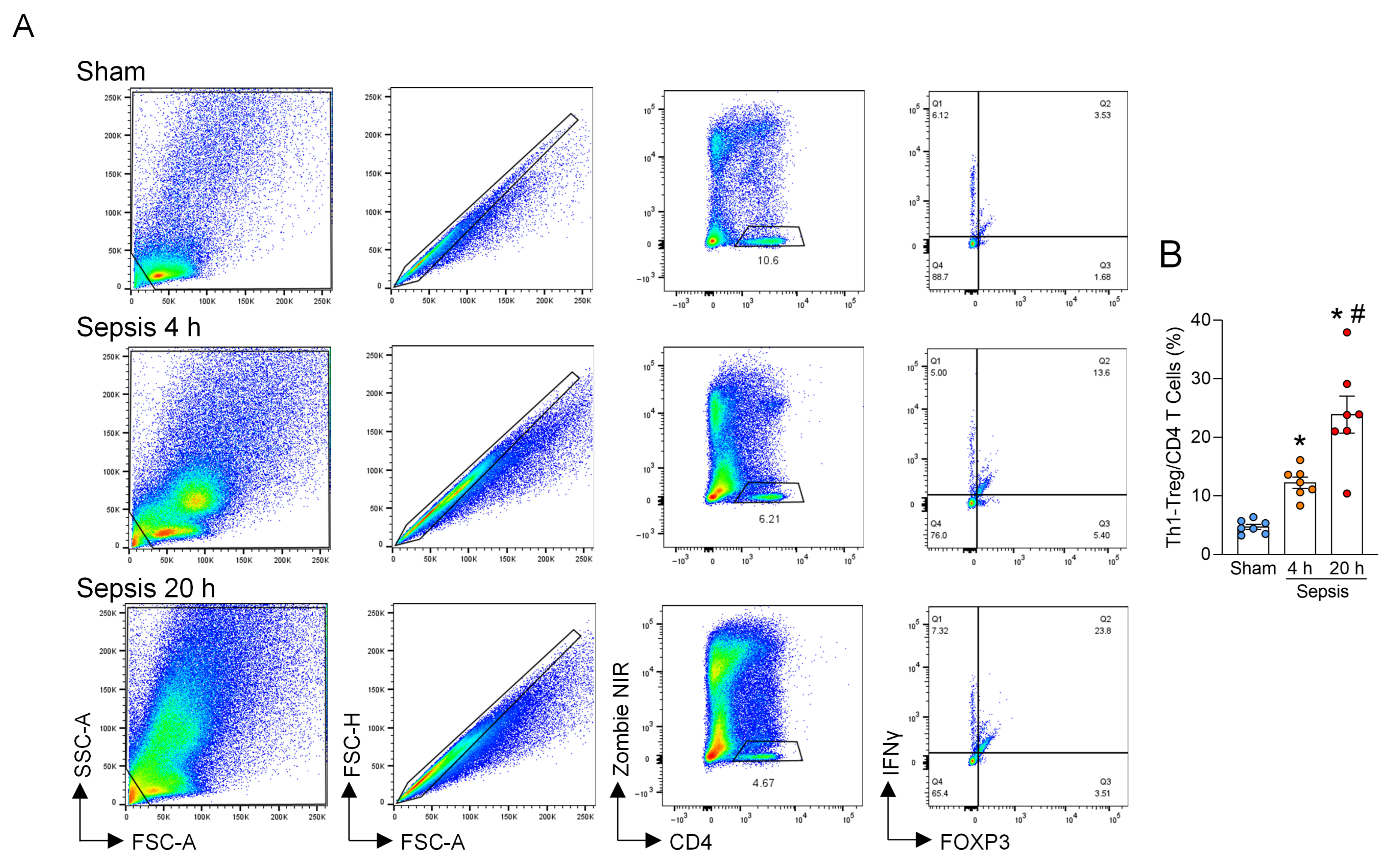
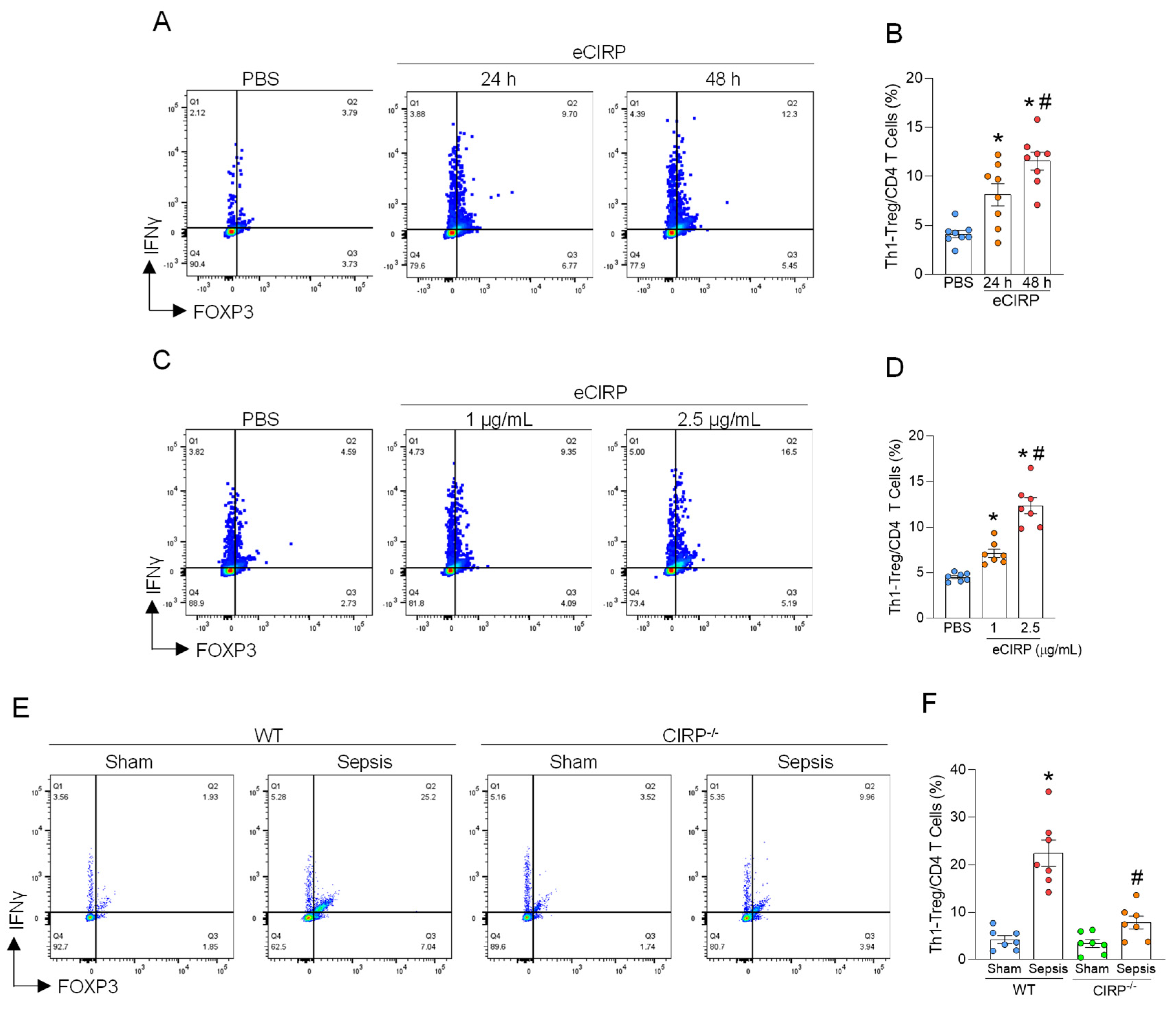
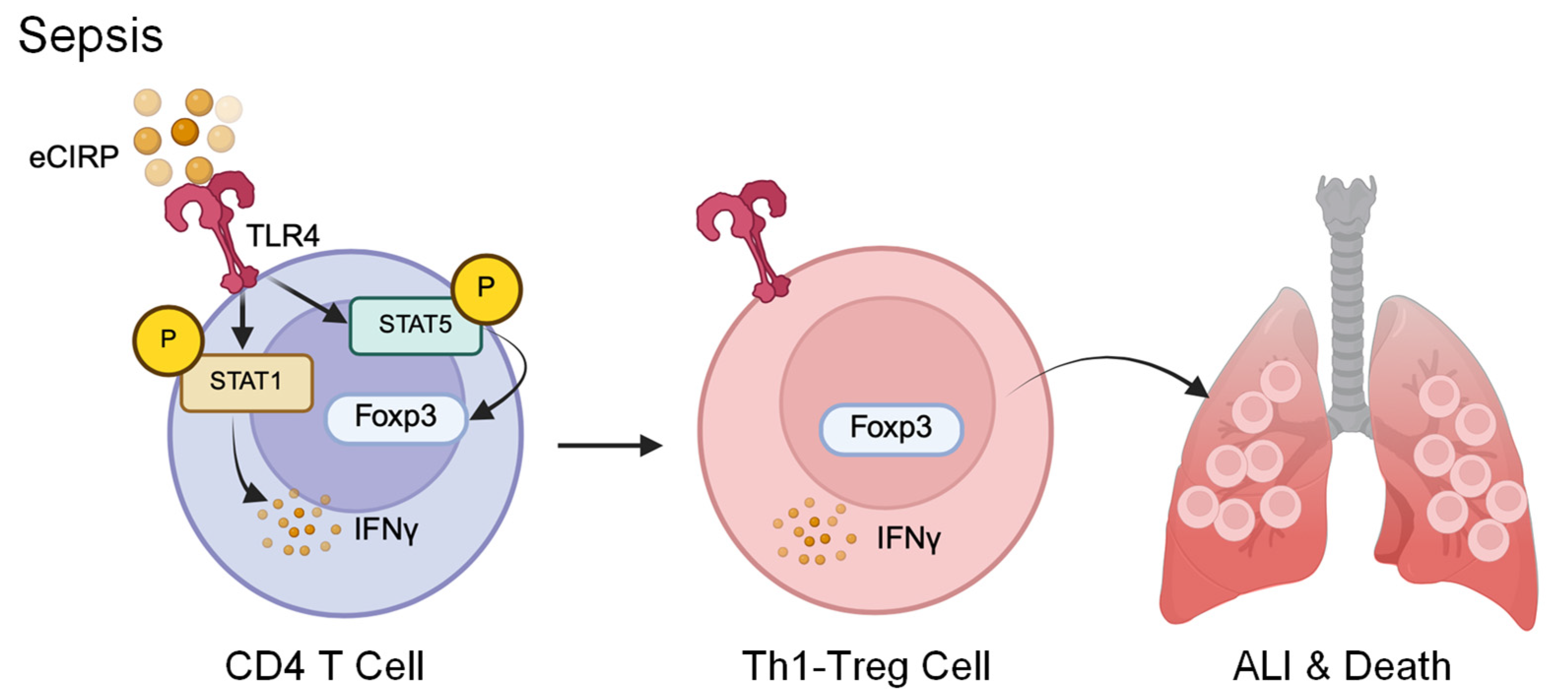
In this study, we investigated the dynamics of Th1-Treg cells during sepsis and found that these cells are markedly increased in the lungs. We further explored the mechanisms underlying their induction and demonstrated that eCIRP drives the generation of Th1-Treg cells via activation of the TLR4–STAT1/5 signaling axis. The accumulation of Th1-Treg cells exacerbates ALI by promoting inflammation and cell death, ultimately contributing to worsened mortality in sepsis. These findings reveal a previously unrecognized role of Th1-Treg cells as pathogenic mediators in septic lung injury and identify the eCIRP–TLR4–STAT1/5 pathway as a critical mechanism driving their induction.
2. Materials and Methods
2.1. Animals
Male 8–12-week-old wild-type (WT) C57BL/6 mice were purchased from Charles River Laboratory (Charles River, Wilmington, MA, USA). CIRP−/− mice on the C57BL/6 background were originally gifted from Prof. Jun Fujita (Kyoto University, Kyoto, Japan), and TLR4−/− mice on the C57BL/6 background were obtained from Dr. Kevin Tracey (The Feinstein Institutes for Medical Research, Manhasset, NY, USA). The mice were housed in a temperature-controlled room under a 12-hour (h) light/dark cycle and provided with standard laboratory chow and water. All experiments were performed in accordance with the National Institutes of Health guidelines for the care and use of laboratory animals and were approved by the Institutional Animal Care and Use Committees (IACUC) of The Feinstein Institutes for Medical Research.
2.2. Isolation of CD4+ T Cells
The spleens were harvested from the 8–12-week-old male WT or TLR4−/− mice. The spleens were minced and filtered through a sterile 70 µm cell strainer (Corning, Corning, NY, USA). The erythrocytes were removed using BD Pharm LyseTM Lysing Buffer (Cat. No.: 555899, BD Science, Franklin Lakes, NJ, USA), and the cells were washed. Then, CD4+ T cells were isolated from the cell suspension using an EasySepTM Mouse CD4+ T cell Isolation Kit (Cat. No.: 19502A, STEMCELL, Vancouver, BC, Canada) according to the manufacturer’s instructions. Naïve CD4+ T cells were isolated using an EasySepTM Mouse Naïve CD4+ T cell Isolation Kit (Cat. No.: 19765A, STEMCELL) according to the manufacturer’s instructions.
2.3. Treatment of CD4+ T Cells with eCIRP
Isolated CD4
+ T cells from the WT or TLR4
−/− mice were cultured in Roswell Park Memorial Institute (RPMI) 1640 medium (Cat. No.: 11875093, Thermo Fisher Scientific, Waltham, MA, USA), supplemented with 10% fetal bovine serum (FBS), 100 U/mL Penicillin and 100 μg/mL Streptomycin, 1% glutamine and 50 μM 2-mercaptaethanol in a 37 °C incubator under humidified conditions containing 5% CO
2. Recombinant mouse CIRP (eCIRP) was prepared in-house, and quality control assays were performed, as previously described [
21]. CD4
+ T cells were treated with 1.0, 2.5 µg/mL of eCIRP for the indicated time periods on CD3/CD28 coated plates (Ultra-LEAF Purified anti-mouse CD3ε, Cat. No.: 100340; Ultra-LEAF Purified anti-mouse CD28, Cat. No.: 102116, Biolegend, San Diego, CA, USA) in the presence and absence of 1 µM of STAT1 inhibitor (Fludarabine; Cat. No.: HY-B0069, MedChemExpress, Monmouth Junction, NJ, USA) and/or 10 μM of STAT5 inhibitor (Cat. No.: HY-101853, MedChemExpress), followed by stimulation with Cell Activation Cocktail (with Brefeldin A) (Cat. No.: 423304, Biolegend) for 4 h for cytokine evaluation.
2.4. Mouse Model of Sepsis
Intra-abdominal sepsis was induced in the 8–12-week-old male WT or CIRP
−/− mice by cecal ligation and puncture (CLP) [
21,
22]. Briefly, the mice were anesthetized with isoflurane, and a midline abdominal incision was made. The cecum was ligated with a 4-0 silk suture 1 cm proximal to its distal extremity, punctured twice (through and through) with a 22 G needle for the 20 h study and once for the survival study. Sham animals underwent laparotomy without CLP. Following the surgery, 1 mL of saline was injected subcutaneously to prevent surgery-induced dehydration, and 0.05 mg/kg buprenorphine was injected subcutaneously as an analgesic. The lungs were harvested 20 h after the surgery. In the survival study, 0.5 mg/kg BW of Imipenem was administered subcutaneously. The mice were monitored for up to 10 days for the survival studies. The mice were euthanized when they met the criteria for humane endpoints approved by the Institutional Animal Care and Use Committee, including failure to eat or drink, grimace score of 2, hunched posture, immobility, labored breathing, non-responsiveness to touch, and reduced response to human touch.
2.5. Isolation of Lung Cells
Lung tissues were minced into small pieces and incubated with 1 mg/mL collagenase type I (Cat. No. LS004197, Worthington Biochemical, Lakewood, NJ, USA) at 37 °C for 30 min with periodic agitation. The digested tissue was then mechanically dissociated, passed through a 70 μm cell strainer (Corning, Corning, NY, USA) and treated with BD Pharm Lyse
TM Lysing Buffer before cell collection [
23]. The isolated cells were stimulated with Cell Activation Cocktail (with Brefeldin A) for 4 h in RPMI 1640 medium, supplemented with 10% fetal bovine serum (FBS), 100 U/mL Penicillin and 100 μg/mL Streptomycin, and 1% glutamine in a 37 °C incubator under humidified conditions containing 5% CO
2, after which they were stained with antibodies and analyzed using flow cytometry. Brefeldin A was used to inhibit IFNγ secretion feasible for intracellular staining of IFNγ.
2.6. Flow Cytometry
Cell suspensions were incubated with a combination of monoclonal fluorescently conjugated antibodies (Abs): FITC anti-mouse CD4 (Cat. No.: 100510, Biolegend), APC anti-mouse CD25 (Cat. No.: 113709, Biolegend), PerCP/Cyanine5.5 anti-mouse CD183 (CXCR3) (Cat. No.: 126514, Biolegend), PE anti-mouse FOXP3 (Cat. No.: 126404, Biolegend), Brilliant Violet 421 anti-mouse IFNγ (Cat. No.: 505830, Biolegend), PE anti-STAT1 Phospho (Ser727) (Cat. No.: 686404, Biolegend), APC anti-STAT5 Phospho (Tyr694) (Cat. No.: 936906, Biolegend), PE Rat IgG2b, κ Isotype Ctrl (Cat. No.: 400636, Biolegend), and Brilliant Violet 421 Rat IgG1, κ Isotype Ctrl (Cat. No.: 400430, Biolegend). To analyze the intracellular expression of IFNγ and Foxp3, the Foxp3 Staining Buffer Set (Cat. No.: 00-5523-00, Thermo Fisher) was used according to the manufacturer’s instructions. To analyze the intracellular expression of phosphorylated (p) STAT1 and pSTAT5, True-PhosTM Perm Buffer (Cat. N0.: 425401, Biolegend) was used according to the manufacturer’s instructions. TrueStain FcX PLUS (Cat. No.: 156604, Biolegend) was used to prevent nonspecific antibody binding, and cell viability was determined using a Zombie NIR Fixable Viability Kit (Cat. No.: 4231006, Biolegend). Flow cytometric analysis was performed on a BD LSRFortessa Cell Analyzer (BD Biosciences, San Jose, CA, USA), and data were processed using FlowJo software (ver. 10; Tree Star, Ashland, OR, USA).
2.7. Adoptive Transfer Experiment of Septic Mice
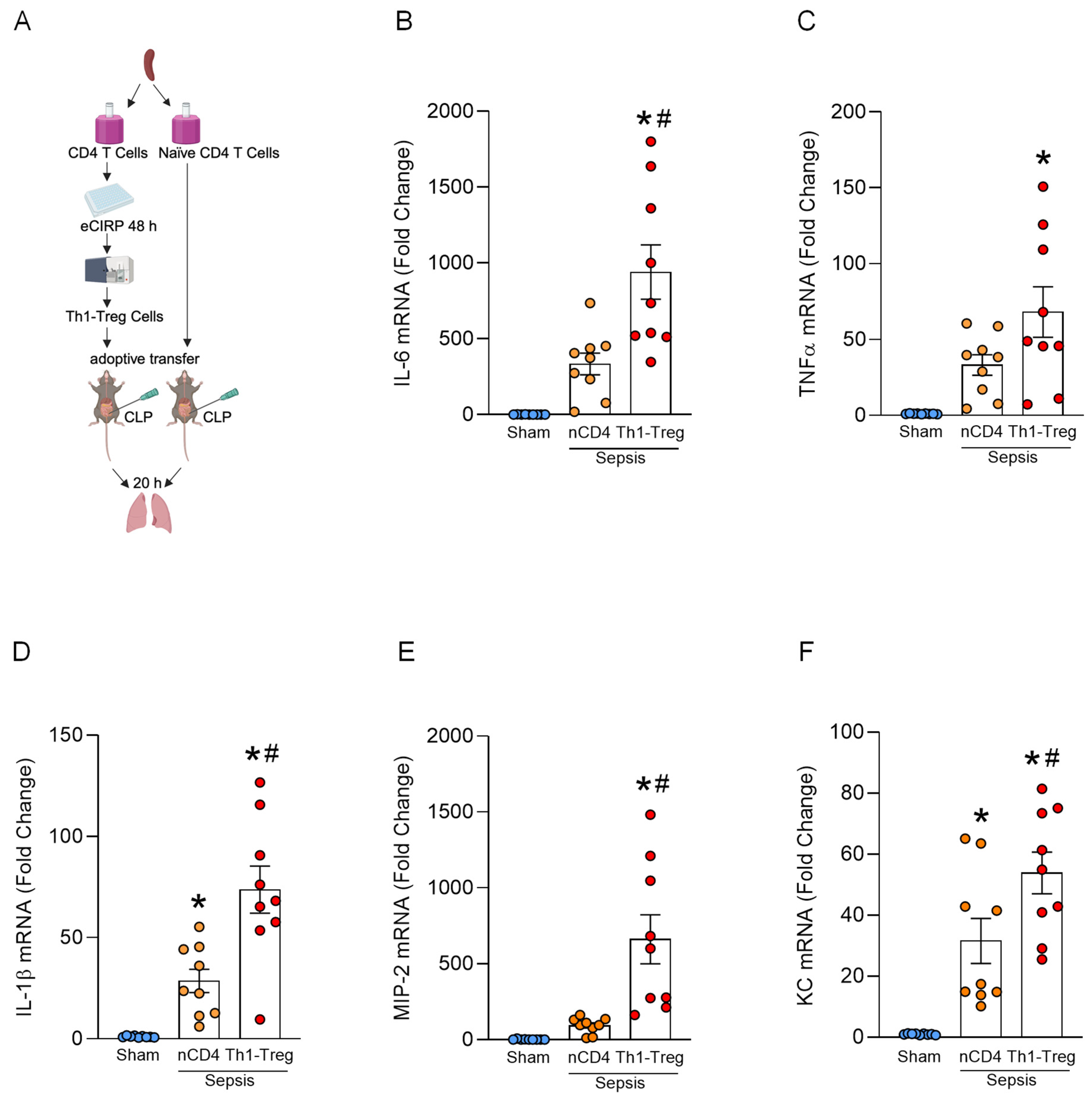
Isolated CD4+ T cells from the WT mice were cultured with 2.5 µg/mL of eCIRP for 48 h in RPMI 1640 medium (Thermo Fisher Scientific), supplemented with 10% FBS, 100 U/mL Penicillin and 100 μg/mL Streptomycin, 1% glutamine and 50 μM 2-mercaptaethanol in a 37 °C incubator under humidified conditions containing 5% CO2, followed by stimulation with Cell Activation Cocktail (without Brefeldin A) (Cat. No.: 423302, Biolegend) for 4 h. Then, Th1-Treg cells, identified by surface CD25 and CXCR3 expression, were sorted by fluorescence-activated cell sorting (FACS) using a BD FACSAria (BD Biosciences). The purity of these sorted Th1-Treg cells consistently exceeded 90% according to FOXP3 and IFNγ expression. The 8–12-week-old male WT mice were subjected to CLP and injected intratracheally (i.t.) with 5 × 10⁵ cells of naïve CD4+ T cells or Th1-Treg cells. Cell viability was confirmed using trypan blue before intratracheal administration. The lungs were harvested 20 h after the surgery.
2.8. Real-Time Quantitative Reverse Transcription PCR
Total RNA was extracted from homogenized lung tissues using TRIzol reagent (Invitrogen, Thermo Fisher Scientific). cDNA was synthesized using murine leukemia virus transcriptase (Biosystems, Barcelona, Spain, Thermo Fisher Scientific). Quantitative Real-Time PCR was performed using gene-specific forward and reverse primers and SYBR Green PCR master mix (Biosystemes) using a StepOnePlusTM real-time PCR machine (Biosystems). The sequences of the primers used are as follows: β-actin, (forward) CGTGAAAAGATGACCCAGATCA, (reverse) TGGTACGACCAGAGGCATACAG; IL-6, (forward) CCGGAGAGGAGACTTCACAG, (reverse) GGAAATTGGGGTAGGAAGGA; TNFα, (forward) AGACCCTCACACTCAGATCATCTTC, (reverse) TTGCTACGACGTGGGCTACA; IL-1β, (forward) CAGGATGAGGACATGAGCACC, (reverse) CTCTGCAGACTCAAACTCCAC; KC, (forward) GCTGGGATTCACCTCAAGAA, (reverse) ACAGGTGCCATCAGAGCAGT; and MIP2, (forward) CCAACCACCAGGCTACAGG, (reverse) GCGTCACACTCAAGCTCTG.
2.9. Lung Myeloperoxidase (MPO) Assay
A total of 50 to 100 mg of lung tissue was pulverized in liquid nitrogen and homogenized in KPO4 buffer containing 0.5% hexadecyltrimethylammonium bromide (Sigma-Aldrich, St. Louis, MO, USA) using a sonicator, with the samples kept on ice. The samples were subjected to 2 freeze/thaw cycles, and the supernatants were collected after centrifugation at 12,000× g for 15 min. The supernatants were diluted in a reaction solution containing O-dianisidine dihydrochloride (Sigma-Aldrich) and H2O2 (Thermo Fisher Scientific) as the substrate. Absorbance was measured at 460 nm, and MPO activity was calculated by determining the rate of change in optical density.
2.10. Lung Histology
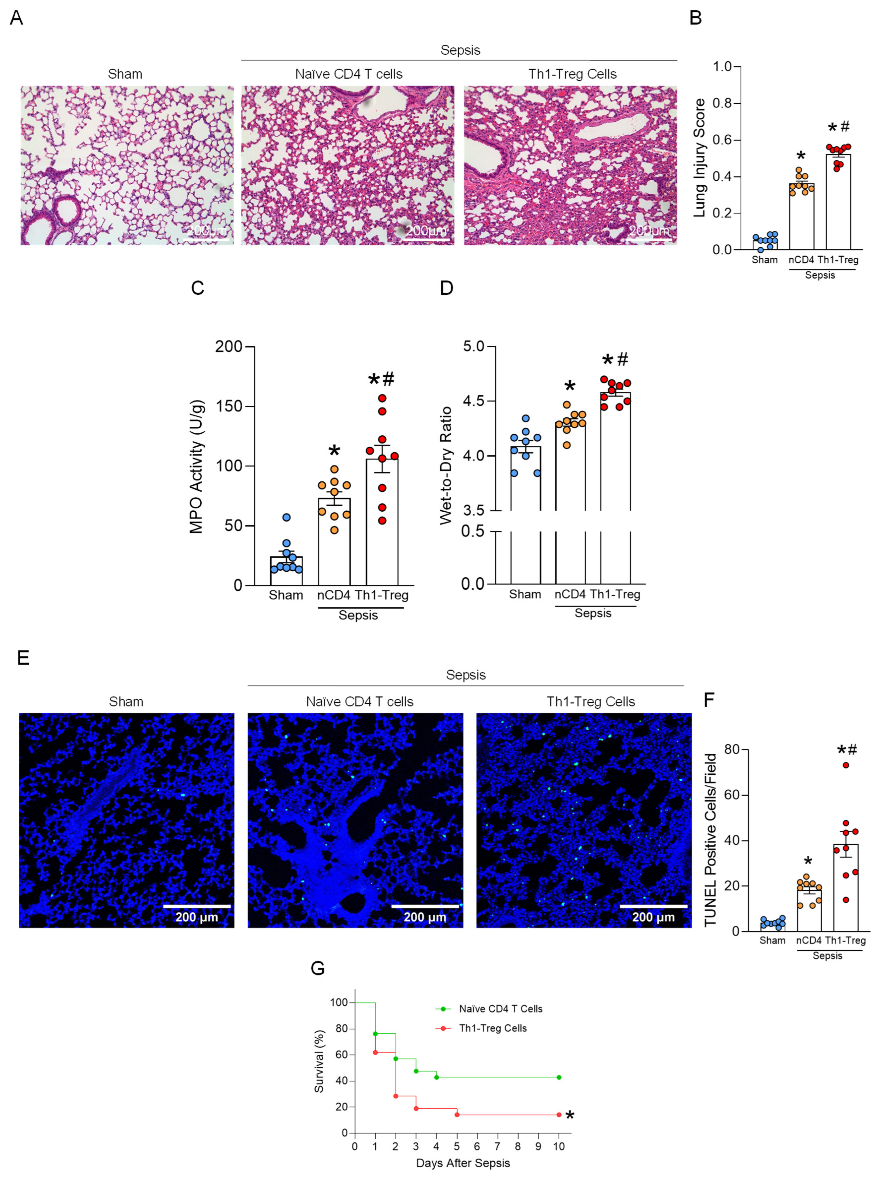
Lung tissues were fixed in 10% formalin and embedded in paraffin. The paraffin-embedded lung tissue blocks were sectioned at 5 μm thickness and mounted on glass slides. The lung tissue sections were stained with hematoxylin and eosin (H&E) and examined using light microscopy. Lung injury scores were assessed using a scoring system established by the American Thoracic Society. Briefly, the scores were evaluated from 0 to 1 based on the presence of neutrophils in the alveolar and interstitial spaces, hyaline membranes, proteinaceous debris in the airspaces, and alveolar septal thickening [
24].
2.11. Terminal Deoxynucleotidyl Transferase dUTP Nick End Labeling (TUNEL) Assay
To evaluate the presence of apoptotic cells in lung tissue, 5 μm lung tissue sections were stained using a commercially available TUNEL assay kit (In Situ Death Detection Kit, Roche Diagnostics, Indianapolis, IN, USA). Paraffin-embedded tissue sections were deparaffinized in xylene, digested with proteinase K, washed, and incubated with an enzyme solution containing terminal deoxynucleotidyl transferase enzyme and fluorescence-labeled nucleotides at 37 °C, according to the manufacturer’s protocol. Nuclei were counterstained with 4′,6-diamidino-2-phenylindole (DAPI, Vectashield Antifade Mounting Media, H-2000, Vector Laboratories, Newark, CA, USA). The slides were imaged using ZEISS LSM 900 confocal microscopy (ZEISS, Oberkochen, Germany). The TUNEL-positive cells in the lung tissue sections were quantified using ImageJ (version 1.54f; Fiji software, National Institute of Health, USA).
2.12. Lung Wet and Dry Ratio
Lung wet and dry weights were measured as previously described [
25]. Briefly, the right lung was harvested and immediately weighed to determine the wet lung weight. The lung samples were then dried at 65 °C for 48 h and weighed again to obtain the dry weight. The wet-to-dry weight ratio was calculated as an indicator of pulmonary edema.
2.13. Statistical Analysis
Data analysis was performed using GraphPad Prism graphing and statistical software (ver 8.0.2; GraphPad Software, LLC, San Diego, CA, USA). The data presented in figures are expressed as mean ± SEM. Comparisons among multiple groups were performed using one-way analysis of variance (ANOVA) followed by Tukey’s multiple comparison test. Comparisons between two groups were performed using an unpaired two-tailed Student’s t-test. Survival rates were analyzed by the Kaplan–Meier estimator and compared with a Log-rank test. A p-value of <0.05 was considered statistically significant for comparisons between experimental groups.
4. Discussion
In the present study, we demonstrate that Th1-Treg cells are significantly increased in the lungs during sepsis and that their accumulation contributes to exacerbated inflammation and enhanced cell death in the lungs and ultimately increased overall mortality. These findings suggest that Th1-Treg cells play a critical role in the dysregulated immune responses that drive lung injury during sepsis, rather than merely being bystanders. In addition, we reveal that, mechanistically, eCIRP, a new DAMP released during sepsis, induces the generation of Th1-Treg cells in septic lungs through TLR4-mediated activation of STAT1 and STAT5. These findings establish Th1-Treg cells as a pathogenic T cell subset in sepsis, linking their presence to organ dysfunction and adverse outcomes. To our knowledge, this is the first study to directly implicate these cells in septic lung injury pathogenesis.
The pathophysiology of sepsis is complex and includes inflammatory imbalance, immune system dysfunction, coagulopathy, and other pathophysiological processes that lead to multiple organ dysfunction [
3]. The immune response to sepsis was classically considered to consist of early pro-inflammatory and later immunosuppressive phases [
28]. In this regard, Th1 cells contribute to tissue injury through the excessive release of pro-inflammatory cytokines such as IFNγ in the state of hyperinflammation [
29], whereas Treg cells contribute to susceptibility to infection by releasing anti-inflammatory cytokines such as IL-10 and TGFβ in the state of immunosuppression [
30]. However, studies have shown that both pro- and anti-inflammatory mediators are elevated from the early stage in sepsis [
31,
32]. These observations suggest that pro-inflammatory and immunosuppressive responses are not strictly sequential but may occur in parallel during disease onset. Therefore, pro-inflammatory and immunosuppressive responses could coordinately contribute to the development of sepsis. Indeed, not only pro-inflammatory mediators, such as IFNγ and IL-6, but also anti-inflammatory cytokines, IL-10 and TGFβ, measured during the acute phase positively correlate with the severity of septic patients [
32,
33]. This indicates the importance of Th1-Treg cells, which may have both pro- and anti-inflammatory phenotypes. In general, pro-inflammatory cells are detrimental in inflammatory disorders but beneficial in cancer [
34,
35,
36]. The multi-faceted property of Th1-Treg cells could be the reason why they exacerbate not only inflammatory disorders, such as sepsis and IBD, but also cancer [
16,
17].
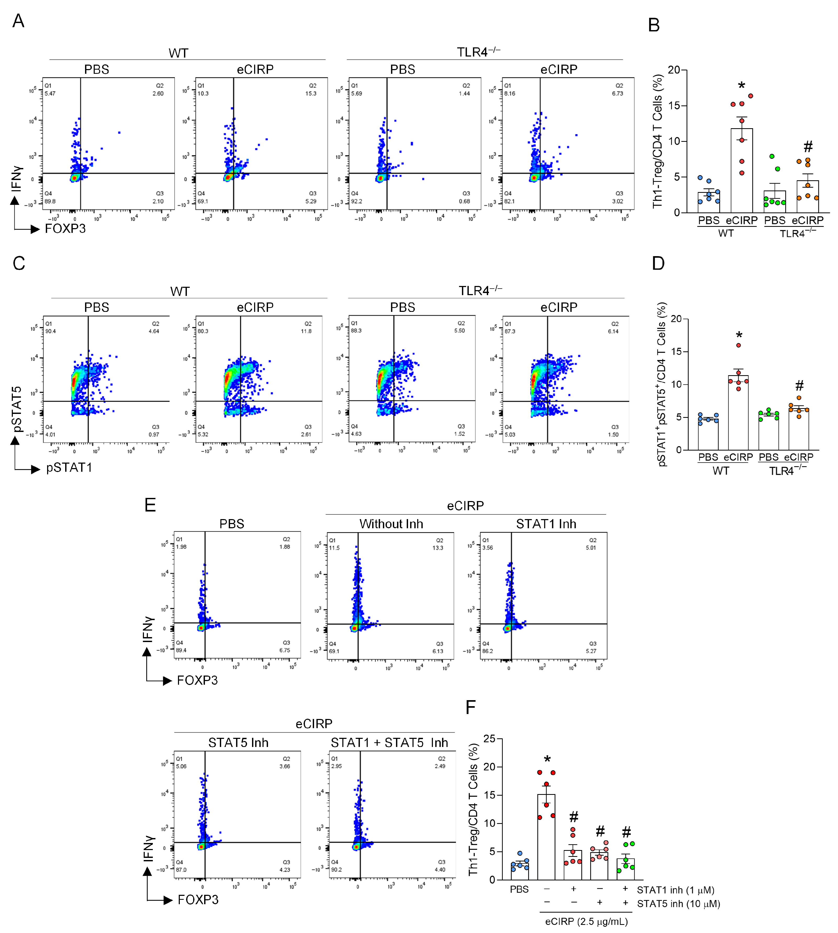
In this study, eCIRP induced Th1-Treg cell differentiation through coordinated activation of STAT1 and STAT5 signaling downstream of TLR4 in CD4
+ T cells. Pharmacological inhibition of either STAT1 or STAT5 equally abrogated the induction of Th1-Treg cells, indicating that both pathways are indispensable for this process. The requirement for both STAT pathways suggests that eCIRP-driven TLR4 activation initiates a STAT1-dependent Th1 program while concurrently promoting STAT5-mediated regulatory features that stabilize a hybrid Th1-Treg phenotype. These findings indicate that activation of both Th1 and Treg signaling within the same CD4
+ T cells resulted in the induction of Th1-Treg cells. Prior studies suggest that Th1-Treg cells are generated from Treg cells by acquiring Th1 phenotypes under disease conditions [
37]. Our study does not rule out the possibility that eCIRP transiently induces Treg cells, which then further transform to Th1-Treg cells. However, while STAT1 is known to be directly activated downstream of TLR4, STAT5 activation is generally mediated indirectly through secondary signaling [
38,
39]. Therefore, in the context of the eCIRP-TLR4 axis, it is possible that following Th1 polarization driven by STAT1 activation, subsequent STAT5 activation promotes the acquisition of regulatory T cell features in these cells. Determining the sequence of STAT1 and STAT5 activations would delineate the detailed mechanism of eCIRP-induced Th1-Treg induction. Moreover, further characterization of the stability and lineage commitment of Th1-Treg cells by assessing Helios and CD127 markers together with epigenetic Foxp3 stability would be informative.
We showed that Th1-Treg cells play an active role in shaping acute lung injury in sepsis. Although we did not determine the exact mechanism by which Th1-Treg cells exacerbate ALI—a limitation of this study—the molecular mechanisms of this tissue injury likely involve multiple aspects, considering their hybrid phenotype. It is plausible that Th1-Treg cells secrete IFNγ and IL-10/TGFβ, as they possess both Th1 and Treg phenotypes. Indeed, Th1-Treg cells have been reported to secrete IFNγ [
16], and our data also demonstrated that IFNγ was elevated intracellularly in Th1-Treg cells. We have previously shown that IFNγ promotes neutrophil extracellular trap (NET) formation and exacerbates pulmonary inflammation and tissue injury during sepsis [
40]. We evaluated ALI in sepsis using the ATS criteria, which include histologic evidence of lung injury, as demonstrated by a lung injury score; an altered alveolar–capillary barrier, as demonstrated by a lung wet-to-dry weight ratio; and an inflammatory response, as demonstrated by elevated levels of pro-inflammatory cytokines and MPO activity [
24]. According to these criteria, we revealed that Th1-Treg cells aggravate ALI in sepsis. Collectively, Th1-Treg cells contribute to lung inflammation and tissue damage during sepsis, potentially through the combined release of pro-inflammatory cytokines, thereby amplifying immune-mediated pulmonary injury.
In our study, we specifically examined the role of eCIRP, a major DAMP in inflammation, in the induction of Th1-Treg cells. However, during sepsis, multiple DAMPs are elevated in the circulation and contribute to the overall immune responses [
41]. Thus, it is possible that other DAMPs capable of activating TLR4 signaling may also participate in the generation of Th1-Treg cells. Our findings indicate that the activation of STAT1 and STAT5 leads to the induction of Th1-Treg cells. We investigated the involvement of STAT proteins in the induction process of Th1-Treg cells, given prior evidence that STAT1 is a key inducer of Th1 differentiation and STAT5 promotes Foxp3 expression [
26,
27]. In addition, other TLR4 downstream signaling pathways, such as NFκB, can be involved in the induction of Th1-Treg cells through secondary signaling. Janus kinase (JAK), another TLR4-mediated signaling, also has the potential to play a role in the induction of Th1-Treg cells, as JAK has been demonstrated to contribute to the activation of STAT proteins [
42,
43]. To determine the direct contribution of Th1-Treg cells to sepsis-induced lung injury, Th1-Treg cells were adoptively transferred into septic lungs. Nevertheless, the cellular origin of the Th1-Treg population under physiological conditions, whether derived from tissue-resident T cells or circulating T cells, was not examined. Future studies employing cell-tracking strategies will be necessary to clarify the origin of these Th1-Treg cells during sepsis.
Adoptive transfer via intratracheal administration has limitations, as it may not completely reflect the body’s natural immune system, potentially causing exaggerated lung tissue damage. However, Th1-Treg cells caused significantly more severe ALI compared to naïve CD4 T cells, even though both cells were injected intratracheally, indicating that Th1-Treg cells’ function is an independent factor in lung tissue damage. In the present study, we investigated the involvement of Th1-Treg cells in sepsis using a CLP model generated to achieve severity approximating the LD50. Although we did not assess less severe CLP models or LPS injection models, it is valuable to explore these conditions in future studies. We used only male mice to reduce internal heterogeneity and experimental variability, as female sex steroids can exert diverse immunomodulatory effects on both humoral and cell-mediated immune responses, potentially necessitating an inhumane number of animals to detect statistical significance [
19]. However, studies using female mice would be valuable in the future. Although we used 8–12-week-old mice, which are considered young adults, future studies including mice of different ages would help to confirm the generality of our findings across a wider age range.
While the present study focused on the lung, which is a particularly vulnerable organ during sepsis, it is important to note that sepsis is a systemic syndrome affecting multiple organs, including the liver and kidneys. Investigating the impact of Th1-Treg cells in these additional organs may provide further insight into their contribution to sepsis-associated organ dysfunction. We used an infectious model of polymicrobial sepsis; however, Th1-Treg cells might be induced in other preclinical models, such as gut ischemia/reperfusion injury, which exhibit elevated levels of circulating eCIRP and are accompanied by organ dysfunction. The role of Th1-Treg cells in other preclinical models can be investigated further in the future.
In summary, the present study demonstrates that eCIRP induces the generation of Th1-Treg cells in the lungs during sepsis through activation of STAT1/5 proteins via TLR4. The accumulation of Th1-Treg cells in the lungs contributes to exacerbated inflammation, enhanced cell death, and ultimately increased mortality, thereby highlighting their pathogenic role in sepsis-induced ALI.