Review

The Interplay Between Ca2+ Homeostasis, Endoplasmic Reticulum Stress, and the Unfolded Protein Response in Human Diseases


DiSIT—Dipartimento di Scienze e Innovazione Tecnologica, University of Piemonte Orientale, Viale Teresa Michel 11, 15121 Alessandria, Italy
*
Author to whom correspondence should be addressed.
Cells 2026, 15(4), 352; https://doi.org/10.3390/cells15040352
Submission received: 9 January 2026 / Revised: 4 February 2026 / Accepted: 13 February 2026 / Published: 15 February 2026
(This article belongs to the Special Issue Regulation of Ca2+ Signals in Human Disease)

Highlights

What are the main findings?
  • The Ca2+-ER Stress–UPR is a pivotal, highly sensitive signaling hub that connects Ca2+ homeostasis within the ER to protein quality control, cell fate, and a variety of downstream pathophysiological responses.
  • The process is bidirectional with a self-amplifying mechanism, such that the result of Ca2+ depletion in the ER, whether attributed to SERCA malfunction, leak channels, or UPR, leads to suppression of PERK-CHOP, a maladaptive component of UPR, which in turn suppresses SERCA, thus entrapping the cell in a Ca2+-depleted, pro-apoptotic state.
What are the implications of the main findings?
  • The Ca2+-ER Stress–UPR system is a common fundamental pathological mechanism that explains several diversified diseases, including neurodegeneration, CVD, and cancer, thus identifying a single, multi-disease target.
  • The targeted therapy should be directed at the modulation of ER Ca2+ homeostasis, e.g., via activators of SERCA, or, alternatively, at the pharmacological redirection of the UPR from the maladaptive, CHOP-mediated phase to the adaptive, pro-survival phase.

Abstract

The maintenance of endoplasmic reticulum (ER) Ca2+ homeostasis is intrinsically linked to the fidelity of protein folding, forming a functional tether that, when disrupted, triggers the Unfolded Protein Response (UPR). This bidirectional axis serves as a critical rheostat for cellular viability, yet its chronic dysregulation underpins the molecular etiology of numerous pathologies, including neurodegeneration, heart failure, and malignant transformation. This review provides a comprehensive interrogation of the Ca2+-ER Stress–UPR network, delineating how primary stress sensors—PERK, IRE1alpha, and ATF6—engage in complex feedback loops that either reinstate equilibrium or commit the cell to apoptosis. We specifically examine the PERK-CHOP-SERCA2b inhibitory circuit as a central driver of persistent Ca2+ depletion and discuss the role of Mitochondria-Associated Membranes (MAMs) in governing lethal Ca2+ transfer. Notably, we move beyond the classical paradigm of CHOP as a terminal apoptotic executioner, incorporating emerging evidence of its context-dependent adaptive functions. By synthesizing mechanistic insights across diverse disease models, this work highlights the transition from adaptive to maladaptive UPR as a universal pathological checkpoint. Ultimately, we evaluate the therapeutic potential of ‘axis-targeted’ interventions, such as SERCA activators and selective UPR modulators, aimed at resolving the underlying Ca2+ signaling defects in ER stress-related disorders.

1. Introduction

The ER is an active membrane network filling the cytoplasm of eukaryotic cells categorized structurally and functionally into the Rough ER, responsible for protein translation and the Smooth ER involved in lipid metabolism and detoxification [1]. Its vast surface area and unique internal environment enable the ER to perform its roles as the cellular site for protein production and quality assurance as well as the principal intracellular Ca2+ store. The operational interaction between these two functions is complex, involving elaborate processes that establish the ER as a crucial component in stress detection and signaling networks that regulate cellular homeostasis and fate [2].

1.1. Interconnected Role of Protein Folding and Ca2+ Signaling

1.1.1. Protein Quality Control and ER Ca2+ Dependence

The lumen of the ER provides an ideal biochemical setting for the folding and post-translational modification of proteins. This is reliant on the presence of Ca2+-dependent chaperone proteins, such as BiP/Grp78 (binding immunoglobulin protein (BiP) also referred to as 78 kDa glucose-regulated protein) and the calnexin/calreticulin network [3].
The activity of these chaperones—responsible for folding and triaging terminally misfolded proteins for Endoplasmic Reticulum-Associated Degradation (ERAD)—is strongly sensitive to luminal Ca2+, which usually falls in the 100- to 500 μM range.

1.1.2. The ER as the Principal Ca2+ Reservoir

The ER maintains a steep gradient of Ca2+ through the active functioning of Sarco/Endoplasmic Reticulum Ca2+-ATPase (SERCA) pumps [4]. The controlled, spatially and temporally precise discharge of this sequestered Ca2+ through Inositol trisphosphate receptors (IP3Rs) and ryanodine receptors (RyRs) produces intracellular cues, for coordinating metabolic activities regulating mitochondrial function through ER-mitochondria junctions or Mitochondria-Associated Membranes (MAMs) and managing gene expression. Any disruption in SERCA activity or Ca2+ channel operation directly impairs chaperone performance. Consequently, it triggers a decrease in protein quality regulation.

1.2. ER Stress and the Unfolded Protein Response Cascade

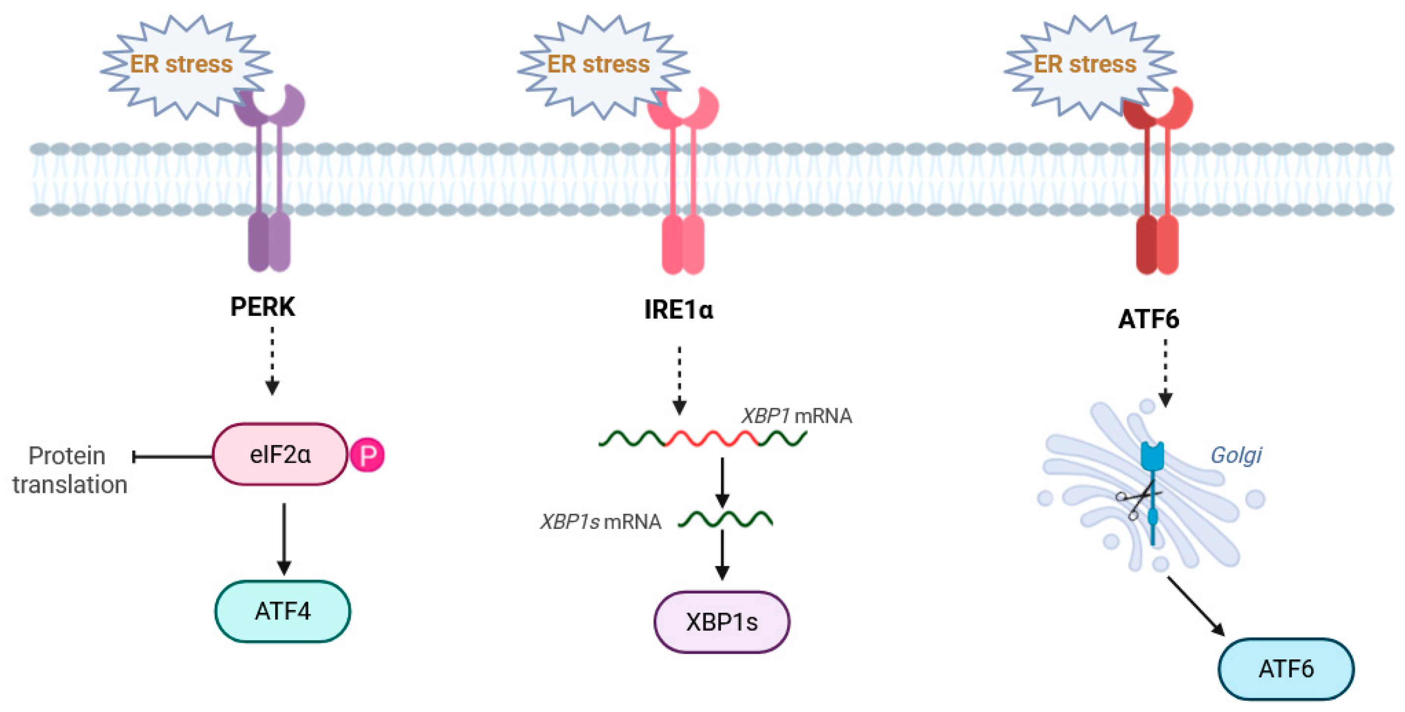
ER stress is typically described as a condition where the ER is overwhelmed by the burden of produced proteins or exposed to harmful stress factors. This situation triggers the Unfolded Protein Response (UPR), a signaling system preserved across multicellular life forms, controlled by three sensors located in the ER [5] (see also Figure 1):
  • PERK (PKR-like ER Kinase) inhibits global translation by phosphorylation of eIF2α but selectively enhances the transcription of the pro-survival factor ATF4 and the pro-apoptotic factor CHOP (C/EBP Homologous Protein).
  • IRE1α, or inositol-requiring enzyme 1α, is an endoribonuclease responsible for the splicing of XBP1 mRNA into its active transcription factor form, XBP1s, which transcriptionally upregulates ERAD components and folding capacity.
  • Activating Transcription Factor 6: ATF6 is cleaved in the Golgi to release an active domain that upregulates ER chaperones.
Although ER stress initially supports survival, prolonged or intense ER stress leads to stimulation of the PERK-ATF4-CHOP pathway, which eventually directs the cell towards apoptosis.

1.3. The Ca2+-ER Stress–UPR Axis in Human Pathophysiology

The tight functional interdependence within the Ca2+-ER Stress–UPR axis establishes it as a convergent pathological checkpoint across diverse disease spectrums. Rather than acting as isolated triggers, Ca2+ dyshomeostasis and protein misfolding form a self-amplifying cycle that dictates cellular fate in neurodegenerative, oncogenic, and cardiovascular contexts [6].

The Ca2+-UPR Axis in Major Human Pathologies

The central nervous system is exquisitely sensitive to ER perturbations, as neuronal signaling and synaptic plasticity rely on the rigorous compartmentalization of Ca2+. In disorders such as Alzheimer’s (AD) and Parkinson’s Disease (PD), the chronic accumulation of proteotoxic aggregates—specifically β-amyloid and α-synuclein—functions as a persistent stressor that destabilizes the ER environment.
Mechanistically, the chronic activation of PERK in neurons leads to the persistent phosphorylation of eIF2α, which causes a global translational shut-off of synaptic proteins. This is compounded by the CHOP-induced downregulation of SERCA2b, which depletes ER Ca2+ stores. The resulting Ca2+ dyshomeostasis triggers the activation of calpains, Ca2+-dependent proteases that further degrade the ER-mitochondria tethering proteins (such as MFN2), leading to mitochondrial fragmentation and an bioenergetic crisis that precedes neuronal death [7].
Clinical evidence suggests that this proteotoxicity is inextricably linked to primary Ca2+ defects; for instance, mutations in Presenilins (PS1/PS2) associated with familial AD do not merely alter proteolytic processing but directly impair SERCA pump efficiency and ER Ca2+ leak dynamics [4]. This chronic store depletion initiates a sustained, low-intensity UPR that eventually recruits the pro-apoptotic PERK-CHOP axis. The resulting synergy between mitochondrial Ca2+ overload, mediated by enhanced efflux through IP3R or Aβ pores, and chronic UPR signaling leads to the synaptic dysfunction and progressive neuronal loss characteristic of these pathologies [8]. In contrast to the degenerative loss of cells, malignancy often involves the co-option of the UPR to favor survival under the hostile conditions of the tumor microenvironment. Cancer cells frequently exhibit a “reprogrammed” Ca2+ metabolism that maintains the UPR in an adaptive phase, thereby avoiding the transition to apoptosis despite high biosynthetic demands, hypoxia, and nutrient deprivation [9].
The IRE1α-XBP1s and ATF4 pathways are typically hijacked to expand ER capacity and promote angiogenesis, while oncogenic adaptations often include the selective upregulation of SERCA isoforms or the modulation of IP3R expression to sustain proliferative signaling [10]. Intriguingly, the role of CHOP in this context appears increasingly complex; while traditionally viewed as a terminal executioner, emerging evidence suggests that transient or non-canonical CHOP signaling may, in specific metabolic landscapes, enhance tumor fitness and therapeutic resistance. Consequently, current pharmacological efforts aim to destabilize this precarious balance by using SERCA inhibitors to force a transition from adaptive survival to CHOP-mediated cell death [11].
In the tumor microenvironment, the IRE1α-XBP1s axis promotes survival by transcriptionally upregulating VEGF, thereby driving angiogenesis to counteract hypoxia. Furthermore, cancer cells often exploit the RIDD (Regulated IRE1-Dependent Decay) activity of IRE1α to selectively degrade mRNAs that encode pro-apoptotic factors, such as caspase-2. This molecular ‘shielding’ prevents the transition to the apoptotic UPR phase, even under conditions of severe proteotoxicity and nutrient deprivation [12].
In the cardiovascular system, where excitation–contraction coupling demands rapid and precise Ca2+ cycling, the ER (or sarcoplasmic reticulum, SR) Stress–UPR axis is a critical determinant of pathogenesis. In heart failure (HF), pathological phosphorylation of RyR channels by PKA or CaMKII induces a chronic “leakage” of Ca2+, which simultaneously impairs contractility and precipitates localized ER stress [13,14,15].
This stress triggers a maladaptive UPR that further depresses SERCA2a expression, creating a vicious cycle that drives myocardial fibrosis and contractile failure.
The initial trigger for UPR activation in HF is the chronic ER Ca2+ depletion caused by mechanical strain and neurohormonal overactivation. At the molecular level, this depletion induces the titration of BiP/GRP78 away from the luminal domains of PERK, IRE1α, and ATF6, as the chaperone is recruited to bind the accumulating misfolded proteins. Once activated, the PERK-ATF4 axis leads to the transcriptional downregulation not only of ATP2A2 (SERCA2a) but also of critical ion channel genes such as HCN4 (Hyperpolarization-Activated Cyclic Nucleotide-Gated channel 4) and KCND3 (potassium voltage-gated channel subfamily D member 3). This broader genomic reprogramming impairs both calcium reuptake and electrical stability, acting as a key driver of the transition to heart failure [16].
A similarly acute manifestation occurs during myocardial infarction; the sudden reperfusion following ischemia-induced energy depletion triggers a massive oxidative burst. This ROS surge inactivates SERCA and promotes exaggerated Ca2+ transfer to the mitochondria via Mitochondria-Associated Membranes (MAMs).
The resulting mitochondrial overload and subsequent opening of the permeability transition pore (mPTP) lead to irreversible cardiomyocyte apoptosis and tissue damage [17,18,19].
In pancreatic β-cells, chronic hyperglycemia induces an overwhelming demand for insulin synthesis, leading to localized ER stress. Mechanistically, this stress activates the PERK-ATF4-CHOP pathway, which increases the expression of TRB3 (Tribbles homolog 3). TRB3 acts as a negative regulator of Akt signaling, effectively blunting insulin sensitivity and promoting β-cell apoptosis. Simultaneously, the excessive Ca2+ efflux from the ER into the cytosol activates the NLRP3 inflammasome, linking ER stress directly to the chronic low-grade inflammation characteristic of Type 2 Diabetes [20].

2. The Ca2+-ER Stress–UPR Axis: An Intricate Molecular Link

The ER lumen is not a passive storage compartment; it is a chemically active environment where the two fundamental processes of protein folding and Ca2+ signaling are fully interrelated [21]. The disruption of one inevitably compromises the other, thus forming the core pathological axis that regulates cellular survival or apoptosis.

2.1. The Essential Role of Ca2+ in Maintaining ER Function

The folding of emerging polypeptides into their functional shapes is a highly coordinated process that relies significantly on a group of ER-resident chaperones, many of which are metalloproteins that need Ca2+, for peak functionality and structural stability [22].
The chaperones Calnexin (CNX) and Calreticulin (CRT) constitute elements of the ER quality control machinery facilitating the folding of N-linked glycoproteins. Their affinity for Ca2+ plays a role: a reduction in luminal Ca2+ levels significantly diminishes their ability to bind and release misfolded proteins effectively decelerating the folding process and causing protein buildup [23].
Serving as the controller of UPR and the most plentiful ER chaperone, the inherent ATPase function of BiP-GRP78 is allosterically influenced by Ca2+. A reduction in Ca2+ alters the shape and stability of BiP weakening its hold on newly formed proteins and UPR sensors (PERK, IRE1α, ATF6) thereby acting as an essential preliminary stage, in triggering UPR [24].
While their role in catalyzing disulfide bond formation largely relies on the redox state, the total folding efficiency facilitated by the Protein Disulfide Isomerases (PDIs) is optimized when ER Ca2+ homeostasis is well-maintained [19].
Therefore, the ER Ca2+ level functions as a rheostat establishing the folding capability of the whole ER network [25].

2.2. Molecular Causes and Consequences of ER Ca2+ Depletion

This essential ER Ca2+ gradient is preserved by an equilibrium between the absorption through the SERCA pumps and controlled discharge through the IP3R and RyR channels. The direct reason for Ca2+ depletion-triggered ER stress is abnormalities that disrupt this fragile balance [26].

2.2.1. Dysfunction of SERCA Pumps

SERCA pumps are very sensitive to pathological insults [27], which results in impaired Ca2+ reuptake.
SERCA is abundant in reactive cysteine residues [28]. Elevated oxidative stress conditions, such as those seen in ischemia–reperfusion injury or chronic inflammation, cause S-nitrosylation or oxidation of these cysteine sites resulting in the loss of function and the pump’s failure to move Ca2+ against the electrochemical gradient [4].
Chronic ER stress and the subsequent activation of the UPR typically trigger a maladaptive signaling cascade, leading to the selective downregulation of SERCA2b transcription [29]. This repression is mediated either by the induction of the pro-apoptotic factor CHOP or through the action of specific microRNAs, such as miR-25 and miR-429, which target the SERCA2b transcript for post-transcriptional silencing [30]. Consequently, a self-perpetuating cycle of luminal Ca2+ depletion and persistent ER stress is established.

2.2.2. Hyperactivity and Leakage of Ca2+ Release Channels

The major mechanism of depletion, especially in excitable cells, involves chronic leakage of Ca2+ [31].
Numerous disease-related signaling routes increase the responsiveness of Inositol trisphosphate receptors (IP3Rs) to IP3 concentrations causing unregulated Ca2+ puffing and sparking that exhausts the stores. IRE1α is also reported to degrade IP3R mRNAs through its RNase function though the resulting effects are intricate and vary depending on the context [32].
The molecular mechanism linking UPR to increased Ca2+ leak involves the induction of the pro-oxidant enzyme ERO1α via the PERK-CHOP pathway [33]. ERO1α-mediated oxidative stress leads to the S-nitrosylation of RyR2 cysteine residues. This post-translational modification causes the dissociation of the stabilizing protein FKBP12.6 (calstabin2) from the RyR2 complex, resulting in a ‘leaky’ channel. The subsequent diastolic Ca2+ leak into the cytosol creates a mitochondrial Ca2+ overload at the MAMs, which stimulates the electron transport chain to produce excessive ROS, thereby establishing a self-amplifying loop of oxidative damage and ER dysfunction.
The bridge between ER stress and ROS production lies at the MAMs. The Ca2+ leaked from the ER via oxidized RyR2 is directly funneled into the mitochondrial matrix through the Mitochondrial Calcium Uniporter (MCU). This mitochondrial Ca2+ overload overstimulates the Krebs cycle and the electron transport chain, leading to a burst of superoxide production. These mitochondrial ROS then diffuse back to the ER, further oxidizing RyR2 thiol groups and exacerbating the Ca2+ leak in a self-perpetuating loop [32].

2.3. The Molecular Interplay at MAMs: ER–Mitochondria Crosstalk

A critical locus for the Ca2+-ER Stress–UPR axis is provided by the Mitochondria-Associated Membranes (MAMs). These constitute micro-regions where the ER and mitochondria are physically connected forming confined zones, for rapid tightly localized Ca2+ exchange [34].
On MAMs, the IP3R channels on the ER membrane are in close proximity (∼10−80 nm) to the Voltage-Dependent Anion Channel on the outer mitochondrial membrane. This spatial coupling enables the high concentration of Ca2+ released from the ER to be taken up rapidly by the mitochondrial calcium uniporter [35]. This transport is crucial for activating TCA cycle enzymes and controlling ATP production.
Pathological ER stress is often correlated with a dramatic alteration in MAM structure and function [36]. Prolonged ER stress boosts the connection between ER and mitochondria (closer contact) causing excessive mitochondrial Ca2+ absorption. This Ca2+ surplus induces depolarization, elevated ROS generation and eventually activation of the intrinsic apoptotic pathway (release of cytochrome c). The ROS produced by the burdened mitochondria subsequently oxidizes SERCA and the lipids in the ER membrane forming a deadly feedback cycle of ER stress [37].

2.4. Adaptive vs. Apoptotic UPR: The CHOP Switch

Since the determination of fate is closely associated with the equilibrium between the adaptive and pro-apoptotic pathways of the UPR this signifies a shift that is greatly reliant on the intensity and duration of the Ca2+ depletion [38].
Mild, short-term ER stress promotes the immediate effects of UPR: global translational attenuation (PERK-eIF2α), increased folding capacity (ATF6), and enhanced degradation (IRE1α-XBP1s). These effects attempt to restore Ca2+ homeostasis indirectly by reducing protein load [39].
When ER stress persists, commonly indicated by Ca2+ depletion and issues, with MAMs the UPR shifts its role. Continuous PERK activation induces the production of CHOP or C/EBP Homologous Protein, known as the effector molecule of the UPR. CHOP facilitates apoptosis by increasing the pro-apoptotic Bcl−2 family proteins (Bim) and decreasing the anti-apoptotic proteins (e.g., Bcl−2) [40].
However, the traditional view of CHOP as a terminal pro-apoptotic executioner has been recently challenged by evidence suggesting a more nuanced, context-dependent role. While chronic induction remains a hallmark of cell death, transient or moderate CHOP expression might contribute to adaptive responses by modulating metabolic genes or facilitating the autophagy-mediated clearance of protein aggregates. This ‘dual-role’ hypothesis suggests that the biological output of CHOP signaling is not binary but is rather fine-tuned by its temporal dynamics and the specific stress landscape of the cell [41].
Additionally, CHOP reduces SERCA2b expression intensifying ER stress and thus reinforcing the condition of Ca2+ deficiency [4].

2.5. ER Stress and Ca2+ Signaling in Disease

The delicate Ca2+-dependent nature of neuronal and cardiac functions renders these tissues exquisitely sensitive to ER homeostatic perturbations [42].
In neurodegenerative disorders such as AD and PD, the chronic accumulation of misfolded proteins does not merely trigger the UPR but exploits the aforementioned PERK-CHOP-SERCA inhibitory loop to lock neurons in a state of persistent Ca2+ depletion [43].
For instance, mutations in Presenilins linked to familial AD impair ER Ca2+ leak, causing a low-level but constitutive UPR activation that eventually collapses the organelle’s folding capacity [44].
Similarly, in heart failure, the pathological phosphorylation of RyR leads to a ‘leaky’ state; this chronic Ca2+ efflux induces localized ER stress, which, via CHOP-mediated repression of SERCA2a, compromises contractile recovery and facilitates the transition to failure [45,46].
Conversely, in the context of malignancy, tumor cells often bypass the apoptotic threshold of this axis. By upregulating specific SERCA isoforms or modulating MAM-tethering proteins, cancer cells harness the adaptive branches of the UPR (IRE1alpha-XBP1s) to support high secretory demands and promote angiogenesis [47].
Understanding these tissue-specific exploitations of the Ca2+-UPR axis is crucial for developing targeted interventions that can either reinstate homeostasis in degenerating cells or force an apoptotic switch in resistant tumors [48,49].

3. Feedback Signaling and Crosstalk: UPR Regulation of Ca2+ Homeostasis

The UPR does not function as a one-way signaling route triggered by Ca2+ depletion; instead, it swiftly forms intricate feedback circuits aiming to recover ER Ca2+ concentrations or, if this fails, deliberately intensifies the depletion to induce cell death. The two-way interaction between UPR sensors and Ca2+ regulation systems is essential for determining cell destiny.

3.1. IRE1α: Modulating ER Biogenesis and Ca2+ Channels

The IRE1α branch, through the active transcription factor XBP1s, exerts powerful control over ER Ca2+ homeostasis.
XBP1s is a powerful transcriptional driver of genes involved in ER membrane synthesis, including components of lipid metabolism [50]. XBP1s acts as a transcriptional regulator for genes linked to ER membrane production encompassing elements of lipid metabolism [50]. By enlarging the ER size XBP1s boosts the protein folding capacity, notably elevating the production of SERCA isoforms (SERCA2b) that facilitate Ca2+ reabsorption. This serves as an adaptive response meant to directly mitigate Ca2+ loss [50].
Regulated beyond splicing XBP1 mRNA, the IRE1α endoribonuclease activity can cleave and degrade other mRNAs, a process termed Regulated IRE1-Dependent Decay (RIDD) [51]. Among the RIDD targets are mRNAs encoding ER membrane proteins, including some SERCA regulators and potentially components of the IP3R channels [52]. The breakdown of proteins that regulate SERCA would effectively block Ca2+ reuptake thereby allowing the UPR to shift from a phase of adaptation to a pro-apoptotic pathway by hindering Ca2+ restoration.

3.2. PERK: The SERCA Repressor and MAM Mediator

The PERK pathway offers the molecular connection between UPR activation and the ultimate disruption of Ca2+ balance [53].
Sustained activation of PERK drives the expression of the pro-apoptotic factor CHOP [54]. CHOP directly acts as a negative regulator of the SERCA2b promoter, thereby repressing the transcription and synthesis of SERCA. This transcriptional repression decreases the cellular capability to pump Ca2+ into the ER and solidifies its depleted state, ensuring the continuance of the ER stress condition, which is a required step for UPR-induced apoptosis.
PERK activation also plays a role in regulating physical tethering between the ER and mitochondria at the MAMs [55]. Persistent PERK signaling enhances the development and maintenance of MAMs resulting in prolonged mitochondrial Ca2+ absorption. This overload of Ca2+ is extremely harmful and triggers Mitochondrial Outer Membrane Permeabilization (MOMP) along with the release of pro-apoptotic molecules such as cytochrome c [56].

3.3. The ATF6 Branch and Ca2+-Dependent Refolding

The ATF6 pathway primarily serves as a mechanism designed to specifically alleviate the stress triggered by Ca2+ depletion.
The functional N-terminal segment of ATF6 moves into the nucleus to trigger the expression of ER chaperones, such as BiP and GRP94. This surge of Ca2+-dependent folding assistants aims to boost the ER’s ability to manage misfolded proteins despite reduced Ca2+ levels, simultaneously allowing time for SERCA function to be restored.

3.4. Ca2+ Signaling as a Regulator of UPR Output

One may note that the crosstalk is not unidirectional. Altered ER Ca2+ levels may also, in a manner independent of BiP dissociation, directly modulate both the activity and output of UPR sensors.
STIM1, the main ER Ca2+ sensor that regulates Store-Operated Calcium Entry (SOCE), physically interacts with the IRE1α luminal domain [57]. Upon ER Ca2+ depletion, the clustering of STIM1 not only triggers SOCE but also activates and induces the clustering of IRE1α, indicating that Ca2+ signaling directly regulates the IRE1α dimerization and RNase activity [58].
Low Ca2+ generally activates the UPR, but various reports suggest that the IRE1α RNase domain requires specific local Ca2+ concentrations for its maximum activity, indicating that the precise spatial and temporal dynamics of Ca2+ release can tune the specific output of the UPR branches, favoring, for example, XBP1 splicing over RIDD [59].
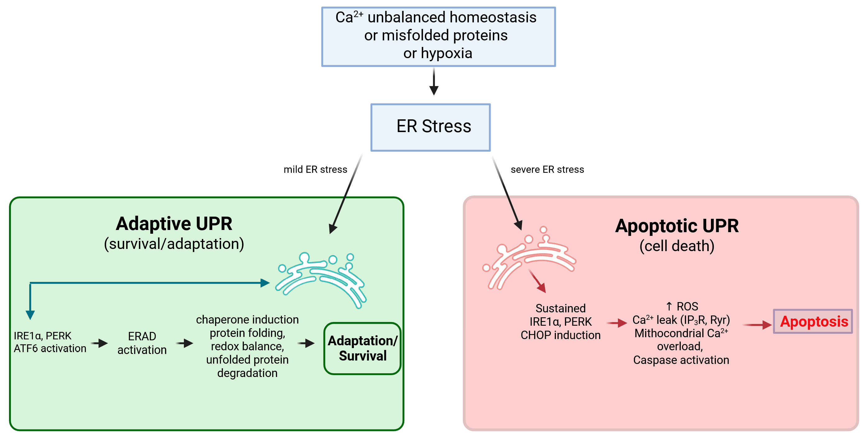
In summary, the UPR is a high-powered signal processor that, in response to Ca2+-mediated stress, strives to reinstate homeostasis but contains robust negative feedback mechanisms, such as the CHOP-SERCA loop, that lock the cell into a pathological, Ca2+-depleted, pro-apoptotic state if the stress is unresolved [41] (see also Figure 2).

3.5. Pathological Implications of UPR-Mediated Ca2+ Dysregulation

The key shift from recovery to pro-apoptotic cell death is often determined by whether the UPR can successfully reestablish ER homeostasis. In significant human diseases the harmful pathway often forms when the UPR positive feedback loops (such as CHOP inhibiting SERCA) remain persistently active, surpassing the original adaptive reactions.
In advancing diseases like AD and PD the continuous existence of misfolded protein accumulations (β-amyloid, α-synuclein) causes sustained mild ER stress. Neurons experiencing this stress frequently show ongoing activation of the PERK signaling pathway. The induced production of CHOP creates a self-sustaining loop of injury: CHOP inhibits the transcription of SERCA2b leading to a lasting reduction, in the neuron’s capacity to replenish its ER Ca2+ reservoirs [60].
This Ca2+ depletion inhibits ER chaperones guaranteeing continued protein misfolding while also preparing the neuron for apoptosis. The inability of the UPR to restore Ca2+ balance is regarded as a mechanism underlying the ongoing neuronal degeneration typical of these disorders.
In multiplying cancer cells, the adaptive arm of the UPR is frequently hijacked to promote unchecked proliferation, involving the adjustment of Ca2+ regulation to aid survival within the challenging tumor microenvironment.
The IRE1alpha/XBP1s axis is frequently hyperactivated in highly aggressive tumors, such as multiple myeloma and triple-negative breast cancer [61]. In addition to enhancing protein folding ability XBP1s promotes the expression of SERCA and lipid synthesis genes aiding the cancer cell in enlarging its ER volume and preserving membrane stability essential, for meeting the biosynthetic needs of rapid proliferation.
This XBP1s-driven maintenance of ER capacity allows the cancer cell to buffer the Ca2+ signaling necessary for cell cycle progression while avoiding the switch to CHOP-mediated apoptosis. Targeting IRE1alpha/XBP1s has become a therapeutic strategy aimed at destabilizing ER Ca2+ balance and pushing the tumor cell towards death.
In conditions of severe metabolic compromise like myocardial ischemia, the tight regulatory control of the UPR over Ca2+ flux at the MAMs becomes highly pathogenic [62].
Acute stress, such as ischemia–reperfusion injury, strongly activates the PERK pathway [63]. Persistent PERK signaling has been shown to increase the physical tethering and stability of the MAMs, leading to an exaggerated proximity between ER IP3R and mitochondrial Ca2+ uptake machinery [64].
This enhanced ER-to-mitochondria coupling, driven by UPR signaling, results in a fatal mitochondrial Ca2+ overload. This overload directly triggers MOMP and the release of cytochrome c, causing massive cardiomyocyte apoptosis and contributing significantly to the irreversible damage following a heart attack [65]. The UPR’s attempt to compartmentalize and manage stress via the MAMs thus ironically serves as a conduit for cell death.

4. Targeting the Ca2+-ER Stress–UPR Axis: Therapeutic Implications

Given the disease-causing involvement of the Ca2+-ER Stress–UPR pathway across a broad range of conditions including neurodegenerative disorders, cardiovascular diseases and cancer, this signaling route has emerged as an attractive target for new treatment approaches [66,67]. The goal would be to redirect the UPR from its harmful/apoptotic stage back to its protective/pro-survival phase or to reestablish normal ER Ca2+ balance. Re-establishing the balance between Ca2+ uptake and release is the most direct means of relieving ER stress [68]. Small-molecule enhancers designed to boost the pump function of SERCA are being developed, such as those aimed at SERCA2a in heart failure. By enhancing Ca2+ uptake these agents replenish the Ca2+ reservoir thereby reactivating Ca2+-dependent chaperones and ultimately addressing the ER stress signal at its origin [69].
Because excessive MAM tethering promotes lethal mitochondrial Ca2+ overload, the disruption of these physical tethers—for example, by targeting the protein complexes such as Miro/Mitofusin2 that mediate the ER—mitochondria contact—can selectively block the pro-apoptotic ROS-amplification loop without globally affecting either ER or mitochondrial function [70].
Inhibitors targeting the IRE1α RNase domain block splicing of XBP1 and RIDD is an approach particularly relevant in malignancies where the product of XBP1s is actively hijacked to support tumor survival (e.g., multiple myeloma). By blocking XBP1s, these agents aim to strip the tumor cells of their adaptive survival mechanism [71].
In certain settings, PERK inhibitors must be used together with XBP1s inhibitors to completely suppress the UPR. A more subtle approach is to enhance the adaptive side while suppressing the maladaptive side; for example, activating ATF6 while inhibiting PERK [72].
The Ca2+-ER Stress–UPR axis offers various points of intervention, beyond general anti-apoptotic or anti-inflammatory drugs. The basis of highly selective therapies will come via understanding the molecular feedback loops that display either a positive or negative influence of Ca2+ on UPR and vice versa. Future treatments for neurodegeneration, cardiovascular failure, and certain cancers may very well rely on molecules that, through pharmacological means, restore ER proteostasis [73,74] by either directly correcting the Ca2+ balance or shifting UPR back toward an adaptive, sustainable response.

5. Conclusions

The evidence synthesized in this review establishes the Ca2+-ER Stress–UPR axis as a critical and bidirectional signaling node. The fundamental conclusion is that the crosstalk between ER Ca2+ dynamics and the UPR is self-amplifying: while Ca2+ depletion acts as a primary trigger for stress, the subsequent maladaptive UPR reinforces this depletion through transcriptional and post-translational feedback loops.
This axis represents a convergent pathological hallmark across neurodegeneration, cardiovascular diseases, and cancer. The transition from an adaptive to a pro-apoptotic UPR serves as a universal checkpoint, often governed by the duration of Ca2+ dyshomeostasis and the stability of ER-mitochondria contacts at the MAMs. Consequently, pharmacological efforts should move beyond broad UPR inhibition. Future therapeutic success likely resides in the development of ‘rheostat-modulators’—such as SERCA activators or selective CHOP-pathway inhibitors—capable of diverting the UPR from its terminal apoptotic phase toward a sustainable, homeostatic response. Resolving the root Ca2+ signaling defect, rather than merely mitigating downstream symptoms, remains the most promising frontier for treating ER stress-related pathologies.

Author Contributions

Conceptualization, E.R. and S.M.; writing—review and editing, E.R. and S.M. All authors have read and agreed to the published version of the manuscript.

Funding

SM was supported by project funding from NextGeneration EU and Compagnia di San Paolo.

Institutional Review Board Statement

Not applicable.

Informed Consent Statement

Not applicable.

Data Availability Statement

No new data were created or analyzed in this study.

Conflicts of Interest

The authors declare no conflicts of interest.

References

  1. Martinotti, S.; Ranzato, E. Targeting the Unfolded Protein Response with Natural Products: Therapeutic Potential in ER Stress-Related Diseases. Int. J. Mol. Sci. 2025, 26, 8814. [Google Scholar] [CrossRef] [PubMed]
  2. Feliziani, C.; Fernandez, M.; Quassollo, G.; Holstein, D.; Bairo, S.M.; Paton, J.C.; Paton, A.W.; de Batista, J.; Lechleiter, J.D.; Bollo, M. Ca2+ signalling system initiated by endoplasmic reticulum stress stimulates PERK activation. Cell Calcium 2022, 106, 102622. [Google Scholar] [CrossRef]
  3. Ibrahim, I.M.; Abdelmalek, D.H.; Elfiky, A.A. GRP78: A cell’s response to stress. Life Sci. 2019, 226, 156–163. [Google Scholar] [CrossRef]
  4. Chemaly, E.R.; Troncone, L.; Lebeche, D. SERCA control of cell death and survival. Cell Calcium 2018, 69, 46–61. [Google Scholar] [CrossRef]
  5. Schwarz, D.S.; Blower, M.D. The endoplasmic reticulum: Structure, function and response to cellular signaling. Cell. Mol. Life Sci. 2016, 73, 79–94. [Google Scholar] [CrossRef]
  6. Makio, T.; Chen, J.; Simmen, T. ER stress as a sentinel mechanism for ER Ca. Cell Calcium 2024, 124, 102961. [Google Scholar] [CrossRef]
  7. Gow, A.; Wrabetz, L. CHOP and the endoplasmic reticulum stress response in myelinating glia. Curr. Opin. Neurobiol. 2009, 19, 505–510. [Google Scholar] [CrossRef] [PubMed]
  8. Hetz, C.; Saxena, S. ER stress and the unfolded protein response in neurodegeneration. Nat. Rev. Neurol. 2017, 13, 477–491. [Google Scholar] [CrossRef] [PubMed]
  9. Bonsignore, G.; Martinotti, S.; Ranzato, E. Endoplasmic Reticulum Stress and Cancer: Could Unfolded Protein Response Be a Druggable Target for Cancer Therapy? Int. J. Mol. Sci. 2023, 24, 1566. [Google Scholar] [CrossRef]
  10. Green, K.N.; Demuro, A.; Akbari, Y.; Hitt, B.D.; Smith, I.F.; Parker, I.; LaFerla, F.M. SERCA pump activity is physiologically regulated by presenilin and regulates amyloid beta production. J. Cell Biol. 2008, 181, 1107–1116. [Google Scholar] [CrossRef]
  11. Rozpedek, W.; Pytel, D.; Mucha, B.; Leszczynska, H.; Diehl, J.A.; Majsterek, I. The Role of the PERK/eIF2α/ATF4/CHOP Signaling Pathway in Tumor Progression During Endoplasmic Reticulum Stress. Curr. Mol. Med. 2016, 16, 533–544. [Google Scholar] [CrossRef]
  12. Cubillos-Ruiz, J.R.; Bettigole, S.E.; Glimcher, L.H. Tumorigenic and Immunosuppressive Effects of Endoplasmic Reticulum Stress in Cancer. Cell 2017, 168, 692–706. [Google Scholar] [CrossRef]
  13. An, Y.; Wang, X.; Guan, X.; Yuan, P.; Liu, Y.; Wei, L.; Wang, F.; Qi, X. Endoplasmic reticulum stress-mediated cell death in cardiovascular disease. Cell Stress Chaperones 2024, 29, 158–174. [Google Scholar] [CrossRef]
  14. Ruan, Y.; Zeng, J.; Jin, Q.; Chu, M.; Ji, K.; Wang, Z.; Li, L. Endoplasmic reticulum stress serves an important role in cardiac ischemia/reperfusion injury (Review). Exp. Ther. Med. 2020, 20, 268. [Google Scholar] [CrossRef] [PubMed]
  15. Li, X.; Zhao, X.; Qin, Z.; Li, J.; Sun, B.; Liu, L. Regulation of calcium homeostasis in endoplasmic reticulum-mitochondria crosstalk: Implications for skeletal muscle atrophy. Cell Commun. Signal. 2025, 23, 17. [Google Scholar] [CrossRef] [PubMed]
  16. Zhao, X.; Zhang, D.Q.; Song, R.; Wang, R.; Zhang, G. The clinical significance of circulating glucose-regulated protein 78, Caspase-3, and C/EBP homologous protein levels in patients with heart failure. Heliyon 2023, 9, e13436. [Google Scholar] [CrossRef]
  17. Ni, L.; Yang, L.; Lin, Y. Recent progress of endoplasmic reticulum stress in the mechanism of atherosclerosis. Front. Cardiovasc. Med. 2024, 11, 1413441. [Google Scholar] [CrossRef]
  18. Yao, S.; Miao, C.; Tian, H.; Sang, H.; Yang, N.; Jiao, P.; Han, J.; Zong, C.; Qin, S. Endoplasmic reticulum stress promotes macrophage-derived foam cell formation by up-regulating cluster of differentiation 36 (CD36) expression. J. Biol. Chem. 2014, 289, 4032–4042. [Google Scholar] [CrossRef] [PubMed]
  19. Wang, L.; Wang, C.C. Oxidative protein folding fidelity and redoxtasis in the endoplasmic reticulum. Trends Biochem. Sci. 2023, 48, 40–52. [Google Scholar] [CrossRef]
  20. Fonseca, S.G.; Gromada, J.; Urano, F. Endoplasmic reticulum stress and pancreatic β-cell death. Trends Endocrinol. Metab. 2011, 22, 266–274. [Google Scholar] [CrossRef]
  21. Walter, P.; Ron, D. The unfolded protein response: From stress pathway to homeostatic regulation. Science 2011, 334, 1081–1086. [Google Scholar] [CrossRef]
  22. Ellgaard, L.; Sevier, C.S.; Bulleid, N.J. How Are Proteins Reduced in the Endoplasmic Reticulum? Trends Biochem. Sci. 2018, 43, 32–43. [Google Scholar] [CrossRef]
  23. Melnyk, A.; Rieger, H.; Zimmermann, R. Co-chaperones of the mammalian endoplasmic reticulum. Subcell. Biochem. 2015, 78, 179–200. [Google Scholar] [CrossRef]
  24. Akinyemi, A.O.; Simpson, K.E.; Oyelere, S.F.; Nur, M.; Ngule, C.M.; Owoyemi, B.C.D.; Ayarick, V.A.; Oyelami, F.F.; Obaleye, O.; Esoe, D.P.; et al. Unveiling the dark side of glucose-regulated protein 78 (GRP78) in cancers and other human pathology: A systematic review. Mol. Med. 2023, 29, 112. [Google Scholar] [CrossRef]
  25. Acosta-Alvear, D.; Harnoss, J.M.; Walter, P.; Ashkenazi, A. Homeostasis control in health and disease by the unfolded protein response. Nat. Rev. Mol. Cell Biol. 2025, 26, 193–212. [Google Scholar] [CrossRef] [PubMed]
  26. Bergner, A.; Kellner, J.; Tufman, A.; Huber, R.M. Endoplasmic reticulum Ca2+-homeostasis is altered in Small and non-small Cell Lung Cancer cell lines. J. Exp. Clin. Cancer Res. 2009, 28, 25. [Google Scholar] [CrossRef]
  27. Hovnanian, A. SERCA pumps and human diseases. Subcell. Biochem. 2007, 45, 337–363. [Google Scholar] [CrossRef] [PubMed]
  28. Tong, X.; Evangelista, A.; Cohen, R.A. Targeting the redox regulation of SERCA in vascular physiology and disease. Curr. Opin. Pharmacol. 2010, 10, 133–138. [Google Scholar] [CrossRef]
  29. Yadav, V.; Nayak, S.; Guin, S.; Mishra, A. Impact of Oxidative Stress and Neuroinflammation on Sarco/Endoplasmic Reticulum Ca. ACS Pharmacol. Transl. Sci. 2025, 8, 173–188. [Google Scholar] [CrossRef]
  30. Çiftçi, Y.C.; Yurtsever, Y.; Akgül, B. Long non-coding RNA-mediated modulation of endoplasmic reticulum stress under pathological conditions. J. Cell. Mol. Med. 2024, 28, e18561. [Google Scholar] [CrossRef] [PubMed]
  31. Boyden, P.A.; Smith, G.L. Ca2+ leak—What is it? Why should we care? Can it be managed? Heart Rhythm. 2018, 15, 607–614. [Google Scholar] [CrossRef]
  32. Ong, G.; Logue, S.E. Unfolding the Interactions between Endoplasmic Reticulum Stress and Oxidative Stress. Antioxidants 2023, 12, 981. [Google Scholar] [CrossRef]
  33. Respress, J.L.; van Oort, R.J.; Li, N.; Rolim, N.; Dixit, S.S.; deAlmeida, A.; Voigt, N.; Lawrence, W.S.; Skapura, D.G.; Skårdal, K.; et al. Role of RyR2 phosphorylation at S2814 during heart failure progression. Circ. Res. 2012, 110, 1474–1483. [Google Scholar] [CrossRef] [PubMed]
  34. Giorgi, C.; Missiroli, S.; Patergnani, S.; Duszynski, J.; Wieckowski, M.R.; Pinton, P. Mitochondria-associated membranes: Composition, molecular mechanisms, and physiopathological implications. Antioxid. Redox Signal. 2015, 22, 995–1019. [Google Scholar] [CrossRef]
  35. Wu, S.; Zou, M.H. Mitochondria-associated endoplasmic reticulum membranes in the heart. Arch. Biochem. Biophys. 2019, 662, 201–212. [Google Scholar] [CrossRef] [PubMed]
  36. Chen, X.; Shi, C.; He, M.; Xiong, S.; Xia, X. Endoplasmic reticulum stress: Molecular mechanism and therapeutic targets. Signal Transduct. Target. Ther. 2023, 8, 352. [Google Scholar] [CrossRef]
  37. Zeeshan, H.M.; Lee, G.H.; Kim, H.R.; Chae, H.J. Endoplasmic Reticulum Stress and Associated ROS. Int. J. Mol. Sci. 2016, 17, 327. [Google Scholar] [CrossRef]
  38. Mekahli, D.; Bultynck, G.; Parys, J.B.; De Smedt, H.; Missiaen, L. Endoplasmic-reticulum calcium depletion and disease. Cold Spring Harb. Perspect. Biol. 2011, 3, a004317. [Google Scholar] [CrossRef]
  39. Groenendyk, J.; Michalak, M. Interplay between calcium and endoplasmic reticulum stress. Cell Calcium 2023, 113, 102753. [Google Scholar] [CrossRef] [PubMed]
  40. Li, Y.; Guo, Y.; Tang, J.; Jiang, J.; Chen, Z. New insights into the roles of CHOP-induced apoptosis in ER stress. Acta Biochim. Biophys. Sin. 2014, 46, 629–640. [Google Scholar] [CrossRef]
  41. Liu, K.; Zhao, C.; Adajar, R.C.; DeZwaan-McCabe, D.; Rutkowski, D.T. A beneficial adaptive role for CHOP in driving cell fate selection during ER stress. EMBO Rep. 2024, 25, 228–253. [Google Scholar] [CrossRef]
  42. Sprenkle, N.T.; Sims, S.G.; Sánchez, C.L.; Meares, G.P. Endoplasmic reticulum stress and inflammation in the central nervous system. Mol. Neurodegener. 2017, 12, 42. [Google Scholar] [CrossRef]
  43. Zhang, H.Y.; Wang, Z.G.; Lu, X.H.; Kong, X.X.; Wu, F.Z.; Lin, L.; Tan, X.; Ye, L.B.; Xiao, J. Endoplasmic reticulum stress: Relevance and therapeutics in central nervous system diseases. Mol. Neurobiol. 2015, 51, 1343–1352. [Google Scholar] [CrossRef] [PubMed]
  44. Huang, H.C.; Tang, D.; Lu, S.Y.; Jiang, Z.F. Endoplasmic reticulum stress as a novel neuronal mediator in Alzheimer’s disease. Neurol. Res. 2015, 37, 366–374. [Google Scholar] [CrossRef] [PubMed]
  45. Calvo-Rodriguez, M.; Hernando-Perez, E.; Nuñez, L.; Villalobos, C. Amyloid β Oligomers Increase ER-Mitochondria Ca. Front. Cell. Neurosci. 2019, 13, 22. [Google Scholar] [CrossRef]
  46. Zhou, R.; Wang, W.; Li, B.; Li, Z.; Huang, J.; Li, X. Endoplasmic Reticulum Stress in Cancer. MedComm 2025, 6, e70263. [Google Scholar] [CrossRef]
  47. Lebeaupin, C.; Yong, J.; Kaufman, R.J. The Impact of the ER Unfolded Protein Response on Cancer Initiation and Progression: Therapeutic Implications. Adv. Exp. Med. Biol. 2020, 1243, 113–131. [Google Scholar] [CrossRef]
  48. Ren, J.; Bi, Y.; Sowers, J.R.; Hetz, C.; Zhang, Y. Endoplasmic reticulum stress and unfolded protein response in cardiovascular diseases. Nat. Rev. Cardiol. 2021, 18, 499–521. [Google Scholar] [CrossRef]
  49. Szegezdi, E.; Duffy, A.; O’Mahoney, M.E.; Logue, S.E.; Mylotte, L.A.; O’brien, T.; Samali, A. ER stress contributes to ischemia-induced cardiomyocyte apoptosis. Biochem. Biophys. Res. Commun. 2006, 349, 1406–1411. [Google Scholar] [CrossRef]
  50. Park, S.M.; Kang, T.I.; So, J.S. Roles of XBP1s in Transcriptional Regulation of Target Genes. Biomedicines 2021, 9, 791. [Google Scholar] [CrossRef]
  51. Coelho, D.S.; Domingos, P.M. Physiological roles of regulated Ire1 dependent decay. Front. Genet. 2014, 5, 76. [Google Scholar] [CrossRef]
  52. Quwaider, D.; Corchete, L.A.; Martín-Izquierdo, M.; Hernández-Sánchez, J.M.; Rojas, E.A.; Cardona-Benavides, I.J.; García-Sanz, R.; Herrero, A.B.; Gutiérrez, N.C. RNA sequencing identifies novel regulated IRE1-dependent decay targets that affect multiple myeloma survival and proliferation. Exp. Hematol. Oncol. 2022, 11, 18. [Google Scholar] [CrossRef]
  53. Pontisso, I.; Ornelas-Guevara, R.; Chevet, E.; Combettes, L.; Dupont, G. Gradual ER calcium depletion induces a progressive and reversible UPR signaling. PNAS Nexus 2024, 3, pgae229. [Google Scholar] [CrossRef]
  54. Keramidas, P.; Papachristou, E.; Papi, R.M.; Mantsou, A.; Choli-Papadopoulou, T. Inhibition of PERK Kinase, an Orchestrator of the Unfolded Protein Response (UPR), Significantly Reduces Apoptosis and Inflammation of Lung Epithelial Cells Triggered by SARS-CoV-2 ORF3a Protein. Biomedicines 2023, 11, 1585. [Google Scholar] [CrossRef]
  55. Peng, W.; Guo, Y.; Wu, X.; Miao, L.; Zhou, X. Crosstalk Between the Endoplasmic Reticulum and Mitochondria in Skeletal Muscle: Implications for Meat Quality. Animals 2025, 15, 3465. [Google Scholar] [CrossRef]
  56. Yapryntseva, M.A.; Zhivotovsky, B.; Gogvadze, V. Permeabilization of the outer mitochondrial membrane: Mechanisms and consequences. Biochim. Biophys. Acta Mol. Basis Dis. 2024, 1870, 167317. [Google Scholar] [CrossRef]
  57. Carreras-Sureda, A.; Zhang, X.; Laubry, L.; Brunetti, J.; Koenig, S.; Wang, X.; Castelbou, C.; Hetz, C.; Liu, Y.; Frieden, M.; et al. The ER stress sensor IRE1 interacts with STIM1 to promote store-operated calcium entry, T cell activation, and muscular differentiation. Cell Rep. 2023, 42, 113540. [Google Scholar] [CrossRef] [PubMed]
  58. Ricci, D.; Marrocco, I.; Blumenthal, D.; Dibos, M.; Eletto, D.; Vargas, J.; Boyle, S.; Iwamoto, Y.; Chomistek, S.; Paton, J.C.; et al. Clustering of IRE1α depends on sensing ER stress but not on its RNase activity. FASEB J. 2019, 33, 9811–9827. [Google Scholar] [CrossRef] [PubMed]
  59. Belyy, V.; Zuazo-Gaztelu, I.; Alamban, A.; Ashkenazi, A.; Walter, P. Endoplasmic reticulum stress activates human IRE1α through reversible assembly of inactive dimers into small oligomers. Elife 2022, 11, e74342. [Google Scholar] [CrossRef] [PubMed]
  60. Bollimuntha, S.; Pani, B.; Singh, B.B. Neurological and Motor Disorders: Neuronal Store-Operated Ca. Adv. Exp. Med. Biol. 2017, 993, 535–556. [Google Scholar] [CrossRef]
  61. Zhang, T.; Zhao, F.; Zhang, Y.; Shi, J.H.; Cui, F.; Ma, W.; Wang, K.; Xu, C.; Zeng, Q.; Zhong, R.; et al. Targeting the IRE1α-XBP1s axis confers selective vulnerability in hepatocellular carcinoma with activated Wnt signaling. Oncogene 2024, 43, 1233–1248. [Google Scholar] [CrossRef]
  62. Chen, C.; Dai, G.; Fan, M.; Wang, X.; Niu, K.; Gao, W. Mitochondria-associated endoplasmic reticulum membranes and myocardial ischemia: From molecular mechanisms to therapeutic strategies. J. Transl. Med. 2025, 23, 277. [Google Scholar] [CrossRef]
  63. Li, Z.; Huang, S.; Li, H.; Liu, Q.; Lu, J.; Liu, P.; Wu, Z. PERK’s novel agonist protects against myocardial ischemia-reperfusion injury by modulating ER-mitochondria contacts and phosphatidic acid transport. Int. J. Cardiol. 2025, 431, 133222. [Google Scholar] [CrossRef]
  64. Fan, Y.; Simmen, T. Mechanistic Connections between Endoplasmic Reticulum (ER) Redox Control and Mitochondrial Metabolism. Cells 2019, 8, 1071. [Google Scholar] [CrossRef] [PubMed]
  65. Zhou, Z.; Arroum, T.; Luo, X.; Kang, R.; Lee, Y.J.; Tang, D.; Hüttemann, M.; Song, X. Diverse functions of cytochrome c in cell death and disease. Cell Death Differ. 2024, 31, 387–404. [Google Scholar] [CrossRef] [PubMed]
  66. Ranzato, E.; Bonsignore, G.; Martinotti, S. ER Stress Response and Induction of Apoptosis in Malignant Pleural Mesothelioma: The Achilles Heel Targeted by the Anticancer Ruthenium Drug BOLD-100. Cancers 2022, 14, 4126. [Google Scholar] [CrossRef]
  67. Bonsignore, G.; Ranzato, E.; Martinotti, S. Unraveling BOLD-100 synergistic potential in pleural mesothelioma treatment: An in vitro study. Investig. New Drugs 2025, 43, 634–645. [Google Scholar] [CrossRef]
  68. Bonsignore, G.; Patrone, M.; Grosso, F.; Martinotti, S.; Ranzato, E. Cancer Therapy Challenge: It Is Time to Look in the “St. Patrick’s Well” of the Nature. Int. J. Mol. Sci. 2021, 22, 10380. [Google Scholar] [CrossRef]
  69. Brinkmann, M.; Wong, T.K.; Roopnarine, O.; Yuen, S.L.; Berg, K.; Cornea, R.L.; Rebbeck, R.T.; Thomas, D.D.; Aldrich, C.C. Development of activators for SERCA2a for heart failure treatments. Eur. J. Med. Chem. 2025, 303, 118408. [Google Scholar] [CrossRef]
  70. Gottschalk, B.; Koshenov, Z.; Bachkoenig, O.A.; Rost, R.; Malli, R.; Graier, W.F. MFN2 mediates ER-mitochondrial coupling during ER stress through specialized stable contact sites. Front. Cell Dev. Biol. 2022, 10, 918691. [Google Scholar] [CrossRef] [PubMed]
  71. Logue, S.E.; McGrath, E.P.; Cleary, P.; Greene, S.; Mnich, K.; Almanza, A.; Chevet, E.; Dwyer, R.M.; Oommen, A.; Legembre, P.; et al. Inhibition of IRE1 RNase activity modulates the tumor cell secretome and enhances response to chemotherapy. Nat. Commun. 2018, 9, 3267. [Google Scholar] [CrossRef] [PubMed]
  72. Mimura, N.; Fulciniti, M.; Gorgun, G.; Tai, Y.T.; Cirstea, D.; Santo, L.; Hu, Y.; Fabre, C.; Minami, J.; Ohguchi, H.; et al. Blockade of XBP1 splicing by inhibition of IRE1α is a promising therapeutic option in multiple myeloma. Blood 2012, 119, 5772–5781. [Google Scholar] [CrossRef]
  73. Bisicchia, E.; Mastrantonio, R.; Nobili, A.; Palazzo, C.; La Barbera, L.; Latini, L.; Millozzi, F.; Sasso, V.; Palacios, D.; D’Amelio, M.; et al. Restoration of ER proteostasis attenuates remote apoptotic cell death after spinal cord injury by reducing autophagosome overload. Cell Death Dis. 2022, 13, 381. [Google Scholar] [CrossRef] [PubMed]
  74. Valenzuela, V.; Jackson, K.L.; Sardi, S.P.; Hetz, C. Gene Therapy Strategies to Restore ER Proteostasis in Disease. Mol. Ther. 2018, 26, 1404–1413. [Google Scholar] [CrossRef] [PubMed]
Figure 1. Schematic representation of UPR signaling pathways. The three primary UPR transducers—IRE1α, PERK, and ATF6—undergo activation upon the accumulation of misfolded proteins. This process is triggered when the chaperone GRP78 (BiP) is recruited to protein aggregates, resulting in its dissociation from the stress sensors. Created in BioRender. https://BioRender.com/0laj4yv (accessed on 25 January 2026).
Cells 15 00352 g001
Figure 2. Molecular mechanisms of the adaptive versus apoptotic Ca2+-ER Stress–UPR interplay. Under physiological conditions or mild stress (green pathway, at the left), the activation of UPR sensors leads to the transcription of chaperones (e.g., BiP/GRP78) and foldases, aiming to restore protein folding capacity and ER Ca2+ homeostasis. Conversely, under chronic or unresolved stress (red pathway, at the right), the signaling shifts toward a terminal UPR program. This involves the sustained activation of PERK and the upregulation of CHOP, which promotes ER Ca2+ leakage (e.g., via IP3R stabilization), leading to mitochondrial Ca2+ overload and the triggering of the apoptotic cascade. Created in BioRender. https://BioRender.com/d3mjswb (accessed on 12 February 2026).
Cells 15 00352 g002
Disclaimer/Publisher’s Note: The statements, opinions and data contained in all publications are solely those of the individual author(s) and contributor(s) and not of MDPI and/or the editor(s). MDPI and/or the editor(s) disclaim responsibility for any injury to people or property resulting from any ideas, methods, instructions or products referred to in the content.