1. Introduction
Tendinopathy is a spectrum of chronic tendon disorders affecting both the general population and athletes, characterised by persistent pain, localised swelling, and impaired function. The condition accounts for approximately 30% of referrals to musculoskeletal practitioners and represents a substantial socioeconomic burden [
1]. Although historically classified as a degenerative disorder driven by mechanical overload and failed healing, increasing evidence has reframed tendinopathy as a disease with a significant inflammatory component, involving complex crosstalk between structural, cellular, and molecular mechanisms [
2,
3]. The transition from healthy to pathological tendon is characterised by extracellular matrix (ECM) disorganisation, hypercellularity, and a profound phenotypic shift in tenocytes, which lose their tenogenic identity and acquire a catabolic and pro-inflammatory profile [
4,
5].
Interleukin-1β (IL-1β) has emerged as a pivotal mediator in tendinopathy pathogenesis. This pro-inflammatory cytokine is upregulated in diseased tendon tissue and promotes a catabolic cascade in tenocytes, including upregulation of cyclooxygenase-2 (COX2), prostaglandin E2 (PGE2), and matrix metalloproteinases (MMPs), alongside downregulation of critical tenogenic transcription factors such as scleraxis (SCX) [
4,
6]. Moreover, IL-1β perpetuates inflammatory cycles through activation of NF-κB and MAPK signalling pathways, and has been implicated in the loss of tendon stem/progenitor cell identity [
7].
Current conservative and surgical treatments for tendinopathy demonstrate limited efficacy, underscoring the need for novel therapeutic strategies. Pulsed electromagnetic fields (PEMFs) represent a non-invasive biophysical intervention with established applications in bone healing and increasing evidence supporting their role in soft tissue repair. In a previous study, our group demonstrate that in a rat model of collagenase-induced Achilles tendinopathy, daily PEMF exposure improved tendon fibre organisation and restored physiological tissue architecture, particularly when applied during the mid-acute phase of disease [
8]. PEMFs are thought to modulate cellular behaviour through interactions with membrane-associated receptors, particularly A
2A adenosine receptors, and the subsequent attenuation of pro-inflammatory signalling pathways [
9,
10]. In vitro studies in tenocytes have already demonstrated PEMF-mediated reductions in IL-6, IL-8, COX-2, and MMP expression, together with increased type I collagen synthesis [
9,
11,
12].
Within the broader landscape of engineering strategies for musculoskeletal tissue regeneration, PEMFs represent a non-genetic approach to directing tenocyte behaviour through controlled physical stimulation, providing a complement to biomaterial and cell-based regenerative strategies.
However, a critical limitation of the existing studies is their reliance on single, acute inflammatory models, which fail to recapitulate the heterogeneity and the different stages of tendinopathy. Chronic tendinopathy encompasses distinct inflammatory phases, from acute reactive stages, characterised by rapid cytokine upregulation and acute ECM remodelling, to prolonged degenerative states associated with sustained low-grade inflammation and loss of tenogenic commitment, which may respond differently to biophysical stimulation [
1,
4]. No study to date has systematically compared PEMF effects across inflammatory models of varying dose and duration in human tenocytes.
The present study therefore aimed to comprehensively evaluate PEMF effects on human tenocytes using three distinct IL-1β-based inflammatory models encompassing inflammatory conditions ranging from acute to more sustained, prolonged stages of tendinopathy: an acute low-dose model, an acute high-dose model, and a prolonged model. PEMF effects were assessed in terms of gene expression profiles, secretion of inflammatory mediators, MMP activity, cell migration, and proliferation, with the overarching aim of defining the inflammatory context in which PEMF stimulation exerts the most relevant biological activity.
2. Materials and Methods
2.1. Cell Culture
Human tenocytes (TCs) were isolated from gracilis and semitendinosus tendon biopsies obtained from n = 4 consenting donors (all male; mean age 25.5 ± 6.2) undergoing anterior cruciate ligament reconstruction, in accordance with institutional ethical approval (“Caratterizzazione e valutazione del potenziale rigenerativo delle cellule progenitrici tessuto specifiche ottenute da tessuto muscoloscheletrici”, approval date 16 December 2020, registered under number 214/int/2020 for surgery room waste material). Tendon fragments were minced into 2–3 mm2 pieces and enzymatically digested with 0.3% type I collagenase (Worthington Biochemical Co., Lakewood, NJ, USA) for 16 h at 37 °C under continuous agitation. After centrifugation (376× g, 5 min at room temperature), cells were resuspended in low-glucose Dulbecco’s Modified Eagle Medium (LG-DMEM, Biowest, Nuaillé, France) containing L-Glutamine and sodium Pyruvate, supplemented with 10% foetal bovine serum (FBS), 1% Penicillin–Streptomycin (PSG; Life Technologies, Carlsbad, CA, USA), and 1% Fungizone (Life Technologies, Carlsbad, CA, USA), seeded at 5 × 10³ cells/cm2 and maintained at 37 °C in a humidified atmosphere containing 5% CO2. TCs were isolated by plastic adherence, expanded to passage 3, and subsequently used for all experimental analyses to ensure phenotypic consistency. Cells from each donor were processed and analysed separately, without pooling, yielding four independent biological replicates per experimental condition.
2.2. Inflammatory Stimulation with IL-1β and PEMF Treatment
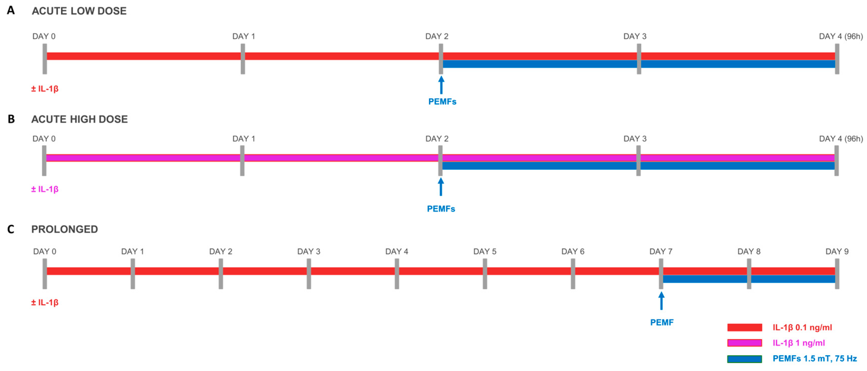
To model both acute and prolonged tendon inflammation, TCs were subjected to IL-1β stimulation according to two experimental protocols, as illustrated in
Scheme 1. In the acute model, cells were cultured in DMEM control medium supplemented with IL-1β at either 0.1 ng/mL (acute low dose) or 1 ng/mL (acute high dose) for a total of 96 h. In the prolonged model, IL-1β was administered at 0.1 ng/mL for 9 consecutive days. The high-dose IL-1β condition (1 ng/mL) replicates the protocol previously used by our group to model tenocyte inflammation [
10]. In both protocols, pulsed electromagnetic field (PEMF) stimulation was applied during the final 48 h, concurrently with the ongoing inflammatory stimulus (
Scheme 1). PEMF stimulation was delivered using a pair of rectangular horizontal coils (18 cm × 13 cm; 1000 turns of copper wire each) positioned opposite each other inside a standard cell culture incubator. The coils were powered by a PEMF generator system (IGEA, Carpi, Italy) set at 1.5 mT, 75 Hz, trapezoidal waveform, pulse length 1.1 ms, and a 10% duty cycle. Culture flasks were placed at the central region between the coils, where field uniformity had been verified by the manufacturer. PEMF exposure was continuous throughout the treatment period at 37 °C. Non-PEMF-exposed control flasks were cultured in a separate incubator without the coil apparatus.
2.3. Gene Expression Analyses
Total RNA was extracted using the RNeasy
® Mini Kit (Qiagen, Venlo, The Netherlands) according to the manufacturer’s instructions, and cDNA was synthesised with the iScript™ cDNA Synthesis Kit (Bio-Rad Laboratories, Segrate, Italy). Gene expression of the following targets was quantified by real-time PCR using iTaq Universal SYBR Green Supermix (Bio-Rad Laboratories, Segrate, Italy) on the QuantStudio™ 12K Flex OpenArray
® Platform (QS12KFlex, Thermo Fisher Scientific, Waltham, MA, USA):
ACAN,
ADAMTS,
BDNF,
CCL2,
CCL5,
CGRP,
COL1,
COL3,
COX2,
DCN,
IDO,
IL1B,
IL1RN,
IL6,
IL8,
MKX,
MMP1,
MMP3,
NANOG,
NGF,
OCT4,
PTGES2,
SCX,
SSEA1,
SOX2,
TAC1,
TIMP1,
TNC, and
VEGF. Reference gene stability was assessed across all experimental conditions by evaluating eight candidate housekeeping genes (
RPL13A,
YWHAZ,
B2M,
GAPDH,
ACTB,
TBP,
RPLP0, and
18S) using the RefFinder tool (
https://www.heartcure.com.au/reffinder/) accessed on 13 May 2026, which integrates the geNorm, NormFinder, BestKeeper, and comparative ΔCt methods.
GAPDH was identified as the most stable reference gene and was used for normalisation in all subsequent analyses. Primer sequences are reported in
Table S3.
2.4. ELISAs
Concentrations of soluble cytokines (CCL2, CCL5, GM-CSF, IL-4, IL-6, IL-8, IL-10, IL-22, IL-23, and IL-28A) in culture supernatants were determined by ELISA using commercial kits from PeproTech (Hamburg, Germany) and Invitrogen (Carlsbad, CA, USA), following the manufacturers’ protocols. The specific detection ranges for PeproTech kits were 15–1000 pg/mL (IL-4/IL-8), 31–2000 pg/mL (IL-6), and 47–3000 pg/mL (IL-10). For Invitrogen kits, the ranges were 7–1000 pg/mL (CCL2), 6–750 pg/mL (GM-CSF), 8–1000 pg/mL (IL-22), 16–2000 pg/mL (IL-23), 15.6–1000 pg/mL (IL-28A), and 47–3000 pg/mL (CCL5). Data were acquired through a PerkinElmer Wallac Victor II plate reader (PerkinElmer, Waltham, MA, USA) and processed and analysed using MyAssays2.com platform (2026). To account for PEMF-induced differences in cell number across conditions, cytokine concentrations were normalised to cell number and expressed as amount per 106 cells.
2.5. Metalloproteinase (MMP) Activity Assay
MMP activity levels were evaluated in culture media using the SensoLyte 520 Generic MMP Assay Kit (AnaSpec, Fremont, CA, USA). Following the manufacturer’s guidelines, pro-MMPs were converted to their active forms via incubation with 1 mM APMA at 37 °C for 3 h. Fluorescence signals (λex = 490 nm; λem = 520 nm) were then recorded utilising a plate reader (PerkinElmer, Waltham, MA, USA).
2.6. Proliferation, Viability and Wound Healing Assays
Cell proliferation and viability were assessed by fluorescence-based counting using Acridine Orange/DAPI solution (A13, ChemoMetec), according to the manufacturer’s instructions. Briefly, 10 µL of staining reagent was added to 190 µL of each cell suspension and analysed with a NucleoCounter® NC-3000™ (ChemoMetec, Allerod, Denmark).
Wound healing capacity was evaluated by scratch assay. TCs were seeded at 5 × 10
3 cells/cm
2 in 24-well plates and, at the onset of PEMF stimulation (Day 2 for the acute model; Day 7 for the prolonged model), a cross-shaped scratch was introduced into the confluent monolayer using a sterile 20 µL pipette tip. Detached cells were removed by PBS washing and fresh medium was added. Images were acquired at T0 (immediately after scratching, 0 h), T1 (8 h), T2 (24 h), T3 (32 h), and T4 (48 h) using an inverted phase-contrast microscope. Wound closure was quantified with ImageJ free software (
https://imagej.net/ij/) and expressed as percentage relative to T0: [(wound area T0 − wound area Tt)/wound area T0] × 100.
2.7. Statistical Analysis
All experiments were performed on
n = 4 independent tendon donors, with cells from each donor processed and analysed separately (no pooling), yielding four independent biological replicates per condition. Technical replicates varied by assay and the mean of the technical replicates was calculated for each donor prior to statistical analysis. All analyses were conducted using GraphPad Prism v9.5.0 (GraphPad Software, San Diego, CA, USA). Data normality was evaluated via the Shapiro–Wilk test. Continuous data are expressed as mean ± SD. For ELISA, MMP activity, gene expression, and proliferation data, differences between groups were analysed by two-way ANOVA (matching across donors; factors: IL-1β dose and PEMF treatment) followed by Sidak’s multiple comparisons test. For wound healing data, a mixed-effects model analysis was applied, followed by Tukey’s multiple comparisons test within each timepoint. For repeated-measures designs, sphericity violations were addressed using the Greenhouse–Geisser correction. Statistical significance was defined as a two-tailed
p-value < 0.05. Effect sizes (η
2, expressed as % of total variation attributable to each source) for two-way ANOVA models are reported in
Table S4; for CCL5 secretion, analysed using a mixed-effects model, F values are reported. Post hoc pairwise comparisons with 95% confidence intervals of the mean difference are reported in
Table S5.
4. Discussion
This study provides a systematic characterisation of PEMF effects on human tenocytes across three IL-1β-based inflammatory models of differing intensity and duration. Our findings demonstrate that PEMFs exert selective, context-dependent biological effects with anti-inflammatory and pro-regenerative activity predominantly during acute, low-grade inflammatory conditions. This observation has important methodological and clinical implications, and may partly explain the heterogeneous outcomes reported in the clinical literature on PEMF therapy in tendinopathy. Notably, this phase-dependent responsiveness is consistent with in vivo evidence from a rat model of Achilles tendinopathy, in which PEMF exposure was more effective in restoring physiological tendon architecture when applied during the mid-acute phase of disease compared to earlier or later stages [
8], suggesting that the inflammatory/reparative stage of the tissue may be a key determinant of PEMF efficacy across experimental systems.
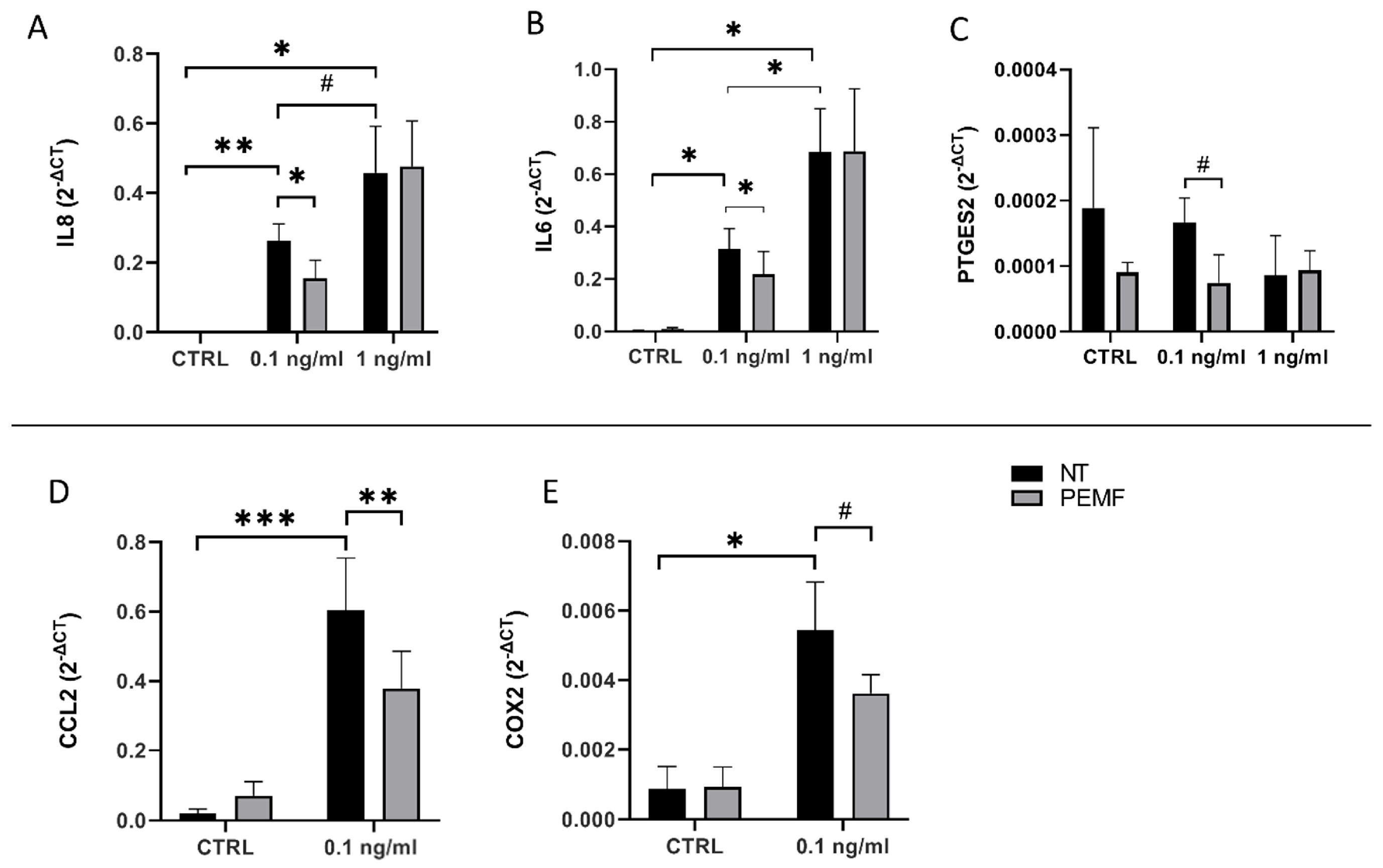
The selective transcriptional effects exerted by PEMFs suggest that biophysical stimulation can modulate specific pathways according to the inflammatory state.
Specifically, the rescue of
IL-6 and
IL-8 in the acute low model is consistent with previous reports describing PEMF-mediated suppression of NF-κB and MAPK signalling in different experimental settings [
9]. The lack of this effect in the acute high model suggests that increasing inflammatory burden progressively reduces the capacity of PEMFs alone to counteract IL-1β-driven signalling. Conversely,
CCL2 rescue in the prolonged model may reflect a distinct mechanism operative under sustained low-grade inflammatory conditions, possibly involving adenosine receptor-mediated pathways [
10]. Consistent with the transcriptional data, PEMFs accounted for the largest share of variance in
CCL2 expression among the sources tested, supporting a robust and reproducible modulatory effect on this specific target, even in a context where IL-1β dominates overall variance across most inflammatory readouts.
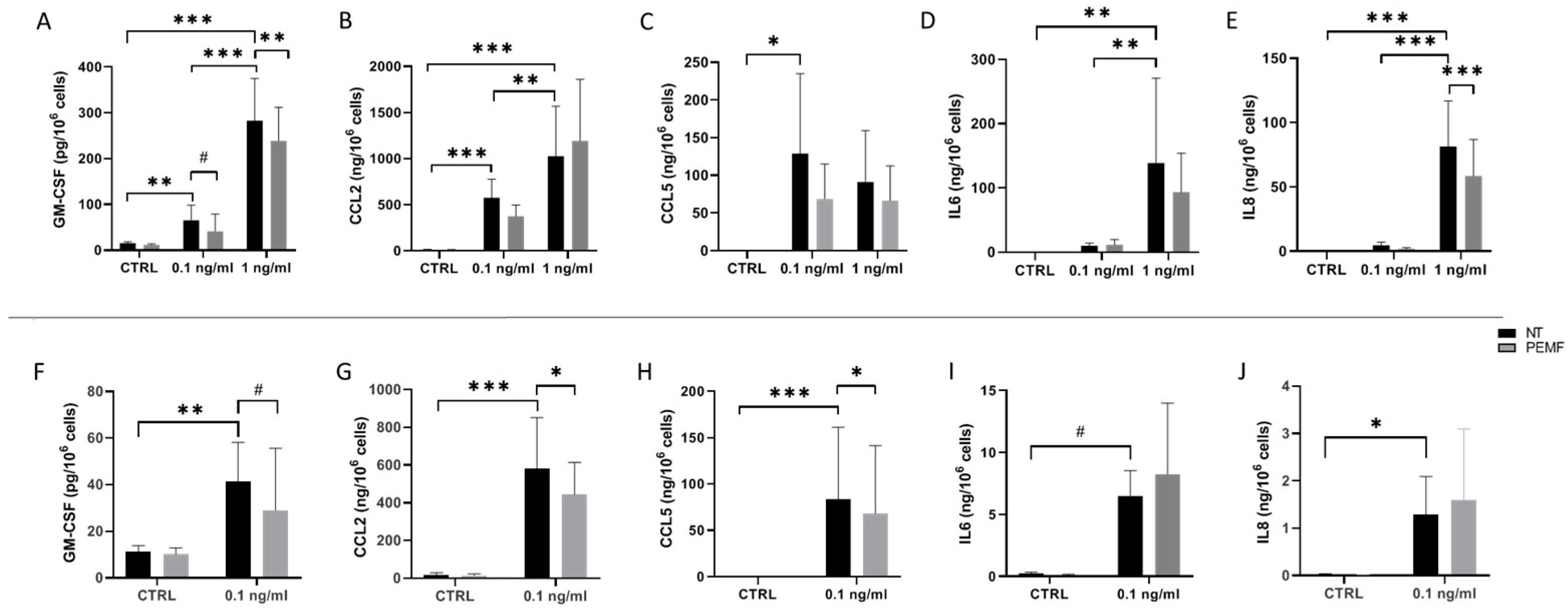
The protein secretome analysis revealed that GM-CSF emerged as one of the most consistently PEMF-responsive secreted mediators. In fact, GM-CSF was significantly elevated by IL-1β in a dose-dependent manner, while PEMF significantly reduced its secretion in the acute high-dose model, with a trend toward reduction in the acute low-dose and prolonged models. This is particularly noteworthy given the central role of GM-CSF in amplifying local inflammation through the activation and differentiation of monocytes and macrophages, and its capacity to drive further production of IL-6, IL-1, and matrix-degrading proteases [
13]. IL-8 secretion was significantly reduced by PEMFs at the higher acute dose.
CCL2 and CCL5 followed a similar pattern across models. Both chemokines were significantly induced by IL-1β in the acute model (CCL2 at both doses, CCL5 at the lower dose only), but PEMFs did not significantly affect their secretion under acute conditions. In the prolonged model, however, the PEMF significantly reduced both CCL2 and CCL5 secretion, suggesting that its immunomodulatory activity may become more evident under sustained, low-grade inflammatory stimulation, potentially limiting the chemokine-driven recruitment of monocytes that characterises prolonged inflammatory states. Nevertheless, as the present in vitro system comprised tenocyte monocultures without immune cell co-culture, these paracrine and chemotactic interpretations remain speculative and warrant direct functional validation in future non-autonomous systems.
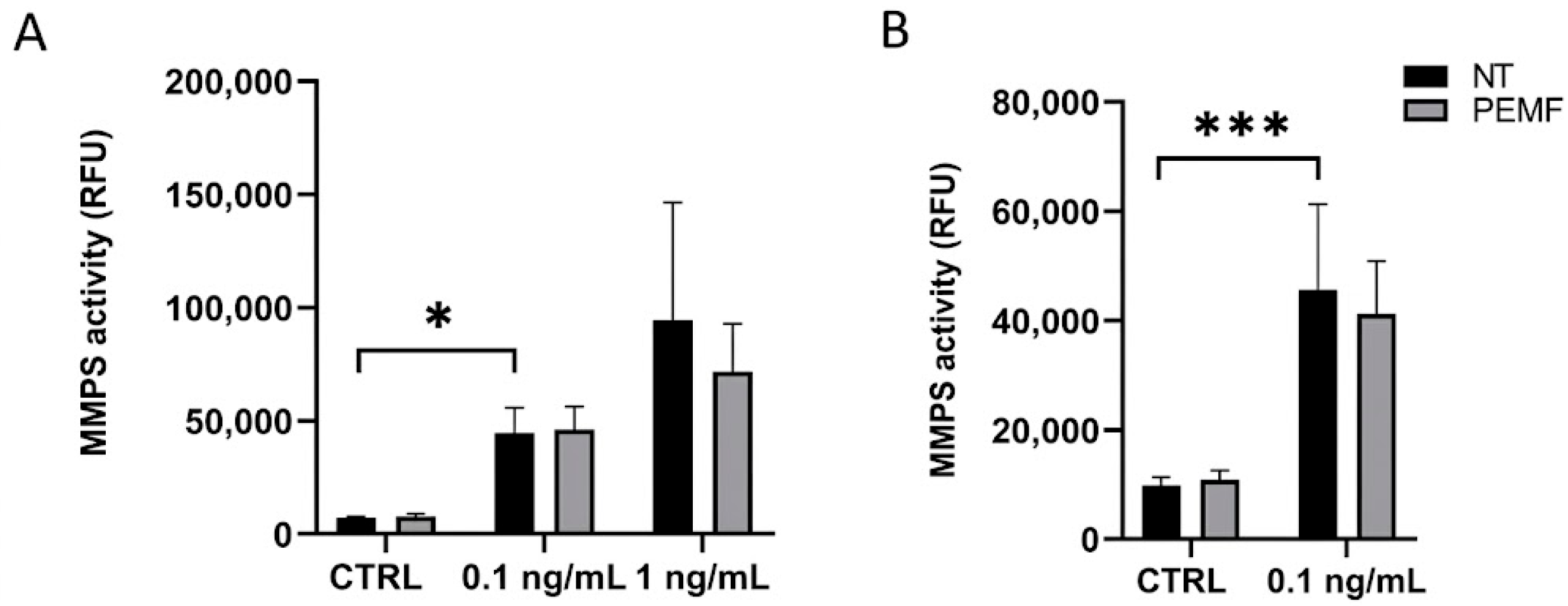
The absence of statistically significant PEMF-mediated MMP modulation in our in vitro models should be interpreted in the context of the well-recognised limitations of static monolayer models in recapitulating tendon pathophysiology. Our system lacks mechanical loading, three-dimensional ECM architecture, vascular interactions, and immune cell crosstalk, all of which critically shape MMP regulation in vivo and may be required to fully unmask biophysical effects on matrix remodelling. Notably, other in vitro studies using IL-1β-stimulated human tenocytes have reported significant PEMF-mediated suppression of MMP-1, MMP-2, and MMP-3 [
9], a discrepancy that may reflect differences in PEMF dosimetry (field intensity, frequency, duty cycle), exposure duration, or cell donor variability between the two experimental settings. Importantly, the absence of MMP modulation in vitro cannot be equated with clinical inefficacy, given the multiple in vivo mechanisms through which PEMFs are known to exert their effects.
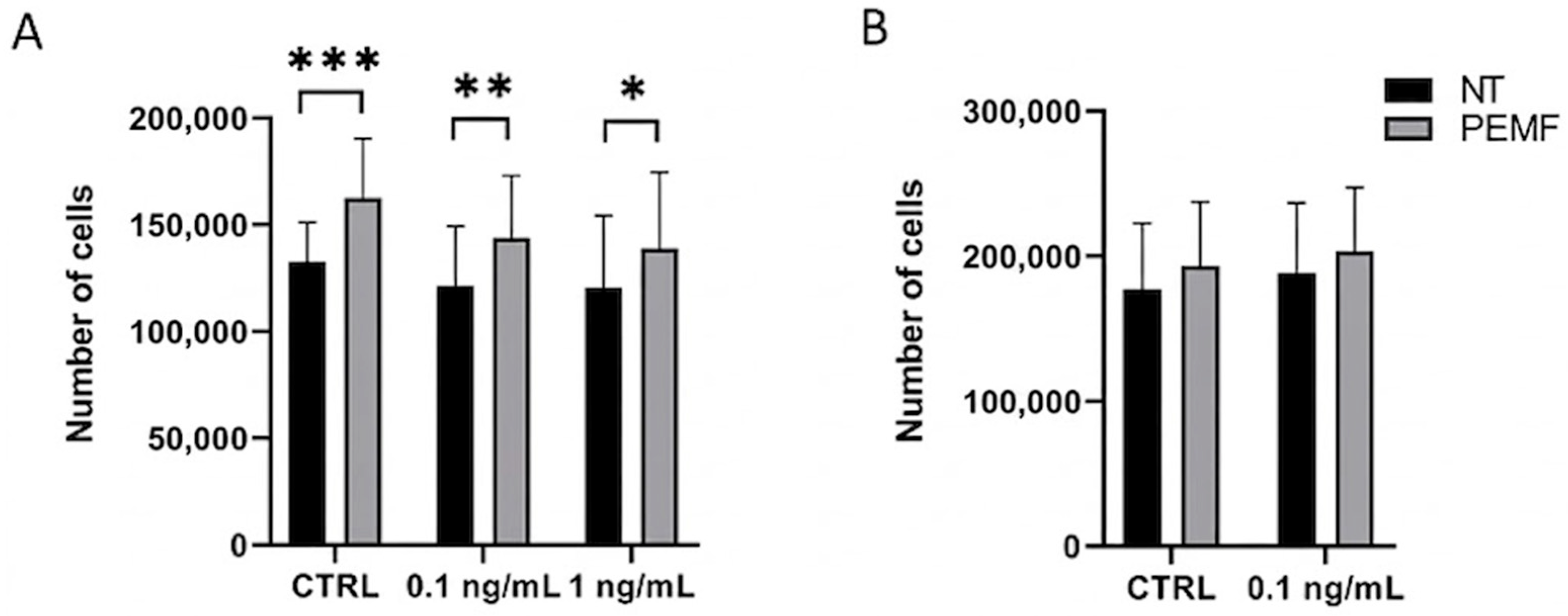
Tenocyte viability, monitored across all conditions, remained consistently high (96–98%) and was not significantly affected by IL-1β or PEMF exposure in any model. The proliferative response of tenocytes to PEMFs was consistently observed in the acute model across all conditions regardless of inflammatory status, suggesting a direct pro-survival response to biophysical stimulation that operates independently of the IL-1β cascade, possibly mediated by A
2A adenosine receptor upregulation [
10,
12] or modulation of growth factor receptor signalling. In the prolonged model, a consistent trend toward increased proliferation was observed, suggesting that this activity may extend to more established inflammatory states, pending confirmation with larger sample sizes. The most parsimonious interpretation is that this could represent a transient proliferative-expansion phase that precedes re-differentiation, consistent with established models of tissue repair biology even if longitudinal and protein-level studies are required to confirm this.
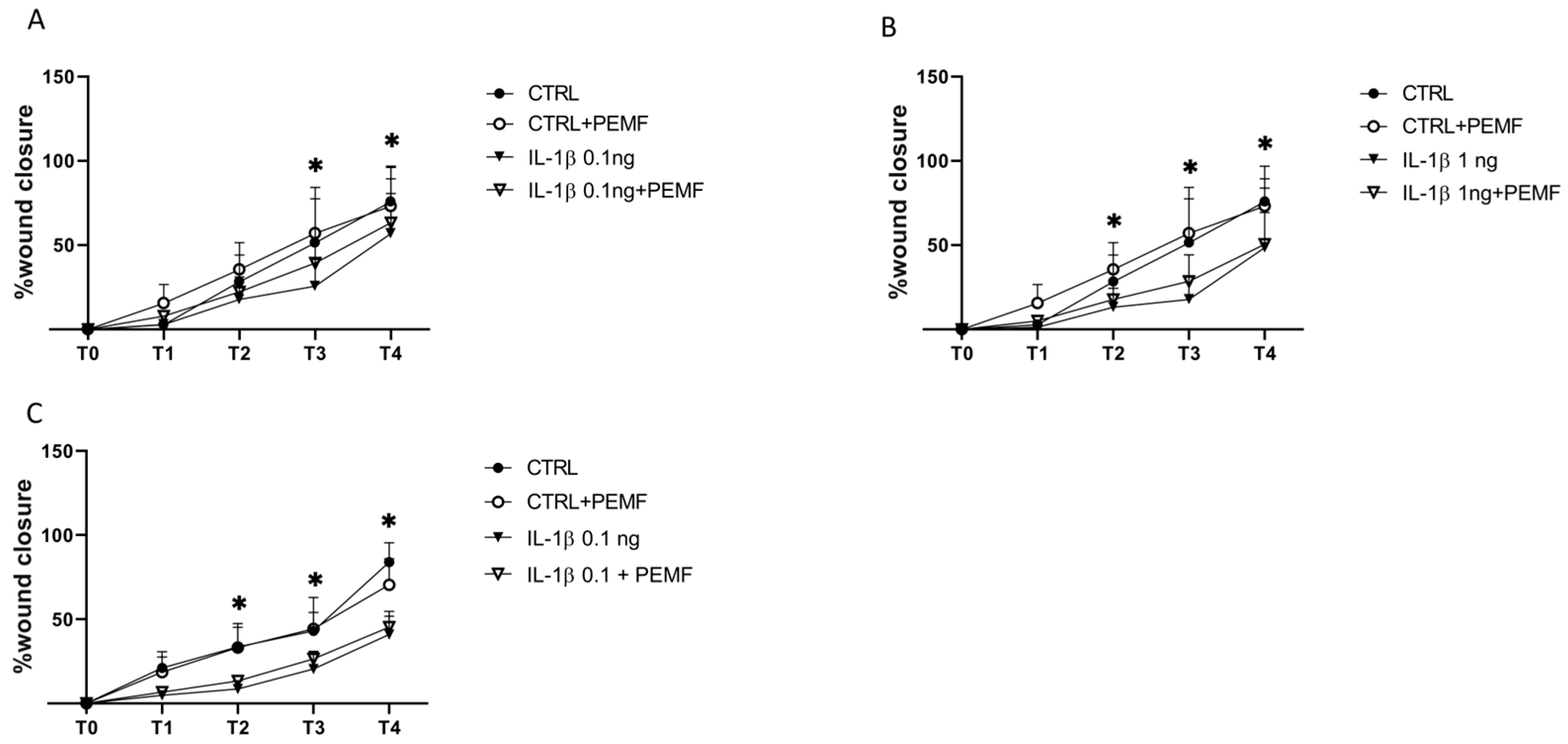
The wound healing analysis provides compelling evidence of PEMF-mediated conditioning of tenocyte wound closure capacity. Since no anti-mitotic agent was employed, the observed wound closure may reflect the combined contribution of PEMF-enhanced proliferation and migration. The consistent advantage in wound closure observed in CTRL + PEMF cells across all three models suggests that the PEMF acts primarily by enhancing the functional competence of tenocytes that retain cytoskeletal plasticity and proliferative responsiveness before severe inflammatory dysfunction becomes established. Notably, PEMFs significantly improved wound closure in IL-1β-treated cells relative to IL-1β alone at T3, before the IL-1β-induced deficit became statistically established (evident only from T4 onward), at which point PEMFs no longer showed a significant effect. This temporal pattern suggests that PEMF-mediated functional benefit may be most readily detectable in the earlier phase of inflammatory stimulation, before cumulative IL-1β exposure imposes a more dominant suppressive effect on cytoskeletal dynamics and focal adhesion signalling, consistent with the hypothesis of a potential narrowing window of cellular responsiveness as inflammatory burden progresses, which requires further validation. These findings suggest the existence of an inflammatory threshold beyond which PEMF-mediated functional recovery becomes progressively limited, possibly reflecting the dominant suppressive effect of IL-1β on cytoskeletal dynamics and focal adhesion signalling [
14], although this mechanism was not directly investigated in the present study. Overall, these findings suggest an in vitro window of cellular responsiveness to PEMFs, whereby the biological responsiveness of tendon cells progressively declines with increasing inflammatory burden. Whether this in vitro observation reflects a clinically relevant window of responsiveness remains a hypothesis to be tested in future in vivo and clinical studies.
Taken together, these findings define a biological profile for PEMFs in human tenocytes characterised by consistent pro-proliferative activity, context-dependent anti-inflammatory modulation at both transcriptional and secretome levels, absence of functional MMP suppression in the current experimental system, and a conditioning effect on wound closure capacity restricted to non-inflamed or mildly inflamed cells.
Several limitations of the present study must be acknowledged. The relatively small sample size and the intrinsic biological variability among human tendon donors may limit the generalisability of the observed trends, particularly for effects that approached but did not reach statistical significance. As already mentioned, the static two-dimensional monolayer system lacks the physiological ECM architecture, dynamic mechanical loading, vascular interactions, and immune cell crosstalk that critically shape tenocyte behaviour and MMP regulation in vivo, and the 9-day prolonged model should not be equated with clinically defined chronic tendinopathy, which develops over months. No formal a priori power calculation was performed. The sample size (n = 4 donors) was chosen based on comparable published in vitro studies of PEMF effects on primary human tenocytes, and on the practical constraints associated with the availability of primary human tendon tissue. However, it remains modest, which limits statistical power and the generalisability of the present findings, particularly with regard to effects of smaller magnitude. A further limitation is the absence of a sham-PEMF control, as untreated cells were maintained in a separate standard incubator. Although this setup does not fully exclude non-specific effects related to device handling, a sham-exposed control will be incorporated in future studies to strengthen attribution of the observed effects to PEMF exposure. Additionally, cytokines for which concentrations fell below the assay detection threshold in all samples (IL-10, IL-22, IL-23, and IL-28A) were excluded from quantitative statistical comparison and are reported descriptively as undetectable, rather than being imputed; this conservative approach may underestimate low-magnitude PEMF effects on these mediators. The absence of immune cells is a particularly relevant constraint, given the central role of macrophage polarisation in orchestrating both the inflammatory and resolution phases of tendon pathology. It is therefore possible that PEMF effects on matrix remodelling and tenocyte phenotype under physiologically relevant conditions differ substantially from those observed here. Furthermore, intracellular signalling pathways were not directly investigated at the protein level (e.g., via Western blot or immunostaining); while secreted levels of selected inflammatory mediators were assessed by ELISA, direct assessment of pathway activation was not performed, limiting mechanistic conclusions regarding the intracellular molecular targets of PEMFs. While cell viability was systematically monitored and remained unaffected across all conditions, dedicated assays for apoptosis and ion channel dynamics were not performed, and represent relevant mechanistic layers for future investigation. More broadly, mechanistic interpretations of intracellular signalling pathways and the contribution of immune cell contributions remain speculative and are proposed as hypotheses for future investigation, rather than as demonstrated findings.
Future studies incorporating three-dimensional bioreactor cultures, mechanical stimulation, and immune cell co-culture systems are required to validate and extend these findings, and to determine whether the hypothesised window of cellular responsiveness identified in vitro translates to clinically meaningful outcomes across the different stages of tendinopathy.




 ,
,