Retinal Biomarkers of Folate and Vitamin B12 Metabolic Dysfunction: A Framework for Machine Learning-Assisted Detection of Cerebral Folate Deficiency


Highlights
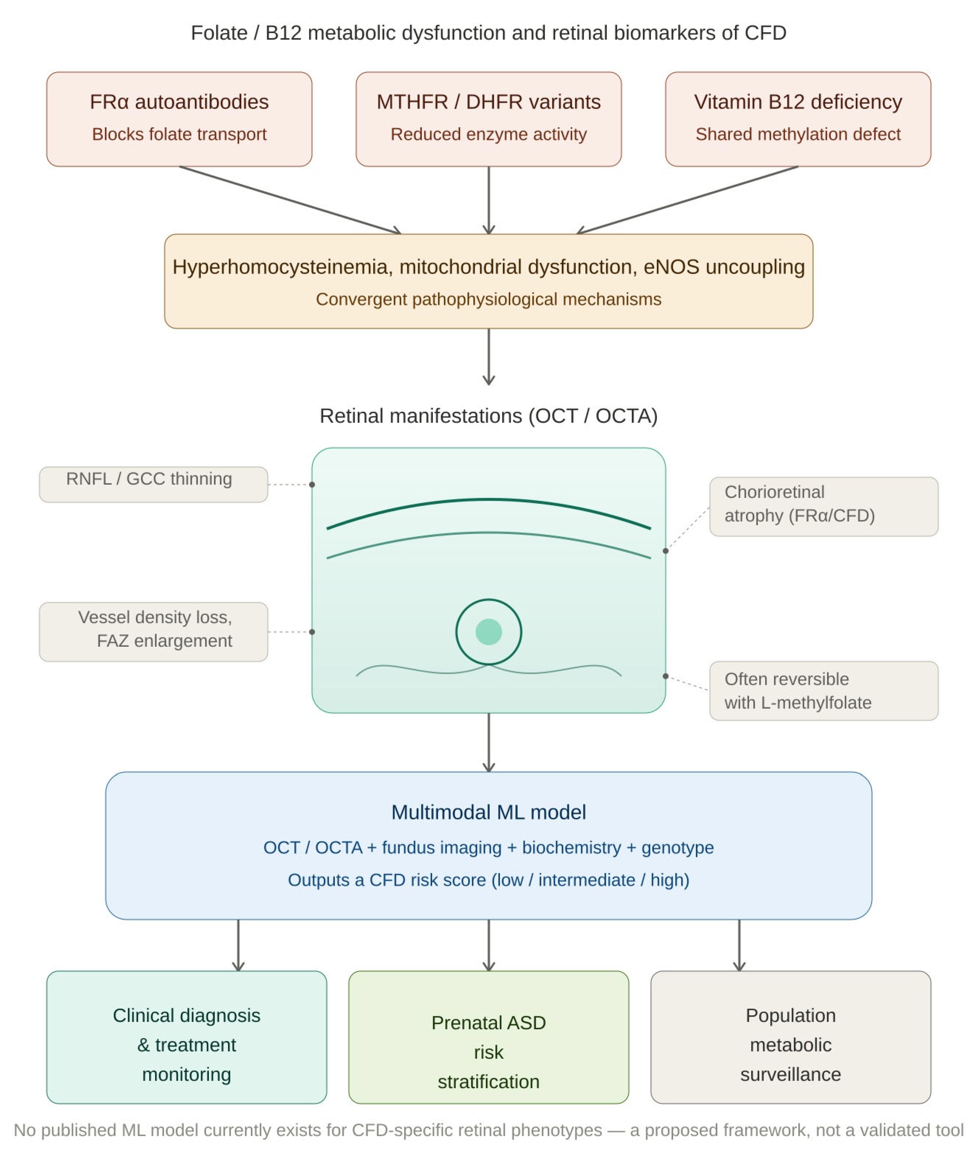
- Folate/B12 metabolic dysfunction (nutritional deficiency, FRα autoantibodies, MTHFR/DHFR variants) produces convergent, quantifiable retinal changes that are detectable with OCT/OCTA and often reversible with early correction.
- The review proposes a conceptual multimodal ML framework, combining retinal imaging with biochemical and genetic data, as a research agenda for non-invasive detection of cerebral folate deficiency (CFD).
- A validated retinal-imaging/ML approach could enable non-invasive prenatal and neonatal screening for CFD in FRAA-positive or genetically high-risk pregnancies, allowing earlier leucovorin treatment.
- Given the folate–ASD risk association reported in FRAA-positive cohorts, earlier identification and treatment of maternal/infant CFD may plausibly reduce neurodevelopmental risk, including autism spectrum disorder, in offspring.
Abstract
1. Introduction
2. Methods
2.1. Literature Search Strategy
2.2. Inclusion and Exclusion Criteria
2.3. Data Extraction and Quality Assessment
3. Pathophysiological Mechanisms of Retinal Injury
3.1. Hyperhomocysteinemia and Endothelial Toxicity
- Direct endothelial cytotoxicity via reactive oxygen species (ROS) generation and thiol-disulfide redox stress, disrupting the inner blood–retinal barrier (BRB) [44].
- Promotion of a prothrombotic state through impaired nitric oxide (NO) bioavailability, platelet hyperactivation, and upregulation of coagulation factors, predisposing to retinal vascular occlusions [46].
- Activation of NMDA (N-methyl D-aspartate) receptors on retinal ganglion cells and Müller glia, inducing excitotoxic calcium influx and apoptosis [46].
- Disruption of retinal pigment epithelium (RPE) structure and function, inducing AMD (age-related macular degeneration)-like changes including basal laminar deposits and complement dysregulation [44].
3.2. Mitochondrial Dysfunction and Energy Failure
3.3. eNOS Uncoupling and Vasoconstriction (DHFR-Related)
3.4. FRα Autoantibody-Mediated Transport Blockade
4. Retinal Manifestations by Etiology
4.1. Nutritional Folate Deficiency
4.2. Components
- Chorioretinal atrophy: Pediatric CFD patients with untreated FRAA syndrome develop massive, progressive chorioretinal degeneration affecting both the RPE and the choroidal vasculature [50]. The degree of atrophy correlates with duration of untreated disease.
- Optic atrophy with diffuse RNFL loss and GCC thinning [50].
- Progressive central visual loss that may stabilize or partially reverse with folinic acid (leucovorin) therapy, which bypasses FRα-mediated transport [17].
4.3. MTHFR Polymorphisms (C677T and A1298C)
- Microvascular structural changes in mouse models: The Mthfr677C>T knock-in mouse demonstrates age- and sex-dependent reductions in retinal vascular density, increased arteriovenous crossings, and vessel tortuosity on high-resolution imaging, directly mirroring human vascular risk [55].
4.4. DHFR Polymorphisms
- Retinal arteriolar vasoconstriction and reduced capillary perfusion pressure [19].
- Elevated retinal venous pressure (RVP), a measurable correlate of impaired retinal outflow [19].
- Potential moderation of susceptibility to retinoblastoma, with epidemiological evidence of genotype–drug interactions involving DHFR in pediatric cancer risk [19].
4.5. Vitamin B12 Deficiency
- Peripapillary RNFL thinning: Temporal quadrant thinning is the most consistent finding. In a pediatric cohort, superior and global RNFL were significantly thinner in B12-deficient children versus controls (p = 0.007 and p = 0.01, respectively), with global and superior RNFL directly correlating with serum B12 levels (r = 0.296 and r = 0.369, respectively) [59]. Adult case series document that there is a mean RNFL reduction of 10–20% in symptomatic patients [54,58].
- Macular GCC/GCL (ganglion cell complex/ganglion cell layer) thinning: Bilateral diffuse GCC loss on macular OCT often accompanies RNFL changes and correlates with centrocecal scotoma location on perimetry [58].
- Reduced peripapillary and macular vessel density on OCTA: Pellegrini et al. provided the first detailed OCTA characterization of B12 deficiency optic neuropathy, demonstrating decreased radial peripapillary capillary (RPC) vessel density at the optic nerve head and maculopapular bundle, with foveal avascular zone (FAZ) enlargement [33].
- Koca et al. [34] confirmed OCTA-detectable RPC vessel density reductions in a B12 deficiency anemia cohort (n = 24), even in patients without overt optic neuropathy, suggesting OCTA may detect preclinical microvascular changes.
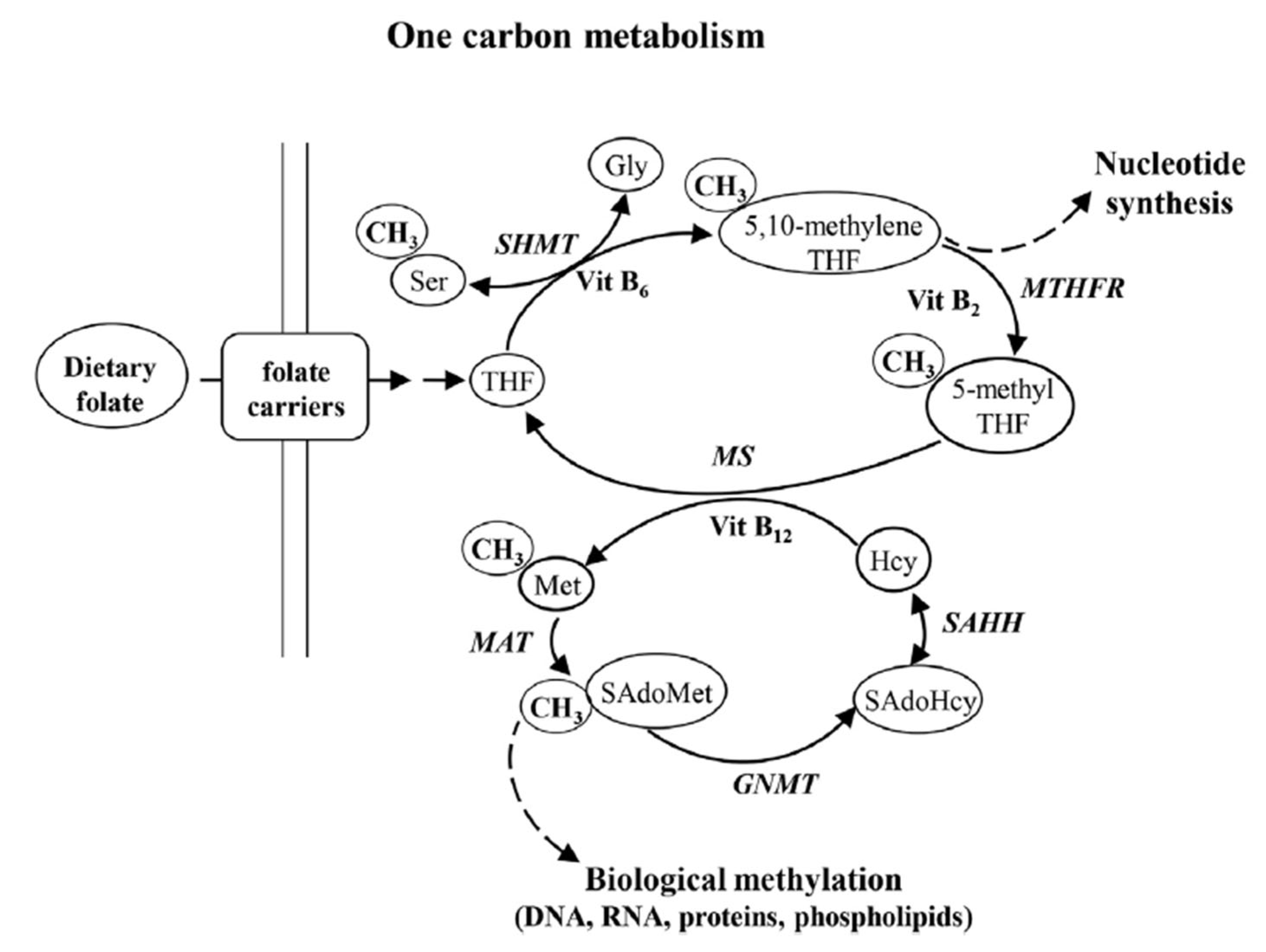
- Methyl trap (or folate trap): Vitamin B12 deficiency stops conversion of MTHF to THF, elevating homocysteine due to the unavailability of folate [29].
5. Multimodal Diagnostic Assessment
5.1. Workflow Integration
6. L-Methylfolate Supplementation: Mechanisms and Retinal Evidence
- OCTA perfusion improvement: Increased vessel density and flow indices; retinal arteriolar blood flow velocity increases as measured by Retinal Function Imager (RFI), with the greatest perfusion gains in the highest-risk genotypes [18].
- Retinal venous pressure (RVP) reduction: In a glaucoma/ocular vascular disease cohort with elevated baseline Hcy (>12 μmol/L), mean RVP dropped by 8.8 mm Hg after 3 months (p < 0.001) [18].
6.1. Retinal Vein Occlusion and AMD Applications
6.2. Clinical Considerations
7. A Machine Learning Framework for Retinal Detection of Cerebral Folate Deficiency
7.1. Current State of AI in Retinal Imaging for Nutritional and Metabolic Disease
- Anemia detection from fundus images: Mitani et al. [35] demonstrated that DL models trained on retinal fundus photographs could detect anemia and estimate hemoglobin concentration with high accuracy (area under the curve (AUC) > 0.88), identifying the optic disc and peripapillary region as the most discriminative areas. Because macrocytic anemia from B12 and folate deficiency produces characteristic vascular and disc changes—pallor, vascular tortuosity, altered vessel caliber—this approach has direct relevance to nutritional deficiency screening [68,69,70].
- Multi-disease retinal screening: Dong et al. [36] demonstrated a DL system capable of simultaneously screening for more than 10 retinal and systemic conditions, including categories encompassing nutritional and toxic optic neuropathies, with high sensitivity and specificity.
- Neuro-ophthalmic disorder classification: Szanto et al. [71] reported a DL system achieving 93–96% accuracy in differentiating optic disc swelling etiologies from fundus photographs, a framework directly applicable to optic neuropathy triage including nutritional causes.
- LLM-assisted differential diagnosis: Shukla et al. [72] evaluated large language models for AI-assisted differential diagnosis of retinal and neuro-ophthalmic disorders, including nutritional optic neuropathy, demonstrating growing AI capacity in this clinical domain.
- Vascular cognitive impairment detection using OCTA measures of retinal vessel density: Zhou et al. [73] evaluated OCTA from cognitively normal and impaired subjects without vision disorders and found a correlation of decreasing retinal vessel density with increasing cognitive impairment.
- Selective ASD determination in children aged 2–6: Zee et al. [43] trained an ML system on color fundus images of children with neurodevelopmental disorders and matched controls (2000+ total subjects). Their ML system successfully isolated three developmental disabilities from control and from each other, using multiple retinal parameters. While they have no FRAA data for these subjects, the ability to identify ASD selectively from global developmental delay or from the combination of these two conditions bodes well for our proposal here to extract CFD evidence from retinal images of ASD-relevant subjects.
7.2. Proposed ML Architecture for CFD Retinal Screening
7.2.1. Input Data Modalities
- Functional metrics (optional): BCVA, Humphrey mean deviation and pattern standard deviation, microperimetry mean sensitivity, contrast sensitivity [49].
7.2.2. Model Architecture
7.2.3. Output and Clinical Integration
7.2.4. Training and Validation Strategy
7.2.5. Extended Applications
8. Application to Prenatal Screening and Autism Spectrum Disorder Risk
8.1. Folate Metabolism as an ASD Risk Pathway
8.2. Retinal Imaging as a Prenatal and Neonatal Biomarker
8.3. An Integrated Prenatal CFD Screening Protocol
- Neonatal retinal screening: Infant OCT (portable handheld devices) in offspring of FRAA-positive mothers to identify early chorioretinal changes and prompt folinic acid initiation [50]. A recent OCT study of very preterm (<32 weeks gestational age) infants found that thicker retinal nerve fiber layers were correlated with higher Bayley Scales of Infant and Toddler Development, with a lower risk of autism at 2 years, thus documenting that even the youngest infants may have measurable retinal differences that can be detected with OCT [77].
9. Research Gaps and Future Directions
9.1. Labeled Retinal Imaging Datasets
9.2. Standardization of OCT/OCTA Metrics
9.3. Longitudinal Recovery Studies
9.4. Pediatric and Neonatal Normative Data
9.5. Randomized Controlled Trials of L-Methylfolate
10. Conclusions
Supplementary Materials
Author Contributions
Funding
Institutional Review Board Statement
Informed Consent Statement
Data Availability Statement
Acknowledgments
Conflicts of Interest
Abbreviations
| 5-MTHF | 5-methyltetrahydrofolate |
| AI | artificial intelligence |
| AMD | age-related macular degeneration |
| ASD | autism spectrum disorder |
| AUC | area under the curve |
| BBB | blood–brain barrier |
| BCVA | best-corrected visual acuity |
| BH4 (BH2) | tetrahydrobiopterin (dihydrobiopterin) |
| BRB | inner blood–retinal barrier |
| CFD | cerebral folate deficiency |
| CNN | convolutional neural network |
| DHFR | dihydrofolate reductase |
| DL | deep learning |
| DR | diabetic retinopathy |
| DVP | deep vascular plexus |
| eNOS | endothelial nitric oxide synthase |
| ERG | electroretinogram |
| ETC | electron transport chain |
| FA | fluorescein angiography |
| FAF | fundus autofluorescence |
| FAZ | foveal avascular zone |
| FOLR1 | gene encoding folate receptor alpha |
| FRAA(s) | folate receptor alpha autoantibody(ies) |
| FRα | folate receptor alpha |
| GCC | ganglion cell complex |
| GCL | ganglion cell layer |
| Hcy | homocysteine |
| HHcy | hyperhomocysteinemia |
| IOVS | Investigative Ophthalmology & Visual Science |
| ML | machine learning |
| MMA | methylmalonic acid |
| MP bundle | maculopapular bundle |
| MTHFR | methylenetetrahydrofolate reductase |
| NMDA | N-methyl-D-aspartate |
| NO | nitric oxide |
| OCT | optical coherence tomography |
| OCTA | optical coherence tomography angiography |
| ONH | optic nerve head |
| ONOO− | peroxynitrite |
| O2− | superoxide anion |
| pERG | pattern electroretinogram |
| PMC | PubMed Central |
| RAO | retinal artery occlusion |
| RBC | red blood cell |
| RCT(s) | randomized controlled trial(s) |
| RFC1/SLC19A1 | reduced folate carrier 1 (gene)/ solute carrier family 19 member 1 (gene) |
| RFI | Retinal Function Imager |
| RGC(s) | retinal ganglion cell(s) |
| RNFL | retinal nerve fiber layer |
| ROS | reactive oxygen species |
| RPC | radial peripapillary capillary |
| RPE | retinal pigment epithelium |
| RVO | retinal vein occlusion |
| RVP | retinal venous pressure |
| SD-OCT/SS-OCT | spectral-domain/ swept-source optical coherence tomography |
| SHMT1/SHMT2 | serine hydroxymethyltransferase (isoform 1)/ serine hydroxymethyltransferase (isoform 2) |
| SVP | superficial vascular plexus |
| THF | tetrahydrofolate |
| UMFA | unmetabolized folic acid |
| VD | vessel density |
| VEGF | vascular endothelial growth factor |
| VEP | visual evoked potential |
| ViT | Vision Transformer |
References
- Ayoub, G. Vitamins, Vascular Health and Disease. Nutrients 2025, 17, 2955. [Google Scholar] [CrossRef] [Scilit] [PubMed]
- Roda, M.; di Geronimo, N.; Pellegrini, M.; Schiavi, C. Nutritional Optic Neuropathies: State of the Art and Emerging Evidences. Nutrients 2020, 12, 2653. [Google Scholar] [CrossRef] [Scilit] [PubMed]
- Wong-Riley, M. Energy Metabolism of the Visual System. Eye Brain 2010, 2, 99–116. [Google Scholar] [CrossRef] [Scilit] [PubMed]
- Ayoub, G. A Window to the Brain—The Enduring Impact of Vision Research. Brain Sci. 2025, 15, 453. [Google Scholar] [CrossRef] [Scilit] [PubMed]
- Abouhish, H.; Shalaby, L.; Elzayat, O.; Peddireddy, N.; Tawfik, A. Modulating One-Carbon Metabolism with B-Vitamins to Protect the Retinal Barrier and Prevent Retinal Degeneration. Nutrients 2026, 18, 236. [Google Scholar] [CrossRef] [Scilit] [PubMed]
- Gu, J.; Lei, C.; Zhang, M. Folate and Retinal Vascular Diseases. BMC Ophthalmol. 2023, 23, 413. [Google Scholar] [CrossRef] [Scilit] [PubMed]
- Ayoub, G.; Luo, Y. Ocular Vascular Health and Disease. Med. Res. Arch. 2026, 14, 1–10. [Google Scholar] [CrossRef] [Scilit]
- Country, M.W. Retinal Metabolism: A Comparative Look at Energetics in the Retina. Brain Res. 2017, 1672, 50–57. [Google Scholar] [CrossRef] [Scilit] [PubMed]
- de Silva, P.; Jayamanne, G.; Bolton, R. Folic Acid Deficiency Optic Neuropathy: A Case Report. J. Med. Case Rep. 2008, 2, 299. [Google Scholar] [CrossRef] [Scilit] [PubMed]
- Smith, A.D.; Refsum, H. Homocysteine—From Disease Biomarker to Disease Prevention. J. Intern. Med. 2021, 290, 826–854. [Google Scholar] [CrossRef] [Scilit] [PubMed]
- Wright, A.D.; Martin, N.; Dodson, P.M. Homocysteine, Folates, and the Eye. Eye 2008, 22, 989–993. [Google Scholar] [CrossRef] [Scilit] [PubMed]
- Peddireddy, N.; Abdelkarim, R.; Elzayat, O.; Hsiung, C.; Tawfik, A.M. One Carbon Metabolism and Aging Eye Disease: Exploring Preventive Therapeutic Targets. Investig. Ophthalmol. Vis. Sci. 2024, 65, 1704. [Google Scholar]
- Tribble, J.R.; Wong, V.H.Y.; Stuart, K.V.; Chidlow, G.; Nicol, A.; Rombaut, A.; Rabiolo, A.; Hoang, A.; Lee, P.Y.; Rutigliani, C.; et al. Dysfunctional One-Carbon Metabolism Identifies Vitamins B6, B9, B12, and Choline as Neuroprotective in Glaucoma. Cell Rep. Med. 2025, 6, 102127. [Google Scholar] [CrossRef] [Scilit] [PubMed]
- Brazionis, L.; Rowley, K., Sr.; Itsiopoulos, C.; Harper, C.A.; O’Dea, K. Homocysteine and Diabetic Retinopathy. Diabetes Care 2008, 31, 50–56. [Google Scholar] [CrossRef] [Scilit] [PubMed]
- Hou, J.; Wen, Y.; Gao, S.; Jiang, Z.; Tao, L. Association of Dietary Intake of B Vitamins with Glaucoma. Sci. Rep. 2024, 14, 8539. [Google Scholar] [CrossRef] [Scilit] [PubMed]
- Liu, Z.; Jiang, H.; Townsend, J.H.; Wang, J. Improved Retinal Microcirculation in Mild Diabetic Retinopathy Patients Carrying MTHFR Polymorphisms Who Received the Medical Food, Ocufolin®. Clin. Ophthalmol. 2022, 16, 1497–1504. [Google Scholar] [CrossRef] [Scilit] [PubMed]
- Ramaekers, V.T.; Häusler, M.; Opladen, T.; Heimann, G.; Blau, N. Psychomotor Retardation, Spastic Paraplegia, Cerebellar Ataxia and Dyskinesia Associated with Low 5-Methyltetrahydrofolate in Cerebrospinal Fluid: A Novel Neurometabolic Condition Responding to Folinic Acid Substitution. Neuropediatrics 2002, 33, 301–308. [Google Scholar] [CrossRef] [Scilit] [PubMed]
- Jiang, H.; Liu, Z.; Townsend, J.H.; Wang, J. Effects of Methylenetetrahydrofolate Reductase (MTHFR) Polymorphisms on Retinal Tissue Perfusion in Mild Diabetic Retinopathy Patients Receiving the Medical Food, Ocufolin®. Clin. Ophthalmol. 2023, 17, 1121–1127. [Google Scholar] [CrossRef] [Scilit] [PubMed]
- Orjuela, M.A.; Cabrera-Muñoz, L.; Paul, L.; Ramirez-Ortiz, M.A.; Liu, X.; Chen, J.; Mejia-Rodriguez, F.; Medina-Sanson, A.; Diaz-Carreño, S.; Suen, I.H.; et al. Risk of Retinoblastoma Is Associated with a Maternal Polymorphism in Dihydrofolatereductase (DHFR) and Prenatal Folic Acid Intake. Cancer 2012, 118, 5912–5919. [Google Scholar] [CrossRef] [Scilit] [PubMed]
- Ducker, G.S.; Rabinowitz, J.D. One-Carbon Metabolism in Health and Disease. Cell Metab. 2017, 25, 27–42. [Google Scholar] [CrossRef] [Scilit] [PubMed]
- Fox, J.T.; Stover, P.J. Chapter 1 Folate-Mediated One-Carbon Metabolism. In Vitamins & Hormones; Academic Press: Cambridge, MA, USA, 2008; Volume 79, pp. 1–44. [Google Scholar]
- Zheng, Y.; Cantley, L.C. Toward a Better Understanding of Folate Metabolism in Health and Disease. J. Exp. Med. 2018, 216, 253–266. [Google Scholar] [CrossRef] [Scilit] [PubMed]
- Friso, S.; Udali, S.; De Santis, D.; Choi, S.-W. One-Carbon Metabolism and Epigenetics. Mol. Asp. Med. 2017, 54, 28–36. [Google Scholar] [CrossRef] [Scilit] [PubMed]
- Shi, C.; Wang, P.; Airen, S.; Brown, C.; Liu, Z.; Townsend, J.H.; Wang, J.; Jiang, H. Nutritional and Medical Food Therapies for Diabetic Retinopathy. Eye Vis. 2020, 7, 33. [Google Scholar] [CrossRef] [Scilit] [PubMed]
- Lyon, P.; Strippoli, V.; Fang, B.; Cimmino, L. B Vitamins and One-Carbon Metabolism: Implications in Human Health and Disease. Nutrients 2020, 12, 2867. [Google Scholar] [CrossRef] [Scilit] [PubMed]
- Dalto, D.B.; Matte, J.-J. Pyridoxine (Vitamin B6) and the Glutathione Peroxidase System; a Link between One-Carbon Metabolism and Antioxidation. Nutrients 2017, 9, 189. [Google Scholar] [CrossRef] [Scilit] [PubMed]
- Stover, P.J.; Field, M.S. Vitamin B-6. Adv. Nutr. 2015, 6, 132–133. [Google Scholar] [CrossRef] [Scilit] [PubMed]
- Shane, B.; Stokstad, E.L.R. Vitamin B12-Folate Interrelationships. Annu. Rev. Nutr. 1985, 5, 115–141. [Google Scholar] [CrossRef] [PubMed]
- Smulders, Y.M.; Smith, D.E.C.; Kok, R.M.; Teerlink, T.; Swinkels, D.W.; Stehouwer, C.D.A.; Jakobs, C. Cellular Folate Vitamer Distribution during and after Correction of Vitamin B12 Deficiency: A Case for the Methylfolate Trap. Br. J. Haematol. 2006, 132, 623–629. [Google Scholar] [CrossRef] [Scilit] [PubMed]
- Palmer, A.M.; Kamynina, E.; Field, M.S.; Stover, P.J. Folate Rescues Vitamin B12 Depletion-Induced Inhibition of Nuclear Thymidylate Biosynthesis and Genome Instability. Proc. Natl. Acad. Sci. USA 2017, 114, E4095–E4102. [Google Scholar] [CrossRef] [Scilit] [PubMed]
- Gurler, G.; Belder, N.; Beker, M.C.; Sever-Bahcekapili, M.; Uruk, G.; Kilic, E.; Yemisci, M. Reduced Folate Carrier 1 Is Present in Retinal Microvessels and Crucial for the Inner Blood Retinal Barrier Integrity. Fluids Barriers CNS 2023, 20, 47. [Google Scholar] [CrossRef] [Scilit] [PubMed]
- Alam, C.; Kondo, M.; O’Connor, D.L.; Bendayan, R. Clinical Implications of Folate Transport in the Central Nervous System. Trends Pharmacol. Sci. 2020, 41, 349–361. [Google Scholar] [CrossRef] [Scilit] [PubMed]
- Pellegrini, F.; Prosdocimo, G.; Papayannis, A.; Cirone, D. Optical Coherence Tomography Angiography Findings in Deficiency Optic Neuropathy. Neuroophthalmology 2019, 43, 401–406. [Google Scholar] [CrossRef] [Scilit] [PubMed]
- Koca, S.; Bozkurt, E.; Dogan, M.; Yavasoglu, F.; Erogul, Ö.; Bulut, A.K. Effects of B12 Deficiency Anemia on Radial Peripapillary and Macular Vessel Density: An Optical Coherence Tomography Angiography (OCTA) Study. Klin. Monatsbl. Augenheilkd. 2023, 240, 92–98. [Google Scholar] [CrossRef] [Scilit] [PubMed]
- Mitani, A.; Huang, A.; Venugopalan, S.; Corrado, G.S.; Peng, L.; Webster, D.R.; Hammel, N.; Liu, Y.; Varadarajan, A.V. Detection of Anaemia from Retinal Fundus Images via Deep Learning. Nat. Biomed. Eng. 2020, 4, 18–27. [Google Scholar] [CrossRef] [Scilit] [PubMed]
- Dong, L.; He, W.; Zhang, R.; Ge, Z.; Wang, Y.X.; Zhou, J.; Xu, J.; Shao, L.; Wang, Q.; Yan, Y.; et al. Artificial Intelligence for Screening of Multiple Retinal and Optic Nerve Diseases. JAMA Netw. Open 2022, 5, e229960. [Google Scholar] [CrossRef] [Scilit] [PubMed]
- Li, M.; Wan, C. The Use of Deep Learning Technology for the Detection of Optic Neuropathy. Quant. Imaging Med. Surg. 2022, 12, 2129–2143. [Google Scholar] [CrossRef] [Scilit] [PubMed]
- Bobrowski-Khoury, N.; Ramaekers, V.T.; Sequeira, J.M.; Quadros, E.V. Folate Receptor Alpha Autoantibodies in Autism Spectrum Disorders: Diagnosis, Treatment and Prevention. J. Pers. Med. 2021, 11, 710. [Google Scholar] [CrossRef] [Scilit] [PubMed]
- Pu, D.; Shen, Y.; Wu, J. Association between MTHFR Gene Polymorphisms and the Risk of Autism Spectrum Disorders: A Meta-Analysis. Autism Res. 2013, 6, 384–392. [Google Scholar] [CrossRef] [Scilit] [PubMed]
- Giorlandino, C.; Mesoraca, A.; Margiotti, K.; Fabiani, M.; Cupellaro, M.; Giorlandino, F.; Mastrandrea, M.L.; Raffio, R.; Pignataro, F.; Mangiafico, L.; et al. Folinic Acid Supplementation in Folate Receptor Alpha Autoantibodies-Positive Pregnancy: A Pilot Randomized Study on Neurodevelopmental Outcomes. Reprod. Female Child Health 2026, 5, e70053. [Google Scholar] [CrossRef] [Scilit]
- Frye, R.E.; Slattery, J.; Delhey, L.; Furgerson, B.; Strickland, T.; Tippett, M.; Sailey, A.; Wynne, R.; Rose, S.; Melnyk, S.; et al. Folinic Acid Improves Verbal Communication in Children with Autism and Language Impairment: A Randomized Double-Blind Placebo-Controlled Trial. Mol. Psychiatry 2018, 23, 247–256. [Google Scholar] [CrossRef] [Scilit] [PubMed]
- Renard, E.; Leheup, B.; Guéant-Rodriguez, R.-M.; Oussalah, A.; Quadros, E.V.; Guéant, J.-L. Folinic Acid Improves the Score of Autism in the EFFET Placebo-Controlled Randomized Trial. Biochimie 2020, 173, 57–61. [Google Scholar] [CrossRef] [Scilit] [PubMed]
- Lai, M.; Lee, J.; Chiu, S.; Charm, J.; So, W.Y.; Yuen, F.P.; Kwok, C.; Tsoi, J.; Lin, Y.; Zee, B. A Machine Learning Approach for Retinal Images Analysis as an Objective Screening Method for Children with Autism Spectrum Disorder. eClinicalMedicine 2020, 28, 100588. [Google Scholar] [CrossRef] [Scilit] [PubMed]
- Mohamed, R.; Sharma, I.; Ibrahim, A.S.; Saleh, H.; Elsherbiny, N.M.; Fulzele, S.; Elmasry, K.; Smith, S.B.; Al-Shabrawey, M.; Tawfik, A. Hyperhomocysteinemia Alters Retinal Endothelial Cells Barrier Function and Angiogenic Potential via Activation of Oxidative Stress. Sci. Rep. 2017, 7, 11952. [Google Scholar] [CrossRef] [Scilit] [PubMed]
- Ibrahim, A.S.; Mander, S.; Hussein, K.A.; Elsherbiny, N.M.; Smith, S.B.; Al-Shabrawey, M.; Tawfik, A. Hyperhomocysteinemia Disrupts Retinal Pigment Epithelial Structure and Function with Features of Age-Related Macular Degeneration. Oncotarget 2016, 7, 8532–8545. [Google Scholar] [CrossRef] [Scilit] [PubMed]
- Tawfik, A.; Al-Shabrawey, M.; Roon, P.; Sonne, S.; Covar, J.A.; Matragoon, S.; Ganapathy, P.S.; Atherton, S.S.; El-Remessy, A.; Ganapathy, V.; et al. Alterations of Retinal Vasculature in Cystathionine-Beta-Synthase Mutant Mice, a Model of Hyperhomocysteinemia. Investig. Ophthalmol. Vis. Sci. 2013, 54, 939–949. [Google Scholar] [CrossRef] [Scilit] [PubMed]
- Ayoub, G. Disease Diagnosis Using Retinal Vasculature: Insights from Flammer Syndrome and AI. Brain Sci. 2025, 15, 919. [Google Scholar] [CrossRef] [Scilit] [PubMed]
- Brawley, H.N.; Arenaz, C.; Mashimo, T.; Rosa, R.H.; Pham, D.L.; Hein, T.W.; Zwart, S.R.; Smith, S.M.; Stover, P.J. In Search of a Mouse Model of Spaceflight Associated Neuro-Ocular Syndrome Using One-Carbon Genetics. Sci. Rep. 2026, 16, 19293. [Google Scholar] [CrossRef] [Scilit] [PubMed]
- Santandrea, E.; Sani, I.; Morbioli, G.; Multari, D.; Marchini, G.; Chelazzi, L. Optic Nerve Degeneration and Reduced Contrast Sensitivity Due to Folic Acid Deficiency: A Behavioral and Electrophysiological Study in Rhesus Monkeys. Investig. Ophthalmol. Vis. Sci. 2018, 59, 6045–6056. [Google Scholar] [CrossRef] [Scilit] [PubMed]
- Kakkassery, V.; Koschmieder, A.; Walther, F.; Lehbrink, R.; Bertsche, A.; Wortmann, S.B.; Buchmann, J.; Jäger, M.; Friedburg, C.; Lorenz, B.; et al. Chorioretinal atrophy in pediatric cerebral folate deficiency—A preventable disease? Ophthalmologe 2021, 118, 383–390. [Google Scholar] [CrossRef] [Scilit] [PubMed]
- Flood, E.; Gnotek, B.; Hilgen, G. Comparison of Folate Receptor-1 (FOLR1) and Reduced Folate Carrier (RFC) Expression in Mouse Retina. Investig. Ophthalmol. Vis. Sci. 2025, 66, 4290. [Google Scholar]
- Pique, K.; Taber, W.; Thompson, A.; Maitland, C.G. Isolated Optic Neuropathy Due to Folate Deficiency with Associated Iron Overload. BMJ Case Rep. 2021, 14, e242399. [Google Scholar] [CrossRef] [Scilit] [PubMed]
- Ceylan, E.; Ekinci, M.; Aksu, N.; Kartal, B.; Tanyıldız, B.; Çinici, E.; Keleş, S.; Çağatay, H.H.; Hüseyinoğlu, N. Peripapillary Retinal Nerve Fibre Layer Thinning Secondary to Nutritional Folic Acid Deficiency. Neuroophthalmology 2014, 38, 135–139. [Google Scholar] [CrossRef] [Scilit] [PubMed]
- Ueda, N.; Kitamura, Y.; Baba, T. A Case of Nutritional Optic Neuropathy Caused by Vitamin B12 and Folate Deficiency Presenting With Bitemporal Hemianopia-Like Visual Field Defects. Cureus 2026, 18, e109561. [Google Scholar] [CrossRef] [Scilit]
- Reagan, A.M.; MacLean, M.; Cossette, T.L.; Howell, G.R. Retinal Vascular Dysfunction in the Mthfr677C>T Mouse Model of Cerebrovascular Disease. Alzheimer’s Dement. 2025, 21, e70501. [Google Scholar] [CrossRef] [Scilit] [PubMed]
- Orct, T.; Kovačić, J.; Peremin, I.; Kljaković-Gašpić, Z.; Pašalić, D.; Sekovanić, A.; Dorotić, A.; Tariba Lovaković, B.; Jurič, A.; Pizent, A.; et al. MTHFR 677C/T Gene Polymorphism and Dietary Habits: Effects on Trace Element Levels, Amino Acids, and Biochemical Parameters. Front. Nutr. 2026, 12, 1710613. [Google Scholar] [CrossRef] [Scilit] [PubMed]
- Moll, S.; Varga, E. Homocysteine and MTHFR Mutations. Circulation 2015, 132, e6–e9. [Google Scholar] [CrossRef] [Scilit] [PubMed]
- Shrinkhal; Garg, P.; Singh, S.P.; Nahar, R. Choosing the Right Investigation: Vitamin B12 Deficiency Optic Neuropathy. Pan-Am. J. Ophthalmol. 2025, 7, 165. [Google Scholar] [CrossRef] [Scilit] [PubMed]
- Comez, A.; Karakucuk, Y.; Beyoglu, A.; Kirik, S.; Belen, F.B. The Evaluation of Retinal Nerve Fiber Layer in Children with Vitamin B12 Deficiency. J. Retin.-Vitr. 2020, 29, 215. [Google Scholar] [CrossRef] [Scilit]
- Srivastav, K.; Saxena, S.; Mahdi, A.A.; Shukla, R.K.; Meyer, C.H.; Akduman, L.; Khanna, V.K. Increased Serum Level of Homocysteine Correlates with Retinal Nerve Fiber Layer Thinning in Diabetic Retinopathy. Mol. Vis. 2016, 22, 1352–1360. [Google Scholar] [PubMed]
- Das Gupta, S.; Dhawan, A.; Kakkar, A.; Shakeel, T.; Verma, A. Effect of Nutritional-Deficiency Anemia on Peripapillary Retinal Nerve Fiber Layer: A North Indian Study. Taiwan J. Ophthalmol. 2023, 13, 210–218. [Google Scholar] [CrossRef] [Scilit] [PubMed]
- Brooks, B.P.; Thompson, A.H.; Sloan, J.L.; Manoli, I.; Carrillo-Carrasco, N.; Zein, W.M.; Venditti, C.P. Ophthalmic Manifestations and Long-Term Visual Outcomes in Patients with Cobalamin C Deficiency. Ophthalmology 2016, 123, 571–582. [Google Scholar] [CrossRef] [Scilit] [PubMed]
- Bonafede, L.; Ficicioglu, C.H.; Serrano, L.; Han, G.; Morgan, J.I.W.; Mills, M.D.; Forbes, B.J.; Davidson, S.L.; Binenbaum, G.; Kaplan, P.B.; et al. Cobalamin C Deficiency Shows a Rapidly Progressing Maculopathy With Severe Photoreceptor and Ganglion Cell Loss. Investig. Ophthalmol. Vis. Sci. 2015, 56, 7875–7887. [Google Scholar] [CrossRef] [Scilit] [PubMed]
- Rutigliani, C.; Rauscher, F.G.; De Nardin, A.; Loeffler, M.; Engel, C.; Peschel, T.; Baber, R.; Kirsten, T.; Wirkner, K.; Elze, T.; et al. The Effect of Serum Vitamin B12 and Folic Acid Levels on Retinal Layer Thickness. Investig. Ophthalmol. Vis. Sci. 2024, 65, 4312. [Google Scholar]
- Merle, B.M.; Schweitzer, C.; Rougier, M.-B.; Cougnard-Grégoire, A.; Gayraud, L.; Delyfer, M.-N.; Korobelnik, J.-F.; Delcourt, C. Associations of Circulating Vitamins with 10-Year Retinal Neurodegeneration: The Alienor Study. J. Nutr. Health Aging 2024, 28, 100286. [Google Scholar] [CrossRef] [Scilit] [PubMed]
- Dai, Y.; Hoover, A.; Wan, L.; Townsend, J.H.; Zohar, Y.; Jiang, H.; Wang, J. Independent Conjunctival and Retinal Microcirculatory Improvements Following Ocufolin® Supplementation in Mild Diabetic Retinopathy. Curr. Eye Res. 2026, 51, 454–460. [Google Scholar] [CrossRef] [Scilit] [PubMed]
- Wang, J.; Hoover, A.; Townsend, J.H.; Yehoshua, Z.; Stremousov, K.; de Rivero Vaccari, J.P.; Jiang, H. Blood Based Vascular Marker Responses to Ocufolin® in Diabetic Retinopathy Patients Carrying MTHFR Polymorphisms. Biomark. Insights 2025, 20, 11772719251378813. [Google Scholar] [CrossRef] [Scilit] [PubMed]
- Olawade, D.B.; Adeniji, Y.J.; Olatunbosun, F.A.; Egbon, E.; Clement David-Olawade, A. Artificial Intelligence for Anemia Screening, Diagnosis, and Management. Curr. Res. Transl. Med. 2026, 74, 103560. [Google Scholar] [CrossRef] [Scilit] [PubMed]
- Khan, R.K.; Pathan, A.Q.M.S.U.; Lin, S.H.; Kelleher, P.; Maseedupally, V.; Raman, R.; Roy, M. Automated Anemia Detection from Retinal Fundus Images Using Artificial Intelligence. Investig. Ophthalmol. Vis. Sci. 2024, 65, 2329. [Google Scholar]
- Khan, R.; Maseedupally, V.; Thakoor, K.A.; Raman, R.; Roy, M. Noninvasive Anemia Detection and Hemoglobin Estimation from Retinal Images Using Deep Learning: A Scalable Solution for Resource-Limited Settings. Transl. Vis. Sci. Technol. 2025, 14, 20. [Google Scholar] [CrossRef] [Scilit] [PubMed]
- Szanto, D.; Garvin, M.; Woods, B.; Wang, J.-K.; Erekat, A.; Johnson, B.A.; Linton, E.; Kupersmith, M.J. Artificial Intelligence Enhances Detection of Neuro-Ophthalmic Disorders|Ophthalmology Times—Clinical Insights for Eye Specialists. Available online: https://www.ophthalmologytimes.com/view/artificial-intelligence-enhances-detection-of-neuro-ophthalmic-disorders (accessed on 21 May 2026).
- Shrinkhal, N.; Shukla, A.; Shukla, M.; Garg, P.; Shukla, R.; Singh, S.P. Artificial Intelligence-Assisted Diagnosis of Retinal and Neuro-Ophthalmic Diseases: A Comparative Evaluation of ChatGPT, Google Gemini and An Ophthalmologist. Maedica 2025, 20, 765–770. [Google Scholar] [CrossRef] [Scilit] [PubMed]
- Zhou, L.; Shahidzadeh, A.; Martinez, J.C.; Martinez, A.C.; Duh, E.J.; Wang, R.K.; Wang, D.J.; Ringman, J.M.; Jiang, X.; Kashani, A.H. OCTA-Based Retinal Vessel Density as a Biomarker for Cognitive Impairment in Adults at Risk for Vascular Cognitive Impairment and Dementia. Investig. Ophthalmol. Vis. Sci. 2025, 66, 80. [Google Scholar]
- Giorlandino, C.; Margiotti, K.; Fabiani, M.; Mesoraca, A.; D’Emidio, L.; Raffio, R.; Coco, C.; Mastrandrea, M.L.; Pasquale, C.; Cupellaro, M.; et al. Maternal Folate Receptor Alpha Autoantibodies and Increased Fetal Nuchal Translucency as Potential Early Markers of Autism Spectrum Disorder. Brain Behav. 2025, 15, e71088. [Google Scholar] [CrossRef] [Scilit] [PubMed]
- Frye, R.E.; McCarty, P.J.; Werner, B.A.; Scheck, A.C.; Collins, H.L.; Adelman, S.J.; Rossignol, D.A.; Quadros, E.V. Binding Folate Receptor Alpha Autoantibody Is a Biomarker for Leucovorin Treatment Response in Autism Spectrum Disorder. J. Pers. Med. 2024, 14, 62. [Google Scholar] [CrossRef] [Scilit] [PubMed]
- Alam, C.; Hoque, M.T.; Finnell, R.H.; Goldman, I.D.; Bendayan, R. Regulation of Reduced Folate Carrier (RFC) by Vitamin D Receptor at the Blood-Brain Barrier. Mol. Pharm. 2017, 14, 3848–3858. [Google Scholar] [CrossRef] [Scilit] [PubMed]
- Gustafson, K.E.; Sarin, N.; He, J.; Ying, G.-S.; Tai, V.; Winter, K.P.; Cotten, C.M.; Toth, C.A.; BabySTEPS Group. Very Preterm Infant Retinal Microanatomy at 36 Weeks’ Postmenstrual Age and 2-Year Neurodevelopment. JAMA Ophthalmol. 2026, 144, 637–646. [Google Scholar] [CrossRef] [Scilit] [PubMed]
- Flynn, C.K.; Carr, K.; Whiteley, P.; Nirmalkar, K.; Bellinghiere, A.; Hahn, J.; Liu, H.; Arici, H.; Hewitson, L.; Devlin, M.; et al. Elevated Microbially-Derived Metabolites in Autism: A Possible Diagnostic Screening Test for a Distinct ASD Phenotype. Mol. Psychiatry 2026, 31, 5323–5333. [Google Scholar] [CrossRef] [Scilit] [PubMed]
- Tang, X.; Ran, X.; Liang, Z.; Zhuang, H.; Yan, X.; Feng, C.; Qureshi, A.; Gao, Y.; Shen, L. Screening Biomarkers for Autism Spectrum Disorder Using Plasma Proteomics Combined with Machine Learning Methods. Clin. Chim. Acta 2025, 565, 120018. [Google Scholar] [CrossRef] [Scilit] [PubMed]
- Temiz, M.; Bakir-Gungor, B.; Ersoz, N.S.; Yousef, M. Machine Learning-Based Prediction of Autism Spectrum Disorder and Discovery of Related Metagenomic Biomarkers with Explainable AI. Appl. Sci. 2025, 15, 9214. [Google Scholar] [CrossRef] [Scilit]
- Wang, A.; Little, I.D.; Carter, D.; Pham, S.; Piper, M.; Ramírez-Renta, G.M.; Telaak, S.; Gunter, C. Provider-Reported Experiences, Barriers, and Perspectives on Genetic Testing as Part of Autism Diagnosis. PLoS ONE 2024, 19, e0296942. [Google Scholar] [CrossRef] [Scilit] [PubMed]
- Briguglio, M.; Turriziani, L.; Currò, A.; Gagliano, A.; Di Rosa, G.; Caccamo, D.; Tonacci, A.; Gangemi, S. A Machine Learning Approach to the Diagnosis of Autism Spectrum Disorder and Multi-Systemic Developmental Disorder Based on Retrospective Data and ADOS-2 Score. Brain Sci. 2023, 13, 883. [Google Scholar] [CrossRef] [Scilit] [PubMed]
- Rossignol, D.A.; Frye, R.E. Early Biomarker for Autism Spectrum Disorder Unveiled—What Are We Learning? Expert. Rev. Mol. Diagn. 2025, 25, 413–418. [Google Scholar] [CrossRef] [Scilit] [PubMed]
- Huda, S.; Khan, D.M.; Masroor, K.; Warda; Rashid, A.; Shabbir, M. Advancements in Automated Diagnosis of Autism Spectrum Disorder through Deep Learning and Resting-State Functional Mri Biomarkers: A Systematic Review. Cogn. Neurodyn. 2024, 18, 3585–3601. [Google Scholar] [CrossRef] [Scilit] [PubMed]
- Abdelrahim, M.; Khudri, M.; Elnakib, A.; Shehata, M.; Weafer, K.; Khalil, A.; Saleh, G.A.; Batouty, N.M.; Ghazal, M.; Contractor, S.; et al. AI-Based Non-Invasive Imaging Technologies for Early Autism Spectrum Disorder Diagnosis: A Short Review and Future Directions. Artif. Intell. Med. 2025, 161, 103074. [Google Scholar] [CrossRef] [Scilit] [PubMed]
- Mertz, L. Using AI and ML to Predict Autism Spectrum Disorder. IEEE Pulse 2024, 15, 11–15. [Google Scholar] [CrossRef] [Scilit] [PubMed]
- Kim, D.Y.; Do, R.; Shin, Y.; Sim, H.; Kim, H.; Cho, S.; Lee, G.; Park, S.; Jang, B.; Lim, H.; et al. Automated AI Based Identification of Autism Spectrum Disorder from Home Videos. npj Digit. Med. 2025, 8, 607. [Google Scholar] [CrossRef] [Scilit] [PubMed]
- Elbattah, M.; Ali Sadek Ibrahim, O.; Dequen, G. Editorial: Improving Autism Spectrum Disorder Diagnosis Using Machine Learning Techniques. Front. Neuroinform. 2024, 18, 1529839. [Google Scholar] [CrossRef] [Scilit] [PubMed]
- Tariq, Q.; Daniels, J.; Schwartz, J.N.; Washington, P.; Kalantarian, H.; Wall, D.P. Mobile Detection of Autism through Machine Learning on Home Video: A Development and Prospective Validation Study. PLoS Med. 2018, 15, e1002705. [Google Scholar] [CrossRef] [Scilit] [PubMed]
- Baran, F.D.E.; Cetin, M. AI-Driven Early Diagnosis of Specific Mental Disorders: A Comprehensive Study. Cogn. Neurodyn. 2025, 19, 70. [Google Scholar] [CrossRef] [Scilit] [PubMed]
- Pandya, S.; Jain, S.; Verma, J. A Comprehensive Analysis towards Exploring the Promises of AI-Related Approaches in Autism Research. Comput. Biol. Med. 2024, 168, 107801. [Google Scholar] [CrossRef] [Scilit] [PubMed]
- Rajagopalan, S.S.; Zhang, Y.; Yahia, A.; Tammimies, K. Machine Learning Prediction of Autism Spectrum Disorder From a Minimal Set of Medical and Background Information. JAMA Netw. Open 2024, 7, e2429229. [Google Scholar] [CrossRef] [Scilit] [PubMed]
- Wolff, N.; Kohls, G.; Mack, J.T.; Vahid, A.; Elster, E.M.; Stroth, S.; Poustka, L.; Kuepper, C.; Roepke, S.; Kamp-Becker, I.; et al. A Data Driven Machine Learning Approach to Differentiate between Autism Spectrum Disorder and Attention-Deficit/Hyperactivity Disorder Based on the Best-Practice Diagnostic Instruments for Autism. Sci. Rep. 2022, 12, 18744. [Google Scholar] [CrossRef] [Scilit] [PubMed]
- Leroy, G.; Andrews, J.G.; KeAlohi-Preece, M.; Jaswani, A.; Song, H.; Galindo, M.K.; Rice, S.A. Transparent Deep Learning to Identify Autism Spectrum Disorders (ASD) in EHR Using Clinical Notes. J. Am. Med. Inf. Assoc. 2024, 31, 1313–1321. [Google Scholar] [CrossRef] [Scilit] [PubMed]
- Li, Y.; Huang, Y.; Yang, S.; Shychuk, E.M.; Shenkman, E.A.; Bian, J.; Angell, A.M.; Guo, Y. Machine Learning Prediction of Autism Spectrum Disorder Through Linking Mothers’ and Children’s Electronic Health Record Data. medRxiv 2024. medRxiv:2024.03.24.24304813. [Google Scholar] [CrossRef] [Scilit] [PubMed]
- Mohammadi, F.; Shahrokhi, H.; Asadzadeh, A.; Pirmoradi, S.; Moghtader, A.; Rezaei-Hachesu, P. Artificial Intelligence in Autism Spectrum Disorder Diagnosis: A Scoping Review of Face, Voice, and Text Analysis Methods. Health Sci. Rep. 2025, 8, e71476. [Google Scholar] [CrossRef] [Scilit] [PubMed]
- Reilly, A.; Walsh, N.; O’Reilly, D.; Smyth, M.; Gorman, K.; Ostadabbas, S.; Power, C. The Role of Machine Learning in Autism Spectrum Disorder Assessment and Management. Pediatr. Res. 2025, 98, 2503–2517. [Google Scholar] [CrossRef] [Scilit] [PubMed]
- Rajagopalan, S.S.; Ghosh, S. Recent Developments in the Application of Artificial Intelligence and Machine Learning in Early Screening and Diagnosis of Autism. Methods Mol. Biol. 2025, 2952, 233–242. [Google Scholar] [CrossRef] [Scilit] [PubMed]
- Xu, W.; Li, H.; Li, J.; Jin, M. Age of Machine Learning: New Trends in Autism Spectrum Disorder Prediction. Front. Microbiol. 2025, 16, 1492484. [Google Scholar] [CrossRef] [Scilit] [PubMed]
- Aldakhil, A.F.; Alasim, K.N. Diagnostic Accuracy of AI-Based Models for Autism Spectrum Disorder: A Systematic Review and Meta-Analysis with a Focus on Arab Populations. Res. Dev. Disabil. 2025, 167, 105166. [Google Scholar] [CrossRef] [Scilit] [PubMed]
- Agrawal, R.; Agrawal, R. Explainable AI in Early Autism Detection: A Literature Review of Interpretable Machine Learning Approaches. Discov. Ment. Health 2025, 5, 98. [Google Scholar] [CrossRef] [Scilit] [PubMed]
- Krishnappa Babu, P.R.; Di Martino, J.M.; Aiello, R.; Eichner, B.; Espinosa, S.; Green, J.; Howard, J.; Perochon, S.; Spanos, M.; Vermeer, S.; et al. Validation of a Mobile App for Remote Autism Screening in Toddlers. NEJM AI 2024, 1. [Google Scholar] [CrossRef] [Scilit] [PubMed]
- Wankhede, N.; Kale, M.; Shukla, M.; Nathiya, D.; R., R.; Kaur, P.; Goyanka, B.; Rahangdale, S.; Taksande, B.; Upaganlawar, A.; et al. Leveraging AI for the Diagnosis and Treatment of Autism Spectrum Disorder: Current Trends and Future Prospects. Asian J. Psychiatr. 2024, 101, 104241. [Google Scholar] [CrossRef] [Scilit] [PubMed]
- 2016 International Meeting for Autism Research: Use of Machine Learning for Behavioral Distinction of Autism and ADHD. Available online: https://imfar.confex.com/imfar/2016/webprogram/Paper22933.html (accessed on 7 July 2026).
- Rakotomanana, H.; Rouhafzay, G. A Scoping Review of AI-Based Approaches for Detecting Autism Traits Using Voice and Behavioral Data. Bioengineering 2025, 12, 1136. [Google Scholar] [CrossRef] [Scilit] [PubMed]
- Eslami, T.; Almuqhim, F.; Raiker, J.S.; Saeed, F. Machine Learning Methods for Diagnosing Autism Spectrum Disorder and Attention- Deficit/Hyperactivity Disorder Using Functional and Structural MRI: A Survey. Front. Neuroinform. 2020, 14, 575999. [Google Scholar] [CrossRef] [Scilit] [PubMed]
- Alsharif, N.; Al-Nefaie, A.H.; Ahmad, S.; Farhah, N.S. Artificial Intelligence-Driven Diagnosis of Autism Spectrum Disorder in Children. Front. Med. 2025, 12, 1723320. [Google Scholar] [CrossRef] [Scilit] [PubMed]
- Demetriou, E.A.; Park, S.H.; Ho, N.; Pepper, K.L.; Song, Y.J.C.; Naismith, S.L.; Thomas, E.E.; Hickie, I.B.; Guastella, A.J. Machine Learning for Differential Diagnosis Between Clinical Conditions With Social Difficulty: Autism Spectrum Disorder, Early Psychosis, and Social Anxiety Disorder. Front. Psychiatry 2020, 11, 545. [Google Scholar] [CrossRef] [Scilit] [PubMed]
- Ramaekers, V.T.; Blau, N. Cerebral Folate Deficiency. Dev. Med. Child Neurol. 2004, 46, 843–851. [Google Scholar] [CrossRef] [Scilit] [PubMed]
- Ramaekers, V.T.; Sequeira, J.M.; Blau, N.; Quadros, E.V. A Milk-Free Diet Downregulates Folate Receptor Autoimmunity in Cerebral Folate Deficiency Syndrome. Dev. Med. Child Neurol. 2008, 50, 346–352. [Google Scholar] [CrossRef] [Scilit] [PubMed]
- Frye, R.E.; Sequeira, J.M.; Quadros, E.V.; James, S.J.; Rossignol, D.A. Cerebral Folate Receptor Autoantibodies in Autism Spectrum Disorder. Mol. Psychiatry 2013, 18, 369–381. [Google Scholar] [CrossRef] [Scilit] [PubMed]
- Frye, R.E.; Delhey, L.; Slattery, J.; Tippett, M.; Wynne, R.; Rose, S.; Kahler, S.G.; Bennuri, S.C.; Melnyk, S.; Sequeira, J.M.; et al. Blocking and Binding Folate Receptor Alpha Autoantibodies Identify Novel Autism Spectrum Disorder Subgroups. Front. Neurosci. 2016, 10, 80. [Google Scholar] [CrossRef] [Scilit] [PubMed]
- Frye, R.E.; Slattery, J.C.; Quadros, E.V. Folate Metabolism Abnormalities in Autism: Potential Biomarkers. Biomark. Med. 2017, 11, 687–699. [Google Scholar] [CrossRef] [Scilit] [PubMed]
- Zhang, C.; Chen, Y.; Hou, F.; Li, Y.; Wang, W.; Guo, L.; Zhang, C.; Li, L.; Lu, C. Safety and Efficacy of High-Dose Folinic Acid in Children with Autism: The Impact of Folate Metabolism Gene Polymorphisms. Nutrients 2025, 17, 1602. [Google Scholar] [CrossRef] [Scilit] [PubMed]
- Soetedjo, F.; Kristijanto, J.A.; Durry, F. Folinic Acid and Autism Spectrum Disorder in Children: A Systematic Review and Meta-Analysis of Two Double-Blind Randomized Placebo-Controlled Trials. AcTion Aceh Nutr. J. 2025, 10, 194. [Google Scholar] [CrossRef] [Scilit]
- Rossignol, D.A.; Frye, R.E. Cerebral Folate Deficiency, Folate Receptor Alpha Autoantibodies and Leucovorin (Folinic Acid) Treatment in Autism Spectrum Disorders: A Systematic Review and Meta-Analysis. J. Pers. Med. 2021, 11, 1141. [Google Scholar] [CrossRef] [Scilit] [PubMed]
- Frye, R.E. Mitochondrial Dysfunction in Autism Spectrum Disorder: Unique Abnormalities and Targeted Treatments. Semin. Pediatr. Neurol. 2020, 35, 100829. [Google Scholar] [CrossRef] [Scilit] [PubMed]
- Ramaekers, V.T.; Quadros, E.V. Cerebral Folate Deficiency Syndrome: Early Diagnosis, Intervention and Treatment Strategies. Nutrients 2022, 14, 3096. [Google Scholar] [CrossRef] [Scilit] [PubMed]


| Domain | Test/Modality | Key Metrics and Clinical Relevance |
|---|---|---|
| Biochemistry | Serum and RBC folate; plasma Hcy; methylmalonic acid (MMA) | Establishes folate/B12 status; elevated Hcy and MMA indicate functional B12 deficiency; RBC folate reflects tissue stores |
| Biochemistry | Vitamin panel (B12, B6, B2) | Identifies overlapping deficiencies amplifying retinal/vascular risk |
| Genetics | MTHFR C677T/A1298C genotyping; DHFR 19 bp deletion; there are rare variants with limited data | Identifies enzymatic inefficiency; guides preference for L-methylfolate over synthetic folic acid |
| Immunology | FRα autoantibody (FRAA assay): blocking and binding | Confirms FRAA-mediated CFD; highest prevalence in ASD and neurological regression populations |
| Structural OCT | SD/SS-OCT: peripapillary RNFL (4 quadrants); macular GCC/GCL; optic disc morphology | RNFL temporal thinning = maculopapular bundle loss; GCC thinning = early macular RGC involvement; serial monitoring tracks progression/recovery |
| Perfusion OCTA | OCTA (preferred): optic nerve head and macular vessel density; FAZ area/perimeter; non-perfusion zones | Quantifies capillary-level ischemia; detects HHcy/eNOS uncoupling-related microvascular loss; ideal for L-methylfolate response monitoring |
| Wide-field imaging | Wide-field fundus photography; fundus autofluorescence (FAF) | Documents disc pallor, hemorrhage, RPE change (chorioretinal atrophy in CFD); FAF highlights RPE metabolic stress |
| Visual function | Best-corrected visual acuity (BCVA); color vision (Ishihara, Farnsworth-Munsell 100-Hue); contrast sensitivity (Pelli-Robson, Mars) | BCVA: overall visual impact; color: dyschromatopsia of maculopapular bundle; contrast: sensitive early functional marker |
| Perimetry | Automated visual fields (Humphrey 24-2); microperimetry (MAIA or MP-3) | Centrocecal scotoma mapping; microperimetry provides fundus-correlated sensitivity data for maculopapular bundle region |
| Electrophysiology | Visual evoked potentials (VEP); pattern ERG (pERG) | VEP: optic nerve conduction; pERG: RGC function; both sensitive for subclinical dysfunction |
| Etiology | Primary OCT Finding | Primary OCTA Finding | Pattern/Selectivity | Key References |
|---|---|---|---|---|
| Folate deficiency | Temporal RNFL thinning; RNFL loss all quadrants (severe) | Not yet characterized by OCTA specifically | Maculopapular bundle; bilateral symmetric | [49,52,53] |
| B12 deficiency | Temporal >> superior RNFL thinning; macular GCC/GCL loss | Reduced RPC VD (ONH); macular VD decrease; FAZ enlargement | Maculopapular bundle; bilateral symmetric | [33,34,59,72] |
| FRα autoantibody/CFD | Diffuse RNFL and GCC thinning; chorioretinal atrophy on OCT-B scan | Choriocapillaris flow deficits; RPE disruption on en face OCT | Diffuse; may be asymmetric; severe in untreated pediatric CFD | [50,51] |
| MTHFR polymorphism | Reduced RNFL/GCC in polymorphic DR patients; vascular tortuosity in mouse model | Reduced macular and peripapillary VD; arteriolar flow velocity decrease | Genotype-dependent; worse in TT homozygotes; DR-context predominant | [16,18,55] |
| Hyperhomocysteinemia (all etiologies) | RNFL thinning correlating with Hcy level; GCC loss | Reduced vessel density; FAZ area increase; BRB disruption | Temporal maculopapular bundle; microvascular; correlates with Hcy | [44,45,46] |
| Input Modality | Feature/Metric | Pathophysiological Correlate | Evidence Strength |
|---|---|---|---|
| Biochemical | FRAA result (blocking/binding) | FRAA-mediated CFD confirmation | High (gold standard) |
| Biochemical | Plasma homocysteine | Severity of metabolic dysfunction | High |
| OCTA | RPC vessel density (ONH) | Peripapillary capillary dropout | High [33,34] |
| SD/SS-OCT | Temporal RNFL thickness | Maculopapular bundle axonal loss | High (multiple cohorts) |
| SD/SS-OCT | Macular GCC/GCL thickness | RGC body loss; early central involvement | High (multiple cohorts) |
| Genetic | MTHFR C677T/A1298C status | Enzymatic risk stratification | Moderate–High |
| SD/SS-OCT | Global RNFL thickness | Diffuse optic neuropathy extent | Moderate–High |
| OCTA | Macular vessel density (SVP/DVP) | HHcy-driven microvascular ischemia | Moderate–High |
| OCTA | FAZ area and perimeter | Central capillary dropout | Moderate [33] |
| OCTA | Choriocapillaris flow deficits | RPE/choroid involvement in CFD | Moderate (emerging) |
| Fundus photo | Disc pallor index | Advanced RGC and axonal loss | Moderate (clinical) |
| Fundus photo | Vascular caliber ratio | HHcy-driven arteriovenous changes | Moderate |
Disclaimer/Publisher’s Note: The statements, opinions and data contained in all publications are solely those of the individual author(s) and contributor(s) and not of MDPI and/or the editor(s). MDPI and/or the editor(s) disclaim responsibility for any injury to people or property resulting from any ideas, methods, instructions or products referred to in the content. |
© 2026 by the authors. Licensee MDPI, Basel, Switzerland. This article is an open access article distributed under the terms and conditions of the Creative Commons Attribution (CC BY) license.
Share and Cite
Ayoub, G.; Brown, C. Retinal Biomarkers of Folate and Vitamin B12 Metabolic Dysfunction: A Framework for Machine Learning-Assisted Detection of Cerebral Folate Deficiency. Cells 2026, 15, 1453. https://doi.org/10.3390/cells15161453
Ayoub G, Brown C. Retinal Biomarkers of Folate and Vitamin B12 Metabolic Dysfunction: A Framework for Machine Learning-Assisted Detection of Cerebral Folate Deficiency. Cells. 2026; 15(16):1453. https://doi.org/10.3390/cells15161453
Chicago/Turabian StyleAyoub, George, and Craig Brown. 2026. "Retinal Biomarkers of Folate and Vitamin B12 Metabolic Dysfunction: A Framework for Machine Learning-Assisted Detection of Cerebral Folate Deficiency" Cells 15, no. 16: 1453. https://doi.org/10.3390/cells15161453
APA StyleAyoub, G., & Brown, C. (2026). Retinal Biomarkers of Folate and Vitamin B12 Metabolic Dysfunction: A Framework for Machine Learning-Assisted Detection of Cerebral Folate Deficiency. Cells, 15(16), 1453. https://doi.org/10.3390/cells15161453