Review

Next-Generation Multi-Engager Complexes Linking Natural Killer Cells to Tumor Cells and Targeting the Proteolytic Checkpoint ADAM17

1
Department of Veterinary and Biomedical Sciences, University of Minnesota, St. Paul, MN 55108, USA
2
Masonic Cancer Center, University of Minnesota, Minneapolis, MN 55455, USA
3
Center for Immunology, University of Minnesota, Minneapolis, MN 55455, USA
4
Stem Cell Institute, University of Minnesota, Minneapolis, MN 55455, USA
*
Author to whom correspondence should be addressed.
Cells 2026, 15(14), 1269; https://doi.org/10.3390/cells15141269
Submission received: 28 May 2026 / Revised: 9 July 2026 / Accepted: 13 July 2026 / Published: 15 July 2026
(This article belongs to the Special Issue Natural Killer (NK) Cells in Immunity: Limitations and Potential)

Simple Summary

Natural killer (NK) cells are innate cytolytic lymphocytes that kill tumor cells. Multi-engager complexes are being developed to link CD16 on NK cells to tumor cell antigens, but their efficacy is limited by the proteolytic checkpoint ADAM17, which cleaves CD16. Novel engager complexes that also target ADAM17 are discussed to preserve key substrates, thereby enhancing NK cell function and suppressing tumor cell growth.

Abstract

Natural killer (NK) cells are innate lymphocytes capable of killing transformed cells without prior sensitization through direct cytolytic activity and, in an antigen-specific manner, by antibody-dependent cellular cytotoxicity (ADCC). Human NK cells mediate ADCC exclusively via the IgG Fc receptor CD16 (FcγRIIIA), triggering degranulation and cytokine production. Multi-engager complexes, such as BiKEs, TriKEs, TetraKEs, ANKETs, and ICEs, have been developed to link CD16 to tumor antigens. However, CD16 expression can be rapidly downregulated upon NK cell activation by the proteolytic checkpoint, a disintegrin and metalloproteinase-17 (ADAM17). ADAM17 induction in NK cells occurs in response to various stimuli, including the potent activating receptor CD16 and cytokine signaling. CD16 downregulation is further exacerbated within the tumor microenvironment, significantly diminishing ADCC potency by tumor-infiltrating NK cells. Moreover, ADAM17 expression and function are upregulated in various solid tumors, leading to the release of NK cell ligands and tumor-promoting factors. Consequently, developing strategies to inhibit ADAM17 to enhance NK cell function and suppress tumor cell growth is paramount to multi-engager efficacy. This review provides an overview of NK cell biology and the role of ADAM17 in regulating NK cell function and tumor cell growth. We examine the current landscape of NK cell multi-engager complexes in clinical development and discuss emerging strategies for incorporating an ADAM17-blocking component to optimize therapeutic outcomes.

1. Introduction

Unlike T cells, NK cells recognize stressed, transformed, and virally infected cells without prior antigen sensitization [1]. NK cells mediate cytotoxicity through two mechanisms: natural cytotoxicity and antibody-dependent cellular cytotoxicity (ADCC) [1,2,3]. Natural cytotoxicity is governed by the balance of signals from activating and inhibitory receptors [2,4]. In contrast, ADCC is triggered in human NK cells solely by CD16 (FcγRIIIA) engagement of cell-bound IgG (primarily IgG1), the only NK cell activating receptor capable of inducing full degranulation without co-stimulation [1,5]. Upon activation, NK cells initiate signaling cascades that mobilize cytolytic granules and facilitate the lysis of target cells [2,4]. In addition, activated NK cells produce pro-inflammatory cytokines such as IFN-γ, TNF-α, and chemokines including CCL3, CCL4, and CCL5 [2,4]. These secreted signals modulate additional innate and adaptive immune responses, such as the maturation of antigen-presenting dendritic cells, MHC class I and II expression, T cell recruitment, and polarization of TH1 cells [1]. These properties have positioned NK cells as attractive candidates for cancer immunotherapy.
Chimeric antigen receptor (CAR) T cell therapies have transformed the treatment of certain hematologic malignancies [6]; however, their application is constrained by significant risks of cytokine release syndrome (CRS) and immune effector cell-associated neurotoxicity syndrome (ICANS), which occur in the majority of treated patients [7,8]. Additionally, autologous manufacturing and administration of CAR T cells add substantial logistical complexity and cost, and CAR single-antigen targeting creates selection pressure for resistance and antigen-escape relapse [9,10]. Clinical evidence demonstrates that autologous NK cell infusions carry substantially lower risks of CRS and ICANS. Moreover, allogeneic NK cells can also kill tumor cells by missing-self mechanisms and do not cause graft-versus-host disease (GvHD), enabling the development of off-the-shelf products that circumvent the manufacturing burdens of individualized therapy [11,12]. Finally, NK cells can be directed against a broad range of tumor antigens by antibody therapies [5,13].
Clinical interests are evolving from monoclonal antibody (mAbs) therapies toward a more flexible, modular system of NK cell multi-engager complexes that directly link NK cells to tumor cell antigens and incorporate cytokine stimulation [14,15,16]. CD16 binding affinity to IgG varies due to genetic polymorphisms, whereas NK cell engager molecules directly bind to CD16 and therefore their induction of signaling is less constrained by CD16 allelic variants [14]. This review provides an overview of NK cell biology, the function and regulation of ADAM17 in NK cells as well as tumor cells, and the current landscape of NK cell multi-engager complexes, with a focus on next-generation versions that incorporate an anti-ADAM17 component for blocking and targeting in NK and tumor cells.

2. NK Cell Biology

2.1. NK Cell Subsets and Maturation

NK cells are innate immune cells of the CD3CD56+ lineage in humans that develop from common lymphoid progenitors [1,4]. NK cells primarily develop and mature in the bone marrow, though they also mature in secondary lymphoid organs [1,4]. Human NK cells are conventionally divided into two major subsets defined by differential expression of CD56 and CD16: the CD56bright (CD16dim/−) subset and the CD56dim (CD16+) subset [1]. CD56bright NK cells represent approximately 5–15% of circulating NK cells and are enriched in secondary lymphoid organs, particularly lymph nodes and tonsils [1]. These cells are less terminally differentiated, express high levels of CD94/NKG2A inhibitory complexes, and characteristically produce abundant pro-inflammatory cytokines, particularly IFN-γ and TNF-α, in response to cytokine stimulation [2]. CD56bright NK cells express low or no surface CD16 and therefore contribute minimally to ADCC [2,17]; however, these cells retain developmental plasticity [17]. Upon appropriate cytokine stimulation, particularly with IL-2 and IL-15, they can differentiate into the CD56dim phenotype [4]. CD56dim NK cells are the major circulating subset, comprising approximately 85–95% of peripheral blood NK cells [1]. These terminally differentiated cells are characterized by high CD16 expression, cytolytic granules containing perforin and granzymes, and cytotoxic capability against tumor and virally infected targets [1]. The CD56dim subset is the primary effector subset that mediates natural cytotoxicity and ADCC [1,3].

2.2. NK Cell Induction of Cytotoxicity

Natural cytotoxicity is triggered when activating receptor signals exceed inhibitory thresholds established by killer cell immunoglobulin-like receptors (KIRs) and CD94/NKG2A heterodimers, which recognize classic MHC class I molecules and the non-classic HLA-E molecule, respectively [2]. The ‘missing self’ principle describes the ability of NK cells to lyse targets with reduced, absent, or different MHC class I expression [18]. Ligands for activating receptors, such as NKG2D, NKp30, NKp46, and DNAM-1, are frequently upregulated on malignant and stressed cells, providing additional stimulatory signals to counteract tumor cell inhibitory signaling [3,19,20]. ADCC by human NK cells is mediated exclusively by CD16, which recognizes the Fc region of IgG antibodies bound to cell-surface antigens on target cells [21]. Because CD16-mediated signaling is uniquely potent, its activation threshold can override dominant inhibitory KIR signals, driving full degranulation even against HLA-sufficient tumors [3,22]. CD16-mediated ADCC represents the primary mechanism of tumor killing for several FDA-approved therapeutic antibodies [23,24,25].
Upon NK cell activation, cytolytic effector mechanisms are initiated through two principal pathways. The granule exocytosis pathway is the dominant mechanism for direct target-cell killing [2]. Cytolytic granules containing perforin and serine proteases (granzymes) polarize toward the immunological synapse via microtubule-organizing center (MTOC) reorientation [2]. Perforin polymerizes within the target cell membrane to form transmembrane pores, facilitating the entry of granzymes A and B [26]. The latter directly cleaves and activates caspase-3, initiating intrinsic apoptosis and mitochondrial cytochrome c release [26]. Granzyme A induces caspase-independent single-stranded DNA damage [26]. A second cytotoxic pathway operates through death receptor ligands. NK cells constitutively express TRAIL and upregulate FasL, which engage DR4/DR5 and Fas, respectively, on susceptible tumor cells to trigger extrinsic apoptosis [2,26]. The relative contribution of each pathway varies with target cell phenotype and activation context, but together they ensure rapid and decisive target cell elimination.
CD16 lacks an intrinsic signaling capacity and instead associates non-covalently with the CD3ζ chain and the FcRγ chain, which form homo- and heterodimers bearing immunoreceptor tyrosine-based activation motifs (ITAMs) in their cytoplasmic tails [2,3]. Engagement of CD16 by IgG-opsonized target cells induces receptor clustering and ITAM phosphorylation by Src-family kinases, including Lck and Fyn [2,13]. Phosphorylated ITAMs serve as docking sites for the protein tyrosine kinases Syk and ZAP-70, which initiate downstream signaling cascades [2]. This includes activation of PLCγ, which generates IP3 and DAG, triggering calcium flux and PKC activation [2]. Parallel activation of the PI3K/Akt and MAP kinase pathways completes the signaling response, collectively driving polarization of cytolytic granules toward the immunological synapse, degranulation of perforin and granzymes, and production of IFN-γ, TNF-α, and other cytokines and chemokines [2].
Genetic polymorphisms affect the affinity with which CD16 binds to IgG [27,28,29,30]. The most clinically significant polymorphism occurs at amino acid position 158, arising from a G-to-T point mutation at nucleotide 559 that produces a valine (V) to phenylalanine (F) substitution [27,30]. CD16 158V binds IgG1 with approximately two-fold higher affinity than 158F, yet the latter allele predominates globally, with the F/F homozygous genotype present in ~25% of individuals [29]. The clinical consequences of this polymorphism are well documented. Patients with non-Hodgkin lymphoma treated with rituximab show significantly improved responses when homozygous for 158V, and similar genotype-dependent differences in response rates have been reported for cetuximab and trastuzumab [31,32,33].
Unlike other activating receptors on NK cells, CD16 surface expression is post-translationally regulated by ADAM17 [34,35,36]. ADAM17 efficiently cleaves cell surface CD16, which is a rapidly induced (within minutes) event that can result in a significant loss of cell-surface CD16 [34,35,36,37]. ADAM17 activity is induced by CD16 during ADCC, other activating receptors, various cytokines, and in the tumor microenvironment [37,38]. This process likely functions as a negative feedback mechanism that regulates the immunological synapse and effector-target conjugation [37]. Srpan et al. reported that blocking ADAM17 cleavage of CD16 decreased the detachment of NK cells from target cells, reducing sequential killing [39]. This effect, however, has not been demonstrated in vivo and how this impacts sequential killing in a tumor microenvironment has not been elucidated. However, because evolutionary pressure rarely preserves a rapid, drastic receptor loss without functional benefit, this mechanism likely evolved to optimize responses and/or mitigate excessive inflammation during infectious diseases rather than malignancy, given that the evolutionary burden of cancer predominantly falls after reproductive age. Indeed, CD16 shedding appears to be detrimental to the anti-tumor function of NK cells [37,40]. This process can be blocked by various approaches, including site-directed mutagenesis of CD16, mAbs directed to its cleavage region, small-molecule inhibitors of ADAM17, and mAbs directed to ADAM17 [34,41,42,43,44]. Notably, preventing CD16 shedding has been shown in multiple studies to enhance ADCC and IFN-γ production by NK cells both in vitro and in vivo [43,45,46,47,48].

3. ADAM17: Function and Regulation

3.1. ADAM17: Discovery and Activation

ADAM17 is a type I transmembrane zinc-dependent metalloprotease that belongs to the adamalysin subfamily of the metzincin metalloproteinase superfamily [49]. ADAM17 is constitutively expressed on the surface of most cell lineages [50]. Studies have demonstrated that ADAM17 processes a broad array of substrates, as reported in other reviews [50,51,52]. A recent study found that ADAM17 had over 100 substrates when evaluated in fibroblasts [53]. The spatial restriction of ADAM17 activity is primarily cis, acting on substrates on the same cell surface and typically cleaving them proximal to the cell membrane, a process referred to as ectodomain shedding [54]. The essential physiological role of ADAM17 is underscored by the lethal consequences of its complete absence. Global Adam17 gene knockout in mice results in perinatal death due to defects in cardiac valvulogenesis, skin barrier malformation, and branching morphogenesis of multiple organ systems, phenotypes that overlap with those of mice lacking EGFR or its ligands, which are substrates of ADAM17, reflecting its critical role in growth factor signaling [55]. In humans, homozygous loss-of-function mutations in ADAM17 cause a severe multisystem disorder characterized by neonatal inflammatory skin and bowel disease, with features including perioral inflammatory dermatitis, bloody diarrhea, and susceptibility to bacterial infection [56]. Due to the ubiquitous expression and broad substrate repertoire, ADAM17 is not dispensable, and blocking its activity systemically for an extended period may pose significant risks, supporting approaches to target specific substrates or to inhibit ADAM17 in a cell-selective manner.
ADAM17 is constitutively expressed and exists in a basal, low-activity state. To traffic to the cell membrane, ADAM17 requires the inactive rhomboid chaperone proteins iRhom1 or iRhom2 for exit from the endoplasmic reticulum [57,58,59]. Notably, while iRhom1 is widely expressed, iRhom2 expression is strictly restricted to hematopoietic cells, dictating leukocyte-specific ADAM17 maturation [58,59]. These iRhoms are essential for the transport of ADAM17 to the Golgi apparatus, where the inhibitory pro-domain of the sheddase is removed by furin-type proprotein convertases, a critical process that renders the enzyme “mature” yet still catalytically inert [58,59,60].
ADAM17 proteolytic activity is highly inducible by cellular activation or stress [54,61,62]. The rate and efficiency of this induction are exemplified in neutrophils, which express 50,000–100,000 surface CD62L (L-selectin) molecules and essentially all are cleaved from the cell surface within minutes upon cell activation [54,63,64]. The induction of ADAM17 activity involves a rapid and reversible conformational shift between “closed” (inactive) and “open” (active) states. In the inactive state, the catalytic site is sterically hindered and inaccessible to substrates [50]. Activation is driven by heterogeneous intracellular signaling pathways that vary by cell type and stimulus. These pathways primarily involve serine and threonine kinases, including MAPKs and PKC [37]. The precise proximal targets of these kinases remain under investigation. In resting cells, ADAM17 may form membrane dimers that associate with the endogenous inhibitor tissue inhibitor of metalloproteinase 3 (TIMP3), which noncovalently blocks the catalytic region [65]. Upon cell activation and MAPK signaling, these inactive dimers convert to active monomers, driving the dissociation of TIMP3 and derepressing the enzyme [65]. ADAM17 conformation has also been reported to be modulated via its disintegrin-like and cysteine-rich domains. These domains contain highly conserved cysteine-X-X-cysteine (CXXC) motifs that function as allosteric disulfide bonds and rapid conformational switches sensitive to reducing/oxidation agents [66]. Together, these regulatory processes ensure that the induction of ADAM17 activity is precisely timed and measured to the cellular response.

3.2. ADAM17 Activity in the Tumor Microenvironment

ADAM17 has been shown to be upregulated in numerous malignancies and is associated with a worse prognosis [38,67]. Increased ADAM17 expression has been reported in ovarian cancer, colon cancer, lung cancer, breast cancer, pancreatic cancer, and prostate cancer [52,68,69,70,71,72,73,74]. In ovarian cancer, ADAM17 has been proposed as an early detection biomarker [75]. In breast cancer, specifically triple-negative, ADAM17 mRNA and protein levels are significantly elevated compared to normal breast tissue, and high expression is a strong indicator of shorter overall survival [72]. The correlation with malignancy and poor survival underscores the role that ADAM17-mediated proteolytic activity plays in orchestrating key tumorigenic processes.
ADAM17 is the primary protease responsible for the cleavage of the EGFR ligands transforming growth factor α (TGF-α), amphiregulin, epiregulin, and HB-EGF [38,67]. These soluble factors bind to EGFR on the same tumor cell or on nearby cells and trigger downstream pathways that drive cell proliferation, survival, and metastatic potential [76]. ADAM17-mediated ligand shedding is a mechanism of clinical resistance to EGFR inhibitors. High levels of TGF-α and amphiregulin can outcompete mAbs like cetuximab for receptor binding, leading to poor clinical outcomes [77,78]. ADAM17-mediated cleavage of tumor cell adhesion molecules remodels the cell surface to promote metastasis. For example, the proteolytic shedding of CD44 by ADAM17 drives cancer stemness in head and neck squamous cell carcinomas [79], and is significantly correlated with nodal metastasis and tumor recurrence in oral squamous cell carcinoma patients [80]. Beyond phenotypic remodeling, this protease shapes a tolerogenic tumor microenvironment. For instance, MICA/B and NR3LG1 (also known as B7-H6), stress-inducible ligands for the NK cell activating receptors NKG2D and NKp30, respectively, are shed from tumor cell surfaces by ADAM17 [38,81,82]. Shed MICA/B and NR3LG1 also function as decoy molecules that competitively inhibit NKG2D and NKp30 signaling, contributing to NK cell exhaustion in the tumor microenvironment [40,82,83]. Continuous exposure to these soluble ligands induces endocytosis and lysosomal degradation of NKG2D, resulting in desensitized NK cells [83]. However, blocking the shedding of these ligands has been shown to increase NK cell-mediated tumor killing [82,83].
ADAM17 activation and upregulation are induced by key components of the tumor microenvironment that promote immunosuppression, including TGF-β and hypoxia [61,84]. Hypoxia has been shown to induce ADAM17 expression and activity through multiple mechanisms, including unfolded protein response activation (caused by misfolded proteins), reactive oxygen species, endoplasmic reticulum stress, and stabilization of hypoxia-inducible factor 1-alpha (HIF-1α) [38,61,85,86]. This transcriptional regulator directly binds to the ADAM17 promoter to upregulate its expression [87], whereas HIF-1α silencing prevents ADAM17 upregulation [88,89]. Reactive oxygen species are abundant in the tumor microenvironment due to myeloid-derived suppressor cells and heightened tumor metabolism [90], which have been shown to activate ADAM17 [38]. Together, these converging signals create a tumor microenvironment milieu that strongly favors ADAM17 induction and upregulation, leading to robust substrate downregulation by immune and tumor cells.

3.3. CD16 Shedding in NK Cells

We and others have shown that ADAM17 is the primary sheddase of CD16, which is cleaved at a specific membrane-proximal extracellular site, between Ala195 and Val196 [34,35]. An adjacent residue, Ser197, plays a critical role as its substitution with proline (S197P) introduces conformational changes in the juxtamembrane region of CD16 that render the receptor resistant to ADAM17 cleavage [34]. This non-cleavable CD16 variant retains adhesion and signaling function and has been combined with the 158V allele to create a higher-affinity, non-cleavable version of CD16 [47,91,92,93], and is currently being examined in iPSC-derived NK cells and T cells for cancer and autoimmune therapies (NCT04551885, NCT04245722, and NCT05950334).
In solid tumors where ADAM17 activity is augmented, CD16 NK cells have been documented in the tumor infiltrates across multiple malignancies, including prostate, ovarian, and colorectal cancers [94,95,96,97,98]. While high densities of NK cell tumor infiltration are generally associated with favorable patient prognosis, this clinical benefit is contingent upon functional NK cell status. CD16 loss renders tumor-infiltrating NK cells less responsive to antibody-based therapies and CD16-dependent engagers [96,99], diminishing their therapeutic potential. Additionally, ADAM17 cleaves CD62L, which governs NK cell homing to lymphoid organs and proliferative niches [100].
The observation that tumor-infiltrating NK cells exhibit reduced surface CD16, together with ADAM17 induction and upregulation occurring by multiple signals from the tumor microenvironment, suggests that CD16 utilization by antibody-based therapies is a significant barrier in the solid tumor setting. Consequently, the strategic preservation of CD16 is essential to maximize the efficacy of multi-engager complexes that utilize CD16 to mediate cytotoxicity.

4. The NK Cell Engager Landscape

4.1. Conceptual Architecture: NK Cell Engagers

The evolution of NK cell engager molecules reflects advances in understanding both NK cell biology and the limitations of earlier approaches. The common architectural principle shared by all NK cell engagers is high-avidity bridging of an NK cell-activating receptor (most commonly CD16) with a tumor-associated antigen, thereby directing NK cell cytotoxicity to tumor cells in a manner analogous to how therapeutic IgG antibodies direct ADCC [14,15,16]. Engager molecules, however, link NK cells directly to tumor targets without Fc receptor competition by other antibodies and variability in IgG binding affinity [14,15]. Moreover, they offer multi-specific antigen recognition, tunable affinity, and cytokine moiety additions [14]. Because CD16 is expressed by all mature peripheral blood NK cells, is the only activation receptor capable of independently driving the full effector program, and is not expressed by T or B cells, it is the preferred anchoring receptor for NK cell engager molecules [15]. This modular flexibility has led to the development of several distinct versions of engager architectures.
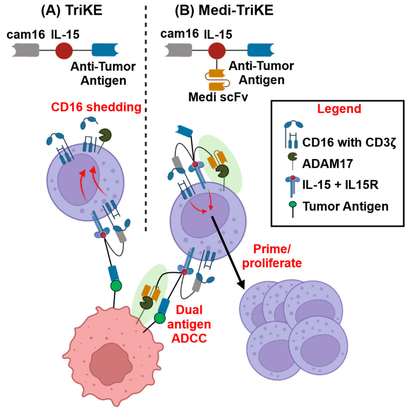
Bi-specific killer engagers (BiKEs) and tri-specific killer engagers (TriKEs): A BiKE consists of single-chain variable fragments (scFvs) and/or variable domains of a heavy chain-only antibody (VHH) targeting CD16 and a tumor antigen [101]. Early BiKE platforms demonstrated proof-of-concept of NK cell-directed killing but were limited by short serum half-lives, lack of cytokine stimulation, and single-antigen targeting which was vulnerable to antigen escape [102]. A TriKE has the same effector and targeting arms and also consists of an IL-15 moiety as a third component [101,103] (Figure 1). IL-15 is a potent NK cell survival and proliferation cytokine [104]. Current antigen targets with the TriKE platform include B7-H3, CD19, CD33, HER2, and EGFR [101,103,105,106,107,108].
An innate cell engager (ICE) consists of tetravalent bispecific antibody-based platforms developed by Affimed GmbH that use a 2 + 2 format, two anti-CD16 domains and two anti-tumor antigen domains, arranged in a tandem diabody structure [109,110]. The bivalent engagement of both CD16 and the tumor antigen provides higher binding avidity and more potent NK cell activation than monovalent formats [109]. Tumor antigens targeted with the ICE platform include EGFR, BCMA, and CD123 [111,112,113].
Other strategies to target NK cells include adding a second effector arm to an NK cell activation receptor. Innate Pharma developed an antibody-based NK cell engager therapeutic (ANKET) targeting CD16 and NKp46 with a modified IL-2 variant for NK cell proliferation [114]. Dragonfly Therapeutics designed a tri-specific NK cell engager therapy (TriNKET) aiming to target CD16 and NKG2D on NK cells [115]. Strategies targeting activating receptors in addition to CD16 have the potential to stabilize the immunological synapse and lower the threshold of CD16 signaling.

4.2. Engager Molecules in Clinical Development

Multi-engager complexes targeting immune cells that are in clinical development have been discussed in other recent reviews; below is an overview of those utilizing CD16 [116]. AFM13, developed by Affimed GmbH in partnership with MD Anderson Cancer Center, is a CD30/CD16 ICE designed for CD30+ lymphomas, including Hodgkin lymphoma and peripheral T-cell lymphoma [117]. As a bispecific antibody, AFM13 demonstrated modest single-agent activity in relapsed/refractory Hodgkin lymphoma [117,118]. However, pre-loading allogeneic, cytokine-expanded NK cells with AFM13 ex vivo dramatically increased anti-tumor activity [119]. The Phase 1 clinical trial of this AFM13-NK cell combination (NCT04074746) enrolled 42 patients with CD30+ lymphomas refractory to multiple therapies, a population with very poor outcomes on conventional therapies [120]. NK cells were derived from donor cord blood, cytokine-preactivated, expanded, and then pre-complexed with AFM13 prior to infusion [120]. Results showed the overall response rate was 92.9% and the complete response rate was 66.7% [120]. Importantly, no CRS, ICANS, or GvHD was observed, consistent with the known favorable safety profile of NK cell therapies [120]. At a median follow-up of 20 months, 11 patients remained in complete remission [120]. These results represent some of the most compelling clinical data for any NK cell engager strategy to date.
AFM24 is an EGFR/CD16 ICE that targets EGFR-expressing solid tumors, representing Affimed’s extension of the ICE platform into the solid tumor setting [111]. Preclinical studies demonstrated robust AFM24-mediated NK cell activation and killing of EGFR+ tumor cell lines, including cell lines with KRAS or other oncogenic mutations that confer resistance to anti-EGFR antibodies such as cetuximab or panitumumab [111]. The first-in-human Phase I/IIa dose-escalation study of AFM24 monotherapy enrolled 35 patients with advanced EGFR-expressing solid tumors across seven dose cohorts (14–720 mg) [121]. AFM24 demonstrated a favorable safety profile, with toxicities limited to manageable infusion-related reactions, including a single reported case of a grade 3 dose-limiting toxicity [121]. Tumor biopsy data demonstrated activation of innate and adaptive immune responses within the tumor, providing a mechanistic rationale for combination strategies with checkpoint inhibitors or adoptive NK cell therapies [121].
The third Affimed ICE is AFM28, which uses an anti-CD16 effector arm with a CD123-targeting scFv (CD123/CD16) to kill stem and progenitor acute myeloid leukemia (AML) and myelodysplastic syndrome (MDS) cells [113]. In a phase I dose-escalation study (NCT05817058), AFM28 is being evaluated as a monotherapy in patients with relapsed or refractory AML and high-risk MDS [113]. Early clinical data presented in late 2024 and early 2025 demonstrate that AFM28 possesses a manageable safety profile with no dose-limiting toxicities observed up to 300 mg [113]. While two cases of CRS occurred, both were low-grade and resolved without the need for tocilizumab (anti-IL-6R) or other anti-cytokine interventions, and no ICANS was reported [113]. Most notably, in the higher-dose cohorts, AFM28 monotherapy achieved a 40% complete remission [113].
The TriKE platform was pioneered by laboratories at the University of Minnesota in partnership with GT Biopharma (Figure 1A) [101]. The first-generation TriKE, designated GTB-3550 incorporated an IL-15 functional domain between anti-CD16 and anti-CD33 scFvs (CD16/IL-15/CD33) to simultaneously crosslink NK cells to CD33+ myeloid malignancy targets and provide in situ IL-15 signaling to drive NK cell activation, persistence, and in vivo expansion. Preclinical studies showed that GTB-3550 induced potent NK cell killing of AML and MDS targets while overcoming myeloid-derived suppressor cells in the tumor microenvironment. In clinical trials, GTB-3550 was shown to be safe and to reduce bone marrow blast levels by more than 60% in AML patients (NCT03214666). GTB-3550 was discontinued in favor of a second-generation TriKE design, GTB-3650. This multi-engager complex replaced the CD16-targeting scFv with a higher-affinity anti-CD16 VHH. The improved design preserves CD33-targeting specificity and cytokine stimulation while increasing CD16 binding. GTB-3650 is being evaluated in Phase 1/2 clinical trials in MDS and AML (NCT06594445).
Innate Pharma and Sanofi have co-developed the ANKET platform, an approach that targets both NKp46 and CD16 on NK cells as well as a tumor-associated antigen [114]. An NKp46/CD16A/CD123 ANKET molecule was under investigation in Phase 1/2 clinical trials for AML and blastic plasmacytoid dendritic cell neoplasm until March 2026, when Innate Pharma announced it was ending work on ANKET.
Dragonfly Therapeutics has developed the TriNKET platform, which targets NKG2D and CD16 simultaneously on NK cells alongside a tumor-associated antigen. DF1001 was designed to engage HER2 on breast and other HER2+ cancers, such as urothelial carcinoma, esophageal, lung, and colorectal cancers. In a clinical trial, DF1001 showed a positive safety profile, with no dose-limiting toxicities, and the only adverse events reported were low-grade infusion-related reactions and fatigue (NCT04143711) [116,122].

5. Strategies to Overcome ADAM17-Mediated CD16 Shedding for NK Engagers

5.1. Anti-ADAM17 Blocking Antibodies

The NK cell engagers discussed rely on CD16 expression to mediate anti-tumor cytotoxicity; however, ADAM17 activation limits the potential of these therapies. As mentioned above, ADAM17 induction occurs in response to various stimuli, including CD16 ligation by engager molecules (Figure 1A) [41,99]. Therefore, it is predicted that the efficacy of NK cell engagers will be enhanced by blocking ADAM17 proteolytic function. Various inhibitors of ADAM17 have been described [51]. Early attempts to inhibit ADAM17 pharmacologically with broad-spectrum small-molecule metalloproteinase inhibitors, including hydroxamate-class compounds that target the zinc-binding active site shared by ADAM17, ADAM10, and MMP family members, were hampered by poor selectivity and dose-limiting off-target toxicities [123,124,125]. For instance, broad-spectrum MMP inhibitors that also target ADAMs, such as marimastat, prinomastat, and tanomastat, failed to progress beyond phase III clinical trials due to lack of efficacy and toxicity [44,126]. Even the two ADAM17-targeted inhibitors, BMS-561392 and TMI-005, retained cross-reactive inhibitory activity and were halted in a phase II clinical trial due to similar efficacy and toxicity issues [44]. These experiences highlight the importance of ADAM17-specific and NK cell-directed ADAM17 inhibition strategies, an objective that can be addressed by the multi-engager complexes described in this section.
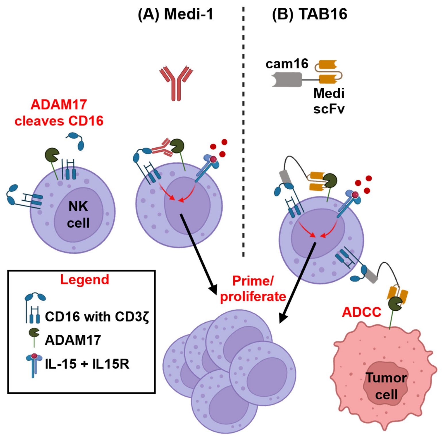
MEDI3622 is a specific ADAM17 function-blocking monoclonal antibody developed by MedImmune to block the release of EGFR ligands in solid tumors [127]. Various Fc variants of this mAb have been generated, including the human isotypes IgG1 (Medi-1), IgG4 (Medi-4), Medi-F(ab′)2 and Medi-PGLALA to impair FcγR binding, and Medi-scFv [41,42]. Medi-1 blocks ADAM17 function in NK cells, preventing CD16 shedding, while its Fc region is simultaneously engaged by CD16, thereby inducing and prolonging CD16 signaling (Figure 2A) [42]. This synergizes with cytokine stimulation, such as IL-15 and IL-2, enhancing NK cell activation and proliferation [42]. The latter process involves a mechanism of enhanced CD137 (4-1BB) upregulation on NK cells and its engagement of CD137L on other PBMCs [42]. Additionally, blocking CD62L shedding, an ADAM17 substrate that governs NK cell trafficking to proliferative niches, enhanced in vivo NK cell expansion [100]. We are aware of only three other anti-human ADAM17 function-blocking mAbs, D1(A12) [128], which does not have the same functional effects on NK cells as Medi-1 [42], and D8P1C1 and C12, which do not block ADAM17 in hematopoietic cells [129,130].

5.2. Next-Generation Engagers Incorporating ADAM17 Inhibition

We developed a novel bispecific engager termed Targeted ADAM17 Blocker CD16 (TAB16), consisting of Medi-scFv to block ADAM17 function linked to a VHH that binds CD16 with high affinity and in a uniform manner (Figure 2B) [41]. TAB16 directed ADAM17 inhibition to NK cells and augmented IL-15-driven activation and proliferation similar to Medi-1 [41]. Importantly, TAB16 also linked NK cells to ADAM17 overexpressed by ovarian cancer cell lines to mediate ADCC, demonstrating dual functionality [41]. The TAB16 engager platform is amenable to modifications, such as the linkage of an IL-15 moiety, which we incorporated into the complex to generate TAB16/15 [41]. By integrating both into a single molecule, TAB16/15 eliminates lot-to-lot variability of the individual components. We show that TAB16/15 maintains NK cell specificity in blocking ADAM17 and in increasing proliferation, while mediating ADCC against ovarian cancer cell lines, with no additional IL-15 required [41]. Our ADAM17-targeting strategy offers a novel means to prevent the shedding of critical receptors in a cell-selective manner.

6. Conclusions and Future Directions

NK cell multi-engager complexes represent an important class of immunotherapeutics that address key limitations of monoclonal antibodies and adoptive cell therapies. CD16 is the only NK cell activation receptor capable of inducing full degranulation and cytokine secretion in the absence of co-stimulatory signals [2]. By linking CD16 on NK cells to tumor targets, these engagers can rapidly direct innate immune cell cytotoxicity and circumvent polymorphic differences in CD16 binding affinities for IgG1. However, ADAM17 induction upon NK cell activation by CD16, other activating receptors, various cytokines, and immunosuppressive components of the tumor microenvironment, represents an important impediment to sustained tumor cell killing by ADCC. The anti-ADAM17 mAb Medi-1 blocks ectodomain shedding and promotes cytokine synergy via CD16 signaling, augmenting NK cell priming and proliferative capacity [42]. To address on-target, off-NK cell issues with Medi-1, we have demonstrated that Medi-scFv can be incorporated into anti-CD16 engager complexes for NK cell-directed ADAM17 inhibition [41].
Medi-scFv could serve as a modular component of multi-engager complexes. For instance, Medi-scFv incorporated into TriKEs (i.e., Medi-TriKEs) would block ADAM17 function in NK cells and target them to assorted tumor antigens (Figure 1B). Khaw et al. recently reported a TriKE comprising a VHH to CD16, an IL-15 moiety, and a novel VHH that binds B7-H3, which is overexpressed in a range of solid tumors [105]. Incorporating Medi-scFv into this TriKE may enhance its function in several ways; for instance, by preventing CD16 downregulation in NK cells, resulting in increased ADCC potency and IL-15 synergy. The Medi-scFv and B7-H3-VHH components would enable dual antigen targeting in solid tumors overexpressing ADAM17 and B7-H3, thereby reducing the potential for antigen-escape variants. Medi-scFv could also inhibit the sheddase in tumor cells, retaining NK cell ligands, such as MICA/B and NR3LG1, and preventing the release of EGFR ligands that promote tumor cell growth. Simultaneously blocking ADAM17 on NK cells and tumor cells could optimize the functional transition between activating pathways. As demonstrated by Srpan et al., prolonged CD16 stimulation eventually diminishes perforin expression; however, switching to alternative pathways like NKG2D stimulation can fully restore perforin release [39]. By utilizing Medi-scFv to inhibit ADAM17 on the tumor cell, the crucial NKG2D ligands (MICA/B) are preserved rather than shed. Thus, if NK cells face CD16-mediated perforin decline, they can effectively switch to a sustained, NKG2D-driven cytotoxic response.
Multi-engager complexes containing Medi-scFv are not restricted to enhancing NK cell function and could be used to link other immune cell populations to tumor cells. Bispecific T-cell Engagers and Tri-specific T-cell Engagers (BiTEs and TriTEs, respectively) link cytotoxic T cells to tumor cells for direct killing. The addition of Medi-1 scFv may also benefit these engager complexes. For instance, by (1) blocking key ADAM17 substrates on T cells, such as CD62L, to enhance their trafficking to secondary lymphoid organs and inflammatory tumor microenvironments; (2) blocking ADAM17 substrates on tumor cells; and (3) targeting T cells to tumors that overexpress ADAM17.
Medi-TriKEs may also have broader applications beyond cancer. They could potentially be used to target pathogenic B or T cells involved in autoimmunity or to treat infectious diseases, such as HIV, by including an envelope protein-targeting arm.
The incorporation of ADAM17 inhibition into multi-engager complexes addresses the consequences of the proteolytic checkpoint in NK cells and its role in immune evasion by tumor cells. This approach does more than enhance cytotoxic output by NK cells; it enhances interaction between the effector cell and the malignancy by stabilizing surface ligands and neutralizing pro-tumorigenic shedding. With further clinical development, these Medi-scFv-containing complexes may be pivotal in driving cell-selective ADAM17 inhibition to enhance success in treatments for solid tumors.

Funding

This work was supported by US Department of Defense Army HT94252510503 and NIH R01CA203348.

Institutional Review Board Statement

Not applicable.

Informed Consent Statement

Not applicable.

Data Availability Statement

No new data were created or analyzed in this study. Data sharing is not applicable to this article.

Conflicts of Interest

The authors declare no conflicts of interest.

References

  1. Mace, E.M. Human natural killer cells: Form, function, and development. J. Allergy Clin. Immunol. 2023, 151, 371–385. [Google Scholar] [CrossRef] [PubMed]
  2. Chen, S.; Zhu, H.; Jounaidi, Y. Comprehensive snapshots of natural killer cells functions, signaling, molecular mechanisms and clinical utilization. Signal Transduct. Target. Ther. 2024, 9, 302. [Google Scholar] [CrossRef] [PubMed]
  3. Long, E.O.; Kim, H.S.; Liu, D.; Peterson, M.E.; Rajagopalan, S. Controlling natural killer cell responses: Integration of signals for activation and inhibition. Annu. Rev. Immunol. 2013, 31, 227–258. [Google Scholar] [CrossRef] [PubMed]
  4. Abel, A.M.; Yang, C.; Thakar, M.S.; Malarkannan, S. Natural Killer Cells: Development, Maturation, and Clinical Utilization. Front. Immunol. 2018, 9, 1869. [Google Scholar] [CrossRef] [PubMed]
  5. Gauthier, M.; Laroye, C.; Bensoussan, D.; Boura, C.; Decot, V. Natural Killer cells and monoclonal antibodies: Two partners for successful antibody dependent cytotoxicity against tumor cells. Crit. Rev. Oncol. Hematol. 2021, 160, 103261. [Google Scholar] [CrossRef] [PubMed]
  6. Cappell, K.M.; Kochenderfer, J.N. Long-term outcomes following CAR T cell therapy: What we know so far. Nat. Rev. Clin. Oncol. 2023, 20, 359–371. [Google Scholar] [CrossRef] [PubMed]
  7. Dimitrov, K.; Merkle, F.; Dimitrov, M.; Merkle, S.; Hoover, A.; Bachanova, V. Major Adverse Events With Chimeric Antigen Receptor T-Cell Therapy: Presentation, Diagnosis, and Resuscitation. Ann. Emerg. Med. 2026, 87, 229–238. [Google Scholar] [CrossRef] [PubMed]
  8. Goto, H. CRS and ICANS in CAR T-cell therapy for large B-cell lymphoma. J. Clin. Exp. Hematop. 2025, 65, 268–275. [Google Scholar] [CrossRef] [PubMed]
  9. Choi, G.; Shin, G.; Bae, S. Price and Prejudice? The Value of Chimeric Antigen Receptor (CAR) T-Cell Therapy. Int. J. Environ. Res. Public Health 2022, 19, 12366. [Google Scholar] [CrossRef] [PubMed]
  10. Lin, H.; Yang, X.; Ye, S.; Huang, L.; Mu, W. Antigen escape in CAR-T cell therapy: Mechanisms and overcoming strategies. Biomed. Pharmacother. 2024, 178, 117252. [Google Scholar] [CrossRef] [PubMed]
  11. Kennedy, P.R.; Felices, M.; Miller, J.S. Challenges to the broad application of allogeneic natural killer cell immunotherapy of cancer. Stem Cell Res. Ther. 2022, 13, 165. [Google Scholar] [CrossRef] [PubMed]
  12. Vivier, E.; Rebuffet, L.; Narni-Mancinelli, E.; Cornen, S.; Igarashi, R.Y.; Fantin, V.R. Natural killer cell therapies. Nature 2024, 626, 727–736. [Google Scholar] [CrossRef] [PubMed]
  13. Dixon, K.J.; Wu, J.; Walcheck, B. Engineering Anti-Tumor Monoclonal Antibodies and Fc Receptors to Enhance ADCC by Human NK Cells. Cancers 2021, 13, 312. [Google Scholar] [CrossRef] [PubMed]
  14. Phung, S.K.; Miller, J.S.; Felices, M. Bi-specific and Tri-specific NK Cell Engagers: The New Avenue of Targeted NK Cell Immunotherapy. Mol. Diagn. Ther. 2021, 25, 577–592. [Google Scholar] [CrossRef] [PubMed]
  15. Nikkhoi, S.K.; Li, G.; Hatefi, A. Natural killer cell engagers for cancer immunotherapy. Front. Oncol. 2024, 14, 1483884. [Google Scholar] [PubMed]
  16. Rolin, C.; Zimmer, J.; Seguin-Devaux, C. Bridging the gap with multispecific immune cell engagers in cancer and infectious diseases. Cell. Mol. Immunol. 2024, 21, 643–661. [Google Scholar] [CrossRef] [PubMed]
  17. Poli, A.; Michel, T.; Theresine, M.; Andres, E.; Hentges, F.; Zimmer, J. CD56bright natural killer (NK) cells: An important NK cell subset. Immunology 2009, 126, 458–465. [Google Scholar] [CrossRef] [PubMed]
  18. Shifrin, N.; Raulet, D.H.; Ardolino, M. NK cell self tolerance, responsiveness and missing self recognition. Semin. Immunol. 2014, 26, 138–144. [Google Scholar] [CrossRef] [PubMed]
  19. Liu, H.; Wang, S.; Xin, J.; Wang, J.; Yao, C.; Zhang, Z. Role of NKG2D and its ligands in cancer immunotherapy. Am. J. Cancer Res. 2019, 9, 2064–2078. [Google Scholar] [PubMed]
  20. Pazina, T.; Shemesh, A.; Brusilovsky, M.; Porgador, A.; Campbell, K.S. Regulation of the Functions of Natural Cytotoxicity Receptors by Interactions with Diverse Ligands and Alterations in Splice Variant Expression. Front. Immunol. 2017, 8, 369. [Google Scholar] [CrossRef] [PubMed]
  21. Mota, G.; Moldovan, I.; Calugaru, A.; Hirt, M.; Kozma, E.; Galatiuc, C.; Brasoveanu, L.; Boltz-Nitulescu, G. Interaction of human immunoglobulin G with CD16 on natural killer cells: Ligand clearance, FcgammaRIIIA turnover and effects of metalloproteinases on FcgammaRIIIA-mediated binding, signal transduction and killing. Scand. J. Immunol. 2004, 59, 278–284. [Google Scholar] [CrossRef] [PubMed]
  22. Beelen, N.A.; Ehlers, F.A.I.; Bos, G.M.J.; Wieten, L. Inhibitory receptors for HLA class I as immune checkpoints for natural killer cell-mediated antibody-dependent cellular cytotoxicity in cancer immunotherapy. Cancer Immunol. Immunother. 2023, 72, 797–804. [Google Scholar] [PubMed]
  23. Golay, J.; Manganini, M.; Facchinetti, V.; Gramigna, R.; Broady, R.; Borleri, G.; Rambaldi, A.; Introna, M. Rituximab-mediated antibody-dependent cellular cytotoxicity against neoplastic B cells is stimulated strongly by interleukin-2. Haematologica 2003, 88, 1002–1012. [Google Scholar] [PubMed]
  24. Baysal, H.; De Pauw, I.; Zaryouh, H.; De Waele, J.; Peeters, M.; Pauwels, P.; Vermorken, J.B.; Smits, E.; Lardon, F.; Jacobs, J.; et al. Cetuximab-induced natural killer cell cytotoxicity in head and neck squamous cell carcinoma cell lines: Investigation of the role of cetuximab sensitivity and HPV status. Br. J. Cancer 2020, 123, 752–761. [Google Scholar] [CrossRef] [PubMed]
  25. Tian, X.; Wei, F.; Wang, L.; Yu, W.; Zhang, N.; Zhang, X.; Han, Y.; Yu, J.; Ren, X. Herceptin Enhances the Antitumor Effect of Natural Killer Cells on Breast Cancer Cells Expressing Human Epidermal Growth Factor Receptor-2. Front. Immunol. 2017, 8, 1426. [Google Scholar] [CrossRef] [PubMed]
  26. Prager, I.; Watzl, C. Mechanisms of natural killer cell-mediated cellular cytotoxicity. J. Leukoc. Biol. 2019, 105, 1319–1329. [Google Scholar] [CrossRef] [PubMed]
  27. Koene, H.R.; Kleijer, M.; Algra, J.; Roos, D.; von dem Borne, A.E.; de Haas, M. Fc gammaRIIIa-158V/F polymorphism influences the binding of IgG by natural killer cell Fc gammaRIIIa, independently of the Fc gammaRIIIa-48L/R/H phenotype. Blood 1997, 90, 1109–1114. [Google Scholar] [CrossRef] [PubMed]
  28. Vance, B.A.; Huizinga, T.W.; Wardwell, K.; Guyre, P.M. Binding of monomeric human IgG defines an expression polymorphism of Fc gamma RIII on large granular lymphocyte/natural killer cells. J. Immunol. 1993, 151, 6429–6439. [Google Scholar] [CrossRef] [PubMed]
  29. Dong, C.; Ptacek, T.S.; Redden, D.T.; Zhang, K.; Brown, E.E.; Edberg, J.C.; McGwin, G., Jr.; Alarcon, G.S.; Ramsey-Goldman, R.; Reveille, J.D.; et al. Fcgamma receptor IIIa single-nucleotide polymorphisms and haplotypes affect human IgG binding and are associated with lupus nephritis in African Americans. Arthritis Rheumatol. 2014, 66, 1291–1299. [Google Scholar] [CrossRef] [PubMed]
  30. Wu, J.; Edberg, J.C.; Redecha, P.B.; Bansal, V.; Guyre, P.M.; Coleman, K.; Salmon, J.E.; Kimberly, R.P. A novel polymorphism of FcgammaRIIIa (CD16) alters receptor function and predisposes to autoimmune disease. J. Clin. Investig. 1997, 100, 1059–1070. [Google Scholar] [CrossRef] [PubMed]
  31. Cartron, G.; Dacheux, L.; Salles, G.; Solal-Celigny, P.; Bardos, P.; Colombat, P.; Watier, H. Therapeutic activity of humanized anti-CD20 monoclonal antibody and polymorphism in IgG Fc receptor FcgammaRIIIa gene. Blood 2002, 99, 754–758. [Google Scholar] [CrossRef] [PubMed]
  32. Musolino, A.; Naldi, N.; Dieci, M.V.; Zanoni, D.; Rimanti, A.; Boggiani, D.; Sgargi, P.; Generali, D.G.; Piacentini, F.; Ambroggi, M.; et al. Immunoglobulin G fragment C receptor polymorphisms and efficacy of preoperative chemotherapy plus trastuzumab and lapatinib in HER2-positive breast cancer. Pharmacogenom. J. 2016, 16, 472–477. [Google Scholar] [CrossRef]
  33. Bibeau, F.; Lopez-Crapez, E.; Di Fiore, F.; Thezenas, S.; Ychou, M.; Blanchard, F.; Lamy, A.; Penault-Llorca, F.; Frebourg, T.; Michel, P.; et al. Impact of FcgammaRIIa-FcgammaRIIIa polymorphisms and KRAS mutations on the clinical outcome of patients with metastatic colorectal cancer treated with cetuximab plus irinotecan. J. Clin. Oncol. 2009, 27, 1122–1129. [Google Scholar] [CrossRef] [PubMed]
  34. Jing, Y.; Ni, Z.; Wu, J.; Higgins, L.; Markowski, T.W.; Kaufman, D.S.; Walcheck, B. Identification of an ADAM17 cleavage region in human CD16 (FcgammaRIII) and the engineering of a non-cleavable version of the receptor in NK cells. PLoS ONE 2015, 10, e0121788. [Google Scholar] [CrossRef] [PubMed]
  35. Lajoie, L.; Congy-Jolivet, N.; Bolzec, A.; Gouilleux-Gruart, V.; Sicard, E.; Sung, H.C.; Peiretti, F.; Moreau, T.; Vie, H.; Clemenceau, B.; et al. ADAM17-mediated shedding of FcgammaRIIIA on human NK cells: Identification of the cleavage site and relationship with activation. J. Immunol. 2014, 192, 741–751. [Google Scholar] [CrossRef] [PubMed]
  36. Romee, R.; Foley, B.; Lenvik, T.; Wang, Y.; Zhang, B.; Ankarlo, D.; Luo, X.; Cooley, S.; Verneris, M.; Walcheck, B.; et al. NK cell CD16 surface expression and function is regulated by a disintegrin and metalloprotease-17 (ADAM17). Blood 2013, 121, 3599–3608. [Google Scholar] [CrossRef] [PubMed]
  37. Wu, J.; Mishra, H.K.; Walcheck, B. Role of ADAM17 as a regulatory checkpoint of CD16A in NK cells and as a potential target for cancer immunotherapy. J. Leukoc. Biol. 2019, 105, 1297–1303. [Google Scholar] [CrossRef] [PubMed]
  38. Wang, K.; Xuan, Z.; Liu, X.; Zheng, M.; Yang, C.; Wang, H. Immunomodulatory role of metalloproteinase ADAM17 in tumor development. Front. Immunol. 2022, 13, 1059376. [Google Scholar] [CrossRef] [PubMed]
  39. Srpan, K.; Ambrose, A.; Karampatzakis, A.; Saeed, M.; Cartwright, A.N.R.; Guldevall, K.; De Matos, G.; Onfelt, B.; Davis, D.M. Shedding of CD16 disassembles the NK cell immune synapse and boosts serial engagement of target cells. J. Cell Biol. 2018, 217, 3267–3283. [Google Scholar] [CrossRef] [PubMed]
  40. Pimenta, R.; Arai, J.; Ribeiro de Lima Brandao, L.; Schmidt, P.; da Silva Bortoleti, B.T.; Ferrari de Andrade, L. Insights into ectodomain shedding as a novel target in natural killer cell-based immunotherapy for cancer. Front. Immunol. 2026, 17, 1753470. [Google Scholar] [CrossRef] [PubMed]
  41. Dixon, K.J.; Hullsiek, R.; Wang, S.; Friess, R.R.; Matson, A.W.; Soignier, Y.; Lenvik, A.J.; Wu, J.; Hart, G.T.; Felices, M.; et al. Dual-functioning Targeted ADAM17 Blocker CD16 (TAB16) mediates selective ADAM17 inhibition in NK cells and engages overexpressed ADAM17 in tumor cells to induce cytotoxicity. Front. Immunol. 2026, 17, 1714022. [Google Scholar] [CrossRef] [PubMed]
  42. Matson, A.W.; Hullsiek, R.; Dixon, K.J.; Wang, S.; Lindstedt, A.J.; Friess, R.R.; Phung, S.K.; Freedman, T.S.; Felices, M.; Truckenbrod, E.N.; et al. Enhanced IL-15-mediated NK cell activation and proliferation by an ADAM17 function-blocking antibody involves CD16A, CD137, and accessory cells. J. Immunother. Cancer 2024, 12, e008959. [Google Scholar] [CrossRef] [PubMed]
  43. da Silva Bortoleti, B.T.; Quasem, S.; Maurer, S.; Zhong, X.; Pimenta, R.; Ribeiro de Lima Brandão, L.; Hernandez, M.; Fraidenburg, M.; Alves da Silva, P.H.; Alvarez, R.; et al. A monoclonal antibody that inhibits the shedding of CD16a and CD16b and promotes antibody-dependent cellular cytotoxicity against tumors. Nat. Commun. 2025, 16, 9915. [Google Scholar] [CrossRef] [PubMed]
  44. Murumkar, P.R.; Giridhar, R.; Yadav, M.R. Novel methods and strategies in the discovery of TACE inhibitors. Expert Opin. Drug Discov. 2013, 8, 157–181. [Google Scholar] [PubMed]
  45. Mishra, H.K.; Pore, N.; Michelotti, E.F.; Walcheck, B. Anti-ADAM17 monoclonal antibody MEDI3622 increases IFNgamma production by human NK cells in the presence of antibody-bound tumor cells. Cancer Immunol. Immunother. 2018, 67, 1407–1416. [Google Scholar] [CrossRef] [PubMed]
  46. Pham, D.H.; Kim, J.S.; Kim, S.K.; Shin, D.J.; Uong, N.T.; Hyun, H.; Yoon, M.S.; Kang, S.J.; Ryu, Y.J.; Cho, J.S.; et al. Effects of ADAM10 and ADAM17 Inhibitors on Natural Killer Cell Expansion and Antibody-dependent Cellular Cytotoxicity Against Breast Cancer Cells In Vitro. Anticancer Res. 2017, 37, 5507–5513. [Google Scholar] [CrossRef] [PubMed]
  47. Zhu, H.; Blum, R.H.; Bjordahl, R.; Gaidarova, S.; Rogers, P.; Lee, T.T.; Abujarour, R.; Bonello, G.B.; Wu, J.; Tsai, P.F.; et al. Pluripotent stem cell-derived NK cells with high-affinity noncleavable CD16a mediate improved antitumor activity. Blood 2020, 135, 399–410. [Google Scholar] [CrossRef] [PubMed]
  48. Pomeroy, E.J.; Hunzeker, J.T.; Kluesner, M.G.; Lahr, W.S.; Smeester, B.A.; Crosby, M.R.; Lonetree, C.L.; Yamamoto, K.; Bendzick, L.; Miller, J.S.; et al. A Genetically Engineered Primary Human Natural Killer Cell Platform for Cancer Immunotherapy. Mol. Ther. 2020, 28, 52–63. [Google Scholar] [CrossRef] [PubMed]
  49. Zunke, F.; Rose-John, S. The shedding protease ADAM17: Physiology and pathophysiology. Biochim. Biophys. Acta Mol. Cell Res. 2017, 1864, 2059–2070. [Google Scholar] [CrossRef] [PubMed]
  50. Saad, M.I.; Jenkins, B.J. The protease ADAM17 at the crossroads of disease: Revisiting its significance in inflammation, cancer, and beyond. FEBS J. 2024, 291, 10–24. [Google Scholar] [PubMed]
  51. Liu, L.; Tian, E.; Quan, S.; Su, C.; Zhou, J.; Hu, S.; Bian, N.; Du, S.; Li, J. ADAM17 as a promising therapeutic target: From structural basis to inhibitor discovery in human diseases. Front. Pharmacol. 2025, 16, 1640090. [Google Scholar] [CrossRef] [PubMed]
  52. Saad, M.I.; Rose-John, S.; Jenkins, B.J. ADAM17: An Emerging Therapeutic Target for Lung Cancer. Cancers 2019, 11, 1218. [Google Scholar] [CrossRef] [PubMed]
  53. Rubina, A.; Patel, M.; Nightingale, K.; Potts, M.; Fielding, C.A.; Kollnberger, S.; Lau, B.; Ladell, K.; Miners, K.L.; Nichols, J.; et al. ADAM17 targeting by human cytomegalovirus remodels the cell surface proteome to simultaneously regulate multiple immune pathways. Proc. Natl. Acad. Sci. USA 2023, 120, e2303155120. [Google Scholar] [CrossRef] [PubMed]
  54. Mishra, H.K.; Ma, J.; Walcheck, B. Ectodomain Shedding by ADAM17: Its Role in Neutrophil Recruitment and the Impairment of This Process during Sepsis. Front. Cell. Infect. Microbiol. 2017, 7, 138. [Google Scholar] [CrossRef] [PubMed]
  55. Peschon, J.J.; Slack, J.L.; Reddy, P.; Stocking, K.L.; Sunnarborg, S.W.; Lee, D.C.; Russell, W.E.; Castner, B.J.; Johnson, R.S.; Fitzner, J.N.; et al. An essential role for ectodomain shedding in mammalian development. Science 1998, 282, 1281–1284. [Google Scholar] [CrossRef] [PubMed]
  56. Bandsma, R.H.; van Goor, H.; Yourshaw, M.; Horlings, R.K.; Jonkman, M.F.; Scholvinck, E.H.; Karrenbeld, A.; Scheenstra, R.; Komhoff, M.; Rump, P.; et al. Loss of ADAM17 is associated with severe multiorgan dysfunction. Hum. Pathol. 2015, 46, 923–928. [Google Scholar] [CrossRef] [PubMed]
  57. Adrain, C.; Zettl, M.; Christova, Y.; Taylor, N.; Freeman, M. Tumor necrosis factor signaling requires iRhom2 to promote trafficking and activation of TACE. Science 2012, 335, 225–228. [Google Scholar] [CrossRef] [PubMed]
  58. Christova, Y.; Adrain, C.; Bambrough, P.; Ibrahim, A.; Freeman, M. Mammalian iRhoms have distinct physiological functions including an essential role in TACE regulation. EMBO Rep. 2013, 14, 884–890. [Google Scholar] [CrossRef] [PubMed]
  59. Issuree, P.D.; Maretzky, T.; McIlwain, D.R.; Monette, S.; Qing, X.; Lang, P.A.; Swendeman, S.L.; Park-Min, K.H.; Binder, N.; Kalliolias, G.D.; et al. iRHOM2 is a critical pathogenic mediator of inflammatory arthritis. J. Clin. Investig. 2013, 123, 928–932. [Google Scholar] [CrossRef] [PubMed]
  60. Wong, E.; Maretzky, T.; Peleg, Y.; Blobel, C.P.; Sagi, I. The Functional Maturation of A Disintegrin and Metalloproteinase (ADAM) 9, 10, and 17 Requires Processing at a Newly Identified Proprotein Convertase (PC) Cleavage Site. J. Biol. Chem. 2015, 290, 12135–12146. [Google Scholar] [CrossRef] [PubMed]
  61. Rzymski, T.; Petry, A.; Kracun, D.; Riess, F.; Pike, L.; Harris, A.L.; Gorlach, A. The unfolded protein response controls induction and activation of ADAM17/TACE by severe hypoxia and ER stress. Oncogene 2012, 31, 3621–3634. [Google Scholar] [PubMed]
  62. Zhu, G.; Liu, J.; Wang, Y.; Jia, N.; Wang, W.; Zhang, J.; Liang, Y.; Tian, H.; Zhang, J. ADAM17 Mediates Hypoxia-Induced Keratinocyte Migration via the p38/MAPK Pathway. BioMed Res. Int. 2021, 2021, 8328216. [Google Scholar] [CrossRef] [PubMed]
  63. Kishimoto, T.K.; Jutila, M.A.; Berg, E.L.; Butcher, E.C. Neutrophil Mac-1 and MEL-14 adhesion proteins inversely regulated by chemotactic factors. Science 1989, 245, 1238–1241. [Google Scholar] [CrossRef] [PubMed]
  64. Walcheck, B.; Kahn, J.; Fisher, J.M.; Wang, B.B.; Fisk, R.S.; Payan, D.G.; Feehan, C.; Betageri, R.; Darlak, K.; Spatola, A.F.; et al. Neutrophil rolling altered by inhibition of L-selectin shedding in vitro. Nature 1996, 380, 720–723. [Google Scholar] [CrossRef] [PubMed]
  65. Xu, P.; Liu, J.; Sakaki-Yumoto, M.; Derynck, R. TACE activation by MAPK-mediated regulation of cell surface dimerization and TIMP3 association. Sci. Signal. 2012, 5, ra34. [Google Scholar] [CrossRef] [PubMed]
  66. Wang, Y.; Herrera, A.H.; Li, Y.; Belani, K.K.; Walcheck, B. Regulation of mature ADAM17 by redox agents for L-selectin shedding. J. Immunol. 2009, 182, 2449–2457. [Google Scholar] [CrossRef] [PubMed]
  67. Arora, S.; Scott, A.M.; Janes, P.W. ADAM Proteases in Cancer: Biological Roles, Therapeutic Challenges, and Emerging Opportunities. Cancers 2025, 17, 1703. [Google Scholar] [CrossRef] [PubMed]
  68. Buchanan, P.C.; Boylan, K.L.M.; Walcheck, B.; Heinze, R.; Geller, M.A.; Argenta, P.A.; Skubitz, A.P.N. Ectodomain shedding of the cell adhesion molecule Nectin-4 in ovarian cancer is mediated by ADAM10 and ADAM17. J. Biol. Chem. 2017, 292, 6339–6351. [Google Scholar] [CrossRef] [PubMed]
  69. Dosch, J.; Ziemke, E.; Wan, S.; Luker, K.; Welling, T.; Hardiman, K.; Fearon, E.; Thomas, S.; Flynn, M.; Rios-Doria, J.; et al. Targeting ADAM17 inhibits human colorectal adenocarcinoma progression and tumor-initiating cell frequency. Oncotarget 2017, 8, 65090–65099. [Google Scholar] [CrossRef] [PubMed]
  70. Fabbi, M.; Costa, D.; Russo, D.; Arenare, L.; Gaggero, G.; Signoriello, S.; Scambia, G.; Pisano, C.; Colombo, N.; Losito, N.S.; et al. Analysis of A Disintegrin and Metalloprotease 17 (ADAM17) Expression as a Prognostic Marker in Ovarian Cancer Patients Undergoing First-Line Treatment Plus Bevacizumab. Diagnostics 2022, 12, 2118. [Google Scholar] [CrossRef] [PubMed]
  71. Liu, J.; Ye, L.; Lin, K.; Zhong, T.; Luo, J.; Wang, T.; Suo, L.; Mo, Q.; Li, S.; Chen, Q.; et al. miR-4299 inhibits tumor progression in pancreatic cancer through targeting ADAM17. Mol. Cell. Biochem. 2023, 478, 1727–1742. [Google Scholar] [PubMed]
  72. McGowan, P.M.; McKiernan, E.; Bolster, F.; Ryan, B.M.; Hill, A.D.; McDermott, E.W.; Evoy, D.; O’Higgins, N.; Crown, J.; Duffy, M.J. ADAM-17 predicts adverse outcome in patients with breast cancer. Ann. Oncol. 2008, 19, 1075–1081. [Google Scholar] [CrossRef] [PubMed]
  73. McGowan, P.M.; Mullooly, M.; Caiazza, F.; Sukor, S.; Madden, S.F.; Maguire, A.A.; Pierce, A.; McDermott, E.W.; Crown, J.; O’Donovan, N.; et al. ADAM-17: A novel therapeutic target for triple negative breast cancer. Ann. Oncol. 2013, 24, 362–369. [Google Scholar] [CrossRef] [PubMed]
  74. Yang, Z.; Chan, K.I.; Kwok, H.F.; Tam, K.Y. Novel Therapeutic Anti-ADAM17 Antibody A9(B8) Enhances EGFR-TKI-Mediated Anticancer Activity in NSCLC. Transl. Oncol. 2019, 12, 1516–1524. [Google Scholar] [CrossRef] [PubMed]
  75. Rogmans, C.; Kuhlmann, J.D.; Hugendieck, G.; Link, T.; Arnold, N.; Weimer, J.P.; Florkemeier, I.; Rambow, A.C.; Lieb, W.; Maass, N.; et al. ADAM17-A Potential Blood-Based Biomarker for Detection of Early-Stage Ovarian Cancer. Cancers 2021, 13, 5563. [Google Scholar] [CrossRef] [PubMed]
  76. Uribe, M.L.; Marrocco, I.; Yarden, Y. EGFR in Cancer: Signaling Mechanisms, Drugs, and Acquired Resistance. Cancers 2021, 13, 2748. [Google Scholar] [CrossRef] [PubMed]
  77. Klupp, F.; Sass, M.; Bergmann, F.; Khajeh, E.; Ghamarnejad, O.; Hassenpflug, M.; Mehrabi, A.; Kulu, Y. Impact of EGFR and EGFR ligand expression on treatment response in patients with metastatic colorectal cancer. Oncol. Lett. 2021, 21, 448. [Google Scholar] [CrossRef] [PubMed]
  78. Yonesaka, K.; Takegawa, N.; Satoh, T.; Ueda, H.; Yoshida, T.; Takeda, M.; Shimizu, T.; Chiba, Y.; Okamoto, I.; Nishio, K.; et al. Combined Analysis of Plasma Amphiregulin and Heregulin Predicts Response to Cetuximab in Metastatic Colorectal Cancer. PLoS ONE 2015, 10, e0143132. [Google Scholar] [CrossRef] [PubMed]
  79. Kamarajan, P.; Shin, J.M.; Qian, X.; Matte, B.; Zhu, J.Y.; Kapila, Y.L. ADAM17-mediated CD44 cleavage promotes orasphere formation or stemness and tumorigenesis in HNSCC. Cancer Med. 2013, 2, 793–802. [Google Scholar] [CrossRef] [PubMed]
  80. Takamune, Y.; Ikebe, T.; Nagano, O.; Nakayama, H.; Ota, K.; Obayashi, T.; Saya, H.; Shinohara, M. ADAM-17 associated with CD44 cleavage and metastasis in oral squamous cell carcinoma. Virchows Arch. 2007, 450, 169–177. [Google Scholar] [PubMed]
  81. Brandt, C.S.; Baratin, M.; Yi, E.C.; Kennedy, J.; Gao, Z.; Fox, B.; Haldeman, B.; Ostrander, C.D.; Kaifu, T.; Chabannon, C.; et al. The B7 family member B7-H6 is a tumor cell ligand for the activating natural killer cell receptor NKp30 in humans. J. Exp. Med. 2009, 206, 1495–1503. [Google Scholar] [CrossRef]
  82. Schlecker, E.; Fiegler, N.; Arnold, A.; Altevogt, P.; Rose-John, S.; Moldenhauer, G.; Sucker, A.; Paschen, A.; von Strandmann, E.P.; Textor, S.; et al. Metalloprotease-mediated tumor cell shedding of B7-H6, the ligand of the natural killer cell-activating receptor NKp30. Cancer Res. 2014, 74, 3429–3440. [Google Scholar] [CrossRef] [PubMed]
  83. Xing, S.; Ferrari de Andrade, L. NKG2D and MICA/B shedding: A ‘tag game’ between NK cells and malignant cells. Clin. Transl. Immunol. 2020, 9, e1230. [Google Scholar] [CrossRef]
  84. Moreno-Caceres, J.; Mainez, J.; Mayoral, R.; Martin-Sanz, P.; Egea, G.; Fabregat, I. Caveolin-1-dependent activation of the metalloprotease TACE/ADAM17 by TGF-beta in hepatocytes requires activation of Src and the NADPH oxidase NOX1. FEBS J. 2016, 283, 1300–1310. [Google Scholar] [CrossRef] [PubMed]
  85. Brill, A.; Chauhan, A.K.; Canault, M.; Walsh, M.T.; Bergmeier, W.; Wagner, D.D. Oxidative stress activates ADAM17/TACE and induces its target receptor shedding in platelets in a p38-dependent fashion. Cardiovasc. Res. 2009, 84, 137–144. [Google Scholar] [CrossRef] [PubMed]
  86. Huner Yigit, M.; Okcu, O.; Atak, M.; Karabulut, S.; Yildiz, G.; Yigit, E. Modulation of Oxidative and ER Stress Pathways by the ADAM17 Inhibitor GW280264X in LPS-Induced Acute Liver Injury. Life 2025, 15, 1877. [Google Scholar] [CrossRef] [PubMed]
  87. Li, R.; Uttarwar, L.; Gao, B.; Charbonneau, M.; Shi, Y.; Chan, J.S.; Dubois, C.M.; Krepinsky, J.C. High Glucose Up-regulates ADAM17 through HIF-1alpha in Mesangial Cells. J. Biol. Chem. 2015, 290, 21603–21614. [Google Scholar] [CrossRef] [PubMed]
  88. Charbonneau, M.; Harper, K.; Grondin, F.; Pelmus, M.; McDonald, P.P.; Dubois, C.M. Hypoxia-inducible factor mediates hypoxic and tumor necrosis factor alpha-induced increases in tumor necrosis factor-alpha converting enzyme/ADAM17 expression by synovial cells. J. Biol. Chem. 2007, 282, 33714–33724. [Google Scholar] [CrossRef] [PubMed]
  89. Obama, T.; Takayanagi, T.; Kobayashi, T.; Bourne, A.M.; Elliott, K.J.; Charbonneau, M.; Dubois, C.M.; Eguchi, S. Vascular induction of a disintegrin and metalloprotease 17 by angiotensin II through hypoxia inducible factor 1alpha. Am. J. Hypertens. 2015, 28, 10–14. [Google Scholar] [PubMed]
  90. Weinberg, F.; Ramnath, N.; Nagrath, D. Reactive Oxygen Species in the Tumor Microenvironment: An Overview. Cancers 2019, 11, 1191. [Google Scholar] [CrossRef] [PubMed]
  91. Woan, K.V.; Kim, H.; Bjordahl, R.; Davis, Z.B.; Gaidarova, S.; Goulding, J.; Hancock, B.; Mahmood, S.; Abujarour, R.; Wang, H.; et al. Harnessing features of adaptive NK cells to generate iPSC-derived NK cells for enhanced immunotherapy. Cell Stem Cell 2021, 28, 2062–2075.E5. [Google Scholar] [CrossRef] [PubMed]
  92. Cichocki, F.; Bjordahl, R.; Goodridge, J.P.; Mahmood, S.; Gaidarova, S.; Abujarour, R.; Davis, Z.B.; Merino, A.; Tuininga, K.; Wang, H.; et al. Quadruple gene-engineered natural killer cells enable multi-antigen targeting for durable antitumor activity against multiple myeloma. Nat. Commun. 2022, 13, 7341. [Google Scholar] [CrossRef] [PubMed]
  93. Cichocki, F.; Goodridge, J.P.; Bjordahl, R.; Mahmood, S.; Davis, Z.B.; Gaidarova, S.; Abujarour, R.; Groff, B.; Witty, A.; Wang, H.; et al. Dual antigen-targeted off-the-shelf NK cells show durable response and prevent antigen escape in lymphoma and leukemia. Blood 2022, 140, 2451–2462. [Google Scholar] [CrossRef] [PubMed]
  94. Belisle, J.A.; Gubbels, J.A.; Raphael, C.A.; Migneault, M.; Rancourt, C.; Connor, J.P.; Patankar, M.S. Peritoneal natural killer cells from epithelial ovarian cancer patients show an altered phenotype and bind to the tumour marker MUC16 (CA125). Immunology 2007, 122, 418–429. [Google Scholar] [CrossRef] [PubMed]
  95. Rocca, Y.S.; Roberti, M.P.; Julia, E.P.; Pampena, M.B.; Bruno, L.; Rivero, S.; Huertas, E.; Sanchez Loria, F.; Pairola, A.; Caignard, A.; et al. Phenotypic and Functional Dysregulated Blood NK Cells in Colorectal Cancer Patients Can Be Activated by Cetuximab Plus IL-2 or IL-15. Front. Immunol. 2016, 7, 413. [Google Scholar] [CrossRef] [PubMed]
  96. Felices, M.; Chu, S.; Kodal, B.; Bendzick, L.; Ryan, C.; Lenvik, A.J.; Boylan, K.L.M.; Wong, H.C.; Skubitz, A.P.N.; Miller, J.S.; et al. IL-15 super-agonist (ALT-803) enhances natural killer (NK) cell function against ovarian cancer. Gynecol. Oncol. 2017, 145, 453–461. [Google Scholar] [CrossRef] [PubMed]
  97. Jewett, A.; Tseng, H.C. Tumor induced inactivation of natural killer cell cytotoxic function; implication in growth, expansion and differentiation of cancer stem cells. J. Cancer 2011, 2, 443–457. [Google Scholar] [CrossRef] [PubMed]
  98. Lai, P.; Rabinowich, H.; Crowley-Nowick, P.A.; Bell, M.C.; Mantovani, G.; Whiteside, T.L. Alterations in expression and function of signal-transducing proteins in tumor-associated T and natural killer cells in patients with ovarian carcinoma. Clin. Cancer Res. 1996, 2, 161–173. [Google Scholar] [PubMed]
  99. Wiernik, A.; Foley, B.; Zhang, B.; Verneris, M.R.; Warlick, E.; Gleason, M.K.; Ross, J.A.; Luo, X.; Weisdorf, D.J.; Walcheck, B.; et al. Targeting natural killer cells to acute myeloid leukemia in vitro with a CD16 x 33 bispecific killer cell engager and ADAM17 inhibition. Clin. Cancer Res. 2013, 19, 3844–3855. [Google Scholar] [CrossRef] [PubMed]
  100. Mishra, H.K.; Dixon, K.J.; Pore, N.; Felices, M.; Miller, J.S.; Walcheck, B. Activation of ADAM17 by IL-15 Limits Human NK Cell Proliferation. Front. Immunol. 2021, 12, 711621. [Google Scholar] [CrossRef] [PubMed]
  101. Felices, M.; Lenvik, T.R.; Davis, Z.B.; Miller, J.S.; Vallera, D.A. Generation of BiKEs and TriKEs to Improve NK Cell-Mediated Targeting of Tumor Cells. Methods Mol. Biol. 2016, 1441, 333–346. [Google Scholar] [CrossRef] [PubMed]
  102. Gleason, M.K.; Verneris, M.R.; Todhunter, D.A.; Zhang, B.; McCullar, V.; Zhou, S.X.; Panoskaltsis-Mortari, A.; Weiner, L.M.; Vallera, D.A.; Miller, J.S. Bispecific and trispecific killer cell engagers directly activate human NK cells through CD16 signaling and induce cytotoxicity and cytokine production. Mol. Cancer Ther. 2012, 11, 2674–2684. [Google Scholar] [CrossRef] [PubMed]
  103. Felices, M.; Kodal, B.; Hinderlie, P.; Kaminski, M.F.; Cooley, S.; Weisdorf, D.J.; Vallera, D.A.; Miller, J.S.; Bachanova, V. Novel CD19-targeted TriKE restores NK cell function and proliferative capacity in CLL. Blood Adv. 2019, 3, 897–907. [Google Scholar] [CrossRef] [PubMed]
  104. Ma, S.; Yu, J.; Caligiuri, M.A. Natural killer cell-based immunotherapy for cancer. J. Immunol. 2025, 214, 1444–1456. [Google Scholar] [CrossRef] [PubMed]
  105. Khaw, M.J.; Zorko, N.A.; Kennedy, P.R.; Bendzick, L.E.; Shackelford, M.; Selleck, C.; Hinderlie, P.; Walker, J.T.; Soignier, Y.; Lyons, R.C.; et al. Novel trispecific killer engager targeting B7-H3 enhances natural killer cell antitumor activity against head and neck cancer. J. Immunother. Cancer 2025, 13, e011370. [Google Scholar] [CrossRef] [PubMed]
  106. Vallera, D.A.; Felices, M.; McElmurry, R.; McCullar, V.; Zhou, X.; Schmohl, J.U.; Zhang, B.; Lenvik, A.J.; Panoskaltsis-Mortari, A.; Verneris, M.R.; et al. IL15 Trispecific Killer Engagers (TriKE) Make Natural Killer Cells Specific to CD33+ Targets While Also Inducing Persistence, In Vivo Expansion, and Enhanced Function. Clin. Cancer Res. 2016, 22, 3440–3450. [Google Scholar] [CrossRef] [PubMed]
  107. Davis, Z.B.; Vallera, D.A.; Miller, J.S.; Felices, M. Natural killer cells unleashed: Checkpoint receptor blockade and BiKE/TriKE utilization in NK-mediated anti-tumor immunotherapy. Semin. Immunol. 2017, 31, 64–75. [Google Scholar] [CrossRef] [PubMed]
  108. Vallera, D.A.; Oh, F.; Kodal, B.; Hinderlie, P.; Geller, M.A.; Miller, J.S.; Felices, M. A HER2 Tri-Specific NK Cell Engager Mediates Efficient Targeting of Human Ovarian Cancer. Cancers 2021, 13, 3994. [Google Scholar] [CrossRef] [PubMed]
  109. Reusch, U.; Burkhardt, C.; Fucek, I.; Le Gall, F.; Le Gall, M.; Hoffmann, K.; Knackmuss, S.H.; Kiprijanov, S.; Little, M.; Zhukovsky, E.A. A novel tetravalent bispecific TandAb (CD30/CD16A) efficiently recruits NK cells for the lysis of CD30+ tumor cells. mAbs 2014, 6, 728–739. [Google Scholar] [CrossRef] [PubMed]
  110. Ellwanger, K.; Reusch, U.; Fucek, I.; Wingert, S.; Ross, T.; Muller, T.; Schniegler-Mattox, U.; Haneke, T.; Rajkovic, E.; Koch, J.; et al. Redirected optimized cell killing (ROCK(R)): A highly versatile multispecific fit-for-purpose antibody platform for engaging innate immunity. mAbs 2019, 11, 899–918. [Google Scholar] [CrossRef] [PubMed]
  111. Wingert, S.; Reusch, U.; Knackmuss, S.; Kluge, M.; Damrat, M.; Pahl, J.; Schniegler-Mattox, U.; Mueller, T.; Fucek, I.; Ellwanger, K.; et al. Preclinical evaluation of AFM24, a novel CD16A-specific innate immune cell engager targeting EGFR-positive tumors. mAbs 2021, 13, 1950264. [Google Scholar] [CrossRef] [PubMed]
  112. Giang, K.A.; Boxaspen, T.; Diao, Y.; Nilvebrant, J.; Kosugi-Kanaya, M.; Kanaya, M.; Krokeide, S.Z.; Lehmann, F.; Svensson Gelius, S.; Malmberg, K.J.; et al. Affibody-based hBCMA x CD16 dual engagers for NK cell-mediated killing of multiple myeloma cells. New Biotechnol. 2023, 77, 139–148. [Google Scholar] [CrossRef]
  113. Schmitt, N.; Siegler, J.J.; Beck, A.; Muller, T.; Kozlowska, I.; Sarlang, S.; Reusch, U.; Knackmuss, S.; Medina-Echeverz, J.; Koch, J.; et al. The bispecific innate cell engager AFM28 eliminates CD123(+) leukemic stem and progenitor cells in AML and MDS. Nat. Commun. 2025, 16, 7793. [Google Scholar] [CrossRef] [PubMed]
  114. Demaria, O.; Gauthier, L.; Vetizou, M.; Blanchard Alvarez, A.; Vagne, C.; Habif, G.; Batista, L.; Baron, W.; Belaid, N.; Girard-Madoux, M.; et al. Antitumor immunity induced by antibody-based natural killer cell engager therapeutics armed with not-alpha IL-2 variant. Cell Rep. Med. 2022, 3, 100783. [Google Scholar] [CrossRef] [PubMed]
  115. Fenis, A.; Demaria, O.; Gauthier, L.; Vivier, E.; Narni-Mancinelli, E. New immune cell engagers for cancer immunotherapy. Nat. Rev. Immunol. 2024, 24, 471–486. [Google Scholar] [CrossRef] [PubMed]
  116. Thalji, M.; Nieto, Y. Recent advances in bispecific natural killer cell engagers for cancer immunotherapy. Immunotherapy 2026, 18, 579–599. [Google Scholar] [CrossRef] [PubMed]
  117. Bartlett, N.L.; Herrera, A.F.; Domingo-Domenech, E.; Mehta, A.; Forero-Torres, A.; Garcia-Sanz, R.; Armand, P.; Devata, S.; Izquierdo, A.R.; Lossos, I.S.; et al. A phase 1b study of AFM13 in combination with pembrolizumab in patients with relapsed or refractory Hodgkin lymphoma. Blood 2020, 136, 2401–2409. [Google Scholar] [CrossRef] [PubMed]
  118. Sasse, S.; Brockelmann, P.J.; Momotow, J.; Plutschow, A.; Huttmann, A.; Basara, N.; Koenecke, C.; Martin, S.; Bentz, M.; Grosse-Thie, C.; et al. AFM13 in patients with relapsed or refractory classical Hodgkin lymphoma: Final results of an open-label, randomized, multicenter phase II trial. Leuk. Lymphoma 2022, 63, 1871–1878. [Google Scholar] [CrossRef] [PubMed]
  119. Kerbauy, L.N.; Marin, N.D.; Kaplan, M.; Banerjee, P.P.; Berrien-Elliott, M.M.; Becker-Hapak, M.; Basar, R.; Foster, M.; Garcia Melo, L.; Neal, C.C.; et al. Combining AFM13, a Bispecific CD30/CD16 Antibody, with Cytokine-Activated Blood and Cord Blood-Derived NK Cells Facilitates CAR-like Responses Against CD30(+) Malignancies. Clin. Cancer Res. Off. J. Am. Assoc. Cancer Res. 2021, 27, 3744–3756. [Google Scholar] [CrossRef]
  120. Kim, W.S.; Shortt, J.; Zinzani, P.L.; Mikhailova, N.; Radeski, D.; Ribrag, V.; Domingo Domenech, E.; Sawas, A.; Alexis, K.; Emig, M.; et al. A Phase II Study of Acimtamig (AFM13) in Patients with CD30-Positive, Relapsed, or Refractory Peripheral T-cell Lymphomas. Clin. Cancer Res. 2025, 31, 65–73. [Google Scholar] [PubMed]
  121. El-Khoueiry, A.; Saavedra, O.; Thomas, J.; Livings, C.; Garralda, E.; Hintzen, G.; Kohlhas, L.; Vanosmael, D.; Koch, J.; Rajkovic, E.; et al. First-in-Human Phase I Study of a CD16A Bispecific Innate Cell Engager, AFM24, Targeting EGFR-Expressing Solid Tumors. Clin. Cancer Res. 2025, 31, 1257–1267. [Google Scholar] [CrossRef] [PubMed]
  122. Shin, M.H.; Oh, E.; Minn, D. Current Developments in NK Cell Engagers for Cancer Immunotherapy: Focus on CD16A and NKp46. Immune Netw. 2024, 24, e34. [Google Scholar] [CrossRef] [PubMed]
  123. DasGupta, S.; Murumkar, P.R.; Giridhar, R.; Yadav, M.R. Current perspective of TACE inhibitors: A review. Bioorg. Med. Chem. 2009, 17, 444–459. [Google Scholar] [CrossRef] [PubMed]
  124. Turk, B. Targeting proteases: Successes, failures and future prospects. Nat. Rev. Drug Discov. 2006, 5, 785–799. [Google Scholar] [CrossRef] [PubMed]
  125. Moss, M.L.; Sklair-Tavron, L.; Nudelman, R. Drug insight: Tumor necrosis factor-converting enzyme as a pharmaceutical target for rheumatoid arthritis. Nat. Clin. Pract. Rheumatol. 2008, 4, 300–309. [Google Scholar] [CrossRef] [PubMed]
  126. Richards, F.M.; Tape, C.J.; Jodrell, D.I.; Murphy, G. Anti-tumour effects of a specific anti-ADAM17 antibody in an ovarian cancer model in vivo. PLoS ONE 2012, 7, e40597. [Google Scholar] [CrossRef] [PubMed]
  127. Rios-Doria, J.; Sabol, D.; Chesebrough, J.; Stewart, D.; Xu, L.; Tammali, R.; Cheng, L.; Du, Q.; Schifferli, K.; Rothstein, R.; et al. A Monoclonal Antibody to ADAM17 Inhibits Tumor Growth by Inhibiting EGFR and Non-EGFR-Mediated Pathways. Mol. Cancer Ther. 2015, 14, 1637–1649. [Google Scholar] [CrossRef] [PubMed]
  128. Caiazza, F.; McGowan, P.M.; Mullooly, M.; Murray, A.; Synnott, N.; O’Donovan, N.; Flanagan, L.; Tape, C.J.; Murphy, G.; Crown, J.; et al. Targeting ADAM-17 with an inhibitory monoclonal antibody has antitumour effects in triple-negative breast cancer cells. Br. J. Cancer 2015, 112, 1895–1903. [Google Scholar] [CrossRef] [PubMed]
  129. Saha, N.; Lee, S.G.; Brockmann, E.C.; de la Cruz, M.J.; Goldgur, Y.; Mendoza, R.P.; Stanchina, E.; Love, T.M.; Marvald, J.; Xu, Y.; et al. Fully human monoclonal antibody targeting the cysteine-rich substrate-interacting region of ADAM17 on cancer cells. Biomed. Pharmacother. 2024, 180, 117605. [Google Scholar] [CrossRef] [PubMed]
  130. Saha, N.; Xu, K.; Zhu, Z.; Robev, D.; Kalidindi, T.; Xu, Y.; Himanen, J.; de Stanchina, E.; Pillarsetty, N.V.K.; Dimitrov, D.S.; et al. Inhibitory monoclonal antibody targeting ADAM17 expressed on cancer cells. Transl. Oncol. 2022, 15, 101265. [Google Scholar] [CrossRef] [PubMed]
Figure 1. (A). TriKE: Anti-CD16 VHH/IL-15/anti-tumor antigen scFv or VHH mediates NK cell linkage to a tumor cell. (B). Medi-TriKE: Medi-scFv attached to a TriKE. The added functionality of Medi-scFv is highlighted in green, which includes blocking ADAM17 function in NK cells and targeting ADAM17 in tumor cells.
Cells 15 01269 g001
Figure 2. (A). ADAM17 cleaves CD16 (left). Medi-1 blocks ADAM17 and its Fc region is engaged by CD16, inducing low-level signaling that synergizes with cytokine stimulation, such as IL-15 (right). (B). TAB16: A Medi-scFv/anti-CD16 VHH bispecific engager that simultaneously binds to CD16 in NK cells and blocks ADAM17 function. TAB16 also links CD16 on NK cells to ADAM17 overexpressed on tumor cells to induce ADCC.
Cells 15 01269 g002
Disclaimer/Publisher’s Note: The statements, opinions and data contained in all publications are solely those of the individual author(s) and contributor(s) and not of MDPI and/or the editor(s). MDPI and/or the editor(s) disclaim responsibility for any injury to people or property resulting from any ideas, methods, instructions or products referred to in the content.