CRP (C-Reactive Protein) Revisited: An Old Yet New Biomarker of Acute and Chronic Inflammation

Abstract
1. Introduction
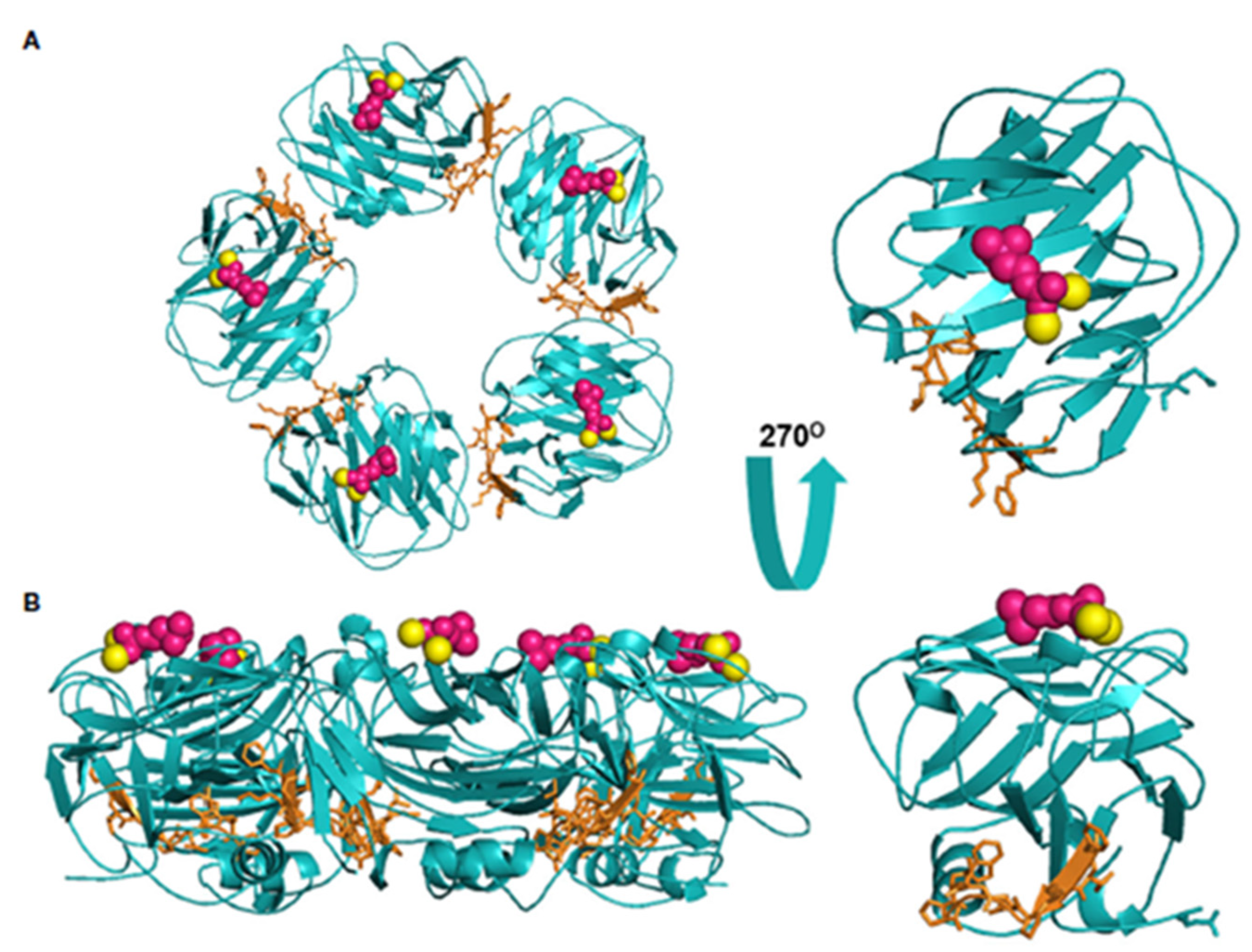
2. Tertiary Structure of CRP and Its Transformation to Dissociation
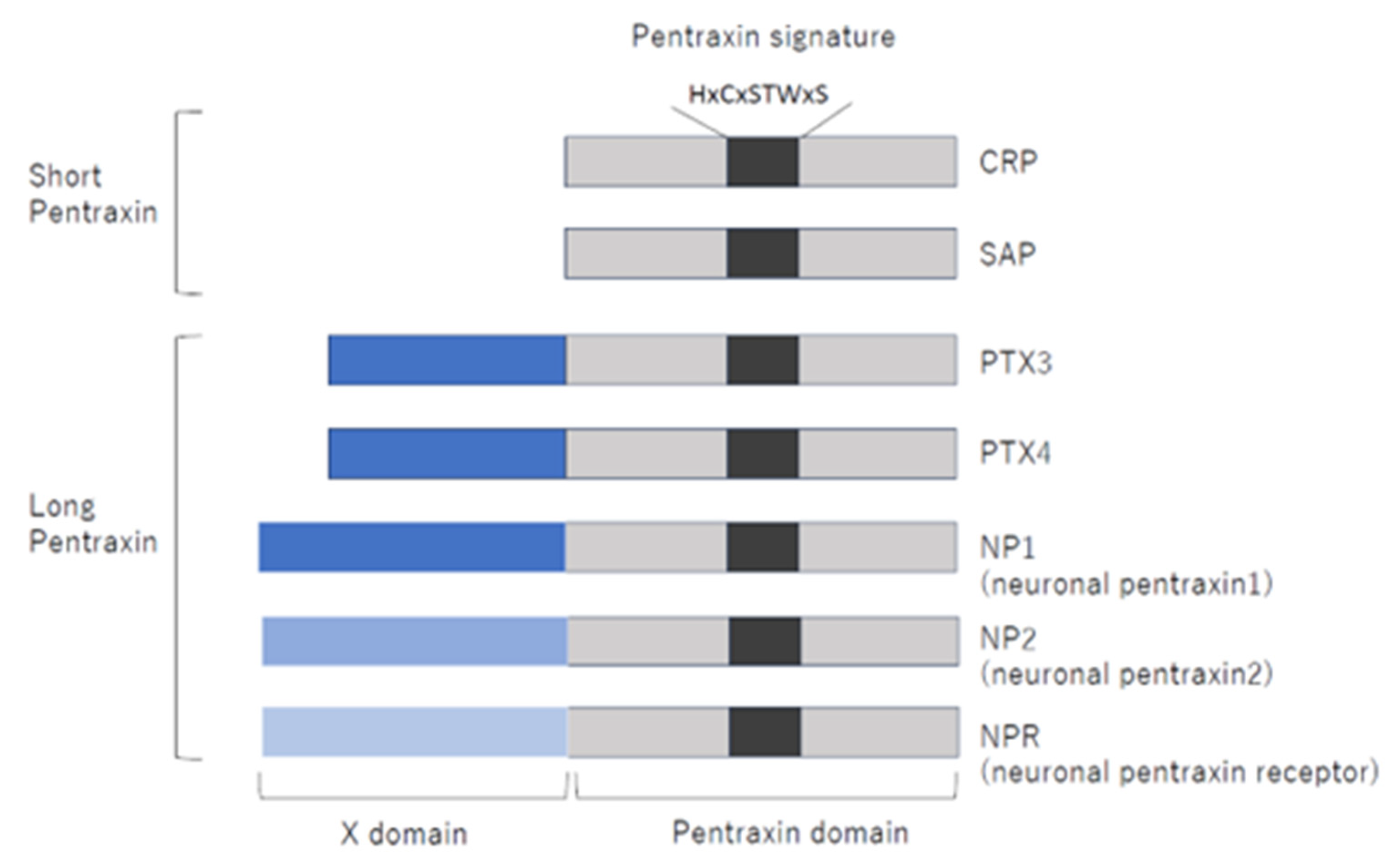
3. CRP Belongs to the Pentraxin Family
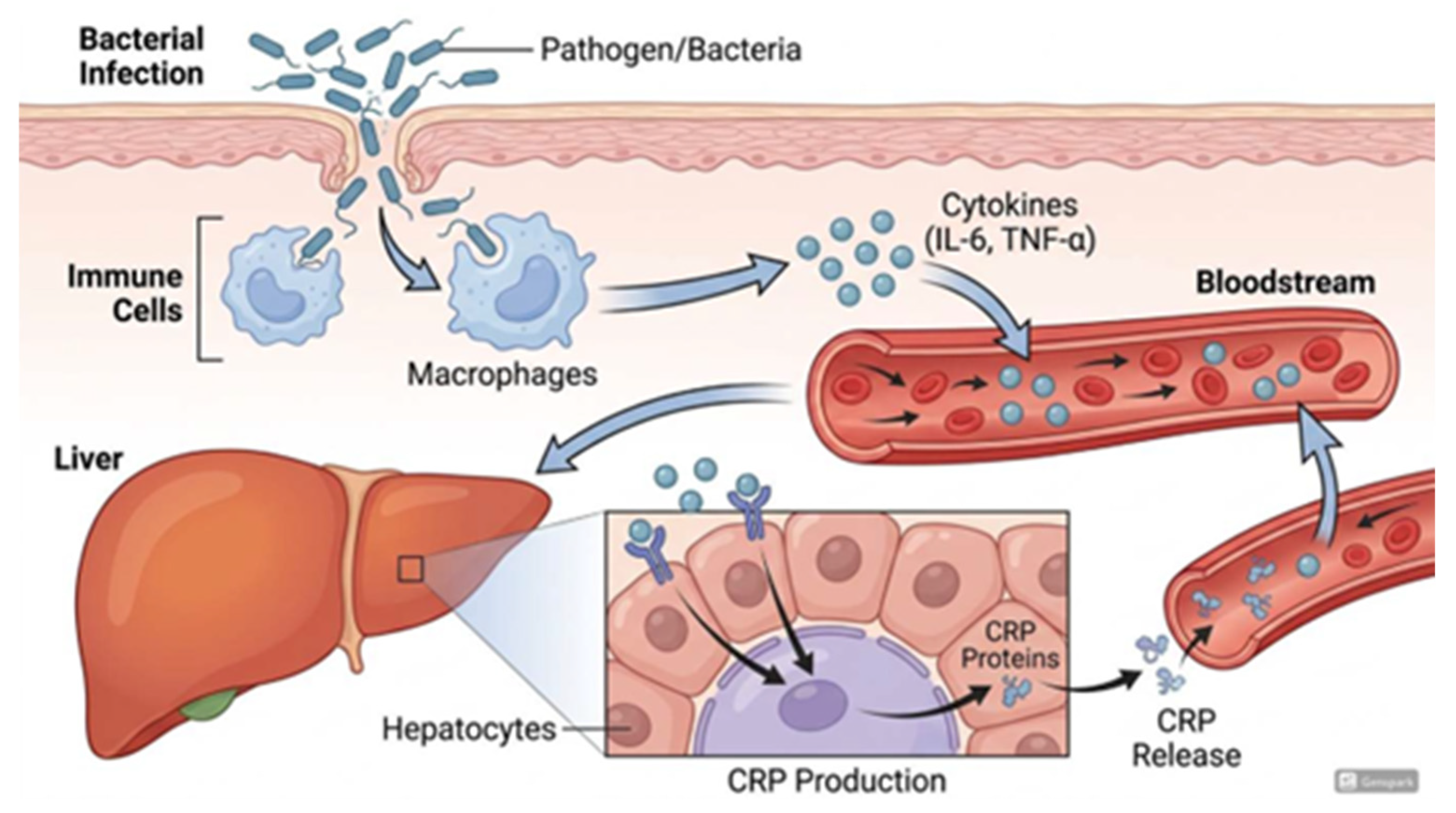
4. Induction of CRP Production
5. Biological Function of CRP
5.1. The Effect of CRP on Neutrophils
5.2. The Effects of CRP on Macrophages and Monocytes
5.3. Effects of CRP on Vascular Endothelial Cells
5.4. Monomeric CRP Causes Platelet Aggregation and Activation
5.5. Complement Activation by CRP and Its Regulation
6. The Association Between CRP Gene Polymorphisms and Diseases
7. Future Outlook of Targeted Therapy for CRP
8. Conclusions
Funding
Institutional Review Board Statement
Informed Consent Statement
Data Availability Statement
Conflicts of Interest
References
- Tillett, W.S.; Francis, T. Serological Reactions in Pneumonia with a Non-Protein Somatic Fraction of Pneumococcus. J. Exp. Med. 1930, 52, 561–571. [Google Scholar] [CrossRef] [PubMed]
- Abernethy, T.J.; Avery, O.T. The Occurrence During Acute Infections of a Protein Not Normally Present in the Blood: I. Distribution of the Reactive Protein in Patients’ Sera and the Effect of Calcium on the Flocculation Reaction with C Polysaccharide of Pneumococcus. J. Exp. Med. 1941, 73, 173–182. [Google Scholar] [CrossRef] [PubMed]
- Yao, Z.; Zhang, Y.; Wu, H. Regulation of C-reactive protein conformation in inflammation. Inflamm. Res. 2019, 68, 815–823. [Google Scholar] [CrossRef]
- Shrive, A.K.; Cheetham, G.M.; Holden, D.; Myles, D.A.; Turnell, W.G.; Volanakis, J.E.; Pepys, M.B.; Bloomer, A.C.; Greenhough, T.J. Three dimensional structure of human C-reactive protein. Nat. Struct. Biol. 1996, 3, 346–354. [Google Scholar] [CrossRef]
- Ji, S.R.; Wu, Y.; Zhu, L.; Potempa, L.A.; Sheng, F.L.; Lu, W.; Zhao, J. Cell membranes and liposomes dissociate C-reactive protein (CRP) to form a new, biologically active structural intermediate: mCRP(m). FASEB J. 2007, 21, 284–294. [Google Scholar] [CrossRef]
- Chang, M.K.; Binder, C.J.; Torzewski, M.; Witztum, J.L. C-reactive protein binds to both oxidized LDL and apoptotic cells through recognition of a common ligand: Phosphorylcholine of oxidized phospholipids. Proc. Natl. Acad. Sci. USA 2002, 99, 13043–13048. [Google Scholar] [CrossRef]
- Thiele, J.R.; Habersberger, J.; Braig, D.; Schmidt, Y.; Goerendt, K.; Maurer, V.; Bannasch, H.; Scheichl, A.; Woollard, K.J.; von Dobschütz, E.; et al. Dissociation of pentameric to monomeric C-reactive protein localizes and aggravates inflammation: In vivo proof of a powerful proinflammatory mechanism and a new anti-inflammatory strategy. Circulation 2014, 130, 35–50. [Google Scholar] [CrossRef]
- Ji, S.R.; Ma, L.; Bai, C.J.; Shi, J.M.; Li, H.Y.; Potempa, L.A.; Filep, J.G.; Zhao, J.; Wu, Y. Monomeric C-reactive protein activates endothelial cells via interaction with lipid raft microdomains. FASEB J. 2009, 23, 1806–1816. [Google Scholar] [CrossRef] [PubMed]
- Heuertz, R.M.; Schneider, G.P.; Potempa, L.A.; Webster, R.O. Native and modified C-reactive protein bind different receptors on human neutrophils. Int. J. Biochem. Cell Biol. 2005, 37, 320–335. [Google Scholar] [CrossRef]
- Rajab, I.M.; Hart, P.C.; Potempa, L.A. How C-Reactive Protein Structural Isoforms with Distinctive Bioactivities Affect Disease Progression. Front. Immunol. 2020, 11, 2126. [Google Scholar] [CrossRef] [PubMed]
- Li, Q.Y.; Li, H.Y.; Fu, G.; Yu, F.; Wu, Y.; Zhao, M.H. Autoantibodies against C-Reactive Protein Influence Complement Activation and Clinical Course in Lupus Nephritis. J. Am. Soc. Nephrol. 2017, 28, 3044–3054. [Google Scholar] [CrossRef]
- Roumenina, L.T.; Ruseva, M.M.; Zlatarova, A.; Ghai, R.; Kolev, M.; Olova, N.; Gadjeva, M.; Agrawal, A.; Bottazzi, B.; Mantovani, A.; et al. Interaction of C1q with IgG1, C-reactive protein and pentraxin 3: Mutational studies using recombinant globular head modules of human C1q A, B, and C chains. Biochemistry 2006, 45, 4093–4104. [Google Scholar] [CrossRef] [PubMed]
- Agrawal, A.; Shrive, A.K.; Greenhough, T.J.; Volanakis, J.E. Topology and structure of the C1q-binding site on C-reactive protein. J. Immunol. 2001, 166, 3998–4004. [Google Scholar] [CrossRef]
- Potempa, L.A.; Maldonado, B.A.; Laurent, P.; Zemel, E.S.; Gewurz, H. Antigenic, electrophoretic and binding alterations of human C-reactive protein modified selectively in the absence of calcium. Mol. Immunol. 1983, 20, 1165–1175. [Google Scholar] [CrossRef]
- Ullah, N.; Wu, Y. Regulation of Conformational Changes in C-reactive Protein Alters its Bioactivity. Cell Biochem. Biophys. 2022, 80, 595–608. [Google Scholar] [CrossRef]
- Eisenhardt, S.U.; Habersberger, J.; Murphy, A.; Chen, Y.C.; Woollard, K.J.; Bassler, N.; Qian, H.; von Zur Muhlen, C.; Hagemeyer, C.E.; Ahrens, I.; et al. Dissociation of pentameric to monomeric C-reactive protein on activated platelets localizes inflammation to atherosclerotic plaques. Circ. Res. 2009, 105, 128–137. [Google Scholar] [CrossRef]
- Slevin, M.; Matou-Nasri, S.; Turu, M.; Luque, A.; Rovira, N.; Badimon, L.; Boluda, S.; Potempa, L.; Sanfeliu, C.; de Vera, N.; et al. Modified C-reactive protein is expressed by stroke neovessels and is a potent activator of angiogenesis in vitro. Brain Pathol. 2010, 20, 151–165. [Google Scholar] [CrossRef]
- Mihlan, M.; Blom, A.M.; Kupreishvili, K.; Lauer, N.; Stelzner, K.; Bergström, F.; Niessen, H.W.; Zipfel, P.F. Monomeric C-reactive protein modulates classic complement activation on necrotic cells. FASEB J. 2011, 25, 4198–4210. [Google Scholar] [CrossRef] [PubMed]
- Strang, F.; Scheichl, A.; Chen, Y.C.; Wang, X.; Htun, N.M.; Bassler, N.; Eisenhardt, S.U.; Habersberger, J.; Peter, K. Amyloid plaques dissociate pentameric to monomeric C-reactive protein: A novel pathomechanism driving cortical inflammation in Alzheimer’s disease? Brain Pathol. 2012, 22, 337–346. [Google Scholar] [CrossRef] [PubMed]
- Yang, X.W.; Tan, Y.; Yu, F.; Zhao, M.H. Interference of antimodified C-reactive protein autoantibodies from lupus nephritis in the biofunctions of modified C-reactive protein. Hum. Immunol. 2012, 73, 156–163. [Google Scholar] [CrossRef]
- Braig, D.; Kaiser, B.; Thiele, J.R.; Bannasch, H.; Peter, K.; Stark, G.B.; Koch, H.G.; Eisenhardt, S.U. A conformational change of C-reactive protein in burn wounds unmasks its proinflammatory properties. Int. Immunol. 2014, 26, 467–478. [Google Scholar] [CrossRef] [PubMed]
- Olson, M.E.; Hornick, M.G.; Stefanski, A.; Albanna, H.R.; Gjoni, A.; Hall, G.D.; Hart, P.C.; Rajab, I.M.; Potempa, L.A. A biofunctional review of C-reactive protein (CRP) as a mediator of inflammatory and immune responses: Differentiating pentameric and modified CRP isoform effects. Front. Immunol. 2023, 14, 1264383. [Google Scholar] [CrossRef]
- Bottazzi, B.; Doni, A.; Garlanda, C.; Mantovani, A. An integrated view of humoral innate immunity: Pentraxins as a paradigm. Annu. Rev. Immunol. 2010, 28, 157–183. [Google Scholar] [CrossRef]
- Garlanda, C.; Bottazzi, B.; Bastone, A.; Mantovani, A. Pentraxins at the crossroads between innate immunity, inflammation, matrix deposition, and female fertility. Annu. Rev. Immunol. 2005, 23, 337–366. [Google Scholar] [CrossRef]
- Latini, R.; Maggioni, A.P.; Peri, G.; Gonzini, L.; Lucci, D.; Mocarelli, P.; Vago, L.; Pasqualini, F.; Signorini, S.; Soldateschi, D.; et al. Prognostic significance of the long pentraxin PTX3 in acute myocardial infarction. Circulation 2004, 110, 2349–2354. [Google Scholar] [CrossRef]
- Brügger-Andersen, T.; Pönitz, V.; Kontny, F.; Staines, H.; Grundt, H.; Sagara, M.; Nilsen, D.W. The long pentraxin 3 (PTX3): A novel prognostic inflammatory marker for mortality in acute chest pain. Thromb. Haemost. 2009, 102, 555–563. [Google Scholar] [CrossRef] [PubMed]
- Botto, M.; Hawkins, P.N.; Bickerstaff, M.C.; Herbert, J.; Bygrave, A.E.; McBride, A.; Hutchinson, W.L.; Tennent, G.A.; Walport, M.J.; Pepys, M.B. Amyloid deposition is delayed in mice with targeted deletion of the serum amyloid P component gene. Nat. Med. 1997, 3, 855–859. [Google Scholar] [CrossRef] [PubMed]
- Dong, Q.; Wright, J.R. Expression of C-reactive protein by alveolar macrophages. J. Immunol. 1996, 156, 4815–4820. [Google Scholar] [CrossRef]
- Calabró, P.; Willerson, J.T.; Yeh, E.T. Inflammatory cytokines stimulated C-reactive protein production by human coronary artery smooth muscle cells. Circulation 2003, 108, 1930–1932. [Google Scholar] [CrossRef]
- Jabs, W.J.; Lögering, B.A.; Gerke, P.; Kreft, B.; Wolber, E.M.; Klinger, M.H.; Fricke, L.; Steinhoff, J. The kidney as a second site of human C-reactive protein formation in vivo. Eur. J. Immunol. 2003, 33, 152–161. [Google Scholar] [CrossRef]
- Calabro, P.; Chang, D.W.; Willerson, J.T.; Yeh, E.T. Release of C-reactive protein in response to inflammatory cytokines by human adipocytes: Linking obesity to vascular inflammation. J. Am. Coll. Cardiol. 2005, 46, 1112–1113. [Google Scholar] [CrossRef] [PubMed]
- Venugopal, S.K.; Devaraj, S.; Jialal, I. Macrophage conditioned medium induces the expression of C-reactive protein in human aortic endothelial cells: Potential for paracrine/autocrine effects. Am. J. Pathol. 2005, 166, 1265–1271. [Google Scholar] [CrossRef]
- Haider, D.G.; Leuchten, N.; Schaller, G.; Gouya, G.; Kolodjaschna, J.; Schmetterer, L.; Kapiotis, S.; Wolzt, M. C-reactive protein is expressed and secreted by peripheral blood mononuclear cells. Clin. Exp. Immunol. 2006, 146, 533–539. [Google Scholar] [CrossRef]
- Vilahur, G.; Hernández-Vera, R.; Molins, B.; Casaní, L.; Duran, X.; Padró, T.; Badimon, L. Short-term myocardial ischemia induces cardiac modified C-reactive protein expression and proinflammatory gene (cyclo-oxygenase-2, monocyte chemoattractant protein-1, and tissue factor) upregulation in peripheral blood mononuclear cells. J. Thromb. Haemost. 2009, 7, 485–493. [Google Scholar] [CrossRef]
- Kang, D.H.; Park, S.K.; Lee, I.K.; Johnson, R.J. Uric acid-induced C-reactive protein expression: Implication on cell proliferation and nitric oxide production of human vascular cells. J. Am. Soc. Nephrol. 2005, 16, 3553–3562. [Google Scholar] [CrossRef] [PubMed]
- Szalai, A.J.; van Ginkel, F.W.; Dalrymple, S.A.; Murray, R.; McGhee, J.R.; Volanakis, J.E. Testosterone and IL-6 requirements for human C-reactive protein gene expression in transgenic mice. J. Immunol. 1998, 160, 5294–5299. [Google Scholar] [CrossRef]
- Majello, B.; Arcone, R.; Toniatti, C.; Ciliberto, G. Constitutive and IL-6-induced nuclear factors that interact with the human C-reactive protein promoter. EMBO J. 1990, 9, 457–465. [Google Scholar] [CrossRef]
- Li, S.P.; Goldman, N.D. Regulation of human C-reactive protein gene expression by two synergistic IL-6 responsive elements. Biochemistry 1996, 35, 9060–9068. [Google Scholar] [CrossRef] [PubMed]
- Zhang, D.; Sun, M.; Samols, D.; Kushner, I. STAT3 participates in transcriptional activation of the C-reactive protein gene by interleukin-6. J. Biol. Chem. 1996, 271, 9503–9509. [Google Scholar] [CrossRef]
- Agrawal, A.; Cha-Molstad, H.; Samols, D.; Kushner, I. Transactivation of C-reactive protein by IL-6 requires synergistic interaction of CCAAT/enhancer binding protein beta (C/EBP beta) and Rel p50. J. Immunol. 2001, 166, 2378–2384. [Google Scholar] [CrossRef]
- Weinhold, B.; Bader, A.; Poli, V.; Rüther, U. Interleukin-6 is necessary, but not sufficient, for induction of the humanC-reactive protein gene in vivo. Biochem. J. 1997, 325, 617–621. [Google Scholar] [CrossRef]
- Voleti, B.; Agrawal, A. Regulation of basal and induced expression of C-reactive protein through an overlapping element for OCT-1 and NF-kappaB on the proximal promoter. J. Immunol. 2005, 175, 3386–3390. [Google Scholar] [CrossRef] [PubMed]
- Patel, D.N.; King, C.A.; Bailey, S.R.; Holt, J.W.; Venkatachalam, K.; Agrawal, A.; Valente, A.J.; Chandrasekar, B. Interleukin-17 stimulates C-reactive protein expression in hepatocytes and smooth muscle cells via p38 MAPK and ERK1/2-dependent NF-kappaB and C/EBPbeta activation. J. Biol. Chem. 2007, 282, 27229–27238. [Google Scholar] [CrossRef] [PubMed]
- Hage, F.G.; Szalai, A.J. C-reactive protein gene polymorphisms, C-reactive protein blood levels, and cardiovascular disease risk. J. Am. Coll. Cardiol. 2007, 50, 1115–1122. [Google Scholar] [CrossRef]
- Devaraj, S.; Venugopal, S.; Jialal, I. Native pentameric C-reactive protein displays more potent pro-atherogenic activities in human aortic endothelial cells than modified C-reactive protein. Atherosclerosis 2006, 184, 48–52. [Google Scholar] [CrossRef]
- Pankow, J.S.; Folsom, A.R.; Cushman, M.; Borecki, I.B.; Hopkins, P.N.; Eckfeldt, J.H.; Tracy, R.P. Familial and genetic determinants of systemic markers of inflammation: The NHLBI family heart study. Atherosclerosis 2001, 154, 681–689. [Google Scholar] [CrossRef]
- Carlson, C.S.; Aldred, S.F.; Lee, P.K.; Tracy, R.P.; Schwartz, S.M.; Rieder, M.; Liu, K.; Williams, O.D.; Iribarren, C.; Lewis, E.C.; et al. Polymorphisms within the C-reactive protein (CRP) promoter region are associated with plasma CRP levels. Am. J. Hum. Genet. 2005, 77, 64–77. [Google Scholar] [CrossRef]
- Okada, Y.; Takahashi, A.; Ohmiya, H.; Kumasaka, N.; Kamatani, Y.; Hosono, N.; Tsunoda, T.; Matsuda, K.; Tanaka, T.; Kubo, M.; et al. Genome-wide association study for C-reactive protein levels identified pleiotropic associations in the IL6 locus. Hum. Mol. Genet. 2011, 20, 1224–1231. [Google Scholar] [CrossRef]
- Ciubotaru, I.; Potempa, L.A.; Wander, R.C. Production of modified C-reactive protein in U937-derived macrophages. Exp. Biol. Med. 2005, 230, 762–770. [Google Scholar] [CrossRef]
- Vigushin, D.M.; Pepys, M.B.; Hawkins, P.N. Metabolic and scintigraphic studies of radioiodinated human C-reactive protein in health and disease. J. Clin. Investig. 1993, 91, 1351–1357. [Google Scholar] [CrossRef] [PubMed]
- Dandona, P.; Nix, D.; Wilson, M.F.; Aljada, A.; Love, J.; Assicot, M.; Bohuon, C. Procalcitonin increase after endotoxin injection in normal subjects. J. Clin. Endocrinol. Metab. 1994, 79, 1605–1608. [Google Scholar] [CrossRef]
- Zacho, J.; Tybjaerg-Hansen, A.; Jensen, J.S.; Grande, P.; Sillesen, H.; Nordestgaard, B.G. Genetically elevated C-reactive protein and ischemic vascular disease. N. Engl. J. Med. 2008, 359, 1897–1908. [Google Scholar] [CrossRef]
- Ridker, P.M.; Moorthy, M.V.; Cook, N.R.; Rifai, N.; Lee, I.M.; Buring, J.E. Inflammation, Cholesterol, Lipoprotein(a), and 30-Year Cardiovascular Outcomes in Women. N. Engl. J. Med. 2024, 391, 2087–2097. [Google Scholar] [CrossRef]
- Liu, Q.; Jiang, C.Y.; Chen, B.X.; Zhao, W.; Meng, D. The association between high-sensitivity C-reactive protein concentration and diabetic nephropathy: A meta-analysis. Eur. Rev. Med. Pharmacol. Sci. 2015, 19, 4558–4568. [Google Scholar] [PubMed]
- Liu, S.H.; Li, Y.J.; Wu, H.H.; Lee, C.C.; Lin, C.Y.; Weng, C.H.; Chen, Y.C.; Chang, M.Y.; Hsu, H.H.; Fang, J.T.; et al. High-sensitivity C-reactive protein predicts mortality and technique failure in peritoneal dialysis patients. PLoS ONE 2014, 9, e93063. [Google Scholar] [CrossRef]
- Kindmark, C.O. Stimulating effect of C-reactive protein on phagocytosis of various species of pathogenic bacteria. Clin. Exp. Immunol. 1971, 8, 941–948. [Google Scholar]
- Mold, C.; Nakayama, S.; Holzer, T.J.; Gewurz, H.; Du Clos, T.W. C-reactive protein is protective against Streptococcus pneumoniae infection in mice. J. Exp. Med. 1981, 154, 1703–1708. [Google Scholar] [CrossRef]
- Holzer, T.J.; Edwards, K.M.; Gewurz, H.; Mold, C. Binding of C-reactive protein to the pneumococcal capsule or cell wall results in differential localization of C3 and stimulation of phagocytosis. J. Immunol. 1984, 133, 1424–1430. [Google Scholar] [CrossRef]
- Shephard, E.G.; Anderson, R.; Strachan, A.F.; Kühn, S.H.; De Beer, F.C. CRP and neutrophils: Functional effects and complex uptake. Clin. Exp. Immunol. 1986, 63, 718–727. [Google Scholar] [PubMed]
- Mold, C.; Edwards, K.M.; Gewurz, H. Effect of C-reactive protein on the complement-mediated stimulated of human neutrophils by Streptococcus pneumoniae serotypes 3 and 6. Infect. Immun. 1982, 37, 987–992. [Google Scholar] [CrossRef] [PubMed]
- Mold, C.; Du Clos, T.W.; Nakayama, S.; Edwards, K.M.; Gewurz, H. C-reactive protein reactivity with complement and effects on phagocytosis. Ann. N. Y. Acad. Sci. 1982, 389, 251–262. [Google Scholar] [CrossRef] [PubMed]
- Mold, C.; Gewurz, H.; Du Clos, T.W. Regulation of complement activation by C-reactive protein. Immunopharmacology 1999, 42, 23–30. [Google Scholar] [CrossRef]
- Edwards, K.M.; Gewurz, H.; Lint, T.F.; Mold, C. A role for C-reactive protein in the complement-mediated stimulation of human neutrophils by type 27 Streptococcus pneumoniae. J. Immunol. 1982, 128, 2493–2496. [Google Scholar] [CrossRef]
- Buchta, R.; Fridkin, M.; Pontet, M.; Contessi, E.; Scaggiante, B.; Romeo, D. Modulation of human neutrophil function by C-reactive protein. Eur. J. Biochem. 1987, 163, 141–146. [Google Scholar] [CrossRef]
- Tatsumi, N.; Hashimoto, K.; Okuda, K.; Kyougoku, T. Neutrophil chemiluminescence induced by platelet activating factor and suppressed by C-reactive protein. Clin. Chim. Acta 1988, 172, 85–92. [Google Scholar] [CrossRef]
- Zurier, R.B.; Weissmann, G.; Hoffstein, S.; Kammerman, S.; Tai, H.H. Mechanisms of lysosomal enzyme release from human leukocytes. II. Effects of cAMP and cGMP, autonomic agonists, and agents which affect microtubule function. J. Clin. Investig. 1974, 53, 297–309. [Google Scholar] [CrossRef] [PubMed]
- Spirer, Z.; Zakuth, V.; Diamant, S.; Stabinsky, Y.; Fridkin, M. Studies on the activity of phorbol myrystate acetate on the human polymorphonuclear leukocytes. Experientia 1979, 35, 830–831. [Google Scholar] [CrossRef]
- Khreiss, T.; József, L.; Potempa, L.A.; Filep, J.G. Loss of pentameric symmetry in C-reactive protein induces interleukin-8 secretion through peroxynitrite signaling in human neutrophils. Circ. Res. 2005, 97, 690–697. [Google Scholar] [CrossRef]
- Pepys, M.B.; Hirschfield, G.M.; Tennent, G.A.; Gallimore, J.R.; Kahan, M.C.; Bellotti, V.; Hawkins, P.N.; Myers, R.M.; Smith, M.D.; Polara, A.; et al. Targeting C-reactive protein for the treatment of cardiovascular disease. Nature 2006, 440, 1217–1221. [Google Scholar] [CrossRef] [PubMed]
- Marnell, L.L.; Mold, C.; Volzer, M.A.; Burlingame, R.W.; Du Clos, T.W. C-reactive protein binds to Fc gamma RI in transfected COS cells. J. Immunol. 1995, 155, 2185–2193. [Google Scholar] [CrossRef]
- Stein, M.P.; Edberg, J.C.; Kimberly, R.P.; Mangan, E.K.; Bharadwaj, D.; Mold, C.; Du Clos, T.W. C-reactive protein binding to FcgammaRIIa on human monocytes and neutrophils is allele-specific. J. Clin. Investig. 2000, 105, 369–376. [Google Scholar] [CrossRef] [PubMed]
- Zahedi, K.; Mortensen, R.F. Macrophage tumoricidal activity induced by human C-reactive protein. Cancer Res. 1986, 46, 5077–5083. [Google Scholar]
- Ballou, S.P.; Lozanski, G. Induction of inflammatory cytokine release from cultured human monocytes by C-reactive protein. Cytokine 1992, 4, 361–368. [Google Scholar] [CrossRef]
- Han, K.H.; Hong, K.H.; Park, J.H.; Ko, J.; Kang, D.H.; Choi, K.J.; Hong, M.K.; Park, S.W.; Park, S.J. C-reactive protein promotes monocyte chemoattractant protein-1--mediated chemotaxis through upregulating CC chemokine receptor 2 expression in human monocytes. Circulation 2004, 109, 2566–2571. [Google Scholar] [CrossRef]
- Cermak, J.; Key, N.S.; Bach, R.R.; Balla, J.; Jacob, H.S.; Vercellotti, G.M. C-reactive protein induces human peripheral blood monocytes to synthesize tissue factor. Blood 1993, 82, 513–520. [Google Scholar] [CrossRef] [PubMed]
- Zouki, C.; Beauchamp, M.; Baron, C.; Filep, J.G. Prevention of In vitro neutrophil adhesion to endothelial cells through shedding of L-selectin by C-reactive protein and peptides derived from C-reactive protein. J. Clin. Investig. 1997, 100, 522–529. [Google Scholar] [CrossRef]
- Hanriot, D.; Bello, G.; Ropars, A.; Seguin-Devaux, C.; Poitevin, G.; Grosjean, S.; Latger-Cannard, V.; Devaux, Y.; Zannad, F.; Regnault, V.; et al. C-reactive protein induces pro- and anti-inflammatory effects, including activation of the liver X receptor alpha, on human monocytes. Thromb. Haemost. 2008, 99, 558–569. [Google Scholar] [CrossRef]
- Yu, S.X.; Chen, W.; Hu, X.Z.; Feng, S.Y.; Li, K.Y.; Qi, S.; Lei, Q.Q.; Hu, G.Q.; Li, N.; Zhou, F.H.; et al. Liver X receptors agonists suppress NLRP3 inflammasome activation. Cytokine 2017, 91, 30–37. [Google Scholar] [CrossRef]
- Higham, A.; Lea, S.; Plumb, J.; Maschera, B.; Simpson, K.; Ray, D.; Singh, D. The role of the liver X receptor in chronic obstructive pulmonary disease. Respir. Res. 2013, 14, 106. [Google Scholar] [CrossRef]
- Pasceri, V.; Willerson, J.T.; Yeh, E.T. Direct proinflammatory effect of C-reactive protein on human endothelial cells. Circulation 2000, 102, 2165–2168. [Google Scholar] [CrossRef] [PubMed]
- Venugopal, S.K.; Devaraj, S.; Yuhanna, I.; Shaul, P.; Jialal, I. Demonstration that C-reactive protein decreases eNOS expression and bioactivity in human aortic endothelial cells. Circulation 2002, 106, 1439–1441. [Google Scholar] [CrossRef] [PubMed]
- Singh, U.; Devaraj, S.; Vasquez-Vivar, J.; Jialal, I. C-reactive protein decreases endothelial nitric oxide synthase activity via uncoupling. J. Mol. Cell Cardiol. 2007, 43, 780–791. [Google Scholar] [CrossRef] [PubMed]
- Khreiss, T.; József, L.; Potempa, L.A.; Filep, J.G. Conformational rearrangement in C-reactive protein is required for proinflammatory actions on human endothelial cells. Circulation 2004, 109, 2016–2022. [Google Scholar] [CrossRef]
- Fiedel, B.A. Platelet agonist synergism by the acute phase reactant C-reactive protein. Blood 1985, 65, 264–269. [Google Scholar] [CrossRef]
- Potempa, L.A.; Zeller, J.M.; Fiedel, B.A.; Kinoshita, C.M.; Gewurz, H. Stimulation of human neutrophils, monocytes, and platelets by modified C-reactive protein (CRP) expressing a neoantigenic specificity. Inflammation 1988, 12, 391–405. [Google Scholar] [CrossRef]
- Boncler, M.; Kehrel, B.; Szewczyk, R.; Stec-Martyna, E.; Bednarek, R.; Brodde, M.; Watala, C. Oxidation of C-reactive protein by hypochlorous acid leads to the formation of potent platelet activator. Int. J. Biol. Macromol. 2018, 107, 2701–2714. [Google Scholar] [CrossRef]
- Vigo, C. Effect of C-reactive protein on platelet-activating factor-induced platelet aggregation and membrane stabilization. J. Biol. Chem. 1985, 260, 3418–3422. [Google Scholar] [CrossRef]
- Khreiss, T.; József, L.; Potempa, L.A.; Filep, J.G. Opposing effects of C-reactive protein isoforms on shear-induced neutrophil-platelet adhesion and neutrophil aggregation in whole blood. Circulation 2004, 110, 2713–2720. [Google Scholar] [CrossRef][Green Version]
- Kaplan, M.H.; Volanakis, J.E. Interaction of C-reactive protein complexes with the complement system. I. Consumption of human complement associated with the reaction of C-reactive protein with pneumococcal C-polysaccharide and with the choline phosphatides, lecithin and sphingomyelin. J. Immunol. 1974, 112, 2135–2147. [Google Scholar] [CrossRef]
- Du Clos, T.W. Pentraxins: Structure, function, and role in inflammation. ISRN Inflamm. 2013, 2013, 379040. [Google Scholar] [CrossRef] [PubMed]
- Mold, C.; Gewurz, H. Inhibitory effect of C-reactive protein on alternative C pathway activation by liposomes and Streptococcus pneumoniae. J. Immunol. 1981, 127, 2089–2092. [Google Scholar] [CrossRef]
- Shephard, E.G.; van Helden, P.D.; Strauss, M.; Böhm, L.; De Beer, F.C. Functional effects of CRP binding to nuclei. Immunology 1986, 58, 489–494. [Google Scholar]
- Haapasalo, K.; Meri, S. Regulation of the Complement System by Pentraxins. Front. Immunol. 2019, 10, 1750. [Google Scholar] [CrossRef]
- Ji, S.R.; Wu, Y.; Potempa, L.A.; Liang, Y.H.; Zhao, J. Effect of modified C-reactive protein on complement activation: A possible complement regulatory role of modified or monomeric C-reactive protein in atherosclerotic lesions. Arterioscler. Thromb. Vasc. Biol. 2006, 26, 935–941. [Google Scholar] [CrossRef]
- Sproston, N.R.; Ashworth, J.J. Role of C-Reactive Protein at Sites of Inflammation and Infection. Front. Immunol. 2018, 9, 754. [Google Scholar] [CrossRef]
- Mold, C.; Kingzette, M.; Gewurz, H. C-reactive protein inhibits pneumococcal activation of the alternative pathway by increasing the interaction between factor H and C3b. J. Immunol. 1984, 133, 882–885. [Google Scholar] [CrossRef]
- O’Flynn, J.; van der Pol, P.; Dixon, K.O.; Prohászka, Z.; Daha, M.R.; van Kooten, C. Monomeric C-reactive protein inhibits renal cell-directed complement activation mediated by properdin. Am. J. Physiol. Ren. Physiol. 2016, 310, F1308–F1316. [Google Scholar] [CrossRef]
- Kathiresan, S.; Larson, M.G.; Vasan, R.S.; Guo, C.Y.; Gona, P.; Keaney, J.F., Jr.; Wilson, P.W.; Newton-Cheh, C.; Musone, S.L.; Camargo, A.L.; et al. Contribution of clinical correlates and 13 C-reactive protein gene polymorphisms to interindividual variability in serum C-reactive protein level. Circulation 2006, 113, 1415–1423. [Google Scholar] [CrossRef]
- Ghattas, M.H.; Abo-Elmatty, D.M.; El-Eraki, A.Z. C-reactive protein 1059G/C gene polymorphism, C-reactive protein levels and acute myocardial infarction. J. Cardiovasc. Med. 2012, 13, 716–719. [Google Scholar] [CrossRef]
- Saratzis, A.; Bown, M.; Wild, B.; Sayers, R.D.; Nightingale, P.; Smith, J.; Johnson, C.; Kitas, G. C-reactive protein polymorphism rs3091244 is associated with abdominal aortic aneurysm. J. Vasc. Surg. 2014, 60, 1332–1339. [Google Scholar] [CrossRef]
- Słomiński, B.; Jankowiak, M.; Maciejewska, A.; Studziński, M.; Mączyńska, A.; Skrzypkowska, M.; Gabig-Cimińska, M.; Myśliwiec, M. Genetic variation in C-reactive protein (CRP) gene is associated with retinopathy and hypertension in adolescents with type 1 diabetes. Cytokine 2022, 160, 156025. [Google Scholar] [CrossRef]
- Jebur, H.B.; Masroor, M.; Ahmad, H.; Khan, N.A.; Akther, J.; Bharali, D.; Singh, V.K.; Verma, A.; Khan, S.; Khan, V.; et al. CRP Gene Polymorphism and Their Risk Association with Type 2 Diabetes Mellitus. Open Access Maced. J. Med. Sci. 2019, 7, 33–37. [Google Scholar] [CrossRef]
- Zee, R.Y.; Germer, S.; Thomas, A.; Raji, A.; Rhees, B.; Ridker, P.M.; Lindpaintner, K.; Williams, G.H.; Nathan, D.M.; Martin, M. C-reactive protein gene variation and type 2 diabetes mellitus: A case-control study. Atherosclerosis 2008, 197, 931–936. [Google Scholar] [CrossRef]
- Balistreri, C.R.; Vasto, S.; Listì, F.; Grimaldi, M.P.; Lio, D.; Colonna-Romano, G.; Caruso, M.; Caimi, G.; Hoffmann, E.; Caruso, C.; et al. Association between +1059G/C CRP polymorphism and acute myocardial infarction in a cohort of patients from Sicily: A pilot study. Ann. N. Y. Acad. Sci. 2006, 1067, 276–281. [Google Scholar] [CrossRef]
- Morita, A.; Nakayama, T.; Soma, M. Association study between C-reactive protein genes and ischemic stroke in Japanese subjects. Am. J. Hypertens. 2006, 19, 593–600. [Google Scholar] [CrossRef]
- Chen, Z.; Jiang, F.; Yang, M.; Yang, J. Relationship between CRP gene polymorphisms and ischemic stroke risk: A systematic review and meta-analysis. Open Life Sci. 2022, 17, 1519–1530. [Google Scholar] [CrossRef] [PubMed]
- Liu, Y.; Geng, P.L.; Yan, F.Q.; Chen, T.; Wang, W.; Tang, X.D.; Zheng, J.C.; Wu, W.P.; Wang, Z.F. C-reactive Protein -717A>G and -286C>T>A Gene Polymorphism and Ischemic Stroke. Chin. Med. J. 2015, 128, 1666–1670. [Google Scholar] [CrossRef] [PubMed]
- Kotlęga, D.; Nowacki, P.; Białecka, M.; Kurzawski, M.; Droździk, M.; Ciećwież, S. Association between CRP gene polymorphism 717A/G, C-reactive protein and neurological deficit in ischemic stroke. J. Clin. Neurosci. 2014, 21, 574–577. [Google Scholar] [CrossRef]
- Geng, P.; Sa, R.; Li, J.; Li, H.; Liu, C.; Liao, Y.; Xiang, L.; Wang, N.; Ou, J.; Xie, G.; et al. Genetic polymorphisms in C-reactive protein increase cancer susceptibility. Sci. Rep. 2016, 6, 17161. [Google Scholar] [CrossRef] [PubMed]
- Chen, C.; Liu, J.N.; Zhao, J.Q.; Zang, B. C-reactive protein gene 1846C>T polymorphism is associated with increased risk and clinical features of lung cancer: A case-control study. Biosci. Rep. 2019, 39, BSR20181936. [Google Scholar] [CrossRef]
- Motoyama, S.; Mori, K.; Kamei, T.; Miura, M.; Hinai, Y.; Sato, Y.; Yoshino, K.; Sasaki, T.; Miyata, G.; Seto, Y.; et al. Evaluation of the risk of lymph node metastasis using CRP 1846C>T genetic polymorphism in submucosal thoracic esophageal squamous cell carcinoma. Ann. Surg. Oncol. 2013, 20, 1978–1984. [Google Scholar] [CrossRef]
- Honda, K.; Taniguchi, K.; Komura, K.; Tanaka, T.; Yoshimoto, H.; Matsuo, K.; Imai, Y.; Tanaka, R.; Ishida, M.; Hirose, Y.; et al. Lymph node metastasis risk in submucosal invasive gastric cancer based on C-reactive protein gene polymorphism analysis. Int. J. Clin. Oncol. 2025, 30, 2549–2558. [Google Scholar] [CrossRef]
- Terata, K.; Motoyama, S.; Kamata, S.; Hinai, Y.; Miura, M.; Sato, Y.; Yoshino, K.; Ito, A.; Imai, K.; Saito, H.; et al. Evaluation of the potential for lymph node metastasis using CRP 1846C>T genetic polymorphism in invasive breast cancer. Tumour Biol. 2014, 35, 5931–5935. [Google Scholar] [CrossRef]
- Michopoulos, V.; Rothbaum, A.O.; Jovanovic, T.; Almli, L.M.; Bradley, B.; Rothbaum, B.O.; Gillespie, C.F.; Ressler, K.J. Association of CRP genetic variation and CRP level with elevated PTSD symptoms and physiological responses in a civilian population with high levels of trauma. Am. J. Psychiatry 2015, 172, 353–362. [Google Scholar] [CrossRef] [PubMed]
- Lin, F.H.; Chu, N.F.; Lee, C.H.; Hung, Y.J.; Wu, D.M. Combined effect of C-reactive protein gene SNP +2147 A/G and interleukin-6 receptor gene SNP rs2229238 C/T on anthropometric characteristics among school children in Taiwan. Int. J. Obes. 2011, 35, 587–594. [Google Scholar] [CrossRef] [PubMed][Green Version]
- Todendi, P.F.; Klinger, E.I.; Ferreira, M.B.; Reuter, C.P.; Burgos, M.S.; Possuelo, L.G.; Valim, A.R. Association of IL-6 and CRP gene polymorphisms with obesity and metabolic disorders in children and adolescents. An. Acad. Bras. Cienc. 2015, 87, 915–924. [Google Scholar] [CrossRef] [PubMed][Green Version]
- Jones, N.R.; Pegues, M.A.; McCrory, M.A.; Singleton, W.; Bethune, C.; Baker, B.F.; Norris, D.A.; Crooke, R.M.; Graham, M.J.; Szalai, A.J. A Selective Inhibitor of Human C-reactive Protein Translation Is Efficacious In Vitro and in C-reactive Protein Transgenic Mice and Humans. Mol. Ther. Nucleic Acids 2012, 1, e52. [Google Scholar] [CrossRef]
- Szalai, A.J.; McCrory, M.A.; Xing, D.; Hage, F.G.; Miller, A.; Oparil, S.; Chen, Y.F.; Mazzone, M.; Early, R.; Henry, S.P.; et al. Inhibiting C-reactive protein for the treatment of cardiovascular disease: Promising evidence from rodent models. Mediat. Inflamm. 2014, 2014, 353614. [Google Scholar] [CrossRef]
- Noveck, R.; Stroes, E.S.; Flaim, J.D.; Baker, B.F.; Hughes, S.; Graham, M.J.; Crooke, R.M.; Ridker, P.M. Effects of an antisense oligonucleotide inhibitor of C-reactive protein synthesis on the endotoxin challenge response in healthy human male volunteers. J. Am. Heart Assoc. 2014, 3, e001084. [Google Scholar] [CrossRef]
- Warren, M.S.; Hughes, S.G.; Singleton, W.; Yamashita, M.; Genovese, M.C. Results of a proof of concept, double-blind, randomized trial of a second generation antisense oligonucleotide targeting high-sensitivity C-reactive protein (hs-CRP) in rheumatoid arthritis. Arthritis Res. Ther. 2015, 17, 80. [Google Scholar] [CrossRef]
- Zeller, J.; Cheung Tung Shing, K.S.; Nero, T.L.; McFadyen, J.D.; Krippner, G.; Bogner, B.; Kreuzaler, S.; Kiefer, J.; Horner, V.K.; Braig, D.; et al. A novel phosphocholine-mimetic inhibits a pro-inflammatory conformational change in C-reactive protein. EMBO Mol. Med. 2023, 15, e16236. [Google Scholar] [CrossRef] [PubMed]



| Allelic Variant | Variant Type | Major > Minor Allele | Minor Allele Frequency | p |
|---|---|---|---|---|
| rs3116653 | 5′-Upstream | C > G | 0.31 | 0.0008 |
| rs3116654 | 5′-Upstream | T > C | 0.12 | 0.04 |
| rs2794517 | 5′-Upstream | C > T | 0.3 | 0.99 |
| rs3122012 | 5′-Upstream | A > G | 0.31 | 0.001 |
| rs3091244 | 5′-Upstream | C > T | 0.31 | <0.0001 |
| C > A | 0.07 | |||
| rs1417938 | Intron | A > T | 0.31 | 0.001 |
| rs1800947 | Synonymous, coding | G > C | 0.06 | 0.09 |
| rs1130864 | 3′-Untranslated | C > T | 0.31 | 0.0006 |
| rs1205 | 3′-Flanking | G > A | 0.33 | <0.0001 |
| rs2808630 | 3′-Flanking | A > G | 0.28 | 0.43 |
| rs3093077 | 3′-Flanking | T > G | 0.07 | 0.002 |
| rs12029262 | 3′-Flanking | C > G | 0.09 | 0.002 |
| rs876538 | 3′-Flanking | G > A | 0.2 | 0.76 |
Disclaimer/Publisher’s Note: The statements, opinions and data contained in all publications are solely those of the individual author(s) and contributor(s) and not of MDPI and/or the editor(s). MDPI and/or the editor(s) disclaim responsibility for any injury to people or property resulting from any ideas, methods, instructions or products referred to in the content. |
© 2026 by the author. Licensee MDPI, Basel, Switzerland. This article is an open access article distributed under the terms and conditions of the Creative Commons Attribution (CC BY) license.
Share and Cite
Nagasawa, M. CRP (C-Reactive Protein) Revisited: An Old Yet New Biomarker of Acute and Chronic Inflammation. Cells 2026, 15, 998. https://doi.org/10.3390/cells15110998
Nagasawa M. CRP (C-Reactive Protein) Revisited: An Old Yet New Biomarker of Acute and Chronic Inflammation. Cells. 2026; 15(11):998. https://doi.org/10.3390/cells15110998
Chicago/Turabian StyleNagasawa, Masayuki. 2026. "CRP (C-Reactive Protein) Revisited: An Old Yet New Biomarker of Acute and Chronic Inflammation" Cells 15, no. 11: 998. https://doi.org/10.3390/cells15110998
APA StyleNagasawa, M. (2026). CRP (C-Reactive Protein) Revisited: An Old Yet New Biomarker of Acute and Chronic Inflammation. Cells, 15(11), 998. https://doi.org/10.3390/cells15110998