Modeling Human Hypertrophic Scars with Induced Pluripotent Stem-Cell-Derived Scar Organoids Versus Skin Organoids

Highlights
- An iPSC-derived scar organoid was established using TGF-β1 and hypoxic culture conditions.
- SCOs reproduced scar-associated features, including collagen accumulation, tissue contraction, and transcriptomic remodeling.
- SCOs may be useful for studying pathological skin remodeling and fibrosis in a human 3D platform.
- This model may support future mechanistic and translational studies related to hypertrophic scars.
Abstract
1. Introduction
2. Materials and Methods
2.1. Generation and Culture of Human iPSCs
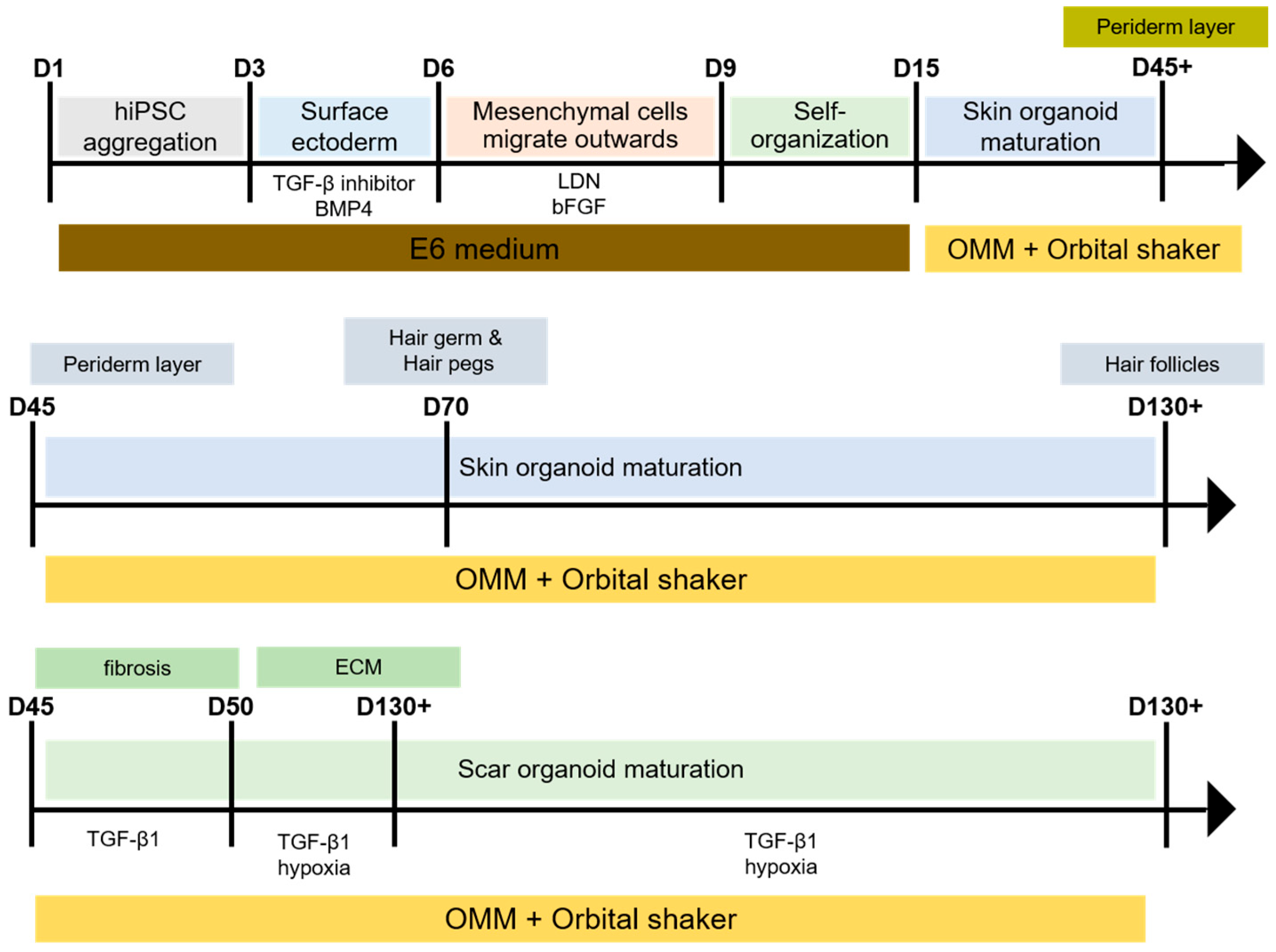
2.2. Generation and Culture of iPSC-Derived SKOs and SCOs
2.3. Morphological and Quantitative Analysis of Organoids
2.4. Histological and Immunofluorescence Analyses
2.5. RNA Sequencing and Transcriptomic Analyses
2.6. Statistical Analysis
3. Results
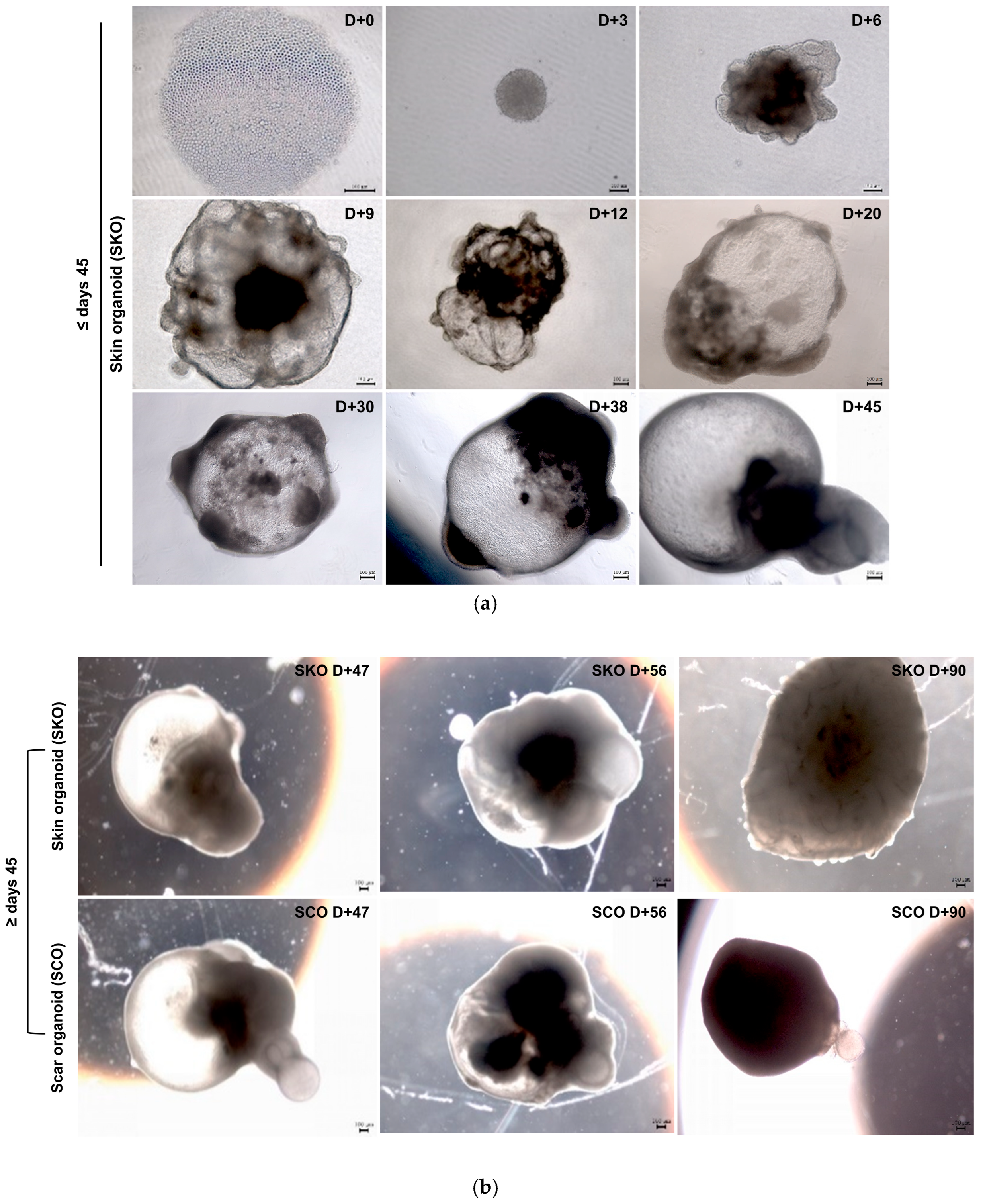
3.1. Generation and Morphological Development of iPSC-Derived SKOs and SCOs
Morphological Development of SKOs and SCOs
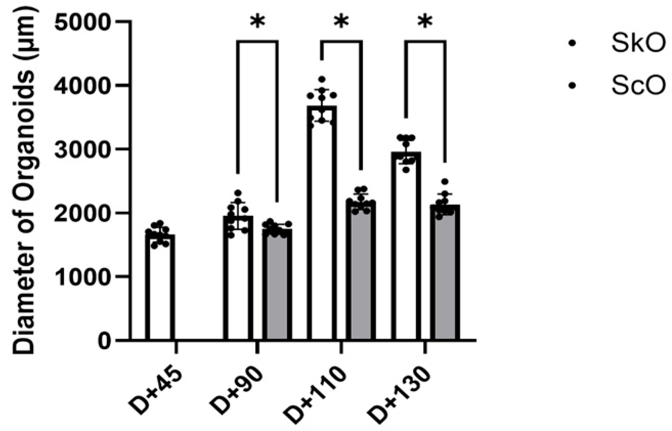
3.2. Quantitative Analysis of Organoid Growth Dynamics
Diameter-Based Comparison of SKOs and SCOs at Defined Time Points
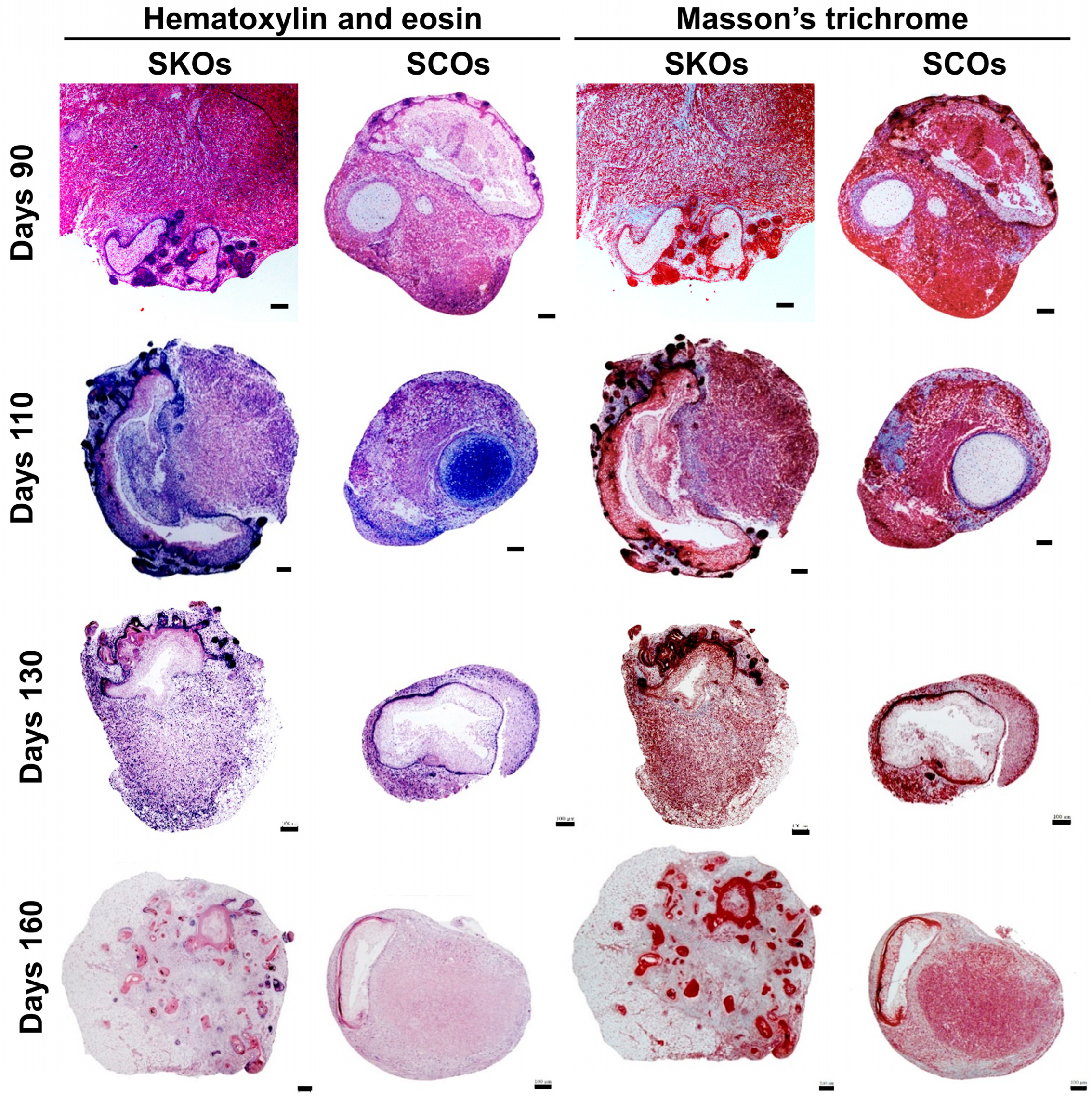
3.3. Histological Characterization of SKOs and SCOs
Tissue Architecture and ECM Remodeling in SKOs and SCOs
3.4. Immunofluorescence Analysis of Fibrotic and Skin-Associated Markers
Differential Expression of COL1 and LHX2 in SKOs and SCOs
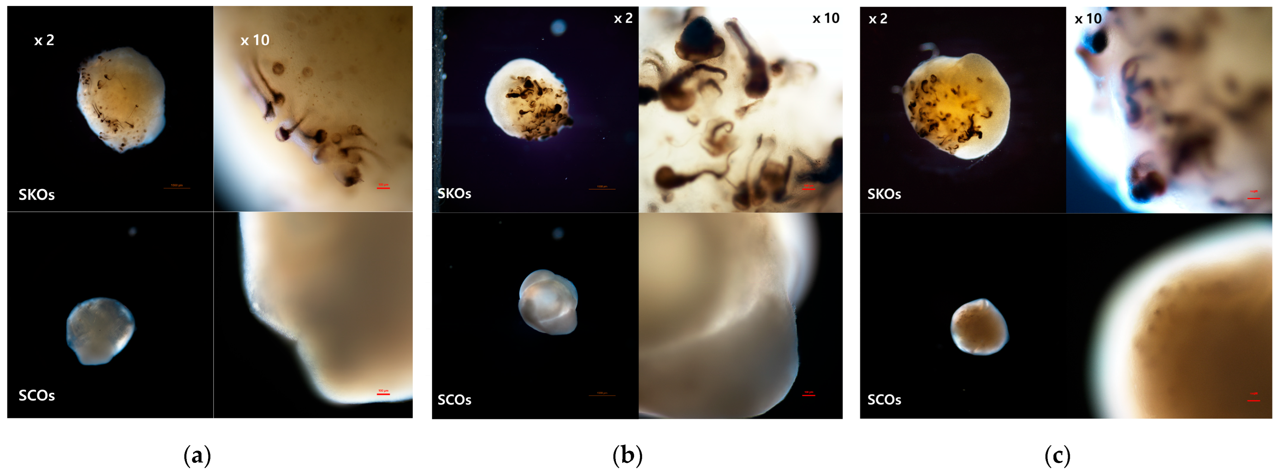
3.5. Morphological Characteristics of SKOs and SCOs
Dark-Field Imaging of SKOs and SCOs During Late-Stage Maturation
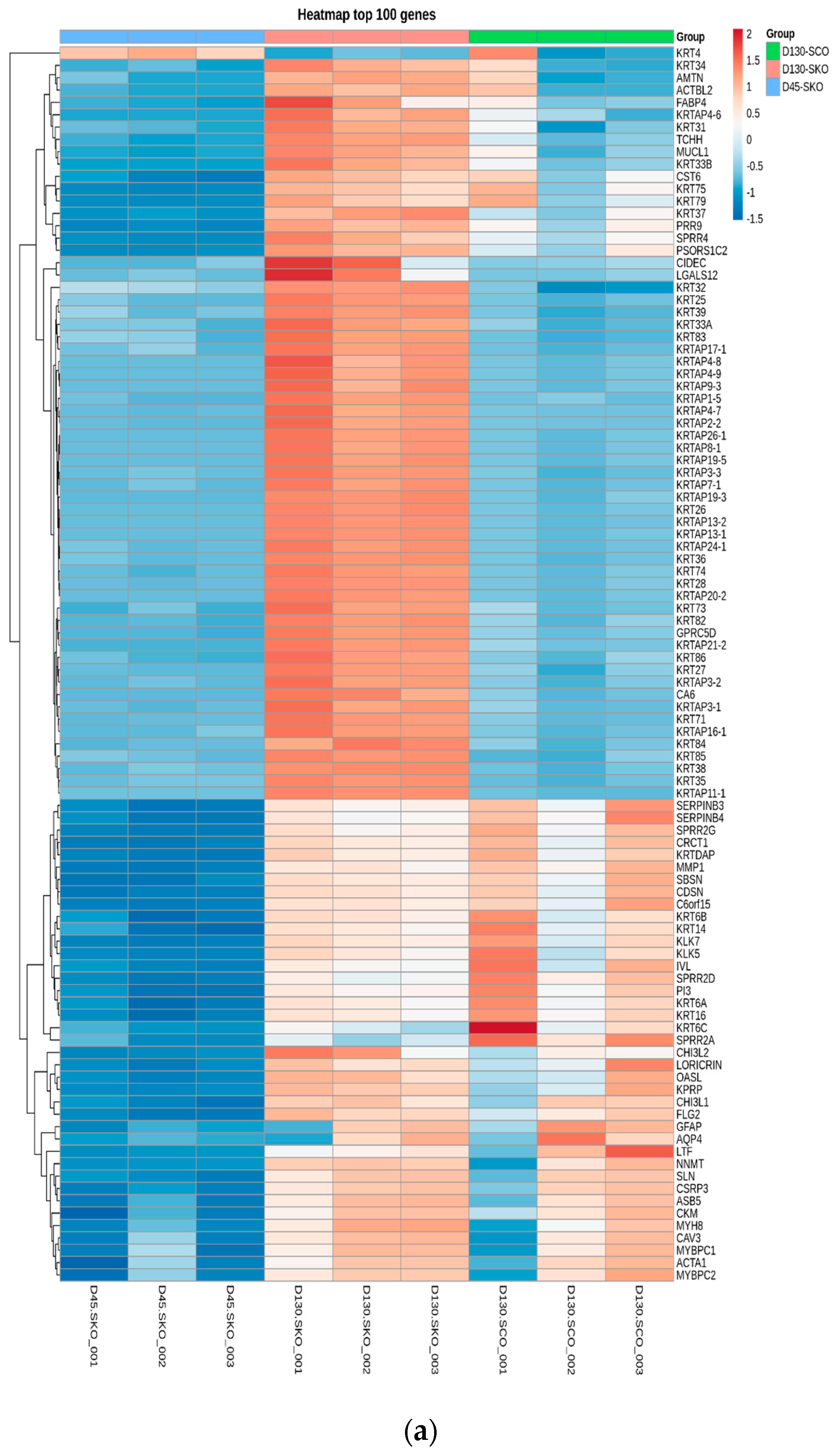
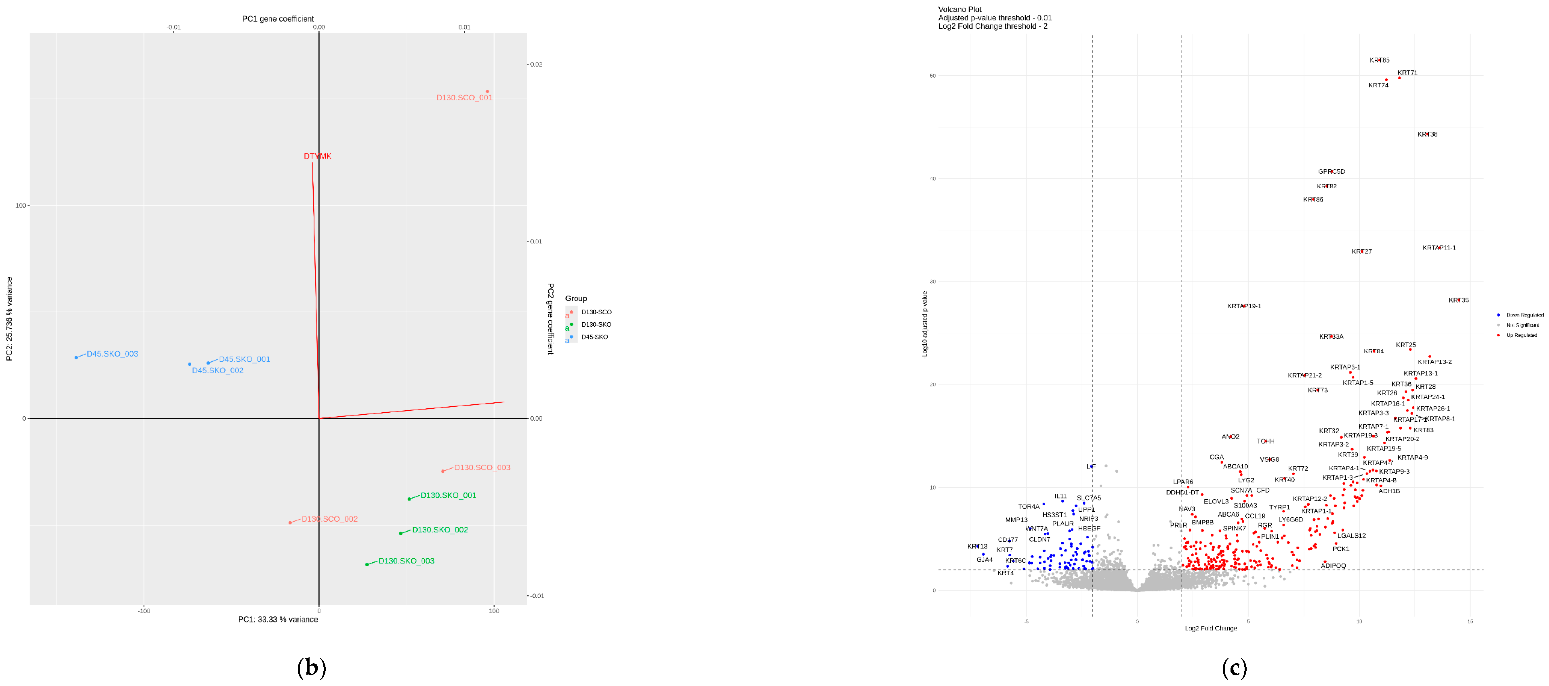
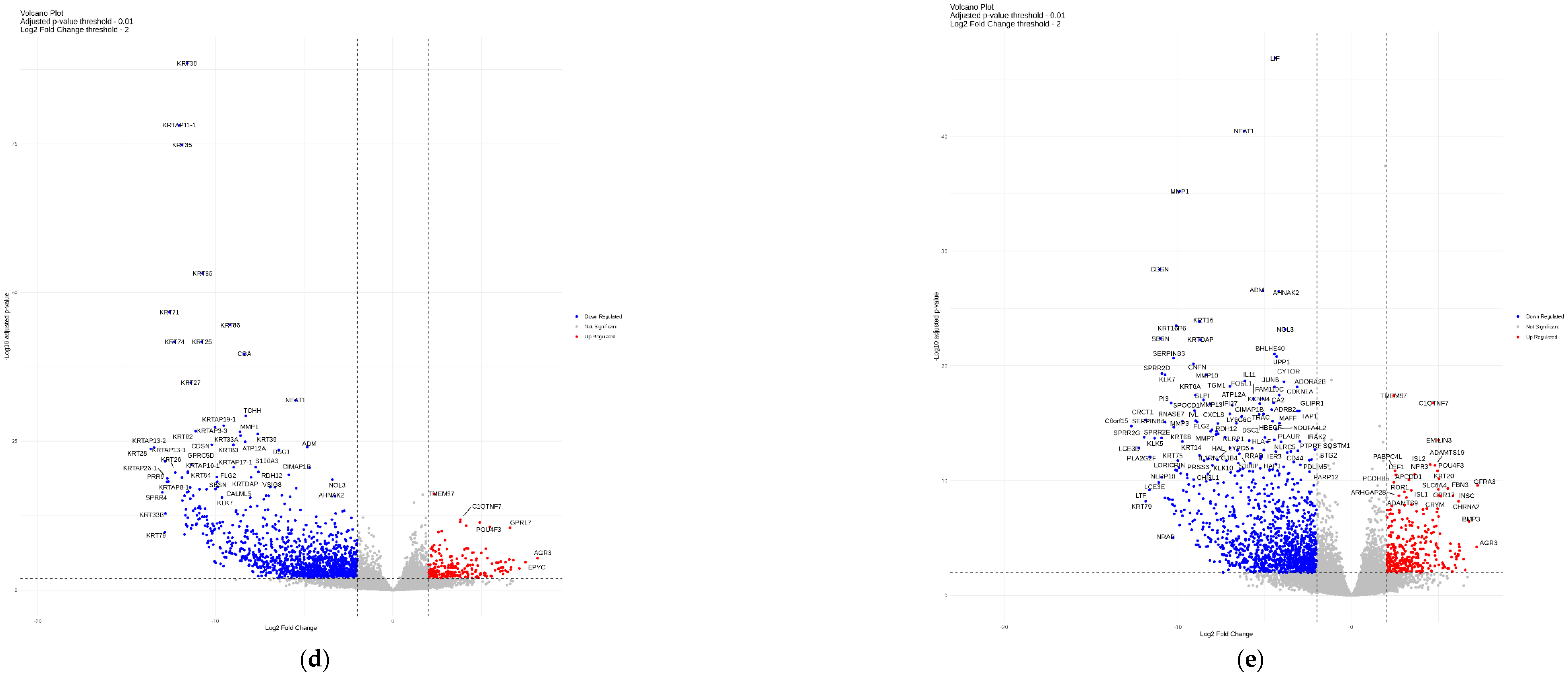
3.6. Transcriptomic Profiling of SKOs and SCOs
Differential Gene Expression and Fibrosis-Associated Pathways in SKOs and SCOs
4. Discussion
5. Conclusions
Supplementary Materials
Author Contributions
Funding
Institutional Review Board Statement
Informed Consent Statement
Data Availability Statement
Acknowledgments
Conflicts of Interest
References
- Jensen, J.; Proksch, E. The skin’s barrier. G. Ital. Di Dermatol. E Venereol. Organo Uff. Soc. Ital. Di Dermatol. E Sifilogr. 2009, 144, 689–700. [Google Scholar] [PubMed]
- Watt, F.M.; Fujiwara, H. Cell-Extracellular Matrix Interactions in Normal and Diseased Skin. Cold Spring Harb. Perspect. Biol. 2011, 3, a005124. [Google Scholar] [CrossRef]
- Ojeh, N.; Pastar, I.; Tomic-Canic, M.; Stojadinovic, O. Stem Cells in Skin Regeneration, Wound Healing, and Their Clinical Applications. Int. J. Mol. Sci. 2015, 16, 25476–25501. [Google Scholar] [CrossRef]
- Guo, S.; DiPietro, L.A. Factors Affecting Wound Healing. J. Dent. Res. 2010, 89, 219–229. [Google Scholar] [CrossRef] [PubMed]
- Krzyszczyk, P.; Schloss, R.; Palmer, A.; Berthiaume, F. The Role of Macrophages in Acute and Chronic Wound Healing and Interventions to Promote Pro-wound Healing Phenotypes. Front. Physiol. 2018, 9, 419. [Google Scholar] [CrossRef]
- Smith, J.; Rai, V. Novel Factors Regulating Proliferation, Migration, and Differentiation of Fibroblasts, Keratinocytes, and Vascular Smooth Muscle Cells during Wound Healing. Biomedicines 2024, 12, 1939. [Google Scholar] [CrossRef] [PubMed]
- Eming, S.A.; Martin, P.; Tomic-Canic, M. Wound repair and regeneration: Mechanisms, signaling, and translation. Sci. Transl. Med. 2014, 6, 265sr6. [Google Scholar] [CrossRef]
- Diegelmann, R.F.; Evans, M.C. Wound healing: An overview of acute, fibrotic and delayed healing. Front. Biosci. A J. Virtual Libr. 2004, 9, 283–289. [Google Scholar] [CrossRef]
- Velnar, T.; Bailey, T.; Smrkolj, V. The Wound Healing Process: An Overview of the Cellular and Molecular Mechanisms. J. Int. Med. Res. 2009, 37, 1528–1542. [Google Scholar] [CrossRef]
- Barone, N.; Safran, T.; Vorstenbosch, J.; Davison, P.G.; Cugno, S.; Murphy, A.M. Current Advances in Hypertrophic Scar and Keloid Management. Semin. Plast. Surg. 2021, 35, 145–152. [Google Scholar] [CrossRef] [PubMed]
- Rubin, J.P.; Neligan, P.C. (Eds.) Plastic Surgery, 4th ed.; Elsevier: London, UK, 2017. [Google Scholar]
- Čoma, M.; Fröhlichová, L.; Urban, L.; Zajíček, R.; Urban, T.; Szabo, P.; Novák, Š.; Fetissov, V.; Dvořánková, B.; Smetana, K., Jr.; et al. Molecular Changes Underlying Hypertrophic Scarring Following Burns Involve Specific Deregulations at All Wound Healing Stages (Inflammation, Proliferation and Maturation). Int. J. Mol. Sci. 2021, 22, 897. [Google Scholar] [CrossRef] [PubMed] [PubMed Central]
- Lee, H.J.; Jang, Y.J. Recent Understandings of Biology, Prophylaxis and Treatment Strategies for Hypertrophic Scars and Keloids. Int. J. Mol. Sci. 2018, 19, 711. [Google Scholar] [CrossRef]
- Wolfram, D.; Tzankov, A.; Pülzl, P.; Piza-Katzer, H. Hypertrophic scars and keloids—A review of their pathophysiology, risk factors, and therapeutic management. Dermatol. Surg. 2009, 35, 171–181. [Google Scholar] [CrossRef] [PubMed]
- Tuan, T.-L.; Nichter, L.S. The molecular basis of keloid and hypertrophic scar formation. Mol. Med. Today 1998, 4, 19–24. [Google Scholar] [CrossRef] [PubMed]
- Werner, S.; Krieg, T.; Smola, H. Keratinocyte–fibroblast interactions in wound healing. J. Investig. Dermatol. 2007, 127, 998–1008. [Google Scholar] [CrossRef] [PubMed]
- Wipff, P.-J.; Rifkin, D.B.; Meister, J.-J.; Hinz, B. Myofibroblast contraction activates latent TGF-β1 from the extracellular matrix. J. Cell Biol. 2007, 179, 1311–1323. [Google Scholar] [CrossRef] [PubMed]
- Tomasek, J.J.; Gabbiani, G.; Hinz, B.; Chaponnier, C.; Brown, R.A. Myofibroblasts and mechano-regulation of connective tissue remodelling. Nat. Rev. Mol. Cell Biol. 2002, 3, 349–363. [Google Scholar] [CrossRef] [PubMed]
- Zhou, X.; Li, J.; Backman, L.J.; Danielson, P. Keratocyte Differentiation Is Regulated by NF-κB and TGFβ Signaling Crosstalk. Int. J. Mol. Sci. 2022, 23, 11073. [Google Scholar] [CrossRef] [PubMed] [PubMed Central]
- Kendall, R.T.; Feghali-Bostwick, C.A. Fibroblasts in fibrosis: Novel roles and mediators. Front. Pharmacol. 2014, 5, 123. [Google Scholar] [CrossRef]
- Crawford, J.; Nygard, K.; Gan, B.S.; O’GOrman, D.B. Periostin induces fibroblast proliferation and myofibroblast persistence in hypertrophic scarring. Exp. Dermatol. 2015, 24, 120–126. [Google Scholar] [CrossRef]
- Bellemare, J.; Roberge, C.J.; Bergeron, D.; Lopez-Vallé, C.A.; Roy, M.; Moulin, V.J. Epidermis promotes dermal fibrosis: Role in the pathogenesis of hypertrophic scars. J. Pathol. 2005, 206, 1–8. [Google Scholar] [CrossRef] [PubMed]
- Gauglitz, G.G.; Korting, H.C.; Pavicic, T.; Ruzicka, T.; Jeschke, M.G. Hypertrophic Scarring and Keloids: Pathomechanisms and Current and Emerging Treatment Strategies. Mol. Med. 2010, 17, 113–125. [Google Scholar] [CrossRef] [PubMed]
- Katano, A.; Minamitani, M.; Yamashita, H. Risk factors for local recurrence of keloids and hypertrophic scars after postoperative electron beam radiotherapy. J. Cancer Res. Ther. 2023, 20, 163–166. [Google Scholar] [CrossRef]
- Li, D.J.; Berry, C.E.; Wan, D.C.; Longaker, M.T. Clinical, mechanistic, and therapeutic landscape of cutaneous fibrosis. Sci. Transl. Med. 2024, 16, eadn7871. [Google Scholar] [CrossRef] [PubMed]
- Ben Abdallah, H.; Bregnhøj, A.; Iversen, L.; Johansen, C. Transcriptomic Analysis of Hidradenitis Suppurativa: A Unique Molecular Signature with Broad Immune Activation. Int. J. Mol. Sci. 2023, 24, 17014. [Google Scholar] [CrossRef]
- Langhans, S.A. Three-Dimensional in Vitro Cell Culture Models in Drug Discovery and Drug Repositioning. Front. Pharmacol. 2018, 9, 6. [Google Scholar] [CrossRef]
- Tung, Y.-C.; Hsiao, A.Y.; Allen, S.G.; Torisawa, Y.-S.; Ho, M.; Takayama, S. High-throughput 3D spheroid culture and drug testing using a 384 hanging drop array. Analyst 2010, 136, 473–478. [Google Scholar] [CrossRef]
- Rogers, D.; Brown, R.D.; Hahn, M. Using Extended-Connectivity Fingerprints with Laplacian-Modified Bayesian Analysis in High-Throughput Screening Follow-Up. J. Biomol. Screen. 2005, 10, 682–686. [Google Scholar] [CrossRef]
- Rössler, S.; Nischwitz, S.P.; Luze, H.; Holzer-Geissler, J.C.J.; Zrim, R.; Kamolz, L.-P. In Vivo Models for Hypertrophic Scars—A Systematic Review. Medicina 2022, 58, 736. [Google Scholar] [CrossRef] [PubMed] [PubMed Central]
- Li, J.; Wang, J.; Wang, Z.; Xia, Y.; Zhou, M.; Zhong, A.; Sun, J. Experimental models for cutaneous hypertrophic scar research. Wound Repair Regen. 2019, 28, 126–144. [Google Scholar] [CrossRef] [PubMed]
- Pingitore, P.; Sasidharan, K.; Ekstrand, M.; Prill, S.; Lindén, D.; Romeo, S. Human Multilineage 3D Spheroids as a Model of Liver Steatosis and Fibrosis. Int. J. Mol. Sci. 2019, 20, 1629. [Google Scholar] [CrossRef] [PubMed]
- Takahashi, K.; Yamanaka, S. Induction of pluripotent stem cells from mouse embryonic and adult fibroblast cultures by defined factors. Cell 2006, 126, 663–676. [Google Scholar] [CrossRef] [PubMed]
- Kim, J.; Koo, B.-K.; Knoblich, J.A. Human organoids: Model systems for human biology and medicine. Nat. Rev. Mol. Cell Biol. 2020, 21, 571–584. [Google Scholar] [CrossRef] [PubMed] [PubMed Central]
- Xu, H.; Lyu, X.; Yi, M.; Zhao, W.; Song, Y.; Wu, K. Organoid technology and applications in cancer research. J. Hematol. Oncol. 2018, 11, 116. [Google Scholar] [CrossRef] [PubMed] [PubMed Central]
- Turhan, A.G.; Hwang, J.W.; Chaker, D.; Tasteyre, A.; Latsis, T.; Griscelli, F.; Desterke, C.; Bennaceur-Griscelli, A. iPSC-Derived Organoids as Therapeutic Models in Regenerative Medicine and Oncology. Front. Med. 2021, 8, 728543. [Google Scholar] [CrossRef] [PubMed] [PubMed Central]
- Yao, Q.; Cheng, S.; Pan, Q.; Yu, J.; Cao, G.; Li, L.; Cao, H. Organoids: Development and applications in disease models, drug discovery, precision medicine, and regenerative medicine. Medcomm 2024, 5, e735. [Google Scholar] [CrossRef] [PubMed] [PubMed Central]
- Verstegen, M.M.A.; Coppes, R.P.; Beghin, A.; De Coppi, P.; Gerli, M.F.M.; de Graeff, N.; Pan, Q.; Saito, Y.; Shi, S.; Zadpoor, A.A.; et al. Clinical applications of human organoids. Nat. Med. 2025, 31, 409–421. [Google Scholar] [CrossRef]
- Tang, X.-Y.; Wu, S.; Wang, D.; Chu, C.; Hong, Y.; Tao, M.; Hu, H.; Xu, M.; Guo, X.; Liu, Y. Human organoids in basic research and clinical applications. Signal Transduct. Target. Ther. 2022, 7, 168. [Google Scholar] [CrossRef]
- Shafiee, A.; Sun, J.; Ahmed, I.A.; Phua, F.; Rossi, G.R.; Lin, C.; Souza-Fonseca-Guimaraes, F.; Wolvetang, E.J.; Brown, J.; Khosrotehrani, K. Development of Physiologically Relevant Skin Organoids from Human Induced Pluripotent Stem Cells. Small 2023, 20, e2304879. [Google Scholar] [CrossRef] [PubMed]
- Sandoval, A.G.W.; Gim, K.Y.; Huang, J.T.; Koehler, K.R. Applications of Human Pluripotent Stem Cell–Derived Skin Organoids in Dermatology. J. Investig. Dermatol. 2023, 143, 1872–1876. [Google Scholar] [CrossRef] [PubMed] [PubMed Central]
- Hong, Z.-X.; Zhu, S.-T.; Li, H.; Luo, J.-Z.; Yang, Y.; An, Y.; Wang, X.; Wang, K. Bioengineered skin organoids: From development to applications. Mil. Med. Res. 2023, 10, 40. [Google Scholar] [CrossRef]
- Itoh, M.; Umegaki-Arao, N.; Guo, Z.; Liu, L.; Higgins, C.A.; Christiano, A.M. Generation of 3D Skin Equivalents Fully Reconstituted from Human Induced Pluripotent Stem Cells (iPSCs). PLoS ONE 2013, 8, e77673. [Google Scholar] [CrossRef]
- Lee, J.; van der Valk, W.; Serdy, S.A.; Deakin, C.; Kim, J.; Le, A.P.; Koehler, K.R. Generation and characterization of hair-bearing skin organoids from human pluripotent stem cells. Nat. Protoc. 2022, 17, 1266–1305. [Google Scholar] [CrossRef] [PubMed] [PubMed Central]
- Lee, J.; Rabbani, C.C.; Gao, H.; Steinhart, M.R.; Woodruff, B.M.; Pflum, Z.E.; Kim, A.; Heller, S.; Liu, Y.; Shipchandler, T.Z.; et al. Hair-bearing human skin generated entirely from pluripotent stem cells. Nature 2020, 582, 399–404. [Google Scholar] [CrossRef] [PubMed]
- Biernacka, A.; Dobaczewski, M.; Frangogiannis, N.G. TGF-β signaling in fibrosis. Growth Factors 2011, 29, 196–202. [Google Scholar] [CrossRef]
- Jinnin, M.; Ihn, H.; Tamaki, K. Characterization of SIS3, a Novel Specific Inhibitor of Smad3, and Its Effect on Transforming Growth Factor-β1-Induced Extracellular Matrix Expression. Mol. Pharmacol. 2006, 69, 597–607. [Google Scholar] [CrossRef] [PubMed]
- Poncelet, A.-C.; De Caestecker, M.P.; Schnaper, H.W. The transforming growth factor-βbgr/SMAD signaling pathway is present and functional in human mesangial cells. Kidney Int. 1999, 56, 1354–1365. [Google Scholar] [CrossRef]
- Zhang, W.; Ou, J.; Inagaki, Y.; Greenwel, P.; Ramirez, F. Synergistic Cooperation between Sp1 and Smad3/Smad4 Mediates Transforming Growth Factor β1 Stimulation of α2(I)-Collagen (COL1A2) Transcription. J. Biol. Chem. 2000, 275, 39237–39245. [Google Scholar] [CrossRef]
- Desmoulière, A.; Geinoz, A.; Gabbiani, F.; Gabbiani, G. Transforming growth factor-beta 1 induces alpha-smooth muscle actin expression in granulation tissue myofibroblasts and in quiescent and growing cultured fibroblasts. J. Cell Biol. 1993, 122, 103–111. [Google Scholar] [CrossRef]
- Matei, A.-E.; Chen, C.-W.; Kiesewetter, L.; Györfi, A.-H.; Li, Y.-N.; Trinh-Minh, T.; Xu, X.; Manh, C.T.; van Kuppevelt, T.; Hansmann, J.; et al. Vascularised human skin equivalents as a novel in vitro model of skin fibrosis and platform for testing of antifibrotic drugs. Ann. Rheum. Dis. 2019, 78, 1686–1692. [Google Scholar] [CrossRef]
- Jiao, H.; Dong, P.; Yan, L.; Yang, Z.; Lv, X.; Li, Q.; Zong, X.; Fan, J.; Fu, X.; Liu, X.; et al. TGF-β1 Induces Polypyrimidine Tract-Binding Protein to Alter Fibroblasts Proliferation and Fibronectin Deposition in Keloid. Sci. Rep. 2016, 6, 38033. [Google Scholar] [CrossRef]
- Marconi, G.D.; Fonticoli, L.; Rajan, T.S.; Lanuti, P.; Della Rocca, Y.; Pierdomenico, S.D.; Trubiani, O.; Pizzicannella, J.; Diomede, F. Transforming Growth Factor-Beta1 and Human Gingival Fibroblast-to-Myofibroblast Differentiation: Molecular and Morphological Modifications. Front. Physiol. 2021, 12, 676512. [Google Scholar] [CrossRef]
- Zhao, M.; Wang, L.; Wang, M.; Zhou, S.; Lu, Y.; Cui, H.; Racanelli, A.C.; Zhang, L.; Ye, T.; Ding, B.; et al. Targeting fibrosis: Mechanisms and clinical trials. Signal Transduct. Target. Ther. 2022, 7, 206. [Google Scholar] [CrossRef]
- Shi, X.; Young, C.D.; Zhou, H.; Wang, X.-J. Transforming Growth Factor-β Signaling in Fibrotic Diseases and Cancer-Associated Fibroblasts. Biomolecules 2020, 10, 1666. [Google Scholar] [CrossRef] [PubMed] [PubMed Central]
- Falanga, V.; Qian, V.S.W.; Danielpour, D.; Katz, M.H.; Roberts, A.B.; Sporn, M.B. Hypoxia upregulates the synthesis of TGF-beta 1 by human dermal fibroblasts. J. Investig. Dermatol. 1991, 97, 634–637. [Google Scholar] [CrossRef] [PubMed]
- Darby, I.A.; Hewitson, T.D. Hypoxia in tissue repair and fibrosis. Cell Tissue Res. 2016, 365, 553–562. [Google Scholar] [CrossRef]
- Ma, Y.; Liu, Z.; Miao, L.; Jiang, X.; Ruan, H.; Xuan, R.; Xu, S. Mechanisms underlying pathological scarring by fibroblasts during wound healing. Int. Wound J. 2023, 20, 2190–2206. [Google Scholar] [CrossRef]
- Nischwitz, S.P.; Fink, J.; Schellnegger, M.; Luze, H.; Bubalo, V.; Tetyczka, C.; Roblegg, E.; Holecek, C.; Zacharias, M.; Kamolz, L.-P.; et al. The Role of Local Inflammation and Hypoxia in the Formation of Hypertrophic Scars—A New Model in the Duroc Pig. Int. J. Mol. Sci. 2022, 24, 316. [Google Scholar] [CrossRef] [PubMed]
- Xu, M.; Peng, Q.; Xu, S.; Ye, C.; Lei, R.; Shen, Y.; Xu, J. Hypoxia-inducible factor-1α activates transforming growth factor-β1/Smad signaling and increases collagen deposition in dermal fibroblasts. Oncotarget 2018, 9, 3188–3197. [Google Scholar] [CrossRef]
- Zhao, B.; Guan, H.; Liu, J.-Q.; Zheng, Z.; Zhou, Q.; Zhang, J.; Su, L.-L.; Hu, D.-H. Hypoxia drives the transition of human dermal fibroblasts to a myofibroblast-like phenotype via the TGF-β1/Smad3 pathway. Int. J. Mol. Med. 2016, 39, 153–159. [Google Scholar] [CrossRef] [PubMed]
- Mallikarjuna, P.; Zhou, Y.; Landström, M. The Synergistic Cooperation between TGF-β and Hypoxia in Cancer and Fibrosis. Biomolecules 2022, 12, 635. [Google Scholar] [CrossRef] [PubMed] [PubMed Central]
- Vang, S.; Helton, E.S.; Guo, Y.; Burpee, B.; Rose, E.; Easter, M.; Bollenbecker, S.; Hirsch, M.J.; Matthews, E.L.; Jones, L.I.; et al. O-GlcNAc transferase regulates collagen deposition and fibrosis resolution in idiopathic pulmonary fibrosis. Front. Immunol. 2024, 15, 1387197. [Google Scholar] [CrossRef] [PubMed] [PubMed Central]
- Linares, H.A.; Kischer, C.W.; Dobrkovsky, M.; Larson, D.L. The Histiotypic organization of the hypertrophic scar in humans. J. Investig. Dermatol. 1972, 59, 323–331. [Google Scholar] [CrossRef]
- Choi, Y.-H.; Kim, K.-M.; Kim, H.-O.; Jang, Y.-C.; Kwak, I.-S. Clinical and Histological Correlation in Post-Burn Hypertrophic Scar for Pain and Itching Sensation. Ann. Dermatol. 2013, 25, 428–433. [Google Scholar] [CrossRef]
- Morris, D.E.; Wu, L.; Zhao, L.L.; Bolton, L.; Roth, S.I.; Ladin, D.A.; Mustoe, T.A. Acute and Chronic Animal Models for Excessive Dermal Scarring: Quantitative Studies. Plast. Reconstr. Surg. 1997, 100, 674–681. [Google Scholar] [CrossRef]
- Gabbiani, G. The myofibroblast in wound healing and fibrocontractive diseases. J. Pathol. 2003, 200, 500–503. [Google Scholar] [CrossRef] [PubMed]
- Zhang, S.; Zhang, Y.; Min, P. Single-Cell and Bulk Transcriptome Data Integration Reveals Dysfunctional Cell Types and Aberrantly Expressed Genes in Hypertrophic Scar. Front. Genet. 2022, 12, 806740. [Google Scholar] [CrossRef] [PubMed] [PubMed Central]
- Tomann, P.; Paus, R.; Millar, S.E.; Scheidereit, C.; Schmidt-Ullrich, R. Lhx2 is a direct NF-κB target gene that promotes primary hair follicle placode down-growth. Development 2016, 143, 1512–1522. [Google Scholar] [CrossRef]
- Mardaryev, A.N.; Meier, N.; Poterlowicz, K.; Sharov, A.A.; Sharova, T.Y.; Ahmed, M.I.; Rapisarda, V.; Lewis, C.; Fessing, M.Y.; Ruenger, T.M.; et al. Lhx2 differentially regulates Sox9, Tcf4 and Lgr5 in hair follicle stem cells to promote epidermal regeneration after injury. Development 2011, 138, 4843–4852. [Google Scholar] [CrossRef] [PubMed]
- Desmoulière, A. Factors influencing myofibroblast differentiation during wound healing and fibrosis. Cell Biol. Int. 1995, 19, 471–476. [Google Scholar] [CrossRef]
- Limandjaja, G.C.; Broek, L.J.; Waaijman, T.; Scheper, R.; Niessen, F.; Gibbs, S. Chapter 6 Tissue Engineered Human Scar Models Identify Extracellular Matrix and inflammatory Cytokine Differences Between Normotrophic, Hypertrophic and Keloid Scars. 2015. Available online: https://pure.amsterdamumc.nl/ws/portalfiles/portal/111272776/hoofdstuk%206.pdf (accessed on 20 April 2026).
- Zheng, Y.; Khan, Z.; Zanfagnin, V.; Correa, L.F.; Delaney, A.A.; Daftary, G.S. Epigenetic Modulation of Collagen 1A1: Therapeutic Implications in Fibrosis and Endometriosis1. Biol. Reprod. 2016, 94, 87. [Google Scholar] [CrossRef]
- Roche, P.; Czubryt, M.P. Transcriptional control of collagen I gene expression. Cardiovasc. Haematol. Dis.-Orders-Drug Targets 2014, 14, 107–120. [Google Scholar] [CrossRef] [PubMed]
- Hou, C.; Huo, J.; Yan, S.; Sun, F.; Yang, X. Identification of fibrosis-associated biomarkers in heart failure and human cancers. J. Transl. Med. 2024, 22, 1042. [Google Scholar] [CrossRef]
- Thiele, B.-J.; Doller, A.; KähNe, T.; Pregla, R.; Hetzer, R.; Regitz-Zagrosek, V. RNA-Binding Proteins Heterogeneous Nuclear Ribonucleoprotein A1, E1, and K Are Involved in Post-Transcriptional Control of Collagen I and III Synthesis. Circ. Res. 2004, 95, 1058–1066. [Google Scholar] [CrossRef]
- Tang, C.-M.; Zhang, M.; Huang, L.; Hu, Z.-Q.; Zhu, J.-N.; Xiao, Z.; Zhang, Z.; Lin, Q.-X.; Zheng, X.-L.; -Yang, M.; et al. CircRNA_000203 enhances the expression of fibrosis-associated genes by derepressing targets of miR-26b-5p, Col1a2 and CTGF, in cardiac fibroblasts. Sci. Rep. 2017, 7, 40342. [Google Scholar] [CrossRef]
- Stamenkovic, I. Extracellular matrix remodelling: The role of matrix metalloproteinases. J. Pathol. 2003, 200, 448–464. [Google Scholar] [CrossRef]
- Ye, M.; Song, Y.; Pan, S.; Chu, M.; Wang, Z.-W.; Zhu, X. Evolving roles of lysyl oxidase family in tumorigenesis and cancer therapy. Pharmacol. Ther. 2020, 215, 107633. [Google Scholar] [CrossRef]
- Klouda, T.; Tsikis, S.T.; Hirsch, T.I.; Kim, Y.; Li, Y.; Gaal, J.; Zhao, Z.; Friehs, I.; Shyy, J.Y.-J.; Raby, B.A.; et al. Smooth muscle Cxcl12 contributions to vascular remodeling in flow and hypoxia-induced pulmonary hypertension. J. Biol. Chem. 2025, 301, 110207. [Google Scholar] [CrossRef] [PubMed]
- Alt, C.; Laschinger, M.; Engelhardt, B. Functional expression of the lymphoid chemokines CCL19 (ELC) and CCL 21 (SLC) at the blood-brain barrier suggests their involvement in G-protein-dependent lymphocyte recruitment into the central nervous system during experimental autoimmune encephalomyelitis. Eur. J. Immunol. 2002, 32, 2133–2144. [Google Scholar] [CrossRef] [PubMed]
- Chavez, B.; Kiaris, H. Insights on the role of the chemokine CCL8 in pathology. Cell. Signal. 2025, 134, 111951. [Google Scholar] [CrossRef]
- Niessen, F.B.; Andriessen, M.P.; Schalkwijk, J.; Visser, L.; Timens, W. Keratinocyte-derived growth factors play a role in the formation of hypertrophic scars. J. Pathol. 2001, 194, 207–216. [Google Scholar] [CrossRef]
- Wikramanayake, T.C.; Stojadinovic, O.; Tomic-Canic, M. Epidermal differentiation in barrier maintenance and wound healing. Adv. Wound Care 2014, 3, 272–280. [Google Scholar] [CrossRef]
- Machesney, M.; Tidman, N.; Waseem, A.; Kirby, L.; Leigh, I. Activated keratinocytes in the epidermis of hypertrophic scars. Am. J. Pathol. 1998, 152, 1133–1141. [Google Scholar] [PubMed] [PubMed Central]
- Rhee, H.; Polak, L.; Fuchs, E. Lhx2 Maintains Stem Cell Character in Hair Follicles. Science 2006, 312, 1946–1949. [Google Scholar] [CrossRef]
- Törnqvist, G.; Sandberg, A.; Hägglund, A.-C.; Carlsson, L. Cyclic Expression of Lhx2 Regulates Hair Formation. PLoS Genet. 2010, 6, e1000904. [Google Scholar] [CrossRef]
- Pasolli, H.; Folgueras, A.R.; Fuchs, E. Architectural Niche Organization by LHX2 is Linked to Hair Follicle Stem Cell Function. Microsc. Microanal. 2014. [Google Scholar] [CrossRef]
- Chen, J.; Ge, S.-J.; Feng, H.-J.; Wu, S.-Z.; Ji, R.; Huang, W.-R.; Huang, W.; Lu, C.-H. KRT17 Promotes the Activation of HSCs via EMT in Liver Fibrosis. J. Clin. Transl. Hepatol. 2021, 10, 207–218. [Google Scholar] [CrossRef]




Disclaimer/Publisher’s Note: The statements, opinions and data contained in all publications are solely those of the individual author(s) and contributor(s) and not of MDPI and/or the editor(s). MDPI and/or the editor(s) disclaim responsibility for any injury to people or property resulting from any ideas, methods, instructions or products referred to in the content. |
© 2026 by the authors. Licensee MDPI, Basel, Switzerland. This article is an open access article distributed under the terms and conditions of the Creative Commons Attribution (CC BY) license.
Share and Cite
Kim, H.M.; Oh, E.J.; Kwak, S.; Han, S.O.; Chung, H.Y. Modeling Human Hypertrophic Scars with Induced Pluripotent Stem-Cell-Derived Scar Organoids Versus Skin Organoids. Cells 2026, 15, 969. https://doi.org/10.3390/cells15110969
Kim HM, Oh EJ, Kwak S, Han SO, Chung HY. Modeling Human Hypertrophic Scars with Induced Pluripotent Stem-Cell-Derived Scar Organoids Versus Skin Organoids. Cells. 2026; 15(11):969. https://doi.org/10.3390/cells15110969
Chicago/Turabian StyleKim, Hyun Mi, Eun Jung Oh, Suin Kwak, Se Ok Han, and Ho Yun Chung. 2026. "Modeling Human Hypertrophic Scars with Induced Pluripotent Stem-Cell-Derived Scar Organoids Versus Skin Organoids" Cells 15, no. 11: 969. https://doi.org/10.3390/cells15110969
APA StyleKim, H. M., Oh, E. J., Kwak, S., Han, S. O., & Chung, H. Y. (2026). Modeling Human Hypertrophic Scars with Induced Pluripotent Stem-Cell-Derived Scar Organoids Versus Skin Organoids. Cells, 15(11), 969. https://doi.org/10.3390/cells15110969