Article

Differential Modulation of Hepatic Akt/mTOR Signaling During Acute and Chronic Toxoplasma gondii Infection in a Murine Model

Stanley Division of Developmental Neurovirology, Department of Pediatrics, Johns Hopkins School of Medicine, Baltimore, MD 21287, USA
Cells 2026, 15(10), 893; https://doi.org/10.3390/cells15100893
Submission received: 3 April 2026 / Revised: 6 May 2026 / Accepted: 9 May 2026 / Published: 14 May 2026

Highlights

What are the main findings?
  • Acute T. gondii infection broadly suppresses hepatic Akt/mTOR signaling.
  • Chronic infection related to cyst burden activates specific Akt/mTOR nodes.
What are the implications of the main findings?
  • T. gondii has distinct strategies to manipulate host survival based on its life stages.
  • The Akt/mTOR pathway may serve as a therapeutic target for the treatment of T. gondii.

Abstract
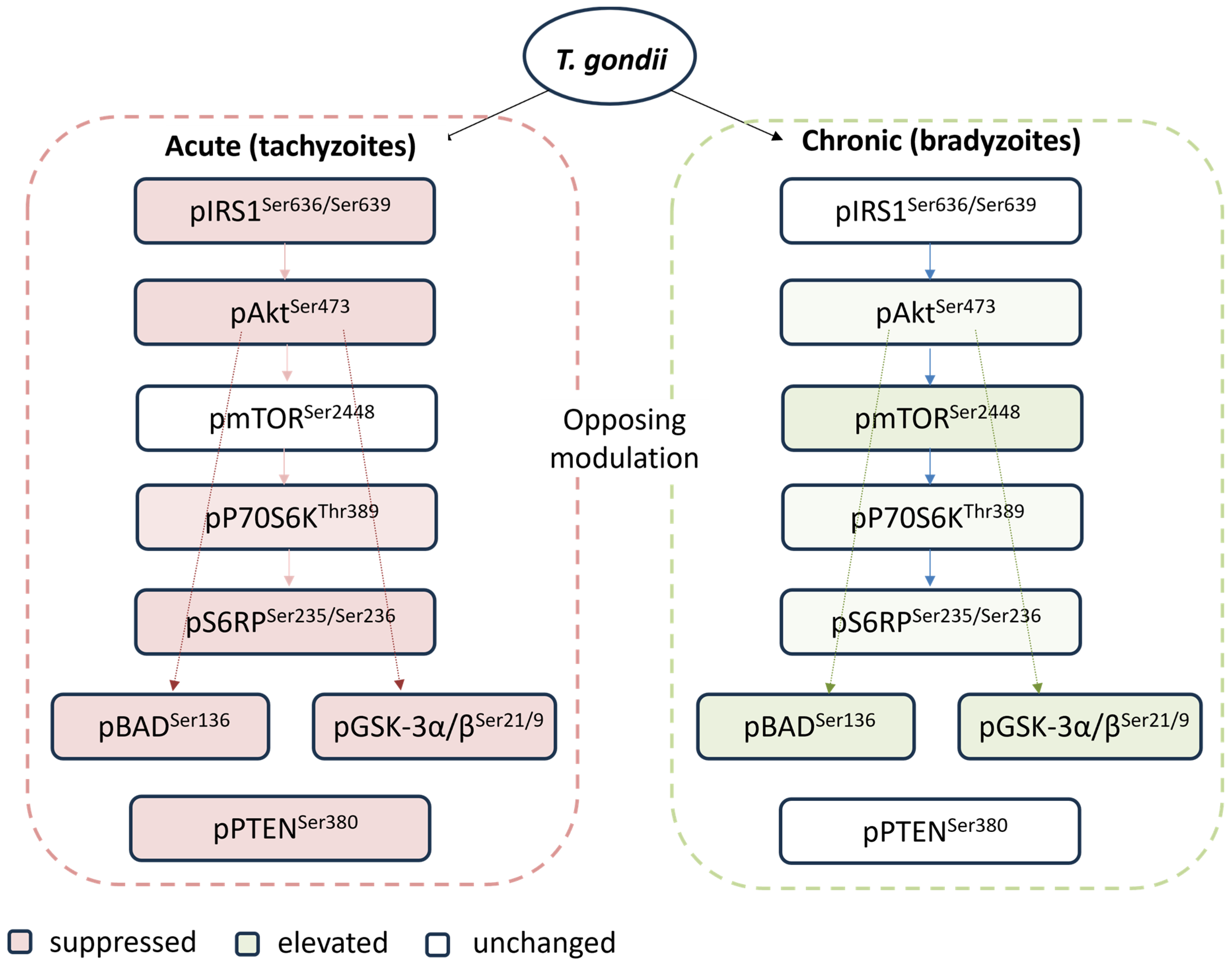
Toxoplasma gondii is an obligate intracellular parasite that infects virtually all warm-blooded animals, progressing through acute and chronic stages. The Akt/mTOR signaling axis plays critical roles in cell survival, proliferation, and metabolism, making it a key target for intracellular pathogens. This study investigated how T. gondii infection modulates this pathway during both infections. Outbred CD-1 mice were infected intraperitoneally with the virulent GT1 strain of T. gondii. Mice for acute studies were sacrificed five days post-infection, while those for chronic studies were treated with sulfadiazine and sacrificed five months post-infection. Phosphoprotein expression of eight Akt/mTOR pathway components was measured in liver tissues using a multiplexed bead-based immunoassay. Acute T. gondii infection caused broad suppression of Akt/mTOR signaling, with 6 of 8 markers significantly downregulated, including pS6RPSer235/236, pAKTS473, pBADSer136, pIRS1S636/639, pPTENSer380, and pGSK-3α/βSer21/9. In contrast, chronic infection related to cyst burden selectively activates specific nodes of the pathway, including pBADSer136, pmTORSer2448, and pGSK-3α/βSer21/9. Infection induced strong correlations between inter-components, which reflect coherent and coordinated pathway-level reprogramming rather than random perturbation. These findings show that acute and chronic T. gondii infections have opposing effects on host Akt/mTOR signaling for their own benefit, which may present new therapeutic targets.

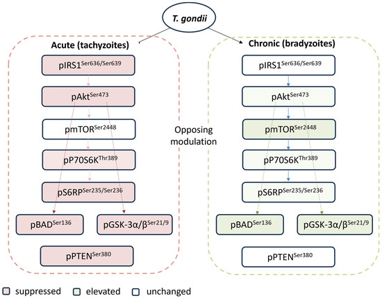
Graphical Abstract

1. Introduction

Toxoplasma gondii infects approximately one-third of the global human population, making it one of the most prevalent parasitic infections worldwide. The infection develops in two stages: acute and chronic [1]. During an acute infection, rapidly dividing tachyzoites disseminate throughout the host, prompting strong innate and adaptive immune responses to control the multiplication of the parasite. This immune response leads to the conversion of tachyzoites into the slower-growing encysted bradyzoites. Tissue cysts can persist for the lifetime of the host and remain infectious if ingested. Currently, available anti-T. gondii drugs are ineffective in clearing the cyst form of the chronic infection [2]. This therapeutic gap largely results from an incomplete understanding of interactions between the host and the parasite during the progression of T. gondii infection. Gaining insight into how T. gondii infection affects cell survival, metabolism, and proliferation during acute and chronic infection may lead to better approaches.
T. gondii is an obligate intracellular parasite that can infect virtually any nucleated cell in warm-blooded animals. The ability to subvert host cell signaling pathways is central to intracellular parasite survival. T. gondii has been shown to hijack host cell biology to survive and replicate by creating a specialized niche, the parasitophorous vacuole (PV), which shields it from lysosomal fusion and degradation [3]. The parasite actively remodels the host cell by secreting effector proteins from specialized organelles (rhoptries and dense granules) directly into the host cytoplasm and nucleus [4]. These effectors manipulate host gene expression, inhibit apoptosis, and subvert host signaling pathways to evade immune responses. Studies on host–parasite interactions have identified numerous signaling pathways altered upon infection.
The phosphatidylinositol 3-kinase (PI3K)/Akt/mammalian target of rapamycin (mTOR) pathway is a master regulator of cell survival, metabolism, and growth [5]. Upon activation by growth factors or insulin receptor signaling, PI3K phosphorylates phosphatidylinositol-4,5-bisphosphate (PIP2) to generate PIP3. This recruits Akt to the plasma membrane, where it is phosphorylated at Thr308 by PDK1 and at Ser473 by the mTOR complex 2 (mTORC2) [6]. Fully activated Akt coordinates diverse downstream responses, including inhibition of apoptosis through phosphorylation of the pro-apoptotic protein BAD at Ser136, which makes BAD dissociate from the Bcl-2/Bcl-X complex and lose the pro-apoptotic function [7]; inhibition of glycogen synthase kinase-3α/β (GSK-3α/β) through phosphorylation at Ser21/9, promoting cell survival and glucose metabolism [8]; and activation of mTOR complex 1 (mTORC1) through multiple mechanisms, leading to phosphorylation of downstream effectors including p70 ribosomal S6 kinase 1 (p70S6K1) at Thr389 and ribosomal protein S6 (S6RP), which drive ribosome biogenesis and protein synthesis. Negative regulation of the pathway is mediated by the tumor suppressor PTEN, which dephosphorylates PIP3 to dampen Akt activation. Additionally, inhibitory phosphorylation of insulin receptor substrate-1 (IRS1) at Ser636/639 acts as a negative feedback mechanism downstream of mTORC1.
Research has suggested that several intracellular pathogens have evolved mechanisms to manipulate the PI3K/Akt/mTOR pathway to their advantage. Mycobacterium tuberculosis [9], Salmonella [10], and Listeria [11] exploit Akt signaling to inhibit apoptosis and promote intracellular survival. PI3K/Akt/mTOR signaling plays a very important role in HPV-induced carcinogenesis by regulating multiple cellular and molecular events [12]. Prior work has demonstrated that T. gondii can activate PI3K/Akt/mTOR signaling to facilitate host cell invasion and inhibit apoptosis in infected cells [13,14]. A recent study suggests that the PI3K/Akt/mTOR signaling pathway plays an important role in T. gondii-induced mitochondrial dysfunction and the reprogramming of cellular energy metabolism [15]. However, systematic profiling of the complete Akt/mTOR signaling network across both acute and chronic infection stages has not been reported.
The current study systematically profiled the phosphorylation of eight components of the Akt/mTOR pathway in mouse liver tissues, during both acute and chronic T. gondii infection, using a multiplexed immunoassay. The hypothesis was that T. gondii modulates hepatic Akt/mTOR signaling differently based on its tachyzoite and bradyzoite life stages. The liver is a key metabolic organ and an important site of T. gondii infection. During the early stages of infection, tachyzoites disseminate via the bloodstream and infect hepatocytes [16,17]. While significant attention has focused on central nervous system toxoplasmosis, the liver can also harbor bradyzoite cysts during chronic infection [18]. Research has found that the liver is the primary site of tissue pathology [16] in severe toxoplasmosis and can cause liver diseases such as hepatitis [19] and hepatomegaly [20,21]. The present study revealed that tachyzoites rapidly replicate within a metabolically suppressed but still viable host cell, while bradyzoites require a stable, long-lived cellular niche for decades-long persistence.

2. Materials and Methods

2.1. Ethics Statement

All mouse specimens were collected from prior projects, and no additional animals were sacrificed for the present study. All protocols were approved by the Animal Care and Use Committee at Johns Hopkins University. All experiments conformed to the U.S. National Institutes of Health Guide for the Care and Use of Laboratory Animals.

2.2. Acute and Chronic Mouse Models of T. gondii Infection

Six- to eight-week-old female outbred CD-1 mice (ICR-Harlan Sprague) were infected intraperitoneally (i.p.) with 500 T. gondii GT1 strain tachyzoites (Type I, virulent). Control mice received vehicle only (PBS). The model employs a type I strain because of its close association with clinical disease and its greater influence on host genes, as demonstrated in our previous research [22,23]. For acute infection, mice were sacrificed at 5 days post infection (dpi), as described previously [24]. For chronic infection, infected mice, including controls, were treated with anti-T. gondii chemotherapy (sulfadiazine sodium) in drinking water (400 mg/liter; Sigma) from days 5 to 30 to control tachyzoite proliferation and prevent animal death. Sulfadiazine is a competitive analogue of PABA and inhibits tachyzoite growth, but not encysted bradyzoites. The GT1 strain was maintained by passage in human fibroblast cells (HFF, ATCC SCRC-1041). Mice were sacrificed at five months postinfection (mpi) [25]. Upon sacrifice, liver tissue was immediately harvested, snap-frozen in liquid nitrogen, and stored at −80 °C.

2.3. Confirmation of Infection and Cyst Burden Assessment

As described previously [25], T. gondii infection in all chronically infected mice was confirmed using a commercial ELISA kit (IB19213, IBL America, Minneapolis, MN, USA) for anti-T. gondii IgG antibodies. Cyst burden was quantified using the MAG1 (matrix antigen 1) assay, which measures antibodies to peptide antigens derived from MAG1 [25,26,27]. The MAG1 protein is an antigen found abundantly in the cyst wall and within the cyst matrix. The serological response to the MAG1 peptide antigen has been validated as a proxy marker of chronic T. gondii infection and cyst burden [25]. The median MAG1 antibody absorbance value (0.5) was the cutoff to stratify mice into two groups: those with a high cyst burden (OD ≥ 0.5) and those with a low cyst burden (OD < 0.5).
For this study, mice (n = 8 per group) were selected based on their antibody profiles. The groups included (i) T. gondii unexposed controls (T. gondii IgG−/MAG1−) and (ii) T. gondii chronically infected mice (T. gondii IgG+/MAG1+; IgG OD: IgG = 3.72 ± 0.09; MAG1 OD = 1.291 ± 1.387). The infected mice were further divided into two subgroups: MAG1-high (OD = 2.394 ± 1.153) and MAG1-low (OD = 0.1884 ± 0.1259).

2.4. Liver Tissue Homogenization and Protein Extraction

Liver tissue samples (approximately 100 mg) were homogenized on ice in RIPA buffer (Sigma, St. Louis, MO, USA), supplemented with a protease and phosphatase inhibitor cocktail (Thermo Scientific, Waltham, MA, USA). The mixture was sonicated at 4 °C for 5 min, followed by centrifugation at 10,000× g for 5 min at 4 °C, and the supernatant was collected. Total protein concentration was determined using a BCA protein assay (Thermo Scientific).

2.5. Multiplexed Phosphoprotein Immunoassay

Phosphorylation levels of Akt/mTOR pathway components were measured using a bead-based multiplex immunoassay (Bio-Plex Pro Cell Signaling Akt Panel 8-plex, LQ00006JK0K0RR, Bio-Rad, Hercules, CA, USA) on the Luminex xMAP platform. The panel comprises eight key phosphoproteins located upstream and downstream from Akt and includes: insulin receptor substrate-1 at Ser636/639 (pIRS1Ser636/Ser639), phosphatase and tensin homolog at Ser380 (pPTENSer380), serine/threonine-protein kinase Akt-1 at Ser 473 (pAktSer473), glycogen synthase kinase-3α/β at Ser21 and Ser9 (pGSK3α/βSer21/Ser9), mTOR at Ser2448 (pmTORSer2448), p70 S6 kinase at Thr389 (pP70S6KThr389), ribosomal protein S6 kinase beta-1 at Ser235/236 (pS6RPSer235/Ser236), and the Bcl2-associated agonist of cell death at Ser136 (pBADSer136). All samples were analyzed according to the manufacturer’s protocols, and the median fluorescence intensity (MFI) values were recorded.

2.6. Statistical Analysis

All data sets were tested for normality by using the Shapiro-Wilk test. Differences between the two groups were analyzed via Welch’s t-test. For multiple groups, ANOVA with Bonferroni’s multiple comparison test was used. Spearman correlation analyses were performed to examine the inter-protein correlation. Multiple testing corrections with a false discovery rate (FDR) of 5% were performed for correlation and infection analysis. Fold change (FC) was calculated as the ratio of the mean MFI of the infection group to the mean MFI of the respective control group. A p-value of <0.05 was considered statistically significant; p < 0.10 was noted as a trend (†). Data are presented as mean ± standard deviation (SD). All statistical analyses were conducted in Graph-Pad Prism V11.0.0.

3. Results

3.1. Acute T. gondii Infection Broadly Suppresses Hepatic Akt/mTOR Signaling

To examine the effects of acute T. gondii infection on host Akt/mTOR signaling, the phosphorylation levels of eight pathway components in liver tissues were compared between mock-infected and acutely infected mice at 5 dpi.
As shown in Table 1, acute infection resulted in significant suppression of Akt/mTOR signaling across most examined nodes. The most pronounced reductions were observed for p70S6KThr389 (FC = 0.616, p = 0.063), pIRS1Ser636/Ser639 (FC = 0.636, p = 0.0277), pS6RPSer235/Ser236 (FC = 0.630, p = 0.0277), and pAKTSer473 (FC = 0.694, p = 0.0277), with each showing a 31–38% decrease compared to controls. A moderate decrease was also observed for pBADSer136 (FC = 0.753, p = 0.0277), pGSK-3α/βSer21/9 (FC = 0.783, p = 0.0277), and pPTENSer380 (FC = 0.868, p = 0.0435). Notably, pmTORSer2448 showed a minimal reduction (FC = 0.862) and did not reach statistical significance (p = 0.1805). Overall, 6 of 8 markers were significantly downregulated in infected mice, with p70S6KThr389 showing a trend towards reduction.

3.2. Chronic T. gondii Infection Related to Cyst Burden Activates Select Hepatic Akt/mTOR Nodes

Chronic T. gondii infection activated specific nodes of the Akt/mTOR signaling pathway (Table 2). Compared to mock-infected control, mice that were chronically infected for 5 months showed increased phosphorylation of all 8 components, with significant changes in four: pGSK-3α/βSer21/9 (FC = 1.72, p = 0.0016), pmTORSer2448 (FC = 1.44, p = 0.0236), pBADSer136 (FC = 1.82, p = 0.0218), and pAKTSer473 (FC = 1.41, p = 0.0218). Additionally, pS6RPSer235/236 showed a trend towards increase (FC = 1.92, p = 0.0971).
Chronic infection is defined by the presence of tissue cysts, making cyst burden a critical factor for understanding its effects. I stratified the chronically infected mice into MAG1-high and MAG1-low groups based on MAG1 antibody levels, which serve as a proxy marker of cyst burden [25]. As shown in Table 3, the results indicated that the elevation was particularly pronounced in the MAG1-high mice, with three pathway components showing a significant increase compared to the control group. Specifically, pGSK-3α/βSer21/9 showed a greater than twofold increase (FC = 2.08, p < 0.0001), as did pBADSer136 (FC = 2.29, p < 0.05) and pmTORSer2448 (FC = 1.73, p < 0.05). Additionally, p70S6KThr389 (FC = 2.16, ANOVA p = 0.097) and pS6RPSer235/Ser236 (FC = 2.57, ANOVA p = 0.073) showed a trend towards increase. Moreover, MAG1-high mice also significantly differed from MAG1-low mice in pmTORSer2448 (FC = 1.51, p < 0.05) and pGSK-3α/βSer21/9 (FC = 1.53, p < 0.01). No significant difference was found between MAG1-low and mock-infected controls.
None of the eight Akt/mTOR pathway components correlate significantly with MAG1 antibody levels in the chronically infected group (all p > 0.05 by Spearman). However, there is a trend indicating a positive correlation between MAG1 antibody levels and pGSK-3α/βSer21/9 (r = 0.46, p = 0.0758) or pBADSer136 (r = 0.43, p = 0.096).

3.3. Phosphorylation Levels of Akt/mTOR Pathway Components Are Significantly Correlated

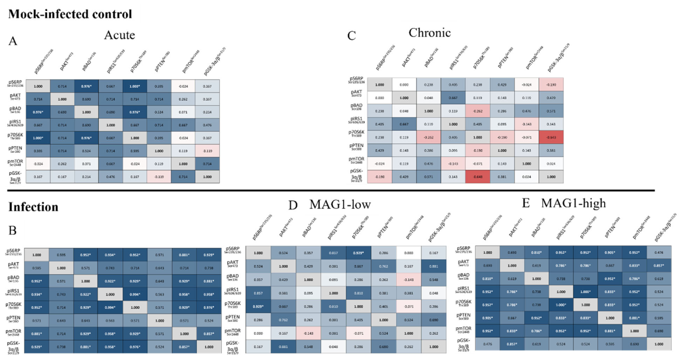
I explored whether activation/inhibition of these Akt/mTOR components was consistent along the pathway. To this aim, correlation analyses were performed among the 8 proteins from both acute and chronic groups, as well as their corresponding controls (Figure 1).
Mice with acute infections or high MAG1 antibody levels show a cohesive phosphoprotein network, with 15 and 17 statistically significant pairwise correlations, respectively. The weakest correlations during acute infection mainly involved pPTENSer380, whereas in mice with high MAG1 levels, the weakest correlations were primarily with pGSK-3α/βSer21/9. The correlation between p70S6KThr389 and pS6RPSer235/Ser236 is the most robustly coupled link in every infection group (acute, MAG1-low, MAG1-high), confirming tight coupling between the kinase and its substrate during infection. For MAG1-low mice, several pairs show moderate r values but fail the FDR correction, suggesting that pathway coherence is partially maintained, although it is notably weaker. In the mock-infected control for chronic infection, all 28 pairwise correlations are non-significant, while 25 are non-significant in the mock-infected control for acute infection. The correlation between pAKTSer473 and pBADSer136 remains nonsignificant across all groups, regardless of the infection status.

4. Discussion

This study provides the first systematic, comparative analysis of hepatic Akt/mTOR signaling during both acute and chronic T. gondii infection in a murine model using the virulent GT1 strain. It revealed that distinct infection stages exert opposing regulatory effects on the host Akt/mTOR pathway: acute infection broadly suppresses activity across most nodes, whereas chronic infection related to cyst burden selectively activates anti-apoptotic and metabolic signaling nodes. These findings are consistent with the distinct functional imperatives of the tachyzoite and bradyzoite life stages [28,29], and support the concept that T. gondii has evolved stage-specific strategies to manipulate host cell biology for its own benefit.

4.1. Broad Suppression of Akt/mTOR Signaling During Acute Infection

The broad suppression of hepatic Akt/mTOR signaling during acute infection—with 6 of 8 markers significantly reduced at 5 dpi—is consistent with metabolic exploitation and immune evasion during the lytic tachyzoite cycle [30,31].
Upstream of Akt, the reduction in pIRS1Ser636/639 appears insufficient to restore downstream Akt signaling, as concurrent activation of pPTENSer380 occurs. PTEN is a widely known negative regulator of insulin/PI3K signaling [32], so its reduction at Ser380 renders PTEN more enzymatically active, leading to further dephosphorylation of PIP3 and reinforcing suppression of the PI3K/Akt axis. Phosphorylation of IRS1 at Ser636/639 normally results from mTORC1/S6K1-mediated negative feedback on insulin signaling [33]; its reduction most likely reflects the loss of this feedback loop, as supported by reduced p70S6KThr389 and pS6RPSer235/236 expression. This reduction would be expected to enhance IRS1 activity and insulin sensitivity [34]. Pro-inflammatory cytokines (TNF-α, IL-1β, IL-6) activated during acute toxoplasmosis are well-known to interfere with insulin receptor signaling [35].
Downstream of Akt, reduced phosphorylation of BADSer136 increases pro-apoptotic pressure on the host cell by allowing BAD to interact with the anti-apoptotic proteins Bcl-2 and Bcl-xL [7]. Suppression of Akt signaling may attenuate NF-κB-dependent inflammatory gene expression, since Akt can phosphorylate and activate IκB kinase [36]. Dampening pro-inflammatory responses could thereby delay the onset of effective Th1 immunity. The phosphorylation of GSK3βSer9 or GSK3αSer21 is known to decrease GSK3α/β enzymatic activity [37,38]. The decrease in pGSK-3α/β indicates GSK-3 activation, which reflects a shift away from anabolic glucose storage and pro-survival signaling. The suppression of p70S6KThr389 and pS6RPSer235/236 may redirect biosynthetic resources away from host cell metabolism [39]. This would favor nutrient availability for the rapidly replicating tachyzoite within the parasitophorous vacuole.
Notably, mTOR phosphorylation at Ser2448 was the only node that did not show a significant change during acute infection. As pmTORSer2448 likely reflects convergent upstream signaling, this implies that mTOR catalytic activity is preserved through Akt-independent mechanisms [40]. This preservation is biologically critical because mTOR is the primary suppressor of autophagy, and its activity prevents autophagic targeting of the parasitophorous vacuole—the host cell’s principal mechanism for intracellular parasite clearance [41,42]. Collectively, this pattern suggests that T. gondii has evolved to uncouple mTOR from its upstream Akt activator and converted the hepatocyte into a nutrient-rich but immunologically crippled replication niche.

4.2. Selective Activation of Akt/mTOR Nodes During Chronic Infection

In contrast to acute infection, chronic T. gondii infection related to cyst burden selectively activates specific nodes of anti-apoptotic (pBAD), metabolic (pmTOR, pGSK-3α/β), and translational (p-70S6K, p-S6RP) pathways. This pattern suggests an overactivated signaling state, supporting the notion that bradyzoites inhibit host cell apoptosis and modify glucose metabolism to create a favorable environment for their survival [28,29]. It is conceivable that MAG1-low mice have insufficient levels of antigens needed to activate these nodes.
The cyst burden-associated activation of pBADSer136 and pGSK-3α/βSer21/Ser9 carries broad functional implications. Phosphorylation of BAD at Ser136 would enhance anti-apoptotic signaling and maintain host cell viability [43]. Previous studies have found that T. gondii-infected cells are broadly resistant to multiple inducers of apoptosis [44]. This resistance may result from Akt and BAD phosphorylation, which, depending on parasite load, inhibit Bax translocation to mitochondria and block apoptosis [14]. Inactivation of GSK-3 may enhance glycolysis and increase glucose availability [45], which is crucial for bradyzoites. GRA18, a dense granule effector protein of T. gondii, has been shown to act as an inhibitor of host GSK3, triggering β-catenin accumulation [46]. β-catenin is a key molecule in the Wnt pathway, and its stable expression on Treg cells promotes cell survival [47]. Notably, our previous study found that the phosphorylation levels of the inhibitory forms of GSK-3α/βSer21/Ser9 were decreased in the cerebellum of MAG1-high mice [48], whereas in this study, a more than two-fold increase in the liver was observed. This tissue-specific divergence likely reflects the well-documented pleiotropy of GSK-3 across different cellular contexts.
The significant upregulation of Akt, mTOR, and mTOR-associated targets (p70S6KThr389 and pS6RPSer235/236) in chronically infected mice—particularly in the MAG1-high subgroup—aligns with previous findings of T. gondii infection on Akt/mTOR pathway activation [15,49]. Hyperactivation of mTOR has been associated with defects in autophagosome formation, and inhibiting mTOR activity suppresses T. gondii replication [50]. A study reports that T. gondii inhibits FOXO3a, a transcription factor that regulates the expression of autophagy-related genes, through AKT-dependent phosphorylation [51]. Interestingly, the upstream regulatory machinery of the Akt/mTOR pathway—pIRS1Ser636/639 and pPTENSer380—was not significantly altered during chronic infection, despite robust changes in downstream nodes. The lack of change suggested that upstream PI3K/PTEN regulation may not be the primary driver of chronic-stage Akt activation. Rather, direct activation at or downstream of Akt itself may be responsible and warrants further investigation. Additionally, although cyst burden is a primary factor in the activation of the hepatic Akt/mTOR pathway, there was no significant correlation between phosphorylation levels and MAG1 antibody levels. This suggests that humoral immunity does not directly reflect the intracellular signaling status within the tissue.

4.3. Opposing Stage-Specific Strategies

The most striking finding of this study is the diametrically opposed regulatory strategies employed by T. gondii between acute and chronic infections. Acute infection broadly suppresses this pathway to redirect host metabolism and dampen immune activation, while chronic infection related to cyst burden selectively activates pro-survival, anti-apoptotic, and anabolic nodes. Similarly, our previous studies also found that T. gondii affects host miR-132 differently in acute and chronic infection [24,52]. A study on neonatal mouse astrocytes found that T. gondii infection triggers both pro-apoptotic and anti-apoptotic signals [53]. The cells activate apoptosis signals shortly after infection; however, the parasite inhibits programmed cell death for up to 24 h. This delay allows the parasite to replicate, egress, and ultimately cause cellular destruction.
There were highly coordinated changes in the phosphorylation levels of adjacent kinases within the Akt/mTOR pathway in infected groups. This indicates that the integrity of the signaling cascade is maintained in the liver and that the change is not a stochastic phenomenon but a coherent event at the pathway level. While pmTORSer2448 showed no change in acute infection, it has significant correlations with all other components except pAKTSer473 and pPTENSer380. Given the correlation between pAKTSer473 and pBADSer136 is consistently non-significant across all groups, this warrants caution in interpreting BAD as a direct readout of Akt activity. Instead, this implies that other regulatory mechanisms may influence BAD phosphorylation during T. gondii infection. Notably, inter-component correlations within control groups differ between the acute and chronic experiments, with acute controls showing more pathway coherence. This difference may reflect age-related changes in hepatic metabolism.

4.4. Clinical and Pathological Implications

Previous clinical and epidemiological studies have established associations between T. gondii infection—including both acute and chronic toxoplasmosis—and chronic liver diseases, including hepatomegaly, granulomas, hepatitis, cirrhosis, and hepatocellular necrosis in both immunocompetent and immunocompromised patients [54,55,56,57,58]. These findings provide novel molecular mechanistic insight into these diseases, identifying the Akt/mTOR signaling axis as a stage-specific, dynamically regulated target of T. gondii infection.
The PI3K/Akt/mTOR pathway is also prominently dysregulated across a wide spectrum of human cancers [59,60]. Given that chronic toxoplasmosis can lead to prolonged overactivation of Akt/mTOR pathways, these findings suggest that long-term T. gondii infection could increase oncogenic risk, as noted in other studies [61,62].
This study has several limitations. One limitation is the lack of correlations with conventional liver injury biomarkers, including alanine transaminase (ALT), aspartate aminotransferase (AST), total bilirubin, and alpha-fetoprotein (AFP). Integrating these clinical-biochemical parameters with the phosphoproteomic data reported here would strengthen the translational relevance of these findings. Another limitation is the use of whole-liver homogenate. Although hepatocytes make up nearly 80% of the total cell population [63], the homogenate may also contain contributions from other cell types. Finally, future studies should include total protein normalization, orthogonal validation, and functional assays to strengthen the results.

5. Conclusions

In conclusion, this study showed that T. gondii exerts stage-specific, opposing regulation of host hepatic Akt/mTOR signaling. These findings reveal the liver as an immunometabolically active site of parasite–host interaction across infection stages. These insights open new avenues for stage-specific therapeutic intervention against toxoplasmosis and provide a molecular framework for understanding the hepatopathological consequences of infection.

Funding

This work was supported by the Stanley Medical Research Institute (SMRI, #907067).

Institutional Review Board Statement

All mouse specimens were collected from prior projects, and no additional animals were sacrificed for the present study. The study was conducted in accordance with the Declaration of Helsinki and approved by the Institutional Review Board of Johns Hopkins University (protocol code #MO15M17, January 2015).

Informed Consent Statement

Not applicable.

Data Availability Statement

The original contributions presented in this study are included in the article. Further inquiries can be directed to the corresponding author.

Acknowledgments

The author thanks Ye Li for technical support.

Conflicts of Interest

The author declares no conflict of interest.

References

  1. Dubey, J.P. Advances in the life cycle of Toxoplasma gondii. Int. J. Parasitol. 1998, 28, 1019–1024. [Google Scholar] [CrossRef]
  2. Konstantinovic, N.; Guegan, H.; Stajner, T.; Belaz, S.; Robert-Gangneux, F. Treatment of toxoplasmosis: Current options and future perspectives. Food Waterborne Parasitol. 2019, 15, e00036. [Google Scholar] [CrossRef]
  3. Manning, B.D.; Toker, A. AKT/PKB Signaling: Navigating the Network. Cell 2017, 169, 381–405. [Google Scholar] [CrossRef] [PubMed]
  4. Hay, N.; Sonenberg, N. Upstream and downstream of mTOR. Genes Dev. 2004, 18, 1926–1945. [Google Scholar] [CrossRef] [PubMed]
  5. Laplante, M.; Sabatini, D.M. mTOR signaling in growth control and disease. Cell 2012, 149, 274–293. [Google Scholar] [CrossRef]
  6. Cantley, L.C. The phosphoinositide 3-kinase pathway. Science 2002, 296, 1655–1657. [Google Scholar] [CrossRef]
  7. Lachmandas, E.; Beigier-Bompadre, M.; Cheng, S.C.; Kumar, V.; van Laarhoven, A.; Wang, X.; Ammerdorffer, A.; Boutens, L.; de Jong, D.; Kanneganti, T.D.; et al. Rewiring cellular metabolism via the AKT/mTOR pathway contributes to host defence against Mycobacterium tuberculosis in human and murine cells. Eur. J. Immunol. 2016, 46, 2574–2586. [Google Scholar] [CrossRef]
  8. Owen, K.A.; Meyer, C.B.; Bouton, A.H.; Casanova, J.E. Activation of focal adhesion kinase by Salmonella suppresses autophagy via an Akt/mTOR signaling pathway and promotes bacterial survival in macrophages. PLoS Pathog. 2014, 10, e1004159. [Google Scholar] [CrossRef]
  9. Gessain, G.; Tsai, Y.H.; Travier, L.; Bonazzi, M.; Grayo, S.; Cossart, P.; Charlier, C.; Disson, O.; Lecuit, M. PI3-kinase activation is critical for host barrier permissiveness to Listeria monocytogenes. J. Exp. Med. 2015, 212, 165–183. [Google Scholar] [CrossRef]
  10. Zhang, L.; Wu, J.; Ling, M.T.; Zhao, L.; Zhao, K.N. The role of the PI3K/Akt/mTOR signalling pathway in human cancers induced by infection with human papillomaviruses. Mol. Cancer 2015, 14, 87. [Google Scholar] [CrossRef] [PubMed]
  11. Seizova, S.; Ruparel, U.; Garnham, A.L.; Bader, S.M.; Uboldi, A.D.; Coffey, M.J.; Whitehead, L.W.; Rogers, K.L.; Tonkin, C.J. Transcriptional modification of host cells harboring Toxoplasma gondii bradyzoites prevents IFN gamma-mediated cell death. Cell Host Microbe 2022, 30, 232–247.e6. [Google Scholar] [CrossRef]
  12. Quan, J.H.; Cha, G.H.; Zhou, W.; Chu, J.Q.; Nishikawa, Y.; Lee, Y.H. Involvement of PI 3 kinase/Akt-dependent Bad phosphorylation in Toxoplasma gondii-mediated inhibition of host cell apoptosis. Exp. Parasitol. 2013, 133, 462–471. [Google Scholar] [CrossRef]
  13. Xu, K.; Zhu, S.; Ma, J.; Zu, M.; Yang, J.; Xu, F.; Dai, L.; Liu, D.; Wang, Y.; Zhang, X.; et al. PI3K/Akt/mTOR signaling pathway mediates energy metabolic reprogramming and regulates mitochondrial homeostasis in host cells exposed to Toxoplasma gondii. Microbiol. Spectr. 2026, 14, e0138525. [Google Scholar] [CrossRef]
  14. Mordue, D.G.; Monroy, F.; La Regina, M.; Dinarello, C.A.; Sibley, L.D. Acute toxoplasmosis leads to lethal overproduction of Th1 cytokines. J. Immunol. 2001, 167, 4574–4584. [Google Scholar] [CrossRef] [PubMed]
  15. Ferguson, D.J. Use of molecular and ultrastructural markers to evaluate stage conversion of Toxoplasma gondii in both the intermediate and definitive host. Int. J. Parasitol. 2004, 34, 347–360. [Google Scholar] [CrossRef]
  16. Skariah, S.; McIntyre, M.K.; Mordue, D.G. Toxoplasma gondii: Determinants of tachyzoite to bradyzoite conversion. Parasitol. Res. 2010, 107, 253–260. [Google Scholar] [CrossRef]
  17. Atilla, A.; Aydin, S.; Demirdoven, A.N.; Kilic, S.S. Severe Toxoplasmic Hepatitis in an Immunocompetent Patient. Jpn. J. Infect. Dis. 2015, 68, 407–409. [Google Scholar] [CrossRef][Green Version]
  18. Huang, J.; Zhang, H.; Liu, S.; Wang, M.; Wan, B.; Velani, B.; Zhu, Y.; Lin, S. Is Toxoplasma gondii infection correlated with nonalcoholic fatty liver disease?—A population-based study. BMC Infect. Dis. 2018, 18, 629. [Google Scholar] [CrossRef] [PubMed]
  19. Babekir, A.; Mostafa, S.; Minor, R.C.; Williams, L.L.; Harrison, S.H.; Obeng-Gyasi, E. The Association of Toxoplasma gondii IgG and Liver Injury in US Adults. Int. J. Environ. Res. Public Health 2022, 19, 7515. [Google Scholar] [CrossRef]
  20. Xiao, J.; Buka, S.L.; Cannon, T.D.; Suzuki, Y.; Viscidi, R.P.; Torrey, E.F.; Yolken, R.H. Serological pattern consistent with infection with type I Toxoplasma gondii in mothers and risk of psychosis among adult offspring. Microbes Infect. 2009, 11, 1011–1018. [Google Scholar] [CrossRef] [PubMed]
  21. Xiao, J.; Jones-Brando, L.; Talbot, C.C., Jr.; Yolken, R.H. Differential effects of three canonical Toxoplasma strains on gene expression in human neuroepithelial cells. Infect. Immun. 2011, 79, 1363–1373. [Google Scholar] [CrossRef] [PubMed]
  22. Xiao, J.; Li, Y.; Prandovszky, E.; Karuppagounder, S.S.; Talbot, C.C., Jr.; Dawson, V.L.; Dawson, T.M.; Yolken, R.H. MicroRNA-132 dysregulation in Toxoplasma gondii infection has implications for dopamine signaling pathway. Neuroscience 2014, 268, 128–138. [Google Scholar] [CrossRef]
  23. Xiao, J.; Li, Y.; Prandovszky, E.; Kannan, G.; Viscidi, R.P.; Pletnikov, M.V.; Yolken, R.H. Behavioral Abnormalities in a Mouse Model of Chronic Toxoplasmosis Are Associated with MAG1 Antibody Levels and Cyst Burden. PLoS Negl. Trop. Dis. 2016, 10, e0004674. [Google Scholar] [CrossRef] [PubMed]
  24. Xiao, J.; Viscidi, R.P.; Kannan, G.; Pletnikov, M.V.; Li, Y.; Severance, E.G.; Yolken, R.H.; Delhaes, L. The Toxoplasma MAG1 peptides induce sex-based humoral immune response in mice and distinguish active from chronic human infection. Microbes Infect. 2013, 15, 74–83. [Google Scholar] [CrossRef]
  25. Xiao, J.; Bhondoekhan, F.; Seaberg, E.C.; Yang, O.; Stosor, V.; Margolick, J.B.; Yolken, R.H.; Viscidi, R.P. Serological Responses to Toxoplasma gondii and Matrix Antigen 1 Predict the Risk of Subsequent Toxoplasmic Encephalitis in People Living With Human Immunodeficiency Virus (HIV). Clin. Infect. Dis. 2021, 73, e2270–e2277. [Google Scholar] [CrossRef]
  26. Cerutti, A.; Blanchard, N.; Besteiro, S. The Bradyzoite: A Key Developmental Stage for the Persistence and Pathogenesis of Toxoplasmosis. Pathogens 2020, 9, 234. [Google Scholar] [CrossRef] [PubMed]
  27. Sullivan, W.J., Jr.; Jeffers, V. Mechanisms of Toxoplasma gondii persistence and latency. FEMS Microbiol. Rev. 2012, 36, 717–733. [Google Scholar] [CrossRef]
  28. Laliberte, J.; Carruthers, V.B. Host cell manipulation by the human pathogen Toxoplasma gondii. Cell. Mol. Life Sci. 2008, 65, 1900–1915. [Google Scholar] [CrossRef]
  29. Blader, I.J.; Saeij, J.P. Communication between Toxoplasma gondii and its host: Impact on parasite growth, development, immune evasion, and virulence. APMIS 2009, 117, 458–476. [Google Scholar] [CrossRef]
  30. Chen, Z.; Dempsey, D.R.; Thomas, S.N.; Hayward, D.; Bolduc, D.M.; Cole, P.A. Molecular Features of Phosphatase and Tensin Homolog (PTEN) Regulation by C-terminal Phosphorylation. J. Biol. Chem. 2016, 291, 14160–14169. [Google Scholar] [CrossRef]
  31. Copps, K.D.; White, M.F. Regulation of insulin sensitivity by serine/threonine phosphorylation of insulin receptor substrate proteins IRS1 and IRS2. Diabetologia 2012, 55, 2565–2582. [Google Scholar] [CrossRef]
  32. Shehata, M.F. Role of the IRS-1 and/or -2 in the pathogenesis of insulin resistance in Dahl salt-sensitive (S) rats. Heart Int. 2009, 4, e6. [Google Scholar] [CrossRef]
  33. Popko, K.; Gorska, E.; Stelmaszczyk-Emmel, A.; Plywaczewski, R.; Stoklosa, A.; Gorecka, D.; Pyrzak, B.; Demkow, U. Proinflammatory cytokines Il-6 and TNF-alpha and the development of inflammation in obese subjects. Eur. J. Med. Res. 2010, 15, 120–122. [Google Scholar] [CrossRef]
  34. Song, Y.S.; Narasimhan, P.; Kim, G.S.; Jung, J.E.; Park, E.H.; Chan, P.H. The role of Akt signaling in oxidative stress mediates NF-kappaB activation in mild transient focal cerebral ischemia. J. Cereb. Blood Flow. Metab. 2008, 28, 1917–1926. [Google Scholar] [CrossRef] [PubMed]
  35. Cross, D.A.; Alessi, D.R.; Cohen, P.; Andjelkovich, M.; Hemmings, B.A. Inhibition of glycogen synthase kinase-3 by insulin mediated by protein kinase B. Nature 1995, 378, 785–789. [Google Scholar] [CrossRef]
  36. Peyrollier, K.; Hajduch, E.; Blair, A.S.; Hyde, R.; Hundal, H.S. L-leucine availability regulates phosphatidylinositol 3-kinase, p70 S6 kinase and glycogen synthase kinase-3 activity in L6 muscle cells: Evidence for the involvement of the mammalian target of rapamycin (mTOR) pathway in the L-leucine-induced up-regulation of system A amino acid transport. Biochem. J. 2000, 350, 361–368. [Google Scholar]
  37. Bahrami, B.F.; Ataie-Kachoie, P.; Pourgholami, M.H.; Morris, D.L. p70 Ribosomal protein S6 kinase (Rps6kb1): An update. J. Clin. Pathol. 2014, 67, 1019–1025. [Google Scholar] [CrossRef] [PubMed]
  38. Bhatt, A.P.; Damania, B. AKTivation of PI3K/AKT/mTOR signaling pathway by KSHV. Front. Immunol. 2012, 3, 401. [Google Scholar] [CrossRef]
  39. Jung, C.H.; Ro, S.H.; Cao, J.; Otto, N.M.; Kim, D.H. mTOR regulation of autophagy. FEBS Lett. 2010, 584, 1287–1295. [Google Scholar] [CrossRef] [PubMed]
  40. Cheng, A.; Zhang, H.; Chen, B.; Zheng, S.; Wang, H.; Shi, Y.; You, S.; Li, M.; Jiang, L. Modulation of autophagy as a therapeutic strategy for Toxoplasma gondii infection. Front. Cell. Infect. Microbiol. 2022, 12, 902428. [Google Scholar] [CrossRef]
  41. Zha, J.; Harada, H.; Yang, E.; Jockel, J.; Korsmeyer, S.J. Serine phosphorylation of death agonist BAD in response to survival factor results in binding to 14-3-3 not BCL-X(L). Cell 1996, 87, 619–628. [Google Scholar] [CrossRef]
  42. Nash, P.B.; Purner, M.B.; Leon, R.P.; Clarke, P.; Duke, R.C.; Curiel, T.J. Toxoplasma gondii-infected cells are resistant to multiple inducers of apoptosis. J. Immunol. 1998, 160, 1824–1830. [Google Scholar] [CrossRef]
  43. Wang, L.; Li, J.; Di, L.J. Glycogen synthesis and beyond, a comprehensive review of GSK3 as a key regulator of metabolic pathways and a therapeutic target for treating metabolic diseases. Med. Res. Rev. 2022, 42, 946–982. [Google Scholar] [CrossRef]
  44. He, H.; Brenier-Pinchart, M.P.; Braun, L.; Kraut, A.; Touquet, B.; Coute, Y.; Tardieux, I.; Hakimi, M.A.; Bougdour, A. Characterization of a Toxoplasma effector uncovers an alternative GSK3/beta-catenin-regulatory pathway of inflammation. Elife 2018, 7, e39887. [Google Scholar] [CrossRef] [PubMed]
  45. Ding, Y.; Shen, S.; Lino, A.C.; Curotto de Lafaille, M.A.; Lafaille, J.J. Beta-catenin stabilization extends regulatory T cell survival and induces anergy in nonregulatory T cells. Nat. Med. 2008, 14, 162–169. [Google Scholar] [CrossRef] [PubMed]
  46. Xiao, J.; Huang, J.; Yolken, R.H. Elevated matrix Metalloproteinase-9 associated with reduced cerebellar perineuronal nets in female mice with toxoplasmosis. Brain Behav. Immun. Health 2024, 36, 100728. [Google Scholar] [CrossRef] [PubMed]
  47. Wang, Y.; Weiss, L.M.; Orlofsky, A. Intracellular parasitism with Toxoplasma gondii stimulates mammalian-target-of-rapamycin-dependent host cell growth despite impaired signalling to S6K1 and 4E-BP1. Cell. Microbiol. 2009, 11, 983–1000. [Google Scholar] [CrossRef]
  48. Leroux, L.P.; Lorent, J.; Graber, T.E.; Chaparro, V.; Masvidal, L.; Aguirre, M.; Fonseca, B.D.; van Kempen, L.C.; Alain, T.; Larsson, O.; et al. The Protozoan Parasite Toxoplasma gondii Selectively Reprograms the Host Cell Translatome. Infect. Immun. 2018, 86, e00244-18. [Google Scholar] [CrossRef]
  49. Diez, A.F.; Leroux, L.P.; Chagneau, S.; Plouffe, A.; Gold, M.; Chaparro, V.; Jaramillo, M. Toxoplasma gondii inhibits the expression of autophagy-related genes through AKT-dependent inactivation of the transcription factor FOXO3a. mBio 2023, 14, e0079523. [Google Scholar] [CrossRef]
  50. Li, Y.E.; Kannan, G.; Pletnikov, M.V.; Yolken, R.H.; Xiao, J. Chronic infection of Toxoplasma gondii downregulates miR-132 expression in multiple brain regions in a sex-dependent manner. Parasitology 2015, 142, 623–632. [Google Scholar] [CrossRef]
  51. Contreras-Ochoa, C.O.; Lagunas-Martinez, A.; Belkind-Gerson, J.; Diaz-Chavez, J.; Correa, D. Toxoplasma gondii invasion and replication within neonate mouse astrocytes and changes in apoptosis related molecules. Exp. Parasitol. 2013, 134, 256–265. [Google Scholar] [CrossRef]
  52. Ustun, S.; Aksoy, U.; Dagci, H.; Ersoz, G. Incidence of toxoplasmosis in patients with cirrhosis. World J. Gastroenterol. 2004, 10, 452–454. [Google Scholar] [CrossRef]
  53. Neves, E.S.; Bicudo, L.N.; Curi, A.L.; Carregal, E.; Bueno, W.F.; Ferreira, R.G.; Amendoeira, M.R.; Benchimol, E.; Fernandes, O. Acute acquired toxoplasmosis: Clinical-laboratorial aspects and ophthalmologic evaluation in a cohort of immunocompetent patients. Mem. Inst. Oswaldo Cruz 2009, 104, 393–396. [Google Scholar] [CrossRef]
  54. Hryzhak, I.H. Infection with Toxoplasma gondii can promote chronic liver diseases in HIV-infected individuals. Folia Parasitol. 2020, 67, 034. [Google Scholar] [CrossRef] [PubMed]
  55. Mohamed, B.M.; Omran, M.M.; Abdelrazek, M.A.; Attallah, A.M.; El-Far, M. Prevalence of Toxoplasma gondii 36-KDa antigen and chronic Hepatitis C: Another evidence of an Association. J. Parasit. Dis. 2021, 45, 1049–1054. [Google Scholar] [CrossRef]
  56. El-Sayed, N.M.; Ramadan, M.E.; Ramadan, M.E. Toxoplasma gondii Infection and Chronic Liver Diseases: Evidence of an Association. Trop. Med. Infect. Dis. 2016, 1, 7. [Google Scholar] [CrossRef]
  57. Janku, F.; Yap, T.A.; Meric-Bernstam, F. Targeting the PI3K pathway in cancer: Are we making headway? Nat. Rev. Clin. Oncol. 2018, 15, 273–291. [Google Scholar] [CrossRef]
  58. Bossler, F.; Hoppe-Seyler, K.; Hoppe-Seyler, F. PI3K/AKT/mTOR Signaling Regulates the Virus/Host Cell Crosstalk in HPV-Positive Cervical Cancer Cells. Int. J. Mol. Sci. 2019, 20, 2188. [Google Scholar] [CrossRef]
  59. Fadel, E.F.; Tolba, M.E.M.; Ahmed, A.M.; El-Hady, H.A. Serological and molecular detection of Toxoplasma gondii among cancer patients in Sohag, Upper Egypt: A case-control study. Sci. Rep. 2025, 15, 5236. [Google Scholar] [CrossRef] [PubMed]
  60. Mostafa, N.E.S.; Abdel Hamed, E.F.; Rashed, H.E.S.; Mohamed, S.Y.; Abdelgawad, M.S.; Elasbali, A.M. The relationship between toxoplasmosis and different types of human tumors. J. Infect. Dev. Ctries. 2018, 12, 137–141. [Google Scholar] [CrossRef] [PubMed]
  61. Blouin, A.; Bolender, R.P.; Weibel, E.R. Distribution of organelles and membranes between hepatocytes and nonhepatocytes in the rat liver parenchyma. A stereological study. J. Cell Biol. 1977, 72, 441–455. [Google Scholar] [CrossRef]
  62. Ihara, F.; Nishikawa, Y. Toxoplasma gondii manipulates host cell signaling pathways via its secreted effector molecules. Parasitol. Int. 2021, 83, 102368. [Google Scholar] [CrossRef] [PubMed]
  63. Hakimi, M.A.; Olias, P.; Sibley, L.D. Toxoplasma Effectors Targeting Host Signaling and Transcription. Clin. Microbiol. Rev. 2017, 30, 615–645. [Google Scholar] [CrossRef] [PubMed]
Figure 1. Correlation analyses in mock-infected controls as well as in acutely and chronically infected mice. Spearman correlation analyses followed by multiple testing corrections were performed separately for each group: mock-infected controls during acute infection (A), acutely infected mice (B), mock-infected controls during chronic infection (C), MAG1-low mice (D), and MAG1-high mice (E) to examine associations among the components measured in the Akt/mTOR pathway. The color scheme of blue, white, and red was utilized to represent the strength and direction of the correlation coefficient. Blue: positive correlation; White: no correlation; Red: negative correlation. bold text with * (p < 0.05).
Cells 15 00893 g001
Table 1. Hepatic Akt/mTOR phosphoprotein levels during acute T. gondii infection.
MarkerControl aInfection aFCp-Value bFDR q-Value cSig.
pS6RPSer235/23617,972 ± 529111,328 ± 35800.6300.01560.0277*
pAKTSer47310,347 ± 25567179 ± 14730.6940.01310.0277*
pBADSer13622,261 ± 280316,763 ± 39860.7530.00980.0277*
pIRS1Ser636/6394596 ± 13062923 ± 9940.6360.01730.0277*
p70S6KThr3892018 ± 8591244 ± 4750.6160.05570.0636
pPTENSer38010,485 ± 12029102 ± 9680.8680.03260.0435*
pmTORSer244821,178 ± 297018,265 ± 45900.8620.18050.1805ns
pGSK-3α/βSer21/910,715 ± 15028387 ± 14730.7830.01100.0277*
a Values are mean ± SD (MFI). b Welch’s t-test. c Multiple testing corrections with a false discovery rate (FDR) set at 5%. † p < 0.10 trend; * p < 0.05; ns, not significant. n = 8 per group. FC, fold change.
Table 2. Hepatic Akt/mTOR phosphoprotein levels during chronic T. gondii infection.
MarkerControl aInfection aFCp-Value bFDR q-
Value c
Sig.
pS6RPSer235/2363740 ± 12807163 ± 65541.920.06070.0971
pAKTSer4732867 ± 3804036 ± 14751.410.00770.0218*
pBADSer1365565 ± 108210,139 ± 59251.820.00820.0218*
pIRS1Ser636/6391031 ± 601454 ± 9981.410.11230.1497ns
p70S6KThr389580 ± 24922 ± 8651.590.13470.1540ns
pPTENSer3808493 ± 7608626 ± 21691.020.83230.8323ns
pmTORSer24484192 ± 5506042 ± 25221.440.01180.0236*
pGSK-3α/βSer21/93975 ± 7656840 ± 23071.720.00020.0016**
a Values are mean ± SD (MFI). b Welch’s t-test. c Multiple testing corrections with a false discovery rate (FDR) set at 5%. † p < 0.10 trend; * p < 0.05; ** p < 0.01; ns, not significant. n = 8 per group. FC, fold change.
Table 3. Hepatic Akt/mTOR phosphoprotein levels stratified based on MAG1 group.
MarkerControl aMAG1-Low aFC (Low/ctr)MAG1-High aFC (High/ctr)ANOVA
p b
Bonferroni Correction
pS6RPSer235/2363740 ± 12804712 ± 26441.269614 ± 75212.570.073
pAKTSer4732867 ± 3803844 ± 5991.344228 ± 17861.470.105
pBADSer1365565 ± 10827560 ± 32101.3612,719 ± 62902.290.013high vs. ctr *
pIRS1Ser636/6391031 ± 601103 ± 3511.071805 ± 11411.750.119
p70S6KThr389580 ± 24593 ± 1101.021252 ± 10882.160.097
pPTENSer3808493 ± 7607950 ± 8930.949302 ± 21781.100.338
pmTORSer24484192 ± 5504812 ± 18251.157272 ± 25851.730.008high vs. ctr *, vs. low *
pGSK-3α/βSer21/93975 ± 7655404 ± 18861.368276 ± 20612.080.0001high vs. ctr ***, vs. low **
a Values are mean ± SD (MFI). b One-way ANOVA followed by Bonferroni’s multiple comparison test among ctr, MAG1-low, and MAG1-high groups. n = 8 per group. FC, fold change. * p < 0.05; ** p < 0.01; *** p < 0.001.
Disclaimer/Publisher’s Note: The statements, opinions and data contained in all publications are solely those of the individual author(s) and contributor(s) and not of MDPI and/or the editor(s). MDPI and/or the editor(s) disclaim responsibility for any injury to people or property resulting from any ideas, methods, instructions or products referred to in the content.