Article

Memory T Cell Subsets Expressing Tissue Homing Receptors and Chemokine Levels in Human Tegumentary Leishmaniasis

by
Julia Pimentel
1,
M. Fernanda García Bustos
1,
Paula Ragone
1,
Jorge D. Marco
1,
Paola Barroso
1,
Andrea Cecilia Mesías
1,
Mercedes Basombrío
1,
María Occhionero
1,
Federico Ramos
1,
Susana Adriana Laucella
2,
Cecilia Pérez Brandán
1 and
Cecilia Parodi
1,*
1
Instituto de Patología Experimental, CONICET/Universidad Nacional de Salta, Salta A4408FVY, Argentina
2
Instituto Nacional de Parasitología Dr. Mario Fatala Chaben, Departamento de Investigación, Buenos Aires C1282AFF, Argentina
*
Author to whom correspondence should be addressed.
Cells 2025, 14(8), 604; https://doi.org/10.3390/cells14080604
Submission received: 12 March 2025 / Revised: 11 April 2025 / Accepted: 15 April 2025 / Published: 16 April 2025

Abstract

:
Tegumentary leishmaniasis (TL) presents two main clinical forms: cutaneous (CL) and mucosal (ML) leishmaniasis affecting skin and nasopharyngeal mucosa. Due to parasite localization through disease stages, recruitment of T cells expressing chemokine receptors and their ligands will influence the generated host responses. The aim of this work was to characterize differential profiles of T cells expressing chemokine receptors and their plasma ligands by flow cytometry and ELISA. CL patients showed increased numbers of effector memory CD4+ T cells expressing skin homing receptors (CLA, CCR4), with the reversion of this effector phenotype observed after achieving clinical recovery. Meanwhile, ML patients showed higher frequencies of effector memory/terminal effector CD4+ and CD8+ T cells expressing chemokine receptors directed to skin (CLA, CCR4, CCR10) and mucosal (CCR6) tissues. Additionally, we reported that plasma amounts of ligands (CCL17, CCL20) vary according to the clinical form of TL. Finally, we demonstrated the ability of Leishmania spp. to modulate chemokine production (CCL17) in vitro. This work highlights the effector T cell response directed to skin and mucosal tissues in TL, emphasizing the role of cytotoxic functions in ML. The studied chemokine receptors could contribute to predicting disease progression and guiding future studies targeting relevant receptors to diminish pathogenic effector functions.

Graphical Abstract

1. Introduction

Tegumentary leishmaniasis (TL) is a disease strongly linked to poverty and unhealthy living conditions with a worldwide incidence and nearly 700,000 to 1,000,000 new cases reported per year (WHO report 2023). The etiological agent is an intracellular parasite from the genus Leishmania, and the disease begins with the bite of an infected phlebotomine of the genus Lutzomyia in the Americas [1]. TL is endemic in Latin America [2] and presents two main clinical forms affecting skin or nasopharyngeal mucosa. The presence of skin ulcers with delimited bounds [3] is a main feature of cutaneous leishmaniasis (CL). Months or years after the onset of primary cutaneous lesions, mucosal leishmaniasis (ML) can appear, compromising oral and nasopharyngeal tissues. These mucosal lesions may provoke facial disfiguration and distress to breath, adding the social burden associated to these types of injuries [1,2,4,5]. In the northwest of Argentina, the most common causative agent is L. (Viannia) braziliensis followed by L. (Leishmania) amazonensis [6]. The mounted immune response to Leishmania spp. infection starts from the first encounter of the parasite with neutrophils which fail in neutralizing the infecting protozoan, acting as Trojan horses and becoming parasite hosts [5]. Then, macrophages and dendritic cells arrive to phagocyte parasites, leading to clearance or acting as vehicles allowing for the dissemination of the pathogen [1]. While CD4+ T cells are critical for the control of Leishmania spp. infections, CD8+ T cells play a controversial role [7]. However, both T cell subsets are implicated in TL outcomes [8]. Moreover, chemokines orchestrate the migration of cell types carrying tissue homing receptors to inflamed tissues. TL involves the skin and nasopharyngeal mucosa, suggesting that the generated chemokine signals and the recruited leukocyte subsets will vary depending on the preferential location of the parasite. The implication of memory T cell subpopulations harboring specific homing receptors and their ligands in human TL has not been fully elucidated [9,10], especially when referring to the mucosal clinical form [11]. Increased expression of skin homing receptors as cutaneous lymphocyte-associated antigen (CLA) [10] and CCR4 [8] was found by analyzing cutaneous lesions of CL patients. However, evaluation of the expression of CCR6 in relation to protection or severity during mice infections with L. major [12,13] and CCR3 in cutaneous lesions [14] led to inconclusive findings.
In the present study, we characterized the peripheral memory T cell subsets expressing a panel of chemokine receptors directed to skin and mucosal tissues, the plasma amounts of their ligands, and the association of these parameters with the clinical form, disease duration, and outcomes in TL. In relation to the role of the parasite, we demonstrated the ability of Leishmania spp. to modulate the production of chemokines by using in vitro assays. This work intends to contribute to improving the patients’ clinical follow up and to be useful in future experimental approaches targeting specific cellular receptors.

2. Materials and Methods

2.1. Study Groups

The study population included patients aged between 12 and 86 years, residing in Salta province—an endemic area for tegumentary leishmaniasis (TL) located in the northwest of Argentina. The sample size (n) of active TL patients (n = 56) was calculated using the statistical formula for estimating proportions in finite populations [15,16], where *p* is the estimated proportion of the population with the desired characteristic, and *q* = 1 − *p*. The calculation was performed with a margin of error (*e*) of 10%, a confidence level (*z*) of 99%, and a population size (*N*) of 130, corresponding to the average number of TL patients diagnosed annually in Salta province over the last 10 years. The formula used for the sample size calculation is as follows [15,16]:
n = (z² × p × q × N)/[e² × (N − 1) + z² × p × q]
TL diagnosis was performed on the basis of clinical evaluation, the detection of amastigotes in Giemsa-stained lesion-scraping smears, parasite isolation from lesions and cultures in blood agar, and Leishmania DNA detection by using k-PCR [kinetoplast DNA (kDNA) PCR] [17]. Patients with comorbidities such as diabetes or HIV were excluded. Study groups were classified as follows: cutaneous leishmaniasis patients (CL, n = 32) presenting single, two, or several cutaneous lesions; CL recovered patients with healed lesions (CL-R, n = 13); and mucosal leishmaniasis patients (ML, n = 24) presenting mucosal nasopharyngeal lesions. Healthy subjects (HS, n = 17) with no leishmaniasis history were chosen as the control group. Demographic and clinical data are shown in Table 1. The analytical approaches were performed independently of the condition for anti-T cruzi antibodies after verifying that there were no significant differences between CL vs. T. cruzi-seropositive CL and ML vs. T. cruzi-seropositive ML groups.

2.2. Ethics Statement

The present study was approved by the Provincial Commission for Biomedical Research, Secretariat for Organizational Development, Ministry of Public Health of Salta (Exp. N° 321-136934/2018-0, approval date 19 June 2019). All subjects gave written informed consent in accordance with the 1964 Declaration of Helsinki.

2.3. Blood Sampling Procedure

Peripheral blood samples (20 mL) were obtained from TL patients and healthy subjects. Plasma samples were obtained by centrifugation at 400× g for 10 min and stored at −80 °C. Peripheral blood mononuclear cells (PBMCs) were isolated following the Ficoll–Paque TM Plus density gradient separation method by centrifugation at 400× g for 40 min. Cells were washed with PBS 1x. Next, an aliquot of fresh PBMCs was used to perform cell culture assays, and the rest of the isolated cells were cryopreserved at −140 °C in aliquots of 5 × 10⁶ cells/mL using a solution of FBS (Fetal Bovine Serum, Gibco™, New York, NY, USA) and 10% DMSO (Dimethyl Sulfoxide, Sigma-Aldrich, Burlington, MA, USA) for later flow cytometry determinations.

2.4. Anti-T. Cruzi Antibody Analysis

To discriminate among TL patients that were also reactive to T. cruzi, anti-T. cruzi antibodies have been tested using a commercial enzyme-linked immunosorbent assay (ELISA) based on the use of recombinant T. cruzi-derived proteins as antigens (recombinant ELISA v.4.0, Wiener, Rosario, Argentina) following the supplier’s instructions.

2.5. Chemokine Receptor Determination

Expression of chemokine receptors was assessed by flow cytometry. PBMCs from TL patients cryopreserved at −140 °C were thawed, viability was measured using Trypan-blue dye exclusion, and then the cells were labeled with anti-human monoclonal antibodies as follows: FITC anti-CD45RO; PE anti-CLA; PE anti-CCR10; PeCy7 anti-CD27; PerCPCy5.5 anti-CCR6; PerCPCy5.5 anti-CCR3; APC anti-CCR4; APCH7 anti-CD4; and APCH7 anti-CD8 (BD Pharmingen TM, New York, NY, USA). For ensuring homing receptor stability on thawed PBMCs, we performed duplicate determinations in chosen TL and HS samples. Data acquisition and analysis were executed using FACs Diva Software 7 in a BD FACSCanto II cytometer.

2.6. Soluble Leishmania Antigens (SLAs)

Leishmania lysates composed of promastigotes derived from the predominant Leishmania spp. found in the northwest of Argentina, L. (V.) braziliensis (MHOM/BR/75/M2903) and L. (L.) amazonensis (MHOM/VE/84/MEL), were obtained. Massive cultures of promastigotes in the exponential phase of growth of both strains were separately processed. Briefly, a first centrifugation step (1500 rpm, 10 min) was performed to remove culture media remnants and parasite pellets were then suspended on PBS 1x followed by a second centrifugation step (1500 rpm, 20 min, 4 °C). Then, a volume of 1.2 mL of lysis buffer (100 mM Tris-HCl, 1mM EDTA, pH = 8) was added to 108 parasites. After a short incubation period of 10 min at 4 °C, three cycles of freezing/thawing in liquid nitrogen containers and in a 37 °C water bath were performed. Then, parasite samples were subjected to sonication (3 pulse cycles of 1 min; 40 watts; 80% amplitude) and complete lysis of the parasite was verified. Finally, a centrifugation step (20,800 rpm, 20 min, 4 °C) with posterior filtration (0.45 μm pore size) of the collected supernatant was performed to obtain the sterile SLAs. Supernatant aliquots were stored at −80 °C until use. Protein content was measured by using the Bradford method.

2.7. In Vitro PBMC Cultures

Fresh isolated PBMCs were cultured (1 × 10⁶ cells/mL) in 24-well plates in the presence or absence of soluble Leishmania antigens composed of equal concentrations (20 μg/ mL) of L. (V.) braziliensis and L. (L.) amazonensis. PBMC cultures were performed over 7 days at 37 °C in a 5% CO2 atmosphere incubator. After that time, supernatants were collected and stored at −80 °C for later determination.

2.8. Chemokine and Cytokine Quantification

To determine in vitro production and peripheral concentrations of chemokines, the levels of CCL17 (pg/mL), CCL20 (pg/ mL), IFN-γ, and IL-10 were assessed in culture supernatants and plasma samples by using commercial enzyme-linked immunosorbent assay (ELISA) kits. The assays were carried out following the manufacturer’s protocol (DuoSet® ELISA for CCL17 and CCL20 determinations, as well as IFN-γ and IL-10 measurements using R&D Systems, Minneapolis, USA and BD OptEIATM, New York, USA).

2.9. Statistical Analysis

A level of p < 0.05 was accepted as being statistically significant. The normality of the variable distribution was assessed by the D’Agostino & Pearson omnibus test. Continuous variables between two groups were compared with the Mann–Whitney U test. A nonparametric Kruskal–Wallis test with Dunn’s post-test and trend analyses with an ANOVA test were used to compare differences among three or more groups. A two-way ANOVA and Bonferroni’s post-test were used to investigate the effects of two different independent variables and verify if there is a significant main effect of each variable. The data were analyzed with GraphPad Prism 9.0.2 Software, California, USA.

3. Results

3.1. Peripheral CLA, CCR4, CCR6, CCR3, and CCR10 Expression on Total CD4+ and CD8+ T Cells in TL Patients

Circulating skin homing populations are defined by the expression of CLA along with receptors such as CCR4, CCR6, and CCR10, whereas CCR3 and CCR6 are also involved in trafficking of T cells to mucosal tissues. Patients with ML showed increased percentages of total CD4+ T cells expressing CLA compared to healthy subjects (Table 2). ML patients also showed higher numbers of total CD4+ and CD8+ T cells expressing CCR3 in comparison with healthy subjects and CL patients. In addition, the ML group showed increased values of total CD4+ and CD8+ T cells expressing CCR6 compared with CL subjects and a positive trend toward higher expression of CCR4+CD4+ T cells and CCR10+CD8+ T cells in comparison with CL and healthy controls (Table 2).

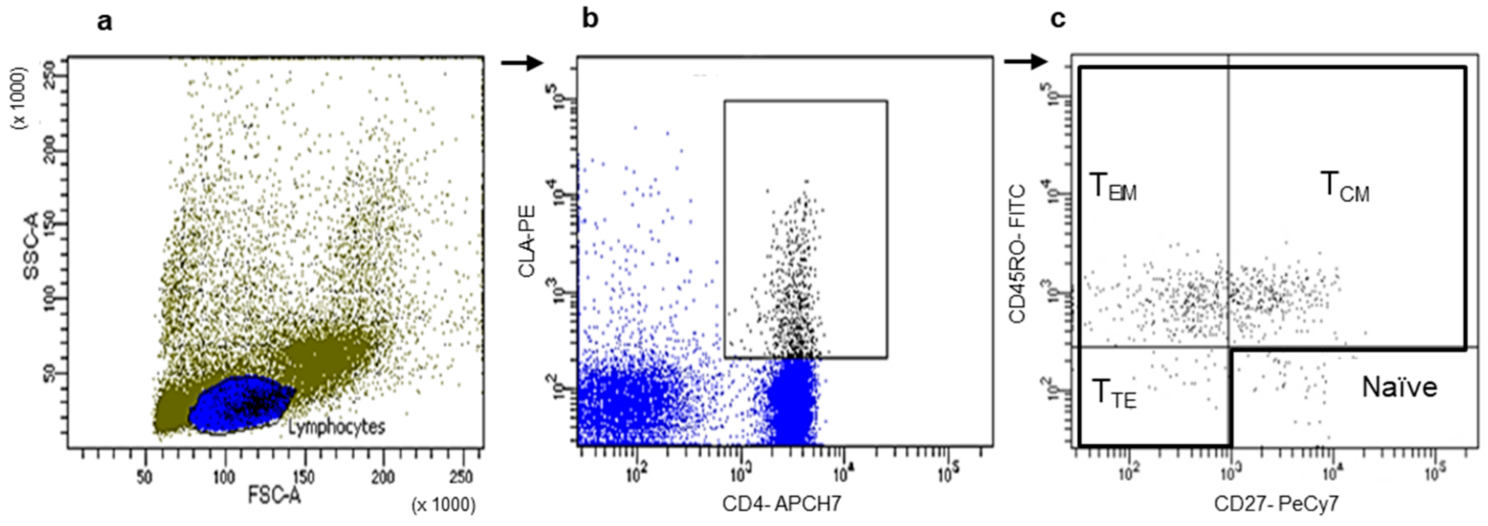
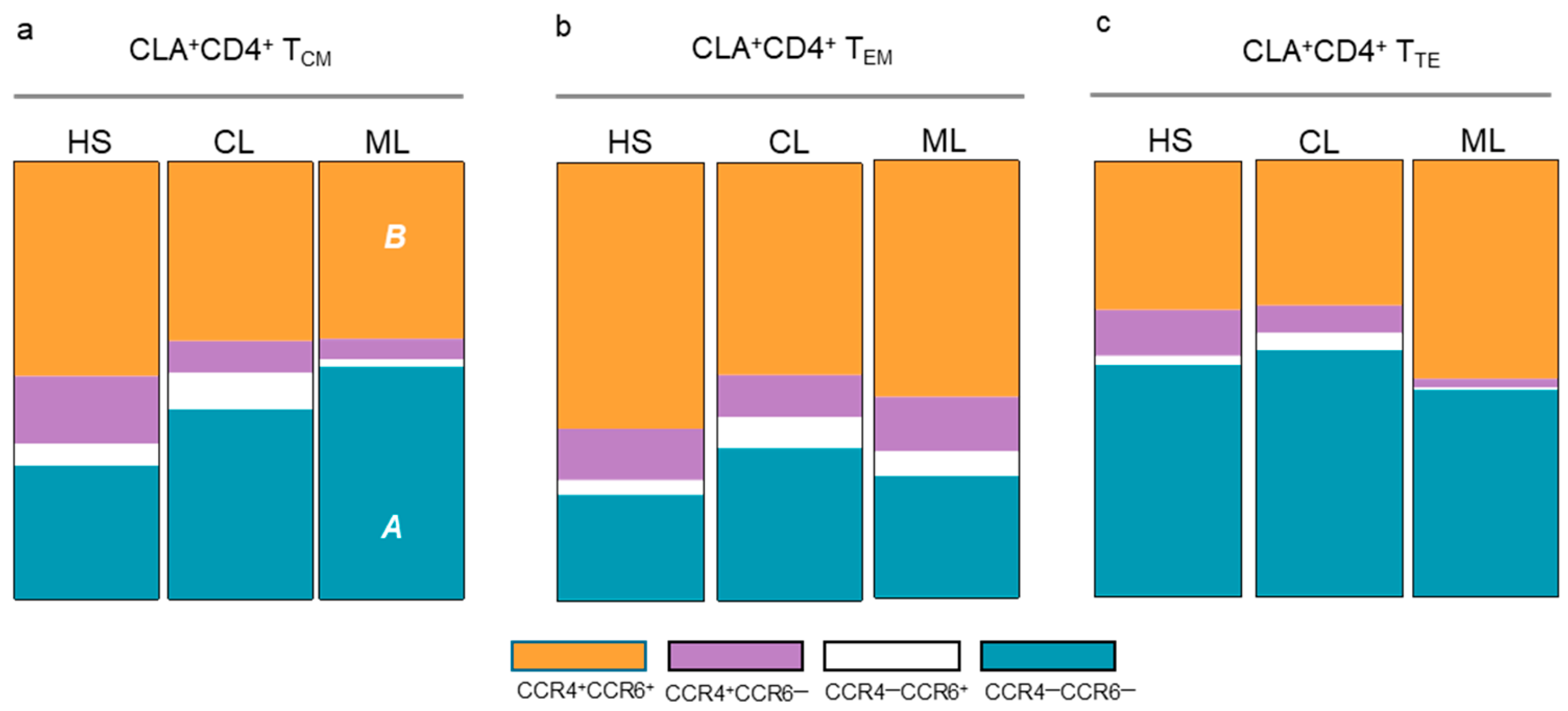
3.2. Memory T Cell Populations Expressing Tissue Tropism Receptors

We selected CD4+ or CD8+ T cells expressing each chemokine receptor and then evaluated the frequencies of the three main memory T cell populations, TCM (central memory), TEM (effector memory), and TTE (terminal effector), according to the expression of CD45RO and CD27 on both T cell subsets (Figure 1).
A similar profile was observed for CLA and CCR4 expression in CL and ML patients, with diminished frequencies of TCM and increased numbers of TEM CD4+ T cells (Figure 2a,b). On CD8+ T cells, the percentages of TCM CD8+ T cells expressing these chemokine receptors significantly decreased and those of TEM increased only in ML patients (Figure 2e,f). In contrast, no alterations of TCM, TEM, and TTE CD8+ T cells expressing these chemokine receptors were observed in CL patients. In addition, ML patients showed diminished percentages of TCM, increased frequencies of TEM, and a positive trend toward higher numbers of TTE CD4+ T cells expressing CCR6 (Figure 2c). Higher percentages of TTE CD4+CCR10+ T cells were also recorded in ML patients (Figure 2d). No differences in CCR6+ and CCR10+ memory T cell populations were recorded in CL patients (Figure 2c,d,g,h), and CCR3 expression did not vary between CL and ML in the different memory T cell subsets analyzed.
Next, co-expression profiles of chemokine receptors were assessed to identify CD4+ T cell subsets with homing characteristics (Th1: CCR4CCR6CLA+; Th22 and Th17: CCR4+CCR6+CLA+). ML patients showed a significant decrease in TCM CCR4+CCR6+CLA+CD4+ T cells and increased numbers of TCM CCR4CCR6CLA+CD4+ T cells compared to healthy subjects (Figure 3a). In contrast, no differences in TEM CLA+CD4+ and TTE CLA+CD4+ T cells were observed between CL patients, LM patients, and healthy subjects (Figure 3b,c).

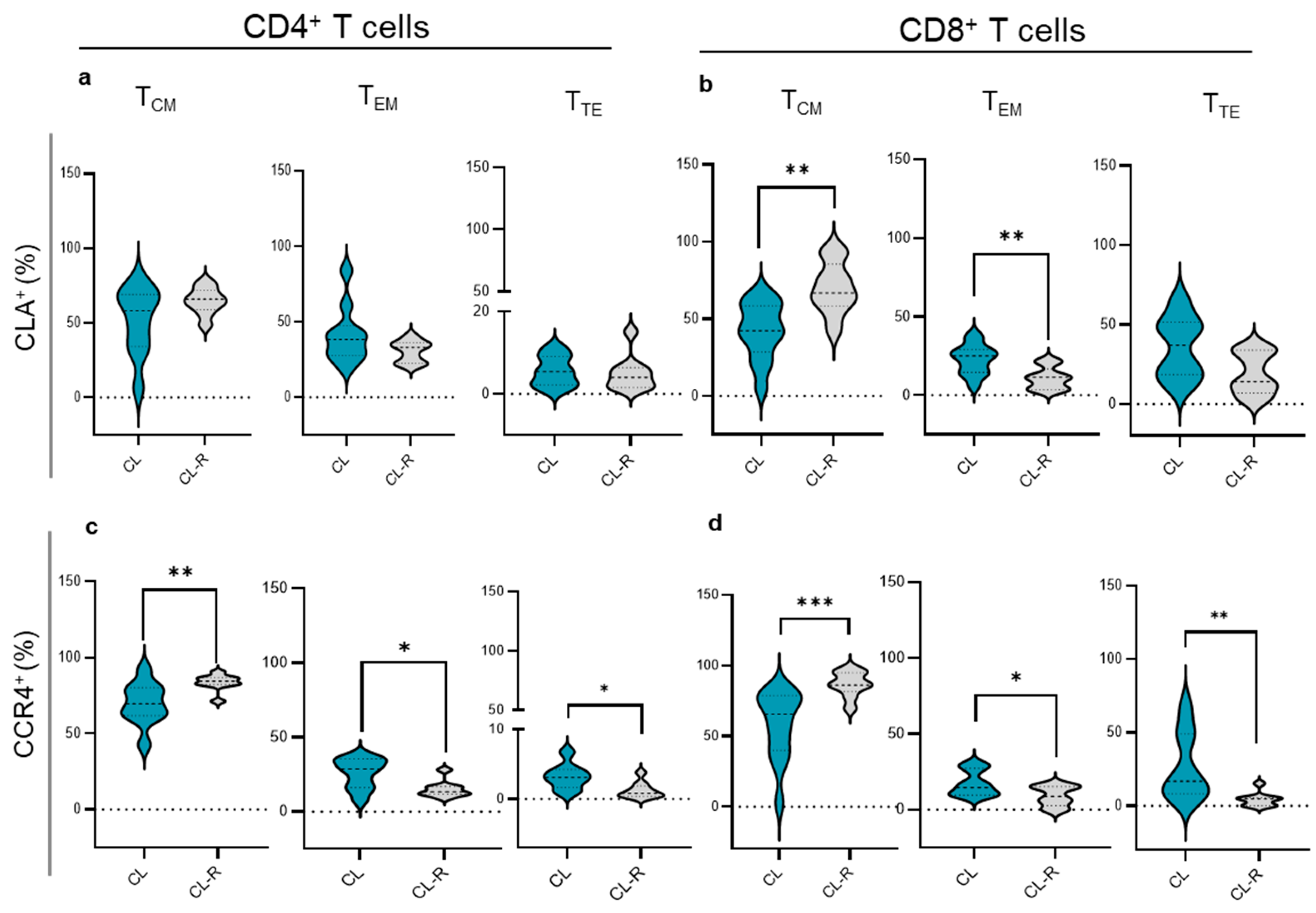
3.3. Memory T Cell Subsets in CL Patients Recovered from Their Cutaneous Lesions

The frequencies of memory T cell subsets expressing chemokine receptors were further analyzed according to the clinical outcome. We found higher frequencies of TCM and decreased TEM CLA+CD8+ T cells in CL recovered patients compared with those with active CL infections (Figure 4b). Similarly, CL recovered patients displayed increased TCM and diminished TEM and TTE CD4+CCR4+ and CD8+CCR4+ (Figure 4c,d) cells compared with patients with skin ulcers (Figure 4d). No variations were detected when analyzing CCR6, CCR10, and CCR3 memory populations.

3.4. Plasma Concentrations of CCL17 and CCL20 Chemokines

Next, we determined the plasma amounts of CCL17 and CCL20. CCL17 in combination with CCR4 mediates homing to cutaneous sites of specific lymphocyte subsets while CCL20 is a chemoattractant for CCR6-expressing cells. Higher plasma CCL17 levels were found in CL patients compared with ML patients and healthy subjects (Figure 5a). In contrast, a positive trend toward higher CCL20 levels was observed in ML patients (Figure 5b). CL recovered cases showed similar concentrations of CCL17 and CCL20 when compared to the active CL group (Figure 5a,b).

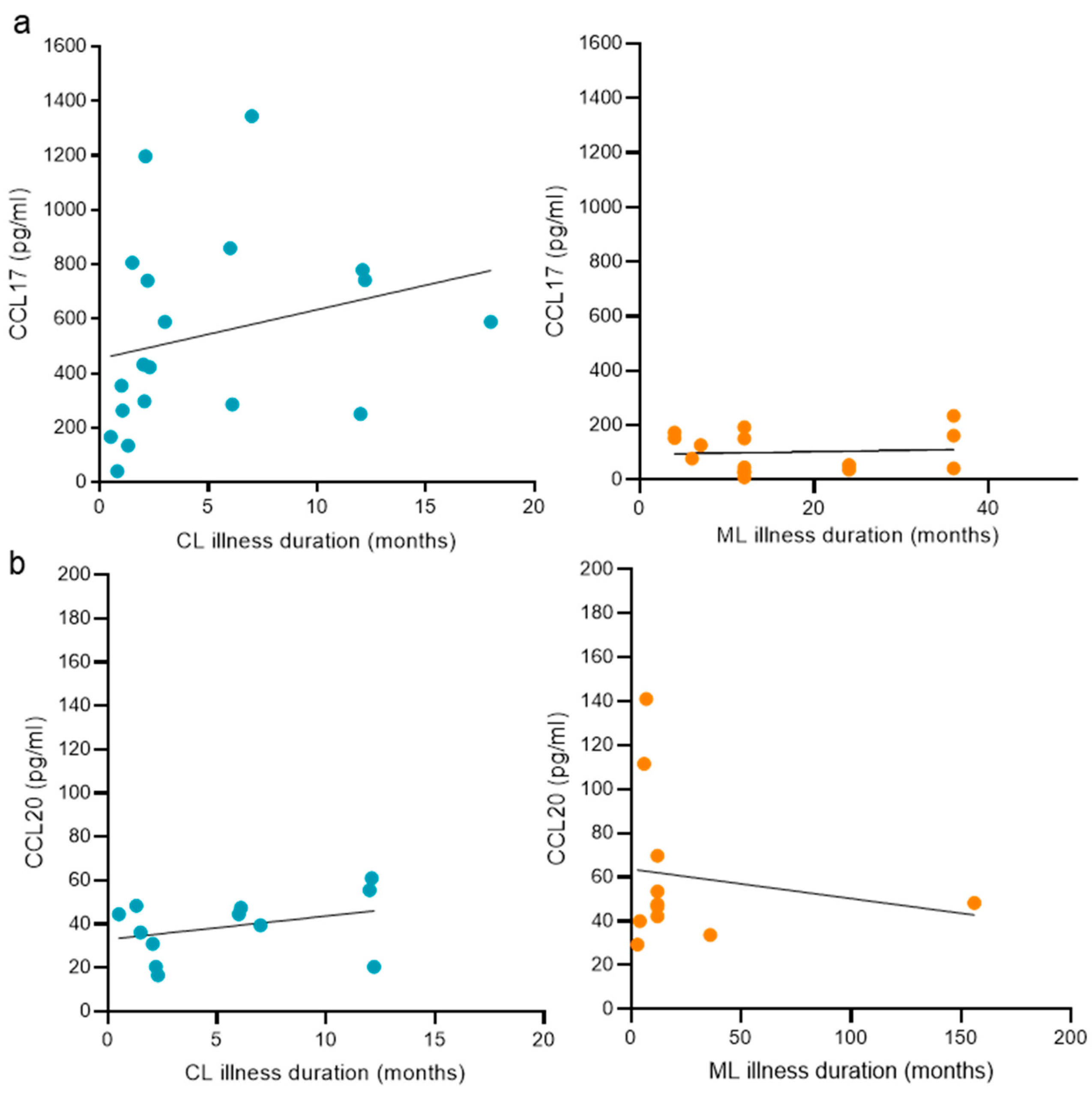
3.5. The Duration of the Disease and Its Relation with Plasma Chemokine Concentrations Targeting Skin and Nasopharyngeal Mucosa

We aimed to analyze the relation between disease duration and CCL17 or CCL20 levels in patients with CL, where cases were comprised within 0.5 months from the appearance of the first skin lesion, and in patients with ML, with samples showing a disease duration of up to 156 months from the onset of the first mucosal lesion. Among the study groups analyzed, we found no association between CCL17 (Figure 6a) or CCL20 (Figure 6b) and the duration of the disease.

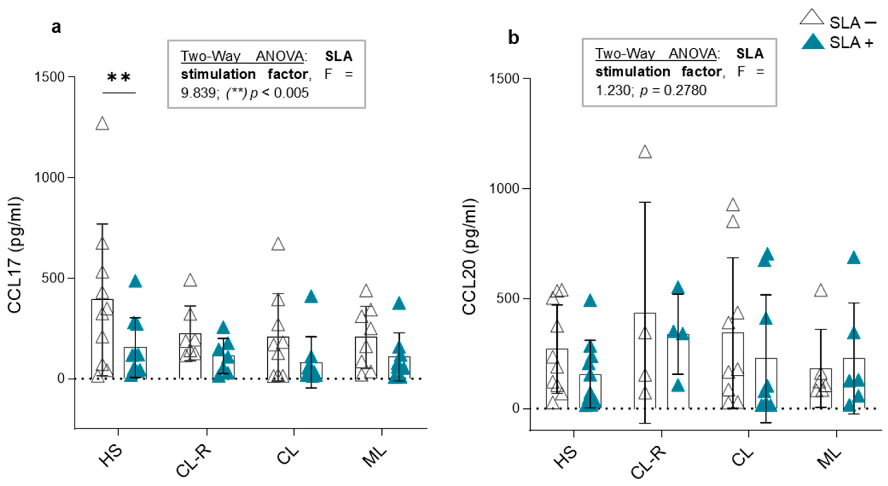
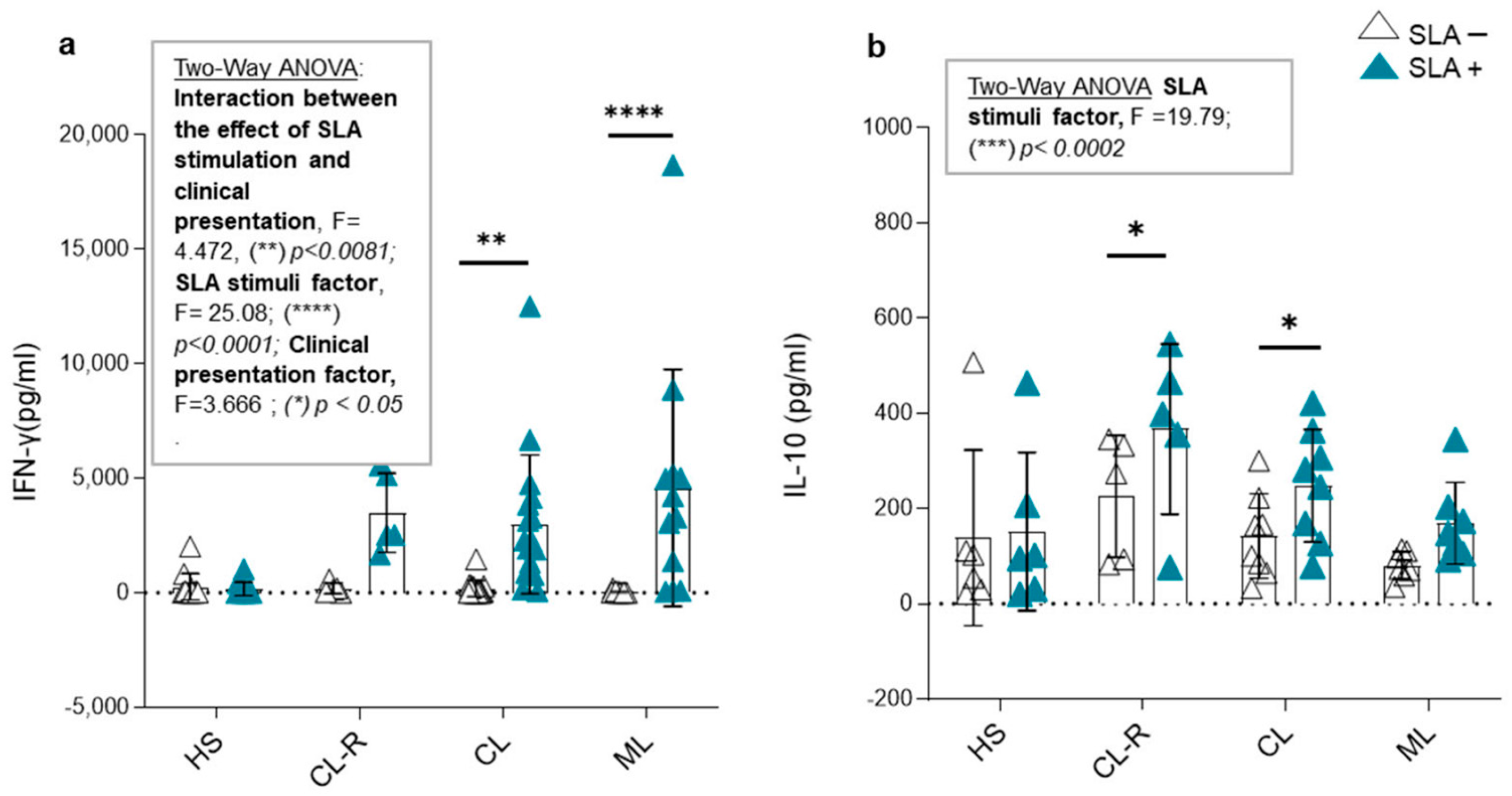
3.6. CCL17 and CCL20 Production in Culture Supernatants and Cytokine Modulation in Response to Leishmania Antigens

Using in vitro cultures, we evaluated whether SLAs have a direct effect on the production of CCL17 and CCL20 by PBMCs in patients with active CL, CL after achieving clinical recovery, or ML and healthy subjects. To analyze this aspect of the study, we applied a two-way ANOVA statistical test, as it allows us to determine how a response variable (CCL17 and CCL20 production) is affected by two factors (clinical presentation and SLA stimulation). The results of this test are shown in Figure 7.
Regarding CCL17 concentration in the supernatant, we determined that stimulation with Leishmania antigens had a significant inhibitory effect on CCL17 production, regardless of clinical presentation or disease outcome (two-way ANOVA: F = 9.839, p < 0.005; Figure 7a). This effect was particularly observed in the healthy subject group, where CCL17 levels significantly decreased in the presence of SLAs (Figure 7a). In contrast, SLA stimulation had no effect on CCL20 production by PBMCs in any of the studied groups (two-way ANOVA: F= 1.230, p = 0.2780; Figure 7b). These results support that soluble proteins of Leishmania spp. might modulate CCL17 expression independently of previous contact with the parasite.
Subsequently, we measured the levels of the pro-inflammatory cytokine IFN-γ and the regulatory cytokine IL-10 in the culture supernatants of PBMCs from the studied groups. The results were analyzed using the two-way ANOVA statistical test. When examining IFN-γ concentration, we identified a significant interaction between the effect of SLA stimulation and clinical presentation, with markedly elevated cytokine levels in ML patients, followed by CL patients (two-way ANOVA: F = 4.472, (**) p < 0.0081; Figure 8a), while levels remained at baseline in healthy subjects.
On the other hand, SLA stimulation induced IL-10 production (two-way ANOVA: F = 19.79, (***) p < 0.0002; Figure 8b), but its interaction with clinical presentation was not significant. However, only patients with active CL and those who achieved recovery from their skin lesions (CL-R) showed a significant increase in IL-10 production ((*) p < 0.05; Figure 8b). Regarding the cytokine environment produced in response to the parasite, PBMCs from patients with cutaneous forms produced a compensatory regulatory effect in the presence of Leishmania sp. antigens. This balance between pro-inflammatory and regulatory cytokines was not observed in patients with mucosal involvement.

4. Discussion

Tegumentary leishmaniasis presents a broad spectrum of manifestations, encompassing single or multiple cutaneous ulcers and nasopharyngeal lesions [18]. Mucosal lesions usually appear months or years after the primary skin lesion, as a possible consequence of the antigen persistence in the host. The failure of complete elimination of the parasite is well documented by the findings of Leishmania sp. amastigotes within cutaneous scars [18,19] or by the reactivation of the infection often observed after immunosuppressive therapies [20]. In this context, the host needs to adapt their immune responses to the location selected by the parasite through the different stages of the disease. Hence, the secreted chemokines and the T lymphocytes recruited to the affected sites play a major role in defining the immune mechanisms that control the infection. The present work demonstrated a modulation of chemokine receptors on total T cells only in the presence of mucosal involvement, with an upregulation of CLA, CCR4, CCR3, and CCR6 on CD4+ T cells and CCR3, CCR6, and CCR10 on CD8+ T cells. The increased expression of a targeted panel of chemokine receptors involved in skin and mucosal tropism in ML might reflect the expansion of the parasite to nasal mucosa, mobilizing more T cell subsets. In accordance with this assertion, when analyzing memory T cell populations, the changes in CL patients were restricted to subsets expressing CLA and CCR4, while ML patients demonstrated alterations in memory T cells expressing CLA, CCR4, CCR6, and CCR10. The expression of CLA and CCR4 has been associated with the pathogenesis of several skin disorders including autoimmune and oncological diseases [21,22,23,24,25]. Meanwhile, CCR10 and CCR6 play a role in homeostasis mechanisms during skin healing processes [26,27,28] and mucosal inflammation [29,30,31,32,33,34,35], respectively. The results of the upregulation of cutaneous chemokine markers on total and memory T cell subpopulations are indicative of possible persistence of the parasite in cutaneous sites during mucosal disease. In addition, the increase in effector memory/terminal effector T cells at expenses of a decrease in central memory T cells expressing chemokine receptors found here in tegumentary leishmaniasis reinforces previous findings of the chronic activation of T cells with effector functions mainly observed during mucosal cases [36]. Herein, we have provided information of the homing direction of these peripheral memory T cell populations, allowing us to recognize the profile of T cells infiltrating lesions, which play a central role in lesion injury.
Previous reports by us and others demonstrate that the increase in effector memory T cells is accompanied by signs of T cell exhaustion in total CD8+ T cells, with a late differentiated (CD27 CD28phenotype, increased expression of senescence markers, and cytotoxic molecules [4,37]. The present work demonstrates that CD4+ T cells represent the main subset implicated in the modulation of the memory profile expressing homing markers in CL, while changes in these markers in CD8+ T cells were only observed in ML, in correspondence with the exacerbation of cytotoxic functions that characterize this clinical form [38,39,40]. The present results demonstrating the predominance of effector T cells expressing chemokine receptors directed to skin and mucosal tissues could be used in future experimental studies to test the selective blocking of chemokine receptors aiming to diminish possible pathogenic effects of T cells. In this sense, Sacramento et al. [41] recently demonstrated the therapeutic potential of blocking CCR5, leading to a decrease in CD8+ T cell recruitment to leishmanial lesions in mice infected with L. (V.) braziliensis, ameliorating the destructive inflammation mediated by these lymphocytes.
The implication of CLA and CCR4 described here during active tegumentary leishmaniasis is in accordance with their increased expression previously reported in cutaneous lesions [10,42] and nasal mucosa samples [11]. Moreover, the significant decrease in the frequencies of effector memory T cells together with an increase in central memory T cells expressing CLA and CCR4 after the healing of cutaneous lesions is consequent with the decrease in the parasitic load achieved after a good response to treatment. It is known that central memory T cells have a high proliferative capacity after a secondary stimulus and constitute the main reservoir of effector memory T cells for future encounters with the antigen [43]. ML recovery could not be evaluated in this work, because the majority of the patients showed poor response and relapses after different therapeutic regimens. However, modulation of the chemokine receptors after CL recovery supports their possible usefulness as markers to corroborate the success of the treatment. In practice, patients diagnosed with tegumentary leishmaniasis receive a medical recommendation of periodical follow up [44]. The possible increase in effector memory T cells expressing CLA and CCR4 could alert us about cutaneous reactivation or the appearance of higher numbers of effector memory T cells expressing other receptors as CCR6 could aid in predicting the risk of lesion dissemination to mucosal sites. A progressive increase in the expression of these receptors over time may serve as a reliable indicator of disease progression, potentially enabling earlier clinical decision-making before further deterioration occurs.
Regarding CLA+ central memory T cells, while active CL patients showed a co-expression profile of CCR4 and CCR6 similar to that in healthy subjects, ML patients demonstrated a predominance of central memory CCR4CCR6 CLA+CD4+ T cells, which is associated with Th1 CD4+ T cells [45]. This profile might be involved in the exacerbation of inflammation, already described in the context of ML, but functional studies, currently being conducted in our laboratory, are needed to confirm this observation.
Additionally, we showed higher levels of CCL17, the ligand of CCR4, in CL patients compared with ML patients. The elevation of CCL17 has been associated with the progression of skin inflammatory diseases such as atopic dermatitis [46] and also reported in patients with post kala azar dermal leishmaniasis [47]. The findings presented here suggest that in tegumentary leishmaniasis, CCL17 is elevated in the presence of dermal lesions, but this elevation is not sustained in plasma when mucosal involvement appears. Meanwhile, the opposite situation occurs when analyzing CCL20, the ligand for CCR6, with a modest increase in ML cases. CCL20 has been associated with disease severity and mucosal inflammation in gastrointestinal diseases and autoimmune disorders [48,49,50]. Thus, CCL17 and CCL20 showed distinct modulation depending on the clinical form of tegumentary leishmaniasis.
Chemokine production serves to facilitate a protective immune response of the host; however, many infections, including those caused by Plasmodium spp., Leishmania spp., and Toxoplasma gondii, can assist their spread throughout the body by modulating chemokine production, leading to leukocyte infiltration to the site of infection [51,52]. Our in vitro assays showed that Leishmania spp. inhibit CCL17 production by PBMCs in CL patients, ML patients, and healthy subjects, but do not alter CCL20 release. Suppression of CCL17 could disrupt Th2-associated immune responses, leading to a shift toward a pro-inflammatory environment which favors the arrival of monocytes able to take up parasites, allowing for their dissemination. According to this explanation, the influx of inflammatory cells was already reported in the in vitro interaction of L. infantum with keratinocytes [53]. Another possible mechanism of Leishmania spp. could be to alter certain chemokine/chemokine receptor migration pathways. In this sense, Pinheiro et al. [9] demonstrated a decrease in CCR4, the CCL17 receptor, after in vitro Leishmania spp. infection, leading to the reduced adhesion of monocytes to connective tissues and suggesting the involvement of this pathway in the migration of these cells from Leishmania infection sites. In addition, IFN-γ production in Leishmania spp. infection is linked to a protective response and has strong immunomodulatory effects. In murine models, high IFN-γ levels indicate resistance, while IL-10 suggests susceptibility [1,7,54,55]. In humans, this dichotomy is less clear, as the immune response depends on the host’s immune status and the Leishmania species involved [55,56,57]. Our study confirms that Leishmania sp. antigens strongly modulate IFN-γ production, especially in patients with ML [58]. This increase in IFN-γ suggests persistent activation of CD4+ and CD8+ T cells. Additionally, the reported results indicate that immune cells are able to maintain their activation capacity even after prolonged infections, as PBMCs from patients with chronic ML demonstrated high antigen-specific inflammatory responses. These findings also support the notion of the pathogenic contribution of pro-inflammatory responses in ML, supporting the deregulated immune activation previously reported [4,56]. Interestingly, both CL and CL-R patients counteracted this pro-inflammatory response through IL-10 production, highlighting the role of regulatory cytokines as a needed balance for lesion resolution and as potential therapeutic strategies for preventing disease progression to chronic mucosal forms [59].

5. Conclusions

Altogether, the present work demonstrates the usefulness of a focused panel of chemokine/chemokine receptors to evaluate cutaneous or mucosal compromise and disease outcomes in tegumentary leishmaniasis. Despite the limited set assayed, these chemokine markers enabled us to obtain clear and valuable results contributing to broadening and strengthening the set of receptors yet described in the literature. In addition, our results emphasize the possible pathogenic role of a sustained effector response in chronic TL cases and provide new insights for experimental studies in an attempt to ameliorate the severity of the disease.

Author Contributions

Conceptualization: J.P. and C.P.; methodology: J.P., C.P., M.F.G.B., C.P.B., F.R. and M.B.; patients diagnosis: J.D.M. and P.B.; formal analysis and investigation: J.P., C.P., M.F.G.B., C.P.B., P.R., A.C.M. and M.O.; writing—original draft preparation: J.P. and C.P.; writing—review and editing: J.P., C.P., S.A.L., M.F.G.B. and C.P.B.; funding acquisition: C.P., M.F.G.B. and C.P.B.; resources: C.P., supervision: C.P. and S.A.L. All authors have read and agreed to the published version of the manuscript.

Funding

This work was supported by a generous donation of equipment (laminar flow cabinet) from the René Barón Foundation, and by grants from the National Council for Scientific and Technical Research (CONICET-PUE-2017) and the National Agency for Scientific and Technological Promotion (FONCYT-PICT-2018-03850), Argentina.

Institutional Review Board Statement

This study was conducted in accordance with the Declaration of Helsinki and approved by the Provincial Commission of Biomedical Research (P-UE 2292017100106 with date of approval 4 June 2019, Salta Argentina).

Informed Consent Statement

Informed consent was obtained from all subjects involved in this study. Written informed consent has been obtained from the patient(s) to publish this paper if applicable.

Data Availability Statement

The raw data supporting the conclusions of this article will be made available by the authors on request.

Acknowledgments

The authors would like to thank Mycology Service of Hospital Señor del Milagro (Ruth Valdez, Yone Chacón); Dermatology/Otorhinolaryngology Services of Hospital San Bernardo (Josefina Beckar); Otorhinolaryngology Services of Hospital Papa Francisco (Sibila Monroig), Salta, Argentina. We are also grateful to the Tropical Diseases Laboratory (Manuela Bono), Orán, Salta and Microbiology Services of Hospital D. Perón (Cristina Bono), Tartagal, Salta.

Conflicts of Interest

The authors declare no conflicts of interest.

Abbreviations

The following abbreviations are used in this manuscript:
TLTegumentary leishmaniasis
CLActive cutaneous leishmaniasis patients
MLMucosal leishmaniasis patients
CL-RRecovered cutaneous leishmaniasis patients
HSHealthy subjects
PBMCsPeripheral blood mononuclear cells
SLASoluble Leishmania antigens
TCMCentral memory T cells
TEMEffector memory T cells
TTETerminal effector T cells

References

  1. Kaye, P.; Scott, P. Leishmaniasis: Complexity at the Host-Pathogen Interface. Nat. Rev. Microbiol. 2011, 9, 604–615. [Google Scholar] [CrossRef]
  2. Amato, V.S.; Tuon, F.F.; Bacha, H.A.; Neto, V.A.; Nicodemo, A.C. Mucosal Leishmaniasis: Current Scenario and Prospects for Treatment. Acta Trop. 2008, 105, 1–9. [Google Scholar] [CrossRef]
  3. de Vries, H.J.C.; Schallig, H.D. Cutaneous Leishmaniasis: A 2022 Updated Narrative Review into Diagnosis and Management Developments. Am. J. Clin. Dermatol. 2022, 23, 823–840. [Google Scholar] [CrossRef]
  4. Parodi, C.; García Bustos, M.F.; Barrio, A.; Ramos, F.; González Prieto, A.G.; Mora, M.C.; Baré, P.; Basombrío, M.A.; de Elizalde de Bracco, M.M. American Tegumentary Leishmaniasis: T-Cell Differentiation Profile of Cutaneous and Mucosal Forms—Co-Infection with Trypanosoma Cruzi. Med. Microbiol. Immunol. 2016, 205, 353–369. [Google Scholar] [CrossRef]
  5. Morgado, F.N.; Schubach, A.; Rosalino, C.M.V.; Quintella, L.P.; Santos, G.; Salgueiro, M.; Conceição-Silva, F. Is the in Situ Inflammatory Reaction an Important Tool to Understand the Cellular Immune Response in American Tegumentary Leishmaniasis? Br. J. Dermatol. 2008, 158, 50–58. [Google Scholar] [CrossRef]
  6. García Bustos, M.F.; González-Prieto, G.; Ramos, F.; Mora, M.C.; Hashiguchi, Y.; Parodi, C.; Basombrío, M.A.; Moreno, S.; Monroig, S.; Beckar, J.; et al. Clinical and Epidemiological Features of Leishmaniasis in Northwestern-Argentina through a Retrospective Analysis of Recent Cases. Acta Trop. 2016, 154, 125–132. [Google Scholar] [CrossRef]
  7. Novais, F.O.; Scott, P. CD8+ T Cells in Cutaneous Leishmaniasis: The Good, the Bad, and the Ugly. Semin. Immunopathol. 2015, 37, 251–259. [Google Scholar] [CrossRef]
  8. de Oliveira, B.C.; da Silva, A.A.; de Andrade Cavalcante, M.K.; de Brito, M.E.F.; de Castro, M.C.A.B.; de Medeiros, V.L.S.; de Freitas e Silva, R.; Pereira, V.R.A. Central and Effector Memory Human CD4+ and CD8+ T Cells during Cutaneous Leishmaniasis and after In Vitro Stimulation with Leishmania (Viannia) Braziliensis Epitopes. Vaccines 2023, 11, 158. [Google Scholar] [CrossRef]
  9. de Araújo, F.F.; Costa-Silva, M.F.; Pereira, A.A.S.; Rêgo, F.D.; Pereira, V.H.S.; de Souza, J.P.; Fernandes, L.O.B.; Martins-Filho, O.A.; Gontijo, C.M.F.; Peruhype-Magalhães, V.; et al. Chemokines in Leishmaniasis: Map of Cell Movements Highlights the Landscape of Infection and Pathogenesis. Cytokine 2021, 147, 155339. [Google Scholar] [CrossRef]
  10. de Oliveira Mendes-Aguiar, C.; Vieira-Gonçalves, R.; Guimarães, L.H.; de Oliveira-Neto, M.P.; Carvalho, E.M.; Da-Cruz, A.M. Effector Memory CD4+ T Cells Differentially Express Activation Associated Molecules Depending on the Duration of American Cutaneous Leishmaniasis Lesions. Clin. Exp. Immunol. 2016, 185, 202–209. [Google Scholar] [CrossRef]
  11. Palmeiro, M.R.; Morgado, F.N.; Valete-Rosalino, C.M.; Martins, A.C.; Moreira, J.; Quintella, L.P.; de Oliveira Schubach, A.; Conceição-Silva, F. Comparative Study of the in Situ Immune Response in Oral and Nasal Mucosal Leishmaniasis. Parasite Immunol. 2012, 34, 23–31. [Google Scholar] [CrossRef]
  12. Lechner, A.; Ritter, U.; Varona, R.; Marquez, G.; Bogdan, C.; Körner, H. Protective Immunity and Delayed Type Hypersensitivity Reaction Are Uncoupled in Experimental Leishmania Major Infection of CCR6-Negative Mice. Microbes Infect. 2007, 9, 291–299. [Google Scholar] [CrossRef]
  13. Barth, T.; Schmidt, D.; Botteron, C.; Nguyen, T.T.T.; Ritter, U.; Männel, D.N.; Lechner, A. An Early Reduction in Treg Cells Correlates with Enhanced Local Inflammation in Cutaneous Leishmaniasis in CCR6-Deficient Mice. PLoS ONE 2012, 7, e44499. [Google Scholar] [CrossRef]
  14. Díaz, N.L.; Zerpa, O.; Tapia, F.J. Chemokines and Chemokine Receptors Expression in the Lesions of Patients with American Cutaneous Leishmaniasis. Memórias Inst. Oswaldo Cruz 2013, 108, 446–452. [Google Scholar] [CrossRef]
  15. Daniel, W.W. Biostatistics: A Foundation for Analysis in the Health Sciences, 7th ed.; John Wiley & Sons: New York, NY, USA, 1999. [Google Scholar]
  16. Lwanga, S.K.; Lemeshow, S. Sample Size Determination in Health Studies: A Practical Manual; World Health Organization: Geneva, Switzerland, 1991. [Google Scholar]
  17. Bracamonte, M.E. Estrategias Noveles de ADN Recombinante Aplicadas al Inmunodiagnóstico de Las Leishmaniasis En Argentina. Doctoral Dissertation, National University of Tucumán, San Miguel de Tucumán, Argentina, 2021. [Google Scholar]
  18. Conceição-Silva, F.; Leite-Silva, J.; Morgado, F.N. The Binomial Parasite-Host Immunity in the Healing Process and in Reactivation of Human Tegumentary Leishmaniasis. Front. Microbiol. 2018, 9, 1308. [Google Scholar] [CrossRef]
  19. Morgado, F.N.; Schubach, A.; Vasconcellos, E.; Azeredo-Coutinho, R.B.; Valete-Rosalino, C.M.; Quintella, L.P.; Santos, G.; Salgueiro, M.; Palmeiro, M.R.; Conceição-Silva, F. Signs of an in Situ Inflammatory Reaction in Scars of Human American Tegumentary Leishmaniasis. Parasite Immunol. 2010, 32, 285–295. [Google Scholar] [CrossRef]
  20. Martínez-Valencia, A.J.; Daza-Rivera, C.F.; Rosales-Chilama, M.; Cossio, A.; Casadiego Rincón, E.J.; Desai, M.M.; Saravia, N.G.; Gómez, M.A. Clinical and Parasitological Factors in Parasite Persistence after Treatment and Clinical Cure of Cutaneous Leishmaniasis. PLoS Negl. Trop. Dis. 2017, 11, e0005713. [Google Scholar] [CrossRef]
  21. de Souza, R.M.; de Andrade Junior, H.F.; Duarte, M.I.S.; Braz, L.M.A.; de Oliveira Schubach, A.; Silva, F.C.; Amato, V.S. Reactivation of Cutaneous and Mucocutaneous Tegumentary Leishmaniasis in Rheumatoid Arthritis Patients: An Emerging Problem? Rev. Inst. Med. Trop. Sao Paulo 2017, 59, e6. [Google Scholar] [CrossRef]
  22. Nicolàs, L.S.D.S.; Czarnowicki, T.; Akdis, M.; Pujol, R.M.; Lozano-Ojalvo, D.; Leung, D.Y.M.; Guttman-Yassky, E.; Santamaria-Babí, L.F. CLA+ Memory T Cells in Atopic Dermatitis: CLA+ T Cells and Atopic Dermatitis. Allergy Eur. J. Allergy Clin. Immunol. 2024, 79, 15–25. [Google Scholar] [CrossRef]
  23. Shin, J.U.; Abaci, H.E.; Herron, L.; Guo, Z.; Sallee, B.; Pappalardo, A.; Jackow, J.; Wang, E.H.C.; Doucet, Y.; Christiano, A.M. Recapitulating T Cell Infiltration in 3D Psoriatic Skin Models for Patient-Specific Drug Testing. Sci. Rep. 2020, 10, 4123. [Google Scholar] [CrossRef]
  24. Peru, S.; Prochazkova-Carlotti, M.; Cherrier, F.; Velazquez, J.; Richard, E.; Idrissi, Y.; Cappellen, D.; Azzi-Martin, L.; Pham-Ledard, A.; Beylot-Barry, M.; et al. Cutaneous Lymphocyte Antigen Is a Potential Therapeutic Target in Cutaneous T-Cell Lymphoma. J. Investig. Dermatol. 2022, 142, 3243–3252.e10. [Google Scholar] [CrossRef] [PubMed]
  25. Jo, Y.; Jo, Y.; Matsumoto, T.; Yada, S.; Fujisawa, K.; Esaki, M.; Onai, N.; Matsushima, K.; Iida, M. CCR4 is an up-regulated chemokine receptor of peripheral blood memory CD4+ T cells in Crohn’s disease. Clin. Exp. Immunol. 2003, 132, 332–338. [Google Scholar] [CrossRef] [PubMed]
  26. Sokolowska-Wojdylo, M.; Wenzel, J.; Gaffal, E.; Steitz, J.; Roszkiewicz, J.; Bieber, T.; Tüting, T. Absence of CD26 Expression on Skin-Homing CLA+ CD4+ T Lymphocytes in Peripheral Blood Is a Highly Sensitive Marker for Early Diagnosis and Therapeutic Monitoring of Patients with Sézary Syndrome. Clin. Exp. Dermatol. 2005, 30, 702–706. [Google Scholar] [CrossRef] [PubMed]
  27. Chen, Z.; Haus, J.M.; Chen, L.; Wu, S.C.; Urao, N.; Koh, T.J.; Minshall, R.D. CCL28-Induced CCR10/ENOS Interaction in Angiogenesis and Skin Wound Healing. FASEB J. 2020, 34, 5838–5850. [Google Scholar] [CrossRef]
  28. Fujimoto, S.; Uratsuji, H.; Saeki, H.; Kagami, S.; Tsunemi, Y.; Komine, M.; Tamaki, K. CCR4 and CCR10 Are Expressed on Epidermal Keratinocytes and Are Involved in Cutaneous Immune Reaction. Cytokine 2008, 44, 172–178. [Google Scholar] [CrossRef]
  29. Xiong, N.; Fu, Y.; Hu, S.; Xia, M.; Yang, J. CCR10 and Its Ligands in Regulation of Epithelial Immunity and Diseases. Protein Cell 2012, 3, 571–580. [Google Scholar] [CrossRef]
  30. Skovdahl, H.K.; Van Beelen Granlund, A.; Østvik, A.E.; Bruland, T.; Bakke, I.; Torp, S.H.; Damas, J.K.; Sandvik, A.K. Expression of CCL20 and Its Corresponding Receptor CCR6 Is Enhanced in Active Inflammatory Bowel Disease, and TLR3 Mediates CCL20 Expression in Colonic Epithelial Cells. PLoS ONE 2015, 10, e0141710. [Google Scholar] [CrossRef]
  31. Ranasinghe, R.; Eri, R. Pleiotropic Immune Functions of Chemokine Receptor 6 in Health and Disease. Medicines 2018, 5, 69. [Google Scholar] [CrossRef]
  32. Meitei, H.T.; Jadhav, N.; Lal, G. CCR6-CCL20 Axis as a Therapeutic Target for Autoimmune Diseases. Autoimmun. Rev. 2021, 20, 102846. [Google Scholar] [CrossRef]
  33. Ito, T.; Carson, W.F.; Cavassani, K.A.; Connett, J.M.; Kunkel, S.L. CCR6 as a Mediator of Immunity in the Lung and Gut. Exp. Cell Res. 2011, 317, 613–619. [Google Scholar] [CrossRef]
  34. Kennedy-Crispin, M.; Billick, E.; Mitsui, H.; Gulati, N.; Fujita, H.; Gilleaudeau, P.; Sullivan-Whalen, M.; Johnson-Huang, L.M.; Suárez-Farĩas, M.; Krueger, J.G. Human Keratinocytes’ Response to Injury Upregulates CCL20 and Other Genes Linking Innate and Adaptive Immunity. J. Investig. Dermatol. 2012, 132, 105–113. [Google Scholar] [CrossRef] [PubMed]
  35. Elnabawi, Y.A.; Garshick, M.S.; Tawil, M.; Barrett, T.J.; Fisher, E.A.; Lo Sicco, K.; Neimann, A.L.; Scher, J.U.; Krueger, J.; Berger, J.S. CCL20 in Psoriasis: A Potential Biomarker of Disease Severity, Inflammation, and Impaired Vascular Health. J. Am. Acad. Dermatol. 2021, 84, 913–920. [Google Scholar] [CrossRef] [PubMed]
  36. Lee, A.Y.S.; Körner, H. The CCR6-CCL20 Axis in Humoral Immunity and T-B Cell Immunobiology. Immunobiology 2019, 224, 449–454. [Google Scholar] [CrossRef] [PubMed]
  37. Pereira-Carvalho, R.; Mendes-Aguiar, C.O.; Oliveira-Neto, M.P.; Covas, C.J.F.; Bertho, Á.L.; Da-Cruz, A.M.; Gomes-Silva, A. Leishmania Braziliensis-Reactive T Cells Are down-Regulated in Long-Term Cured Cutaneous Leishmaniasis, but the Renewal Capacity of T Effector Memory Compartments Is Preserved. PLoS ONE 2013, 8, e81529. [Google Scholar] [CrossRef]
  38. Egui, A.; Ledesma, D.; Pérez-Antón, E.; Montoya, A.; Gómez, I.; Robledo, S.M.; Infante, J.J.; Vélez, I.D.; López, M.C.; Thomas, M.C. Phenotypic and Functional Profiles of Antigen-Specific CD4+ and CD8+ T Cells Associated with Infection Control in Patients with Cutaneous Leishmaniasis. Front. Cell. Infect. Microbiol. 2018, 8, 393. [Google Scholar] [CrossRef]
  39. Jaigirdar, S.A.; MacLeod, M.K.L. Development and Function of Protective and Pathologic Memory CD4 T Cells. Front. Immunol. 2015, 6, 456. [Google Scholar] [CrossRef]
  40. Novais, F.O.; Carvalho, L.P.; Graff, J.W.; Beiting, D.P.; Ruthel, G.; Roos, D.S.; Betts, M.R.; Goldschmidt, M.H.; Wilson, M.E.; de Oliveira, C.I.; et al. Cytotoxic T Cells Mediate Pathology and Metastasis in Cutaneous Leishmaniasis. PLoS Pathog. 2013, 9, e1003504. [Google Scholar] [CrossRef]
  41. Sacramento, L.A.; Amorim, C.F.; Lombana, C.G.; Beiting, D.; Novais, F.; Carvalho, L.P.; Carvalho, E.M.; Scott, P. CCR5 Promotes the Migration of Pathological CD8+ T Cells to the Leishmanial Lesions. PLoS Pathog. 2024, 20, e1012211. [Google Scholar] [CrossRef]
  42. Campanelli, A.P.; Brodskyn, C.I.; Boaventura, V.; Silva, C.; Roselino, A.M.; Costa, J.; Saldanha, A.C.; Rodrigues de Freitas, L.A.Ô.; De Oliveira, C.I.; Barral-Netto, M.; et al. Chemokines and Chemokine Receptors Coordinate the Inflammatory Immune Response in Human Cutaneous Leishmaniasis. Hum. Immunol. 2010, 71, 1220–1227. [Google Scholar] [CrossRef]
  43. Sallusto, F.; Geginat, J.; Lanzavecchia, A. Central Memory and Effector Memory T Cell Subsets: Function, Generation, and Maintenance. Annu. Rev. Immunol. 2004, 22, 745–763. [Google Scholar] [CrossRef]
  44. Ministerio de Salud Pública de La República Argentina. InfoLEG—Información Legislativa y Documental—Ministerio de Justicia y Derechos Humanos—Presidencia de La Nación de La República Argentina. In Resolución 386/2004: Manual de Procedimientos Para El Nivel Gerencial y Profesional Sobre Leshmaniosis; Ministerio de Salud Pública de La República Argentina: Buenos Aires, Argentina, 2004. [Google Scholar]
  45. Sabat, R.; Wolk, K.; Loyal, L.; Döcke, W.D.; Ghoreschi, K. T Cell Pathology in Skin Inflammation. Semin. Immunopathol. 2019, 41, 359–377. [Google Scholar] [CrossRef] [PubMed]
  46. Kataoka, Y. Thymus and Activation-Regulated Chemokine as a Clinical Biomarker in Atopic Dermatitis. J. Dermatol. 2014, 41, 221–229. [Google Scholar] [CrossRef] [PubMed]
  47. Mukherjee, S.; Sengupta, R.; Mukhopadhyay, D.; Braun, C.; Mitra, S.; Roy, S.; Kanti Das, N.; Chatterjee, U.; von Stebut-Borschitz, E.; Chatterjee, M. Impaired Activation of Lesional CD8+ T-Cells Is Associated with Enhanced Expression of Programmed Death-1 in Indian Post Kala-Azar Dermal Leishmaniasis. Sci. Rep. 2019, 9, 762. [Google Scholar] [CrossRef]
  48. Lee, H.J.; Choi, S.C.; Lee, M.H.; Oh, H.M.; Choi, E.Y.; Choi, E.J.; Yun, K.J.; Seo, G.S.; Kim, S.W.; Lee, J.G.; et al. Increased Expression of MIP-3α/CCL20 in Peripheral Blood Mononuclear Cells from Patients with Ulcerative Colitis and Its down-Regulation by Sulfasalazine and Glucocorticoid Treatment. Inflamm. Bowel Dis. 2005, 11, 1070–1079. [Google Scholar] [CrossRef]
  49. Ranasinghe, R.; Eri, R. Modulation of the CCR6-CCl20 Axis: A Potential Therapeutic Target in Inflammation and Cancer. Medicina 2018, 54, 88. [Google Scholar] [CrossRef]
  50. Furue, K.; Ito, T.; Tsuji, G.; Nakahara, T.; Furue, M. The CCL20 and CCR6 Axis in Psoriasis. Scand. J. Immunol. 2020, 91, e12846. [Google Scholar] [CrossRef]
  51. Glennie, N.D.; Scott, P. Memory T Cells in Cutaneous Leishmaniasis. Cell. Immunol. 2016, 309, 50–54. [Google Scholar] [CrossRef]
  52. Scorza, B.M.; Wacker, M.A.; Messingham, K.; Kim, P.; Klingelhutz, A.; Fairley, J.; Wilson, M.E. Differential Activation of Human Keratinocytes by Leishmania Species Causing Localized or Disseminated Disease. J. Investig. Dermatol. 2017, 137, 2149–2156. [Google Scholar] [CrossRef]
  53. Pinheiro, N.F.; Hermida, M.D.R.; Macedo, M.P.; Mengel, J.; Bafica, A.; Dos-Santos, W.L.C. Leishmania Infection Impairs Β1-Integrin Function and Chemokine Receptor Expression in Mononuclear Phagocytes. Infect. Immun. 2006, 74, 3912–3921. [Google Scholar] [CrossRef]
  54. Samant, M.; Sahu, U.; Pandey, S.C.; Khare, P. Role of Cytokines in Experimental and Human Visceral Leishmaniasis. Front. Cell. Infect. Microbiol. 2021, 11, 624009. [Google Scholar] [CrossRef]
  55. Zaph, C.; Uzonna, J.; Beverley, S.M.; Scott, P. Central Memory T Cells Mediate Long-Term Immunity to Leishmania Major in the Absence of Persistent Parasites. Nat. Med. 2004, 10, 1104–1110. [Google Scholar] [CrossRef] [PubMed]
  56. Kaye, P.M.; Cruz, I.; Picado, A.; Van Bocxlaer, K.; Croft, S.L. Leishmaniasis Immunopathology—Impact on Design and Use of Vaccines, Diagnostics and Drugs. Semin. Immunopathol. 2020, 42, 247–264. [Google Scholar] [CrossRef] [PubMed]
  57. Niedzielska, I.; Cierpka, S. Interferon Gamma in the Etiology of Atherosclerosis and Periodontitis. Thromb. Res. 2010, 126, 324–327. [Google Scholar] [CrossRef] [PubMed]
  58. Santos, C.D.S.; Boaventura, V.; Ribeiro Cardoso, C.; Tavares, N.; Lordelo, M.J.; Noronha, A.; Costa, J.; Borges, V.M.; De Oliveira, C.I.; Van Weyenbergh, J.; et al. CD8+ Granzyme B+—Mediated Tissue Injury vs. CD4+IFNγ+ -Mediated Parasite Killing in Human Cutaneous Leishmaniasis. J. Investig. Dermatol. 2013, 133, 1533–1540. [Google Scholar] [CrossRef]
  59. da Silva Santos, C.; Brodskyn, C.I. The Role of CD4 and CD8 T Cells in Human Cutaneous Leishmaniasis. Front. Public Health 2014, 2, 165. [Google Scholar] [CrossRef]
Figure 1. Representative gating strategy of memory T cell subsets expressing chemokines receptors. (a) Lymphocytes (blue) were initially gated by forward and side light scatter, (b) CLA+CD4+ co-expression was selected (dark gray), and (c) a dot plot graph of CD45RO and CD27 staining of the gated CLA+CD4+ T cells are shown. Memory T cell populations were determined as central memory (TCM, CD45RO+CD27+), effector memory (TEM, CD45RO+ CD27), and terminal effector (TTE, CD45ROCD27) cells.
Cells 14 00604 g001
Figure 2. CLA, CCR4, CCR6, and CCR10 memory T cell populations in TL patients. (ad) CD4+ T cells and (eh) CD8+ T cells from (HS, gray bars) healthy subjects, (CL, blue bars) cutaneous leishmaniasis patients, and (ML, orange bars) mucosal leishmaniasis (ML) patients. Memory T cell populations were determined as central memory (TCM, CD45RO+CD27+), effector memory (TEM, CD45RO+CD27), and terminal effector (TTE, CD45ROCD27) cells. Significant differences between memory T cell subsets were recorded using the Kruskal–Wallis test and Dunn’s post-test. (*) p < 0.05; (**) p < 0.01, (***) p < 0.001 The asterisk over the diagonal line indicates a positive linear trend using a one-way ANOVA with a post-test for linear trends (p < 0.05).
Cells 14 00604 g002
Figure 3. Co-expression of CCR4 and CCR6 on CLA+CD4+ TCM, CLA+CD4+ TEM, and CLA+CD4+ TTE fractions in TL patients. CCR4+CCR6+ (orange), CCR4+CCR6 (violet), CCR4CCR6+ (white), and CCR4CCR6 (blue) fractions of (a) CLA+CD4+ TCM cells and (b) CLA+CD4+ TEM cells and (c) CLA+CD4+ TTE cells from (HS) healthy subjects, (CL) cutaneous leishmaniasis patients, and (ML) mucosal leishmaniasis patients. (A) CCR4CCR6CLA+, p < 0.05, ML vs. HS; (B) p < 0.01, CCR4+CCR6+CLA+, ML vs. HS. Significant differences were assessed using the Kruskal–Wallis test and Dunn’s post-test.
Cells 14 00604 g003
Figure 4. Memory T cell subsets after recovery of CL patients. (CL) Cutaneous leishmaniasis patients (blue) and (CL-R) CL recovered patients (light gray). Memory T cell populations expressing CLA or CCR4 were determined as central memory (TCM, CD45RO+CD27+), effector memory (TEM, CD45RO+CD27), and terminal effector (TTE, CD45ROCD27) CD4+ T cells (ac) and CD8+ T cells (b,d). Significant differences between active disease and recovery were assessed using the Mann–Whitney test. (*) p < 0.05; (**) p < 0.01; (***) p < 0.001.
Cells 14 00604 g004
Figure 5. Plasma levels of skin and mucosal homing chemokines. (a) CCL17 and (b) CCL20 chemokine plasma concentration. (HS) Healthy subjects (gray), (CL-R) cutaneous leishmaniasis recovered patients (light gray), (CL) cutaneous leishmaniasis patients (blue), and (ML) mucosal leishmaniasis patients (orange). Differences between groups were recorded using the Kruskal–Wallis test and Dunn’s post- test. (*) p < 0.05; (**) p < 0.01. The asterisk over the diagonal line indicates a positive trend using a one-way ANOVA with a post-test for linear trends (p < 0.05).
Cells 14 00604 g005
Figure 6. Disease duration and plasma concentration of CCL17 and CCL20 in TL. Linear correlation analysis between disease duration in patients with cutaneous leishmaniasis (CL) or mucosal leishmaniasis (ML) and (a) CCL17 and (b) CCL20. Healthy subjects (HS), patients with cutaneous leishmaniasis (CL), and patients with mucosal leishmaniasis (ML). Simple linear regression analysis: (a) CL, R² = 0.0659; ML, R² = 0.0718. (b) CL, R² = 0.1151; ML, R² = 0.0289.
Cells 14 00604 g006
Figure 7. In vitro production of CCL17 and CCL20 in the presence or absence of Leishmania sp. antigens. Cytokine levels in culture supernatants of (a) CCL17 and (b) CCL20 in the presence (blue triangles) or absence (white triangles) of soluble Leishmania sp. antigens (SLAs) from peripheral blood mononuclear cells (PBMCs) of healthy subjects (HS), recovered cutaneous leishmaniasis patients (CL-R), cutaneous leishmaniasis patients (CL), and mucosal leishmaniasis patients (ML). Two-way ANOVA with Bonferroni’s post-test.
Cells 14 00604 g007
Figure 8. In vitro production of IFN-γ and IL-10 in the presence or absence of Leishmania sp. antigens. Cytokine levels in culture supernatants of (a) IFN-γ and (b) IL-10 in the presence (blue triangles) or absence (white triangles) of soluble Leishmania sp. antigens (SLAs) from peripheral blood mononuclear cells (PBMCs) of healthy subjects (HS), recovered cutaneous leishmaniasis patients (CL-R), cutaneous leishmaniasis patients (CL), and mucosal leishmaniasis patients (ML). Two-way ANOVA with Bonferroni’s post-test.
Cells 14 00604 g008
Table 1. Demographic and clinical characteristics of tegumentary leishmaniasis patients.
Study GroupsNAge (Years)Male/
Female
Subjects with Detectable Anti-T. cruzi Antibodies (%)Cutaneous/Mucosal Illness Duration (months)Time of Complete Recovery (months)
Cutaneous leishmaniasis (CL)3240 [12–72]26/69.32 [0.5–18]NA
Cutaneous leishmaniasis recovered (CL-R)1342 [19–72]9/46.25NA31 [2–72]
Mucosal leishmaniasis (LM)2457 [27–86]17/741.6636 [12–156]NA
Healthy subjects (HS)1737 [29–61]10/70NANA
Results of age, illness duration, and time of recovery are expressed as median [range]. NA, not applicable.
Table 2. Expression of chemokine receptors on CD4+ and CD8+ T cells in patients with cutaneous leishmaniasis and mucosal leishmaniasis.
Cell MarkerCD4+ T Cells (%)CD8+ T Cells (%)
HSCLMLHSCLML
CLA10.30 [7.70–12.80] 10.30 [6.70–28.80]14.80 [7.30–32.80] *4.65 [2.00–8.70]3.80 [1.30–13.40]8.70 [1.30–11.60]
CCR414.50 [8.00–30.50]13.30 [4.30–24.80]18.40 [7.50–49.30] **2.60 [1.50–6.00]3.10 [0.80–6.50]3.40 [1.70–9.00]
CCR66.70 [2.70–9.90]2.40 [0.50–11.80] 8.40 [0.40–19.70] ≠1.90 [0.70–7.70]1.70 [0.70–3.40] 3.30 [0.50–9.60] ≠
CCR30.90 [0.50–3.10] 1.45 [0.50–3.80]2.10 [0.80–4.10] €0.70 [0.30–2.90] 1.40 [0.40–3.80] 2.60 [1.20–3.10] €
CCR106.15 [4.60–9.70]6.30 [3.20–17.30]8.10 [4.60–13.30]2.40 [1.50–4.40]2.50 [1.50–6.40]3.75 [1.60–7.70] **
Percentages of expression of CLA, CCR4, CCR6, CCR3, and CCR10 on CD4+ and CD8+ T cells from HS, healthy subjects; CL, cutaneous leishmaniasis patients; ML, mucosal leishmaniasis patients. Significant differences among groups were evaluated using the Kruskal–Wallis test with Dunn’s post-test (p < 0.05: (*) ML vs. HS; (€) ML vs. HS and CL; and (≠) ML vs. CL) and a one-way ANOVA with a post-test for linear trends, p < 0.05. (**) A positive trend toward higher expression for ML vs. HS and CL. The results are expressed as the median [range].
Disclaimer/Publisher’s Note: The statements, opinions and data contained in all publications are solely those of the individual author(s) and contributor(s) and not of MDPI and/or the editor(s). MDPI and/or the editor(s) disclaim responsibility for any injury to people or property resulting from any ideas, methods, instructions or products referred to in the content.

