Review

The Role of Perineuronal Nets in Physiology and Disease: Insights from Recent Studies

1
Institute of Functional and Clinical Anatomy, Friedrich-Alexander-Universität Erlangen-Nürnberg, 91054 Erlangen, Germany
2
Department of Neuropathology, Universitätsklinikum Erlangen, Friedrich-Alexander-Universität Erlangen-Nürnberg, Partner of the European Reference Network (ERN) EpiCARE, 91054 Erlangen, Germany
3
Institute of Continuum Mechanics and Biomechanics, Friedrich-Alexander-Universität Erlangen-Nürnberg, 91058 Erlangen, Germany
4
Department of Biology, Animal Physiology, Friedrich-Alexander-Universität Erlangen-Nürnberg, 91058 Erlangen, Germany
*
Authors to whom correspondence should be addressed.
Cells 2025, 14(5), 321; https://doi.org/10.3390/cells14050321
Submission received: 28 January 2025 / Revised: 12 February 2025 / Accepted: 19 February 2025 / Published: 20 February 2025
(This article belongs to the Section Cells of the Nervous System)

Abstract

:
Perineuronal nets (PNNs) are specialized extracellular matrix structures that predominantly surround inhibitory neurons in the central nervous system (CNS). They have been identified as crucial regulators of synaptic plasticity and neuronal excitability. This literature review aims to summarize the current state of knowledge about PNNs, their molecular composition and structure, as well as their functional roles and involvement in neurological diseases. Furthermore, future directions in PNN research are proposed, and the therapeutic potential of targeting PNNs to develop novel treatment options for various neurological disorders is explored. This review emphasizes the importance of PNNs in CNS physiology and pathology and underscores the need for further research in this area.

1. Introduction

In the central nervous system (CNS), neural cells are embedded within the extracellular space (ECS), a fluid-filled environment that contains the extracellular matrix (ECM), which serves as a structural scaffold for brain tissue [1]. During mammalian brain development, the ECS occupies approximately 40% of the total brain volume, decreasing to about 20% in adulthood [2,3]. The ECM is integral to the structural and functional integrity of the brain. Beside the “loose” matrix formed by the ECM, specialized forms of the ECM can be found, such as the so-called perineuronal net (PNN). Perineuronal nets (PNNs) are extracellular matrix structures that densely surround certain neurons in the central nervous system [4], e.g., fast-spiking inhibitory parvalbumin-expressing (PV+) GABAergic interneurons [5]. It has been shown that PNNs play a crucial role in regulating synaptic plasticity and neuronal excitability [6,7,8,9,10]. Their composition, including chondroitin sulfate proteoglycans (CSPGs), hyaluronan (HA), and linking proteins, contributes to the stabilization of synaptic contacts and protection of neurons from oxidative stress [11,12]. Furthermore, studies have suggested that alterations in PNNs may be involved in neurological disorders, such as epilepsy, Alzheimer’s disease, and schizophrenia [13,14,15,16]. Understanding the role of PNNs in the human brain is therefore essential for gaining insights into the pathophysiology of these conditions and potentially identifying novel therapeutic targets.

2. Distribution of PNNs in the Mammalian CNS

PNNs have mainly been observed in regions associated with sensory and cognitive function, such as the cerebral neocortex, the hippocampus, or the amygdala. However, PNNs have also been identified in the spinal cord [17]. Their distribution patterns vary within different mammalian species and brain regions, although most studies were conducted in rodents. The distribution pattern of PNNs and their colocalization with parvalbumin was systematically described for the adult mouse brain, showing an enrichment in layer IV and upper layer V of primary sensory cortex areas. In contrast, they were confined to layers II and III of the medial entorhinal cortex [18,19]. In the hippocampus, medial entorhinal cortex, and the visual cortex of rat and mouse brains, PNNs mainly surround fast-spiking PV+ inhibitory neurons. Beside inhibitory enwrapped neurons, PNNs were shown to be present around excitatory pyramidal cells in the hippocampal cornu ammonis 2 (CA2) region of mice [18,20]. Yamada and Jinno demonstrated the molecular heterogeneity of aggrecan-based PNNs in the mouse hippocampus, showing that several subclasses of PV+ GABAergic neurons are enwrapped in the CA1 and CA3 regions [21]. In the human brain, PNNs accumulate in layers III and V of the motor and somatosensory cortex, where they covered both non-pyramidal and pyramidal neurons [22]. In the human visual cortex, PNNs are also present in a subpopulation of PV+ interneurons, primarily in layers IV and VI of Brodmann area 17 (BA17) [23]. In the human hippocampus, PNNs were visible in all anatomical subfields (dentate gyrus, CA-subfields, and subiculum). However, the density varies within the anatomical regions and layers of the subregions [24]. The heterogeneity of PNNs across different species, brain regions, and neuronal populations adds complexity to the understanding and study of PNNs.

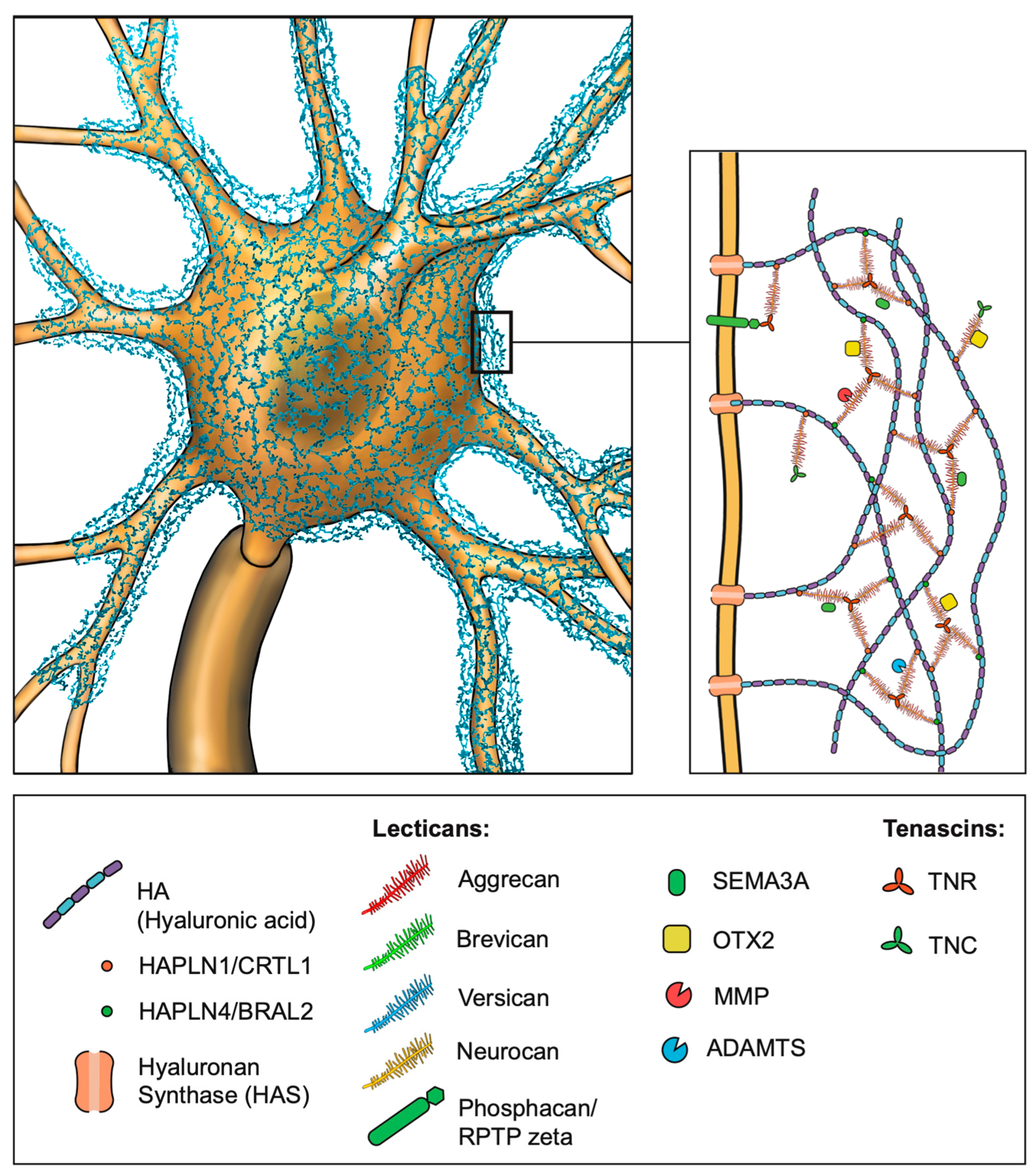
3. Molecular Structure of PNNs

PNNs were initially described in 1893 by Camillo Golgi [25]. However, their significance began to rise much later in the late 19th century as the techniques for studying PNNs became more advanced (for a detailed description of the discovery of PNNs, refer to [25]). PNNs are specialized forms of the ECM surrounding the soma, the axonal initial segment, and proximal dendrites of neurons. The major components of PNNs are hyaluronic acid (HA), chondroitin sulfate proteoglycans (CSGPs), tenascins, in particular, tenascin-R (TNR), and link proteins (CRTL1/HAPLN1 and BRAL2/HAPLN4), which are produced by various neuronal cell types (Figure 1). The hyaluronic backbone of the PNN provides structural stability and anchors the PNN to the cellular surface by interaction with the receptor CD44, expressed on both neurons and astrocytes [26]. HA is produced by hyaluronan synthases (HAS1–3) located at the membrane of the enveloped neuron [27]. Compared to the “loose” ECM, the PNNs display a denser structure with greater amounts of CSPG lecticans. These lecticans are produced by the surrounded neurons, but additionally by astrocytes or oligodendrocytes, and represent the organizers of the brain extracellular matrix with domains on the N- and C-terminals attached to the core protein [28]. The amino terminal regions facilitate binding to HA, while the carboxy terminal regions establish a connection with TNR. In addition to the globular domains, a varying amount of chondroitin sulfate glycosaminoglycan (GAG) is covalently bound to the core protein. Repeated chains of glucuronic acid (GlcA) and N-acetyl-galactosamine (GalNAc) form the structural basis of the GAGs. The number of GAG chains, the sulfation pattern of GlcA and GalNAc, and the core protein structure led to the identification of four CSPG lecticans: aggrecan, versican, brevican, and neurocan [29,30,31,32,33]. An additional CSPG is named phosphacan, a splice variant of the gene PTPRZ1 encoding the receptor protein tyrosine phosphatase-zeta (RPTPzeta) [34]. Recently, it was shown that phosphacan may connect with TNR and function as an anchor for the PNN to the cellular surface, in addition to HA attachment [35]. The connection between CSPGs and HA is maintained by the link proteins cartilage link protein-/hyaluronan and proteoglycan link protein 1 (CRTL1/HAPLN1) and brain link protein-2/hyaluronan and proteoglycan link protein 4 (BRAL2/HAPLN4) [36,37]. The CSPGs are connected to each other in triads through TNR, which in turn contributes to the structural integrity of the PNNs [38,39]. Lastly, semaphorin3A (SEMA3A) and orthodenticle homeobox2 (OTX2) are molecules binding to GAG side chains and thereby controlling plasticity [40,41] (Figure 2).

4. Development and Plasticity

During brain development, the formation of PNNs occurs in a highly regulated manner, coinciding with critical periods of synaptic refinement and maturation that are unique to each brain region [54,55,56]. It has been shown that some of the main components of the PNNs are already expressed in the embryonic nervous system. However, their assembly into PNNs occurs later in development when the expression of PNN components is upregulated [57,58]. In the human developing brain, PNNs were shown to first be present in the second month of life, varying across different brain regions [57]. Their maturation is a gradual process, with them reaching complete maturation by the age of eight years [57]. In coincidence with this, parvalbumin (PV) was shown to be present at birth, but reaches its peak levels around the age of 2 years [57,59]. In addition, neuronal activity also has an impact on the PNNs’ formation. This was demonstrated by the fact that rearing rats without visual stimuli in the dark from birth prevented the formation of PNNs. As a result, reintroducing the animals into a normal light/dark environment could restore PNNs [60,61]. The maturation of the PNNs also depends on the expression of the two molecules SEMA3A and OTX2. Both play a crucial role in initiating and closing the critical period of plasticity in the developing brain [41,62,63,64]. The formation of PNNs is a dynamic process, and physiological remodeling persists even after the PNNs are fully matured. During formation and remodeling, the enzymes MMP (matrix metalloproteinase) and ADAMTS (matrix metalloproteinase and a disintegrin and metalloproteinase with thrombospondin motifs) play a significant role. MMPs are a family of zinc-dependent endopeptidases that are capable of degrading the PNN structure. From the over 20 members of MMPs, MMP-1–3, 7–17, 24, and 28 are expressed in the brain. MMP-9 is the most studied endopeptidase in the brain, being expressed by neurons and glial cells [65]. Several studies have demonstrated that MMP-9 expression is upregulated under conditions with higher neural plasticity or activity [66,67,68]. Furthermore, enhanced MMP-9 activity has been observed in several pathological conditions, for example, epilepsy, traumatic brain injury and focal cerebral ischemia [69,70,71,72]. Another group of enzymes involved in PNN remodeling is ADAMTS. ADAMTS forms a family of metalloproteinases that share structural similarities with MMPs [73]. The proteinases ADAMTS-1, 4, 5, 9, and 15 are expressed in the brain. They are capable in degrading CSPGs, mainly aggrecan and brevican [74,75,76,77]. Moreover, alterations in their expression levels have been associated with acute CNS injuries, such as stroke and spinal cord injury [77,78,79]. In addition to changes in the expression levels of MMPs and ADAMTS due to pathologies, their activity can also be internally regulated by molecules known as TIMPs (tissue inhibitors of metalloproteinases). TIMPs were shown to contribute to the restriction of ECM proteolysis by inhibiting the proteolytic activity of MMPs and ADAMTS [80]. Taken together, PNNs are dynamic structures that undergo certain remodeling processes in both physiological and pathological conditions.

5. Function of PNNs

Continuous scientific research adds to the growing list of physiological functions assigned to PNNs in the mammalian brain.

5.1. Stabilization of Synaptic Sites and Regulation of Synaptic Transmission

Above all, they play a crucial role in the stabilization of synaptic contacts by forming a mesh-like matrix around the cell bodies and proximal dendrites of neurons. The structural framework provided by the PNNs includes “holes” that offer specific sites for synaptic contacts, ensuring the proper functioning of synaptic transmission by restricting the reorganization of the transmission machinery [81]. Recent studies utilizing high-resolution and 3D electron microscopy further elucidated the role of PNNs in synaptic stabilization. The studies have revealed that the “holes” within PNN-surrounded neurons are occupied by synaptic contacts, highlighting the critical role of PNNs in maintaining synaptic contact sites [82]. Beyond the pre- and postsynaptic compartments and the nearby astrocytes, the ECM plays a significant role in physiological synaptic transmission [83]. A study conducted by Blümcke and colleagues in 1995 provided early evidence of the interaction between astrocytic processes and PNNs [84]. Recent advances by Tewari and colleagues (2024) further emphasize this by demonstrating the functional synergy between PNNs and astrocytic processes within the somatosensory cortex of adult mice, underscoring the important role of PNNs in sustaining physiological synaptic function [85]. Collectively, these observations reinforce the concept of the tetrapartite synapse, where the ECM, particularly PNNs, acts as a key participant in structural and functional organization of synaptic networks (Figure 3). The neurotransmitter receptors at the postsynaptic site are highly dynamic in the neuronal plasma membrane [86,87]. The mobility of AMPA-type-glutamate receptors (α-amino-3-hydroxy-5-methyl-4-isoxazolepropionic acid-receptors, AMPAR) represents one example for the importance of lateral diffusion in terms of modulating synaptic transmission. It was shown that PNNs can restrict this lateral diffusion, which demonstrates the control of the availability of synaptic receptors [7]. Moreover, the degradation of hyaluronan results in disruption of the perisynaptic ECM followed by a restoration of the juvenile mobility [7]. This gave rise to the assumption that the ECM plays an important role in modulating synaptic plasticity. In addition to the postsynaptic AMPAR, integrin receptors interacting with ECM components were shown to contribute to long-term potentiation (LTP) by modulating NMDA receptor (N-methyl-D-aspartate receptor, NMDAR) currents [88]. Furthermore, their interaction with the surrounding actin cytoskeleton provides stability [89,90,91,92]. The pre- and postsynaptic hyaluronan receptor CD44 is involved in functional and structural plasticity of dendritic spines, as well as in synaptic stabilization [93]. Another important factor influencing synaptic plasticity is the ECM protein reelin. It was shown that reelin regulates NMDAR and thereby impacts LTP responses [94]. For a more detailed description of the synaptic extracellular matrix, please refer to the review by Dankovich and Rizzoli [81]. Taken together, the ECM has a major impact on the stabilization of synaptic sites and in maintaining proper synaptic transmission.

5.2. Regulation of Neural Plasticity

Since PNNs influence the synaptic sites in the CNS, it is reasonable to assume that PNNs contribute to the regulation of neural plasticity. Neural plasticity is a fundamental mechanism driving learning, memory, and cognitive processes, which encompasses the brain’s ability to adapt and reorganize in response to experiences and changes in the environment. These changes rely on the dynamic nature of the PNN structure. The impact of PNNs on neural plasticity becomes particularly prominent during critical periods of various stages of life. These critical periods represent windows of enhanced neural plasticity during which neural circuits exhibit considerable structural and functional changes in response to sensory experiences. Crucially, PNNs exert control over the closure of these critical periods. It was demonstrated that raising animals in complete darkness from birth prevents the formation of PNNs in the adult visual cortex. Moreover, following chondroitin sulfate proteoglycan (CSPG) degradation in adult rats, the ocular dominance plasticity was restored [60]. Additionally, De Vivo et al. showed that enzymatic degradation of CSPG leads to more motile cortical spines with a higher structural and functional plasticity in the adult visual cortex [95]. PNNs were shown to be responsible for the transition from juvenile to adult forms of learning in sensory systems. In adult mice, PNNs provided protection against fear memory extinction. Consequently, removal of PNNs in adults leads to faster erasure of fear memory [56]. Moreover, auditory learning is limited in adult mammals; however, auditory cortex-dependent relearning can restore the agility of the juvenile state following ECM digestion [96]. Collectively, these findings illustrate the impact of PNNs on neural plasticity and thus the role of PNN in orchestrating the closure of critical periods in neural development.

5.3. Protection Against Oxidative Stress

Parvalbumin-positive interneurons exhibit fast-spiking properties. To maintain this high neuronal activity, a high metabolic activity is required, which leaves them vulnerable to oxidative damage. PNNs possess highly charged structures with polyanionic characteristics, allowing them to directly influence the local ion homeostasis of the enwrapped neurons and thus providing neuroprotective effects [12]. Suttkus and colleagues demonstrated that their ability to bind redox-active molecules result in a reduction in the local oxidative potential. When analyzing the brains of mice post-injection of FeCl3, a significant reduction in neuronal degeneration was observed in neurons surrounded by PNNs compared to KO-mice lacking various PNN components. The findings display that the neuroprotective effects of PNNs are mediated by both the polyanionic charge and the proper interaction of the PNN components [97]. Moreover, the interaction between PNN-ensheathed parvalbumin-positive interneurons and oxidative stress was investigated in mice carrying a genetic redox dysregulation. Oxidative stress profoundly affects parvalbumin-positive cells with immature or impaired PNNs, while parvalbumin-positive cells surrounded by intact PNNs were shown to be protected against excessive reactive oxygen species (ROS) production [11]. Lipofuscin is generated via iron-catalyzed oxidative processes with an accumulation that can be observed in aging and Alzheimer’s disease. PNNs in the human cerebral cortex were shown to provide protective effects against lipofuscin accumulation [12]. Taken together, these finding demonstrate that PNNs exhibit a neuroprotective effect by shielding neurons from oxidative stress and promoting neuronal survival.

5.4. Regulation of Neural Circuity Activity

The central nervous system (CNS) contains neurons that exhibit different inhibitory, excitatory, or modulatory characteristics, depending primarily on their respective neurotransmitters. These neurons play a crucial role in regulating neural network activity by maintaining a balanced state of inhibition and excitation, thereby ensuring proper network functioning. In the cerebral cortex, excitation is provided by glutamatergic cells and inhibition is implemented by GABAergic interneurons. Thereby, GABAergic interneurons play a crucial role in regulating the excitatory/inhibitory (E/I) balance and preventing hyperexcitability, which in turn can lead to epilepsy [98]. Due to their positioning around primarily parvalbumin-positive interneurons, PNNs possess the ability to regulate the balance between excitation and inhibition. Moreover, an interneuron dysfunction was shown to be implicated in a variety of psychiatric disorders, including schizophrenia (SZ), autism spectrum disorder (ASD), and bipolar disorder (BPD), and in neurodegenerative disorders, like Alzheimer’s disease (AD), and aging (for a detailed review, see [99]). Parvalbumin-positive interneurons, and thus PNNs, play a critical role in regulating and sustaining gamma oscillations owing to their distinctive inhibitory properties and their involvement in synchronizing neural firing patterns. Gamma oscillations, typically occurring at frequencies between 30 and 90 Hz, are rhythmic patterns of neural activity. These oscillations are integral to cognitive processes, like the formation of memories and the procession of sensory information (reviewed in [100,101]). Consequently, disturbances within gamma oscillations might be linked to psychiatric disorders like schizophrenia [102]. The research on the impact of depletion of PNNs on neural circuit activity mainly involved two approaches: enzymatic degradation using chondroitinase ABC (ChABC) treatment and genetic knockdown of PNN components. The main findings is that the absence of PNNs resulted in a decline in the high firing rate of parvalbumin-positive interneurons (for a detailed overview, refer to [9]).

6. Scientific Methods to Study PNNs in the Mammalian Brain

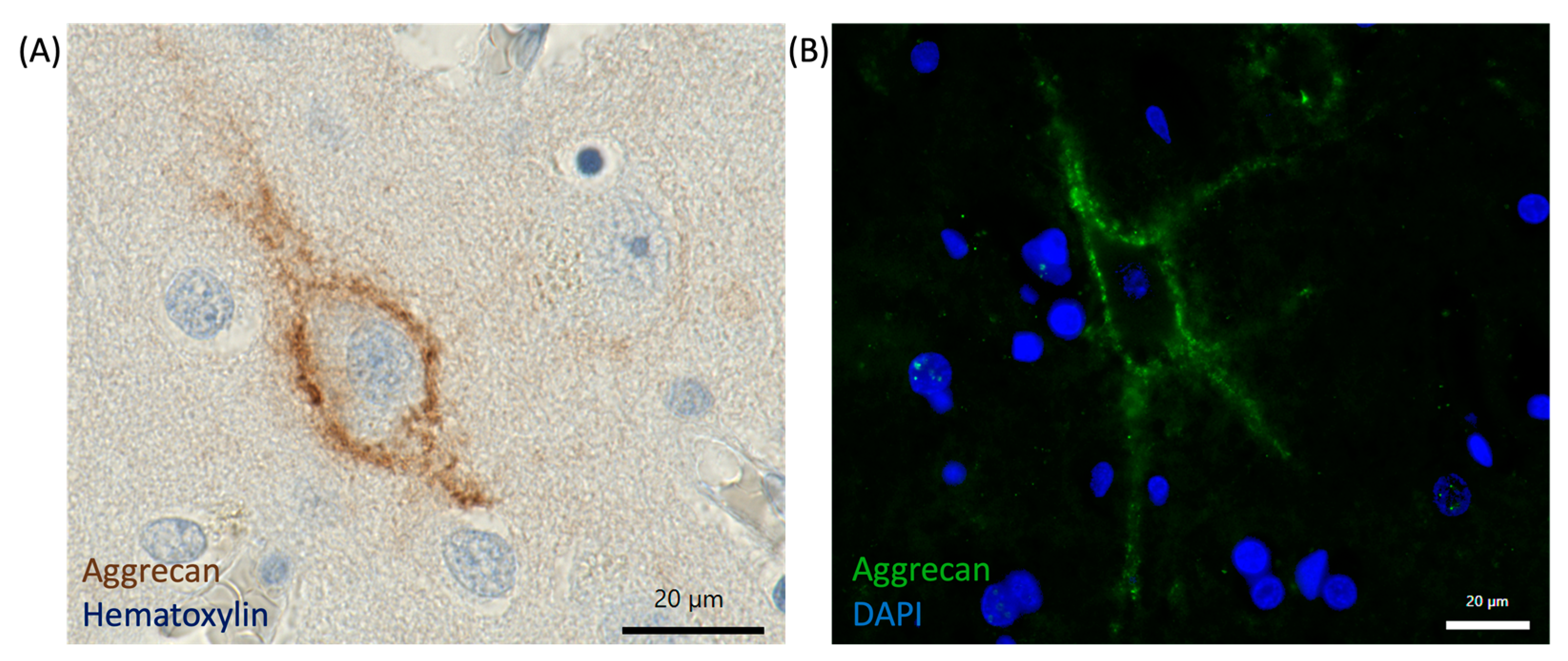
6.1. Immunohistochemistry and Imaging Techniques

A common technique for visualization of PNNs is the use of the plant-derived lectin Wisteria floribunda agglutinin (WFA), which binds to GalNAc present in chondroitin sulfate proteoglycans (CSPGs) [103]. The incubation of tissue sections or cultured cells with WFA conjugated to biotin or a fluorophore allows for a visualization of PNNs. Of note, not all PNNs can be visualized by using lectin detection, due to structural variation in chondroitin sulfate chains, as well as regional variability [21,104]. Instead, antibodies against CSPG components, mainly aggrecan, can be used in immunohistochemical or immunofluorescence approaches [4,24,105] (Figure 4). The analysis with antibodies is much more robust than WFA, supported by the fact that treatment with ChABC cannot abolish the reactivity with an antibody targeted against aggrecan [21,106]. This indicates that there might be a structural heterogeneity of PNNs. Therefore, it is recommended that various markers should be used, to explore this heterogeneity when studying PNNs. Fluorescence microscopy allows for visualizing PNNs labeled with either lectins or antibodies conjugated to a fluorophore. This technique was used in various studies and offers high-resolution imaging of the PNN distribution and structure within tissue sections (Figure 4). By using double or triple staining, researchers can assess the colocalization of different molecular components and investigate the cellular subtypes enwrapped by PNNs [107,108,109,110,111]. Transmission electron microscopy (TEM) can be used to gain an even deeper insight into PNNs. TEM provides ultrastructural details of PNNs in the nanometer range [82,112,113]. This enables visualization of the fine structure of PNNs, as well as their interaction with surrounding neuronal elements and other extracellular matrix components. In combination with immunogold labeling or DAB labeling, TEM permits selective localization and analysis of various PNN components. These immunohistochemistry and imaging techniques offer valuable tools in the research on PNNs and provide insight into their distribution, structure, and role in neural development, plasticity, and disease.

6.2. Knocking out Different PNN Components in Rodents

Various studies in rodents using knock-out techniques have been performed to specifically target and understand the function of each PNN component. In 2010, Giamanco and others studied the role of aggrecan in PNN formation by using aggrecan-deficient cmd mice (cartilage matrix deficiency in mice). The evaluation of neuronal cultures as well as organotypic slice cultures in immunohistochemistry (IHC) or immunocytochemistry (ICC) approaches showed that staining with the lectin Wisteria floribunda agglutinin (WFA) was absent in mice lacking aggrecan. On the contrary, the other components hyaluronan and proteoglycan link protein 1 (HAPLN1), brevican, hyaluronan (HA), and tenascin R (TNR) remained unaffected. Additionally, the binding of HAPLN1 to the cell surface was still intact after treatment with the enzyme chondroitinase ABC, indicating hyaluronan-dependent binding. In contrast, brevican, TNR, and HA were shown to be bound to the cellular surface in an hyaluronan- and CS-dependent manner [114]. In a recent study by Rowlands and colleagues, the aggrecan gene Acan was genetically deleted in an established Acan-IoxP mouse model. This resulted in a reactivation of the critical-period plasticity in the visual cortices of adult mice, suggesting a major role of aggrecan in PNN formation, plasticity, and memory function [115]. The PNN component brevican was also studied using knocking-out techniques. Brevican KO mice showed a normal induction of long-term potentiation (LTP) but had deficits in the maintenance of hippocampal LTP [116]. Favuzzi and coworkers demonstrated that brevican is an essential regulator of interneuron plasticity. This was substantiated by the ability of brevican to control the localization of potassium channels and AMPA-type receptors [117]. The role of neurocan in PNN development has been studied by using neurocan-deficient mice [118]. The medial nucleus of the trapezoid body (MNTB) in the auditory brainstem was analyzed via immunohistochemical approaches. In conclusion, the study provided evidence that neurocan controls the regulation of PNN development by influencing the mRNA and protein quantity of various PNN components in the MNTB [118]. In addition to the study of CSPGs, the role of TNR was also analyzed in knock-out studies. Mice deficient in TNR showed an attenuated PNN structure, implying the crucial role of TNR in both formation and stabilization of PNNs by clustering with aggrecan [119]. To understand the basis of the neuroprotective effect of PNNs, four different mice strains lacking PNN components were treated with FeCl3 to induce neurodegeneration. Aggrecan, link protein, and TNR were identified as essential in maintaining the neuroprotective effect of PNNs, whereas the contribution of brevican was insignificant [97]. Knocking out the link protein CRTL1 resulted in a weakened PNN structure and a retention of the critical period visual plasticity. The expression of other PNN components was not altered [61]. The link protein BRAL2 was shown to be indispensable for proper brevican localization, indicating a pivotal role in the organization of PNNs [120]. In a recent study, the interaction of TNR and RPTPζ/phosphacan was studied by comparing the PNN phenotype of Tnr −/− and Ptprz1 −/− mice. Both displayed a disrupted PNN structure indicating a cooperation of TNR and RPTPζ/phosphacan in the proper assembly of PNNs [121]. This interaction was also proven by a study of Eill and others, analyzing the role of the protein tyrosin phosphatase RPTPζ/phosphacan in Ptprz1 −/− mice [35]. In studies using quadruple KO mice lacking TNC, TNR, brevican, and neurocan, PNNs were analyzed. The results of the studies displayed a reduction in PNN-sheathed cells and a disrupted PNN structure. Moreover, the ratio of inhibitory and excitatory synapses was imbalanced, with a reduction in inhibitory synaptic elements within PNNs. In addition, the number of parvalbumin-positive cells was reduced in the KO mice. Finally, the transcription factor OTX2 (orthodenticle homeobox 2) was shown to be decreased. Overall, this confirms that TNR, TNC, brevican, and neurocan play a significant role in modulating the dynamic interaction among PNNs, synaptic integrity, inhibitory neurons, and the transcription factor OTX2 [122,123].

6.3. Enzymatic Degradation of PNN Components (ChABC, Hyaluronidase)

In addition to the genetic ablation of PNN components, enzymatic degradation of PNNs offers a further, more locally applicable option for investigating the influence of PNN changes. The enzymatic disruption of PNNs involves the application of enzymes to degrade molecular components of PNNs. This provides a valuable tool for studying the functional consequences on neural function or plasticity following PNN alterations. The enzyme chondroitinase ABC (ChABC) is commonly used for this purpose. ChABC induces the ablation of PNNs by enzymatic degrading of sugar chains (glycosaminoglycans or GAGs) of CSPGs. The efficacy of this ability can be validated through the elimination of WFA staining, which is a commonly used marker for PNNs [124]. The enzymatic manipulation of PNNs was used in various studies (reviewed in [125]). In a recent study, the importance of PNNs in the hippocampus and cortex in terms of contextual fear memories was shown [126]. Hence, the elimination of PNNs through ChABC administration led to impaired memory consolidation. In recent work by Willis and coworkers, the disruption of PNNs via ChABC administration facilitated juvenile-like plasticity by enhancing structural flexibility [127]. Further, treating parvalbumin-positive interneurons in the primary visual cortex with ChABC resulted in changes in neuronal excitability and synaptic transmission [128]. Within the hippocampal CA1 region, removal of PNNs via ChABC injection led to a reduced firing rate of parvalbumin-positive neurons and decreased synaptic transmission. Moreover, ChABC treatment interfered with the maintenance of long-term contextual fear memory [129]. In mice lacking CRTL1, administration of ChABC made fear memories more susceptible to erasure, suggesting a role of PNNs in regulating the boundaries of the critical period for fear extinction [130]. Another opportunity to disrupt the structure of PNNs enzymatically arises with the application of the enzyme hyaluronidase. Hyaluronidase cleaves the HA backbone of the PNNs. In one study, it was shown that the disruption of PNNs via HA treatment had an impact on the neurophysiology of hippocampal cells [131]. However, the enzymatic degradation of PNNs using either ChABC or hyaluronidase displays several limitations; for example, the digestion cannot be specifically targeted to PNN-ECM. Here, the recently developed Cre-LoxP system by Carstens and coworkers offers an alternative tool. It allows for cell-type-specific targeting and for longer-lasting ChABC degradation [132]. This system can be used to further study the effect of the durable loss of PNNs and to identify strategies to potentially treat neurological disorders.

6.4. Proteomic Studies

A novel approach for studying the composition of the brain’s ECM is proteomic analysis. In this way, the use of mass spectrometry allows for the identification and quantification of the proteins present in the tissue and provides valuable insights into the role of ECM alterations in neurological diseases. Recent studies have employed this approach to uncover differences in the proteome in epilepsy. For instance, proteomic differences could be identified in the hippocampus and cortex of epilepsy brain tissue, displaying significant differences in the expression of 777 proteins in the hippocampus and 296 proteins in the cortex, indicating that proteins associated with protein synthesis, mitochondrial function, G-protein signaling, and synaptic plasticity are altered in epilepsy [133]. However, this study did not specifically target ECM-associated proteins. In studies focusing on the ECM, proteomic analyses in mice and human revealed a link between ECM alterations and cerebrovascular diseases [134]. In addition, the identification of the proteomic profile of ECM proteins in aging and stroke could reveal an increased activation of genes encoding proteins related to ECM remodeling. Moreover, CSPGs, syndecans, and link proteins were upregulated in aged murine models [135]. In a recent study by Leitner and colleagues, the proteomic signatures of Alzheimer’s disease (AD) and epilepsy were compared, demonstrating significant overlap in the differentially expressed proteins involved in synaptic and mitochondrial dysfunction [136]. Finally, a study by Srivastava and colleagues analyzed the molecular basis for mesial temporal lobe epilepsy (MTLE). By employing proteomics and protein co-expression network analysis, the study analyzed brain tissue from patients with MTLE and revealed altered protein networks and pathways. These disruptions were linked to pathways such as synaptic vesicle neurotransmitter release, synaptic plasticity, metabolic and mitochondrial dysfunction, as well as extracellular matrix organization and cell signaling. This study provided insights into the pathogenesis of MTLE. The advances in proteomics in epilepsy alongside discussions of mass spectrometry methods for analyzing ECM components in neurological disorders have been reviewed in various papers [137,138]. While many recent studies have not prioritized ECM-specific proteins, ECM-targeted proteomic approaches offer valuable tools for investigating alterations in the ECM composition and provide insights into the role of ECM remodeling under pathological conditions.

7. PNNs and Their Implications for Neurological Disorders

7.1. Schizophrenia and Bipolar Disorder

Schizophrenia (SZ) represents a multifaceted psychiatric disorder, marked by a combination of positive symptoms, like hallucinations or delusions, negative symptoms, such as deficits in the expression of emotions, as well as cognitive impairments. Recent research suggests a potential link between PNNs and the pathophysiology of schizophrenia. Post-mortem studies have revealed abnormalities in the ECM in brain regions like the amygdala and entorhinal cortex. Specifically, individuals with schizophrenia exhibited an increase in CSPG-positive glial cells and a reduction in PNN density in the lateral nucleus of the amygdala and layer II of the entorhinal cortex. These findings underscore the significance of the interaction of the ECM and glial cells in the development of schizophrenia [139]. Moreover, post-mortem studies of the olfactory epithelium revealed differences in the expression of CSPGs [140]. Analysis of post-mortem human brains with schizophrenia showed a reduction in PNN density in layers III and V of the prefrontal cortex [141]. However, PNN density remain unchanged in the primary visual cortex. These findings suggest that PNNs might contribute to the dysfunction of the prefrontal cortex seen in schizophrenia. Additionally, the timing of PNN development coincides with the emergence of schizophrenia symptoms, implying a potential role for PNN formation in the onset of the illness [141]. Abnormalities of aggrecan and chondroitin-6-sulfate were examined in a study by the research group around Pantazopoulos. The use of post-mortem tissue from the amygdala of schizophrenia patients showed that the number of aggrecan PNNs was decreased, and CSPG abnormalities could be observed [105]. In the dorsolateral prefrontal cortex, the density of parvalbumin-positive cells remained unchanged in individuals with schizophrenia. However, the fluorescence intensity of parvalbumin labeling was notably lower in comparison to controls. In addition, there was a decrease in the immunoreactivity of PNNs positive for WFA and aggrecan in the schizophrenia group. These observations suggest distinct differences in the levels of parvalbumin protein and PNN components among individuals with schizophrenia [111]. In an experimental model of schizophrenia using ketamine-treated rats, researchers investigated the ECM expression. A reduced number of WFA-labeled PNNs and a decrease in PV fluorescence intensity were observed in interneurons within the prefrontal cortex [142]. In the thalamic reticular nucleus of transgenic mice with a redox dysregulation, the numbers of parvalbumin-positive neurons and PNNs were decreased in the rodent model for schizophrenia and bipolar disorders compared to wild-type (WT) mice [143]. Another mouse model used for studying schizophrenia is Gclm KO mice with a genetically compromised glutathione system. Here, a deficit in parvalbumin neurons was found due to their high susceptibility to oxidative stress [144]. By using RNA sequencing approaches, the transcriptome revealed differences of more than 25% in differential splicing or expression levels in subjects with autism spectrum disorder (ASD), schizophrenia, and bipolar disorders (BPDs) [145]. Post-mortem sections of BPD patients revealed a significantly lower density of PNNs in the dorsolateral prefrontal cortex, whereas schizophrenia patients showed only a slight reduction in PNN density in this brain region [146]. PNN remodeling by endopeptidases was also shown to be affected in schizophrenia, as indicated by a dysregulation of MMP-9 expression [147]. In more recent studies using the ketamine mouse model of schizophrenia, treated mice exhibited fine structure abnormalities and a quantitative reduction in PNNs [148,149]. Analyzing the molecular signature with microarray analysis, as was examined in a study by Pantazopoulos and colleagues, revealed a differential expression of several ECM components, e.g., brevican, neurocan, and a disintegrin and metalloprotease with thrombospondin motifs 1 (ADAMTS1), in schizophrenia [150]. In the prefrontal cortex of a schizophrenia mouse model (disruption in schizophrenia, DISC1), both PNNs and parvalbumin-positive neurons were decreased. Following digestion with ChABC, the behavior of the control mice matched that of the DISC1 mice. Moreover, the number of high-frequency firing neurons was reduced, whereas the ratio of irregularly firing neurons was increased [151]. In a recent study by Liang and Zhang, the effects of an inhibition of NMDAR in young-aged mice, combined with social stress in adulthood, were assessed. The number of PV+ cells enwrapped by PNNs was increased, and the activity of PV+ neurons was decreased in the prefrontal cortex in mice with an inhibition of NMDAR [152]. Overall, substantial evidence indicates the crucial role of PNNs in the pathophysiology of schizophrenia and bipolar disorders (for a review, see [153,154,155,156,157]).

7.2. Alzheimer’s Disease (AD)

Alzheimer’s disease is a progressive, neurodegenerative disease and represents the most common underlying cause of dementia. The symptoms include successive memory impairment, changes in behavior and personality, and difficulties in reasoning, language, and handling of complex tasks. The underlying reason for AD is not yet fully understood. However, the abnormal accumulation of proteins such as amyloid plaques and Tau protein tangles in the brain can lead to the death of brain cells. To date, there is no curative treatment option for AD available. Yet, some medications can slow the progression of dementia symptoms. Immense efforts are being made to research the causes of AD and to find novel treatment options. Several studies have investigated the involvement of PNNs in the pathophysiology of AD. In a study using samples of AD patients, it was shown that neurons, which are enwrapped by aggrecan-based PNNs, displayed protection against the accumulation of Tau fibers. In regions that were heavily affected by Tau pathology, aggrecan-based ECM was absent. This suggests that the aggrecan ECM may contribute to the protection of neurons from Tau protein accumulation in AD [158]. On the contrary, in an Alzheimer model using transgenic Tg2576 mice, PNNs were largely unaffected when investigating aggrecan-based PNNs. However, with advanced progression of the disease, PNNs were removed after cell death [159]. By detecting the main components of PNNs in temporal and occipital lobes of AD patient tissue, a study by Morawski and colleagues confirmed that aggrecan-based PNNs provide protection against Tau pathology. Conversely, the PNN structure and distribution remained unchanged within the amyloid pathology in AD tissue [160]. In the hippocampi of AD patients, loss of PNNs was observed. Additionally, the formation of axonal coats was shown to contribute to the preservation of synaptic integrity within AD-related change [109]. Several studies conducted proteome analysis to investigate protein alterations in AD. Various ECM proteins were shown to be dysregulated within AD conditions [157]. In a novel mouse model TauP301L, which lacks aggrecan, high levels of Tau protein were observed [161]. Using mass spectrometry analysis, a change in the sulfation patterns of the PNN-associated CS-GAGs was observed in patients with AD. These findings correlate with AD progression and with Tau accumulation, as well as with the impairment of cognition [162]. A possible neuroprotective function of aggrecan and other PNN components was demonstrated in a study using a Tau/aggrecan double transgenic model [163]. Studying the proteomic signature of AD and epilepsy tissue revealed overlapping changes in the protein composition, with 89% of the proteins altered in the epileptic hippocampus also shown to undergo changes in AD patients [136]. In summary, PNNs may have a substantial role in the development and progression of AD and potentially offer protective effects in relation to the disease’s pathology. This suggests a potential of compounds targeting PNNs as a promising therapeutic strategy for treating AD (for a review, see [15]).

7.3. Epilepsy

Epilepsy is a chronic neurological disorder marked by recurrent, spontaneous seizures, affecting approximately 50 million people worldwide [164]. The main cause is an imbalance in the electrical activity of the brain. Given that PNNs are involved in regulating and maintaining the balance between excitation and inhibition, as well as in regulating the excitability of PV+ neurons, it is plausible to assume that PNNs are involved in the pathogenesis of epilepsy. Moreover, by modulating synaptic plasticity, PNNs are implicated in the pathophysiology of epilepsy. Numerous studies investigated the ongoing remodeling of the ECM in epileptic brain tissue. In a rodent model of temporal lobe epilepsy (TLE), PNN components were analyzed following status epilepticus (SE). A persistent decrease in PNN components including aggrecan, hyaluronan and proteoglycan link protein 1 (HAPLN1), and hyaluronan synthetase 3 (HAS3) was detected in the epileptic hippocampus [165]. The degradation of PNNs was analyzed in an epilepsy rodent model in a study by Rankin-Gee and colleagues [70]. By inducing SE, the MMP proteolysis of aggrecan was increased. Moreover, a loss of PNNs was associated with a higher number of seizures [70]. Further studies showed that MMP activity, in particular, MMP-3 and MMP-13, was increased in the hippocampi of rats after the induction of SE. In contrast, the activity of ADAMTS was not significantly increased after status epilepticus [166]. Thus, these studies provide evidence that the PNN remodeling in epilepsy is primarily driven by MMP activity. In epilepsy associated with brain tumors, the ability of PNNs to modulate the firing frequency of cells has been demonstrated using rodent models. PNNs were shown to be able to reduce the membrane capacity of fast-spiking interneurons, letting them fire action potentials in a more than physiological manner. Therefore, the loss of PNNs led to a reduced firing rate of fast-spiking interneurons, which represents the underlying cause of tumor-associated seizures [167]. A study comparing the developmental timeline of PNNs during aging suggested no differences between controls and epilepsy patients. However, this conclusion is limited due to the small sample number of epileptic surgical specimens examined and the inclusion of different epilepsy types, leaving the findings inconclusive [57]. In contrast, a more recent study observed an age-dependent increase in PNNs in the human hippocampus [24]. Additionally, precocious aging effects could be determined in TLE patients with hippocampus sclerosis, characterized by a higher PNN density in immunohistochemical analyses of surgical specimens. This increase was not accompanied by an increase in the PV+ neuronal cell density, raising questions about the source of PNN material and their target cell population. These observations corroborated, however, that PNNs are involved in the formation of memory engrams in the human hippocampus [24]. In summary, these studies underscore the role of PNNs in the pathophysiology of epilepsy by modulating synaptic plasticity and neuronal excitability. The ongoing PNN degradation is predominantly driven by increased MMP activity and a higher seizure frequency. Moreover, age-dependent changes in PNN density could be found in the epileptic hippocampus. Consequently, targeting PNNs with drugs may hold potential as a therapeutic intervention in epilepsy (for a detailed review, see [14]).

8. Therapeutic Potential and Future Directions

The integrity of the ECM, particularly PNNs, plays an important role in regulating synaptic plasticity and maintaining neuronal excitability. Recent studies have increasingly highlighted the role of the ECM in the pathogenesis of various diseases of the CNS. Consequently, targeting ECM components could represent a novel treatment option for neurological disorders.
In epilepsy, the ongoing ECM remodeling is mainly driven by MMP activity. Hence, inhibiting MMP enzymatic activity might offer a promising option for treating epilepsy. By employing doxycycline as an MMP inhibitor, PNN degradation could be prevented, and seizure occurrence could be reduced in an epilepsy rodent model [168]. Similarly, the MMP inhibitor IPR-179 demonstrated antiseizure effects in a rat temporal lobe epilepsy model. The seizure intensity could be reduced by IPR-179 treatment without severe side effects. These findings suggest the potential of MMP inhibition as a promising therapeutic approach against epilepsy, though further investigation is needed [169].
In the context of AD, PNNs may represent a promising target, as they are involved in memory and cognition. Manipulating the PNNs could reactivate plasticity and restore memory functions impaired in AD pathogenesis. Various strategies targeting PNNs have been proposed, including enzymatic degradation, genetic therapy to inhibit PNN formation, and the use of compounds or molecules to block PNNs [170]. However, it is also important to consider the protective effects of PNNs in preventing the accumulation of Tau or amyloid-beta proteins in AD pathology (see Section 7.2). This protective role could have a beneficial effect in slowing the progression of the disease.
While therapeutic approaches targeting PNNs in neurological disorders like epilepsy and AD appear promising, the precise role of PNNs in the pathophysiology of these and other neurological diseases is not yet fully understood. Further research is, therefore, needed to elucidate the molecular mechanisms by which PNNs contribute to the pathogenesis. In addition, further studies should focus on improving the methods used to study PNNs in their physiological state, which is critical for gaining a deeper understanding of PNN dynamics and functions. Understanding PNNs under normal conditions is essential to better understand how alterations might contribute to neurological disorders, and thus, to explore potential therapeutic interventions.

Author Contributions

Ideation, S.A. and F.P.; literature search, S.A.; writing—original draft preparation, S.A.; writing—review and editing, F.P., M.S., L.H., S.B., R.F. and I.B.; funding acquisition, F.P., I.B. and S.B. All authors have read and agreed to the published version of the manuscript.

Funding

We gratefully acknowledge the financial support by the German Research Foundation (DFG), under project 460333672 CRC1540 EBM.

Data Availability Statement

No new data were created or analyzed in this study. Data sharing is not applicable to this article.

Acknowledgments

The present article is part of the first author’s (S.A.) doctoral thesis. The underlying work was performed in fulfillment of the requirement for obtaining the degree at the Natural Science Faculty of the Friedrich-Alexander-Universität Erlangen-Nürnberg. We would like to thank Jörg Pekarsky, illustrator at the Institute of Functional and Clinical Anatomy, for creating the graphics.

Conflicts of Interest

The authors declare no conflict of interest.

Abbreviations

The following abbreviations are used in this manuscript:
PNNPerineuronal net
CNSCentral nervous system
ECMExtracellular matrix
PV+Parvalbumin expressing
GABAGamma-aminobutyric acid
HAHyaluronic acid
CSPGChondroitin sulfate proteoglycan
CACornu ammonis
BABrodmann area
TNRTenascin R
CRTL1Cartilage link protein 1
HAPLNHyaluronan and proteoglycan link protein
BRAL2Brain link protein 2
CDCluster of differentiation
HASHyaluronan synthase
GAGGlycosaminoglycan
GlcAGlucoronic acid
GalNAcN-acetlyl-galactosamine
RPTPReceptor protein tyrosine phosphatase
SEMA3ASemaphorin 3A
OTX2Orthodenticle homeobox 2
MMPMatrix metalloproteinase
ADAMTSA disintegrin and metalloproteinase with thrombospondin motifs
TNCTenascin C
TIMPTissue inhibitor of metalloproteinases
AMPARα-amino-3-hydroxy-5-methyl-4-isoxazolepropionic acid receptor
NMDARN-methyl-D-aspartate-receptor
LTPLong-term potentiation
ROSReactive oxygen species
E/IExcitatory/inhibitory
SZSchizophrenia
ASDAutism spectrum disorder
BPDBipolar disorder
ADAlzheimer’s disease
ChABCChondroitinase ABC
WFAWisteria floribunda agglutinin
TEMTransmission electron microscopy
DAB3,3′-diaminobenzidine
IHCImmunohistochemistry
IFImmunofluorescence
DAPI4′,6-diamidin-2-phenylindol
CmdCartilage matrix deficiency
ICCImmunocytochemistry
KOKnock-out
MNTBMedial nucleus of the trapezoid body
MTLEMesial temporal lobe epilepsy
SEStatus epilepticus
TLETemporal lobe epilepsy

References

  1. Nicholson, C.; Hrabětová, S. Brain Extracellular Space: The Final Frontier of Neuroscience. Biophys. J. 2017, 113, 2133–2142. [Google Scholar] [CrossRef] [PubMed]
  2. Syková, E.; Nicholson, C. Diffusion in Brain Extracellular Space. Physiol. Rev. 2008, 88, 1277–1340. [Google Scholar] [CrossRef] [PubMed]
  3. Lehmenkühler, A.; Sykova, E.; Svoboda, Q.J.; Zilles, K.; Nicholson, C. Extracellular space parameters in the rat neocortex and subcortical white matter during postnatal development determined by diffusion analysis. Neuroscience 1993, 55, 339–351. [Google Scholar] [CrossRef] [PubMed]
  4. Härtig, W.; Brauer, K.; Bigl, V.; Brückner, G. Chondroitin Sulfate Proteoglycan-Immunoreactivity of Lectin-Labeled Perineuronal Nets around Parvalbumin-Containing Neurons. Brain Res. 1994, 635, 307–311. [Google Scholar] [CrossRef]
  5. Härtig, W.; Brauer, K.; Brückner, G. Wisteria Floribunda Agglutinin-Labelled Nets Surround Parvalbumin-Containing Neurons. Neuroreport 1992, 3, 869–872. [Google Scholar] [CrossRef]
  6. Dityatev, A.; Schachner, M. Extracellular Matrix Molecules and Synaptic Plasticity. Nat. Rev. Neurosci. 2003, 4, 456–468. [Google Scholar] [CrossRef]
  7. Frischknecht, R.; Heine, M.; Perrais, D.; Seidenbecher, C.I.; Choquet, D.; Gundelfinger, E.D. Brain Extracellular Matrix Affects AMPA Receptor Lateral Mobility and Short-Term Synaptic Plasticity. Nat. Neurosci. 2009, 12, 897–904. [Google Scholar] [CrossRef]
  8. Sorg, B.A.; Berretta, S.; Blacktop, J.M.; Fawcett, J.W.; Kitagawa, H.; Kwok, J.C.F.; Miquel, M. Casting a Wide Net: Role of Perineuronal Nets in Neural Plasticity. J. Neurosci. 2016, 36, 11459–11468. [Google Scholar] [CrossRef]
  9. Wingert, J.C.; Sorg, B.A. Impact of Perineuronal Nets on Electrophysiology of Parvalbumin Interneurons, Principal Neurons, and Brain Oscillations: A Review. Front. Synaptic Neurosci. 2021, 13, 673210. [Google Scholar] [CrossRef]
  10. Balmer, T.S. Perineuronal Nets Enhance the Excitability of Fast-Spiking Neurons. eNeuro 2016, 3, 745–751. [Google Scholar] [CrossRef]
  11. Cabungcal, J.H.; Steullet, P.; Morishita, H.; Kraftsik, R.; Cuenod, M.; Hensch, T.K.; Do, K.Q. Perineuronal Nets Protect Fast-Spiking Interneurons against Oxidative Stress. Proc. Natl. Acad. Sci. USA 2013, 110, 9130–9135. [Google Scholar] [CrossRef] [PubMed]
  12. Morawski, M.; Brückner, M.K.; Riederer, P.; Brückner, G.; Arendt, T. Perineuronal Nets Potentially Protect against Oxidative Stress. Exp. Neurol. 2004, 188, 309–315. [Google Scholar] [CrossRef]
  13. Wen, T.H.; Binder, D.K.; Ethell, I.M.; Razak, K.A. The Perineuronal ‘Safety’ Net? Perineuronal Net Abnormalities in Neurological Disorders. Front. Mol. Neurosci. 2018, 11, 270. [Google Scholar] [CrossRef]
  14. Chaunsali, L.; Tewari, B.P.; Sontheimer, H. Perineuronal Net Dynamics in the Pathophysiology of Epilepsy. Epilepsy Curr. 2021, 21, 273–281. [Google Scholar] [CrossRef]
  15. Scarlett, J.M.; Hu, S.J.; Alonge, K.M. The “Loss” of Perineuronal Nets in Alzheimer’s Disease: Missing or Hiding in Plain Sight? Front. Integr. Neurosci. 2022, 16, 896400. [Google Scholar] [CrossRef]
  16. Pantazopoulos, H.; Berretta, S. In Sickness and in Health: Perineuronal Nets and Synaptic Plasticity in Psychiatric Disorders. Neural Plast. 2016, 2016, 9847696. [Google Scholar] [CrossRef]
  17. Bertolotto, A.; Manzardo, E.; Guglielmone, R. Immunohistochemical Mapping of Perineuronal Nets Containing Chondroitin Unsulfate Proteoglycan in the Rat Central Nervous System. Cell Tissue Res. 1996, 283, 283–295. [Google Scholar] [CrossRef]
  18. Lensjø, K.K.; Christensen, A.C.; Tennøe, S.; Fyhn, M.; Hafting, T. Differential Expression and Cell-Type Specificity of Perineuronal Nets in Hippocampus, Medial Entorhinal Cortex, and Visual Cortex Examined in the Rat and Mouse. eNeuro 2017, 4. [Google Scholar] [CrossRef]
  19. Lupori, L.; Totaro, V.; Cornuti, S.; Ciampi, L.; Carrara, F.; Grilli, E.; Viglione, A.; Tozzi, F.; Putignano, E.; Mazziotti, R.; et al. A Comprehensive Atlas of Perineuronal Net Distribution and Colocalization with Parvalbumin in the Adult Mouse Brain. Cell Rep. 2023, 42, 112788. [Google Scholar] [CrossRef]
  20. Carstens, K.E.; Phillips, M.L.; Pozzo-Miller, L.; Weinberg, R.J.; Dudek, S.M. Perineuronal Nets Suppress Plasticity of Excitatory Synapses on CA2 Pyramidal Neurons. J. Neurosci. 2016, 36, 6312–6320. [Google Scholar] [CrossRef]
  21. Yamada, J.; Jinno, S. Molecular Heterogeneity of Aggrecan-Based Perineuronal Nets around Five Subclasses of Parvalbumin-Expressing Neurons in the Mouse Hippocampus. J. Comp. Neurol. 2017, 525, 1234–1249. [Google Scholar] [CrossRef] [PubMed]
  22. Hausen, D.; Brückner, G.; Drlicek, M.; Härtig, W.; Brauer, K.; Bigl, V. Pyramidal Cells Ensheathed by Perineuronal Nets in Human Motor and Somatosensory Cortex. Neurochemistry 1996, 7, 1725–1729. [Google Scholar] [CrossRef] [PubMed]
  23. Seeger, G.; Lüth, H.J.; Winkelmann, E.; Brauer, K. Distribution Patterns of Wisteria Floribunda Agglutinin Binding Sites and Parvalbumin-Immunoreactive Neurons in the Human Visual Cortex: A Double-Labelling Study. J. Hirnforsch. 1996, 37, 351–366. [Google Scholar] [PubMed]
  24. Lehner, A.; Hoffmann, L.; Rampp, S.; Coras, R.; Paulsen, F.; Frischknecht, R.; Hamer, H.; Walther, K.; Brandner, S.; Hofer, W.; et al. Age-Dependent Increase of Perineuronal Nets in the Human Hippocampus and Precocious Aging in Epilepsy. Epilepsia Open 2024, 9, 1372–1381. [Google Scholar] [CrossRef]
  25. Celio, M.R.; Blümcke, I. Perineuronal Nets-a Specialized Form of Extracellular Matrix in the Adult Nervous System. Brain Res. Rev. 1994, 19, 12–14. [Google Scholar] [CrossRef]
  26. Aruffo, A.; Stamenkovic, I.; Melnick, M.; Underhill, C.B.; Seed, B. CD44 is the principal cell surface receptor for hyaluronate. Cell 1990, 61, 1303–1313. [Google Scholar] [CrossRef]
  27. Weigel, P.H.; Hascall, V.C.; Tammi, M. Hyaluronan Synthases. J. Biol. Chem. 1997, 272, 13997–14000. [Google Scholar] [CrossRef]
  28. Yamaguchi, Y. Lecticans: Organizers of the Brain Extracellular Matrix. CMLS Cell. Mol. Life Sci. 2000, 57, 276–289. [Google Scholar] [CrossRef]
  29. Rauch, U.; Karthikeyan, L.; Maurel, P.; Margolis, R.U.; Margolis, R.K. Cloning and Primary Structure of Neurocan, a Developmentally Regulated, Aggregating Chondroitin Sulfate Proteoglycan of Brain. J. Biol. Chem. 1992, 267, 19536–19547. [Google Scholar] [CrossRef]
  30. Doegesq, K.J.; Sasakill, M.; Kimurajj, T.; Yamada, Y. Complete Coding Sequence and Deduced Primary Structure of the Human Cartilage Large Aggregating Proteoglycan, Aggrecan. J. Biol. Chem. 1991, 266, 894–902. [Google Scholar]
  31. Yamada, H.; Watanabe, K.; Shimonaka, M.; Yamaguchis, Y. Molecular Cloning of Brevican, a Novel Brain Proteoglycan of the Aggrecadersican Family. J. Biol. Chem. 1994, 269, 10119–10126. [Google Scholar] [CrossRef] [PubMed]
  32. Zimmermann, D.R.; Ruoslahti, E. Multiple Domains of the Large Fibroblast Proteoglycan, Versican. EMBO J. 1989, 8, 2975–2981. [Google Scholar] [CrossRef] [PubMed]
  33. Testa, D.; Prochiantz, A.; Di Nardo, A.A. Perineuronal Nets in Brain Physiology and Disease. Semin. Cell Dev. Biol. 2019, 89, 125–135. [Google Scholar] [CrossRef]
  34. Maurel, P.; Rauch, U.; Flad, M.; Margolis, R.K.; Margolis, R.U. Phosphacan, a Chondroitin Sulfate Proteoglycan of Brain That Interacts with Neurons and Neural Cell-Adhesion Molecules, Is an Extracellular Variant of a Receptor-Type Protein Tyrosine Phosphatase. Biochemistry 1994, 91, 2512–2516. [Google Scholar] [CrossRef]
  35. Eill, G.J.; Sinha, A.; Morawski, M.; Viapiano, M.S.; Matthews, R.T. The Protein Tyrosine Phosphatase RPTPξ/Phosphacan Is Critical for Perineuronal Net Structure. J. Biol. Chem. 2020, 295, 955–968. [Google Scholar] [CrossRef]
  36. Spicer, A.P.; Joo, A.; Bowling, R.A. A Hyaluronan Binding Link Protein Gene Family Whose Members Are Physically Linked Adjacent to Chrondroitin Sulfate Proteoglycan Core Protein Genes. The Missing Links. J. Biol. Chem. 2003, 278, 21083–21091. [Google Scholar] [CrossRef]
  37. Oohashi, T.; Edamatsu, M.; Bekku, Y.; Carulli, D. The Hyaluronan and Proteoglycan Link Proteins: Organizers of the Brain Extracellular Matrix and Key Molecules for Neuronal Function and Plasticity. Exp. Neurol. 2015, 274, 134–144. [Google Scholar] [CrossRef]
  38. Aspberg, A.; Miura, R.; Bourdoulous, S.; Shimonaka, M.; Heinegård, D.; Schachner, M.; Ruoslahti, E.; Yamaguchi, Y.U. The C-Type Lectin Domains of Lecticans, a Family of Aggregating Chondroitin Sulfate Proteoglycans, Bind Tenascin-R by Protein-Protein Interactions Independent of Carbohydrate Moiety. Proc. Natl. Acad. Sci. USA 1997, 94, 10116–10121. [Google Scholar] [CrossRef]
  39. Jakovljević, A.; Tucić, M.; Blažiková, M.; Korenić, A.; Missirlis, Y.; Stamenković, V.; Andjus, P. Structural and Functional Modulation of Perineuronal Nets: In Search of Important Players with Highlight on Tenascins. Cells 2021, 10, 1345. [Google Scholar] [CrossRef]
  40. Dick, G.; Liktan, C.; Alves, J.N.; Ehlert, E.M.E.; Miller, G.M.; Hsieh-Wilson, L.C.; Sugahara, K.; Oosterhof, A.; Van Kuppevelt, T.H.; Verhaagen, J.; et al. Semaphorin 3A Binds to the Perineuronal Nets via Chondroitin Sulfate Type E Motifs in Rodent Brains. J. Biol. Chem. 2013, 288, 27384–27395. [Google Scholar] [CrossRef]
  41. Beurdeley, M.; Spatazza, J.; Lee, H.H.C.; Sugiyama, S.; Bernard, C.; Di Nardo, A.A.; Hensch, T.K.; Prochiantz, A. Otx2 Binding to Perineuronal Nets Persistently Regulates Plasticity in the Mature Visual Cortex. J. Neurosci. 2012, 32, 9429–9437. [Google Scholar] [CrossRef] [PubMed]
  42. Lander, C.; Zhang, H.; Hockfield, S. Neurons Produce a Neuronal Cell Surface-Associated Chondroitin Sulfate Proteoglycan. J. Neurosci. 1998, 18, 174–183. [Google Scholar] [CrossRef] [PubMed]
  43. Miyata, S.; Nishimura, Y.; Hayashi, N.; Oohira, A. Construction of Perineuronal Net-like Structure by Cortical Neurons in Culture. Neuroscience 2005, 136, 95–104. [Google Scholar] [CrossRef] [PubMed]
  44. Giamanco, K.A.; Matthews, R.T. Deconstructing the Perineuronal Net: Cellular Contributions and Molecular Composition of the Neuronal Extracellular Matrix. Neuroscience 2012, 218, 367–384. [Google Scholar] [CrossRef]
  45. Carulli, D.; Rhodes, K.E.; Brown, D.J.; Bonnert, T.P.; Pollack, S.J.; Oliver, K.; Strata, P.; Fawcett, J.W. Composition of Perineuronal Nets in the Adult Rat Cerebellum and the Cellular Origin of Their Components. J. Comp. Neurol. 2006, 494, 559–577. [Google Scholar] [CrossRef]
  46. Bartsch, U. The extracellular matrix molecule tenascin-C: Expression in vivo and functional characterization in vitro. Pergamon Prog. Neurobiol. 1996, 49, 145–168. [Google Scholar] [CrossRef]
  47. Wiese, S.; Karus, M.; Faissner, A. Astrocytes as a Source for Extracellular Matrix Molecules and Cytokines. Front. Pharmacol. 2012, 3, 120. [Google Scholar] [CrossRef]
  48. Kelwick, R.; Desanlis, I.; Wheeler, G.N.; Edwards, D.R. The ADAMTS (A Disintegrin and Metalloproteinase with Thrombospondin Motifs) Family. Genome Biol. 2015, 16, 113. [Google Scholar] [CrossRef]
  49. Larsen, P.H.; Yong, V.W. The Expression of Matrix Metalloproteinase-12 by Oligodendrocytes Regulates Their Maturation and Morphological Differentiation. J. Neurosci. 2004, 24, 7597–7603. [Google Scholar] [CrossRef]
  50. Gorter, R.P.; Baron, W. Matrix Metalloproteinases Shape the Oligodendrocyte (Niche) during Development and upon Demyelination. Neurosci. Lett. 2020, 729, 134980. [Google Scholar] [CrossRef]
  51. Hamel, M.G.; Mayer, J.; Gottschall, P.E. Altered Production and Proteolytic Processing of Brevican by Transforming Growth Factor β in Cultured Astrocytes. J. Neurochem. 2005, 93, 1533–1541. [Google Scholar] [CrossRef] [PubMed]
  52. Song, I.; Dityatev, A. Crosstalk between Glia, Extracellular Matrix and Neurons. Brain Res. Bull. 2018, 136, 101–108. [Google Scholar] [CrossRef] [PubMed]
  53. Bosiacki, M.; Gąssowska-Dobrowolska, M.; Kojder, K.; Fabiańska, M.; Jeżewski, D.; Gutowska, I.; Lubkowska, A. Perineuronal Nets and Their Role in Synaptic Homeostasis. Int. J. Mol. Sci. 2019, 20, 4108. [Google Scholar] [CrossRef] [PubMed]
  54. Ueno, H.; Takao, K.; Suemitsu, S.; Murakami, S.; Kitamura, N.; Wani, K.; Okamoto, M.; Aoki, S.; Ishihara, T. Age-Dependent and Region-Specific Alteration of Parvalbumin Neurons and Perineuronal Nets in the Mouse Cerebral Cortex. Neurochem. Int. 2018, 112, 59–70. [Google Scholar] [CrossRef]
  55. Mafi, A.M.; Hofer, L.N.; Russ, M.G.; Young, J.W.; Mellott, J.G. The Density of Perineuronal Nets Increases With Age in the Inferior Colliculus in the Fischer Brown Norway Rat. Front. Aging Neurosci. 2020, 12, 27. [Google Scholar] [CrossRef]
  56. Gogolla, N.; Caroni, P.; Lüthi, A.; Herry, C. Perineuronal Nets Protect Fear Memories from Erasure. Science 2009, 325, 1258–1261. [Google Scholar] [CrossRef]
  57. Rogers, S.L.; Rankin-Gee, E.; Risbud, R.M.; Porter, B.E.; Marsh, E.D. Normal Development of the Perineuronal Net in Humans; In Patients with and without Epilepsy. Neuroscience 2018, 384, 350–360. [Google Scholar] [CrossRef]
  58. Gao, R.; Wang, M.; Lin, J.; Hu, L.; Li, Z.; Chen, C.; Yuan, L. Spatiotemporal Expression Patterns of Chondroitin Sulfate Proteoglycan MRNAs in the Developing Rat Brain. Neuroreport 2018, 29, 517–523. [Google Scholar] [CrossRef]
  59. Fung, S.J.; Webster, M.J.; Sivagnanasundaram, S.; Duncan, C.; Elashoff, M.; Weickert, C.S. Interneuron Markers in the Developing Human and in Schizophrenia. Am. J. Psychiatry 2010, 167, 1479–1488. [Google Scholar] [CrossRef]
  60. Pizzorusso, T.; Medini, P.; Berardi, N.; Chierzi, S.; Fawcett, J.W.; Maffei, L. Reactivation of Ocular Dominance Plasticity in the Adult Visual Cortex. Science 2002, 298, 1248–1251. [Google Scholar] [CrossRef]
  61. Carulli, D.; Pizzorusso, T.; Kwok, J.C.F.; Putignano, E.; Poli, A.; Forostyak, S.; Andrews, M.R.; Deepa, S.S.; Glant, T.T.; Fawcett, J.W. Animals Lacking Link Protein Have Attenuated Perineuronal Nets and Persistent Plasticity. Brain 2010, 133, 2331–2347. [Google Scholar] [CrossRef] [PubMed]
  62. Sugiyama, S.; Di Nardo, A.A.; Aizawa, S.; Matsuo, I.; Volovitch, M.; Prochiantz, A.; Hensch, T.K. Experience-Dependent Transfer of Otx2 Homeoprotein into the Visual Cortex Activates Postnatal Plasticity. Cell 2008, 134, 508–520. [Google Scholar] [CrossRef] [PubMed]
  63. De Winter, F.; Kwok, J.C.F.; Fawcett, J.W.; Vo, T.T.; Carulli, D.; Verhaagen, J. The Chemorepulsive Protein Semaphorin 3A and Perineuronal Net-Mediated Plasticity. Neural Plast. 2016, 2016, 3679545. [Google Scholar] [CrossRef]
  64. Boggio, E.M.; Ehlert, E.M.; Lupori, L.; Moloney, E.B.; De Winter, F.; Vander Kooi, C.W.; Baroncelli, L.; Mecollari, V.; Blits, B.; Fawcett, J.W.; et al. Inhibition of Semaphorin3A Promotes Ocular Dominance Plasticity in the Adult Rat Visual Cortex. Mol. Neurobiol. 2019, 56, 5987–5997. [Google Scholar] [CrossRef]
  65. Ethell, I.M.; Ethell, D.W. Matrix Metalloproteinases in Brain Development and Remodeling: Synaptic Functions and Targets. J. Neurosci. Res. 2007, 85, 2813–2823. [Google Scholar] [CrossRef]
  66. Bozdagi, O.; Nagy, V.; Kwei, K.T.; Huntley, G.W. In Vivo Roles for Matrix Metalloproteinase-9 in Mature Hippocampal Synaptic Physiology and Plasticity. J. Neurophysiol. 2007, 98, 334–344. [Google Scholar] [CrossRef]
  67. Nagy, V.; Bozdagi, O.; Matynia, A.; Balcerzyk, M.; Okulski, P.; Dzwonek, J.; Costa, R.M.; Silva, A.J.; Kaczmarek, L.; Huntley, G.W. Matrix Metalloproteinase-9 Is Required for Hippocampal Late-Phase Long-Term Potentiation and Memory. J. Neurosci. 2006, 26, 1923–1934. [Google Scholar] [CrossRef]
  68. Vafadari, B.; Salamian, A.; Kaczmarek, L. MMP-9 in Translation: From Molecule to Brain Physiology, Pathology, and Therapy. J. Neurochem. 2016, 139, 91–114. [Google Scholar] [CrossRef]
  69. Wilczynski, G.M.; Konopacki, F.A.; Wilczek, E.; Lasiecka, Z.; Gorlewicz, A.; Michaluk, P.; Wawrzyniak, M.; Malinowska, M.; Okulski, P.; Kolodziej, L.R.; et al. Important Role of Matrix Metalloproteinase 9 in Epileptogenesis. J. Cell Biol. 2008, 180, 1021–1035. [Google Scholar] [CrossRef]
  70. Rankin-Gee, E.K.; McRae, P.A.; Baranov, E.; Rogers, S.; Wandrey, L.; Porter, B.E. Perineuronal Net Degradation in Epilepsy. Epilepsia 2015, 56, 1124–1133. [Google Scholar] [CrossRef]
  71. Hobohm, C.; Günther, A.; Grosche, J.; Roßner, S.; Schneider, D.; Brückner, G. Decomposition and Long-Lasting Downregulation of Extracellular Matrix in Perineuronal Nets Induced by Focal Cerebral Ischemia in Rats. J. Neurosci. Res. 2005, 80, 539–548. [Google Scholar] [CrossRef] [PubMed]
  72. Hsieh, T.H.; Cheong Lee, H.H.; Hameed, M.Q.; Pascual-Leone, A.; Hensch, T.K.; Rotenberg, A. Trajectory of Parvalbumin Cell Impairment and Loss of Cortical Inhibition in Traumatic Brain Injury. Cereb. Cortex 2017, 27, 5509–5524. [Google Scholar] [CrossRef] [PubMed]
  73. Kuno, K.; Kanada, N.; Nakashima, E.; Fujiki, F.; Ichimura, F.; Matsushima, K. Molecular Cloning of a Gene Encoding a New Type of Metalloproteinase-Disintegrin Family Protein with Thrombospondin Motifs as an Inflammation Associated Gene. J. Biol. Chem. 1997, 272, 556–562. [Google Scholar] [CrossRef] [PubMed]
  74. Stanton, H.; Melrose, J.; Little, C.B.; Fosang, A.J. Proteoglycan Degradation by the ADAMTS Family of Proteinases. Biochim. Biophys. Acta Mol. Basis Dis. 2011, 1812, 1616–1629. [Google Scholar] [CrossRef]
  75. Tang, B.L. ADAMTS: A Novel Family of Extracellular Matrix Proteases. Int. J. Biochem. Cell Biol. 2001, 33, 33–44. [Google Scholar] [CrossRef]
  76. Apte, S.S. A Disintegrin-like and Metalloprotease (Reprolysin Type) with Thrombospondin Type 1 Motifs: The ADAMTS Family. Int. J. Biochem. Cell Biol. 2004, 36, 981–985. [Google Scholar] [CrossRef]
  77. Lemarchant, S.; Pruvost, M.; Montaner, J.; Emery, E.; Vivien, D.; Kanninen, K.; Koistinaho, J. ADAMTS Proteoglycanases in the Physiological and Pathological Central Nervous System. J. Neuroinflammation 2013, 10, 133. [Google Scholar] [CrossRef]
  78. Tauchi, R.; Imagama, S.; Natori, T.; Ohgomori, T.; Muramoto, A.; Shinjo, R.; Matsuyama, Y.; Ishiguro, N.; Kadomatsu, K. The Endogenous Proteoglycan-Degrading Enzyme ADAMTS-4 Promotes Functional Recovery after Spinal Cord Injury. J. Neuroinflammation 2012, 9, 53. [Google Scholar] [CrossRef]
  79. Rolls, A.; Shechter, R.; Schwartz, M. The Bright Side of the Glial Scar in CNS Repair. Nat. Rev. Neurosci. 2009, 10, 235–241. [Google Scholar] [CrossRef]
  80. Arpino, V.; Brock, M.; Gill, S.E. The Role of TIMPs in Regulation of Extracellular Matrix Proteolysis. Matrix Biol. 2015, 44–46, 247–254. [Google Scholar] [CrossRef]
  81. Dankovich, T.M.; Rizzoli, S.O. The Synaptic Extracellular Matrix: Long-Lived, Stable, and Still Remarkably Dynamic. Front. Synaptic Neurosci. 2022, 14, 854956. [Google Scholar] [CrossRef] [PubMed]
  82. Lev-Ram, V.; Lemieux, S.P.; Deerinck, T.J.; Bushong, E.A.; Perez, A.J.; Pritchard, D.R.; Toyama, B.H.; Park, S.K.R.; McClatchy, D.B.; Savas, J.N.; et al. Do Perineuronal Nets Stabilize the Engram of a Synaptic Circuit? Cells 2024, 13, 1627. [Google Scholar] [CrossRef] [PubMed]
  83. Dityatev, A.; Schachner, M. The Extracellular Matrix and Synapses. Cell Tissue Res. 2006, 326, 647–654. [Google Scholar] [CrossRef] [PubMed]
  84. Blümcke, I.; Eggli, P.; Celio, M.R. Relationship between Astrocytic Processes and “Perineuronal Nets” in Rat Neocortex. Glia 1995, 15, 131–140. [Google Scholar] [CrossRef] [PubMed]
  85. Tewari, B.P.; Woo, A.L.M.; Prim, C.E.; Chaunsali, L.; Patel, D.C.; Kimbrough, I.F.; Engel, K.; Browning, J.L.; Campbell, S.L.; Sontheimer, H. Astrocytes Require Perineuronal Nets to Maintain Synaptic Homeostasis in Mice. Nat. Neurosci. 2024, 27, 1475–1488. [Google Scholar] [CrossRef]
  86. Thomas, P.; Mortensen, M.; Hosie, A.M.; Smart, T.G. Dynamic Mobility of Functional GABAA Receptors at Inhibitory Synapses. Nat. Neurosci. 2005, 8, 889–897. [Google Scholar] [CrossRef]
  87. Triller, A.; Choquet, D. Synaptic Structure and Diffusion Dynamics of Synaptic Receptors. Biol. Cell 2003, 95, 465–476. [Google Scholar] [CrossRef]
  88. Lin, B.; Arai, A.C.; Lynch, G.; Gall, C.M. Integrins Regulate NMDA Receptor-Mediated Synaptic Currents. J. Neurophysiol. 2003, 89, 2874–2878. [Google Scholar] [CrossRef]
  89. Sloan Warren, M.; Bradley, W.D.; Gourley, S.L.; Lin, Y.C.; Simpson, M.A.; Reichardt, L.F.; Greer, C.A.; Taylor, J.R.; Koleske, A.J. Integrin Β1 Signals through Arg to Regulate Postnatal Dendritic Arborization, Synapse Density, and Behavior. J. Neurosci. 2012, 32, 2824–2834. [Google Scholar] [CrossRef]
  90. Liao, H.; Huang, W.; Schachner, M.; Guan, Y.; Guo, J.; Yan, J.; Qin, J.; Bai, X.; Zhang, L. β 1 Integrin-Mediated Effects of Tenascin-R Domains EGFL and FN6-8 on Neural Stem/Progenitor Cell Proliferation and Differentiation in Vitro. J. Biol. Chem. 2008, 283, 27927–27936. [Google Scholar] [CrossRef]
  91. Bernard-Trifilo, J.A.; Kramár, E.A.; Torp, R.; Lin, C.Y.; Pineda, E.A.; Lynch, G.; Gall, C.M. Integrin Signaling Cascades Are Operational in Adult Hippocampal Synapses and Modulate NMDA Receptor Physiology. J. Neurochem. 2005, 93, 834–849. [Google Scholar] [CrossRef] [PubMed]
  92. Tan, C.L.; Kwok, J.C.F.; Patani, R.; Ffrench-Constant, C.; Chandran, S.; Fawcett, J.W. Integrin Activation Promotes Axon Growth on Inhibitory Chondroitin Sulfate Proteoglycans by Enhancing Integrin Signaling. J. Neurosci. 2011, 31, 6289–6295. [Google Scholar] [CrossRef] [PubMed]
  93. Roszkowska, M.; Skupien, A.; Wójtowicz, T.; Konopka, A.; Gorlewicz, A.; Kisiel, M.; Bekisz, M.; Ruszczycki, B.; Dolezyczek, H.; Rejmak, E.; et al. CD44: A Novel Synaptic Cell Adhesion Molecule Regulating Structural and Functional Plasticity of Dendritic Spines. Mol. Biol. Cell 2016, 27, 4055–4066. [Google Scholar] [CrossRef]
  94. Rogers, J.T.; Rusiana, I.; Trotter, J.; Zhao, L.; Donaldson, E.; Pak, D.T.S.; Babus, L.W.; Peters, M.; Banko, J.L.; Chavis, P.; et al. Reelin Supplementation Enhances Cognitive Ability, Synaptic Plasticity, and Dendritic Spine Density. Learn. Mem. 2011, 18, 558–564. [Google Scholar] [CrossRef]
  95. De Vivo, L.; Landi, S.; Panniello, M.; Baroncelli, L.; Chierzi, S.; Mariotti, L.; Spolidoro, M.; Pizzorusso, T.; Maffei, L.; Ratto, G.M. Extracellular Matrix Inhibits Structural and Functional Plasticity of Dendritic Spines in the Adult Visual Cortex. Nat. Commun. 2013, 4, 1484. [Google Scholar] [CrossRef]
  96. Happel, M.F.K.; Niekisch, H.; Castiblanco Rivera, L.L.; Ohl, F.W.; Deliano, M.; Frischknecht, R. Enhanced Cognitive Flexibility in Reversal Learning Induced by Removal of the Extracellular Matrix in Auditory Cortex. Proc. Natl. Acad. Sci. USA 2014, 111, 2800–2805. [Google Scholar] [CrossRef]
  97. Suttkus, A.; Rohn, S.; Weigel, S.; Glöckner, P.; Arendt, T.; Morawski, M. Aggrecan, Link Protein and Tenascin-R Are Essential Components of the Perineuronal Net to Protect Neurons against Iron-Induced Oxidative Stress. Cell Death Dis. 2014, 5, e1119. [Google Scholar] [CrossRef]
  98. Treiman, D.M. GABAergic Mechanisms in Epilepsy. Epilepsia 2001, 42, 8–12. [Google Scholar] [CrossRef]
  99. Ruden, J.B.; Dugan, L.L.; Konradi, C. Parvalbumin Interneuron Vulnerability and Brain Disorders. Neuropsychopharmacology 2021, 46, 279–287. [Google Scholar] [CrossRef]
  100. Bartos, M.; Vida, I.; Jonas, P. Synaptic Mechanisms of Synchronized Gamma Oscillations in Inhibitory Interneuron Networks. Nat. Rev. Neurosci. 2007, 8, 45–56. [Google Scholar] [CrossRef]
  101. Guan, A.; Wang, S.; Huang, A.; Qiu, C.; Li, Y.; Li, X.; Wang, J.; Wang, Q.; Deng, B. The Role of Gamma Oscillations in Central Nervous System Diseases: Mechanism and Treatment. Front. Cell Neurosci. 2022, 16, 962957. [Google Scholar] [CrossRef] [PubMed]
  102. Xu, M.Y.; Wong, A.H.C. GABAergic Inhibitory Neurons as Therapeutic Targets for Cognitive Impairment in Schizophrenia. Acta Pharmacol. Sin. 2018, 39, 733–753. [Google Scholar] [CrossRef] [PubMed]
  103. Brückner, G.; Bringmann, A.; Köppe, G.; Härtig, W.; Brauer, K. In vivo and in vitro Labelling of Perineuronal Nets in Rat Brain. Brain Res. 1996, 720, 84–92. [Google Scholar] [CrossRef] [PubMed]
  104. Belliveau, C.; Théberge, S.; Netto, S.; Rahimian, R.; Fakhfouri, G.; Hosdey, C.; Davoli, M.A.; Hendrickson, A.; Hao, K.; Giros, B.; et al. Chondroitin Sulfate Glycan Sulfation Patterns Influence Histochemical Labeling of Perineuronal Nets: A Comparative Study of Interregional Distribution in Human and Mouse Brain. Glycobiology 2024, 34, cwae049. [Google Scholar] [CrossRef]
  105. Pantazopoulos, H.; Markota, M.; Jaquet, F.; Ghosh, D.; Wallin, A.; Santos, A.; Caterson, B.; Berretta, S. Aggrecan and Chondroitin-6-Sulfate Abnormalities in Schizophrenia and Bipolar Disorder: A Postmortem Study on the Amygdala. Transl. Psychiatry 2015, 5, e496. [Google Scholar] [CrossRef]
  106. Köppe, G.; Brückner, G.; Härtig, W.; Delpech, B.; Bigl, V. Characterization of Proteoglycan-Containing Perineuronal Nets by Enzymatic Treatments of Rat Brain Sections. Histochem. J. 1996, 29, 11–20. [Google Scholar] [CrossRef]
  107. Virgintino, D.; Perissinotto, D.; Girolamo, F.; Mucignat, M.T.; Montanini, L.; Errede, M.; Kaneiwa, T.; Yamada, S.; Sugahara, K.; Roncali, L.; et al. Differential Distribution of Aggrecan Isoforms in Perineuronal Nets of the Human Cerebral Cortex. J. Cell Mol. Med. 2009, 13, 3151–3173. [Google Scholar] [CrossRef]
  108. Banovac, I.; Prkačin, M.V.; Kirchbaum, I.; Trnski-Levak, S.; Bobić-Rasonja, M.; Sedmak, G.; Petanjek, Z.; Jovanov-Milosevic, N. Morphological and Molecular Characteristics of Perineuronal Nets in the Human Prefrontal Cortex—A Possible Link to Microcircuitry Specialization. Mol. Neurobiol. 2024, 62, 1094–1111. [Google Scholar] [CrossRef]
  109. Lendvai, D.; Morawski, M.; Négyessy, L.; Gáti, G.; Jäger, C.; Baksa, G.; Glasz, T.; Attems, J.; Tanila, H.; Arendt, T.; et al. Neurochemical Mapping of the Human Hippocampus Reveals Perisynaptic Matrix around Functional Synapses in Alzheimer’s Disease. Acta Neuropathol. 2013, 125, 215–229. [Google Scholar] [CrossRef]
  110. Härtig, W.; Meinicke, A.; Michalski, D.; Schob, S.; Jäger, C. Update on Perineuronal Net Staining With Wisteria Floribunda Agglutinin (WFA). Front. Integr. Neurosci. 2022, 16, 851988. [Google Scholar] [CrossRef]
  111. Enwleft, J.F.; Sanapala, S.; Foglio, A.; Berry, R.; Fish, K.N.; Lewis, D.A. Reduced Labeling of Parvalbumin Neurons and Perineuronal Nets in the Dorsolateral Prefrontal Cortex of Subjects with Schizophrenia. Neuropsychopharmacology 2016, 41, 2206–2214. [Google Scholar] [CrossRef] [PubMed]
  112. Brückner, G.; Grosche, J.; Schmidt, S.; Härtig, W.; Margolis, R.U.; Delpech, B.; Seidenbecher, C.I.; Czaniera, R.; Schachner, M. Postnatal Development of Perineuronal Nets in Wild-Type Mice and in a Mutant Deficient in Tenascin-R. J. Comp. Neurol. 2000, 428, 616–629. [Google Scholar] [CrossRef] [PubMed]
  113. Brückner, G.; Härtig, W.; Kacza, J.; Seeger, J.; Welt, K.; Brauer, K. Extracellular Matrix Organization in Various Regions of Rat Brain Grey Matter. J. Neurocytol. 1996, 25, 333–346. [Google Scholar] [CrossRef] [PubMed]
  114. Giamanco, K.A.; Morawski, M.; Matthews, R.T. Perineuronal Net Formation and Structure in Aggrecan Knockout Mice. Neuroscience 2010, 170, 1314–1327. [Google Scholar] [CrossRef]
  115. Rowlands, D.; Lensjø, K.K.; Dinh, T.; Yang, S.; Andrews, M.R.; Hafting, T.; Fyhn, M.; Fawcett, J.W.; Dick, G. Aggrecan Directs Extracellular Matrix-Mediated Neuronal Plasticity. J. Neurosci. 2018, 38, 10102–10113. [Google Scholar] [CrossRef]
  116. Brakebusch, C.; Seidenbecher, C.I.; Asztely, F.; Rauch, U.; Matthies, H.; Meyer, H.; Krug, M.; Böckers, T.M.; Zhou, X.; Kreutz, M.R.; et al. Brevican-Deficient Mice Display Impaired Hippocampal CA1 Long-Term Potentiation but Show No Obvious Deficits in Learning and Memory. Mol. Cell Biol. 2002, 22, 7417–7427. [Google Scholar] [CrossRef]
  117. Favuzzi, E.; Marques-Smith, A.; Deogracias, R.; Winterflood, C.M.; Sánchez-Aguilera, A.; Mantoan, L.; Maeso, P.; Fernandes, C.; Ewers, H.; Rico, B. Activity-Dependent Gating of Parvalbumin Interneuron Function by the Perineuronal Net Protein Brevican. Neuron 2017, 95, 639–655.e10. [Google Scholar] [CrossRef]
  118. Schmidt, S.; Arendt, T.; Morawski, M.; Sonntag, M. Neurocan Contributes to Perineuronal Net Development. Neuroscience 2020, 442, 69–86. [Google Scholar] [CrossRef]
  119. Morawski, M.; Dityatev, A.; Hartlage-Rübsamen, M.; Blosa, M.; Holzer, M.; Flach, K.; Pavlica, S.; Dityateva, G.; Dityateva, G.; Brückner, G.; et al. Tenascin-R Promotes Assembly of the Extracellular Matrix of Perineuronal Nets via Clustering of Aggrecan. Philos. Trans. R. Soc. B Biol. Sci. 2014, 369, 20140046. [Google Scholar] [CrossRef]
  120. Bekku, Y.; Saito, M.; Moser, M.; Fuchigami, M.; Maehara, A.; Nakayama, M.; Kusachi, S.; Ninomiya, Y.; Oohashi, T. Bral2 Is Indispensable for the Proper Localization of Brevican and the Structural Integrity of the Perineuronal Net in the Brainstem and Cerebellum. J. Comp. Neurol. 2012, 520, 1721–1736. [Google Scholar] [CrossRef]
  121. Sinha, A.; Kawakami, J.; Cole, K.S.; Ladutska, A.; Nguyen, M.Y.; Zalmai, M.S.; Holder, B.L.; Broerman, V.M.; Matthews, R.T.; Bouyain, S. Protein–Protein Interactions between Tenascin-R and RPTPζ/Phosphacan Are Critical to Maintain the Architecture of Perineuronal Nets. J. Biol. Chem. 2023, 299, 104952. [Google Scholar] [CrossRef] [PubMed]
  122. Gottschling, C.; Wegrzyn, D.; Denecke, B.; Faissner, A. Elimination of the Four Extracellular Matrix Molecules Tenascin-C, Tenascin-R, Brevican and Neurocan Alters the Ratio of Excitatory and Inhibitory Synapses. Sci. Rep. 2019, 9, 13939. [Google Scholar] [CrossRef]
  123. Mueller-Buehl, C.; Reinhard, J.; Roll, L.; Bader, V.; Winklhofer, K.F.; Faissner, A. Brevican, Neurocan, Tenascin-C, and Tenascin-R Act as Important Regulators of the Interplay Between Perineuronal Nets, Synaptic Integrity, Inhibitory Interneurons, and Otx2. Front. Cell Dev. Biol. 2022, 10, 886527. [Google Scholar] [CrossRef]
  124. Brückner, G.; Bringmann, A.; Härtig, W.; Köppe, G.; Delpech, B.; Brauer, K. Acute and Long-Lasting Changes in Extracellular-Matrix Chondroitin-Sulphate Proteoglycans Induced by Injection of Chondroitinase ABC in the Adult Rat Brain. Exp. Brain Res. 1998, 121, 300–310. [Google Scholar] [CrossRef]
  125. Fawcett, J.W. The Extracellular Matrix in Plasticity and Regeneration after CNS Injury and Neurodegenerative Disease. Prog. Brain Res. 2015, 218, 213–226. [Google Scholar] [CrossRef]
  126. Shi, W.; Wei, X.; Wang, X.; Du, S.; Liu, W.; Song, J.; Wang, Y. Perineuronal Nets Protect Long-Term Memory by Limiting Activity-Dependent Inhibition from Parvalbumin Interneurons. Proc. Natl. Acad. Sci. USA 2019, 116, 27063–27073. [Google Scholar] [CrossRef]
  127. Willis, A.; Pratt, J.A.; Morris, B.J. Enzymatic Degradation of Cortical Perineuronal Nets Reverses GABAergic Interneuron Maturation. Mol. Neurobiol. 2022, 59, 2874–2893. [Google Scholar] [CrossRef]
  128. Liu, L.; Zhang, Y.; Ju, J. Removal of Perineuronal Nets Leads to Altered Neuronal Excitability and Synaptic Transmission in the Visual Cortex with Distinct Time Courses. Neurosci. Lett. 2022, 785, 136763. [Google Scholar] [CrossRef]
  129. Liu, L.; Zhang, Y.; Men, S.; Li, X.; Hou, S.T.; Ju, J. Elimination of Perineuronal Nets in CA1 Disrupts GABA Release and Long-Term Contextual Fear Memory Retention. Hippocampus 2023, 33, 862–871. [Google Scholar] [CrossRef]
  130. Poli, A.; Viglione, A.; Mazziotti, R.; Totaro, V.; Morea, S.; Melani, R.; Silingardi, D.; Putignano, E.; Berardi, N.; Pizzorusso, T. Selective Disruption of Perineuronal Nets in Mice Lacking Crtl1 Is Sufficient to Make Fear Memories Susceptible to Erasure. Mol. Neurobiol. 2023, 60, 4105–4119. [Google Scholar] [CrossRef]
  131. Sun, Z.Y.; Bozzelli, P.L.; Caccavano, A.; Allen, M.; Balmuth, J.; Vicini, S.; Wu, J.Y.; Conant, K. Disruption of Perineuronal Nets Increases the Frequency of Sharp Wave Ripple Events. Hippocampus 2018, 28, 42–52. [Google Scholar] [CrossRef] [PubMed]
  132. Carstens, K.E.; Gloss, B.R.; Alexander, G.M.; Dudek, S.M. Modified Adeno-Associated Virus Targets the Bacterial Enzyme Chondroitinase ABC to Select Mouse Neuronal Populations in Vivo Using the Cre-LoxP System. Eur. J. Neurosci. 2021, 53, 4005–4015. [Google Scholar] [CrossRef] [PubMed]
  133. Pires, G.; Leitner, D.; Drummond, E.; Kanshin, E.; Nayak, S.; Askenazi, M.; Faustin, A.; Friedman, D.; Debure, L.; Ueberheide, B.; et al. Proteomic Differences in the Hippocampus and Cortex of Epilepsy Brain Tissue. Brain Commun. 2021, 3, fcab021. [Google Scholar] [CrossRef]
  134. Pokhilko, A.; Brezzo, G.; Handunnetthi, L.; Heilig, R.; Lennon, R.; Smith, C.; Allan, S.M.; Granata, A.; Sinha, S.; Wang, T.; et al. Global Proteomic Analysis of Extracellular Matrix in Mouse and Human Brain Highlights Relevance to Cerebrovascular Disease. J. Cereb. Blood Flow. Metab. 2021, 41, 2423–2438. [Google Scholar] [CrossRef]
  135. Chmelova, M.; Androvic, P.; Kirdajova, D.; Tureckova, J.; Kriska, J.; Valihrach, L.; Anderova, M.; Vargova, L. A View of the Genetic and Proteomic Profile of Extracellular Matrix Molecules in Aging and Stroke. Front. Cell Neurosci. 2023, 17, 1296455. [Google Scholar] [CrossRef]
  136. Leitner, D.; Pires, G.; Kavanagh, T.; Kanshin, E.; Askenazi, M.; Ueberheide, B.; Devinsky, O.; Wisniewski, T.; Drummond, E. Similar Brain Proteomic Signatures in Alzheimer’s Disease and Epilepsy. Acta Neuropathol. 2024, 147, 27. [Google Scholar] [CrossRef]
  137. do Canto, A.M.; Donatti, A.; Geraldis, J.C.; Godoi, A.B.; da Rosa, D.C.; Lopes-Cendes, I. Neuroproteomics Epilepsy: What Do We Know So Far? Front. Mol. Neurosci. 2021, 13, 604158. [Google Scholar] [CrossRef]
  138. Downs, M.; Zaia, J.; Sethi, M.K. Mass Spectrometry Methods for Analysis of Extracellular Matrix Components in Neurological Diseases. Mass. Spectrom. Rev. 2023, 42, 1848–1875. [Google Scholar] [CrossRef]
  139. Pantazopoulos, H.; Woo, T.U.W.; Lim, M.P.; Lange, N.; Berretta, S. Extracellular Matrix-Glial Abnormalities in the Amygdala and Entorhinal Cortex of Subjects Diagnosed with Schizophrenia. Arch. Gen. Psychiatry 2010, 67, 155–166. [Google Scholar] [CrossRef]
  140. Pantazopoulos, H.; Boyer-Boiteau, A.; Holbrook, E.H.; Jang, W.; Hahn, C.G.; Arnold, S.E.; Berretta, S. Proteoglycan Abnormalities in Olfactory Epithelium Tissue from Subjects Diagnosed with Schizophrenia. Schizophr. Res. 2013, 150, 366–372. [Google Scholar] [CrossRef]
  141. Mauney, S.A.; Athanas, K.M.; Pantazopoulos, H.; Shaskan, N.; Passeri, E.; Berretta, S.; Woo, T.U.W. Developmental Pattern of Perineuronal Nets in the Human Prefrontal Cortex and Their Deficit in Schizophrenia. Biol. Psychiatry 2013, 74, 427–435. [Google Scholar] [CrossRef] [PubMed]
  142. Matuszko, G.; Curreli, S.; Kaushik, R.; Becker, A.; Dityatev, A. Extracellular Matrix Alterations in the Ketamine Model of Schizophrenia. Neuroscience 2017, 350, 13–22. [Google Scholar] [CrossRef]
  143. Steullet, P.; Cabungcal, J.H.; Bukhari, S.A.; Ardelt, M.I.; Pantazopoulos, H.; Hamati, F.; Salt, T.E.; Cuenod, M.; Do, K.Q.; Berretta, S. The Thalamic Reticular Nucleus in Schizophrenia and Bipolar Disorder: Role of Parvalbumin-Expressing Neuron Networks and Oxidative Stress. Mol. Psychiatry 2018, 23, 2057–2065. [Google Scholar] [CrossRef] [PubMed]
  144. Cabungcal, J.H.; Steullet, P.; Kraftsik, R.; Cuenod, M.; Do, K.Q. A Developmental Redox Dysregulation Leads to Spatio-Temporal Deficit of Parvalbumin Neuron Circuitry in a Schizophrenia Mouse Model. Schizophr. Res. 2019, 213, 96–106. [Google Scholar] [CrossRef] [PubMed]
  145. Gandal, M.J.; Zhang, P.; Hadjimichael, E.; Walker, R.L.; Chen, C.; Liu, S.; Won, H.; Van Bakel, H.; Varghese, M.; Wang, Y.; et al. Transcriptome-Wide Isoform-Level Dysregulation in ASD, Schizophrenia, and Bipolar Disorder. Science 2018, 362, eaat8127. [Google Scholar] [CrossRef]
  146. Alcaide, J.; Guirado, R.; Crespo, C.; Blasco-Ibáñez, J.M.; Varea, E.; Sanjuan, J.; Nacher, J. Alterations of Perineuronal Nets in the Dorsolateral Prefrontal Cortex of Neuropsychiatric Patients. Int. J. Bipolar Disord. 2019, 7, 24. [Google Scholar] [CrossRef]
  147. Bitanihirwe, B.K.Y.; Woo, T.U.W. A Conceptualized Model Linking Matrix Metalloproteinase-9 to Schizophrenia Pathogenesis. Schizophr. Res. 2020, 218, 28–35. [Google Scholar] [CrossRef]
  148. Kaushik, R.; Lipachev, N.; Matuszko, G.; Kochneva, A.; Dvoeglazova, A.; Becker, A.; Paveliev, M.; Dityatev, A. Fine Structure Analysis of Perineuronal Nets in the Ketamine Model of Schizophrenia. Eur. J. Neurosci. 2021, 53, 3988–4004. [Google Scholar] [CrossRef]
  149. Fujikawa, R.; Yamada, J.; Jinno, S. Subclass Imbalance of Parvalbumin-Expressing GABAergic Neurons in the Hippocampus of a Mouse Ketamine Model for Schizophrenia, with Reference to Perineuronal Nets. Schizophr. Res. 2021, 229, 80–93. [Google Scholar] [CrossRef]
  150. Pantazopoulos, H.; Katsel, P.; Haroutunian, V.; Chelini, G.; Klengel, T.; Berretta, S. Molecular Signature of Extracellular Matrix Pathology in Schizophrenia. Eur. J. Neurosci. 2021, 53, 3960–3987. [Google Scholar] [CrossRef]
  151. Sultana, R.; Brooks, C.B.; Shrestha, A.; Ogundele, O.M.; Lee, C.C. Perineuronal Nets in the Prefrontal Cortex of a Schizophrenia Mouse Model: Assessment of Neuroanatomical, Electrophysiological and Behavioral Contributions. Int. J. Mol. Sci. 2021, 22, 11140. [Google Scholar] [CrossRef]
  152. Liang, Y.R.; Zhang, X.H. Inhibition of GluN2B-Containing NMDA Receptors in Early Life Combined with Social Stress in Adulthood Leads to Alterations in Prefrontal PNNs in Mice. Acta Physiol. Sin. 2024, 76, 1–11. [Google Scholar] [CrossRef]
  153. Berretta, S.; Pantazopoulos, H.; Markota, M.; Brown, C.; Batzianouli, E.T. Losing the Sugar Coating: Potential Impact of Perineuronal Net Abnormalities on Interneurons in Schizophrenia. Schizophr. Res. 2015, 167, 18–27. [Google Scholar] [CrossRef] [PubMed]
  154. Bitanihirwe, B.K.Y.; Woo, T.U.W. Perineuronal Nets and Schizophrenia: The Importance of Neuronal Coatings. Neurosci. Biobehav. Rev. 2014, 45, 85–99. [Google Scholar] [CrossRef] [PubMed]
  155. Bitanihirwe, B.K.Y.; Mauney, S.A.; Woo, T.U.W. Weaving a Net of Neurobiological Mechanisms in Schizophrenia and Unraveling the Underlying Pathophysiology. Biol. Psychiatry 2016, 80, 589–598. [Google Scholar] [CrossRef] [PubMed]
  156. Lisboa, J.R.F.; Costa, O.; Pakes, G.H.; Colodete, D.A.E.; Gomes, F.V. Perineuronal Net Density in Schizophrenia: A Systematic Review of Postmortem Brain Studies. Schizophr. Res. 2024, 271, 100–109. [Google Scholar] [CrossRef]
  157. Sethi, M.K.; Zaia, J. Extracellular Matrix Proteomics in Schizophrenia and Alzheimer’s Disease. Anal. Bioanal. Chem. 2017, 409, 379–394. [Google Scholar] [CrossRef]
  158. Morawski, M.; Brückner, G.; Jäger, C.; Seeger, G.; Arendt, T. Neurons Associated with Aggrecan-Based Perineuronal Nets Are Protected against Tau Pathology in Subcortical Regions in Alzheimer’s Disease. Neuroscience 2010, 169, 1347–1363. [Google Scholar] [CrossRef]
  159. Morawski, M.; Pavlica, S.; Seeger, G.; Grosche, J.; Kouznetsova, E.; Schliebs, R.; Brückner, G.; Arendt, T. Perineuronal Nets Are Largely Unaffected in Alzheimer Model Tg2576 Mice. Neurobiol. Aging 2010, 31, 1254–1256. [Google Scholar] [CrossRef]
  160. Morawski, M.; Brückner, G.; Jäger, C.; Seeger, G.; Matthews, R.T.; Arendt, T. Involvement of Perineuronal and Perisynaptic Extracellular Matrix in Alzheimer’s Disease Neuropathology. Brain Pathol. 2012, 22, 547–561. [Google Scholar] [CrossRef]
  161. Schmidt, S.; Stapf, C.; Schmutzler, S.; Lachmann, I.; Arendt, T.; Holzer, M.; Sonntag, M.; Morawski, M. Aggrecan Modulates the Expression and Phosphorylation of Tau in a Novel Bigenic TauP301L–Acan Mouse Model. Eur. J. Neurosci. 2021, 53, 3889–3904. [Google Scholar] [CrossRef] [PubMed]
  162. Logsdon, A.F.; Francis, K.L.; Richardson, N.E.; Hu, S.J.; Faber, C.L.; Phan, B.A.; Nguyen, V.; Setthavongsack, N.; Banks, W.A.; Woltjer, R.L.; et al. Decoding Perineuronal Net Glycan Sulfation Patterns in the Alzheimer’s Disease Brain. Alzheimer’s Dement. 2021, 18, 942–954. [Google Scholar] [CrossRef] [PubMed]
  163. Schmidt, S.; Holzer, M.; Arendt, T.; Sonntag, M.; Morawski, M. Tau Protein Modulates Perineuronal Extracellular Matrix Expression in the TauP301L-Acan Mouse Model. Biomolecules 2022, 12, 505. [Google Scholar] [CrossRef] [PubMed]
  164. World Health Organisation. Epilepsy A Public Health Imperative International League Against Epilepsy. Available online: https://www.who.int/publications/i/item/epilepsy-a-public-health-imperative (accessed on 20 January 2025).
  165. Mcrae, P.A.; Baranov, E.; Rogers, S.L.; Porter, B.E. Persistent Decrease in Multiple Components of the Perineuronal Net Following Status Epilepticus. Eur. J. Neurosci. 2012, 36, 3471–3482. [Google Scholar] [CrossRef]
  166. Dubey, D.; McRae, P.A.; Rankin-Gee, E.K.; Baranov, E.; Wandrey, L.; Rogers, S.; Porter, B.E. Increased Metalloproteinase Activity in the Hippocampus Following Status Epilepticus. Epilepsy Res. 2017, 132, 50–58. [Google Scholar] [CrossRef]
  167. Tewari, B.P.; Chaunsali, L.; Campbell, S.L.; Patel, D.C.; Goode, A.E.; Sontheimer, H. Perineuronal Nets Decrease Membrane Capacitance of Peritumoral Fast Spiking Interneurons in a Model of Epilepsy. Nat. Commun. 2018, 9, 4724. [Google Scholar] [CrossRef]
  168. Pollock, E.; Everest, M.; Brown, A.; Poulter, M.O. Metalloproteinase Inhibition Prevents Inhibitory Synapse Reorganization and Seizure Genesis. Neurobiol. Dis. 2014, 70, 21–31. [Google Scholar] [CrossRef]
  169. Broekaart, D.W.M.; Bertran, A.; Jia, S.; Korotkov, A.; Senkov, O.; Bongaarts, A.; Mills, J.D.; Anink, J.J.; Seco, J.; Baayen, J.C.; et al. The Matrix Metalloproteinase Inhibitor IPR-179 Has Antiseizure and Antiepileptogenic Effects. J. Clin. Investig. 2021, 131, e138332. [Google Scholar] [CrossRef]
  170. Duncan, J.A.; Foster, R.; Kwok, J.C.F. The Potential of Memory Enhancement through Modulation of Perineuronal Nets. Br. J. Pharmacol. 2019, 176, 3611–3621. [Google Scholar] [CrossRef]
Figure 1. Cell-type-specific production of perineuronal net components. The figure illustrates the sources of various PNN components: oligodendrocytes [42,43,44,45], neurons [27,42,43,44,45,46,47,48,49,50], astrocytes [27,42,43,44,45,46,47,48,49,50], and reactive astrocytes [44,45,46,47,51]. Each cell type is represented with its characteristic morphology, accompanied by a list of the corresponding PNN components produced. Summarized in [39,52,53]. (Illustration by Jörg Pekarsky, Institute of Functional and Clinical Anatomy, FAU Erlangen-Nürnberg).
Cells 14 00321 g001
Figure 2. Schematic representation of the structure and composition of perineuronal nets (PNNs). The backbone of perineuronal nets is formed by hyaluronic acid (HA), which facilitates the binding of chondroitin sulfate proteoglycan (CSPG) lecticans via the link proteins HAPLN1 (CRTL1) and HAPLN4 (BRAL2). HA is produced by hyaluronan synthases (HASs) embedded in the neuronal membrane. The CSPG triads are further stabilized by tenascin-R (TNR), whereas tenascin-C (TNC) interacts with individual CSPGs. In addition to the HA attachment, CSPGs are linked to the cell membrane via interaction with the phosphacan/RPTPzeta receptor. The physiological remodeling of PNNs is mediated by two families of proteolytic enzymes: matrix metalloproteinases (MMPs) and a disintegrin and metalloproteinase with thrombospondin motifs (ADAMTSs). Additional molecules, such as OTX2 and SEMA3A, play a role in PNN maturation by binding to the glycosaminoglycan (GAG) side chains of CSPGs. Abbreviations: CSPG = chondroitin sulfate proteoglycans; HA = hyaluronic acid; TNR = tenascin-R; HAPLN = hyaluronan and proteoglycan link protein; ADAMTS = a disintegrin and metalloproteinase with thrombospondin motifs; OTX2 = orthodenticle homeobox 2; SEMA3A = semaphorin 3A; GAG = glycosaminoglycan; MMP = matrix metalloproteinase. (Illustration by Jörg Pekarsky, Institute of Functional and Clinical Anatomy, FAU Erlangen-Nürnberg).
Cells 14 00321 g002
Figure 3. Schematic illustration of the tetrapartite synapse concept. In addition to the presynaptic terminal, postsynaptic terminal, and adjacent astrocytic process, the ECM plays a critical role in modulating synaptic transmission. The presynaptic terminal (yellow) contains CD44 and integrin receptors within its membrane and releases neurotransmitters via synaptic vesicles into the synaptic cleft. At the postsynaptic site (orange), neurotransmitter receptors, such as glutamate receptors NMDAR and AMPAR, are present. The enzyme HAS produces the HA backbone to which lecticans are attached, and these lecticans are linked in triads via TNR. Integrin receptors in the postsynaptic membrane are hypothesized to interact with TNR, while reelin modulates synaptic plasticity through its interaction with NMDAR. The astrocytic process (gray) contains the potassium channel Kir4.1 and the glutamate transporter GLT-1 and features the CD44 receptor, which anchors the HA backbone [81,82,83,84,85]. (Illustration by Jörg Pekarsky, Institute of Functional and Clinical Anatomy, FAU Erlangen-Nürnberg).
Cells 14 00321 g003
Figure 4. Representative visualization of aggrecan-based PNNs using immunohistochemistry (IHC) and immunofluorescence (IF). (A) Immunohistochemical detection of aggrecan-based PNNs in human occipital lobe cortical tissue, stained in brown using rabbit-anti-aggrecan antibody (Proteintech, #13880-1-AP, 1:300), followed by incubation with goat-anti-rabbit-biotinylated secondary antibody (Invitrogen, #31820, 1:200) and DAB substrate reaction. Tissue was counterstained with hematoxylin for visualization of cell nuclei. (B) Immunofluorescent staining of aggrecan (green) in human occipital lobe cortical tissue, with DAPI staining (blue) marking cell nuclei, revealing the distribution of PNNs in the cortex. PNNs were detected using rabbit-anti-aggrecan antibody (Proteintech, #13880-1-AP, 1:200), followed by incubation with chicken-anti-rabbit-A488 secondary antibody (Invitrogen, #A21441, 1:1000). Cell nuclei were stained using DAPI (1:1000). Scale bar = 20 µm.
Cells 14 00321 g004
Disclaimer/Publisher’s Note: The statements, opinions and data contained in all publications are solely those of the individual author(s) and contributor(s) and not of MDPI and/or the editor(s). MDPI and/or the editor(s) disclaim responsibility for any injury to people or property resulting from any ideas, methods, instructions or products referred to in the content.

