Article

Quiescent OXPHOS-High Triple-Negative Breast Cancer Cells That Persist After Chemotherapy Depend on BCL-XL for Survival

by
Slawomir Andrzejewski
1,2,3,†,
Marie Winter
1,2,3,†,
Leandro Encarnacao Garcia
1,2,3,†,
Olusiji Akinrinmade
1,2,3,
Francisco Madeira Marques
2,3,4,
Emmanouil Zacharioudakis
2,3,4,
Anna Skwarska
2,5,
Julio Aguirre-Ghiso
2,3,6,7,8,9,10,
Marina Konopleva
1,2,3,5,
Guangrong Zheng
11,
Susan A. Fineberg
2,12,
Daohong Zhou
13,
Evripidis Gavathiotis
2,3,4,5,7,
Tao Wang
2,14 and
Eugen Dhimolea
1,2,3,8,10,*
1
Department of Molecular Pharmacology, Albert Einstein College of Medicine, Bronx, NY 10461, USA
2
Montefiore Einstein Comprehensive Cancer Center, Albert Einstein College of Medicine, Bronx, NY 10461, USA
3
Cancer Dormancy Institute, Albert Einstein College of Medicine, Bronx, NY 10461, USA
4
Department of Biochemistry, Albert Einstein College of Medicine, Bronx, NY 10461, USA
5
Department of Medicine, Albert Einstein College of Medicine, Bronx, NY 10461, USA
6
Department of Cell Biology, Albert Einstein College of Medicine, Bronx, NY 10461, USA
7
Department of Oncology, Albert Einstein College of Medicine, Bronx, NY 10461, USA
8
Ruth L. and David S. Gottesman Institute for Stem Cell Research and Regenerative Medicine, Albert Einstein College of Medicine, Bronx, NY 10461, USA
9
Institute for Aging Research, Albert Einstein College of Medicine, Bronx, NY 10461, USA
10
Marilyn and Stanley M. Katz Institute for Immunotherapy for Cancer and Inflammatory Disorders, Albert Einstein College of Medicine, Bronx, NY 10461, USA
11
Department of Medicinal Chemistry, College of Pharmacy, University of Florida, Gainesville, FL 32610, USA
12
Department of Pathology, Montefiore Medical Center, New York, NY 10461, USA
13
Department of Biochemistry and Structural Biology, University of Texas Health San Antonio, San Antonio, TX 78229, USA
14
Department of Epidemiology & Population Health, Albert Einstein College of Medicine, Bronx, NY 10461, USA
*
Author to whom correspondence should be addressed.
These authors contributed equally to this work.
Cells 2025, 14(19), 1557; https://doi.org/10.3390/cells14191557
Submission received: 21 August 2025 / Revised: 29 September 2025 / Accepted: 29 September 2025 / Published: 8 October 2025
(This article belongs to the Section Cell Proliferation and Division)

Abstract

The persistent residual tumor cells that survive after chemotherapy are a major cause of treatment failure, but their survival mechanisms remain largely elusive. These cancer cells are typically characterized by a quiescent state with suppressed activity of MYC and MTOR. We observed that the MYC-suppressed persistent triple-negative breast cancer (TNBC) cells are metabolically flexible and can upregulate mitochondrial oxidative phosphorylation (OXPHOS) genes and respiratory function (“OXPHOS-high” cell state) in response to DNA-damaging anthracyclines such as doxorubicin, but not to taxanes. The elevated biomass and respiratory function of mitochondria in OXPHOS-high persistent cancer cells were associated with mitochondrial elongation and remodeling, suggestive of increased mitochondrial fusion. A genome-wide CRISPR editing screen in doxorubicin-persistent OXPHOS-high TNBC cells revealed the BCL-XL gene as the top survival dependency in these quiescent tumor cells, but not in their untreated proliferating counterparts. Quiescent OXPHOS-high TNBC cells were highly sensitive to BCL-XL inhibitors, but not to inhibitors of BCL2 and MCL1. Interestingly, inhibition of BCL-XL in doxorubicin-persistent OXPHOS-high TNBC cells rapidly abrogated mitochondrial elongation and respiratory function, followed by caspase 3/7 activation and cell death. The platelet-sparing proteolysis-targeted chimera (PROTAC) BCL-XL degrader DT2216 enhanced the efficacy of doxorubicin against TNBC xenografts in vivo without induction of thrombocytopenia that is often observed with the first-generation BCL-XL inhibitors, supporting the development of this combinatorial treatment strategy for eliminating dormant tumor cells that persist after treatment with anthracycline-based chemotherapy.

1. Introduction

Cytotoxic chemotherapy remains a cornerstone in the treatment of many highly lethal cancers, including triple-negative breast cancer (TNBC). However, a fraction of tumor cells can survive chemotherapy as viable “seed” for the eventual relapse. These residual cancer cells are a crucial link in the disease evolution as they represent the reservoir of malignant cells from which future genetic, epigenetic, and other forms of treatment resistance emerge. The eradication of persistent cells could achieve prolonged remissions or cures. Recent work by us [1,2] and others [3,4] in multiple cancer types has shown that chemotherapy-persistent cancer cells in preclinical models and in patients can respond to drug-induced cytotoxic stress by adopting a reversible biosynthetically/proliferatively quiescent state, distinct from baseline tumors and not genetically selected [1,2,4]. This rapid transition of residual cancer cells to chemo-tolerant quiescence is enabled by suppression of MYC [1,2] and MTOR [1,3], which are global drivers of cell growth and biosynthesis in proliferating cells [5]. In particular, inactivation of MYC induces a biosynthetically paused state with reduced redox stress and attenuated apoptotic priming, which partially mitigates drug-induced cytotoxicity [1,2,3]. Interestingly, this drug persistence mechanism mimics the “survival-mode” transcriptional reprogramming in embryonic diapause [6], a reversible quiescence stage in early embryos driven by adaptive MYC suppression in response to environmental perturbagens and physiological factors [7].
Anthracyclines (e.g., doxorubicin) and taxanes (e.g., docetaxel) are commonly used for the treatment of TNBC in the neoadjuvant and adjuvant settings [8]. Anthracyclines are nucleic acid intercalators that induce cancer cell death by causing DNA damage, which eventually leads to activation of apoptosis [9]. When the anthracycline-induced DNA damage is not sufficient for reaching the apoptotic threshold, the residual cancer cells can enter a G0/G1 quiescent state and activate DNA damage repair mechanisms [1,10]. Distinctly, taxanes are mitotic spindle poisons that are thought to kill cancer cells during mitosis through induction of multipolar spindles and chromosomal instability [11]. Taxane-persistent cancer cells can evade drug-induced mitotic cell death by exiting mitosis (a phenotype referred to as “mitotic slippage” [12]) and entering a diapause-like quiescence until proliferation resumes [1,13]. Both anthracycline- and taxane-persistent cancer cells presumably resume proliferation after drug-induced cellular damage has been repaired.
While the transient dormant state after treatment contributes to drug tolerance, recent published work has demonstrated that persistent cells can also evade cell death by rewiring their metabolism [14,15]. For instance, detoxifying enzymes such as ALDH and GPX4 can be essential for the survival of drug-persistent cancer cells and render residual tumors sensitive to their inhibition [16,17,18,19]. More notably, increased OXPHOS function has been linked to reduced drug sensitivity in breast and other cancers [20,21,22,23,24,25]. For instance, cytarabine-persistent residual acute myeloid leukemia (AML) cells increase their mitochondrial mass, consistent with a high OXPHOS state [26]. Upregulation of peroxisomal fatty acid β-oxidation (FAO) and a metabolic shift from glycolysis to oxidative respiration have also been recently reported in drug-tolerant persistent cells in lung cancer, melanoma, and AML [4,27,28]. An increase in mitochondrial respiratory function in response to treatment has been reported in several other solid and hematological cancers [20,22,23,24,25]. The increasing recognition of the role of OXPHOS in cancer has prompted the development of OXPHOS inhibitors with excellent selectivity against electron transport chain (ETC) complex I [21,29]. Preclinical evaluation of these OXPHOS inhibitors had shown promising results in several cancer models, including chemotherapy-persistent OXPHOS-high TNBC [20]. Despite their target selectivity and significant therapeutic potential, the advancement in OXPHOS inhibitors in the clinic has been hampered by high levels of adverse effects, likely due to the inhibition of cellular respiration in normal cells [30,31], highlighting the need for alternative approaches targeting the quiescent OXPHOS-high persistent cancer cell state.
Here, we examined the mitochondrial OXPHOS-high state in MYC-suppressed chemotherapy-persistent TNBC cells in preclinical models and patients and used genetic and pharmacological tools to interrogate its therapeutic vulnerabilities. We observed that the quiescent persistent cancer cell state is metabolically heterogeneous across chemotherapeutic classes and cellular models. Notably, the activation of OXPHOS in quiescent post-treatment cancer cells is associated with acquired dependency on BCL-XL and can be targeted therapeutically using a proteolysis-targeted chimera (PROTAC)-based approach with minimal toxicity.

2. Materials and Methods

2.1. Cell Culture

TNBC cell lines MDAMB-231, SUM159, and HCC1806 were obtained from ATCC and maintained in DMEM (Dulbeco’s Minimal Essential Medium, Gibco, Grand Island, NY, USA) supplemented with 10% FBS (Fetal Bovine Serum), 40 UI/mL penicillin (Gibco), 40 μ g/mL streptomycin (Gibco) at 37 °C in 5% CO2 humidity-controlled atmosphere. MDA-MB-468 TNBC cell line (from ATCC) was cultured in RPMI 1640 culture medium supplemented with 10% FBS, and 40 UI/mL penicillin, 40 μ g/mL streptomycin at 37 °C in 5% CO2 humidity-controlled atmosphere. All the cell lines were stably transfected with lentiviral vector expressing luciferase and mCherry (FUW-Luc-mCherry-puro, Addgene #183685).

2.2. Generation of Drug-Persistent Cells

The cancer cells were plated in 6-well low-adherence plates (1 million cells/cm2, Corning, Corning, NY, USA) and treated with DMSO, or 150 nM or 250 nM doxorubicin (Selleckem, Houston, TX, USA) or 15 nM docetaxel (Selleckem, Houseton, TX, USA) for 2 h at 37 °C. After the pulse treatment, the cells were washed with PBS, followed by centrifugation (5 min, 300× g) and plating in 10 cm dish (100,000 cells/cm2) for 48 h or 72 h. The quiescent state of the surviving cells was confirmed by cell cycle analysis using flow cytometry. The resulting quiescent drug-persistent cells were used for subsequent experiments.

2.3. In Vitro Drug Response Assays

Chemotherapy-persistent and chemo-naïve cells were seeded (1500 cells/well) in 384-well plates (Corning) 24 h before the treatment using cell culture medium containing 5% luciferin. The evaluated drugs were added the next day, and cell viability was evaluated every 24 h for 72–96 h treatment using a plate reader (SynergyH1, Biotek, Winooski, VT, USA) and bioluminescence as readout.

2.4. Western Blot

The cells were washed with cold PBS and lysed in Radio Immuno Precipitation Assay (RIPA) buffer (New England Biolabs, Ipswich, MA, USA) containing protease and phosphatase inhibitors (78446, Thermo Scientific, Waltham, MA, USA). Cell lysates were centrifuged, and supernatants were collected. Protein concentration was measured using a BCA Protein Assay Kit (Thermo Fisher Scientific, Waltham, MA, USA). The protein samples were mixed with Laemmli buffer (Bio-Rad, Hercules, CA, USA) and loaded (5 μg/well) on 4% to 12% SDS-PAGE gels, followed by transfer to PVDF membranes. Membranes were blocked in 5% milk and incubated with primary antibodies at 4 °C overnight. After washing with PBST, membranes were incubated with the appropriate secondary antibodies for 2 h at room temperature. Membranes were incubated with SuperSignal West Femto Stable Peroxide and Luminol/Enhancer (Thermo Fisher Scientific) and developed using the ECL Detection System (Thermo Fisher Scientific). The following antibodies were used: BCL-XL (66020-1), MFN2 (82673-2-RR), anti-rabbit antibody (A8914), and anti-mouse antibody (A10516).

2.5. RNA Sequencing

Chemotherapy-persistent or chemo-naïve cell pellets were harvested during the proliferation pause phase (3–5 days after the suspension treatment). RNA was extracted using the RNAeasy kit (Qiagen, Hilden, Germany, 74004) following the manufacturer’s instructions. Briefly, the cells were initially counted and then lysed with the RLT buffer supplemented with 5% β-mercaptoethanol. ERCC RNA Spike-In Mix (1/10 dilution, Thermo Fisher Scientific, 4456740) was added in each condition to adjust for the cell number. The extracted RNA was measured using NanodropTM spectrophotometers (Thermo Fisher Scientific, Waltham, MA, USA, ND2000). RNA integrity was assessed using a Qubit fluorometer (Thermo Fisher, Waltham, MA, USA). Libraries were prepared using a library prep kit (Illumina, San Diego, CA, USA) and sequenced using Illumina NextSeq 500 Next Gen with 150 bp paired-end reads (Novogene, Sacramento, CA, USA). Trimmed raw reads were aligned to the reference genome GRCh38 using STAR [32,33]. Reads are counted for each gene with featureCounts (reference), and differentially expressed analysis was performed in DESeq2 [34]. In addition to the results, gene set enrichment analysis (GSEA) and ingenuity pathway analysis (IPA) were also performed.

2.6. Genome-Wide CRISPR-Editing Screen

MDA-MB-231 cells that stably expressed the Cas9 DNA endonuclease were transfected with the Brunello lentiviral LentiCRISPR v2 library (#73178-LV, Addgene, Watertown, MA, USA) at an MOI of 70% in medium containing polybrene (8 µg/mL) for 12 h, similarly to our previous studies [35]. The transfected cells were divided into 2 groups: the baseline group and the test group. The cells in the baseline group were stored in a dry pellet at −80 °C 1 week after the transfection. The cells in the test group were treated for 2 h in suspension with doxorubicin (125 nM), docetaxel (10 nM), or nothing (naïve cells). Twenty-four hours after lentivirus washout, the cells were selected through treatment with puromycin (1 µg/mL) for 2 days. The baseline samples were collected and stored as cell pellets immediately after puromycin selection was completed. The remaining transfected cells were treated with DMSO or 250 nM doxorubicin (2 h pulse treatment as described above). The cells were collected after 2 weeks, and the gDNA was extracted using the QIAamp Blood Maxi kit (Qiagen, Hilden, Germany) according to the manufacturer’s instructions. The sgRNA loci were selectively amplified by PCR using the NEBNext Ultra II Q5 Master Mix (NEB, #M0544, New England Biolabs, Ipswich, MA, USA), P5 primers mix (500 nM), and P7 primer (500 nM). Each replicate was barcoded with a specific P7 primer, which also contained the Illumina adaptor sequences. PCR reactions were pooled and purified using AMPure XP magnetic beads (#A63880, Beckman Coulter, Brea, CA, USA). The quantity of gDNA obtained after PCR purification was measured by Qubit. Multiplexed samples were then sequenced using an Illumina NextSeq 500 (Novogene, Sacramento, CA, USA), allowing ~4 × 10⁸ individual reads per multiplexed sample.
Relative sgRNA abundance was performed by applying a one-sided test for enrichment and depletion of the sgRNAs and sgRNA rank aggregation for each gene using the MaGECK-RRA algorithm and default parameter settings [36]. Analysis was performed for three readcount normalization techniques (total, median, and non-targeting readcount normalization). The non-targeting sgRNAs were used as a control distribution for the rank aggregation procedure based on the RRA algorithm.

2.7. BH3 Profiling

BH3 profiling was performed as previously described [37]. Briefly, alamethicin (25 µM), CCCP (10 µM), and the synthetic peptides BID (final concentrations of 10–0.1 µM) or HRK-y (final concentrations of 10, 1, and 0.5 µM) were added to the JC1-MEB staining solution (150 mM mannitol, 10 mM HEPES-KOH, 50 mM KCl, 0.02 mM EGTA, 0.02 mM EDTA, 0.1% BSA, 5 mM succinate, pH 7.5) in a black 384-well plate. Approximately 40,000 Doxo-P or chemo-naïve MDAMB-231 cells in single-cell format were suspended in 15 µL JC1-MEB staining solution. The cells were incubated for 10 min at room temperature in the dark before being dispensed in the 384-well plates containing the peptides. JC1 fluorescence was then monitored using a TECAN M1000 microplate reader (excitation: 545 nm; emission: 590 nm, TECAN, Grödig/Salzburg, Austria) at 30 °C, every 15 min for a total of 3 h. Mitochondrial depolarization was quantified by calculating the area under the curve using the GraphPad Prism software (version 8.0.1.244; GraphPad Software, La Jolla, CA, USA).

2.8. Mitochondrial Network Analysis

Mitochondrial structures were stained using the mitochondria-specific fluorescent dye MitoTracker CMXRos (M7514, Thermo Fisher Scientific, Waltham, MA, USA). Chemotherapy-persistent and chemo-naïve cells were seeded in clear-bottom 96-well plates (Corning) at approximately 60% confluency the day before. Following treatment, the cells were washed twice and incubated in serum-free, pre-warmed medium containing MitoTracker CMXRos (Thermo Fisher Scientific, Waltham, MA, USA) at 2 µM for 40 min at 37 °C in a humidity-controlled atmosphere with 5% CO2 and protected from light. After incubation, the cells were washed twice and maintained in serum-free, pre-warmed medium at 37 °C. Image acquisition was performed using an ECHO microscope (Evolve, San Diego, CA, USA) with a 10× objective, capturing 10 images per condition. Raw images were processed using ImageJ (version 1.51, NIH, Bethesda, MD, USA) with the MiNA plugin [38] for mitochondrial network analysis and ridge detection [39], following the authors’ instructions. At least 60 cells were analyzed for each experimental condition.

2.9. Caspase Activation Staining

The cells were stained using CellEvent™ Caspase-3/7 Detection Reagent (FITC fluorescence; #C10430, Thermo Fisher Scientific, Waltham, MA, USA), following the manufacturer’s instructions. The day before the assay, persistent and naïve cells expressing mCherry were seeded in 96-well plates at approximately 50% confluency in culture medium. On the day of the experiment, the cells were incubated with the CellEvent™ Caspase-3/7 Detection Reagent (10× working solution, Thermo Fisher Scientific, Waltham, MA, USA) and simultaneously treated with either vehicle (untreated), A-1155463, or the mitochondrial complex I inhibitor IACS-010759 (Sigma, Saint Louis, MO, USA) at a final concentration of 10 µM. The cells were incubated for at least 1 h at 37 °C in a humidified atmosphere with 5% CO2 and protected from light. Caspase-3/7 activation was evaluated at 1, 3, 6, and 24 h post-staining using fluorescence microscopy (ECHO, Evolve, San Diego, CA, USA). For each condition, 10 images were acquired per well, across 3 independent wells, using a 10× objective with Texas Red and FITC channels. Raw images were processed using ImageJ. The total number of cells was quantified using a cell-counting plugin in the Texas Red channel, and cells positive for Caspase-3/7 activation were manually counted on the FITC channel. For each time point, the percentage of Caspase-3/7-positive cells was calculated as the ratio of FITC-positive cells to the total number of cells.

2.10. Oxygen Consumption Rate (OCR) Measurement

Oxygen consumption rate (OCR) was measured using the XFe96 Seahorse analyzer (Seahorse Bioscience, Billerica, MA, USA) as previously reported [40]. Briefly, the cells were suspended in DMEM (Merck, Darmstadt, Germany) with L-glutamine (2 mM), glucose (10 mM), and pyruvate (1 mM) for oxygen consumption and seeded at 25 × 104 cells∙100 µL−1∙well−1. Each port was loaded with 20 µL (XFe96) inhibitor diluted in OXPHOS medium. The OCR was assessed at baseline and after injection of each of the following molecules: oligomycin A (1 µM), FCCP (0.2 and 0.3 µM), rotenone, and antimycin A (1 µM each). To evaluate the impact of BCL-XL on OCR, after the washes with Seahorse medium, the cells were treated with 10 μ M of Bcl-XL inhibitor A-1155463 (Selleckem, Houston, TX, USA) 1 h before the Seahorse experiment and maintained under Bcl-XL inhibition during the experiment. After the Seahorse assay, the cells were stained using Hoestch33342 (1 mM; Invitrogen, Carlsbad, CA, USA) for 30 min in the dark at room temp. The staining intensity was measured using the SynergyH1 (Biotek, Winooski, VT, USA) plate reader, and the readout values were used as a surrogate for the cell number to normalize the Seahorse output data.

2.11. Animal Studies

The in vivo experiments were conducted in accordance with the experimental and ethical guidelines of the Institute of Animal Studies of the Albert Einstein College of Medicine. MDAMB-231 cells were suspended in medium containing 50% matrigel and injected subcutaneously in both flanks of NU(NCr)-Foxn1nu mice (106 cells per injection). When the average tumor diameter reached approximately 0.5 cm, mice were separated into treatment vs. control groups as indicated and treated biweekly (intraperitoneal injections) with either DMSO, doxorubicin (3 mg/kg), DT2216 (15 mg/kg, provided by Dialectic Therapeutics), or doxorubicin plus DT2216. Tumor growth and response to treatment were quantified by longitudinal tumor size measurements and in vivo bioluminescence measurements. Statistical analyses (GraphPad Prism 8.2.0) compared all the tumor size values between the treatment vs. vehicle cohorts via two-way analysis of variance (ANOVA) with correction for multiple comparisons (Sidak test, default option of the statistical package). Platelets were measured after the last drug injection using a standard automated hematology analyzer.

2.12. Statistical Analysis

Data are presented as the mean values ± standard deviation, from at least 3 independent experiments. Statistical significance was measured with the GraphPad Prism software (GraphPad software ver 8.0.1.244, Inc., La Jolla, CA, USA) using the unpaired two-tailed Student’s t-test, or ANOVA test with a p value of 0.05 or less considered as statically significant. The patient outcome analysis was performed using the KM-plotter online survival analysis tool [41] accessed on 31 July 2025.

3. Results

3.1. Mitochondrial Respiration Is Upregulated in Doxorubicin-Persistent TNBC

To determine the levels of mitochondrial OXPHOS in cancer cells that persist after chemotherapy, we initially examined the transcriptional profiles of quiescent, MYC-suppressed tumor cells from breast cancer patient samples collected after neoadjuvant chemotherapy (NAC) of combined anthracycline and taxane treatment [42]. As expected, these residual tumor cells had suppressed transcriptional modules related to proliferation and biosynthesis. Interestingly, despite the global transcriptional suppression in this quiescent cell state, a large proportion of the MYC-suppressed residual tumors had increased expression of mitochondrial OXPHOS genes (“MYC-low/OXPHOS-high” cell state, Figure 1A), largely reflecting the components of the ETC complexes. The patients with OXPHOS-high post-NAC residual tumors were not restricted to a single breast cancer subtype, but, in general, tended to have baseline (pre-treatment) tumors with more aggressive clinical features, with many being TNBC. Because increased OXPHOS has been linked with reduced drug sensitivity in breast and other cancers [20,21,22,23,24,25], we reasoned that targeting this cancer cell state could enhance the efficacy of treatment. To dissect this tumor cell state, we first examined whether the OXPHOS-high transcriptional signature can be modeled in chemotherapy-persistent TNBC cell lines grown in vitro. Pulse treatment (2 h) of MDAMB-231 cells with clinically relevant concentrations of doxorubicin or docetaxel induced fractional killing, followed by proliferative arrest of the surviving chemotherapy-persistent cells ~48 h after drug exposure (Figure S1. We compared the transcriptional profiles of docetaxel-persistent (Dtx-P) and doxorubicin-persistent (Doxo-P) cells with their untreated (chemo-naïve) counterparts. The Dtx-P cells had suppressed MYC activity and downregulated OXPHOS genes (Figure 1B), similar to the “MYC-low/OXPHOS-low” residual tumors in the clinical dataset (Figure 1A). Interestingly, Doxo-P cancer cells also had suppressed Myc activity and overall proliferation modules, but upregulated OXPHOS transcriptional signature (Figure 1B), similar to the respective subset of “MYC-low/OXPHOS-high” post-NAC patient tumors (Figure 1A). To determine the effect of OXPHOS gene upregulation on mitochondrial biogenesis and function, we microscopically quantified the mitochondrial length and area (“footprint”) in the quiescent chemotherapy-persistent MDAMB-231 cells (Figure 1C). Qualitative and quantitative microscopic analyses determined that the quiescent OXPHOS-high Doxo-P (but not the OXPHOS-low Dtx-P) cells have increased mitochondrial length, overall footprint, and network branching (Figure 1D), which are morphological changes associated with increased mitochondrial fusion and shown to increase OXPHOS function and attenuate apoptosis [22,43,44].
Given that the upregulated OXPHOS transcriptional signature of quiescent chemotherapy-persistent TNBC cells included many components of the mitochondrial ETC complexes (Figure 2A), we also examined the respiratory function in this experimental condition. Using the fluorescent dye JC1, which accumulates in the mitochondrial matrix and is used to estimate the outer mitochondrial membrane (OMM) polarization state [45], we found that the OMM potential was higher in quiescent Doxo-P MDAMB-231 cells compared to Dtx-P and chemo-naïve counterparts (Figure 2B). Measurement of oxygen consumption rate (OCR) using a Seahorse analyzer (Seahorse Bioscience, Billerica, MA, USA) demonstrated that quiescent OXPHOS-high Doxo-P (but not OXPHOS-low Dtx-P) MDAMB-231 cells have significantly increased respiratory function compared to chemo-naïve cells (e.g., Figure 2C). This increased OXPHOS function is congruent with the upregulation of OXPHOS genes and the mitochondrial elongation/remodeling in this quiescent cancer cell state. Additional Seahorse-based metabolic analyses in TNBC HC1806 (Figure S2A) and SUM159 (Figure S2B) cell lines confirmed that Doxo-P (but not Dtx-P) cells had increased respiratory function, supporting that this phenotype is frequent among persistent TNBC cell types. Furthermore, inhibition of OXPHOS using the highly specific OXPHOS inhibitor IACS-010759 [21] rapidly induced apoptosis in quiescent OXPHOS-high Doxo-P (but not in proliferative chemo-naïve or in OXPHOS-low Dtx-P) TNBC cells (e.g., Figure 2D)—a finding concordant with previous studies showing that anthracycline-persistent residual tumors acquire dependency on OXPHOS [21].

3.2. Quiescent OXPHOS-High TNBC Cells Depend on BCL-XL for Survival

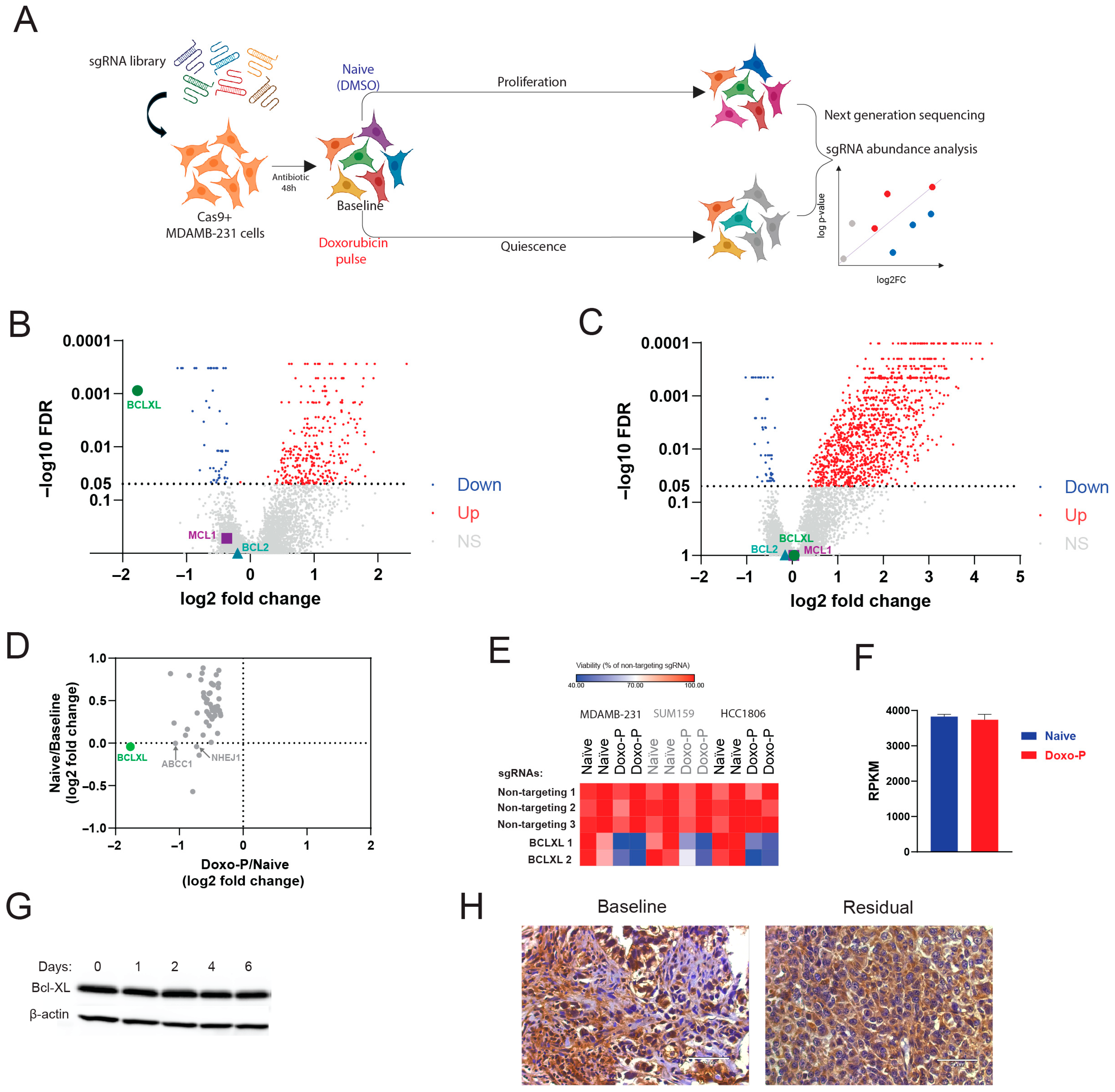
To determine the therapeutic vulnerabilities of the OXPHOS-high TNBC cells through an unbiased approach, we performed genome-wide CRISPR editing screens in proliferative chemo-naïve and quiescent Doxo-P MDAMB231 cells (schematically depicted in Figure 3A). We quantified the depletion of sgRNAs in chemo-naïve cells (vs. baseline) and in Doxo-P cells (vs. chemo-naïve) to identify the genes that are essential for survival in the quiescent persistent state but not in the proliferative state. BCL-XL was the top essential gene for survival in Doxo-P quiescent MDAMB-231 cells (Figure 3B). Remarkably, BCL-XL was not required for proliferation or survival in chemo-naïve cells (Figure 3C). Other notable anti-apoptotic protein genes, such as BCL2 and MCL1, were not essential for survival in Doxo-P cells. The sgRNAs targeting tumor suppressor genes tend to be enriched in chemo-naïve cells (vs. baseline) due to the anti-proliferative function of these genes. Consequently, these sgRNAs against tumor suppressor genes were more abundant in proliferating cells than in quiescent cells at the end of the CRISPR editing experiment and therefore scored as depleted in the proliferatively arrested Doxo-P condition (vs. chemo-naïve). To account for the sgRNAs that score as depleted in Doxo-P (vs. chemo-naïve) due to the quiescent state of Doxo-P cells, we compared the fold changes in all statistically significant (FDR < 0.05) depleted sgRNAs in the Doxo-P condition (vs. chemo-naïve) with the respective fold changes in the chemo-naïve condition (vs. baseline). This comparison confirmed that BCL-XL sgRNA depletion reflected its survival role in Doxo-P cells and not tumor suppressor function in chemo-naive cells (Figure 3D). Additional genes that were essential for survival in Doxo-P cells (but did not affect proliferation of chemo-naïve cells) included membrane drug transporter ABCC1 (also known as multidrug resistance-associated protein 1 [MRP1]), DNA double-strand break repair protein NHEJ1, and the synthetase of detoxifying molecule selenophosphate SEPHS2. The relevance of these additional hits to the mechanism of action of doxorubicin further supports the validity of our CRISPR screen results. The pro-survival function of BCL-XL in quiescent Doxo-P cells was confirmed by single-gene knock-out (KO) experiments in TNBC cell lines MDAMB-231, SUM159, and HC1806 (Figure 3E). Notably, BCL-XL transcript levels were associated with worse outcomes in triple-negative (but not in ER-positive) breast cancer patients, supporting the clinical relevance of our findings (Figure S3). Interestingly, BCL-XL transcript and protein levels were not upregulated in Doxo-P cancer cells (Figure 3F,G). Congruently, qualitative immunohistochemical analysis of paired (pre- and post-treatment) tumor samples from 10 patients treated with anthracycline-based NAC indicated that BCL-XL immunostaining intensity in residual tumors was similar to baseline (representative example shown in Figure 3H).

3.3. Inhibition of BCL-XL Disrupts Mitochondrial Remodeling and OXPHOS Function in Doxo-P TNBC Cells

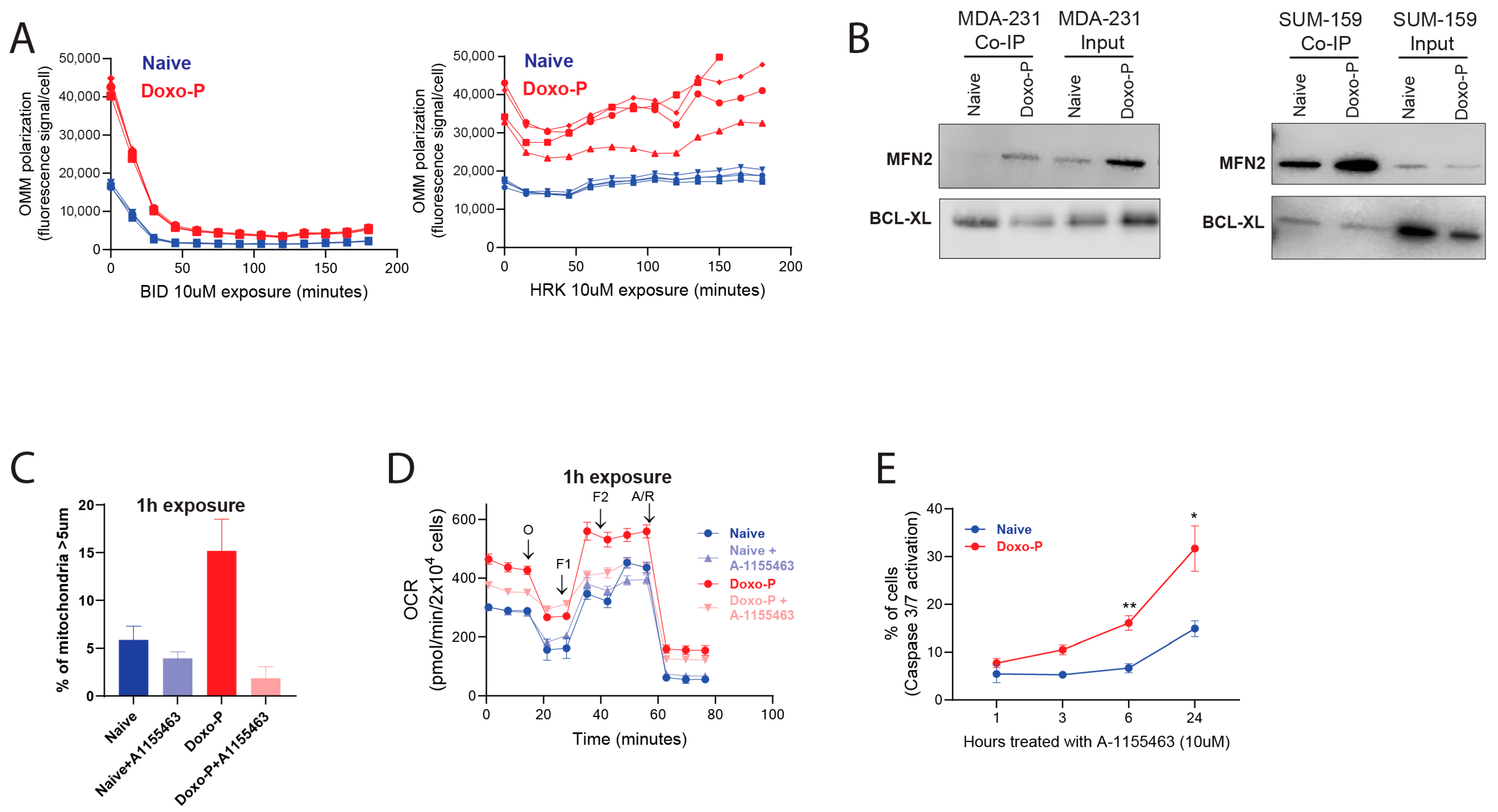
We initially evaluated the apoptotic priming of Doxo-P MDAMB-231 cells using the BH3 profiling method, which is based on longitudinal OMM potential measurement as an indication of OMM permeabilization [46,47,48]. As expected, the OXPHOS-high Doxo-P cells had higher baseline OMM potential compared to their chemo-naïve counterparts. Notably, the Doxo-P cells were sensitive to the apoptotic activator BID (e.g., Figure 4A left), indicating that these persistent quiescent cells are apoptotically competent. However, BCL-XL-specific pro-apoptotic sensitizer peptide HRK did not induce apoptosis in Doxo-P cells (Figure 4A right), indicating that the protective role of BCL-XL may not necessarily be through its canonical role as a negative regulator of pro-apoptotic proteins. Interestingly, previous work in various cell types and biological contexts has demonstrated that BCL-XL can co-localize and interact with mitofusins 1 and/or 2 in the OMM to enable mitochondrial fusion/elongation and apoptosis evasion [49,50,51,52,53]. Concordant with these previous observations, BCL-XL co-immunoprecipitated with MFN2 (but not MFN1) in quiescent Doxo-P (but not in chemo-naïve) MDAMB-231 and SUM-159 TNBC cells (Figure 4B). Importantly, 1 h exposure to the selective BCL-XL inhibitor A-1155463 abrogated co-immunoprecipitation of BCL-XL with MFN2 (Figure S4A), disrupted mitochondrial elongation, and inhibited respiratory function in Doxo-P (but not in chemo-naïve) MDAMB-231 cells, which was followed by caspase activation only after several hours (Figure 4C–E). These data suggest that BCL-XL may be involved in the mitochondrial remodeling and the upregulation of OXPHOS function in Doxo-P cells, ultimately enabling cell survival.

3.4. Inhibition of BCL-XL Abrogates Survival of Doxo-P TNBC Cells

Based on our functional genomics results, we hypothesized that therapeutic agents targeting BCL-XL would kill the quiescent anthracycline-persistent TNBC cells. We initially evaluated BCL-XL as a therapeutic target in chemo-persistent TNBC cells using the well-validated BCL-XL and BCL2 dual inhibitor navitoclax [54]; the BCL-XL-specific inhibitors A1155463 [55] and A1331852 [56]; and the cell line models MDAMB-231, HCC1806, and SUM-159. These inhibitors were highly effective against quiescent OXPHOS-high Doxo-P cells, but not against OXPHOS-low Dtx-P or chemo-naïve cells (Figure 5A–C). Similar results were obtained using the TNBC patient-derived organoid model HCI-002 (Figure 5D) that we developed and characterized in our previous studies [1]. The cytotoxic effect of A1155463 was partially rescued by pan-caspase inhibitor Z-VAD-FMK (Figure S4B), indicating that the cell death of persistent cells treated with the BCL-XL inhibitor is, at least in part, through the apoptotic pathway. Congruent with the CRISPR screen data, the sensitivity of Doxo-P TNBC cells to BCL2 and MCL1 inhibitors was similar to that of chemo-naïve and Dtx-P cells (Figure S4C). Quiescent TNBC cells that persisted after epirubicin or cyclophosphamide treatment were also highly sensitive to BCL-XL inhibitors (Figure S4D), suggesting that the dependency of residual cancer cells on BCL-XL may be generalizable across DNA-damaging chemotherapeutics. Importantly, the cancer cells that exited the Doxo-P quiescent state and resumed proliferation after ~4 weeks in culture had similar BCL-XL inhibitor sensitivity to chemo-naïve cells (Figure 5E), indicating that the BCL-XL dependency of the OXPHOS-high persistent cancer cell state may be transient.
Distinctly from the other TNBC cell models, the quiescent MDAMB-468 TNBC cells that survived after doxorubicin treatment did not have elevated OXPHOS gene expression, mitochondrial elongation, or respiratory function (Figure S5A–C). Notably, Doxo-P MDAMB-468 did not depend on BCL-XL for survival (Figure S5D,E), even though the BCL-XL gene was expressed in both baseline and Doxo-P conditions This observation indicates that the OXPHOS regulation and associated BCL-XL dependency may be heterogeneous among residual Doxo-P TNBC tumors.
Our data suggests that targeting BCL-XL in residual TNBC tumors in combination with anthracyclines can enhance the efficacy of chemotherapy. Existing inhibitors of BCL-XL have potent efficacy against tumor cells but are limited by thrombocytopenia-related adverse effects due to the dependence of platelets on BCL-XL for survival [57,58]. To overcome this on-target and dose-limiting toxicity, we recently developed DT2216, a BCL-XL selective proteolysis-targeting chimera (PROTAC) that targets BCL-XL to the Von Hippel-Lindau (VHL) E3 ligase for ubiquitination and subsequent proteasome-mediated degradation [59,60,61]. Given that VHL is poorly expressed in platelets, DT2216 minimizes platelet loss and is currently being evaluated in phase I/II clinical trials (NCT06620302 and NCT06964009). DT2216 was able to degrade BCL-XL in Doxo-P MDAMB-231 cells, indicating that the proteasome-mediated degradation system is functional in quiescent MYC-suppressed OXPHOS-high cells (Figure 6A). As expected, in vitro Doxo-P cells were highly sensitive to DT2216, compared to chemo-naïve cells (Figure 6B). Combined biweekly treatment of MDAMB-231 xenografts with doxorubicin and DT-2216 (with the first DT2216 dose starting 3 days after the first doxorubicin dose) was superior to either of the two drugs alone (Figure 6C–H). Notably, the combination of doxorubicin plus DT2216 did not cause significant platelet loss compared to untreated mice (Figure 6I), suggesting that this therapeutic combination may avoid the dose-limiting thrombocytopenia observed with other BCL-XL inhibitors.

4. Discussion

Chemotherapy is the first-line standard of care treatment for TNBC, but often fails to eliminate all malignant cells, which can later cause disease relapse. Eradicating the tumor cells that survive the cytotoxic treatment could prevent relapses and prolong remissions. Thus, identifying the mechanisms that enable the survival of persistent cells is critical for the development of more efficient treatment strategies. We and others have shown that tumor cells can survive chemotherapy through a quiescent diapause-like cell state with suppressed global drivers of biosynthesis such as MYC and MTOR [1,2,3]. Here we find that this diapause-like treatment-persistent cancer cell state is not merely a uniform biosynthetic nadir as a consequence of MYC/MTOR suppression, but is actively regulated at transcriptional, metabolic, and structural levels in response to distinct chemotherapeutics. Intriguingly, although the expression of mitochondrial OXPHOS genes in proliferating cells requires MYC function [5], OXPHOS was remarkably upregulated in MYC-suppressed anthracycline-persistent TNBC cells. This observation indicates that despite the strongly suppressed MYC transcriptional program, persistent cells can selectively upregulate the OXPHOS gene set through mechanisms that remain to be discovered. Notably, upregulation of OXPHOS has been linked to drug resistance and persistence in several cancers, including breast [20], pancreatic [24], lung [4], melanoma [25], and leukemia [23,26]. While the upregulation of OXPHOS genes can explain the increased mitochondrial footprint, we also observed that the mitochondria of the Doxo-P cells were elongated and remodeled, confirming similar previous reports [62]. Mitochondrial remodeling could be another mechanism contributing to elevated OXPHOS function in Doxo-P TNBC cells [62]. Distinctly, the TNBC cells that survived treatment with docetaxel did not have upregulated OXPHOS or remodeled mitochondria, indicating that these mitochondrial adaptation mechanisms may not be necessary for persistence to anti-microtubule drugs. These distinct adaptive cell fates upon treatment with anthracyclines and taxanes might have implications regarding the order in which these two drug classes are administered in a chemotherapy regimen.
Accumulating evidence indicates that metabolic rewiring in drug-persistent cancer cells is critical for evading cell death [14,15]. For instance, OXPHOS-high TNBC cells can use glucose or fatty acids as energy sources that contribute to survival [63,64]. Inhibition of OXPHOS, using, e.g., ETC complex I inhibitors, can kill drug-resistant cancer cells in many preclinical cancer models [20,21,23,24,25]. Despite its promise as a therapeutic target, direct inhibition of OXPHOS has shown prohibitory systemic toxicity in the clinic [30,31]. We reason that alternative approaches targeting OXPHOS-high persisting TNBC cells could enable the necessary therapeutic index to specifically kill the chemo-persisting cancer cells while sparing normal tissues. Our genomics and pharmacological approaches identified BCL-XL as a key survival dependency in quiescent MYC-low/OXPHOS-high chemotherapy-persistent cancer cell state, adding to the growing evidence that implicates this protein in TNBC chemotherapy persistence [65]. Interestingly, only the OXPHOS-high Doxo-P, but not the chemo-naïve or OXPHOS-low Dtx-P, TNBC cells were sensitive to BCL-XL ablation or inhibition, indicating a potential link between the OXPHOS-high state and BCL-XL dependency. Although the death of Doxo-P cells treated with BCL-XL inhibitors seems to occur primarily through the apoptotic pathway, the activation of other cell death programs cannot be excluded. The surprising insensitivity of Doxo-P cells to BCL-XL-specific HRK pro-apoptotic peptide turned our attention towards previous reports of a potential role of BCL-XL in mitochondrial remodeling through interactions with mitofusins, which localize in the OMM and are key mediators of mitochondrial fusion [49,50]. Mitochondrial elongation is a complex process that requires coordinated fusion of both inner and outer mitochondrial membranes. For instance, mitochondrial cristae protein OPA1, which mediates inner membrane fusion, is important for the survival of chemotherapy-persistent cancer cells [62]. Our data indicate that BCL-XL interacts with MFN2 and may be involved in OMM fusion, which could explain the rapid mitochondrial fragmentation and collapse of mitochondrial respiration after BCL-XL inhibition in persistent cells. Interestingly, MNF1 and MNF2 did not score as essential for the survival of Doxo-P TNBC cells in the genome-wide CRISPR editing screen, likely reflecting their redundancy for OMM fusion and OXPHOS upregulation in these cells. Our data suggest that BCL-XL-enabled survival of Doxo-P TNBC cells may not necessarily involve its canonical inhibition of pro-apoptotic proteins (e.g., BID/BIM) but could be mediated through a structural role of BCL-XL during mitochondrial aggregation/elongation, which contributes to elevated OXPHOS and thus to cancer cell survival. Overall, these findings add to accumulating evidence suggesting that mitochondrial fusion and elongation contribute to drug resistance in breast and other cancers [22,62] (schematic representation in Figure S6).
MYC suppression is necessary for the induction of the quiescent molecular profile in the chemotherapy-persistent cancer cells [1]. However, the link between MYC-suppressed transcriptional reprogramming and OXPHOS upregulation and BCL-XL dependency remains to be elucidated. The activation of OXPHOS in Doxo-P (but not in Dtx-P cells) may indicate that the repair of cellular damage induced by anthracyclines requires increased amounts of ATP or other metabolic byproducts of mitochondrial respiration. Interestingly, quiescent Dtx-P TNBC cells did not depend on BCL-XL for survival–a finding that seemingly contradicts reports of an anti-apoptotic role of BCL-XL in taxane-treated cells [66]. Crucially, our Dtx-P model reflects a G0/G1 quiescent cell state after drug withdrawal, whereas the anti-apoptotic function of BCL-XL during taxane treatment is exerted during the drug-induced prolonged mitotic arrest and not in non-mitotic cells [66]. Distinctly from anthracycline-persistent cells, the role of BCL-XL during treatment with taxanes suggests that simultaneous (as opposed to sequential) administration of taxanes and a BCL-XL inhibitor may be necessary for the elimination of persistent cells. Additionally, although several Doxo-P TNBC cell lines (MDAMB-231, SUM159, and HCC1806) were OXPHOS-high and dependent on BCL-XL for survival, MDAB-468 Doxo-P cells were OXPHOS-low and were insensitive to BCL-XL ablation or inhibition. Furthermore, Doxo-P cells that exited quiescence lost their sensitivity to BCL-XL inhibitors. These observations highlight the need for predictive tools to assess OXPHOS dependency in residual chemo-persistent tumors, which can be used to identify the patients that would benefit from targeting BCL-XL after chemotherapy, and to correctly time the administration of such treatment.
The management of chemotherapy-induced clinical remission typically involves monitoring for signs of recurrence. Our data support a modification of this clinical practice through administration of a sequential “two-hit” strategy to eliminate residual tumors, with the first treatment (e.g., anthracycline) killing a proportion of tumor cells and inducing transition into OXPHOS-high quiescence of the remaining, followed by a second treatment targeting BCL-XL in residual cells and eliminating them. Despite their strong anti-tumor effect, the efficacy of conventional BXL-XL inhibitors was limited by on-target and dose-limiting platelet toxicity, due to the survival dependency of platelets on BCL-XL. On the other hand, thrombocytopenia is also a common adverse effect of anthracyclines, which would render a combination of these two drug classes highly toxic. Here we show that the BCL-XL-selective PROTAC DT2216 can be safely combined with doxorubicin to enhance its anti-tumor efficacy. Importantly, this combination did not increase thrombocytopenia rates, indicating that this DT2216 will be better tolerated by patients than other BCL-XL inhibitors. Although this study focuses on TNBC, the derived conclusions are likely to apply to other cancer types treated with anthracyclines and other DNA-damaging drugs, therefore potentially impacting a large number of patients.

Supplementary Materials

The following supporting information can be downloaded at https://www.mdpi.com/article/10.3390/cells14191557/s1. Figure S1. Quiescent state of chemotherapy-persistent TNBC cells. Cell cycle analysis of chemo-naïve and chemotherapy-persistent MDAMB-231 cells 3 days after 2 h pulse treatment with indicated drugs. Figure S2. Upregulated mitochondrial respiration in Doxo-P TNBC cells. (A,B) Mitochondrial respiration in chemo-naïve and chemotherapy-persistent HCC1806 (A) and SUM159 (B) TNBC cells measured using the Seahorse analyzer; values normalized to cell number; **** p < 0.0001, *** < 0.001, ** < 0.01, * < 0.05, one-way ANOVA. Figure S3. Prognostic role of BCL-XL in breast cancer. Correlation of BCL-XL expression with Overall Survival in triple-negative and ER-positive breast cancer patients. KM-plotter online survival analysis tool [41] accessed on 31 July 2025. Figure S4. Phenotypic characterization of chemotherapy-persistent MDAMB-231 cells treated with inhibitors of BCL2, MCL1 and BCL-XL. (A) Immunoprecipitation of BCL-XL in Doxo-P TNBC cells treated with A-1155463 (10 µM, 1 h exposure) and analysis by western blot using antibodies against MFN2 and BCL-XL. (B) Viability of chemo-naïve and Doxo-P MDAMB-231 cells after co-treatment with BCL-XL inhibitor A-1155463 (5 µM) and pan-caspase inhibitor Z-VAD-FMK (20 µM) for 24 h. (C) Sensitivity of chemo-naïve, Doxo-P and Dtx-P MDAMB-231 cells to BCL2 inhibitor venetoclax and MCL1 inhibitor S63845. (D) Sensitivity of chemo-naïve, epirubicin-persistent (Epi-P) and cyclophosphamide-persistent (Cyclo-P) MDAMB-231 cells to BCL-XL inhibitor A1155463. Figure S5. Doxo-P MDAMB-468 cells have low OXPHOS levels and do not depend on BCL-XL for survival. (A) Mitochondrial respiration in chemo-naïve and chemotherapy-persistent MDAMB-468 cells measured using the Seahorse analyzer; values normalized to cell number. (B) GSEA analysis of transcriptional changes in MDAMB-468 cells surviving treatment with doxorubicin (Doxo-P) or docetaxel (Dtx-P), compared to chemo-naïve counterparts. (C) OMM polarization levels in individual cells from chemo-naïve and Doxo-P MDAMB-468 cells, measured using the JC-1 staining assay. (D) Viability of chemo-naïve and Doxo-P MDAMB-468 after BCL-XL gene knockout. (E) Sensitivity of chemo-naïve, Doxo-P and Dtx-P MDAMB-468 cells to BCL-XL inhibitor A1155463. Figure S6. Schematic representation of chemotherapy persistence model.

Author Contributions

Conceptualization: E.D.; methodology: E.D., S.A., M.W., L.E.G., O.A., F.M.M., E.Z., A.S., J.A.-G., M.K., G.Z., S.A.F., D.Z., E.G., and T.W.; investigation: E.D., S.A., M.W., L.E.G., O.A., S.A.F., and T.W.; visualization: E.D., S.A., M.W., and L.E.G.; funding acquisition: E.D.; project administration: E.D.; supervision: E.D.; writing—original draft: E.D.; writing—review and editing: all authors. All authors have read and agreed to the published version of the manuscript.

Funding

This work was supported by the Montefiore Einstein Comprehensive Cancer Center Pilot Grant on Molecular Variations as Determinants of Outcomes (E.D.), Montefiore Einstein Cancer Dormancy Institute Developmental Fund Award (E.D., E.G.), Montefiore Einstein Comprehensive Cancer Center grant P30CA013330, US National Institutes of Health (NIH) grants R21CA267539 (E.D.), NCI R01CA178394 (E.G.) and R01CA260239 (G.Z. and D.Z.).

Institutional Review Board Statement

The animal study protocol was approved by the Institute of Animal Studies of the Albert Einstein College of Medicine (protocol code 00001590 and 8 April 2025 of approval).

Data Availability Statement

The raw data supporting the conclusions of this article will be made available by the authors on request.

Acknowledgments

We thank Dialectic Therapeutics for providing DT2216 for the study.

Conflicts of Interest

G.Z. and D.Z. are inventors on patents for the use of BCL-XL PROTACs as antitumor agents and are cofounders of and have equity in Dialectic Therapeutics, which develops BCL-XL/2 PROTACs to treat cancer. J.A.-G. is a co-founder, advisory board member, and equity holder in HiberCell, a Mount Sinai spin-off developing cancer recurrence prevention therapies. He consults for HiberCell and Astrin Biosciences and serves as Chief Mission Advisor for the Samuel Waxman Cancer Research Foundation, and he has an ownership interest in patent number WO2019191115A1/EP-3775171-B1. The remaining authors declare no relevant competing interests.

References

  1. Dhimolea, E.; de Matos Simoes, R.; Kansara, D.; Al’Khafaji, A.; Bouyssou, J.; Weng, X.; Sharma, S.; Raja, J.; Awate, P.; Shirasaki, R.; et al. An Embryonic Diapause-like Adaptation with Suppressed Myc Activity Enables Tumor Treatment Persistence. Cancer Cell 2021, 39, 240–256.e11. [Google Scholar] [CrossRef]
  2. Duy, C.; Li, M.; Teater, M.; Meydan, C.; Garrett-Bakelman, F.E.; Lee, T.C.; Chin, C.R.; Durmaz, C.; Kawabata, K.C.; Dhimolea, E.; et al. Chemotherapy induces senescence-like resilient cells capable of initiating AML recurrence. Cancer Discov. 2021, 11, 1542–1561. [Google Scholar] [CrossRef]
  3. Rehman, S.K.; Haynes, J.; Collignon, E.; Brown, K.R.; Wang, Y.; Nixon, A.M.L.; Bruce, J.P.; Wintersinger, J.A.; Singh Mer, A.; Lo, E.B.L.; et al. Colorectal Cancer Cells Enter a Diapause-like DTP State to Survive Chemotherapy. Cell 2021, 184, 226–242.e221. [Google Scholar] [CrossRef] [PubMed]
  4. Oren, Y.; Tsabar, M.; Cuoco, M.S.; Amir-Zilberstein, L.; Cabanos, H.F.; Hütter, J.C.; Hu, B.; Thakore, P.I.; Tabaka, M.; Fulco, C.P.; et al. Cycling cancer persister cells arise from lineages with distinct programs. Nature 2021, 596, 576–582. [Google Scholar] [CrossRef] [PubMed]
  5. Lin, C.Y.; Lovén, J.; Rahl, P.B.; Paranal, R.M.; Burge, C.B.; Bradner, J.E.; Lee, T.I.; Young, R.A. Transcriptional amplification in tumor cells with elevated c-Myc. Cell 2012, 151, 56–67. [Google Scholar] [CrossRef] [PubMed]
  6. Fenelon, J.C.; Banerjee, A.; Murphy, B.D. Embryonic diapause: Development on hold. Int. J. Dev. Biol. 2014, 58, 163–174. [Google Scholar] [CrossRef]
  7. Scognamiglio, R.; Cabezas-Wallscheid, N.; Thier, M.C.; Altamura, S.; Reyes, A.; Prendergast, A.M.; Baumgartner, D.; Carnevalli, L.S.; Atzberger, A.; Haas, S.; et al. Myc Depletion Induces a Pluripotent Dormant State Mimicking Diapause. Cell 2016, 164, 668–680. [Google Scholar] [CrossRef]
  8. Marra, A.; Curigliano, G. Adjuvant and Neoadjuvant Treatment of Triple-Negative Breast Cancer with Chemotherapy. Cancer J. 2021, 27, 41–49. [Google Scholar] [CrossRef]
  9. Wallace, K.B.; Sardão, V.A.; Oliveira, P.J. Mitochondrial Determinants of Doxorubicin-Induced Cardiomyopathy. Circ. Res. 2020, 126, 926–941. [Google Scholar] [CrossRef]
  10. Nesic, K.; Parker, P.; Swisher, E.M.; Krais, J.J. DNA repair and the contribution to chemotherapy resistance. Genome Med. 2025, 17, 62. [Google Scholar] [CrossRef]
  11. Scribano, C.M.; Wan, J.; Esbona, K.; Tucker, J.B.; Lasek, A.; Zhou, A.S.; Zasadil, L.M.; Molini, R.; Fitzgerald, J.; Lager, A.M.; et al. Chromosomal instability sensitizes patient breast tumors to multipolar divisions induced by paclitaxel. Sci. Transl. Med. 2021, 13, eabd4811. [Google Scholar] [CrossRef] [PubMed]
  12. Brito, D.A.; Rieder, C.L. Mitotic checkpoint slippage in humans occurs via cyclin B destruction in the presence of an active checkpoint. Curr. Biol. 2006, 16, 1194–1200. [Google Scholar] [CrossRef]
  13. Rieder, C.L.; Maiato, H. Stuck in division or passing through: What happens when cells cannot satisfy the spindle assembly checkpoint. Dev. Cell 2004, 7, 637–651. [Google Scholar] [CrossRef] [PubMed]
  14. Shen, S.; Vagner, S.; Robert, C. Persistent Cancer Cells: The Deadly Survivors. Cell 2020, 183, 860–874. [Google Scholar] [CrossRef]
  15. Russo, M.; Chen, M.; Mariella, E.; Peng, H.; Rehman, S.K.; Sancho, E.; Sogari, A.; Toh, T.S.; Balaban, N.Q.; Batlle, E.; et al. Cancer drug-tolerant persister cells: From biological questions to clinical opportunities. Nat. Rev. Cancer 2024, 24, 694–717. [Google Scholar] [CrossRef]
  16. Raha, D.; Wilson, T.R.; Peng, J.; Peterson, D.; Yue, P.; Evangelista, M.; Wilson, C.; Merchant, M.; Settleman, J. The cancer stem cell marker aldehyde dehydrogenase is required to maintain a drug-tolerant tumor cell subpopulation. Cancer Res. 2014, 74, 3579–3590. [Google Scholar] [CrossRef]
  17. Lu, Y.; Travnickova, J.; Badonyi, M.; Rambow, F.; Coates, A.; Khan, Z.; Marques, J.; Murphy, L.C.; Garcia-Martinez, P.; Marais, R.; et al. ALDH1A3-acetaldehyde metabolism potentiates transcriptional heterogeneity in melanoma. Cell Rep. 2024, 43, 114406. [Google Scholar] [CrossRef]
  18. Hangauer, M.J.; Viswanathan, V.S.; Ryan, M.J.; Bole, D.; Eaton, J.K.; Matov, A.; Galeas, J.; Dhruv, H.D.; Berens, M.E.; Schreiber, S.L.; et al. Drug-tolerant persister cancer cells are vulnerable to GPX4 inhibition. Nature 2017, 551, 247–250. [Google Scholar] [CrossRef]
  19. Viswanathan, V.S.; Ryan, M.J.; Dhruv, H.D.; Gill, S.; Eichhoff, O.M.; Seashore-Ludlow, B.; Kaffenberger, S.D.; Eaton, J.K.; Shimada, K.; Aguirre, A.J.; et al. Dependency of a therapy-resistant state of cancer cells on a lipid peroxidase pathway. Nature 2017, 547, 453–457. [Google Scholar] [CrossRef] [PubMed]
  20. Echeverria, G.V.; Ge, Z.; Seth, S.; Zhang, X.; Jeter-Jones, S.; Zhou, X.; Cai, S.; Tu, Y.; McCoy, A.; Peoples, M.; et al. Resistance to neoadjuvant chemotherapy in triple-negative breast cancer mediated by a reversible drug-tolerant state. Sci. Transl. Med. 2019, 11, eaav0936. [Google Scholar] [CrossRef] [PubMed]
  21. Molina, J.R.; Sun, Y.; Protopopova, M.; Gera, S.; Bandi, M.; Bristow, C.; McAfoos, T.; Morlacchi, P.; Ackroyd, J.; Agip, A.A.; et al. An inhibitor of oxidative phosphorylation exploits cancer vulnerability. Nat. Med. 2018, 24, 1036–1046. [Google Scholar] [CrossRef]
  22. Chen, X.; Glytsou, C.; Zhou, H.; Narang, S.; Reyna, D.E.; Lopez, A.; Sakellaropoulos, T.; Gong, Y.; Kloetgen, A.; Yap, Y.S.; et al. Targeting Mitochondrial Structure Sensitizes Acute Myeloid Leukemia to Venetoclax Treatment. Cancer Discov. 2019, 9, 890–909. [Google Scholar] [CrossRef]
  23. Kuntz, E.M.; Baquero, P.; Michie, A.M.; Dunn, K.; Tardito, S.; Holyoake, T.L.; Helgason, G.V.; Gottlieb, E. Targeting mitochondrial oxidative phosphorylation eradicates therapy-resistant chronic myeloid leukemia stem cells. Nat. Med. 2017, 23, 1234–1240. [Google Scholar] [CrossRef]
  24. Viale, A.; Pettazzoni, P.; Lyssiotis, C.A.; Ying, H.; Sánchez, N.; Marchesini, M.; Carugo, A.; Green, T.; Seth, S.; Giuliani, V.; et al. Oncogene ablation-resistant pancreatic cancer cells depend on mitochondrial function. Nature 2014, 514, 628–632. [Google Scholar] [CrossRef] [PubMed]
  25. Haq, R.; Shoag, J.; Andreu-Perez, P.; Yokoyama, S.; Edelman, H.; Rowe, G.C.; Frederick, D.T.; Hurley, A.D.; Nellore, A.; Kung, A.L.; et al. Oncogenic BRAF regulates oxidative metabolism via PGC1α and MITF. Cancer Cell 2013, 23, 302–315. [Google Scholar] [CrossRef] [PubMed]
  26. Farge, T.; Saland, E.; de Toni, F.; Aroua, N.; Hosseini, M.; Perry, R.; Bosc, C.; Sugita, M.; Stuani, L.; Fraisse, M.; et al. Chemotherapy-Resistant Human Acute Myeloid Leukemia Cells Are Not Enriched for Leukemic Stem Cells but Require Oxidative Metabolism. Cancer Discov. 2017, 7, 716–735. [Google Scholar] [CrossRef] [PubMed]
  27. Shen, S.; Faouzi, S.; Souquere, S.; Roy, S.; Routier, E.; Libenciuc, C.; André, F.; Pierron, G.; Scoazec, J.Y.; Robert, C. Melanoma Persister Cells Are Tolerant to BRAF/MEK Inhibitors via ACOX1-Mediated Fatty Acid Oxidation. Cell Rep. 2020, 33, 108421. [Google Scholar] [CrossRef]
  28. Stevens, B.M.; Jones, C.L.; Pollyea, D.A.; Culp-Hill, R.; D’Alessandro, A.; Winters, A.; Krug, A.; Abbott, D.; Goosman, M.; Pei, S.; et al. Fatty acid metabolism underlies venetoclax resistance in acute myeloid leukemia stem cells. Nat. Cancer 2020, 1, 1176–1187. [Google Scholar] [CrossRef] [PubMed]
  29. Baccelli, I.; Gareau, Y.; Lehnertz, B.; Gingras, S.; Spinella, J.F.; Corneau, S.; Mayotte, N.; Girard, S.; Frechette, M.; Blouin-Chagnon, V.; et al. Mubritinib Targets the Electron Transport Chain Complex I and Reveals the Landscape of OXPHOS Dependency in Acute Myeloid Leukemia. Cancer Cell 2019, 36, 84–99.e88. [Google Scholar] [CrossRef]
  30. Yap, T.A.; Daver, N.; Mahendra, M.; Zhang, J.; Kamiya-Matsuoka, C.; Meric-Bernstam, F.; Kantarjian, H.M.; Ravandi, F.; Collins, M.E.; Francesco, M.E.D.; et al. Complex I inhibitor of oxidative phosphorylation in advanced solid tumors and acute myeloid leukemia: Phase I trials. Nat. Med. 2023, 29, 115–126. [Google Scholar] [CrossRef]
  31. Machado, N.D.; Heather, L.C.; Harris, A.L.; Higgins, G.S. Targeting mitochondrial oxidative phosphorylation: Lessons, advantages, and opportunities. Br. J. Cancer 2023, 129, 897–899. [Google Scholar] [CrossRef]
  32. Dobin, A.; Davis, C.A.; Schlesinger, F.; Drenkow, J.; Zaleski, C.; Jha, S.; Batut, P.; Chaisson, M.; Gingeras, T.R. STAR: Ultrafast universal RNA-seq aligner. Bioinformatics 2013, 29, 15–21. [Google Scholar] [CrossRef]
  33. Liao, Y.; Smyth, G.K.; Shi, W. featureCounts: An efficient general purpose program for assigning sequence reads to genomic features. Bioinformatics 2014, 30, 923–930. [Google Scholar] [CrossRef]
  34. Love, M.I.; Huber, W.; Anders, S. Moderated estimation of fold change and dispersion for RNA-seq data with DESeq2. Genome Biol. 2014, 15, 550. [Google Scholar] [CrossRef] [PubMed]
  35. Dhimolea, E.; de Matos Simoes, R.; Kansara, D.; Weng, X.; Sharma, S.; Awate, P.; Liu, Z.; Gao, D.; Mitsiades, N.; Schwab, J.H.; et al. Pleiotropic Mechanisms Drive Endocrine Resistance in the Three-Dimensional Bone Microenvironment. Cancer Res. 2021, 81, 371–383. [Google Scholar] [CrossRef] [PubMed]
  36. Li, W.; Xu, H.; Xiao, T.; Cong, L.; Love, M.I.; Zhang, F.; Irizarry, R.A.; Liu, J.S.; Brown, M.; Liu, X.S. MAGeCK enables robust identification of essential genes from genome-scale CRISPR/Cas9 knockout screens. Genome Biol. 2014, 15, 554. [Google Scholar] [CrossRef] [PubMed]
  37. Lopez, A.; Reyna, D.E.; Gitego, N.; Kopp, F.; Zhou, H.; Miranda-Roman, M.A.; Nordstrøm, L.U.; Narayanagari, S.R.; Chi, P.; Vilar, E.; et al. Co-targeting of BAX and BCL-XL proteins broadly overcomes resistance to apoptosis in cancer. Nat. Commun. 2022, 13, 1199. [Google Scholar] [CrossRef]
  38. Valente, A.J.; Maddalena, L.A.; Robb, E.L.; Moradi, F.; Stuart, J.A. A simple ImageJ macro tool for analyzing mitochondrial network morphology in mammalian cell culture. Acta Histochem. 2017, 119, 315–326. [Google Scholar] [CrossRef]
  39. Steger, C. An unbiased detector of curvilinear structures. IEEE Trans. Pattern Anal. Mach. Intell. 1998, 20, 113–125. [Google Scholar] [CrossRef]
  40. Winter, M.; Nait Eldjoudi, A.; Guette, C.; Hondermarck, H.; Bourette, R.P.; Fovez, Q.; Laine, W.; Ghesquiere, B.; Adriaenssens, E.; Kluza, J.; et al. Mitochondrial adaptation decreases drug sensitivity of persistent triple negative breast cancer cells surviving combinatory and sequential chemotherapy. Neoplasia 2023, 46, 100949. [Google Scholar] [CrossRef] [PubMed]
  41. Győrffy, B. Survival analysis across the entire transcriptome identifies biomarkers with the highest prognostic power in breast cancer. Comput. Struct. Biotechnol. J. 2021, 19, 4101–4109. [Google Scholar] [CrossRef]
  42. Kimbung, S.; Markholm, I.; Bjohle, J.; Lekberg, T.; von Wachenfeldt, A.; Azavedo, E.; Saracco, A.; Hellstrom, M.; Veerla, S.; Paquet, E.; et al. Assessment of early response biomarkers in relation to long-term survival in patients with HER2-negative breast cancer receiving neoadjuvant chemotherapy plus bevacizumab: Results from the Phase II PROMIX trial. Int. J. Cancer 2018, 142, 618–628. [Google Scholar] [CrossRef] [PubMed]
  43. Sugioka, R.; Shimizu, S.; Tsujimoto, Y. Fzo1, a protein involved in mitochondrial fusion, inhibits apoptosis. J. Biol. Chem. 2004, 279, 52726–52734. [Google Scholar] [CrossRef] [PubMed]
  44. Neuspiel, M.; Zunino, R.; Gangaraju, S.; Rippstein, P.; McBride, H. Activated mitofusin 2 signals mitochondrial fusion, interferes with Bax activation, and reduces susceptibility to radical induced depolarization. J. Biol. Chem. 2005, 280, 25060–25070. [Google Scholar] [CrossRef]
  45. Sivandzade, F.; Bhalerao, A.; Cucullo, L. Analysis of the Mitochondrial Membrane Potential Using the Cationic JC-1 Dye as a Sensitive Fluorescent Probe. Bio-protocol 2019, 9, e3128. [Google Scholar] [CrossRef]
  46. Certo, M.; Del Gaizo Moore, V.; Nishino, M.; Wei, G.; Korsmeyer, S.; Armstrong, S.A.; Letai, A. Mitochondria primed by death signals determine cellular addiction to antiapoptotic BCL-2 family members. Cancer Cell 2006, 9, 351–365. [Google Scholar] [CrossRef] [PubMed]
  47. Vo, T.T.; Ryan, J.; Carrasco, R.; Neuberg, D.; Rossi, D.J.; Stone, R.M.; Deangelo, D.J.; Frattini, M.G.; Letai, A. Relative mitochondrial priming of myeloblasts and normal HSCs determines chemotherapeutic success in AML. Cell 2012, 151, 344–355. [Google Scholar] [CrossRef]
  48. Ni Chonghaile, T.; Sarosiek, K.A.; Vo, T.T.; Ryan, J.A.; Tammareddi, A.; Moore Vdel, G.; Deng, J.; Anderson, K.C.; Richardson, P.; Tai, Y.T.; et al. Pretreatment mitochondrial priming correlates with clinical response to cytotoxic chemotherapy. Science 2011, 334, 1129–1133. [Google Scholar] [CrossRef]
  49. Du, M.; Yu, S.; Su, W.; Zhao, M.; Yang, F.; Liu, Y.; Mai, Z.; Wang, Y.; Wang, X.; Chen, T. Mitofusin 2 but not mitofusin 1 mediates Bcl-XL-induced mitochondrial aggregation. J. Cell Sci. 2020, 133, jcs245001. [Google Scholar] [CrossRef]
  50. Berman, S.B.; Chen, Y.B.; Qi, B.; McCaffery, J.M.; Rucker, E.B., 3rd; Goebbels, S.; Nave, K.A.; Arnold, B.A.; Jonas, E.A.; Pineda, F.J.; et al. Bcl-x L increases mitochondrial fission, fusion, and biomass in neurons. J. Cell Biol. 2009, 184, 707–719. [Google Scholar] [CrossRef]
  51. Hoppins, S.; Edlich, F.; Cleland, M.M.; Banerjee, S.; McCaffery, J.M.; Youle, R.J.; Nunnari, J. The soluble form of Bax regulates mitochondrial fusion via MFN2 homotypic complexes. Mol. Cell 2011, 41, 150–160. [Google Scholar] [CrossRef] [PubMed]
  52. Delivani, P.; Adrain, C.; Taylor, R.C.; Duriez, P.J.; Martin, S.J. Role for CED-9 and Egl-1 as regulators of mitochondrial fission and fusion dynamics. Mol. Cell 2006, 21, 761–773. [Google Scholar] [CrossRef]
  53. Hardwick, J.M.; Chen, Y.B.; Jonas, E.A. Multipolar functions of BCL-2 proteins link energetics to apoptosis. Trends Cell Biol. 2012, 22, 318–328. [Google Scholar] [CrossRef] [PubMed]
  54. Oltersdorf, T.; Elmore, S.W.; Shoemaker, A.R.; Armstrong, R.C.; Augeri, D.J.; Belli, B.A.; Bruncko, M.; Deckwerth, T.L.; Dinges, J.; Hajduk, P.J.; et al. An inhibitor of Bcl-2 family proteins induces regression of solid tumours. Nature 2005, 435, 677–681. [Google Scholar] [CrossRef] [PubMed]
  55. Tao, Z.F.; Hasvold, L.; Wang, L.; Wang, X.; Petros, A.M.; Park, C.H.; Boghaert, E.R.; Catron, N.D.; Chen, J.; Colman, P.M.; et al. Discovery of a Potent and Selective BCL-XL Inhibitor with in Vivo Activity. ACS Med. Chem. Lett. 2014, 5, 1088–1093. [Google Scholar] [CrossRef]
  56. Wang, L.; Doherty, G.A.; Judd, A.S.; Tao, Z.F.; Hansen, T.M.; Frey, R.R.; Song, X.; Bruncko, M.; Kunzer, A.R.; Wang, X.; et al. Discovery of A-1331852, a First-in-Class, Potent, and Orally-Bioavailable BCL-X(L) Inhibitor. ACS Med. Chem. Lett. 2020, 11, 1829–1836. [Google Scholar] [CrossRef]
  57. Zhang, H.; Nimmer, P.M.; Tahir, S.K.; Chen, J.; Fryer, R.M.; Hahn, K.R.; Iciek, L.A.; Morgan, S.J.; Nasarre, M.C.; Nelson, R.; et al. Bcl-2 family proteins are essential for platelet survival. Cell Death Differ. 2007, 14, 943–951. [Google Scholar] [CrossRef]
  58. Mason, K.D.; Carpinelli, M.R.; Fletcher, J.I.; Collinge, J.E.; Hilton, A.A.; Ellis, S.; Kelly, P.N.; Ekert, P.G.; Metcalf, D.; Roberts, A.W.; et al. Programmed anuclear cell death delimits platelet life span. Cell 2007, 128, 1173–1186. [Google Scholar] [CrossRef]
  59. Khan, S.; Zhang, X.; Lv, D.; Zhang, Q.; He, Y.; Zhang, P.; Liu, X.; Thummuri, D.; Yuan, Y.; Wiegand, J.S.; et al. A selective BCL-X(L) PROTAC degrader achieves safe and potent antitumor activity. Nat. Med. 2019, 25, 1938–1947. [Google Scholar] [CrossRef]
  60. He, Y.; Koch, R.; Budamagunta, V.; Zhang, P.; Zhang, X.; Khan, S.; Thummuri, D.; Ortiz, Y.T.; Zhang, X.; Lv, D.; et al. DT2216-a Bcl-xL-specific degrader is highly active against Bcl-xL-dependent T cell lymphomas. J. Hematol. Oncol. 2020, 13, 95. [Google Scholar] [CrossRef]
  61. Zhang, X.; Thummuri, D.; Liu, X.; Hu, W.; Zhang, P.; Khan, S.; Yuan, Y.; Zhou, D.; Zheng, G. Discovery of PROTAC BCL-X(L) degraders as potent anticancer agents with low on-target platelet toxicity. Eur. J. Med. Chem. 2020, 192, 112186. [Google Scholar] [CrossRef] [PubMed]
  62. Baek, M.L.; Lee, J.; Pendleton, K.E.; Berner, M.J.; Goff, E.B.; Tan, L.; Martinez, S.A.; Mahmud, I.; Wang, T.; Meyer, M.D.; et al. Mitochondrial structure and function adaptation in residual triple negative breast cancer cells surviving chemotherapy treatment. Oncogene 2023, 42, 1117–1131. [Google Scholar] [CrossRef] [PubMed]
  63. Park, J.H.; Vithayathil, S.; Kumar, S.; Sung, P.L.; Dobrolecki, L.E.; Putluri, V.; Bhat, V.B.; Bhowmik, S.K.; Gupta, V.; Arora, K.; et al. Fatty Acid Oxidation-Driven Src Links Mitochondrial Energy Reprogramming and Oncogenic Properties in Triple-Negative Breast Cancer. Cell Rep. 2016, 14, 2154–2165. [Google Scholar] [CrossRef] [PubMed]
  64. Camarda, R.; Zhou, A.Y.; Kohnz, R.A.; Balakrishnan, S.; Mahieu, C.; Anderton, B.; Eyob, H.; Kajimura, S.; Tward, A.; Krings, G.; et al. Inhibition of fatty acid oxidation as a therapy for MYC-overexpressing triple-negative breast cancer. Nat. Med. 2016, 22, 427–432. [Google Scholar] [CrossRef]
  65. Saleh, T.; Carpenter, V.J.; Tyutyunyk-Massey, L.; Murray, G.; Leverson, J.D.; Souers, A.J.; Alotaibi, M.R.; Faber, A.C.; Reed, J.; Harada, H.; et al. Clearance of therapy-induced senescent tumor cells by the senolytic ABT-263 via interference with BCL-X(L) -BAX interaction. Mol. Oncol. 2020, 14, 2504–2519. [Google Scholar] [CrossRef]
  66. Qin, X.; Presser, A.; Johnson, L.; Matoba, Y.; Shay, B.S.; Xu, W.; Choiniere, J.; Fraser, C.; Garbicz, F.; Spetz, J.; et al. Paclitaxel-induced mitotic arrest results in a convergence of apoptotic dependencies that can be safely exploited by BCL-X(L) degradation to overcome cancer chemoresistance. bioRxiv 2025. [Google Scholar] [CrossRef]
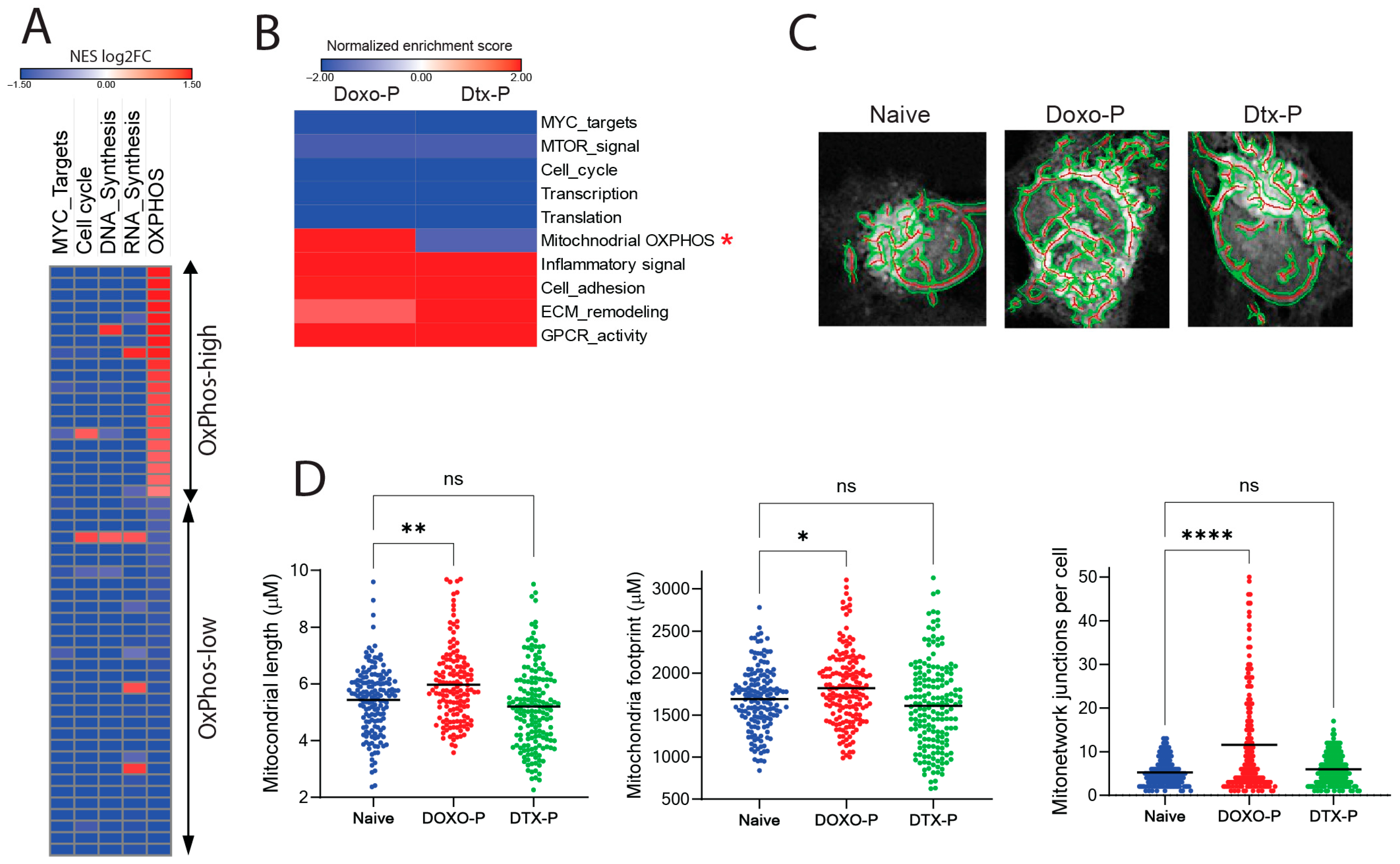
Figure 1. Altered mitochondrial biogenesis and structure in chemotherapy-persistent TNBC cells: (A) Bulk RNA-seq gene set enrichment analysis (GSEA) of quiescent residual vs. baseline breast tumor cells after chemotherapy in the PROMIX trial patient dataset (GSE87455; each row represents a patient). (B) GSEA of transcriptional changes in MDAMB-231 cells surviving treatment with doxorubicin (Doxo-P) or docetaxel (DTX-P) compared to chemo-naïve counterparts. OXPHOS transcriptional module is marked with an asterisk. (C) Mitochondrial network in individual chemo-naïve, Doxo-P, and Dtx-P MDAMB-231 cells visualized using the Mito Tracker dye. Red depicts central mitochnorial axis and green depicts mitochnodrial outline. (D) Quantification of average mitochondrial length, footprint, and network in individual chemo-naïve, Doxo-P, or Dtx-P MDAMB-231 cells (**** p < 0.0001, ** <0.01, * <0.05, ns = non-significant, one-way ANOVA).
Cells 14 01557 g001
Figure 2. Upregulated mitochondrial respiration in Doxo-P TNBC cells: (A) Transcriptional changes (RNA sequencing) in electron transport chain genes in Doxo-P MDAMB-231 cells (compared to chemo-naive). Upregulated genes depicted in red; downregulated in green. (B) OMM polarization levels in individual cells from chemo-naïve and chemotherapy-persistent MDAMB-231 cells, measured using the JC-1 staining assay [45] (one-way ANOVA). (C) Mitochondrial respiration in chemo-naïve and chemotherapy-persistent MDAMB-231 cells measured using the Seahorse analyzer; values normalized to cell number (**** p < 0.0001, *** <0.001, ns = non-significant, one-way ANOVA). (D) Caspase 3/7 activation in chemo-naïve and chemotherapy-persistent MDAMB-231 cells treated with OXPHOS inhibitor IACS-10759 (1 µM); * <0.05, mixed effects analysis.
Cells 14 01557 g002
Figure 3. Doxo-P cancer cells depend on BCL-XL for survival: (A) Schematic diagram depicting the genome-wide CRISPR-editing screen in chemo-naïve and Doxo-P MDAMB-231 cells. (B,C) Changes in sgRNA abundance in Doxo-P (B) and chemo-naïve (C) MDAMB-231 cells. Analysis performed using the MAGECK pipeline [36]. Genes with significant (FDR < 0.05) sgRNA depletion are highlighted in blue or red. Dashed line at Y value 0.05; ns = non-significant. (D) SgRNA fold changes for significantly depleted genes in the Doxo-P vs. chemo-naïve analysis and the respective changes in the chemo-naïve vs. baseline analysis. (E) Viability of chemo-naïve and Doxo-P MDAMB-231, SUM159, and HCC1806 TNBC cells after BCL-XL gene knockout. (F) BCL-XL transcript levels in chemo-naïve and Doxo-P MDAMB-231 cells. (G) BCL-XL protein levels in MDAMB-231 after treatment with doxorubicin. (H) BCL-XL protein immunostaining in baseline and post-NAC residual tumor from a TNBC patient (magnification 200×).
Cells 14 01557 g003
Figure 4. Inhibition of BCL-XL abrogates mitochondrial elongation and respiratory function in Doxo-P TNBC cells: (A) The effect of BID and HRK on OMM polarization level in chemo-naïve and Doxo-P MDAMB-231 cells; BH3 profiling assay [48]. (B) Immunoprecipitation of BCL-XL in chemo-naïve and Doxo-P TNBC cells and analysis by Western blot using antibodies against MFN2 and BCL-XL. (CE) Effects of BCL-XL inhibitor A-1155463 (10 µM, 1 h exposure) on mitochondrial length (C), respiratory function measured using Seahorse analyzer (D), and apoptosis (E) in chemo-naïve and Doxo-P MDAMB-231 cells.(** p < 0.01, * <0.05, one-way ANOVA).
Cells 14 01557 g004
Figure 5. Doxo-P TNBC cells are sensitive to inhibitors of BCL-XL: (AD) Sensitivity of chemo-naïve, Doxo-P, and Dtx-P TNBC cell lines MDAMB-231 (A), HCC1806 (B), SUM159 (C), and TNBC patient-derived organoid line HCI002 (D) to BCL-XL inhibitors navitoclax, A-1155463, and A-1331852. (E) Sensitivity of chemo-naïve, Doxo-P, and regrown (3–4 weeks post doxorubicin washout) MDAMB-231 cells to Bcl-XL inhibitor A-1155463.
Cells 14 01557 g005
Figure 6. TNBC tumors are sensitive to combination treatment of doxorubicin and DT2216: (A) DT2216-induced degradation of BCL-XL protein in Doxo-P MDAMB-231 cells. (B) In vitro sensitivity of chemo-naïve and Doxo-P MDAMB-231 cells to DT2216. (C) Schematic diagram of animal experiment. (DH) Response of MDAMB-231 xenografts to combination treatment (twice weekly, IP) of doxorubicin (3 mg/kg) and DT2216 (15 mg/kg) or each drug alone, estimated by longitudinal measurements of tumor size (D), as well as tumor weight (E,F) and in vivo bioluminescence (GH) after the last drug administration; two-way ANOVA. (I) Platelet levels in all treatment groups after the last drug administration; two-way ANOVA. **** p < 0.0001, ** <0.01, * <0.05, ns = non-significant.
Cells 14 01557 g006
Disclaimer/Publisher’s Note: The statements, opinions and data contained in all publications are solely those of the individual author(s) and contributor(s) and not of MDPI and/or the editor(s). MDPI and/or the editor(s) disclaim responsibility for any injury to people or property resulting from any ideas, methods, instructions or products referred to in the content.