Review

Maternal Thymus Adaptations and Hormone Regulation During Pregnancy

School of Life Sciences and Food Engineering, Hebei University of Engineering, Handan 056038, China
*
Author to whom correspondence should be addressed.
Cells 2025, 14(19), 1534; https://doi.org/10.3390/cells14191534
Submission received: 8 September 2025 / Revised: 28 September 2025 / Accepted: 29 September 2025 / Published: 30 September 2025
(This article belongs to the Special Issue Cellular Mechanisms in Pregnancy and Foetal Development)

Abstract

The thymus is necessary for the development of T lymphocytes and central tolerance, and adaptations in the maternal thymus are required during pregnancy. In the present paper, maternal thymic cellular anatomy, T-cell development in the thymus, and related progress are reviewed. In addition, the recent progress in maternal thymic adaptations during pregnancy is discussed, including adaptations in thymic cellular anatomy, T-cell development, and immune-related cytokines. Finally, the latest information about hormones that regulate thymic immunology during pregnancy is summarized. In summary, there are many factors, including a lot of hormones, which are involved in maternal thymic immunological adaptations during pregnancy, and may be used to prevent pregnancy-related thymic diseases and preterm birth.

Graphical Abstract

1. Introduction

In humans, there are 5 to 18% of pregnancies associated with preterm birth, and the breakdown of pregnancy immune tolerance at the maternal–fetal interface is a leading cause of preterm birth [1]. The thymus plays a central role in the lymphoid system, which is essential for lymphocyte production and normal immune functions [2]. The development of T lymphocytes in the thymus is associated with immune responses and central tolerance in humans [3]. The thymic microenvironment mediates the maturation and selection of T-cells, which is related to thymic epithelial cells (TECs), dendritic cells (DCs), and mesenchymal cells [4]. T-cell development is dependent on the TECs within the cortical and medullary areas, and non-epithelial stromal cells indirectly regulate TEC development and/or function to influence T-cell development [5].
There are adaptations in the maternal immune system, including the thymus, during successful pregnancy, and the osteoclast differentiation receptor and female sex hormones are involved in regulating the development of thymic regulatory T-cells during pregnancy [6]. Pregnancy increases the expression of progesterone receptor (PGR) in the cortical TECs, which is necessary for thymic involution during murine pregnancy [7]. The thymus undergoes significant involution, a profound loss of early thymic progenitors, as well as decreases in thymocyte proliferation and thymic emigrants, which are associated with the elevating levels of hormones, such as estrogen, during normal pregnancy [8]. Progesterone contributes to the severe reduction in thymus size and thymocyte output, which is involved in maternal–fetal tolerance during pregnancy [9]. It has been reported that pregnancy changes the expression of the complement system, nod-like receptors, nuclear factor kappa B subunits, ikappaB protein, toll-like receptors, interferon-stimulated genes, and prostaglandin synthases in the maternal thymus [10,11,12,13,14,15]. The changes are essential for the maternal immune regulation and successful pregnancy in an animal model.
The maternal thymic immune adaptations during pregnancy are necessary for the mother to avoid detrimental immune responses against the allogeneic fetus, and prevent her thymic diseases related to pregnancy. However, at present, there is no systemic review that focuses on the maternal thymus adaptations during pregnancy. In the present review, thymic cellular anatomy and T-cell development in the thymus are first reviewed. Furthermore, the research progresses and the maternal thymic adaptations during pregnancy are summarized. Finally, the hormones that regulate thymic immunology during pregnancy are highlighted, which may be used to enhance pregnancy rate in humans and animals.
This review was conducted following systematic review processes and standards described in the updated guideline for Preferred Reporting Items for Systematic Reviews (PRISMA) using the electronic bibliographic databases (Pubmed (https://pubmed.ncbi.nlm.nih.gov/ (accessed on 29 August 2025)) and Web of Science (https://www.webofscience.com/wos/ (accessed on 29 August 2025)). Outcomes of interest were impacts on thymus, thymic cellular anatomy, pregnancy, and hormones. After the removal of duplicates and screening of literatures in fetus, infancy, male, and aging, as well as not in mammals, a total of 106 studies were included in this review.

2. Adult Thymic Cellular Anatomy

Thymic mesenchymal septae divide the thymus into two lobes that consist of cortical and medullary areas, and specialized stromal cells make up microenvironments, which influence the maturation program of immature T-cell precursors [16]. The thymus comprises subcapsule, cortex, corticomedullary junction (CMJ) and medulla, DCs, vasculature, and mesenchymal cells, and TECs include cortical TECs (cTECs) and medullary TECs (mTECs) [17]. The lymphoid precursors from the bone marrow enter the thymus via blood vessels in CMJ which is related to self-tolerance [18]. Thymocyte development depends on interactions with thymic microenvironments, which include cTECs, mTECs, endothelial cells, mesenchymal/fibroblast cells, DCs, and B cells in the cortex and medulla. In addition, the thymus plays crucial roles in thymocytes development and differentiation, and generates mature T-cells able to respond to foreign antigens but maintains tolerance to self-antigens [19].

3. T-Cell Development in the Thymus

T and B cells share a common lymphoid progenitor, and B cells develop in the confines of the bone marrow. However, T-cell development occurs in the thymus that provides specialized stromal or epithelial cells, as well as unique signals, which are required for proper thymocyte differentiation [20]. T-cells undergo a series of differentiation steps defined based on the cell-surface expression of CD4 and CD8. At the first step, hematopoietic precursor cells from the bone marrow enter the thymus at CMJ and migrate to the outer cortex to constitute the CD4 CD8 double negative (DN) thymocytes via interacting between notch receptor-expressing cortical epithelial cells. Secondly, DN thymocytes become CD4+CD8+ double positive (DP) in the cortex under the interaction between cortical microenvironments. Finally, the DP thymocytes mature into single-positive (SP) CD4+ or CD8+ T-cells in the medulla under the interaction between medullary microenvironments [21]. SP CD4+ or CD8+ T-cells emigrate from the thymus to establish the peripheral T-cell pool (Figure 1).
The cTECs are involved in the positive selection of thymocytes, whereas the mTECs support negative selection to induce self-tolerance [22]. Type 2 cytokines activate Sirpα+ DCs to mediate thymocyte selection in the thymus, which enforces central tolerance [23]. The autoimmune regulator expressed by mTECs plays a critical role in self-tolerance, which is through deleting autoreactive T-cells and promoting thymic regulatory T (Treg)-cell development in the thymus [24]. Thymoproteasomes expressed by cTECs are essential for optimal positive selection of CD8+ T-cells, whereas immunoproteasomes expressed by mTECs contribute to the establishment of self-tolerance in the T-cells. In addition, immunoproteasomes expressed by DCs and developing thymocytes are associated with T-cell development in the thymus [25].
As a major histocompatibility complex (MHC) class II peptide-editing molecular chaperon, H2-O has effects on the selection of thymic Tregs. H2-O deficiency in the thymic medulla promotes regulatory T-cell differentiation and increases basal auto-stimulation of CD4 T-cells [26]. The affinity between T-cell receptors and self-peptides associated with MHC molecules mediates positive T-cell selection in the thymic cortex [27]. The fine-tuning of key transcriptional regulators downstream of TCR signaling can control innate-like γδ T-cell effector commitment in the thymus [28]. TCRs expressed by T-cell precursors interact with self-peptide MHC complexes in the thymic mTECs and DCs directly and indirectly, which is implicated in the T-cell selection process [29]. The mature B cells act as specialized antigen-presenting cells in the thymic medulla, which mediate the negative selection of self-reactive T-cells in the thymus [30]. Self-reactive SP cells are deleted by ‘negative selection’, mediated by thymic DCs and mTECs [22]. As a transcriptional regulatory protein, Pax1 expressed in adult cTECs is necessary for establishing the thymus microenvironment, which is required for normal T-cell maturation and maintaining the total size of the thymus [31]. Therefore, there are lots of factors that are involved in T-cell development in the thymus.

4. Maternal Thymic Adaptations During Pregnancy

During pregnancy, the thymic cortex shrinks, and the medulla enlarges and rearranges, which creates a microenvironment containing increased numbers of mature thymocytes, and contributes to the immune suppression of the mother to paternal and fetal antigens [32]. The thymus shrinks in size and the cortex at the ultrastructural level, and the epithelial cells of the subcapsular cortex become wrinkled and more phagocytosis occurs. Thymus and body weight during pregnancy and the number of progeny are affected by strain difference in the rat [33]. In addition, the mTECs increase in mitosis, and DCs in the medulla become more conspicuous and phagocytic during pregnancy in mice [34]. The number of T helper cells and cytotoxic T-cells is lower in the first and third trimesters of pregnancy, but the number of suppressor T-cells is higher in the first trimester of pregnancy compared to pre-pregnancy in humans [35]. Thymus-derived regulatory T-cells play an important immunosuppressive role during pregnancy, which is essential for maternal–fetal tolerance [36].
Maternal thymus cell populations and mitogenic responsiveness are significantly changed during pregnancy [37]. Pregnancy results in profound loss of early thymic progenitors and suppressed proliferation of developing thymocytes in the mammalian thymus [8]. Gonadotropin-releasing hormone (GnRH) and patterns of prohibitin are implicated in pregnancy-induced thymic involution, which is important for the maturation of T lymphocytes during pregnancy in rats [38]. Pregnancy-induced thymic involution reduces all major thymic lymphoid cell populations, including the early T-lymphoid progenitors, thymic regulatory T-cells, and all major nonlymphoid cell populations. In addition, pregnancy also decreases the expression of chemokines in the thymic nonlymphoid cells and can be downregulated by short-term treatment with progesterone but not estrogen [9]. There is an upregulation of PGR in the cTECs during pregnancy, which is related to thymocyte maturation and thymic involution in mice [7]. Thymic adaptations to the semi-allogeneic fetus lead to acute thymic involution during pregnancy, which is mainly induced by progesterone, and associated with the shrinkage of thymus volume [39]. Pregnancy induces shrinking in the thymic cortex and size, and enlarges and rearranges in the medulla, deceasing in populations of cTECs, mTECs, and mesenchymal cells (Figure 2).
Maternal TRegs derived from the thymus suppress reactive effector T-cells, which contribute to the potential role of maternal–fetal tolerance during successful pregnancy [40]. Pregnancy reduces the output of recent thymic emigrant-regulatory T-cells (RTE-Treg), decreases the ratio of RTE-Treg/mature naïve Tregs, which contributes to maternal immune tolerance to the semi-allogeneic fetus and the maintenance of pregnancy in humans. In addition, pregnancy changes the expression of CD4 protein [41], and modulates the expression of helper T cytokines, including tumor necrosis factor beta, interferon-gamma, interleukin-2 (IL-2), IL-4, IL-5, IL-6, and IL-10 in the ovine maternal thymus [42].
Pregnancy causes a severe decrease in the number of all thymocyte subsets and qualitative changes in cTECs, which is triggered by progesterone. However, Klf4 can protect cTEC’s integrity, and mitigate thymic involution during late pregnancy [43]. The production of DN T-cells is mediated by estrogen receptor-α (ERα) located in thymic mast cells, and the numbers of DN T-cells increase dramatically in pregnant females. In addition, the cytokines produced by gamma/delta DN T-cells are necessary for the maintenance of pregnancy [44]. Apart from regulatory T-cells, DN T regulatory cells also participate in the immune regulation and tolerance of the female reproductive system, which is essential for ovulation, implantation, and pregnancy maintenance [45]. The thymus is involved in the development of CD4+Foxp3+ TReg cells that have capital effects on modulating embryo implantation and fetal growth by progesterone in mice [46]. In addition, the autoimmune regulator is expressed in mTECs, which is essential for thymic selection and maintaining maternal–fetal tolerance in humans and mice [47].
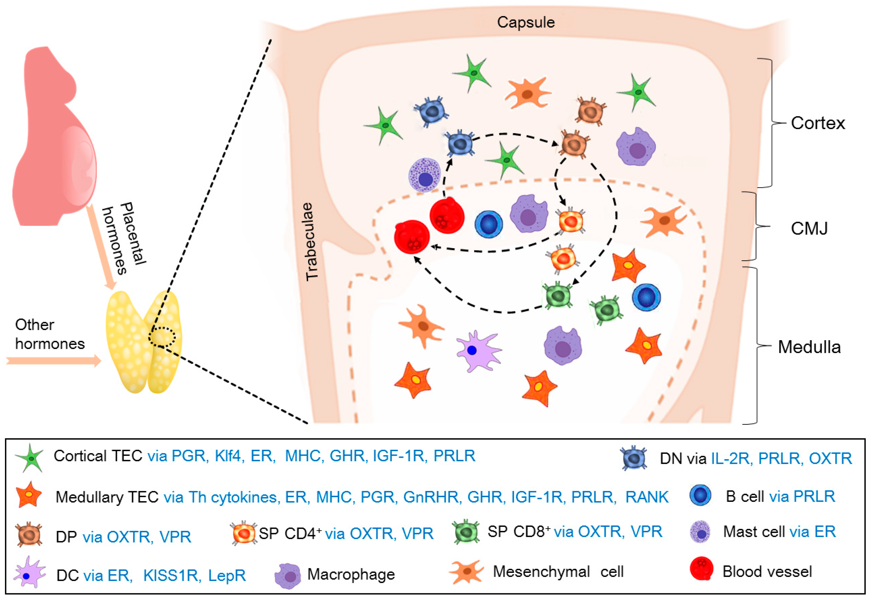
During pregnancy, T-cell development under the thymic microenvironments is regulated by cTECs, mTECs, B cells, and mast cells, which are via many cytokines to regulate maternal thymic adaptations. In addition, the T-cell regulates its development in an autocrine or paracrine manner (Figure 3). However, more studies on the cytokines related to maternal thymic adaptations are needed.

5. Hormones That Regulate Thymic Immunology During Pregnancy

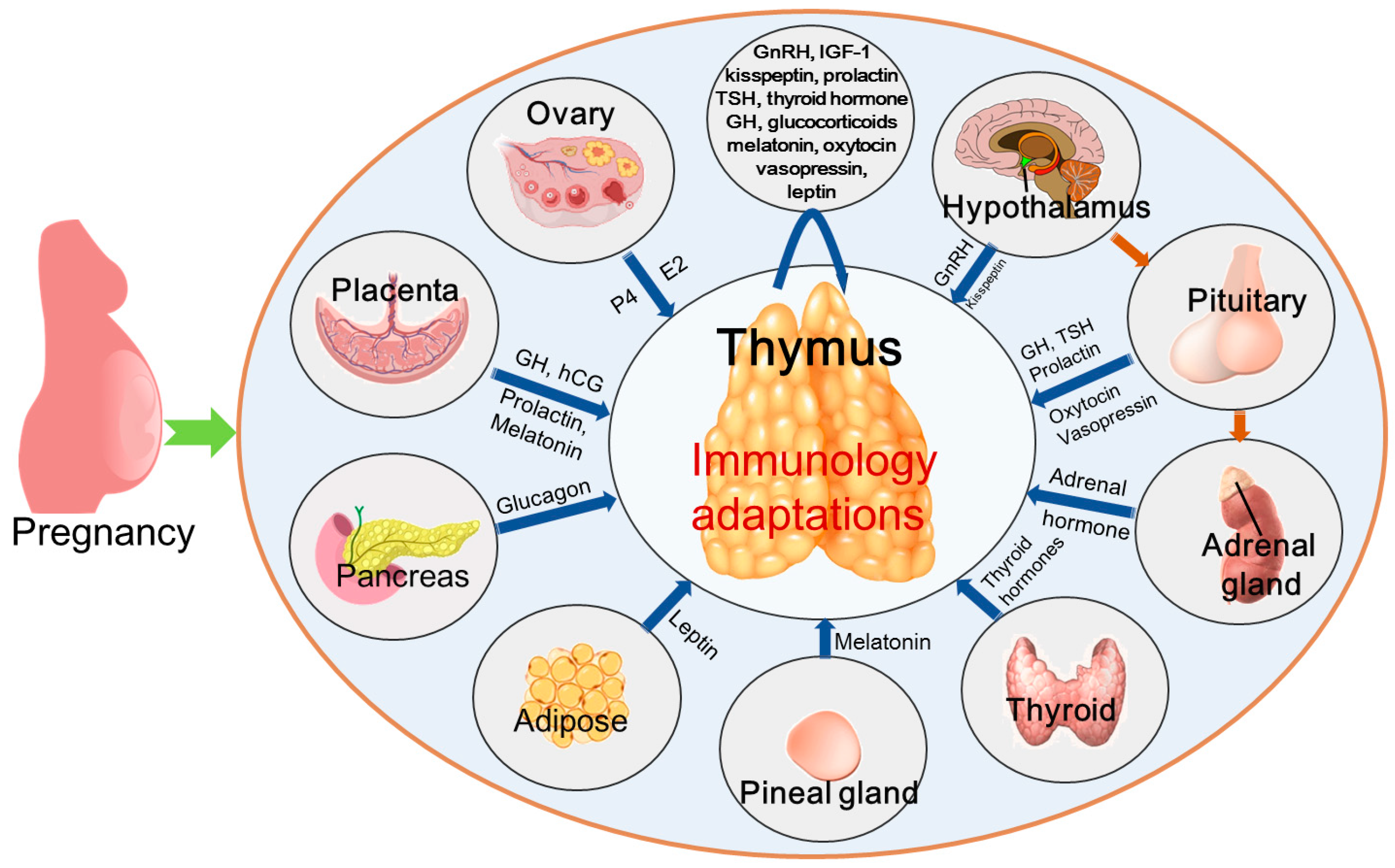
Hormones act on thymic microenvironment cells and thymocytes in both endocrine and paracrine/autocrine pathways, which modulate the proliferation and survival of thymic microenvironment cells, selection of the T-cell repertoire, as well as the migration and export of developing T-cells. The hormones include growth hormone (GH)/insulin-like growth factor 1 (IGF-1), prolactin (PRL), leptin, thyroid hormone, sex hormones, and glucocorticoids [48]. The hormones that regulate thymic immunology during pregnancy include human chorionic gonadotropin (HCG), estrogen, progesterone, GnRH, GH/IGF-1, kisspeptin, PRL, thyroid-stimulating hormone (TSH), thyroid hormone, glucocorticoids, melatonin, oxytocin, vasopressin, leptin, and insulin (Figure 4). However, it is also possible that other hormones are involved in modulating thymic immunology during pregnancy.

5.1. Human Chorionic Gonadotropin

HCG has multiple endocrine, paracrine, and autocrine actions on a variety of gestational cells and tissues, including the immune system, to promote and maintain pregnancy in humans [49]. HCG administration inhibits thymic T-cell development and peripheral T-cell populations, which has negative effects on immune function in mice [50]. Chorionic gonadotrophin affects the thymic secretory function in pubertal female mice [51], and HCG enhances the effect of hormones on the in vitro differentiation of thymocytes in the presence of TECs in humans [52]. However, HCG does not affect the phenotype of the human thymocytes but influences the production of autocrine growth factors by these cells, which regulates antigen-independent differentiation of T-lymphocytes during pregnancy [53]. HCG is detectable from 10 days after fertilization, increased to a peak in weeks 9 to 10 of pregnancy, and subsequently declined. HCG can increase the frequency of Treg cells and restrict pregnancy-harmful proinflammatory Th17 responses in mice [54]. Therefore, HCG has effects on thymus functions, and further research is needed on the molecular mechanism that HCG regulates for thymic function during pregnancy.

5.2. Estrogen

Estrogens can modulate atrophy and phenotypic alterations of the thymus, and T-cell development via ERα [55]. Estrogen treatment can inhibit the production of thymic factors and change T-cell subpopulations via its receptors in the thymus to modulate thymic immune function [56]. In addition, estrogen regulates the development and differentiation of T-cells and the immune functions of the TECs through ERs, which contributes to autoimmunity [57]. Estriol modulates the processes of myeloid DC maturation in the thymus during the first half of pregnancy and also is an element of steroid-induced involution of the thymus during this period [58]. In addition, estrogen has effects on thymocytes and thymic stromal cells (mainly TECs), which are involved in the modulation of T-cell development and repertoire selection for central tolerance [59].
Estrogen treatment reduces thymic size and cellularity, as well as defined T-cell subsets of CD4 and CD8 in the mouse thymus [60]. The high serum level of estradiol inhibits the percentage of CD4+CD8+ DP T-cells in the mouse thymus during the proestrus phase, but CD4+CD8 or CD4CD8+ SP T-cells are significantly increased in the proestrus phase via ERα [61]. Basal estrogens promote normal thymus growth but inhibit cortical double negative development via ERα, and reduce expression of MHC in the TECs [62]. The elevated estrogen levels during pregnancy reduce thymocyte proliferation, which contributes to thymic involution and loss of thymocyte cellularity during pregnancy [8]. In addition, early pregnancy regulates the expression of ERα and ERβ in the maternal thymus, which is involved in the regulation of maternal immune function during early pregnancy in ewes [63]. Thus, estrogen is related to the establishment of immune tolerance during normal pregnancy, and expression of ERα and ERβ in the maternal thymus during mid and late pregnancy needs to be investigated further.

5.3. Progesterone

There are specific intracellular PGRs that interact with progesterone to perform pleiotropic reproductive activities of progesterone in the female thymus [64]. Progesterone has effects on thymulin-secreting TECs, which modulates proliferation, secretion, and the degree of interactions between TECs and thymocytes through both cytoplasmic and membrane-bound PGRs [65]. Pregnancy induces the expression of PGR in thymic stromal cells (mainly TECs), which is specifically essential for thymic involution and block of T-cell development [66]. Progesterone promotes the development of thymic Treg cells through differentiation receptor RANK in mTECs during pregnancy, and depletion of RANK in the mouse thymic epithelium results in fetal loss and maternal glucose intolerance [6]. It has been reported that there is an upregulation of the 60-kDa PGR isoform and the 62-kDa progesterone-induced blocking factor variant in the maternal thymus during early pregnancy in ewes [67], and the upregulation of PGR in maternal cTECs during pregnancy is required for thymic involution and successful pregnancy in mice [7]. Therefore, upregulation of PGR in the maternal thymus is essential for normal pregnancy, but the molecular mechanism that progesterone regulates, thymic involution, needs to be studied further.

5.4. GnRH

GnRH is mainly secreted by the hypothalamus and also synthesized by the thymus, which regulates the development and function of thymic T lymphocytes [68]. There is a thymus–hypothalamus–pituitary–gonadal axis through which the thymus gland secretes factors to regulate the release of GnRH, the production of luteinizing hormone, and the secretion of gonadal steroids [69]. Both GnRH and GnRH receptors (GnRHRs) are mainly expressed in the thymic medulla, which exert direct actions on the immune modulation in the immune cells [38,70]. GnRHR agonist leuprolide treatment induces immunosuppression, which is related to the levels of thymus nitric oxide and changes in immunological parameters [71]. GnRHR agonist leuprolide treatment also enhances thymus weight and increases secretion of thymosin alpha 1 in the thymus [72]. Furthermore, GnRH agonist infusions attenuate pregnancy-induced thymic involution, and increase thymic weight in pregnant rats. On the other hand, the maternal thymus expresses GnRH and GnRHR, and early pregnancy can stimulate the expression of GnRH, but inhibit the expression of GnRHR in the maternal thymus in sheep [73]. Taken together, these findings suggest that GnRH and GnRHR are involved in modulating maternal thymus function during pregnancy. Thus, further research may focus on the molecular mechanism that GnRH regulates, maternal thymus function, during pregnancy.

5.5. Growth Hormone/Insulin-Like Growth Factor 1

GH acts directly on target cells or indirectly by stimulating the production of IGF-1 to promote body growth and metabolism. GH and IGF-1 are expressed in the thymus to regulate the development and function of immune cells and thymus involution [74]. GH can improve thymocyte proliferation and migration, and also affect the secretion of cytokines and thymic hormones in humans and mice [75]. GH is involved in the regulation of the thymic microenvironment [48], which is mediated by IGF-1 in the thymus [75]. GH and IGF-1 secreted by human thymocytes enhance the proliferation of thymocytes and TECs to influence immune function [76]. The IGF-1 receptor is expressed in human thymic cells and murine TECs and thymocytes, which are involved in intrathymic T-cell differentiation and migration [77]. Furthermore, GH enhances total thymocyte numbers and the secretion of thymulin from TECs mediated by IGF-1, which are via GH and IGF-1 receptors on thymocytes and TECs [78]. Exogenous IGF-1 improves thymopoiesis through TECs expansion in mice [79]. The IGF-1 receptor expressed in the thymus is involved in thymic rejuvenation and involution in pigs [80]. The placental GH increases, and gradually replaces pituitary GH, and maternal IGF-1 levels also upregulate during pregnancy [81]. On the other hand, the changes in the expression of GH and GH receptors in the maternal thymus are implicated in the adaptations of the maternal thymus in ewes [82]. Thus, GH and IGF-1 participate in modulating maternal thymus function during pregnancy. However, the molecular mechanism that GH and IGF-1 modulate for thymic adaptation during pregnancy needs to be explored further.

5.6. Kisspeptin

Kisspeptin is secreted by the hypothalamus, and is also found in extra-hypothalamic areas, which is a critical regulatory factor of GnRH release [83]. The KISS1 gene encodes kisspeptins that bind to kisspeptin receptors, including GPR54 [84], and GPR54 mRNA, which are expressed in the thymus [85]. GPR54 deficiency results in thymus enlargement, an increase in thymocytes, and altered thymic micro-architecture, which is involved in T-cell development and self-tolerant immunity in mice [86]. Kisspeptin has effects on the processes of differentiation of thymic myeloid DCs during the second–third trimesters [58]. There is a dramatic increase in circulating levels of kisspeptin during a healthy pregnancy, which plays a fundamental role in the regulation of GnRH secretion and placentation [87]. Thus, the upregulation of kisspeptin levels is implicated in the T-cell development of the maternal thymus during pregnancy.

5.7. Prolactin

Lactotroph cells in the anterior pituitary gland synthesize PRL, and the principal role of PRL is to regulate lactation via the PRL receptor (PRLR) [88]. PRL has beneficial effects on the survival and differentiation of T-cell progenitors [89], and administration of PRL antiserum modulates the developmental pattern of T-lymphocytes in the thymus [90]. In addition, PRL can stimulate the secretion of thymulin by TECs, and maintain thymocyte viability during the DP stage of thymocyte differentiation, which is through PRLR on thymocytes and TECs [79]. Developing thymocytes produce PRL, and PRLR is expressed in T-cells, TECs, and B cells [91], and PRL treatment has effects on the migration of total DP, CD4-positive, and CD8-positive thymocytes [92]. It has been reported that there is an upregulation of PRL and PRLR in the maternal thymus during early pregnancy in ewes, and PRLR protein is located in the epithelial reticular cells, capillaries, and thymic corpuscles [93]. Therefore, the changes in the expression of PRL and PRLR in the maternal thymus are associated with T-cell development and proliferation during early pregnancy. However, the effect of PRL on the maternal thymus during mid and late pregnancy needs to be studied further.

5.8. Thyroid-Stimulating Hormone and Thyroid Hormone

TSH can bind and activate the TSH receptor (TSHR) in thymocytes to enhance T-cell development, and lacking functional TSHR expression is associated with lower frequencies of DP and SP thymocytes in mice [94]. It has been reported that β-TSH is expressed in the subcapsular and cortical thymic zones [95], and TSH increases the frequencies of CD4+ and CD8+ SP thymocytes, and protects thymocytes from apoptosis [96].
Triiodothyronine has a pleiotropic effect on thymus physiology [97], and triodothyronine treatment of cultured TECs increases thymulin synthesis and secretion in mice [98]. On the other hand, the thyrotropin receptor is expressed in the thymus, which enhances thymocyte proliferation in an autocrine/paracrine manner [48]. Free thyroxine levels increase during the first trimester and then decrease after the first trimester. However, TSH concentrations decrease, and then return to normal during the same stages [99]. Therefore, TSH and thyroid hormone, as well as their receptors, are expressed in the maternal thymus, which are related to T-cell development and thymocyte output. However, the effects of TSH and thyroid hormone on the maternal thymus during pregnancy need to be studied.

5.9. Glucocorticoids

Glucocorticoids are mainly synthesized in the adrenal glands and have anti-inflammatory and immune-suppressive actions [100]. The thymus also produces glucocorticoids which are critical modulators in the immune system [101]. Endogenous glucocorticoids have pleiotropic effects on different T-cell populations [102], and endogenous glucocorticoids play a positive role in thymocyte selection via glucocorticoid receptors (GRs) [103]. Elevated serum glucocorticoid levels induced by stress responses result in thymocyte apoptosis and thymic involution in humans and mice [104]. Glucocorticoid treatment increases the production of immunosuppressive cytokines by thymic Treg cells and immunosuppressive cytokine expression, which contributes to the immunosuppressive effects of glucocorticoids [105]. Maternal glucocorticoid concentrations critically rise during pregnancy, which promotes immune tolerance via GR-mediated pathways [106]. Taken together, glucocorticoids are associated with maternal immune-suppressive actions, but the function of glucocorticoids in the maternal thymus during pregnancy needs to be investigated further.

5.10. Melatonin

The pineal gland is the major site for melatonin production, and melatonin is also synthesized by other organs, including the thymus. Melatonin exerts its effects through melatonin receptors expressed in many mammalian organs [107]. Endogenous thymic melatonin synthesis is regulated by circulating levels of melatonin [108], and melatonin treatment in physiological doses exerts effects on cell proliferation and differentiation of T-cells in the thymus [109]. In addition, melatonin treatment decreases mast cell densities in the thymus, which is involved in immune system regulation and proinflammatory cytokine production [110]. There is a progressive increase both in maternal and placental melatonin levels during normal pregnancy [111]. On the other hand, melatonin receptors are upregulated in the maternal thymus during early pregnancy, which is related to immune regulation of the maternal thymus in an animal model [41]. Thus, the progressive increase in maternal and placental melatonin levels may be involved in immunoregulation of the maternal thymus via melatonin receptors during pregnancy, which needs to be investigated further.

5.11. Oxytocin and Vasopressin

Oxytocin and vasopressin released by the neurohypophysis have effects on thymus physiology, and their transcripts are found in the thymus. In addition, oxytocin receptors are detected in all thymocyte subsets, whereas vasopressin receptor is only found in DP and SP CD8 cells [112]. Oxytocin and vasopressin are peptides with high physiological relevance, and play a central role in bodily homeostatic regulation via banding to oxytocin receptors and vasopressin receptors [113]. Oxytocin and vasopressin play a role in regulating the thymus microenvironment [114], and oxytocins, including intrathymic oxytocin, are involved in the central immunological self-tolerance of T-cells [115]. Inhibition of the oxytocin receptor is related to apoptosis of CD8+ mature T-cells, while the vasopressin receptor is associated with T-cell differentiation in the thymus [48]. Neurophysin levels of vasopressin and oxytocin elevate, and the release of these hormones also increases during pregnancy [116]. Oxytocins released from the neurohypophysis and peripheral organs (including the thymus) play a key role in the initiation of pregnancy in rodents [117]. However, the function of vasopressin in the maternal thymus during pregnancy needs to be investigated further.

5.12. Leptin

Leptin is mainly synthesized in white adipose tissue [118]. The thymus also expresses leptin, which has a protective effect on thymocytes from apoptosis via the leptin receptor in the thymus [48]. The intrathymic role of leptin includes maintaining healthy thymic epithelium and promoting thymopoiesis [119], and leptin can regulate thymic plasmacytoid DC abilities, and influence the thymocyte distribution [120]. There is an increase in the leptin concentration in the maternal circulation during pregnancy, which contributes to adaptations of the energy demands of the growing fetus [121]. An immunosuppressive fraction of boar seminal vesicle fluid lowers the concentration of leptin in blood plasma and adipose tissue, which prevents thymus involution during the first period of pregnancy in mice [122]. In addition, as a proinflammatory adipocytokine, leptin can improve thymopoiesis and regulate T-cell immune responses, which is involved in restoring thymic involution associated with obesity in mice [123]. However, the studies that leptin and its receptors are related to maternal thymus involution during pregnancy in humans and domestic animals need to be investigated further.

5.13. Insulin

Insulin can be used for the management of hyperglycemia and also serves as an immunomodulatory hormone [124]. Insulin is expressed in the thymus, which is related to the induction of immune tolerance [125], and susceptibility to type 1 autoimmune diabetes in humans and mice [126]. In addition, thymic insulin levels play a pivotal role in the self-tolerance of insulin-specific T-cells in mice [127]. Insulin expressed by thymic epithelial cells is involved in trimming and removing high-affinity insulin-specific T-cells in mice [128], and the autoimmune regulator controls the expression of insulin in the thymus in humans [129]. There is an increasing demand for glucose, which results in a rise in insulin levels, as well as site-specific central insulin resistance during pregnancy [130]. On the other hand, early pregnancy modulates the expression of insulin receptor β gene and protein in ewes (our unpublicized data). However, the function of insulin in the maternal thymus during mid and late pregnancy needs to be investigated further.

6. Conclusions and Future Prospects

Pregnancy induces maternal thymus adaptations that are necessary for successful pregnancy, which results in thymus involution, changes in major thymic lymphoid cell populations, and T-cell development in the thymus. The factors and cells involved in the maternal thymus adaptations during pregnancy are summarized in Table 1. In addition, the hormones that modulate thymic immunology are reviewed, which may be used for avoiding pregnancy-related thymic diseases, and preventing embryo loss and abortion. However, there are differences in early pregnancy recognition signals among different species and placental types. Therefore, in different species, pregnancy-induced maternal thymus adaptation may vary in early pregnancy. Nevertheless, the difference may not be significant in the middle and later stages of pregnancy. Thus, many mysteries still exist in modulating thymic immune functions at different stages of pregnancy, and more studies in these areas are needed to figure out the regulatory mechanisms of maternal thymic immunology adaptations.

Author Contributions

Conceptualization, L.Y., L.Z. and X.W.; writing—original draft preparation, L.Y.; writing—review and editing, L.Y.; supervision, L.Y. All authors have read and agreed to the published version of the manuscript.

Funding

This research was funded by Natural Science Foundation of Hebei Province, grant number C2024402023.

Institutional Review Board Statement

Not applicable.

Informed Consent Statement

Not applicable.

Data Availability Statement

Not applicable.

Conflicts of Interest

The authors declare no conflicts of interest.

Abbreviations

AIREAutoimmune regulator
CGChorionic gonadotrophin
cTECsCortical thymic epithelial cells
DCsDendritic cells
DNDouble negative
DNTregsDouble negative T regulatory cells
DPDouble positive
ERαEstrogen receptor-α
GHGrowth hormone
GnRHGonadotropin-releasing hormone
GnRHRGonadotropin-releasing hormone receptor
GRsGlucocorticoid receptors
HCGHuman chorionic gonadotropin
IGF-1Insulin-like growth factor 1
IL-2Interleukin-2
MHCMajor histocompatibility complex
mTECsMedullary thymic epithelial cells
NESNon-epithelial stromal cells
PGRProgesterone receptor
PRLProlactin
RTE-TregRecent thymic emigrant-regulatory T-cell
SPSingle-positive
TCRsT-cell receptors
TECsThymic epithelial cells
ThHelper T
TregRegulatory T
TSHThyroid-stimulating hormone
TSHRThyroid-stimulating hormone receptor
tTRegsTRegs derived from the thymus

References

  1. Romero, R.; Dey, S.K.; Fisher, S.J. Preterm labor: One syndrome, many causes. Science 2014, 345, 760–765. [Google Scholar] [CrossRef] [PubMed]
  2. Miller, J.F.A.P. The function of the thymus and its impact on modern medicine. Science 2020, 369, eaba2429. [Google Scholar] [CrossRef] [PubMed]
  3. Thapa, P.; Farber, D.L. The role of the thymus in the immune response. Thorac. Surg. Clin. 2019, 29, 123–131. [Google Scholar] [CrossRef] [PubMed]
  4. Park, J.E.; Botting, R.A.; Domínguez Conde, C.; Popescu, D.M.; Lavaert, M.; Kunz, D.J.; Goh, I.; Stephenson, E.; Ragazzini, R.; Tuck, E.; et al. A cell atlas of human thymic development defines T cell repertoire formation. Science 2020, 367, eaay3224. [Google Scholar] [CrossRef]
  5. James, K.D.; Jenkinson, W.E.; Anderson, G. Non-epithelial stromal cells in thymus development and function. Front. Immunol. 2021, 12, 634367. [Google Scholar] [CrossRef]
  6. Paolino, M.; Koglgruber, R.; Cronin, S.J.F.; Uribesalgo, I.; Rauscher, E.; Harreiter, J.; Schuster, M.; Bancher-Todesca, D.; Pranjic, B.; Novatchkova, M.; et al. RANK links thymic regulatory T cells to fetal loss and gestational diabetes in pregnancy. Nature 2021, 589, 442–447. [Google Scholar] [CrossRef]
  7. Ahn, S.H.; Nguyen, S.L.; Kim, T.H.; Jeong, J.W.; Arora, R.; Lydon, J.P.; Petroff, M.G. Nuclear progesterone receptor expressed by the cortical thymic epithelial cells dictates thymus involution in murine pregnancy. Front. Endocrinol. 2022, 13, 846226. [Google Scholar] [CrossRef]
  8. Zoller, A.L.; Schnell, F.J.; Kersh, G.J. Murine pregnancy leads to reduced proliferation of maternal thymocytes and decreased thymic emigration. Immunology 2007, 121, 207–215. [Google Scholar] [CrossRef]
  9. Laan, M.; Haljasorg, U.; Kisand, K.; Salumets, A.; Peterson, P. Pregnancy-induced thymic involution is associated with suppression of chemokines essential for T-lymphoid progenitor homing. Eur. J. Immunol. 2016, 46, 2008–2017. [Google Scholar] [CrossRef]
  10. Li, N.; Wang, L.; Cao, N.; Zhang, L.; Han, X.; Yang, L. Early pregnancy affects the expression of toll-like receptor pathway in ovine thymus. Reprod. Biol. 2020, 20, 547–554. [Google Scholar] [CrossRef]
  11. Yang, L.; Cai, C.; Fang, S.; Hao, S.; Zhang, T.; Zhang, L. Changes in expression of nuclear factor kappa B subunits in the ovine thymus during early pregnancy. Sci. Rep. 2022, 12, 17683. [Google Scholar] [CrossRef]
  12. Zhang, L.; Li, Y.; Zhao, Z.; Cai, J.; Zhao, S.; Yang, L. Modulation of nod-like receptor expression in the thymus during early pregnancy in ewes. Vaccines 2022, 10, 2128. [Google Scholar] [CrossRef]
  13. Zhang, L.; Zhang, Q.; Wang, H.; Feng, P.; Yang, G.; Yang, L. Effects of early pregnancy on the complement system in the ovine thymus. Vet. Res. Commun. 2022, 46, 137–145. [Google Scholar] [CrossRef]
  14. Meng, Y.; Yang, Z.; Quan, Y.; Zhao, S.; Zhang, L.; Yang, L. Regulation of ikappaB protein expression by early gestation in the thymus of ewes. Vet. Sci. 2023, 10, 462. [Google Scholar] [CrossRef]
  15. Yang, L.; Lv, W.; Liu, Y.; Chen, K.; Xue, J.; Wang, Q.; Wang, B.; Zhang, L. Effect of early pregnancy on the expression of prostaglandin synthases in the ovine thymus. Theriogenology 2019, 136, 166–171. [Google Scholar] [CrossRef] [PubMed]
  16. Anderson, G.; Jenkinson, E.J. Lymphostromal interactions in thymic development and function. Nat. Rev. Immunol. 2001, 1, 31–40. [Google Scholar] [CrossRef] [PubMed]
  17. Manley, N.R.; Richie, E.R.; Blackburn, C.C.; Condie, B.G.; Sage, J. Structure and function of the thymic microenvironment. Front. Biosci. 2011, 16, 2461–2477. [Google Scholar] [CrossRef]
  18. Sharma, H.; Moroni, L. Recent advancements in regenerative approaches for thymus rejuvenation. Adv. Sci. 2021, 8, 2100543. [Google Scholar] [CrossRef]
  19. Bosticardo, M.; Notarangelo, L.D. Human thymus in health and disease: Recent advances in diagnosis and biology. Semin. Immunol. 2023, 66, 101732. [Google Scholar] [CrossRef]
  20. Petrie, H.T. Role of thymic organ structure and stromal composition in steady-state postnatal T-cell production. Immunol. Rev. 2002, 189, 8–19. [Google Scholar] [CrossRef]
  21. Zúñiga-Pflücker, J.C. T-cell development made simple. Nat. Rev. Immunol. 2004, 4, 67–72. [Google Scholar] [CrossRef]
  22. Figueiredo, M.; Zilhão, R.; Neves, H. Thymus inception: Molecular network in the early stages of thymus organogenesis. Int. J. Mol. Sci. 2020, 21, 5765. [Google Scholar] [CrossRef]
  23. Breed, E.R.; Vobořil, M.; Ashby, K.M.; Martinez, R.J.; Qian, L.; Wang, H.; Salgado, O.C.; O’Connor, C.H.; Hogquist, K.A. Type 2 cytokines in the thymus activate Sirpα+ dendritic cells to promote clonal deletion. Nat. Immunol. 2022, 23, 1042–1051. [Google Scholar] [CrossRef]
  24. Akagbosu, B.; Tayyebi, Z.; Shibu, G.; Paucar Iza, Y.A.; Deep, D.; Parisotto, Y.F.; Fisher, L.; Pasolli, H.A.; Thevin, V.; Elmentaite, R.; et al. Novel antigen-presenting cell imparts Treg-dependent tolerance to gut microbiota. Nature 2022, 610, 752–760. [Google Scholar] [CrossRef] [PubMed]
  25. Frantzeskakis, M.; Takahama, Y.; Ohigashi, I. The role of proteasomes in the thymus. Front. Immunol. 2021, 12, 646209. [Google Scholar] [CrossRef] [PubMed]
  26. Welsh, R.A.; Song, N.; Park, C.S.; Peske, J.D.; Sadegh-Nasseri, S. H2-O deficiency promotes regulatory T cell differentiation and CD4 T cell hyperactivity. Front. Immunol. 2024, 14, 1304798. [Google Scholar] [CrossRef] [PubMed]
  27. Takada, K.; Kondo, K.; Takahama, Y. Generation of peptides that promote positive selection in the thymus. J. Immunol. 2017, 198, 2215–2222. [Google Scholar] [CrossRef]
  28. Parker, M.E.; Ciofani, M. Regulation of γδ T cell effector diversification in the thymus. Front. Immunol. 2020, 11, 42. [Google Scholar] [CrossRef]
  29. Březina, J.; Vobořil, M.; Filipp, D. Mechanisms of direct and indirect presentation of self-antigens in the thymus. Front. Immunol. 2022, 13, 926625. [Google Scholar] [CrossRef]
  30. Castañeda, J.; Hidalgo, Y.; Sauma, D.; Rosemblatt, M.; Bono, M.R.; Núñez, S. The multifaceted roles of B cells in the thymus: From immune tolerance to autoimmunity. Front. Immunol. 2021, 12, 766698. [Google Scholar] [CrossRef]
  31. Wallin, J.; Eibel, H.; Neubüser, A.; Wilting, J.; Koseki, H.; Balling, R. Pax1 is expressed during development of the thymus epithelium and is required for normal T-cell maturation. Development 1996, 122, 23–30. [Google Scholar] [CrossRef] [PubMed]
  32. Clarke, A.G.; Kendall, M.D. The thymus in pregnancy: The interplay of neural, endocrine and immune influences. Immunol. Today 1994, 15, 545–551. [Google Scholar] [CrossRef] [PubMed]
  33. Leeming, G.; McLean, J.; Gibbs, A.C. Thymic and body weight during first syngeneic and allogeneic pregnancy in the rat, and the effects of strain difference. Thymus 1984, 6, 153–165. [Google Scholar] [PubMed]
  34. Kendall, M.D.; Clarke, A.G. The thymus in the mouse changes its activity during pregnancy: A study of the microenvironment. J. Anat 2000, 197 Pt 3, 393–411. [Google Scholar] [CrossRef]
  35. Abu-Raya, B.; Michalski, C.; Sadarangani, M.; Lavoie, P.M. Maternal immunological adaptation during normal pregnancy. Front. Immunol. 2020, 11, 575197. [Google Scholar] [CrossRef]
  36. Li, Z.; Liang, X.; Chen, X.; Chen, Y.; Wang, F.; Wang, S.; Liao, Y.; Li, L. The role of thymus- and extrathymus-derived regulatory T cells in maternal-fetal tolerance. Front. Immunol. 2023, 14, 1109352. [Google Scholar] [CrossRef]
  37. Gambel, P.I.; Cleland, A.W.; Ferguson, F.G. Alterations in thymus and spleen cell populations and immune reactivity during syngeneic pregnancy and lactation. J. Clin. Lab. Immunol. 1980, 3, 115–119. [Google Scholar]
  38. Dixit, V.D.; Sridaran, R.; Edmonsond, M.A.; Taub, D.; Thompson, W.E. Gonadotropin-releasing hormone attenuates pregnancy-associated thymic involution and modulates the expression of antiproliferative gene product prohibitin. Endocrinology 2003, 144, 1496–1505. [Google Scholar] [CrossRef]
  39. Shu, Y.Y.; Xu, X.; Zhang, Z.W.; Gao, J.L. Mechanism of pregnancy-induced thymus involution and regeneration and medication rules of postpartum prescriptions. Zhongguo Zhong Yao Za Zhi 2023, 48, 4275–4284. [Google Scholar] [CrossRef]
  40. Ahn, S.H.; Nguyen, S.L.; Petroff, M.G. Exploring the origin and antigenic specificity of maternal regulatory T cells in pregnancy. Front. Immunol. 2020, 11, 1302. [Google Scholar] [CrossRef]
  41. Bai, J.; Zhang, L.; Zhao, Z.; Li, N.; Wang, B.; Yang, L. Expression of melatonin receptors and CD4 in the ovine thymus, lymph node, spleen and liver during early pregnancy. Immunology 2020, 160, 52–63. [Google Scholar] [CrossRef]
  42. Zhang, L.; Zhao, Z.; Mi, H.; Liu, B.; Wang, B.; Yang, L. Modulation of helper t cytokines in thymus during early pregnancy in ewes. Animals 2019, 9, 245. [Google Scholar] [CrossRef]
  43. Depoërs, L.; Dumont-Lagacé, M.; Trinh, V.Q.; Houques, C.; Côté, C.; Larouche, J.D.; Brochu, S.; Perreault, C. Klf4 protects thymus integrity during late pregnancy. Front. Immunol. 2023, 14, 1016378. [Google Scholar] [CrossRef] [PubMed]
  44. Chapman, J.C.; Chapman, F.M.; Michael, S.D. The production of alpha/beta and gamma/delta double negative (DN) T-cells and their role in the maintenance of pregnancy. Reprod. Biol. Endocrinol. 2015, 13, 73. [Google Scholar] [CrossRef] [PubMed]
  45. Bafor, E.E.; Valencia, J.C.; Young, H.A. Double negative T regulatory cells: An emerging paradigm shift in reproductive immune tolerance? Front. Immunol. 2022, 13, 886645. [Google Scholar] [CrossRef] [PubMed]
  46. Green, E.S.; Moldenhauer, L.M.; Groome, H.M.; Sharkey, D.J.; Chin, P.Y.; Care, A.S.; Robker, R.L.; McColl, S.R.; Robertson, S.A. Regulatory T cells are paramount effectors in progesterone regulation of embryo implantation and fetal growth. JCI Insight 2023, 8, e162995. [Google Scholar] [CrossRef]
  47. Sun, I.H.; Gillis-Buck, E.; Mackenzie, T.C.; Gardner, J.M. Thymic and extrathymic Aire-expressing cells in maternal-fetal tolerance. Immunol. Rev. 2022, 308, 93–104. [Google Scholar] [CrossRef]
  48. Savino, W.; Mendes-da-Cruz, D.A.; Lepletier, A.; Dardenne, M. Hormonal control of T-cell development in health and disease. Nat. Rev. Endocrinol. 2016, 12, 77–89. [Google Scholar] [CrossRef]
  49. Paulesu, L.; Rao, C.V.; Ietta, F.; Pietropolli, A.; Ticconi, C. hCG and its disruption by environmental contaminants during human pregnancy. Int. J. Mol. Sci. 2018, 19, 914. [Google Scholar] [CrossRef]
  50. Yoon, J.; Sun, S.; Moon, S.; Yang, H. Repeated gonadotropin administration suppresses T cell development in the mouse thymus. Dev. Reprod. 2025, 29, 1–11. [Google Scholar] [CrossRef]
  51. Labunets, I.F. Effect of chorionic gonadotropin on the structure and endocrine function of the thymus gland in mice. Fiziol. Zh 1991, 37, 75–80. [Google Scholar] [PubMed]
  52. Kuklina, E.M.; Shirshev, S.V.; Sharova, N.I.; Iarilin, A.A. Effect of chorionic gonadotropin on thymocyte differentiation in the presence of thymus epithelial cells. Ontogenez 2003, 34, 36–42. [Google Scholar] [CrossRef] [PubMed]
  53. Kuklina, E.M.; Shirshev, S.V.; Sharova, N.I.; Iarilin, A.A. Rol’ khorionicheskogo gonadotropina v differentsirovke timotsitov [Role of chorionic gonadotropin in the differentiation of thymocytes]. Ontogenez 1999, 30, 341–345. [Google Scholar] [PubMed]
  54. Lentz, L.S.; Stutz, A.J.; Meyer, N.; Schubert, K.; Karkossa, I.; von Bergen, M.; Zenclussen, A.C.; Schumacher, A. Human chorionic gonadotropin promotes murine Treg cells and restricts pregnancy-harmful proinflammatory Th17 responses. Front. Immunol. 2022, 13, 989247. [Google Scholar] [CrossRef]
  55. Staples, J.E.; Gasiewicz, T.A.; Fiore, N.C.; Lubahn, D.B.; Korach, K.S.; Silverstone, A.E. Estrogen receptor alpha is necessary in thymic development and estradiol-induced thymic alterations. J. Immunol. 1999, 163, 4168–4174. [Google Scholar] [CrossRef]
  56. Seiki, K.; Sakabe, K. Sex hormones and the thymus in relation to thymocyte proliferation and maturation. Arch. Histol. Cytol. 1997, 60, 29–38. [Google Scholar] [CrossRef]
  57. Merrheim, J.; Villegas, J.; Van Wassenhove, J.; Khansa, R.; Berrih-Aknin, S.; le Panse, R.; Dragin, N. Estrogen, estrogen-like molecules and autoimmune diseases. Autoimmun. Rev. 2020, 19, 102468. [Google Scholar] [CrossRef]
  58. Shirshev, S.V.; Orlova, E.G.; Loginova, O.A.; Nekrasova, I.V.; Gorbunova, O.L.; Maslennikova, I.L. Hormonal regulation of dendritic cell differentiation in the thymus. Bull. Exp. Biol. Med. 2018, 165, 230–234. [Google Scholar] [CrossRef]
  59. Moulton, V.R. Sex hormones in acquired immunity and autoimmune disease. Front. Immunol. 2018, 9, 2279. [Google Scholar] [CrossRef]
  60. Rijhsinghani, A.G.; Thompson, K.; Bhatia, S.K.; Waldschmidt, T.J. Estrogen blocks early T cell development in the thymus. Am. J. Reprod. Immunol. 1996, 36, 269–277. [Google Scholar] [CrossRef]
  61. Lee, H.; Kim, H.; Chung, Y.; Kim, J.; Yang, H. Thymocyte differentiation is regulated by a change in estradiol levels during the estrous cycle in mouse. Dev. Reprod. 2013, 17, 441–449. [Google Scholar] [CrossRef]
  62. Taves, M.D.; Ashwell, J.D. Effects of sex steroids on thymic epithelium and thymocyte development. Front. Immunol. 2022, 13, 975858. [Google Scholar] [CrossRef]
  63. Yang, Z.; Zhang, Y.; Cao, Z.; Li, Z.; Zhang, L.; Yang, L. Expression of estrogen receptors in main immune organs in sheep during early pregnancy. Int. J. Mol. Sci. 2025, 26, 3528. [Google Scholar] [CrossRef]
  64. Conneely, O.M.; Mulac-Jericevic, B.; Lydon, J.P. Progesterone-dependent regulation of female reproductive activity by two distinct progesterone receptor isoforms. Steroids 2003, 68, 771–778. [Google Scholar] [CrossRef] [PubMed]
  65. Head, G.M.; Downing, J.E.; Brucker, C.; Mentlein, R.; Kendall, M.D. Rapid progesterone actions on thymulin-secreting epithelial cells cultured from rat thymus. Neuroimmunomodulation 1999, 6, 31–38. [Google Scholar] [CrossRef] [PubMed]
  66. Tibbetts, T.A.; DeMayo, F.; Rich, S.; Conneely, O.M.; O’Malley, B.W. Progesterone receptors in the thymus are required for thymic involution during pregnancy and for normal fertility. Proc. Natl. Acad. Sci. USA 1999, 96, 12021–12026. [Google Scholar] [CrossRef] [PubMed]
  67. Zhang, L.; Xue, J.; Wang, Q.; Lv, W.; Mi, H.; Liu, Y.; Yang, L. Changes in expression of ISG15, progesterone receptor and progesterone-induced blocking factor in ovine thymus during early pregnancy. Theriogenology 2018, 121, 153–159. [Google Scholar] [CrossRef]
  68. Zakharova, L.; Sharova, V.; Izvolskaia, M. Mechanisms of reciprocal regulation of gonadotropin-releasing hormone (GnRH)-producing and immune systems: The role of GnRH, cytokines and their receptors in early ontogenesis in normal and pathological conditions. Int. J. Mol. Sci. 2020, 22, 114. [Google Scholar] [CrossRef]
  69. Golsteyn, E.J.; Fritzler, M.J. The role of the thymus-hypothalamus-pituitary-gonadal axis in normal immune processes and autoimmunity. J. Rheumatol. 1987, 14, 982–990. [Google Scholar]
  70. Jacobson, J.D.; Crofford, L.J.; Sun, L.; Wilder, R.L. Cyclical expression of GnRH and GnRH receptor mRNA in lymphoid organs. Neuroendocrinology 1998, 67, 117–125. [Google Scholar] [CrossRef]
  71. Ullewar, M.P.; Umathe, S.N. Gonadotropin-releasing hormone agonist prevents l-arginine induced immune dysfunction independent of gonadal steroids: Relates with a decline in elevated thymus and brain nitric oxide levels. Nitric. Oxide 2016, 57, 40–47. [Google Scholar] [CrossRef]
  72. Blacker, C.M.; Ataya, K.M.; Savoy-Moore, R.T.; Subramanian, M.G.; Mutchnick, M.G.; Dunbar, J.C. The gonadotropin-releasing hormone agonist leuprolide affects the thymus and other non-reproductive systems of female rats. Acta Endocrinol. 1991, 125, 581–589. [Google Scholar] [CrossRef]
  73. Cao, N.; Cao, L.; Gao, M.; Wang, H.; Zhang, L.; Yang, L. Changes in mRNA and protein levels of gonadotropin releasing hormone and receptor in ovine thymus, lymph node, spleen, and liver during early pregnancy. Domest. Anim. Endocrinol. 2021, 76, 106607. [Google Scholar] [CrossRef]
  74. Reis, M.D.D.S.; Veneziani, L.P.; Porto, F.L.; Lins, M.P.; Mendes-da-Cruz, D.A.; Savino, W. Intrathymic somatotropic circuitry: Consequences upon thymus involution. Front. Immunol. 2023, 14, 1108630. [Google Scholar] [CrossRef] [PubMed]
  75. Savino, W.; de Mello-Coelho, V.; Dardenne, M. Control of the thymic microenvironment by growth hormone/insulin-like growth factor-I-mediated circuits. Neuroimmunomodulation 1995, 2, 313–318. [Google Scholar] [CrossRef] [PubMed]
  76. Sabharwal, P.; Varma, S. Growth hormone synthesized and secreted by human thymocytes acts via insulin-like growth factor I as an autocrine and paracrine growth factor. J. Clin. Endocrinol. Metab. 1996, 81, 2663–2669. [Google Scholar] [CrossRef] [PubMed]
  77. de Mello Coelho, V.; Villa-Verde, D.M.; Farias-de-Oliveira, D.A.; de Brito, J.M.; Dardenne, M.; Savino, W. Functional insulin-like growth factor-1/insulin-like growth factor-1 receptor-mediated circuit in human and murine thymic epithelial cells. Neuroendocrinology 2002, 75, 139–150. [Google Scholar] [CrossRef]
  78. de Mello Coelho, V.; Savino, W.; Postel-Vinay, M.C.; Dardenne, M. Role of prolactin and growth hormone on thymus physiology. Dev. Immunol. 1998, 6, 317–323. [Google Scholar] [CrossRef]
  79. Chu, Y.W.; Schmitz, S.; Choudhury, B.; Telford, W.; Kapoor, V.; Garfield, S.; Howe, D.; Gress, R.E. Exogenous insulin-like growth factor 1 enhances thymopoiesis predominantly through thymic epithelial cell expansion. Blood 2008, 112, 2836–2846. [Google Scholar] [CrossRef]
  80. Tasaki, M.; Villani, V.; Shimizu, A.; Sekijima, M.; Yamada, R.; Hanekamp, I.M.; Hanekamp, J.S.; Cormack, T.A.; Moran, S.G.; Kawai, A.; et al. Role of bone marrow maturity, insulin-like growth factor 1 receptor, and forkhead box protein N1 in thymic involution and rejuvenation. Am. J. Transpl. 2016, 16, 2877–2891. [Google Scholar] [CrossRef]
  81. Vila, G.; Luger, A. Growth hormone deficiency and pregnancy: Any role for substitution? Minerva Endocrinol. 2018, 43, 451–457. [Google Scholar] [CrossRef]
  82. Li, Z.; Ma, X.; Du, Z.; Li, J.; Zhang, L.; Yang, L. Gestation regulates growth hormone and its receptor expression in sheep immune organs. Biology 2025, 14, 1318. [Google Scholar] [CrossRef]
  83. Xie, Q.; Kang, Y.; Zhang, C.; Xie, Y.; Wang, C.; Liu, J.; Yu, C.; Zhao, H.; Huang, D. The role of kisspeptin in the control of the hypothalamic-pituitary-gonadal axis and reproduction. Front. Endocrinol. 2022, 13, 925206. [Google Scholar] [CrossRef]
  84. Tng, E.L. Kisspeptin signalling and its roles in humans. Singap. Med. J. 2015, 56, 649–656. [Google Scholar] [CrossRef] [PubMed]
  85. Li, S.; Ren, J.; Yang, G.; Guo, Y.; Huang, L. Characterization of the porcine Kisspeptins receptor gene and evaluation as candidate for timing of puberty in sows. J. Anim. Breed. Genet. 2008, 125, 219–227. [Google Scholar] [CrossRef] [PubMed]
  86. Xing, R.; Liu, F.; Yang, Y.; Cui, X.; Wang, T.; Xie, L.; Zhao, Y.; Fang, L.; Yi, T.; Zheng, B.; et al. GPR54 deficiency reduces the Treg population and aggravates experimental autoimmune encephalomyelitis in mice. Sci. China Life Sci. 2018, 61, 675–687. [Google Scholar] [CrossRef] [PubMed]
  87. Tsoutsouki, J.; Patel, B.; Comninos, A.N.; Dhillo, W.S.; Abbara, A. Kisspeptin in the prediction of pregnancy complications. Front. Endocrinol. 2022, 13, 942664. [Google Scholar] [CrossRef]
  88. Bernard, V.; Young, J.; Binart, N. Prolactin—A pleiotropic factor in health and disease. Nat. Rev. Endocrinol. 2019, 15, 356–365. [Google Scholar] [CrossRef]
  89. Carreño, P.C.; Sacedón, R.; Jiménez, E.; Vicente, A.; Zapata, A.G. Prolactin affects both survival and differentiation of T-cell progenitors. J. Neuroimmunol. 2005, 160, 135–145. [Google Scholar] [CrossRef]
  90. Russell, D.H.; Mills, K.T.; Talamantes, F.J.; Bern, H.A. Neonatal administration of prolactin antiserum alters the developmental pattern of T- and B-lymphocytes in the thymus and spleen of BALB/c female mice. Proc. Natl. Acad. Sci. USA 1988, 85, 7404–7407. [Google Scholar] [CrossRef]
  91. Legorreta-Haquet, M.V.; Santana-Sánchez, P.; Chávez-Sánchez, L.; Chávez-Rueda, A.K. The effect of prolactin on immune cell subsets involved in SLE pathogenesis. Front. Immunol. 2022, 13, 1016427. [Google Scholar] [CrossRef]
  92. Medeiros, N.V.C.; Porto, F.L.; De Menezes, C.A.; Santos Reis, M.D.D.; Smaniotto, S.; Lins, M.P. CXCL12-driven thymocyte migration is increased by thymic epithelial cells treated with prolactin in vitro. J. Biosci. 2021, 46, 103. [Google Scholar] [CrossRef] [PubMed]
  93. Feng, P.; Wu, J.; Ren, Y.; Zhang, L.; Cao, J.; Yang, L. Early pregnancy regulates the expression of prolactin and its receptor in the thymus, the liver, the spleen and lymph nodes in sheep. Domest. Anim. Endocrinol. 2022, 81, 106731. [Google Scholar] [CrossRef] [PubMed]
  94. van der Weerd, K.; van Hagen, P.M.; Schrijver, B.; Heuvelmans, S.J.; Hofland, L.J.; Swagemakers, S.M.; Bogers, A.J.; Dik, W.A.; Visser, T.J.; van Dongen, J.J.; et al. Thyrotropin acts as a T-cell developmental factor in mice and humans. Thyroid 2014, 24, 1051–1061. [Google Scholar] [CrossRef] [PubMed]
  95. Batanero, E.; de Leeuw, F.E.; Jansen, G.H.; van Wichen, D.F.; Huber, J.; Schuurman, H.J. The neural and neuro-endocrine component of the human thymus. II. Hormone immunoreactivity. Brain Behav. Immun. 1992, 6, 249–264. [Google Scholar] [CrossRef]
  96. Wu, K.; Zhao, M.; Ma, C.; Zhang, H.; Liu, X.; Zhou, L.; Zhao, J.; Gao, L.; Wang, D. Thyrotropin alters T cell development in the thymus in subclinical hypothyroidism mouse model. Scand. J. Immunol. 2017, 85, 35–42. [Google Scholar] [CrossRef]
  97. Villa-Verde, D.M.; de Mello-Coelho, V.; Farias-de-Oliveira, D.A.; Dardenne, M.; Savino, W. Pleiotropic influence of triiodothyronine on thymus physiology. Endocrinology 1993, 133, 867–875. [Google Scholar] [CrossRef]
  98. Dardenne, M.; Savino, W.; Bach, J.F. Modulation of thymic endocrine function by thyroid and steroid hormones. Int. J. Neurosci. 1988, 39, 325–334. [Google Scholar] [CrossRef]
  99. Moleti, M.; Trimarchi, F.; Vermiglio, F. Thyroid physiology in pregnancy. Endocr. Pract. 2014, 20, 589–596. [Google Scholar] [CrossRef]
  100. Vandewalle, J.; Luypaert, A.; De Bosscher, K.; Libert, C. Therapeutic mechanisms of glucocorticoids. Trends Endocrinol. Metab. 2018, 29, 42–54. [Google Scholar] [CrossRef]
  101. Salehzadeh, M.; Hamden, J.E.; Li, M.X.; Bajaj, H.; Wu, R.S.; Soma, K.K. Glucocorticoid production in lymphoid organs: Acute effects of lipopolysaccharide in neonatal and adult mice. Endocrinology 2022, 163, bqab244. [Google Scholar] [CrossRef]
  102. Taves, M.D.; Ashwell, J.D. Glucocorticoids in T cell development, differentiation and function. Nat. Rev. Immunol. 2021, 21, 233–243. [Google Scholar] [CrossRef]
  103. Mittelstadt, P.R.; Monteiro, J.P.; Ashwell, J.D. Thymocyte responsiveness to endogenous glucocorticoids is required for immunological fitness. J. Clin. Investig. 2012, 122, 2384–2394. [Google Scholar] [CrossRef] [PubMed]
  104. Collins, C.P.; Khuat, L.T.; Sckisel, G.D.; Vick, L.V.; Minnar, C.M.; Dunai, C.; Le, C.T.; Curti, B.D.; Crittenden, M.; Merleev, A.; et al. Systemic immunostimulation induces glucocorticoid-mediated thymic involution succeeded by rebound hyperplasia which is impaired in aged recipients. Front. Immunol. 2024, 15, 1429912. [Google Scholar] [CrossRef] [PubMed]
  105. Ugor, E.; Prenek, L.; Pap, R.; Berta, G.; Ernszt, D.; Najbauer, J.; Németh, P.; Boldizsár, F.; Berki, T. Glucocorticoid hormone treatment enhances the cytokine production of regulatory T cells by upregulation of Foxp3 expression. Immunobiology 2018, 223, 422–431. [Google Scholar] [CrossRef] [PubMed]
  106. Solano, M.E.; Arck, P.C. Steroids, pregnancy and fetal development. Front. Immunol. 2020, 10, 3017. [Google Scholar] [CrossRef]
  107. Ahmad, S.B.; Ali, A.; Bilal, M.; Rashid, S.M.; Wani, A.B.; Bhat, R.R.; Rehman, M.U. Melatonin and health: Insights of melatonin action, biological functions, and associated disorders. Cell. Mol. Neurobiol. 2023, 43, 2437–2458. [Google Scholar] [CrossRef]
  108. Rezzani, R.; Franco, C.; Hardeland, R.; Rodella, L.F. Thymus-pineal gland axis: Revisiting its role in human life and ageing. Int. J. Mol. Sci. 2020, 21, 8806. [Google Scholar] [CrossRef]
  109. Michurina, S.V.; Miroshnichenko, S.M.; Ishchenko, I.Y.; Serykh, A.E.; Rachkovskaya, L.N. Effect of melatonin on the content of CD3low and CD3hi T cells in the thymus of mice with functional pinealectomy. Bull. Exp. Biol. Med. 2023, 174, 754–757. [Google Scholar] [CrossRef]
  110. Ozkanlar, S.; Kara, A.; Sengul, E.; Simsek, N.; Karadeniz, A.; Kurt, N. Melatonin modulates the immune system response and inflammation in diabetic rats experimentally-induced by alloxan. Horm. Metab. Res. 2016, 48, 137–144. [Google Scholar] [CrossRef]
  111. Sagrillo-Fagundes, L.; Assunção Salustiano, E.M.; Yen, P.W.; Soliman, A.; Vaillancourt, C. Melatonin in pregnancy: Effects on brain development and CNS programming disorders. Curr. Pharm. Des. 2016, 22, 978–986. [Google Scholar] [CrossRef]
  112. Hansenne, I.; Rasier, G.; Péqueux, C.; Brilot, F.; Renard, C.; Breton, C.; Greimers, R.; Legros, J.J.; Geenen, V.; Martens, H.J. Ontogenesis and functional aspects of oxytocin and vasopressin gene expression in the thymus network. J. Neuroimmunol. 2005, 158, 67–75. [Google Scholar] [CrossRef]
  113. Rae, M.; Lemos Duarte, M.; Gomes, I. Oxytocin and vasopressin: Signalling, behavioural modulation and potential therapeutic effects. Br. J. Pharmacol. 2022, 179, 1544–1564. [Google Scholar] [CrossRef]
  114. Francelin, C.; Veneziani, L.P.; Farias, A.D.S. Neurotransmitters modulate intrathymic T-cell development. Front. Cell. Dev. Biol. 2021, 9, 668067. [Google Scholar] [CrossRef]
  115. Li, T.; Wang, P.; Wang, S.C.; Wang, Y.F. Approaches mediating oxytocin regulation of the immune system. Front. Immunol. 2017, 7, 693. [Google Scholar] [CrossRef] [PubMed]
  116. van der Post, J.A.; van Buul, B.J.; Hart, A.A.; van Heerikhuize, J.J.; Pesman, G.; Legros, J.J.; Steegers, E.A.; Swaab, D.F.; Boer, K. Vasopressin and oxytocin levels during normal pregnancy: Effects of chronic dietary sodium restriction. J. Endocrinol. 1997, 152, 345–354. [Google Scholar] [CrossRef] [PubMed]
  117. Borrow, A.P.; Cameron, N.M. The role of oxytocin in mating and pregnancy. Horm. Behav. 2012, 61, 266–276. [Google Scholar] [CrossRef]
  118. Pereira, S.; Cline, D.L.; Glavas, M.M.; Covey, S.D.; Kieffer, T.J. Tissue-specific effects of leptin on glucose and lipid metabolism. Endocr. Rev. 2021, 42, 1–28. [Google Scholar] [CrossRef]
  119. Gruver, A.L.; Ventevogel, M.S.; Sempowski, G.D. Leptin receptor is expressed in thymus medulla and leptin protects against thymic remodeling during endotoxemia-induced thymus involution. J. Endocrinol. 2009, 203, 75–85. [Google Scholar] [CrossRef]
  120. Orlova, E.; Loginova, O.; Shirshev, S. Leptin regulates thymic plasmacytoid dendritic cell ability to influence the thymocyte distribution in vitro. Int. Immunopharmacol. 2023, 117, 109912. [Google Scholar] [CrossRef]
  121. Khant Aung, Z.; Grattan, D.R.; Ladyman, S.R. Pregnancy-induced adaptation of central sensitivity to leptin and insulin. Mol. Cell. Endocrinol. 2020, 516, 110933. [Google Scholar] [CrossRef]
  122. Veselský, L.; Holán, V.; Dostál, J.; Zelezná, B. Boar seminal immunosuppressive fraction attenuates the leptin concentration and restores the thymus mass during pregnancy in mice. Reproduction 2004, 127, 581–585. [Google Scholar] [CrossRef]
  123. Lamas, A.; Lopez, E.; Carrio, R.; Lopez, D.M. Adipocyte and leptin accumulation in tumor-induced thymic involution. Int. J. Mol. Med. 2016, 37, 133–138. [Google Scholar] [CrossRef]
  124. van Niekerk, G.; Christowitz, C.; Conradie, D.; Engelbrecht, A.M. Insulin as an immunomodulatory hormone. Cytokine Growth Factor. Rev. 2020, 52, 34–44. [Google Scholar] [CrossRef]
  125. Vafiadis, P.; Bennett, S.T.; Todd, J.A.; Nadeau, J.; Grabs, R.; Goodyer, C.G.; Wickramasinghe, S.; Colle, E.; Polychronakos, C. Insulin expression in human thymus is modulated by INS VNTR alleles at the IDDM2 locus. Nat. Genet. 1997, 15, 289–292. [Google Scholar] [CrossRef]
  126. Mendes-da-Cruz, D.A.; Lemos, J.P.; Passos, G.A.; Savino, W. Abnormal T-cell development in the thymus of non-obese diabetic mice: Possible relationship with the pathogenesis of type 1 autoimmune diabetes. Front. Endocrinol. 2018, 9, 381. [Google Scholar] [CrossRef]
  127. Chentoufi, A.A.; Polychronakos, C. Insulin expression levels in the thymus modulate insulin-specific autoreactive T-cell tolerance: The mechanism by which the IDDM2 locus may predispose to diabetes. Diabetes 2002, 51, 1383–1390. [Google Scholar] [CrossRef] [PubMed]
  128. Smith, J.A.; Yuen, B.T.K.; Purtha, W.; Balolong, J.M.; Phipps, J.D.; Crawford, F.; Bluestone, J.A.; Kappler, J.W.; Anderson, M.S. Aire mediates tolerance to insulin through thymic trimming of high-affinity T cell clones. Proc. Natl. Acad. Sci. USA 2024, 121, e2320268121. [Google Scholar] [CrossRef]
  129. Sabater, L.; Ferrer-Francesch, X.; Sospedra, M.; Caro, P.; Juan, M.; Pujol-Borrell, R. Insulin alleles and autoimmune regulator (AIRE) gene expression both influence insulin expression in the thymus. J. Autoimmun. 2005, 25, 312–318. [Google Scholar] [CrossRef]
  130. Ladyman, S.R.; Brooks, V.L. Central actions of insulin during pregnancy and lactation. J. Neuroendocr. 2021, 33, e12946. [Google Scholar] [CrossRef]
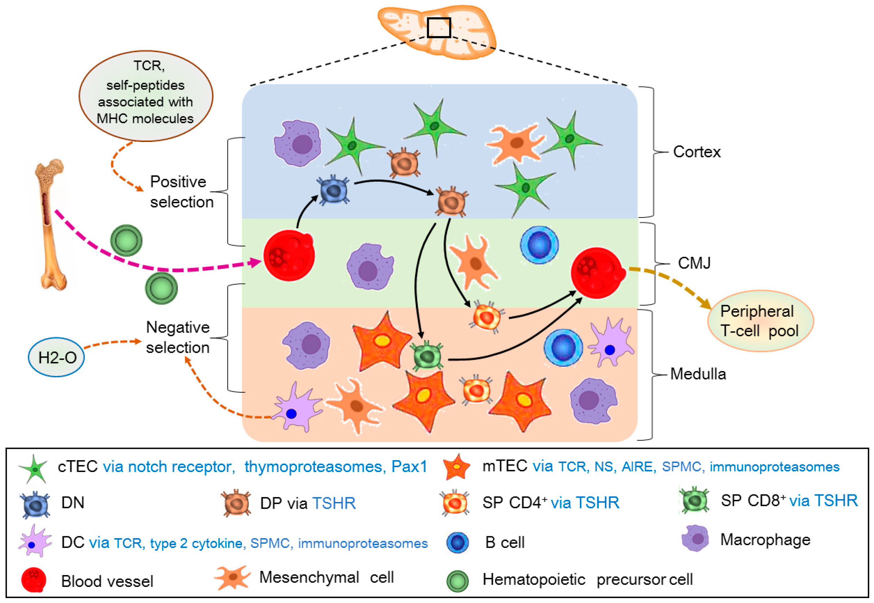
Figure 1. T-cell development in the thymus. First of all, hematopoietic precursor cells from the bone marrow enter the thymus at corticomedullary junction (CMJ) and migrate to the cortex to constitute the CD4 CD8 double negative (DN) thymocytes via interacting with cortical thymic epithelial cells (cTECs). Secondly, DN thymocytes become CD4+CD8+ double positive (DP) in the cortex under the interaction between cortical microenvironments. The positive selection is involved in cTECs via notch receptor, thymoproteasomes, and transcriptional regulatory protein (Pax1), as well as T-cell receptor (TCR) and self-peptides associated with MHC molecules. Finally, the DP thymocytes mature into single-positive (SP) CD4+ or CD8+ T-cells in the medulla under the interaction between medullary microenvironments. The negative selection is related to medulla thymic epithelial cells (mTECs) via TCRs, self-peptide MHC (SPMC), autoimmune regulator (AIRE) and immunoproteasomes, DC via TCR, type 2 cytokine, SPMC and immunoproteasomes, and MHC class II peptide-editing molecular chaperon (H2-O). SP CD4+ or CD8+ T-cells emigrate from the thymus through CMJ to establish the peripheral T-cell pool. In addition, DP, SP CD4+, and CD8+ T-cells are under the influence of thyroid-stimulating hormone (TSH) via its receptor (TSHR).
Cells 14 01534 g001
Figure 2. Pregnancy induces changes in the thymic cortex and size, the medulla, and cell populations. During pregnancy, the thymic cortex and size shrink, but the medulla enlarges and rearranges, and populations of cTECs, mTECs, and mesenchymal cells decease. In addition, populations of the CD4 CD8 double negative (DN), CD4+CD8+ double positive (DP), and single-positive (SP) CD4+ or CD8+ T-cells also decrease. These changes are involved in the modulation of T-cell development in the thymus.
Cells 14 01534 g002
Figure 3. T-cell development is regulated in the maternal thymus during pregnancy. During pregnancy, T-cell development under the thymic microenvironments is regulated by cTECs via PGR, Klf4, ER, MHC, GHR, IGF-1R, and PRLR, and mTECs via Th cytokines, ER, MHC, PGR, GnRHR, GHR, IGF-1R, PRLR, and RANK. In addition, the microenvironments are modulated by B cells via PRLR, mast cells via ER, and DCs via ER, KISS1R, and LepR. Furthermore, T-cell development is regulated in autocrine or paracrine manners through DN T-cells via IL-2R, PRLR, OXTR, DP T-cells via OXTR, VPR, and SP CD4+ and SP CD8+ T-cells via OXTR and VPR.
Cells 14 01534 g003
Figure 4. Hormones regulate maternal thymic immunological adaptations during pregnancy. During pregnancy, placental hormones (including human chorionic gonadotropin (hCG), growth hormone (GH), prolactin, and melatonin) exert effects on the maternal thymus. In addition, ovaries (progesterone (P4) and estrogen (E2)), hypothalamus (gonadotropin-releasing hormone (GnRH) and kisspeptin), and the pituitary gland (GH, thyroid-stimulating hormone (TSH), prolactin, oxytocin, and vasopressin), participate in regulating maternal thymic immunological adaptations during pregnancy. Furthermore, pineal gland (melatonin), adrenal glands (adrenal hormone), pancreas (insulin and glucagon), thyroid (thyroid hormones) and white adipose tissue (leptin) are also involved in maternal thymic immunological adaptations. Moreover, GnRH, insulin-like growth factor 1 (IGF-1), kisspeptin, prolactin, TSH, thyroid hormone, GH, glucocorticoids, melatonin, oxytocin, vasopressine, leptin, and insulin are produced by the thymus, which regulate thymic immunological adaptations in an autocrine or paracrine manner.
Cells 14 01534 g004
Table 1. Summary of the factors and cells in the maternal thymus adaptations during pregnancy.
Factors and CellsEffects on Thymic FunctionEffects of PregnancySpeciesRef.
Osteoclast differentiation receptor RANKDevelopment of thymic regulatory T-cellsFetal loss and gestational diabetesMice[6]
Nuclear progesterone receptorRegulation of thymus involutionIncreases in expression of progesterone receptorMice[7]
Estrogen and thymocyteRegulation of thymus involutionElevating levels of hormonesMice[8]
Thymic nonlymphoid cells, CCL25, CXCL12, CCL21, and CCL19Thymic involutionMaternal–fetal toleranceMice[9]
Toll-like receptorThymic immuneMaternal immune toleranceSheep[10]
NF-κB subunitsThymic immuneMaternal central immune toleranceSheep[11]
Nod-like receptorThymic immune responsesMaternal immunomodulationSheep[12]
Complement componentsThymic immuneMaternal immune regulationSheep[13]
IkappaB proteinThymic immune regulationMaternal immunologic toleranceSheep[14]
Interferon-stimulated genesThymic immuneMaternal immunologic toleranceSheep[67]
Prostaglandin synthasesThymic immune regulationMaternal immunologic toleranceSheep[15]
Sex steroidsCortical involution of the thymusImmune suppression of the mother to paternal and fetal antigensHumans and mice[32]
Strain differenceThymic weightNumber of progenyRats[33]
Epithelial cells of the subcapsular cortex, mTECsThymus shrinks in size and the cortexMaternal tolerance to fetal antigensMice[34]
Estrogen and progesteroneThymic involutionMaternal immune system maintains tolerance towards the allogeneic fetusHumans[35]
Treg cellsImmunosuppressive role of the thymusMaternal–fetal toleranceMice[36]
Cell populationsThymic immune Maternal immune reactivityMice[37]
Gonadotropin-releasing hormoneThymic involutionMaturation of T lymphocytes during pregnancyRats[38]
ProgesteroneThymic involutionAdaptations to the semi-allogeneic fetusHumans[39]
Thymus regulatory T-cells, T-cell receptor, autoimmune regulator, and mTECsThymocyte development and differentiation in the thymusMaternal–fetal tolerance to the fetusHumans[40]
CD4, MT1, and MT2Thymic immune regulationImmune regulation of the maternal immune system Sheep[41]
Helper T cytokinesThymic immune regulationImmune tolerance in maternal immune systemSheep[42]
Klf4, thymic epithelial cellsThymic involutionMaintaining cTEC numbers during pregnancyMice[43]
α/β and γ/δ double negative T-cellsThymocyte loss and thymic involutionMaintenance of pregnancyHumans and mice[44]
Double negative T regulatory cellsThymic developmentImplantation failure, and pregnancy lossHumans and mice[45]
Progesterone, CD4+Foxp3+ TReg cellsThymic involutionEmbryo implantation and fetal growthMice[46]
Aire and mTECsThymic selectionMaintaining maternal–fetal toleranceHumans and mice[47]
Human chorionic gonadotropinAntigen-independent differentiation of T-lymphocytes Production of autocrine growth factors during pregnancyHumans[53]
Estriol and kisspeptinMyeloid DC maturation in the thymusMaintaining systemic tolerance of the motherHumans[58]
EstrogenT-cell developmentImmune suppression for a potential pregnancyHumans and mice[59]
EstrogenInhibit cortical double negative developmentPregnancy-driven involutionHumans and mice[62]
Estrogen receptor α and βThymic immune regulationRegulation of maternal immune function [63]
ProgesteroneThymic involution Normal fertilityHumans, rats, and mice[64]
ProgesteroneThymic involution and T-cell developmentT-cell lymphopoiesis during pregnancyMice[66]
Progesterone receptor and PIBFThymic immunoregulatory functionsMaternal immune toleranceSheep[67]
GnRH and GnRHRModulation of thymus functionBlockade of lymphocyte development in maternal thymusSheep[73]
Prolactin and PRLRThymic innate immune Pregnancy increases expression of prolactin and PRLRSheep[93]
GlucocorticoidThymic immune tolerancePregnancy increases expression of glucocorticoid receptorsHumans and mice[106]
OxytocinsThymic immune regulationInitiation of pregnancyMice and rats[116]
LeptinThymus involutionLoss of thymus mass during pregnancy Mice [122]
Disclaimer/Publisher’s Note: The statements, opinions and data contained in all publications are solely those of the individual author(s) and contributor(s) and not of MDPI and/or the editor(s). MDPI and/or the editor(s) disclaim responsibility for any injury to people or property resulting from any ideas, methods, instructions or products referred to in the content.