Review

m6A mRNA Methylation in Hematopoiesis: The Importance of Writing, Erasing, and Reading

by
Antonia-Gerasimina Vasilopoulou
,
Eleni Kalafati
,
Ekati Drakopoulou
and
Nicholas P. Anagnou
*
Laboratory of Cell and Gene Therapy, Centre of Basic Research, Biomedical Research Foundation of the Academy of Athens (BRFAA), 11527 Athens, Greece
*
Author to whom correspondence should be addressed.
Cells 2025, 14(17), 1388; https://doi.org/10.3390/cells14171388
Submission received: 30 July 2025 / Revised: 26 August 2025 / Accepted: 31 August 2025 / Published: 5 September 2025
(This article belongs to the Section Stem Cells)

Abstract

Over recent years, epitranscriptomic research has provided a new layer of gene regulation during hematopoietic development and aberrant hematopoiesis. Among the 170 identified RNA chemical marks, N6-methyladenosine (m6A) is the most abundant in eukaryotic cells and plays a critical role in various biological processes. This dynamic modification is regulated by a series of methyltransferases, demethylases, and m6A binding proteins, known as writers, erasers, and readers, respectively. Emerging evidence suggests that m6A modification and its regulators are involved in every aspect of normal hematopoietic development, from the emergence of hematopoietic stem cells to the generation of mature blood cells. Also, it has been established that abnormal expression of m6A regulators is implicated in the initiation of blood diseases. In this review, we summarize the latest findings regarding the role of m6A in erythropoiesis and highlight its implications in the pathophysiology of hemoglobin disorders.

1. Introduction

The Biochemistry of N6-Methyladenosine RNA Modification

During the last few decades, advances in epigenetic research have provided novel insights into the mechanisms that control either normal hematopoiesis or lead to blood disorders. The production and development of hematopoietic stem cells (HSCs) is a complex process regulated at multiple levels by epigenetic and epitranscriptomic factors, including DNA methylation, histone modification, chromatin modification, and RNA methylation [1,2,3]. So far, a significant number of post-transcriptional modifications of mRNA have been identified, among which the nitrogen-6-methyladenosine (m6A) is the most common. The m6A marking is a dynamic RNA modification, abundant in endogenous eukaryotic messenger RNAs [4]. Although m6A was first discovered in the early 1970s [5,6], only recent advances in sequencing technologies have allowed for mapping the m6A-modified sites in mammalian cells and tissues and thus have begun deciphering its role in physiological processes and disease [7,8]. Marks of m6A occurring in adenines of RRACH sequence (R=G or A; H=A, C, or U) are in abundance in the 3′ untranslated regions (3′ UTRs), in long internal exons, and around stop codons [7]. The location of these m6A marks is functionally decisive. For instance, when present within the 3′ UTRs or near stop codons, transcript turnover is usually promoted [4,9], while enrichment of m6A in the 5′ UTR region facilitates cap-independent translation initiation by recruiting eukaryotic initiation factor 3 (eIF3), a mechanism particularly important under conditions of cellular stress [10].
The m6A marks are deposited in the nucleus by the m6A methyltransferase (MTase) complex (writers), consisting of methyltransferase-like 3 (METTL3), which is the catalytic subunit; methyltransferase-like 14 (METTL14), which provides structural support and RNA-binding stabilization; Wilm’s tumor 1-associating protein (WTAP), which acts as a regulatory subunit [11]; and other cofactors. Although WTAP lacks inherent methylation activity, it interacts with the METTL3-METTL14 heterodimer to substantially affect the efficiency and specificity of m6A deposition [12]. Alongside the core components of the MTase complex, several co-factors modulate the efficiency and selectivity of m6A deposition. Specifically, vir-like m6A methyltransferase (VIRMA, also known as KIAA1429) guides the writer complex to methylation sites, enriched in 3′ UTR and stop codons [13]; RNA-binding motif protein 15 (RBM15) is a major RNA-binding adaptor that recruits the MTase complex to specific targets [14]; E3 ubiquitin-protein ligase Hakai (CBLL1) contributes to the assembly and stability of the writer complex [13]; and zinc finger CCCH domain-containing protein 13 (ZC3H13) stabilizes the interaction among WTAP, VIRMA, and RBM15 [15]. While the majority of m6A modifications are deposited by the MTase complex, methyltransferase-like 16 (METTL16) has also emerged as a versatile regulator of RNA processing [16,17]. In the nucleus, METTL16 deposits m6A into mRNA targets, whereas in the cytoplasm it promotes mRNA translation through an m6A-independent mechanism [17]. This RNA methylation process is reversed by the m6A demethylases (erasers), fat mass and obesity-associated protein (FTO) [18] and α-ketoglutarate-dependent dioxygenase ALKB homolog 5 (ALKBH5) [19], which thereby reduce the global m6A levels [16,17]. Finally, m6A-binding proteins, known as readers, are located both in the nucleus and in the cytoplasm, and determine the fate of the m6A-modified transcripts by affecting mRNA nuclear export, splicing, stability, and translation efficiency, as depicted in Figure 1. The best-described readers belong to the YT521-B homology (YTH) domain family, which includes the YTHDF1, YTHDF2, YTHDF3, YTHDC1, and YTHDC2 proteins [20]. Among them, the YTHDF proteins located in the cytoplasm are primarily associated with the regulation of mRNA stability and translation, with YTHDF2 accelerating transcript degradation [9] and YTHDF1 and YTHDF3 promoting translation and cooperating in maintaining mRNA turnover balance [21]. In the nucleus, YTHDC1 has been shown to affect alternative splicing of the m6A-modified mRNAs by recruiting splicing factors [22]. Most recently, insulin-like growth factor 2 mRNA-binding proteins (IGF2BPs) and proteins of the heterogeneous nuclear ribonucleoprotein (HNRNP) family were found to impact the stability and translation of targeted mRNAs, as well as the alternative splicing and microRNA processing, respectively [20].
Regarding the biological role of m6A, accumulating evidence suggests that it is involved in a broad spectrum of biological processes, such as tissue development, circadian rhythm, apoptosis, autophagy, and tumorigenesis [23,24,25,26,27]. For instance, dynamic changes in the topology of the m6A marks between fetal and adult stages have been reported, with these modifications being closely linked to genes governing embryonic development and tissue-specific functions [23]. Furthermore, it has been reported that m6A modification regulates circadian rhythms by modulating the processing and stability of key clock gene transcripts. Inhibition of the m6A methyltransferase METTL3 disrupts RNA processing and lengthens the circadian period [24]. In addition, m6A has been shown to influence cell fate decisions by modulating apoptotic pathways, for example, through the regulation of transcripts encoding pro- and anti-apoptotic proteins, thereby affecting cell survival under stress conditions [26]. It also contributes to autophagy by controlling the methylation status and translation efficiency of autophagy-related genes [25]. Regarding its implication in tumorigenesis, several studies suggest that aberrant expression of m6A regulators disrupts normal gene expression programs. These changes can stabilize oncogenic transcripts; destabilize tumor suppressors; and ultimately drive malignant transformation, progression, and therapeutic resistance [27].
Recent studies revealed that m6A-methyladenosine RNA modification plays a crucial role in hematopoietic cell fate decisions by regulating both self-renewal and HSC differentiation [28,29,30,31]. This review highlights insights into how m6A RNA modification regulates hematopoiesis, emphasizing its critical role in erythropoiesis and its impact on the development of hemoglobin disorders.

2. The Role of m6A RNA Methylation in Normal Hematopoiesis and Disease

2.1. HSCs and Endothelial-to-Hematopoietic Transition

The blood system is comprised of more than ten different blood cell types, termed lineages, with numerous functions, from immune surveillance and defense to O2 transport and wound healing [32]. All blood cell types are generated from HSCs that reside mostly in the bone marrow, a major site of adult hematopoiesis [32]. The primary stages of embryonic development of blood tissue literally take place in the absence of any blood cells [33]. The sites of the de novo hematopoietic stem and progenitor cell (HSPC) generation, as well as the maintenance and expansion, are constantly changing in the developing embryo [34]. The first hematopoietic cells are generated in the extraembryonic yolk sac, before the first heartbeat, and later in the allantois and placenta [34]. All adult and many of the later embryonic blood cells are generated from HSCs. However, there is a transient population of primitive blood cells in the early embryo generated from the endothelium through a highly conserved process known as endothelial-to-hematopoietic transition (EHT) [35,36]. Molecular signaling by Notch ligands, hedgehog, bone morphogenetic protein (BMP), vascular endothelial growth factor (VEGF), and nitric oxide signaling play a crucial role in the induction of the hemogenic program in endothelial cells [34].
During EHT, cells undergo significant morphological changes, including the breaking of tight connections with surrounding endothelial cells and rounding up, before escaping into the bloodstream [37]. A complex interplay of critical transcription factors, such as runt-related transcription factor-1 (RUNX1) and c-MYB; signaling pathways, including the Notch pathway; as well as inputs from adjacent tissues, coordinates the entire process [38]. In brief, RUNX1, a key regulator of hematopoiesis, promotes the expression of genes required for endothelial cells to acquire hematopoietic identity [39], whereas c-MYB supports the survival and proliferation of emerging HSCs [40]. Globin transcription factor-2 (GATA-2) further facilitates the process by promoting EHT, even though it is not directly required for the specification of the hemogenic endothelium [41]. Accumulating evidence so far suggests that these transcription factors are regulated by m6A modification, which acts as an additional layer of epitranscriptomic control over the expression of key EHT regulators and plays a pivotal role in the successful generation of HSCs.
Specifically, Zhang and colleagues [42] showed that reduced METTL3 protein, and hence reduced m6A levels in zebrafish embryos, lead to EHT disruption and eventually to complete loss of specialized blood cells. Interestingly, epitranscriptome mapping revealed that the lack of HSC emergence was caused by reduced m6A mRNA methylation of neurogenic locus notch homolog protein 1 (Notch1) and of other arterial endothelial genes, which are normally under YTHDF2-mediated degradation [42]. Consistently, lower levels of the HSPC markers RUNX1 and c-myb were detected, and analogous results were obtained by ythdf2 zebrafish morphants [42]. Additionally, m6A implication in EHT regulation was confirmed in Mettl3-knockdown hematopoietic stem and progenitor cells acquired from the aorta-gonad-mesonephros region (AGM) of mice [42]. Employment of m6A-RNA immunoprecipitation (RIP)-qPCR assays and conventional qPCR analysis revealed that Notch1 is m6A-targeted, and its expression is enhanced following Mettl3 knockdown. Furthermore, in vivo experiments using the Cre/LoxP system to knockout the expression of Mettl3 specifically in endothelial cells of the mouse AGM region provided consistent results [42]. Collectively, METTL3 and YTHDF2 operate synergistically to suppress the Notch signaling pathway and thus affect the EHT process [42]. Similarly, a study by Lv et al. [43] showed that METTL3 depletion in vascular endothelial cells of mouse AGM leads to a significant suppression of HSPC function. Moreover, there seems to be an interplay between epigenetic and epitranscriptomic mechanisms that are involved in EHT, such as microRNAs (miRNAs), known to affect HSC production and control m6A levels via modulating METTL3 binding to mRNAs [44]. The above findings clearly show that m6A modification plays an essential role during vertebrate definitive hematopoiesis by fine-tuning the expression of key transcription factors such as RUNX1, c-MYB, and GATA2 as well as signaling molecules like Notch1 to ensure precise HSC emergence [44].

2.2. HSC Self-Renewal and Maintenance

Hematopoietic stem cells are multipotent precursors that have the dual ability for self-renewal and for giving rise to myeloid, lymphoid, and megakaryocytic-erythroid cell lineages throughout life [32]. This equilibrium is maintained by cell fate decisions made during cellular division [32]. It has not been fully elucidated yet which signals determine whether a cellular division will result in lineage commitment and differentiation or in self-renewal. The expansion and maintenance of HSC self-renewal are coordinated by signaling pathways, including Notch [45], Wnt [46], BMP [47], and Hedgehog [48] pathways. For instance, Notch signaling promotes HSC quiescence and prevents premature HSC differentiation, whereas canonical Wnt signaling is activated by extracellular proteins and supports symmetric self-renewal divisions. These signaling pathways are regulated by both intrinsic and extrinsic mechanisms [49]. Intrinsic mechanisms include transcription factors, such as RUNX1, GATA-2, c-MYB, and T-cell acute lymphocytic leukemia protein 1 (TAL1), as well as epigenetic and epitranscriptomic regulators, including DNA methylation enzymes and m6A-related proteins. Extrinsic mechanisms are mediated by the HSC niche, which provides physical interactions, growth factors, and chemical cues that trigger the different signaling pathways regulating HSC fate decisions [49]. According to recent studies, m6A modification and its regulators seem to be key participants in HSC homeostasis [50,51,52,53].
Among the m6A readers, YTHDF2, which mediates targeted m6A-mRNA degradation, was reported to uniquely influence HSC regeneration and expansion. Recent studies showed that the loss of YTHDF2 leads to increased proliferation of HSC without altering lineage bias [50]. Particularly, Wang et al. [50] exploited Cre/LoxP technology to create hematopoietic-specific Ythdf2-knockout mice. According to their study, Ythdf2 expression in HSCs expedites the decay of m6A-marked mRNAs of Wnt target genes, leading to suppression of Wnt signaling at steady state. Given the fact that aberrant activation of Wnt signaling leads to an increase in the number of HSCs with lower functional response to hematological stressors, YTHDF2 probably has a protective role. Interestingly, upon YTHDF2 loss, not only was the number of HSCs increased, but so was the regenerative capacity of HSCs under stress circumstances [50]. Furthermore, Mapperley et al. [51] showed that YTHDF2 protects HSCs from proinflammatory signals, which cause excessive proliferation and promote myeloid-biased differentiation. These data also suggest that Ythdf2 deletion results in the expansion of HSCs and multipotent progenitors (MPPs) [51]. Similar observations were made by Li et al. [52] after the depletion of YTHDF2 in mouse HSPCs and human umbilical cord blood HSCs. Data obtained by Ythdf2 knockout mice indicated that YTHDF2 normally suppresses HSC self-renewal through RNA degradation. Furthermore, m6A mapping revealed that mRNAs of transcription factors important for stem cell self-renewal, such as TAL1 and GATA-2, are m6A-labeled and recognized by YTHDF2, which drives them to decay sites [52]. Moreover, Ythdf2 knockdown in human umbilical cord blood HSCs led to decreased apoptotic rates and enhanced expansion ex vivo, providing possible new strategies for future stem cell-based treatments [52]. Reader control of hematopoiesis extends beyond YTHDF2. Another YTH domain containing an m6A reader, the YTHDF3, cooperates with METTL3 to recognize and promote the translation of the m6A-modified Cyclin D1 mRNA, which is a key regulator of cell cycle progression, thereby supporting HSC self-renewal [54]. In addition to cytoplasmic readers, the nuclear m6A reader YTHDC1 is also essential for normal hematopoiesis and HSPC maintenance according to recent studies, partly by regulating miRNA maturation [53,55]. Loss of YTHDC1 results in HSC apoptosis, highlighting its critical role in HSC survival [55]. In line with the previous studies, Yin et al. [31] proposed that m6A could be considered as a quality control system for the preservation of HSC integrity. Their findings suggest that the reader protein IGF2BP2 plays a critical role in maintaining HSC stemness by regulating the expression state of m6A-methylated mRNAs as well as mitochondrial activity of HSCs [31]. Taken together, these studies suggest that the m6A readers cooperatively regulate transcript stability, signaling, and metabolic homeostasis to preserve HSC self-renewal and functional integrity.
Other m6A regulators whose role has been studied in mammalian hematopoietic development include the m6A writers METTL3 and METTL14. Their role in the control of HSC self-renewal was assessed in adult mouse bone marrow utilizing Cre/LoxP technology to create Mettl3-/Mettl14-deficient mice [56]. Deletion of these genes led to the accumulation of HSCs and a reduction of their self-renewal capacity. Specifically, it was shown that METTL3 enhances the expression of genes related to HSC quiescence, maintenance, and self-renewal, such as nuclear receptor subfamily 4 group A member 2 (Nr4a2), cyclin-dependent kinase inhibitor 1A (CDK1A), Bmi-1 proto-oncogene polycomb ring finger, and PR domain-containing 16 (Prdm16) [56]. Notably, qPCR analysis showed no difference in the expression of genes related to HSC differentiation, such as Ikaros, PU.1, and Mef2c [56]. Consistent with these results, deletion of Mettl3 in adult mouse HSCs, and therefore lack of m6A, leads to an accumulation of HSCs in the bone marrow while having no important effect on HSC self-renewal and quiescence [29]. On the contrary, a recent study demonstrated that the previously identified accumulated HSCs are actually blocked HSC-multipotent progenitor-like cells, which fail to differentiate due to loss of Myc, which controls HSC symmetric commitment and is normally under m6A regulation [3]. Moreover, it was shown that Mettl3 knock-out HSCs are less quiescent and have increased metabolism, as well as mitochondrial activity, indicating that m6A is essential for HSC quiescence and self-renewal [3]. In addition, loss of METTL3-mediated m6A modification decreases HSC quiescence, increases metabolism and mitochondrial activity, and enhances the formation of endogenous double-stranded RNAs and abnormal stimulation of melanoma differentiation-associated protein 5 (MDA5)-retinoic acid-inducible gene I (RIG-I), protein kinase R (PKR)-eukaryotic initiation factor 2α (eIF2α), and 2′,5′-oligoadenylate synthetase/ribonuclease L (OAS-RNase L) pathways in HSPCs, leading to accumulation of dysfunctional immature cells in vitro and in vivo [57]. Finally, m6A erasers are also implicated in the regulation of HSC self-renewal. ALKBH5 was found to stabilize transcripts of key metabolic enzymes, such as oxoglutarate dehydrogenase (OGDH) [58]. Loss of ALKBH5 results in destabilization of Ogdh mRNA; impaired mitochondrial oxidative phosphorylation and ATP production; and consequently reduced HSC metabolic activity, self-renewal, and long-term repopulation potential [58]. More recently, FTO-mediated regulation of m6A levels was shown to be essential for HSC expansion and homing [59].
Taken together, m6A RNA modifications and their regulators play a central role in maintaining HSC homeostasis, self-renewal, and functional integrity, as summarized in Table 1. The MTase complex is critical for controlling HSC differentiation and preventing the accumulation of dysfunctional progenitors, while the m6A eraser ALKBH5 ensures metabolic stability by regulating key enzymatic transcripts. Finally, m6A readers, such as YTHDF2, YTHDF3, YTHDC1, and IGF2BP2, cooperatively regulate transcript stability, signaling pathways, and metabolic activity to preserve HSC quiescence and regenerative capacity. Collectively, these findings highlight that m6A-mediated epitranscriptomic regulation functions as a quality control system, coordinating intrinsic and extrinsic signals to safeguard HSC fate decisions, lineage commitment, and stress response.

2.3. Erythropoiesis

Hematopoietic stem cells replenish all blood cell types on a continuous basis through a series of differentiation stages and recurrent cellular divisions that entail the production of lineage-committed progenitors. Daily, the erythroid progenitors expand enormously to generate 200 billion red blood cells in the bone marrow during a stepwise maturation process known as erythropoiesis [60]. Erythropoiesis is controlled by a network of factors, such as oxygen sensors, cytokines, and regulators of iron metabolism [60]. Any abnormalities in critical components of this process can result in severe disorders such as anemia or myelodysplastic syndromes [60]. Briefly, HSCs initially give rise to multipotent progenitors (MPPs), which in turn give rise either to common myeloid precursors (CMPs) or common lymphoid precursors (CLPs), through cytokine signaling and activation of several transcription factors [61,62]. MPPs differentiate into CLPs that originate lymphocytes and natural killer cells that rely mainly on activation by SPI1, Ikaros, and GATA-3 transcription factors [61,63,64,65]. MPP fate-decision differentiation into CMP generates granulocyte-macrophage (GMP) and megakaryocyte-erythroid progenitors (MEPs), modulated by SPI1 and GATA-1 [61,65]. The differentiation of CMP relies on the secretion of granulocyte-macrophage-colony stimulating factor (GM-CSF) and subsequently on macrophage-colony stimulating factor (M-CSF), which regulates the differentiation of monocytes/macrophages, while granulocyte-colony stimulating factor (G-CSF) regulates the differentiation of neutrophils, basophils, and eosinophils. During erythropoiesis, MEP differentiates into burst-forming unit-erythroid (BFU-E) and, finally, into colony-forming unit-erythroid (CFU-E) [60]. This process is modulated by soluble factors such as erythropoietin (EPO), stem cell factor (SCF), interleukin-3 (IL-3), and interleukin-6 (IL-6), as well as by the activation of GATA-1, signal transducer and activator of transcription 5 (STAT-5), and Krüppel-like factor-1 (KLF-1) pathways [60].
Despite the extensive knowledge in this field, many of the regulatory features that control HSC differentiation, as well as the production and maturation of erythrocytes, remain unknown. Numerous studies have shown that m6A RNA methylation plays a pivotal role in normal and pathological erythropoiesis [3,66,67,68]. Epitranscriptomic sequencing, either with m6A-sequencing or by MeRIP-seq technology, has enabled the mapping of m6A sites on the mRNA molecules of mammalian cells. Notably, transcripts of transcription factors that coordinate erythroid differentiation, such as KLF1, GATA-1, GATA-2, Friend leukemia integration 1 transcription factor (FLI1), and myeloproliferative leukemia protein (MPL), and of genes involved in erythroid disorders, were found to be enriched in m6A RNA methylation [66]. KLF1 and GATA-1 are known to work together, as well as with other cofactors and chromatin modifiers, to drive the erythroid transcriptional program [66]. Low levels or complete loss of GATA-1 have been linked with impaired terminal erythroid differentiation and anemia development [69]. KLF1 is a principal regulator of erythropoiesis that directly regulates the expression of genes involved in α and β-globin and iron-bound heme biosynthesis [70]. Particularly, KLF1 has been demonstrated to activate the expression of β-globin and regulate the expression of genes that participate in the production of hemoglobin molecules, such as B-cell lymphoma/leukemia 11A (BCL11A), which is known to control the production of fetal γ-globin [69]. Erythropoiesis is substantially hampered upon loss of KLF1 [70]. As a result, the reduction or absence of β-globin chain production leads to the accumulation of an excess of α-globin chains, which precipitate, forming insoluble aggregates in the red blood cell precursors, leading to apoptosis at the polychromatophilic erythroblast stage [71].
Genome-wide CRISPR screening in human erythroleukemia (HEL) cells first established the requirement of the MTase complex in erythropoiesis [66]. Specifically, transduction of HEL cells with lentiviral sgRNA reagents, followed by sorting of CD235- cells out of the population of the outgrown HEL cells, disclosed that METTL3, METTL14, and WTAP were among the genes required for normal erythroid differentiation. Next, CRISPR-Cas9 gene targeting was carried out to knock out each of the main components of the MTase complex and thus to elicit a reduction in global m6A levels. The data documented that elimination of the m6A MTase complex results in erythroid differentiation blockage without affecting the development of other non-lymphoid cell lineages. Further analysis showed that m6A marking enhanced the translation of genes involved in erythroblast differentiation and heme biosynthesis as well as of genes that constitute the H3K4 MTase network [66]. Intriguingly, METTL3 and WTAP knockdown led to the loss of H3K4me3 marking in KLF1-bound promoters, leading to decreased binding capacity [66]. Similar results were obtained following m6A loss in human adult bone marrow HSPCs. Knockdown of MTase complex components revealed that their activity is required for early erythropoiesis but not for megakaryocytic differentiation [66]. Also, it was revealed that WTAP enhances the translation of polyadenylate-binding protein 1 and 4 (PABPC1, PABPC4), which promote the expression of β-globin in human HSPCs. Taken together, the data suggest that m6A modification is implicated in the regulation of erythropoiesis by maintaining the H3K4me3 marking at erythroid gene targets and promoting erythroid lineage development [66].
Beyond these m6A writers, METTL16, which preferentially deposits m6A marks in structured RNAs, has emerged as an additional m6A methyltransferase with distinct targets relevant to erythroid homeostasis [68,72]. Notably, METTL16 targets the methionine adenosytransferase 2A (MAT2A) transcript, which is responsible for producing S-adenosylmethionine (SAM) [72]. SAM is a universal methyl donor essential for various methylation reactions in cells. By modulating MAT2A expression, METTL16 indirectly influences SAM levels, thereby impacting global methylation capacity and cellular methylation-dependent processes [72]. Because SAM is required for METTL3-mediated m6A installation and for histone methyltransferases that deposit marks such as H3K4me3, METTL16 can indirectly influence both RNA methylation and chromatin states that are critical for erythroid gene expression. Yoshinaga et al. [68] showed that METTL16 depletion in erythroblasts causes loss of m6A on DNA-repair transcripts, accumulation of DNA damage, and increased apoptosis [68,73]. Interestingly, supplementing cells with SAM can mitigate some defects, demonstrating that METTL16’s control of SAM contributes functionally to erythropoiesis [68]. Collectively, these findings position METTL16 as a crucial regulator of erythropoiesis.
The significance of the m6A writers in erythropoiesis is further highlighted in the regulation of MYC, a proto-oncogenic protein known to control the balance between HSC self-renewal and differentiation. During terminal erythroid differentiation, its protein levels are rapidly decreased [74]. Transcripts of c-MYC, BCL-2, and phosphatase and tensin homolog (PTEN) are enriched in m6A marks [74]. Specifically, METTL3-mediated m6A targeting of these transcripts promotes their translation and subsequently blocks differentiation of HSPCs [74]. Importantly, the functional outcome of these modifications depends on their positional placement within the sequence of mRNAs. In the case of c-MYC, m6A methylation within a 250 bp region at the 3′ end of the coding sequence and 3′ UTR enhances transcript stability and translation through the recognition by the reader family IGF2BPs and YTHDF1, thereby sustaining HSC self-renewal [75,76,77]. Similarly, m6A in the 3′ UTRs of BCL-2 and PTEN promotes efficient translation and stability, further supporting survival and blocking differentiation of HSPCs [54,77]. Although a complete positional mapping of these transcripts remains unresolved, accumulating evidence underscores that the location of m6A marks is critical in dictating their fate during hematopoietic regulation.
Furthermore, key components of the MTase complex seem to regulate Myc RNA stability and expression in HSPCs [3]. Specifically, siRNA depletion of Mettl3 in normal murine HSPCs resulted in decreased MYC protein levels and blockage of HSC symmetric commitment, while in Mettl3-knockout mice, accumulation of immature erythroblasts, leading to spleen enlargement and disruption of its architecture, was observed. METTL14 was found to be negatively regulated by the SPI1 transcription factor both in normal and pathological conditions such as acute myeloid leukemia [78]. For the first time, Weng and coworkers [78] described the signaling axis SPI1-METTL14-MYB/MYC in normal myelopoiesis and leukemogenesis and proposed that METTL14 could be a new therapeutic target to treat acute myeloid leukemia (AML). This view is further supported by the fact that upon its deletion, HSCs are expanded, while AML malignant stem cells are selectively compromised [79].
Moreover, c-Myc expression is regulated by the RBM15, which was recently identified as a component of the MTase complex [80]. RBM15 was found to be highly expressed in the hematopoietic system [80,81]. Particularly, in vitro studies of cultured AML cells showed that knockdown of RBM15 leads to impaired differentiation and increased apoptosis rate. Also, knockout mouse studies revealed that RBM15 is implicated in HSC expansion and differentiation, probably by affecting Notch signaling and c-Myc expression, respectively [81]. Furthermore, it was found that the expression of METTL14 and FTO is significantly lower in mature hematopoietic cells. No significant expression pattern was found for METTL3, WTAP, or ALKBH5. Consistent with these data, recent studies showed that ALKBH5 is not required for maintaining the function of normal HSCs [82,83]. Moreover, METTL14 knockdown in normal CD34+ cells resulted in enhanced myeloid differentiation, colony formation inhibition, and decreased expression of the oncogenic transcription factors MYB and MYC [3]. Normally, METTL14 and FTO were found to promote the stability of MYC mRNA via IGF2BP-mediated regulation of its expression [3]. Both YTHDF2 and IGF2BPs act as regulators of mRNA stability. However, contrary to the mRNA degradation-promoting function of YTHDF2, IGF2BPs were reported to promote mRNA stability [77]. In vivo models of murine AML and analysis of primary samples from patients with AML revealed that the RNA-binding protein Y-box binding protein 1 (YBX1) affects the stability of m6A-enriched transcripts of apoptotic genes, such as Bcl2 and c-Myc, by interacting with IGF2BPs [84]. However, two independent studies demonstrated that YBX1 does not affect normal hematopoiesis or the function of HSCs [73,84]. It is also noteworthy that a recent study in cancer cells documented that the m6A reader protein YTHDF1 affects transferrin receptor-mediated iron metabolism [85].
Collectively, these studies highlight that m6A RNA methylation is a critical regulator of erythropoiesis, controlling HSPC differentiation, erythroid lineage commitment, and terminal maturation. m6A writers, such as METTL3, METTL14, WTAP, and METTL16, as well as m6A readers (YTHDF1, IGF2BPs) coordinate the differentiation of HSPCs into erythroid lineages by modulating factors like GATA-1, KLF1, and MYC as well as ensuring proper heme biosynthesis and globin production. Dysregulation of this epitranscriptomic network can lead to impaired erythroid maturation, anemia, or hematologic malignancies, emphasizing the therapeutic potential of targeting m6A pathways in blood disorders.

2.4. The Role of m6A Modification in Malignant Myeloid Hematopoiesis
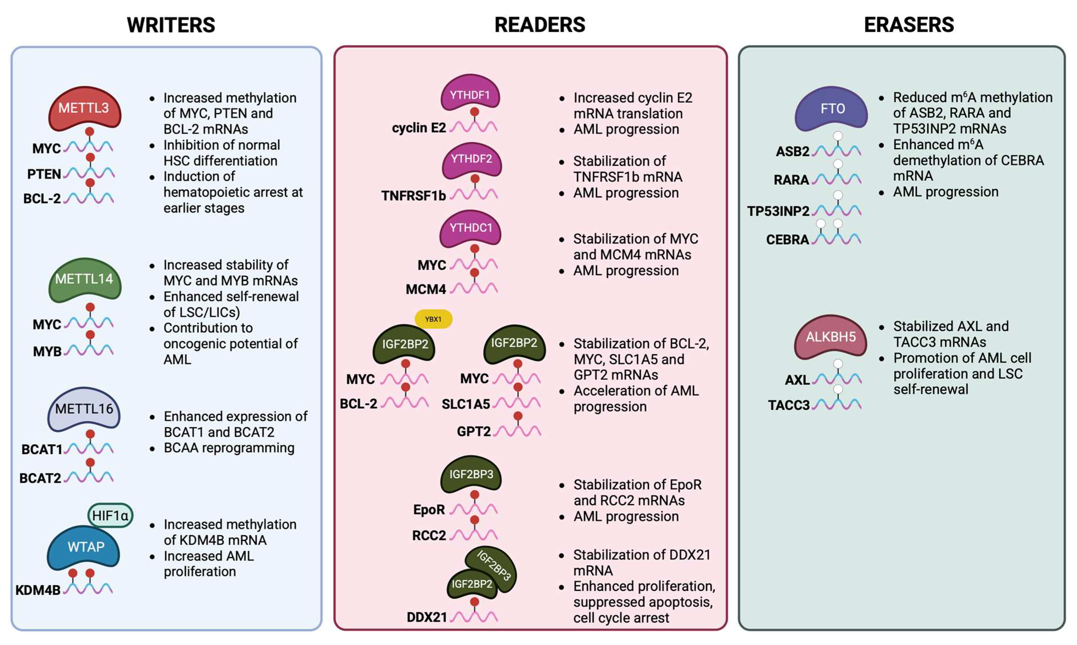
Studies investigating the role of m6A modification in hematologic malignancies have demonstrated that it profoundly affects gene expression, signaling networks, and cell fate decisions. Acute myeloid leukemia, the most prevalent and aggressive form of acute leukemia in adults, is characterized by the abnormal clonal proliferation of primitive HSPCs. Dysregulation of m6A marking was found to significantly alter gene expression programs, thereby sustaining leukemia stem cell (LSC) self-renewal, blocking normal HSC differentiation, and driving leukemogenesis. The basic m6A modifications that have been reported in AML are summarized in Figure 2.
Figure 2. The basic m6A modifications occurring in acute myeloid leukemia (AML). Methyltransferases, demethylases, and reader proteins contribute to AML progression by regulating the stability and/or decay of different crucial mRNA transcripts, thus interfering with translation and eventual protein production. MYC, myelocytomatosis oncogene (also known as c-Myc); PTEN, phosphatase and tensin homolog; BCL-2, B-cell lymphoma 2; MYB, myeloblastosis oncogene; LSC, leukemia stem cell; LIC, leukemia-initiating cell; BCAA, branched-chain amino acid; BCAT1/2, branched-chain amino acid transaminase 1/2; HIF1α, hypoxia-inducible factor 1-alpha; KDM4B, lysine demethylase 4B; TNFRSF1b, tumor necrosis factor receptor superfamily member 1b; MCM4, minichromosome maintenance complex component 4; YBX1, Y box-binding protein 1; SLC1A5, solute carrier family 1 member 5; GPT2, glutamic–pyruvic transaminase 2; EpoR, erythropoietin receptor; RCC2, regulator of chromosome condensation 2; DDX21, DEAD-box helicase 21; ASB2, ankyrin repeat and SOCS box containing 2; RARA, retinoic acid receptor alpha; TP53INP2, tumor protein P53 inducible nuclear protein 2; CEBPA, CCAAT enhancer-binding protein alpha; AXL, AXL receptor tyrosine kinase; TACC3, transforming acidic coiled-coil-containing protein 3. Adapted from Chen et al. [93]. Created in Biorender. Vasilopoulou, A-G. (2025) https://BioRender.com/nk1sx3l (accessed on 26 August 2025).
Cells 14 01388 g002
Aberrant expression of m6A writers in AML compared to normal HSCs alters the methylation levels of key transcripts, promoting malignant transformation. In AML patients, METTL3 was found to be upregulated at diagnosis and during relapse compared to patients in complete remission [86]. This overexpression of METTL3 results in increased methylation of MYC, PTEN, and BCL-2 transcripts, and, thus, upregulation of their expression and inhibition of normal HSC differentiation [74]. Clinical and translation studies further link upregulated METTL3 expression to the development of AML chemoresistance by regulating the homing and engraftment of AML cells [86]. Similarly, METTL14 was found to contribute to AML development by stabilizing transcripts of MYC and MYB, which leads to enhanced self-renewal of LSCs. Furthermore, Han et al. [87] showed that METTL16 is essential for survival and stemness of the AML cells by regulating the branched-chain amino acid (BCAA) metabolic pathway and particularly the expression of branched-chain amino acid transaminases 1 and 2 (BCAT1 and BCAT2) [87]. BCAA metabolism sustains the anabolic demands of LSCs, thereby supporting their rapid proliferation [87]. In AML, WTAP is also activated by the hypoxia-inducible factor 1α (HIF1α) and consequently increases the m6A methylation of lysine-specific demethylase 4B (KDM4B) transcripts [88]. This modification stabilizes KDM4B transcripts and further enhances the proliferation of LSCs [88].
m6A demethylases were also found to be upregulated in AML, contributing to its pathogenesis. This upregulation leads to decreased m6A methylation levels of mRNAs that drive myeloid differentiation, including retinoic acid receptor alpha (RARA) and ankyrin repeat and SOCS box protein 2 (ASB2) [89]. More specifically, increased expression of FTO results in destabilization and reduced translation of these transcripts, thereby blocking normal differentiation and promoting AML progression [89]. Additionally, ALKBH5 is required for LSC maintenance and function during the leukemogenesis of AML [82]. By erasing m6A marks on specific target mRNAs, ALKBH5 protects oncogenic transcripts from YTHDF-mediated decay, thereby supporting AML pathogenesis [82].
m6A reader proteins also play an important role in AML by modulating the fate of the modified mRNAs and thereby influencing leukemogenesis. The cytoplasmic readers YTHDF1 and YTHDF2 promote AML progression by enhancing the translation of cyclin E2 and inhibiting the expression of TNF-α, respectively [79]. Increased expression of cyclin E2 promotes LSC proliferation, whereas downregulation of TNF-α results in suppressed apoptosis, further supporting LSC self-renewal. Nuclear m6A readers, including YTHDC1 and HNRNPC, regulate pre-mRNA splicing and processing of genes involved in cell cycle control and differentiation, such as MYC, thereby supporting AML cell survival [53,90]. Additionally, the IGF2BP family (IGF2BP1–3) stabilizes oncogenic mRNAs such as MYC and BCL-2, enhancing their translation and sustaining LSC self-renewal [76,91]. Taken together, these readers ensure that oncogenic transcripts are efficiently translated and protected from decay, reinforcing the block in differentiation and promoting leukemogenesis.
Table 1. The role of different m6A regulators in the renewal, differentiation, and maintenance of HSCs.
m6A RegulatorTypeRole in HSCsEffect on Self-Renewal, Differentiation and MaintainanceReferences
METTL3Writer
(m6A methyltransferase)
Catalyzes m6A deposition with METTL14Essential for HSC self-renewal; loss leads to impaired HSC maintenance, defective differentiation, and bone marrow failure[56,57]
METTL14Writer
(METTL3 partner)
Stabilizes METTL3 and regulates target mRNAsRequired for HSC self-renewal; deletion causes loss of HSC quiescence and differentiation bias[29,56]
WTAPWriter complex componentRegulatory subunit for METTL3/METTL14Contributes to HSC survival and lineage commitment[66]
FTOEraser
(demethylase)
Removes m6A from target transcriptsOverexpression reduces differentiation, maintaining stemness; depletion promotes myeloid differentiation[59]
ALKBH5Eraser
(demethylase)
Regulates mRNA stability and splicingHelps maintain stem cell pool under stress;
linked to leukemogenesis
[58]
YTDHF1Reader
(cytoplasmic)
Enhances translation of m6A-marked transcriptsPromotes lineage-specific differentiation by boosting translation of key regulators[54]
YTDHF2Reader
(cytoplasmic)
Mediates degradation of m6A-modified RNAsCritical for HSC self-renewal; deletion leads to increased HSC numbers but reduced long-term function[50,51,52]
YTHDF3Reader
(cooperates with YTHDF1/2)
Balances translation vs decayCoordinates with YTHDF1/2 in HSC maintenance[54]
YTHDC1Reader
(nuclear)
Controls mRNA splicing and exportEnsures proper transcript processing in HSC renewal;
deletion causes impaired hematopoiesis
[53,55]
IGF2BP1/2/3Readers
(stabilizers)
Bind and stabilize m6A-marked mRNAsEnhance expression of stemness-associated genes, supporting HSC maintenance and survival[31]
HNRNPCReader
(nuclear)
Binds to m6A-switch structuresControls transcript fate, influencing HSC homeostasis[90]

2.5. Ineffective Erythropoiesis and Apoptosis

Apoptosis and autophagy are cell death machineries widely associated with ineffective erythropoiesis and blood cancers. More specifically, apoptosis eliminates defective or excess erythroid progenitors, preventing accumulation of abnormal cells, while autophagy degrades and recycles damaged organelles and proteins, facilitating erythroblast maturation and enucleation. Dysregulation of either process can lead to ineffective erythropoiesis, as observed in disorders such as β-thalassemia and myelodysplastic syndromes, and contributes to hematologic malignancies [92,93]. Notably, transcripts of pro-apoptotic and apoptotic genes, for instance MYC, BCL-2, and PTEN, as well as autophagy-related genes, for instance, the autophagy-related 7 gene, ATG7, were found to be enriched in m6A marks [25,94,95]. The presence or absence of m6A on these RNAs substantially alters their translation, stability, or decay with direct consequences for cell survival.
Several studies have shown that METTL3, which is the core component of the MTase complex, plays an important role in apoptosis mainly by regulating the expression of Bcl-2 [96]. Specifically, METTL3 promotes the translation of pro-survival mRNAs in myeloid cells, including MYC and BCL-2, thereby favoring cell survival and blocking differentiation. Additionally, the demethylases FTO and ALKBH5 primarily function to prevent apoptosis. FTO knockdown in AML cell lines led to increased m6A levels of MYC mRNA, promoting its degradation and thus enhancing apoptosis [97]. m6A regulators are also involved in the autophagy pathway [98,99]. Specifically, it has been demonstrated that m6A RNA methylation controls the expression of autophagy-related gene (ATG) transcripts [98,99]. The m6A-marked ATG transcripts are under YTHDF2-mediated degradation and, therefore, autophagy is inhibited [98]. Further research is needed to decipher how the relationship between m6A and cell death could be implicated in the pathophysiology of hematologic diseases.

2.6. The Role of m6A Modification in Thalassemia
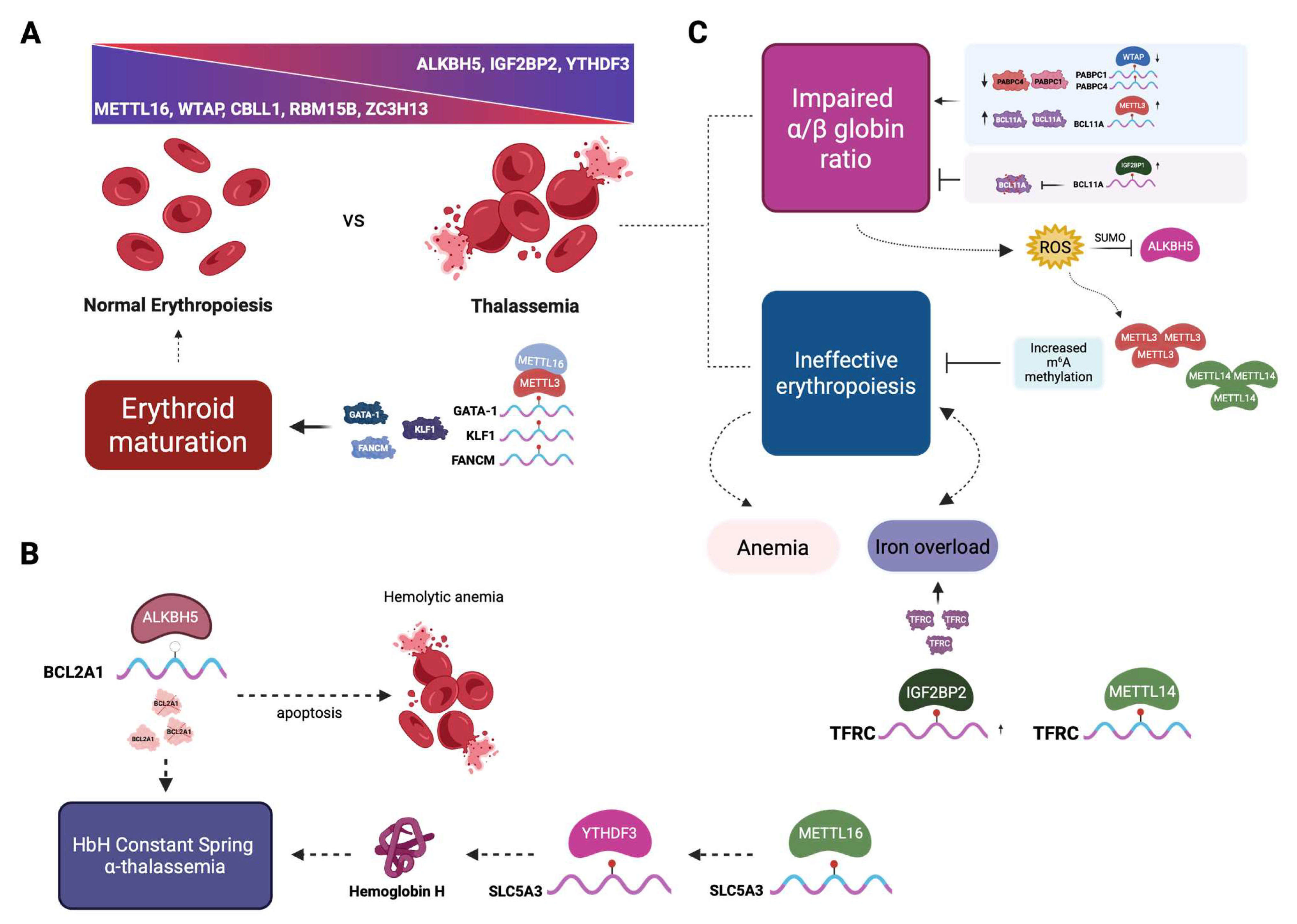
Recent studies revealed that m6A RNA modification also affects the expression of genes involved in hemoglobin production, heme biosynthesis, iron metabolism, and cell death mechanisms, such as apoptosis and autophagy [85,97]. These findings imply that m6A modification could also be a key participant in the pathogenesis of thalassemic hematopoiesis (Figure 3). Recently, Ruan et al. [100] demonstrated for the first time that m6A modification is linked to the phenotype of the Hemoglobin H Constant Spring variant, a severe form of α-thalassemia caused by mutations of the duplicated α-globin genes (--/αCSα) that affect the production of α-globin chains. For their study, immature erythrocytes were derived from the peripheral blood of 16 thalassemia patients and 15 healthy individuals. Data acquired from qRT-PCR and m6A mRNA and lncRNA epitranscriptomic microarrays revealed that the expression pattern of m6A-related factors is significantly different between the two groups. Specifically, they discovered that the expression of the MTase complex components METTL16, WTAP, CBLL1, RBM15B, and ZC3H13 was lower in this type of α-thalassemia compared to normal erythroid cells, whereas the eraser protein ALKBH5, as well as the readers IGF2BP2 and YTHDF3, were overexpressed (Figure 3A). However, these findings were based on transcriptomic analyses and the decreased expression of the above genes reflects changes at the mRNA level. The impact of these transcript-level alterations on protein abundance remains unverified, as proteomic assessment was beyond the scope of that study and no published data are currently available addressing this issue. Furthermore, the authors employed m6A mapping to identify the differentially hypo-m6A-methylated transcripts in the two cohorts. Interestingly, the transcript of the anti-apoptotic protein BCL2A1 was downregulated in thalassemia patients. Therefore, in HbH Constant Spring α-thalassemia, apoptosis remains uninhibited as a result of the decreased expression of the BCL2A1, eventually leading eventually to hemolytic anemia (Figure 3B). According to their findings, the ALKBH5-mediated alteration of the m6A methylation status of BCL2A1 contributes to the pathogenesis of α-thalassemia. However, the exact mechanism by which m6A modification regulates BCL2A1 expression remains to be elucidated.
Further evidence for the role of METTL16 in the pathogenesis of Hemoglobin H disease was provided recently [16], where the METTL16, YTHDF3, and solute carrier family 5 member 3 (SLC5A3) mRNAs were downregulated in HbH patients. Modification of the METTL16 levels in human erythroleukemia K562 cells provided evidence that METTL16 affects the expression of hemoglobin via the modification of SLC5A3 mRNA and its sequential reading by YTHDF3 [16], as shown in Figure 3B. Further research is needed to elucidate the mechanisms by which m6A modification influences thalassemic hematopoiesis and to explore potential therapeutic strategies targeting these pathways.
Figure 3. The impact of m6A modifications in thalassemia and their role in the disease pathophysiology. (A). Normal erythroblasts demonstrate high levels of METTL16, WTAP, CBLL1, RBM15B, and ZC3H13, while erythroblasts of HbH disease Constant Spring α-thalassemia overexpress ALKBH5, IGF2BP2, and YTHDF3. Furthermore, in normal erythropoiesis, increased expression of METTL16 and METTL3 positively regulates the production of the erythroid regulatory proteins GATA-1, KLF1, and FANCM, preventing DNA damage and apoptosis, thus ensuring efficient erythroid maturation. (B). Ιn HbH Constant Spring α-thalassemia, the anti-apoptotic protein BCL2A1 is downregulated via the action of ALKBH5, and as a result, apoptosis remains uninhibited, leading eventually to hemolytic anemia. Furthermore, it has been speculated that the expression of Hemoglobin H and hence the disease phenotype is affected by the modification of SLC5A3 mRNA by METTL16 and its sequential reading by YTHDF3. (C). On the other hand, the β-thalassemia phenotype is primarily the result of impaired α/β ratio of globin synthesis and ineffective erythropoiesis. In the former case, reduced m6A methylation of PABPC1 and PABPC4 mRNAs by WTAP leads to reduced PABPC1 and PABPC4 protein levels and deteriorates the detrimental effects of the impaired α/β ratio since these two proteins affect mRNA isoform diversity during erythropoiesis and stabilize a subset of erythroid mRNAs, shielding them from degradation, a crucial feature for terminal erythroid maturation [101]. Furthermore, elevated METTL3 expression stabilizes BCL11A, a major γ-globin repressor, inhibiting the capacity for the beneficial reactivation of the fetal γ-globin genes for HbF production. Interestingly, overexpression of the reader IGF2BP1 has been recently shown to induce fetal hemoglobin in cultured erythroid cells from patients with thalassemia or sickle cell disease, reducing the α/β globin imbalance, probably by inhibiting the translation of BCL11A mRNA via binding [102]. With regards to ineffective erythropoiesis, as a result of the impaired α/β globin ratio, there is an increased precipitation of excess α-globins that leads to the destruction of erythroid precursors, releasing free iron that ultimately leads to ROS production. In turn, ROS enhances the expression of METTL3 and METTL14, also inhibiting the demethylase activity of ALKBH5 via SUMO. Elevated levels of the aforementioned methyltransferases result in increased m6A methylation levels that demonstrate a protective role for cell genome integrity, reducing ineffective erythropoiesis and thus ameliorating the clinical manifestations of thalassemia. Lastly, elevated METTL14 expression in thalassemia leads to increased IGF2BP2-binding TFRC mRNAs, contributing to iron overload. KLF1; Krüppel-like factor 1; FANCM, FA complementation group M; PABPC, poly(A)-binding protein; TFRC, transferrin receptor; SUMO, small ubiquitin-like modifier; ROS, reactive oxygen species; BCL-2, B-cell lymphoma 2. Partially adapted from Zheng et al. [103]. Created in BioRender. Vasilopoulou, A-G. (2025) https://BioRender.com/2uz1er2 (accessed on 26 August 2025).
Cells 14 01388 g003

3. Future Perspectives

Over the past decade, a significant number of studies have confirmed the importance of m6A RNA modification in normal hematopoiesis and the pathogenesis of hematological malignancies, particularly AML. However, there is limited knowledge regarding the role of m6A modification in non-malignant erythroid disorders, such as the thalassemia syndromes. Thalassemia, an autosomal recessive disease, is characterized by reduced or absent synthesis of the globin chains and is strongly influenced by genetic modifiers and epigenetic regulation. To date, there are only two studies [16,100] that have demonstrated the significant role of m6A modification in α-thalassemia, suggesting that altered methylation may influence globin mRNA stability and ineffective erythropoiesis. Moreover, m6A regulators modulate the expression of genes involved in heme biosynthesis, iron metabolism, and cell death mechanisms. Therefore, elucidating whether m6A modification contributes to other forms of thalassemia is critical, as such insights could be leveraged to reduce ineffective erythropoiesis and enhance globin mRNA stability.
Furthermore, several fundamental questions regarding the molecular basis of m6A are yet to be addressed, including m6A stoichiometry and topology as well as regulation of the MTase complex [104]. Further research is needed to elucidate the exact function of each component of the m6A regulatory network in governing cell fate decisions and to explore the underlying mechanisms by which altered m6A levels affect gene expression. Indeed, recent studies have documented that m6A modification is a potent inducer of ribosome stalling, leading to ribosome collisions at the m6A sites, which in turn is followed by recruitment of the YTHDF reader proteins, promoting RNA degradation [105]. The distribution of m6A marks is very consistent among tissues, probably due to the conserved gene architecture [104]. For that reason, any detected differentially regulated m6A sites could be used to explore the role of other molecular pathways to m6A formation [104,106].
Looking to the future, targeting the m6A pathway could be exploited as a new therapeutic tool for treating hematopoietic disorders. However, focus should be given to developing therapeutic strategies that do not have any adverse effects on healthy cells. Lastly, among the remaining major challenges to be addressed is to determine experimentally whether these key m6A regulators, the newly discovered coding sequence-m6A decay (CMD) pathway [106] and/or the enhancer RNAs (eRNAs) m6A modifications [107] could be efficiently targeted by small molecule inhibitors [101] before moving the translation of such findings to the clinical stage.

4. Conclusions

This review highlights the role of m6A RNA modification in normal hematopoiesis, erythroid differentiation, malignant myeloid hematopoiesis, and its potential implication in thalassemia syndromes. Epitranscriptomic regulation through m6A marking has emerged as a fundamental mechanism modulating gene expression in both normal and pathological contexts. In vertebrate hematopoiesis, m6A plays an important role in regulating HSC self-renewal, lineage commitment, and terminal erythroid differentiation, in part through affecting the expression and stability of key transcripts such as MYC, MYB, KLF1, and GATA1. In malignant myeloid hematopoiesis, particularly in AML, dysregulation of m6A regulators promotes leukemic stem cell self-renewal and blocks normal HSC differentiation. While the importance of m6A in malignant hematopoiesis is well established, knowledge of the role of m6A in non-malignant erythroid disorders, such as thalassemia, remains limited. Early studies on a severe form of α-thalassemia indicated that altered methylation patterns affect globin mRNA stability, ineffective erythropoiesis, and iron metabolism. These findings suggest that m6A may play a broader role in modifying disease severity across thalassemia genotypes and related anemias, though systematic investigations are lacking. Taken together, m6A RNA modification represents a central epitranscriptomic regulator of hematopoiesis, and its dysregulation not only drives malignant transformation but also holds promise as a potential therapeutic target in non-malignant erythroid disorders, such as thalassemia.

Author Contributions

A.-G.V. and E.K. wrote the manuscript. E.D. prepared the Figures. N.P.A. and E.D. critically read the manuscript. All authors contributed to the article and approved the submitted version. All authors have read and agreed to the published version of the manuscript.

Funding

A.-G.V. was supported by a Graduate Scholarship (Contract No. BDA-1479) from the Bodossaki Foundation, Athens, Greece.

Institutional Review Board Statement

Not applicable.

Informed Consent Statement

Not applicable.

Data Availability Statement

No new data were created or analyzed in this study. Data sharing is not applicable to this article.

Acknowledgments

The figures were created with Biorender.com.

Conflicts of Interest

The authors declare no conflicts of interest.

References

  1. Cui, K.; Zang, C.; Roh, T.Y.; Schones, D.E.; Childs, R.W.; Peng, W.; Zhao, K. Chromatin Signatures in Multipotent Human Hematopoietic Stem Cells Indicate the Fate of Bivalent Genes during Differentiation. Cell Stem Cell 2009, 4, 80–93. [Google Scholar] [CrossRef]
  2. Gore, A.V.; Athans, B.; Iben, J.R.; Johnson, K.; Russanova, V.; Castranova, D.; Pham, V.N.; Butler, M.G.; Williams-Simons, L.; Nichols, J.T.; et al. Epigenetic regulation of hematopoiesis by DNA methylation. eLife 2016, 5, e11813. [Google Scholar] [CrossRef]
  3. Cheng, Y.; Luo, H.; Izzo, F.; Pickering, B.F.; Nguyen, D.; Myers, R.; Schurer, A.; Gourkanti, S.; Brüning, J.C.; Vu, L.P.; et al. m6A RNA Methylation Maintains Hematopoietic Stem Cell Identity and Symmetric Commitment. Cell Rep. 2019, 28, 1703–1716.e6. [Google Scholar] [CrossRef]
  4. Wang, X.; Lu, Z.; Gomez, A.; Hon, G.C.; Yue, Y.; Han, D.; Fu, Y.; Parisien, M.; Dai, Q.; Jia, G.; et al. m6A-dependent regulation of messenger RNA stability. Nature 2013, 505, 117–120. [Google Scholar] [CrossRef]
  5. Wei, C.M.; Moss, B. Nucleotide Sequences at the N6-Methyladenosine Sites of HeLa Cell Messenger Ribonucleic Acid. Biochemistry 1977, 16, 1672–1676. [Google Scholar] [CrossRef]
  6. Desrosiers, R.; Friderici, K.; Rottman, F. Identification of methylated nucleosides in messenger RNA from Novikoff hepatoma cells. Proc. Natl. Acad. Sci. USA 1974, 71, 3971–3975. [Google Scholar] [CrossRef]
  7. Dominissini, D.; Moshitch-Moshkovitz, S.; Schwartz, S.; Salmon-Divon, M.; Ungar, L.; Osenberg, S.; Cesarkas, K.; Jacob-Hirsch, J.; Amariglio, N.; Kupiec, M.; et al. Topology of the human and mouse m6A RNA methylomes revealed by m6A-seq. Nature 2012, 485, 201–206. [Google Scholar] [CrossRef]
  8. Chen, K.; Lu, Z.; Wang, X.; Fu, Y.; Luo, G.Z.; Liu, N.; Han, D.; Dominissini, D.; Dai, Q.; Pan, T.; et al. High-resolution N6-methyladenosine (m6A) map using photo-crosslinking-assisted m6A sequencing. Angew. Chem.—Int. Ed. 2015, 54, 1587–1590. [Google Scholar] [CrossRef] [PubMed]
  9. Du, H.; Zhao, Y.; He, J.; Zhang, Y.; Xi, H.; Liu, M.; Ma, J.; Wu, L. YTHDF2 destabilizes m6A-containing RNA through direct recruitment of the CCR4–NOT deadenylase complex. Nat. Commun. 2016, 7, 12626. [Google Scholar] [CrossRef] [PubMed]
  10. Meyer, K.D.; Patil, D.P.; Zhou, J.; Zinoviev, A.; Skabkin, M.A.; Elemento, O.; Pestova, T.V.; Qian, S.B.; Jaffrey, S.R. 5′ UTR m6A Promotes Cap-Independent Translation. Cell 2015, 163, 999–1010. [Google Scholar] [CrossRef] [PubMed]
  11. Yan, X.; Liu, F.; Yan, J.; Hou, M.; Sun, M.; Zhang, D.; Gong, Z.; Dong, X.; Tang, C.; Yin, P. WTAP–VIRMA counteracts dsDNA binding of the m6A writer METTL3–METTL14 complex and maintains N 6-adenosine methylation activity. Cell Discov. 2023, 9, 100. [Google Scholar] [CrossRef] [PubMed]
  12. Liu, J.; Yue, Y.; Han, D.; Wang, X.; Fu, Y.; Zhang, L.; Jia, G.; Yu, M.; Lu, Z.; Deng, X.; et al. A METTL3-METTL14 complex mediates mammalian nuclear RNA N6-adenosine methylation. Nat. Chem. Biol. 2013, 10, 93–95. [Google Scholar] [CrossRef]
  13. Liu, J.; Yue, Y.; Liu, J.; Cui, X.; Cao, J.; Luo, G.; Zhang, Z.; Cheng, T.; Gao, M.; Shu, X.; et al. VIRMA mediates preferential m6A mRNA methylation in 3′UTR and near stop codon and associates with alternative polyadenylation. Cell Discov. 2018, 4, 10. [Google Scholar] [CrossRef]
  14. Patil, D.P.; Chen, C.K.; Pickering, B.F.; Chow, A.; Jackson, C.; Guttman, M.; Jaffrey, S.R. M6 A RNA methylation promotes XIST-mediated transcriptional repression. Nature 2016, 537, 369–373. [Google Scholar] [CrossRef] [PubMed]
  15. Wen, J.; Lv, R.; Ma, H.; Shen, H.; He, C.; Wang, J.; Jiao, F.; Liu, H.; Yang, P.; Tan, L.; et al. Zc3h13 Regulates Nuclear RNA m6A Methylation and Mouse Embryonic Stem Cell Self-Renewal. Mol. Cell 2018, 69, 1028–1038.e6. [Google Scholar] [CrossRef] [PubMed]
  16. Liao, Y.; Zhang, F.; Yang, F.; Huang, S.; Su, S.; Tan, X.; Zhong, L.; Deng, L.; Pang, L. METTL16 participates in haemoglobin H disease through m6A modification. PLoS ONE 2024, 19, e0306043. [Google Scholar] [CrossRef]
  17. Su, R.; Dong, L.; Li, Y.; Gao, M.; He, P.C.; Liu, W.; Wei, J.; Zhao, Z.; Gao, L.; Han, L.; et al. METTL16 exerts an m6A-independent function to facilitate translation and tumorigenesis. Nat. Cell Biol. 2022, 24, 205–216. [Google Scholar] [CrossRef]
  18. Jia, G.; Fu, Y.; Zhao, X.; Dai, Q.; Zheng, G.; Yang, Y.; Yi, C.; Lindahl, T.; Pan, T.; Yang, Y.G.; et al. N6-Methyladenosine in Nuclear RNA is a Major Substrate of the Obesity-Associated FTO. Nat. Chem. Biol. 2011, 7, 885–887, Erratum in Nat. Chem. Biol. 2012, 8, 1008. [Google Scholar] [CrossRef]
  19. Zheng, G.; Dahl, J.A.; Niu, Y.; Fedorcsak, P.; Huang, C.M.; Li, C.J.; Vågbø, C.B.; Shi, Y.; Wang, W.L.; Song, S.H.; et al. ALKBH5 Is a Mammalian RNA Demethylase that Impacts RNA Metabolism and Mouse Fertility. Mol. Cell 2013, 49, 18–29. [Google Scholar] [CrossRef]
  20. Zhen, D.; Wu, Y.; Zhang, Y.; Chen, K.; Song, B.; Xu, H.; Tang, Y.; Wei, Z.; Meng, J. m6A Reader: Epitranscriptome Target Prediction and Functional Characterization of N6-Methyladenosine (m6A) Readers. Front. Cell Dev. Biol. 2020, 8, 741. [Google Scholar] [CrossRef]
  21. Shi, H.; Wang, X.; Lu, Z.; Zhao, B.S.; Ma, H.; Hsu, P.J.; Liu, C.; He, C. YTHDF3 facilitates translation and decay of N6-methyladenosine-modified RNA. Cell Res. 2017, 27, 315–328. [Google Scholar] [CrossRef] [PubMed]
  22. Xiao, W.; Adhikari, S.; Dahal, U.; Chen, Y.S.; Hao, Y.J.; Sun, B.F.; Sun, H.Y.; Li, A.; Ping, X.L.; Lai, W.Y.; et al. Nuclear m6A Reader YTHDC1 Regulates mRNA Splicing. Mol. Cell 2016, 61, 507–519. [Google Scholar] [CrossRef] [PubMed]
  23. Li, S.; Yang, Q.; Jiao, R.; Xu, P.; Sun, Y.; Li, X. m6A Topological Transition Coupled to Developmental Regulation of Gene Expression During Mammalian Tissue Development. Front. Cell Dev. Biol. 2022, 10, 916423. [Google Scholar] [CrossRef]
  24. Fustin, J.M.; Doi, M.; Yamaguchi, Y.; Hida, H.; Nishimura, S.; Yoshida, M.; Isagawa, T.; Morioka, M.S.; Kakeya, H.; Manabe, I.; et al. RNA-methylation-dependent RNA processing controls the speed of the circadian clock. Cell 2013, 155, 793–806. [Google Scholar] [CrossRef]
  25. Wang, X.; Wu, R.; Liu, Y.; Zhao, Y.; Bi, Z.; Yao, Y.; Liu, Q.; Shi, H.; Wang, F.; Wang, Y. m6A mRNA methylation controls autophagy and adipogenesis by targeting Atg5 and Atg7. Autophagy 2020, 16, 1221–1235. [Google Scholar] [CrossRef] [PubMed]
  26. Guo, S.; Lin, T.; Chen, G.; Shangguan, Z.; Zhou, L.; Chen, Z.; Shi, T.; Chen, D.; Wang, Z.; Liu, W. METTL3 Affects Spinal Cord Neuronal Apoptosis by Regulating Bcl-2 m6A Modifications After Spinal Cord Injury. Neurospine 2023, 20, 623–636. [Google Scholar] [CrossRef]
  27. Liu, S.; Li, Q.; Chen, K.; Zhang, Q.; Li, G.; Zhuo, L.; Zhai, B.; Sui, X.; Hu, X.; Xie, T. The emerging molecular mechanism of m6A modulators in tumorigenesis and cancer progression. Biomed. Pharmacother. 2020, 127, 110098. [Google Scholar] [CrossRef]
  28. Hong, Y.-G.; Yang, Z.; Chen, Y.; Liu, T.; Zheng, Y.; Wu, G.-C.; Chen, Y.; Xia, J.; Wen, R.; Liu, W.; et al. The RNA m6A reader YTHDF1 promotes hematopoietic malignancy by maintaining oncogenic translation. bioRxiv 2022. [Google Scholar] [CrossRef]
  29. Lee, H.; Bao, S.; Qian, Y.; Geula, S.; Leslie, J.; Zhang, C.; Hanna, J.H.; Ding, L. Stage-specific requirement for Mettl3-dependent m6A mRNA methylation during haematopoietic stem cell differentiation. Nat. Cell Biol. 2019, 21, 700–709. [Google Scholar] [CrossRef]
  30. Gao, Y.; Vasic, R.; Tebaldi, T.; Song, Y.; Teng, R.; Joshi, P.; Viero, G.; Xiao, A.; Batista, P.; Li, H.; et al. Mettl3 Mediated m6A Modification Is Essential in Fetal Hematopoiesis. Blood 2018, 132 (Suppl. S1), 3825. [Google Scholar] [CrossRef]
  31. Yin, R.; Chang, J.; Li, Y.; Gao, Z.; Qiu, Q.; Wang, Q.; Han, G.; Chai, J.; Feng, M.; Wang, P.; et al. Differential m6A RNA landscapes across hematopoiesis reveal a role for IGF2BP2 in preserving hematopoietic stem cell function. Cell Stem Cell 2022, 29, 149–159.e7. [Google Scholar] [CrossRef] [PubMed]
  32. Rieger, M.A.; Schroeder, T. Hematopoiesis. Cold Spring Harb. Perspect. Biol. 2012, 4, a008250. [Google Scholar] [CrossRef] [PubMed]
  33. Baron, M.H. Embryonic origins of mammalian hematopoiesis. Exp. Hematol. 2003, 31, 1160–1169. [Google Scholar] [CrossRef]
  34. Gekas, C.; Rhodes, K.E.; van Handel, B.; Chhabra, A.; Ueno, M.; Mikkola, H.K.A. Hematopoietic stem cell development in the placenta. Int. J. Dev. Biol. 2010, 54, 1089–1098. [Google Scholar] [CrossRef]
  35. Chen, M.J.; Li, Y.; De Obaldia, M.E.; Yang, Q.; Yzaguirre, A.D.; Yamada-Inagawa, T.; Vink, C.S.; Bhandoola, A.; Dzierzak, E.; Speck, N.A. Erythroid/myeloid progenitors and hematopoietic stem cells originate from distinct populations of endothelial cells. Cell Stem Cell 2011, 9, 541–552, Erratum in Cell Stem Cell 2012, 10, 345–346. [Google Scholar] [CrossRef]
  36. Swiers, G.; Baumann, C.; O’Rourke, J.; Giannoulatou, E.; Taylor, S.; Joshi, A.; Moignard, V.; Pina, C.; Bee, T.; Kokkaliaris, K.D.; et al. Early dynamic fate changes in haemogenic endothelium characterized at the single-cell level. Nat. Commun. 2013, 4, 2924. [Google Scholar] [CrossRef]
  37. Kissa, K.; Herbomel, P. Blood stem cells emerge from aortic endothelium by a novel type of cell transition. Nature 2010, 464, 112–115. [Google Scholar] [CrossRef]
  38. Ottersbach, K. Endothelial-to-haematopoietic transition: An update on the process of making blood. Biochem. Soc. Trans. 2019, 47, 591–601. [Google Scholar] [CrossRef] [PubMed]
  39. Chen, M.J.; Yokomizo, T.; Zeigler, B.M.; Dzierzak, E.; Speck, N.A. Runx1 is required for the endothelial to hematopoietic cell transition but not thereafter. Nature 2009, 457, 887–891. [Google Scholar] [CrossRef]
  40. Lieu, Y.K.; Reddy, E.P. Conditional c-myb knockout in adult hematopoietic stem cells leads to loss of self-renewal due to impaired proliferation and accelerated differentiation. Proc. Natl. Acad. Sci. USA 2009, 106, 21689–21694. [Google Scholar] [CrossRef]
  41. Kang, H.J.; Mesquitta, W.T.; Jung, H.S.; Moskvin, O.V.; Thomson, J.A.; Slukvin, I.I. GATA2 Is Dispensable for Specification of Hemogenic Endothelium but Promotes Endothelial-to-Hematopoietic Transition. Stem Cell Rep. 2018, 11, 197–211. [Google Scholar] [CrossRef]
  42. Zhang, C.; Chen, Y.; Sun, B.; Wang, L.; Yang, Y.; Ma, D.; Lv, J.; Heng, J.; Ding, Y.; Xue, Y.; et al. m6A modulates haematopoietic stem and progenitor cell specification. Nature 2017, 549, 273–276. [Google Scholar] [CrossRef] [PubMed]
  43. Lv, J.; Zhang, Y.; Gao, S.; Zhang, C.; Chen, Y.; Li, W.; Yang, Y.G.; Zhou, Q.; Liu, F. Endothelial-specific m6A modulates mouse hematopoietic stem and progenitor cell development via Notch signaling. Cell Res. 2018, 28, 249–252. [Google Scholar] [CrossRef] [PubMed]
  44. Chen, T.; Hao, Y.J.; Zhang, Y.; Li, M.M.; Wang, M.; Han, W.; Wu, Y.; Lv, Y.; Hao, J.; Wang, L.; et al. m6A RNA Methylation Is Regulated by MicroRNAs and Promotes Reprogramming to Pluripotency. Cell Stem Cell 2015, 16, 289–301, Erratum in Cell Stem Cell 2012, 10, 345–346. [Google Scholar] [CrossRef]
  45. Butler, J.M.; Nolan, D.J.; Vertes, E.L.; Varnum-Finney, B.; Kobayashi, H.; Hooper, A.T.; Seandel, M.; Shido, K.; White, I.A.; Kobayashi, M.; et al. Endothelial Cells Are Essential for the Self-Renewal and Repopulation of Notch-Dependent Hematopoietic Stem Cells. Cell Stem Cell 2010, 6, 251–264. [Google Scholar] [CrossRef] [PubMed]
  46. Reya, T.; Duncan, A.W.; Ailles, L.; Domen, J.; Scherer, D.C.; Willert, K.; Hintz, L.; Nusse, R.; Weissman, I.L. A role for Wnt signalling in self-renewal of haematopoietic stem cells. Nature 2003, 423, 409–414. [Google Scholar] [CrossRef]
  47. Karlsson, G.; Blank, U.; Moody, J.L.; Ehinger, M.; Singbrant, S.; Deng, C.-X.; Karlsson, S. Smad4 is critical for self-renewal of hematopoietic stem cells. J. Exp. Med. 2007, 204, 467–474. [Google Scholar] [CrossRef]
  48. Karlsson, G.; Blank, U.; Moody, J.L.; Ehinger, M.; Singbrant, S.; Deng, C.X.; Karlsson, S. Sonic hedgehog induces the proliferation of primitive human hematopoietic cells via BMP regulation. Nat. Immunol. 2001, 2, 172–180. [Google Scholar] [CrossRef]
  49. Zon, L.I. Intrinsic and extrinsic control of haematopoietic stem-cell self-renewal. Nature 2008, 453, 306–313. [Google Scholar] [CrossRef]
  50. Wang, H.; Zuo, H.; Liu, J.; Wen, F.; Gao, Y.; Zhu, X.; Liu, B.; Xiao, F.; Wang, W.; Huang, G.; et al. Loss of YTHDF2-mediated m6A-dependent mRNA clearance facilitates hematopoietic stem cell regeneration. Cell Res. 2018, 28, 1035–1038. [Google Scholar] [CrossRef]
  51. Mapperley, C.; van de Lagemaat, L.N.; Lawson, H.; Tavosanis, A.; Paris, J.; Campos, J.; Wotherspoon, D.; Durko, J.; Sarapuu, A.; Choe, J.; et al. The mRNA m6A reader YTHDF2 suppresses proinflammatory pathways and sustains hematopoietic stem cell function. J. Exp. Med. 2021, 218, e20200829. [Google Scholar] [CrossRef]
  52. Li, Z.; Qian, P.; Shao, W.; Shi, H.; He, X.C.; Gogol, M.; Yu, Z.; Wang, Y.; Qi, M.; Zhu, Y.; et al. Suppression of m6A reader Ythdf2 promotes hematopoietic stem cell expansion. Cell Res. 2018, 28, 904–917, Erratum in Cell Res. 2018, 28, 1042. [Google Scholar] [CrossRef]
  53. Sheng, Y.; Wei, J.; Yu, F.; Xu, H.; Yu, C.; Wu, Q.; Liu, Y.; Li, L.; Cui, X.L.; Gu, X.; et al. A critical role of nuclear m6A reader YTHDC1 in leukemogenesis by regulating MCM complex–mediated DNA replication. Blood 2021, 138, 2838–2852. [Google Scholar] [CrossRef]
  54. Zhang, X.; Cong, T.; Wei, L.; Zhong, B.; Wang, X.; Sun, J.; Wang, S.; Xu, M.M.; Zhu, P.; Jiang, H.; et al. YTHDF3 modulates hematopoietic stem cells by recognizing RNA m6A modification on Ccnd1. Haematologica 2022, 107, 2381–2394. [Google Scholar] [CrossRef] [PubMed]
  55. Zuo, H.; Liu, J.; Shen, B.; Sheng, Y.; Ju, Z.; Wang, H. YTHDC1-mediated microRNA maturation is essential for hematopoietic stem cells maintenance. Cell Death Discov. 2024, 10, 439. [Google Scholar] [CrossRef] [PubMed]
  56. Yao, Q.J.; Sang, L.; Lin, M.; Yin, X.; Dong, W.; Gong, Y.; Zhou, B.O. Mettl3–Mettl14 methyltransferase complex regulates the quiescence of adult hematopoietic stem cells. Cell Res. 2018, 28, 952–954. [Google Scholar] [CrossRef] [PubMed]
  57. Gao, Y.; Vasic, R.; Song, Y.; Teng, R.; Liu, C.; Gbyli, R.; Biancon, G.; Nelakanti, R.; Lobben, K.; Kudo, E.; et al. m6A Modification Prevents Formation of Endogenous Double-Stranded RNAs and Deleterious Innate Immune Responses during Hematopoietic Development. Immunity 2020, 52, 1007–1021.e8. [Google Scholar] [CrossRef]
  58. Gao, Y.; Zimmer, J.T.; Vasic, R.; Liu, C.; Gbyli, R.; Zheng, S.J.; Patel, A.; Liu, W.; Qi, Z.; Li, Y.; et al. ALKBH5 modulates hematopoietic stem and progenitor cell energy metabolism through m6A modification-mediated RNA stability control. Cell Rep. 2023, 42, 113163. [Google Scholar] [CrossRef]
  59. Pasala, A.R.; Hassan, T.; Pineault, N.; Maganti, H. Fat Mass Obesity-Associated (FTO) Driven Gene Regulatory Networks Control Hematopoietic Stem Cell Expansion and Homing. Blood 2024, 14 (Suppl. S1), 2652. [Google Scholar] [CrossRef]
  60. Dzierzak, E.; Philipsen, S. Erythropoiesis: Development and Differentiation. Cold Spring Harb. Perspect. Med. 2013, 3, a011601. [Google Scholar] [CrossRef]
  61. Akashi, K.; Traver, D.; Miyamoto, T.; Weissman, I.L. A clonogenic common myeloid progenitor that gives rise to all myeloid lineages. Nature 2000, 404, 193–197. [Google Scholar] [CrossRef]
  62. Adolfsson, J.; Månsson, R.; Buza-Vidas, N.; Hultquist, A.; Liuba, K.; Jensen, C.T.; Bryder, D.; Yang, L.; Borge, O.J.; Thoren, L.A.M.; et al. Identification of Flt3+ lympho-myeloid stem cells lacking erythro-megakaryocytic potential: A revised road map for adult blood lineage commitment. Cell 2005, 121, 295–306. [Google Scholar] [CrossRef]
  63. Ting, C.N.; Olson, M.C.; Barton, K.P.; Leiden, J.M. Transcription factor GATA-3 is required for development of the T-cell lineage. Nature 1996, 384, 474–475. [Google Scholar] [CrossRef]
  64. Georgopoulos, K.; Bigby, M.; Wang, J.H.; Molnar, A.; Wu, P.; Winandy, S.; Sharpe, A. The ikaros gene is required for the development of all lymphoid lineages. Cell 1994, 79, 143–156. [Google Scholar] [CrossRef]
  65. Scott, E.W.; Simon, M.C.; Anastasi, J.; Singh, H. Requirement of Transcription Factor PU.1 in the Development of Multiple Hematopoietic Lineages. Science 1994, 265, 1573–1577. [Google Scholar] [CrossRef] [PubMed]
  66. Kuppers, D.A.; Arora, S.; Lim, Y.; Lim, A.R.; Carter, L.M.; Corrin, P.D.; Plaisier, C.L.; Basom, R.; Delrow, J.J.; Wang, S.; et al. N6-methyladenosine mRNA marking promotes selective translation of regulons required for human erythropoiesis. Nat. Commun. 2019, 10, 4596. [Google Scholar] [CrossRef] [PubMed]
  67. Zhang, L.; Zhao, H.; Wang, S.; Wu, X.; Yang, Q.; Cheng, Y.; Zhao, J.; Zhang, S.; Zhang, H.; Chen, L.; et al. Mettl3-Mediated m6a Modification Is Essential for the Maintenance of Genomic Stability of Erythroid Cells. Blood 2024, 144 (Suppl. 1), 2449. [Google Scholar] [CrossRef]
  68. Yoshinaga, M.; Han, K.; Morgens, D.W.; Horii, T.; Kobayashi, R.; Tsuruyama, T.; Hia, F.; Yasukura, S.; Kajiya, A.; Cai, T.; et al. The N6-methyladenosine methyltransferase METTL16 enables erythropoiesis through safeguarding genome integrity. Nat. Commun. 2022, 13, 6435. [Google Scholar] [CrossRef]
  69. Barbarani, G.; Fugazza, C.; Strouboulis, J.; Ronchi, A.E. The pleiotropic effects of GATA1 and KLF1 in physiological erythropoiesis and in dyserythropoietic disorders. Front. Physiol. 2019, 10, 91. [Google Scholar] [CrossRef]
  70. Tallack, M.R.; Perkins, A.C. KLF1 directly coordinates almost all aspects of terminal erythroid differentiation. IUBMB Life 2010, 62, 886–890. [Google Scholar] [CrossRef]
  71. Lithanatudom, P.; Leecharoenkiat, A.; Wannatung, T.; Svasti, S.; Fucharoen, S.; Smith, D.R. A mechanism of ineffective erythropoiesis in β-thalassemia/Hb E disease. Haematologica 2010, 95, 716–723. [Google Scholar] [CrossRef] [PubMed]
  72. Flaherty, J.N.; Sivasudhan, E.; Tegowski, M.; Xing, Z.; McGinnis, M.M.; Hunter, O.V.; Featherston, K.M.; Sethia, K.; Tu, B.P.; Meyer, K.D.; et al. The catalytic efficiency of METTL16 affects cellular processes by governing the intracellular S-adenosylmethionine setpoint. Cell Rep. 2025, 44, 115966. [Google Scholar] [CrossRef]
  73. Jayavelu, A.K.; Schnöder, T.M.; Perner, F.; Herzog, C.; Meiler, A.; Krishnamoorthy, G.; Huber, N.; Mohr, J.; Edelmann-Stephan, B.; Austin, R.; et al. Splicing factor YBX1 mediates persistence of JAK2-mutated neoplasms. Nature 2020, 588, 157–163. [Google Scholar] [CrossRef]
  74. Vu, L.P.; Pickering, B.F.; Cheng, Y.; Zaccara, S.; Nguyen, D.; Minuesa, G.; Chou, T.; Chow, A.; Saletore, Y.; Mackay, M.; et al. The N6-methyladenosine (m6A)-forming enzyme METTL3 controls myeloid differentiation of normal hematopoietic and leukemia cells. Nat. Med. 2017, 23, 1369–1376. [Google Scholar] [CrossRef]
  75. Yang, X.; Shao, F.; Guo, D.; Wang, W.; Wang, J.; Zhu, R.; Gao, Y.; He, J.; Lu, Z. WNT/β-catenin-suppressed FTO expression increases m6A of c-Myc mRNA to promote tumor cell glycolysis and tumorigenesis. Cell Death Dis. 2021, 12, 462. [Google Scholar] [CrossRef]
  76. Zhang, N.; Shen, Y.; Li, H.; Chen, Y.; Zhang, P.; Lou, S.; Deng, J. The m6A reader IGF2BP3 promotes acute myeloid leukemia progression by enhancing RCC2 stability. Exp. Mol. Med. 2022, 54, 194–205. [Google Scholar] [CrossRef] [PubMed]
  77. Huang, H.; Weng, H.; Sun, W.; Qin, X.; Shi, H.; Wu, H.; Zhao, B.S.; Mesquita, A.; Liu, C.; Yuan, C.L.; et al. Recognition of RNA N6-methyladenosine by IGF2BP Proteins Enhances mRNA Stability and Translation. Nat. Cell Biol. 2018, 20, 285–295. [Google Scholar] [CrossRef] [PubMed]
  78. Weng, H.; Huang, H.; Wu, H.; Qin, X.; Zhao, B.S.; Dong, L.; Shi, H.; Skibbe, J.; Shen, C.; Hu, C.; et al. METTL14 Inhibits Hematopoietic Stem/Progenitor Differentiation and Promotes Leukemogenesis via mRNA m6A Modification. Cell Stem Cell 2018, 22, 191–205.e9. [Google Scholar] [CrossRef] [PubMed]
  79. Paris, J.; Morgan, M.; Campos, J.; Spencer, G.J.; Shmakova, A.; Ivanova, I.; Mapperley, C.; Lawson, H.; Wotherspoon, D.A.; Sepulveda, C.; et al. Targeting the RNA m6A Reader YTHDF2 Selectively Compromises Cancer Stem Cells in Acute Myeloid Leukemia. Cell Stem Cell 2019, 25, 137–148.e6. [Google Scholar] [CrossRef]
  80. Raffel, G.D.; Mercher, T.; Shigematsu, H.; Williams, I.R.; Cullen, D.E.; Akashi, K.; Bernard, O.A.; Gilliland, D.G. Ott1(Rbm15) has pleiotropic roles in hematopoietic development. Proc. Natl. Acad. Sci. USA 2007, 104, 6001–6006. [Google Scholar] [CrossRef]
  81. Niu, C.; Zhang, J.; Breslin, P.; Onciu, M.; Ma, Z.; Morris, S.W. c-Myc is a target of RNA-binding motif protein 15 in the regulation of adult hematopoietic stem cell and megakaryocyte development. Blood 2009, 114, 2087–2096. [Google Scholar] [CrossRef]
  82. Shen, C.; Sheng, Y.; Zhu, A.C.; Robinson, S.; Jiang, X.; Dong, L.; Chen, H.; Su, R.; Yin, Z.; Li, W.; et al. RNA Demethylase ALKBH5 Selectively Promotes Tumorigenesis and Cancer Stem Cell Self-Renewal in Acute Myeloid Leukemia. Cell Stem Cell 2020, 27, 64–80.e9. [Google Scholar] [CrossRef] [PubMed]
  83. Wang, J.; Li, Y.; Wang, P.; Han, G.; Zhang, T.; Chang, J.; Yin, R.; Shan, Y.; Wen, J.; Xie, X.; et al. Leukemogenic Chromatin Alterations Promote AML Leukemia Stem Cells via a KDM4C-ALKBH5-AXL Signaling Axis. Cell Stem Cell 2020, 27, 81–97.e8. [Google Scholar] [CrossRef] [PubMed]
  84. Feng, M.; Xie, X.; Han, G.; Zhang, T.; Li, Y.; Li, Y.; Yin, R.; Wang, Q.; Zhang, T.; Wang, P.; et al. YBX1 is required for maintaining myeloid leukemia cell survival by regulating BCL2 stability in an m6A-dependent manner. Blood 2021, 138, 71–85. [Google Scholar] [CrossRef]
  85. Ye, J.; Wang, Z.; Chen, X.; Jiang, X.; Dong, Z.; Hu, S.; Li, W.; Liu, Y.; Liao, B.; Han, W.; et al. YTHDF1-enhanced iron metabolism depends on TFRC m6A methylation. Theranostics 2020, 10, 12072–12089. [Google Scholar] [CrossRef]
  86. Li, M.; Ye, J.; Xia, Y.; Li, M.; Li, G.; Hu, X.; Su, X.; Wang, D.; Zhao, X.; Lu, F.; et al. METTL3 mediates chemoresistance by enhancing AML homing and engraftment via ITGA4. Leukemia 2022, 36, 2586–2595. [Google Scholar] [CrossRef]
  87. Han, L.; Dong, L.; Leung, K.; Zhao, Z.; Li, Y.; Gao, L.; Chen, Z.; Xue, J.; Qing, Y.; Li, W.; et al. METTL16 drives leukemogenesis and leukemia stem cell self-renewal by reprogramming BCAA metabolism. Cell Stem Cell 2023, 30, 52–68.e13. [Google Scholar] [CrossRef]
  88. Shao, Y.L.; Li, Y.Q.; Li, M.Y.; Wang, L.L.; Zhou, H.S.; Liu, D.H.; Yu, L.; Lin, J.; Gao, X.N. HIF1α-mediated transactivation of WTAP promotes AML cell proliferation via m6A-dependent stabilization of KDM4B mRNA. Leukemia 2023, 37, 1254–1267. [Google Scholar] [CrossRef]
  89. Li, Z.; Weng, H.; Su, R.; Weng, X.; Zuo, Z.; Li, C.; Huang, H.; Nachtergaele, S.; Dong, L.; Hu, C.; et al. FTO Plays an Oncogenic Role in Acute Myeloid Leukemia as a N6-Methyladenosine RNA Demethylase. Cancer Cell 2017, 31, 127–141. [Google Scholar] [CrossRef] [PubMed]
  90. Vijay, V.; Meacham, A.; Katzell, L.M.; Winer, A.J.; Terrell, J.; Archibald, V.L.; Vaughn, L.T.; Drusbosky, L.; Thach, E.; Welmaker, G.; et al. Heterogeneous Nuclear Ribonucleoprotein C is an Indispensable Target in Acute Myeloid Leukemia. bioRxiv 2023. [Google Scholar] [CrossRef]
  91. Elcheva, I.A.; Wood, T.; Chiarolanzio, K.; Chim, B.; Wong, M.; Singh, V.; Gowda, C.P.; Lu, Q.; Hafner, M.; Dovat, S.; et al. RNA-binding protein IGF2BP1 maintains leukemia stem cell properties by regulating HOXB4, MYB, and ALDH1A1. Leukemia 2020, 34, 1354–1363. [Google Scholar] [CrossRef]
  92. Mathias, L.A.; Fisher, T.C.; Zeng, L.; Meiselman, H.J.; Weinberg, K.I.; Hiti, A.L.; Malik, P. Ineffective erythropoiesis in β-thalassemia major is due to apoptosis at the polychromatophilic normoblast stage. Exp. Hematol. 2000, 28, 1343–1353. [Google Scholar] [CrossRef]
  93. Chaichompoo, P.; Nithipongvanitch, R.; Kheansaard, W.; Tubsuwan, A.; Srinoun, K.; Vadolas, J.; Fucharoen, S.; Smith, D.R.; Winichagoon, P.; Svasti, S. Increased autophagy leads to decreased apoptosis during β-thalassaemic mouse and patient erythropoiesis. Sci. Rep. 2022, 12, 18628. [Google Scholar] [CrossRef] [PubMed]
  94. Chen, J.; Wang, C.; Fei, W.; Fang, X.; Hu, X. Epitranscriptomic m6A modification in the stem cell field and its effects on cell death and survival. Am. J. Cancer Res. 2019, 9, 752–764. [Google Scholar] [PubMed] [PubMed Central]
  95. Chen, X.; Gong, W.; Shao, X.; Shi, T.; Zhang, L.; Dong, J.; Shi, Y.; Shen, S.; Qin, J.; Jiang, Q.; et al. METTL3-mediated m6A modification of ATG7 regulates autophagy-GATA4 axis to promote cellular senescence and osteoarthritis progression. Ann. Rheum. Dis. 2022, 81, 85–97. [Google Scholar] [CrossRef] [PubMed]
  96. Liu, S.; Zhuo, L.; Wang, J.; Zhang, Q.; Li, Q.; Li, G.; Yan, L.; Jin, T.; Pan, T.; Sui, X.; et al. METTL3 plays multiple functions in biological processes. Am. J. Cancer Res. 2020, 10, 1631–1646. [Google Scholar] [CrossRef] [PubMed Central]
  97. Xu, L.; Zhang, C.; Yin, H.; Gong, S.; Wu, N.; Ren, Z.; Zhang, Y. RNA modifications act as regulators of cell death. RNA Biol. 2021, 18, 2183–2193. [Google Scholar] [CrossRef]
  98. Chen, X.; Wang, J.; Tahir, M.; Zhang, F.; Ran, Y.; Liu, Z.; Wang, J. Current insights into the implications of m6A RNA methylation and autophagy interaction in human diseases. Cell Biosci. 2021, 11, 147. [Google Scholar] [CrossRef]
  99. Liu, S.; Li, Q.; Li, G.; Zhang, Q.; Zhuo, L.; Han, X.; Zhang, M.; Chen, X.; Pan, T.; Yan, L.; et al. The mechanism of m6A methyltransferase METTL3-mediated autophagy in reversing gefitinib resistance in NSCLC cells by β-elemene. Cell Death Dis. 2020, 11, 969. [Google Scholar] [CrossRef]
  100. Ruan, H.; Yang, F.; Deng, L.; Yang, D.; Zhang, X.; Li, X.; Pang, L. Human m6A-mRNA and lncRNA epitranscriptomic microarray reveal function of RNA methylation in hemoglobin H-constant spring disease. Sci. Rep. 2021, 11, 20478, Erratum in Sci. Rep. 2021, 11, 22339. [Google Scholar] [CrossRef]
  101. Feng, G.; Wu, Y.; Hu, Y.; Shuai, W.; Yang, X.; Li, Y.; Ouyang, L.; Wang, G. Small molecule inhibitors targeting m6A regulators. J. Hematol. Oncol. 2024, 17, 30. [Google Scholar] [CrossRef]
  102. Chambers, C.B.; Gross, J.; Pratt, K.; Guo, X.; Byrnes, C.; Lee, Y.T.; Lavelle, D.; Dean, A.; Miller, J.L.; Wilber, A. The mRNA-Binding Protein IGF2BP1 Restores Fetal Hemoglobin in Cultured Erythroid Cells from Patients with β-Hemoglobin Disorders. Mol. Ther. Methods Clin. Dev. 2020, 17, 429–440. [Google Scholar] [CrossRef]
  103. Zheng, Y.; Lin, S.; Chen, M.; Xu, L.; Huang, H. Regulation of N6-methyladenosine modification in erythropoiesis and thalassemia. Clin. Genet. 2024, 106, 3–12. [Google Scholar] [CrossRef]
  104. Murakami, S.; Jaffrey, S.R. Hidden codes in mRNA: Control of gene expression by m6A. Mol. Cell 2022, 82, 2236–2251. [Google Scholar] [CrossRef] [PubMed]
  105. Murakami, S.; Olarerin-George, A.O.; Liu, J.F.; Zaccara, S.; Hawley, B.; Jaffrey, S.R. m6A alters ribosome dynamics to initiate mRNA degradation. Cell 2025, 188, 3728–3743.e20. [Google Scholar] [CrossRef] [PubMed]
  106. Ćorović, M.; Hoch-Kraft, P.; Zhou, Y.; Hallstein, S.; König, J.; Zarnack, K. m6A in the coding sequence: Linking deposition, translation, and decay. Trends Genet. 2025. [Google Scholar] [CrossRef] [PubMed]
  107. Han, Y.; Sun, J.; Yao, M.; Miao, L.; Li, M. Biological roles of enhancer RNA m6A modification and its implications in cancer. Cell Commun. Signal. 2025, 23, 254. [Google Scholar] [CrossRef]
Figure 1. Features of the m6A RNA modification mechanism and its role on hematopoietic stem cell (HSC) development. (A) m6A methylation occurs in the nucleus by the MTase complex (writers), consisting of METTL3, which is the catalytic subunit; METTL14; WTAP; and other cofactors, and is erased by the demethylases ALKBH5 and FTO (erasers). The fate of the m6A-tagged mRNAs is determined by the m6A-binding proteins known as readers (e.g., YTH family of proteins; IGF2BP1, 2, and 3; HNRNPA2B1), whose actions can confer either mRNA stability or induce mRNA degradation. Created in BioRender. Vasilopoulou, A-G. (2025) https://BioRender.com/u0vton0 (accessed on 26 August 2025). (B) Recent findings suggest that m6A regulators are key players during HSC development and erythroid differentiation. The expression pattern and the role of several writers (METTL3/METTL14/WTAP), readers (YTHDF and YTHDC), and erasers (FTO and ALKBH5) during the different stages of human erythropoiesis are shown in detail. Circular arrow denotes self-renewal and proliferation at the specific stage. Upward arrows indicate increased expression. HSC, hematopoietic stem cell; MPP, multipotent progenitor; CMP, common myeloid progenitor; MEP, megakaryocyte-erythroid progenitor; GATA-1, GATA binding protein 1; KLF1, Krüppel-like factor 1; BFU-E, burst-forming unit-erythroid; CFU-E, colony-forming unit-erythroid; JAK2, Janus kinase 2; STAT5, signal transducer and activator of transcription 5; Pro-E, proerythroblast. Created in BioRender. Vasilopouou, A-G. (2025) https://BioRender.com/3l0ezm9 (accessed on 26 August 2025).
Cells 14 01388 g001
Disclaimer/Publisher’s Note: The statements, opinions and data contained in all publications are solely those of the individual author(s) and contributor(s) and not of MDPI and/or the editor(s). MDPI and/or the editor(s) disclaim responsibility for any injury to people or property resulting from any ideas, methods, instructions or products referred to in the content.