Review

Cellular and Molecular Mechanisms Explaining the Link Between Inflammatory Bowel Disease and Heart Failure

1
Department of Medicine, University of British Columbia, Vancouver, BC V6T 1Z4, Canada
2
Division of Cardiology, University of British Columbia, 9th Floor 2775 Laurel St., Vancouver, BC V5Z 1M9, Canada
*
Author to whom correspondence should be addressed.
These authors contributed equally to this work.
Cells 2025, 14(14), 1124; https://doi.org/10.3390/cells14141124
Submission received: 16 June 2025 / Revised: 14 July 2025 / Accepted: 16 July 2025 / Published: 21 July 2025

Abstract

Inflammatory bowel disease (IBD), encompassing Crohn’s disease and ulcerative colitis, is increasingly recognized as a systemic condition with cardiovascular implications. Among these, heart failure has emerged as a significant complication. The aim of this narrative review was to explore the cellular and molecular pathways that link IBD and heart failure. Drawing upon findings from epidemiologic studies, experimental models, and clinical research, we examined the pathways through which IBD may promote cardiac dysfunction. Chronic systemic inflammation in IBD, driven by cytokines such as TNF-α and IL-1β, can impair myocardial structure and function. Furthermore, intestinal barrier dysfunction and gut dysbiosis can facilitate the translocation of proinflammatory microbial metabolites, including lipopolysaccharide and phenylacetylglutamine, and deplete cardioprotective metabolites like short-chain fatty acids, thereby exacerbating heart failure risk. Additional contributing factors include endothelial and microvascular dysfunction, autonomic dysregulation, nutritional deficiencies, shared genetic susceptibility, and adverse pharmacologic effects. IBD contributes to heart failure pathogenesis through multifactorial and interrelated mechanisms. Recognizing the role of the gut–heart axis in IBD is crucial for the early identification of cardiovascular risk, providing guidance for integrating care and developing targeted therapies to reduce the risk of heart failure in this vulnerable population.

1. Introduction

Inflammatory bowel disease (IBD), primarily composed of Crohn’s disease (CD) and ulcerative colitis (UC), is classically defined as a gastrointestinal disorder but is increasingly recognized as a systemic disease with numerous extraintestinal manifestations affecting numerous organ systems [1,2]. Among these, cardiovascular complications, including myocarditis, pericarditis, arrhythmias, myocardial infarction, and heart failure (HF), have emerged as clinical concerns [3,4].
HF, a clinical syndrome marked by impaired cardiac function, has emerged as an important cardiovascular outcome in the IBD population. Multiple large-scale epidemiological studies have consistently demonstrated this association across diverse populations and healthcare systems. A population-based study in Minnesota found that IBD patients had significantly higher rates of new-onset HF despite a lower prevalence of traditional cardiovascular risk factors [4]. This was further confirmed in a large Swedish cohort study following over 80,000 patients with biopsy-confirmed IBD, which demonstrated a 19% higher risk of developing HF that persisted across all IBD subtypes and patient demographics [5]. Also compelling, a Danish study of over 5 million patients found that the risk of HF was particularly elevated during periods of active IBD flares, with a 2.5-fold increase in the incidence of HF during disease exacerbations [6]. A recent meta-analysis of over 59,000 patients with IBD found consistent evidence of impaired diastolic function, reflected by a reduced E/A ratio, elevated E/e′, and decreased global longitudinal strain [7]. Structural changes were also observed, including increased left atrial size and abnormalities in atrial electromechanical conduction [7].
These epidemiological findings underscore the critical importance of understanding the mechanistic links between IBD and HF. While observational data robustly support this association, the underlying biological pathways remain incompletely characterized. This review examines several key molecular and cellular mechanisms that may link IBD to HF development: systemic inflammation, intestinal barrier dysfunction, gut microbiome alteration, endothelial and vascular abnormalities, thrombotic risk, hemodynamic and autonomic perturbations, metabolic and nutritional consequences, shared genetic susceptibility, and pharmacological influences (Figure 1).

2. Methods

Relevant citations were identified through a non-systematic search of PubMed and Google Scholar up to June 2025. Search terms included, but were not limited to, “inflammatory bowel disease,” “heart failure,” “cardiac dysfunction,” and “gut–heart axis”, as well as other specific molecular targets. Artificial intelligence (AI) tools were secondarily used to assist in screening for citations of potential interest; however, any articles identified through AI-assisted search were manually reviewed, and the full original source material was thoroughly evaluated for accuracy and relevance. The reference list of key citations was also screened for additional sources. Priority was given to human studies, meta-analyses, and experimental research directly examining molecular links between IBD and HF, although no formal inclusion or exclusion criteria were applied.

3. Systemic Inflammation

3.1. Cytokine-Mediated Myocardial Dysfunction

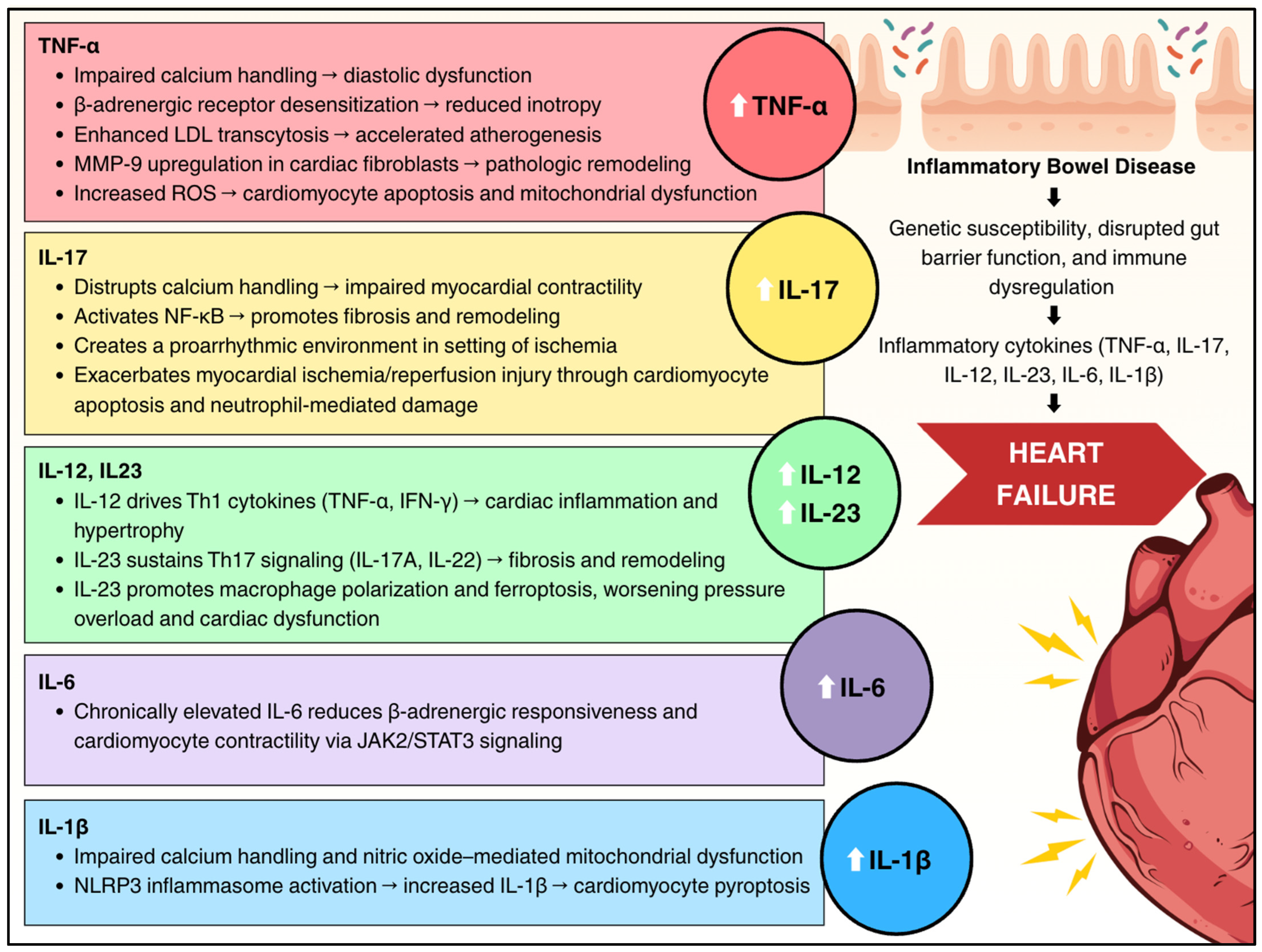
IBD is characterized by dysregulated cytokine production with prominent elevation of proinflammatory cytokines. These cytokines create a systemic inflammatory milieu that directly impacts cardiac structure and function through multiple interconnected pathways. Figure 2 summarizes the key cytokines that are elevated and involved in IBD and are also implicated in HF, including tumor necrosis factor-alpha (TNF-α), interleukin-1β (IL-1β), interleukin-6 (IL-6), interleukin-12 (IL-12), interleukin-23 (IL-23), and interleukin-17 (IL-17).

3.2. Tumor Necrosis Factor-Alpha

TNF-α is an extensively studied cytokine in IBD and cardiac dysfunction and is a common therapeutic target in IBD management. Elevated TNF-α drives adverse left ventricular remodeling through downregulation of sarcoplasmic reticulum Ca2+ ATPase gene expression in cardiomyocytes; the impaired calcium reuptake into the sarcoplasmic reticulum during diastole results in diastolic dysfunction [8,9]. TNF-α blunts the inotropic response, and subsequently cardiac contractility, through GRK2-mediated desensitization of β-adrenergic receptors [10]. Additionally, TNF-α amplifies intracellular reactive oxygen species generation, inducing endothelial dysfunction [11], which triggers cardiomyocyte apoptosis [12]. Oxidative stress also contributes to mitochondrial dysfunction, compromising cardiac function [13]. Within the vasculature, TNF-α accelerates atherogenesis by increasing LDL transcytosis and foam cell formation, thereby enlarging atherosclerotic plaques and increasing susceptibility to ischemia [14]. TNF-α also upregulates matrix metalloproteinase-9 in cardiac fibroblasts [15], accelerating extracellular matrix degradation and promoting pathologic ventricular remodeling [16].

3.3. Interleukin-1β

IL-1β exerts cardiodepressant effects by impairing calcium handling and inducing nitric oxide (NO)-mediated mitochondrial dysfunction [17]. In IBD, dysregulation of the NLRP3 inflammasome, a multiprotein complex involved in innate immunity, leads to enhanced IL-1β release and induction of pyroptosis, a form of inflammatory programmed cell death [18]. While pyroptosis serves as a defense mechanism by eliminating infected or damaged cells, excessive activation can disrupt the intestinal barrier, promoting further inflammation in IBD [18]. Similarly, in HF, activation of the NLRP3 inflammasome within cardiomyocytes has been shown to induce pyroptosis, contributing to myocardial cell loss and disease progression [19]. Although direct experimental evidence is limited, it is plausible that chronic intestinal inflammation and NLRP3 inflammasome activation in IBD may have systemic effects, promoting cardiac dysfunction.

3.4. Interleukin-6

IL-6 levels are also elevated in IBD and HF [20,21]. In IBD, IL-6 is produced by activated immune cells in the intestinal mucosa, promoting chronic inflammation [20]. IL-6 levels are increased in both HF with reduced (HFrEF) and preserved (HFpEF) ejection fraction [22,23]. Chronic elevation in IL-6 depresses β-adrenergic responsiveness and cardiomyocyte contractility, in part through the JAK2/STAT3 pathway [24]. IL-6 binds to its receptor IL-6R, forming a complex that activates the gp130 receptor, which in turn triggers the JAK tyrosine kinase pathway. Activated JAK phosphorylates STAT3, allowing it to dimerize and translocate into the nucleus, where it binds DNA to upregulate genes involved in cardiac inflammation and fibrosis [25].

3.5. Interleukin-12 and Interleukin-23

Interleukin-12 (IL-12) and interleukin-23 (IL-23) are proinflammatory cytokines that play an important role in intestinal homeostasis and inflammation in IBD [26,27]. The production of both cytokines is mediated by antigen-presenting cells (APC), including activated dendritic cells and phagocytes [26]. IL-12 promotes intestinal tissue damage through the modulation of Th1 responses, leading to TNF-α and interferon-γ (IFN-γ) secretion, increasing intestinal inflammation [28]. IL-23 is involved in the growth and maintenance of Th17 cells [29]. Together, the IL-23/Th17 axis mediates chronic inflammation through the production of IL-17A, IL-17F, IL-22, IL-26, TNF-a, and IFNγ [26]. IL-12 and IL-23 share a common p40 subunit, which is a therapeutic target for monoclonal antibodies such as ustekinumab [30]. Elevated IL-12 and IL-23 have been previously shown to be associated with cardiovascular diseases [31]. Recent studies have now recognized the role of cardiac inflammation in the pathogenesis of HF and cardiac remodeling through cytokine recruitment and its interplay with the neurohormonal system and endothelial dysfunction [32]. Experimental studies in mice demonstrated that IL-12α KO significantly attenuated pressure overload-induced cardiac inflammation, hypertrophy, fibrosis, and HF progression [33]. Knockout of IL-23p19 in pressure overload mice models has also shown improved cardiac dysfunction and remodeling via attenuation of M1 macrophage polarization and the reduction in ferroptosis [34]. Together, these findings link IL-12- and IL-23-driven inflammation to both intestinal disease and cardiac remodeling, highlighting their role in the pathogenesis of HF.

3.6. Interleukin-17

In IBD patients, IL-17A and -17F are significantly upregulated both in inflamed intestinal mucosa and systemically in circulation, with Th17 cells serving as the primary source [35]. Mendelian randomization studies have established causal relationships between specific IL-17 subtypes and IBD development [36]. Additionally, IL17RA promoter polymorphisms directly correlate with increased cardiovascular mortality, demonstrating the significance of IL-17-mediated immune activation [37]. The cardiac consequences of sustained IL-17 elevation are multifaceted. Experimental models reveal that IL-17 directly impairs myocardial contractility through disrupted calcium handling and activates NF-κB-mediated pathways that drive pathological cardiac remodeling [38]. In ischemic contexts, IL-17 creates a proarrhythmic setting by impairing electrical conduction, prolonging repolarization, and promoting both myocardial fibrosis and inflammation—effects that are reversible with IL-17 neutralization [39]. Additionally, IL-17A exacerbates myocardial ischemia/reperfusion injury through dual mechanisms of enhanced cardiomyocyte apoptosis and neutrophil-mediated tissue damage [40]. Collectively, these findings establish IL-17 as a biomarker increased in IBD and a direct mediator of the cardiac complications that contribute to HF in this patient population.

3.7. Interleukin-33

IL-33 is produced by various cells, including epithelial cells, antigen-presenting cells (APCs), and fibroblasts [41,42]. IL-33 influences the immune system, including Th2 cell induction and Th1 cell activation, as well as the modulation of group 2 innate lymphoid cells and regulatory T cells [41]. IL-33 binds to its receptor, the growth stimulus-expressed gene 2 protein (ST2), which produces downstream cascades that can phosphorylate extracellular signal-regulated kinase 1/2, p38 MAPK, and JNKs and activate NF-κB [43]. IL-33 is active in IBD. Its effect on the heart is complex and not fully understood. Interestingly, although IL-33 activates NF-κB, it decreases the NF-κB activation caused by angiotensin II or phenylephrine in cardiomyocytes [43]. Experimental models have demonstrated that IL-33 knockout causes pathologic cardiac remodeling in mice with impaired cardiac function [44]. IL-33 may produce myocardial damage and increase fibrosis. Soluble ST2 (sST2) has been shown to decrease the beneficial effects of IL-33 [45]. In IBD patients, increased sST2 is secreted by intestinal proinflammatory T cells [46], and IBD patients have been found to have decreased serum levels of IL-33 [47]. Put together, the dysregulation of the IL-33/ST2L/sST2 pathway may serve as a shared link between IBD and HF pathogenesis.

4. Experimental Evidence

A recent murine model provides mechanistic evidence linking IBD to HF [48]. Two mouse models were used. In the first, chronic colitis was induced in mice by administering 2% dextran sodium sulfate (DSS). In another model, interleukin 10 (IL-10) knockout mice on a 129/Sv background developed spontaneous colitis. Chronic intestinal inflammation reproducibly precipitated a similar cardiac phenotype in both models. Transthoracic echocardiography revealed a significant decrease in left ventricular ejection fraction and fractional shortening, accompanied by inflammatory cell infiltration, myofibrillar disorganization, interstitial edema, and increased cardiac fibrosis [48]. Molecular profiling corroborated a profibrotic switch with an increase in collagen fiber deposition and increased fibrotic proteins, suggesting that chronic colitis promotes cardiac fibrosis [48]. Cardiac fibrosis is an important mechanism leading to HFpEF [49]. Therapeutic fecal microbiota transplantation (FMT) is the process of delivering a gut microbiome from a healthy donor to a recipient in order to restore the gut microbiome in the recipient and manage various disease processes [50]. Studies have demonstrated that FMT from a healthy donor mouse attenuated both gut and cardiac pathology. FMT dampened colonic IL-1β expression and myeloperoxidase activity, improved ventricular systolic indices, and reduced myocardial collagen burden. The reversal in symptoms was near-complete in DSS mice and partial in the IL-10 double knockout model, highlighting a cardioprotective role for endogenous IL-10 [48].

Intestinal Barrier Dysfunction and Gut Microbiome Alteration

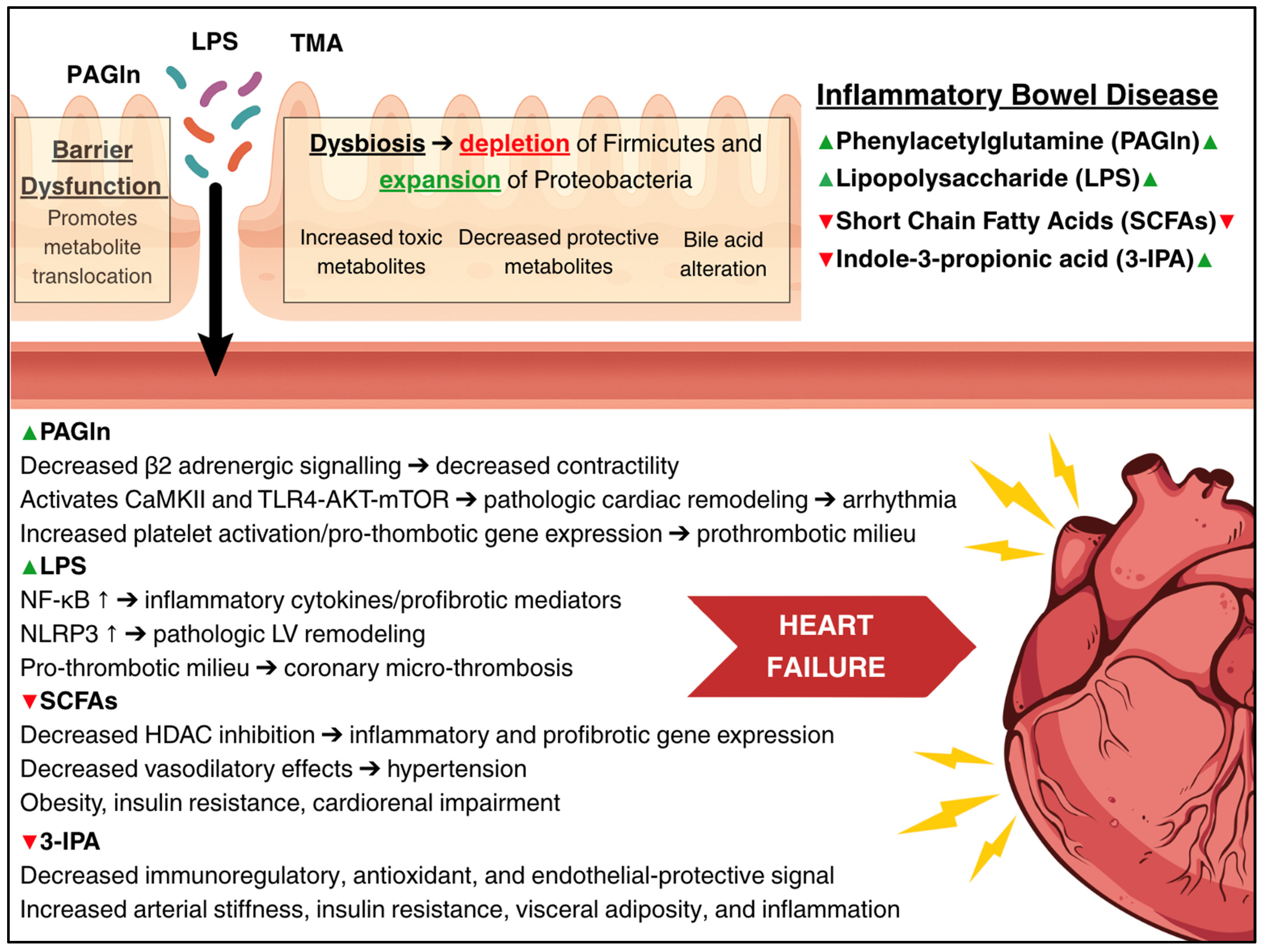
The gut–heart axis represents a pathway connecting intestinal and cardiac health [51,52]. Two primary mechanisms appear particularly important: (1) intestinal barrier dysfunction, often termed “leaky gut,” and (2) gut microbiome dysbiosis. Together, these alterations result in increased translocation of metabolites into the systemic circulation, enhanced production of toxic metabolites, reduced production of protective metabolites, and disruption of metabolite homeostasis. Figure 3 summarizes the key metabolites that are altered in IBD and have established roles in HF pathogenesis.

5. Intestinal Barrier Dysfunction

5.1. Structural and Functional Changes

The intestinal barrier comprises a multilayered selectively permeable system including protective mucus, epithelial cells joined by tight junctions, antimicrobial molecules, and mucosal immune networks [53]. IBD is characterized by intestinal inflammation, leading to barrier dysfunction that may clinically manifest as diarrhea and impaired absorptive capacity that can result in nutritional deficiencies [54]. In IBD, several factors contribute to the disruption of the intestinal barrier. These include alterations to tight junctions [55], increased epithelial cell apoptosis, erosions, and mucosal abnormalities [54,56]. Increased enteric permeability has been described in healthy first-degree relatives of patients with CD, supporting a genetic basis for barrier dysfunction [57]. Additionally, intestinal barrier dysfunction appears to scale with inflammatory activity in IBD [58,59]. Increased intestinal permeability results in translocation of luminal pathogens and agents into systemic circulation, triggering an inflammatory response [60]. The translocation of toxic microbial products and associated immune activation contributes to the development of HF.

5.2. Cardiovascular Implications

Intestinal barrier dysfunction has been observed in cardiovascular disease (CVD) states. A recent meta-analysis demonstrated that patients with CVD have significantly higher circulating levels of lipopolysaccharide (LPS), d-lactate, zonulin, and serum diamine oxidase, markers of intestinal permeability, relative to healthy controls [61]. This suggests that a breached intestinal barrier, whether caused by gut inflammation or by cardiac disease, correlates with systemic inflammation. In chronic HF, intestinal wall edema from venous congestion can itself induce intestinal barrier dysfunction, exacerbating systemic inflammation via microbial translocation [62]. Intestinal barrier dysfunction in IBD allows luminal contents to bypass epithelial tight junctions and evade mucosal immune surveillance, leading to increased systemic exposure to microbial products and endotoxins. This promotes endotoxemia, drives systemic inflammation, and facilitates translocation of toxins to distant organs, including the heart [63].

5.3. Lipopolysaccharide and Cardiac Dysfunction

LPS, an endotoxin from Gram-negative bacterial outer membranes, represents a key mediator linking barrier dysfunction in IBD to HF [64,65]. Circulating LPS binds Toll-like receptor-4 on endothelial cells and cardiomyocytes, triggering NF-κB–driven proinflammatory cytokine release, profibrotic signaling, a pro-thrombotic milieu, and mitochondrial dysfunction. Animal models demonstrate that LPS exposure promotes myocardial fibrosis and HFpEF through multiple mechanisms, including increased TNF-α and IL-6 [66]. Inhibition of this pathway in an LPS-treated mouse model led to cardioprotective outcomes [67]. In rodent and in vitro models, LPS exposure amplified components of the NLRP3 inflammasome and increased NLRP3 gene expression, respectively [68]. Activation of the NLRP3 inflammasome in these models produced HFpEF-like phenotypes characterized by concentric remodeling, LV stiffening, and impaired relaxation [68]. Additionally, LPS creates a pro-thrombotic environment, primarily through contact pathway and platelet activation [69,70,71]. This promotes coronary microvascular thrombosis [72], accelerating adverse ventricular remodeling and HF progression. In a sepsis model, LPS led to cardiac dysfunction through mitochondrial electron transport chain disruption, increased ROS generation, and impaired antioxidant defenses, resulting in oxidative damage and lipid peroxidation [73]. Clinically, LPS levels increase significantly during decompensated HF episodes, likely due to splanchnic venous congestion, and decrease with successful decongestion [74,75]. This temporal relationship suggests that LPS may serve both as a biomarker and mediator of HF progression.

5.4. Does TMAO Play a Role?

Intestinal barrier dysfunction in IBD also allows for translocation of other gut-derived metabolites with well-established roles in HF pathophysiology. Some authors have speculated that in the setting of IBD-related gut barrier dysfunction, increased trimethylamine (TMA) absorption, and thus trimethylamine N-oxide (TMAO) production, may contribute to increased HF risk [76]. Increased serum TMAO is associated with an increased risk of all-cause mortality in HF [77] and has been associated with increased HF incidence [78]. TMAO promotes cardiac hypertrophy and fibrosis by activating profibrotic signaling pathways, including TGF-β/Smad3 [79] and JAK2-STAT3 [80], and by stimulating the NLRP3 inflammasome in cardiac fibroblasts [81]. TMAO also impairs myocardial energetics [82] and induces oxidative stress and endothelial dysfunction [83]. Interestingly, IBD has been shown to be accompanied by decreased circulating TMAO relative to healthy controls [84]. Additionally, plasma TMAO further decreases in active UC relative to inactive disease [84,85]. This finding of “low TMAO despite leaky gut” may be explained by two converging mechanisms: (1) dysbiosis-driven depletion of choline- and carnitine-metabolizing microbes that encode the cutC and cntA genes, limiting luminal production of trimethylamine (TMA) [84,86]; (2) systemic inflammation suppresses hepatic expression of flavin-containing monooxygenase 3 (FMO3), the key enzyme responsible for converting TMA to TMAO [87]. In the dextran sulfate sodium (DSS) colitis model, colonic tissue TMA levels increased while TMAO concentrations declined, a pattern mediated by downregulation of hepatic FMO3 expression—highlighting an inflammation-induced impairment of TMAO synthesis during active IBD [88]. As a result, although barrier dysfunction enhances the translocation of many gut-derived molecules, the systemic TMAO pool contracts in active IBD, distinguishing it from other forms of gut-derived metabolic injury. Despite lower circulating TMAO levels in IBD, recent evidence suggests that even elevations within this suppressed range may still contribute to vascular dysfunction [89]. In particular, higher TMAO levels were associated with impaired coronary microvascular function in patients with UC [89], highlighting a potential role for TMAO in IBD-related cardiovascular risk even when absolute concentrations are reduced. Interestingly, a metabolomics study of Italian UC patients demonstrated increased fecal TMAO [90], although it is unclear if fecal TMAO correlates with plasma TMAO. Further research is needed to clarify TMAO’s role at the intersection of IBD and HF pathogenesis.

6. Gut Microbiome Dysbiosis

6.1. Microbial Composition Changes

IBD is characterized by significant dysbiosis, with depletion of Firmicutes (including butyrate-producing genera like Faecalibacterium and Roseburia) and expansion of Proteobacteria such as Enterobacteriaceae [91,92]. This disrupts the metabolic output of the gut microbiome, promoting downstream cardiovascular consequences.

6.2. Short-Chain Fatty Acid Depletion

Short-chain fatty acids (SCFAs) such as acetate, propionate, and butyrate are beneficial microbial metabolites produced by fermentation of dietary fiber [93]. SCFAs function as primary energy substrates for colonocytes, enhance epithelial barrier integrity, and regulate both innate and adaptive immune responses to mitigate excessive inflammation [93]. Following absorption, SCFAs participate in hepatic and systemic metabolic processes, modulating gluconeogenesis, lipogenesis, and appetite regulation to maintain glucose and lipid homeostasis [94]. At elevated concentrations, particularly in the colonic microenvironment, SCFAs exert antineoplastic effects [94]. Additionally, SCFAs engage in cellular signaling through multiple pathways, including activation of G-protein-coupled receptors (GPCRs), inhibition of histone deacetylases (HDAC), and stimulation of peroxisome proliferator-activated receptor gamma (PPARγ). Through these mechanisms, they serve as critical mediators linking dietary intake, gut microbial composition, gastrointestinal pathology, and systemic physiological responses [94].
IBD-associated dysbiosis is characterized by a loss of SCFA-producing bacteria and significantly reduced SCFA levels in the gut [93]. A recent meta-analysis of 12 studies found that patients with IBD exhibited significantly lower fecal concentrations of acetate, propionate, butyrate, and valerate, alongside elevated lactate, relative to healthy controls [95]. Reduced SCFAs in IBD perpetuates impaired epithelial barrier integrity, diminished mucus production, and dysregulation of immune cell activity, including reduced differentiation of regulatory T cells. These deficits contribute to increased intestinal permeability, heightened proinflammatory cytokine release, and impaired tissue repair, thereby perpetuating chronic gut inflammation and mucosal injury [96].
Beyond the gastrointestinal tract, the consequences of SCFA depletion affect cardiovascular and metabolic function, including insulin resistance, blood pressure control, and HF development or progression [97]. SCFAs exert multiple cardioprotective effects relevant to HF progression. SCFAs, specifically butyrate, promote macrophage mitochondrial function and dampen inflammation [98,99]. In a rat model of myocardial fibrosis, dietary supplementation with butyric acid ameliorated myocardial fibrosis by promoting macrophage polarization from a pro- to anti-inflammatory state and restoring mitochondrial function [99]. Additionally, SCFAs have been shown to have a vasorelaxant or vasodilatory effect in human and animal models [100,101,102]. A study of older adults found that increased serum SCFA concentrations, specifically acetic acid and valeric acid, were inversely associated with systolic blood pressure [103], suggesting that reduced SCFA levels in the context of IBD may increase the risk of hypertensive heart disease. SCFAs also exert cardioprotective effects through HDAC inhibition. HDAC inhibition reverses diastolic dysfunction in various animal models [104,105,106]. HDAC inhibition reduces proinflammatory cytokines [107], reduces profibrotic signaling [108], reverses maladaptive protein acetylation, promotes autophagy-related gene expression, and improves energy metabolism [109]. Together, these effects contribute to improved myocardial structure and function. In a diabetic mouse model, sodium butyrate treatment led to significant improvements in ejection fraction, fractional shortening, and reduced myocardial fibrosis and cardiomyocyte hypertrophy [110]. This suggests that the low SCFA levels observed in IBD may impair endogenous HDAC inhibition, linking gut dysbiosis in IBD to adverse cardiac remodeling. Animal models have shown that SCFA supplementation reduces body weight [111,112,113], stimulates insulin sensitivity, reduces hepatic steatosis [112], improves mitochondrial biogenesis, and inhibits chronic inflammation [111]. Dietary interventions resulting in low fecal SCFA levels have been shown to promote adiposity in mice [114]. SCFA depletion may promote obesity by disrupting adipokine signaling and metabolic homeostasis [98]. Obesity is a well-established risk factor for both systolic and diastolic dysfunction, with a particularly strong association with HFpEF [115,116]. Reduced levels of acetate, propionate, and butyrate in HF is associated with lower left ventricular ejection fraction, elevated NT-proBNP, and a reduced glomerular filtration rate, linking SCFA deficiency to cardiac dysfunction, ventricular stress, and cardiorenal impairment [117]. Taken collectively, these mechanisms establish a mechanistic link between microbiome-driven SCFA depletion in IBD and the pathogenesis of HF.

6.3. Do Bile Acids Play a Role?

Bile acids play a multifaceted role in the pathogenesis and clinical course of IBD. In IBD, gut microbial imbalance interferes with the normal microbial conversion of bile acids secreted by hepatocytes. As a result, primary bile acids accumulate while levels of secondary bile acids, which exert anti-inflammatory effects, are markedly reduced [118]. The abnormal bile acid profile feeds back to further disturb the gut microbiome, contributing to further dysbiosis and inflammation [118]. These alterations impair the integrity of the intestinal barrier and disrupt immune regulation through impaired signaling of bile acid-activated receptors. Bile acid-activated receptors, particularly the nuclear receptor farnesoid X receptor (FXR) and the G protein-coupled receptor TGR5, are also expressed in cardiomyocytes, endothelial cells, and vascular smooth muscle cells [119]. Dysfunctional FXR and TGR5 signaling pathways can adversely impact cardiac health by promoting atherosclerosis, systemic inflammation, and myocardial metabolic disturbances, which collectively contribute to cardiac dysfunction [120,121,122]. Conversely, in chronic HF, congestion-related intestinal hypoperfusion and hepatobiliary dysfunction disrupt enterohepatic bile acid circulation, leading to a relative increase in circulating secondary bile acids and a reduction in primary bile acids [123]. An elevated secondary-to-primary bile acid ratio has been associated with adverse clinical outcomes, including increased mortality [123]. While IBD and HF exhibit opposite alterations in bile acid profiles, with primary dominant in IBD and secondary dominant in HF, both conditions converge on disrupted bile acid receptor signaling and downstream inflammatory and fibrotic pathways. The net impact of IBD-related changes in bile acid metabolism on the heart is not fully established, and the precise role of bile acid dysregulation in IBD-related HF remains speculative and warrants further investigation.

6.4. Phenylacetylglutamine

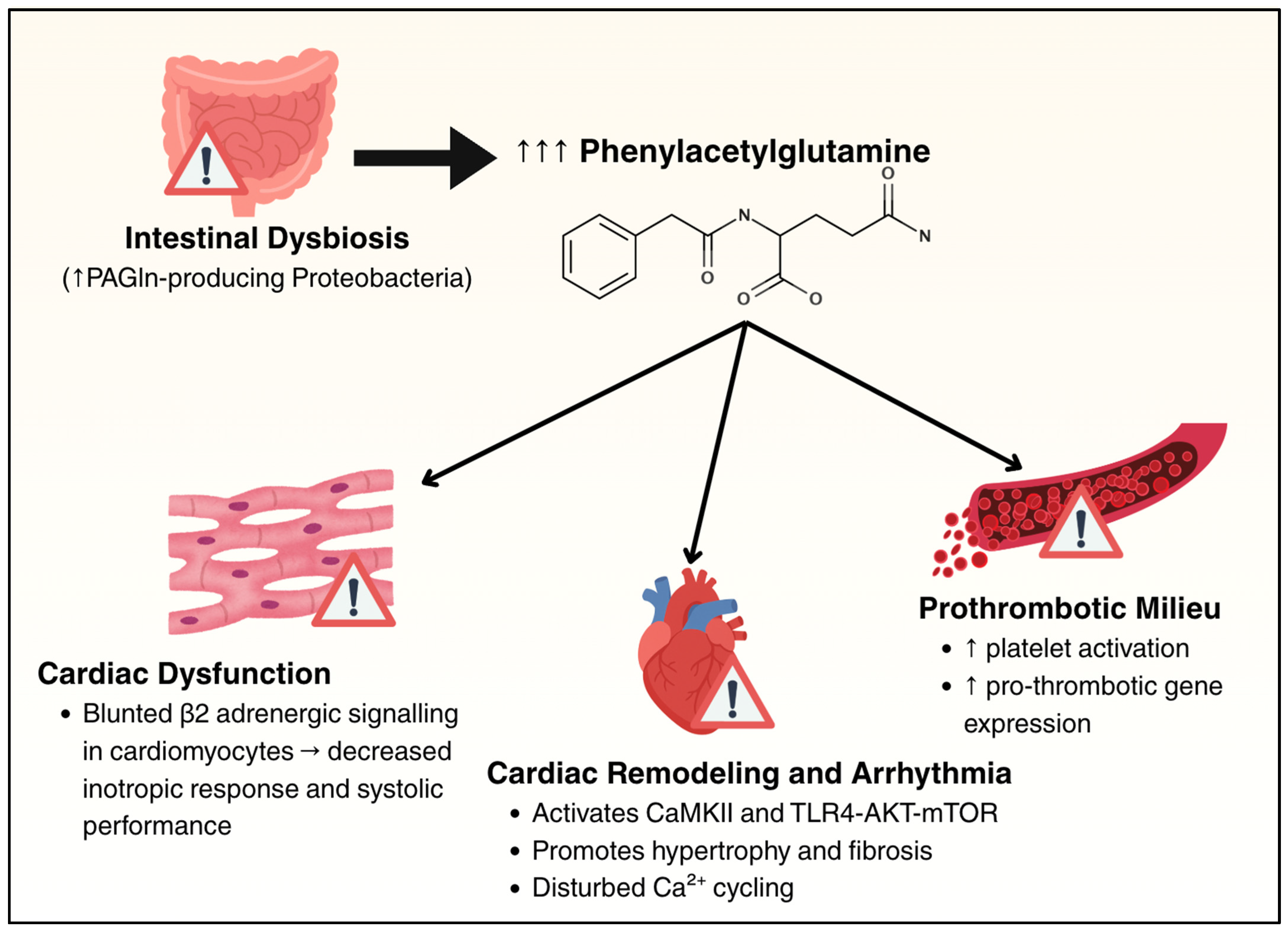
PAGln is derived from dietary phenylalanine, which is converted to phenylacetic acid by gut microbes and then conjugated with glutamine in hepatocytes and renal cells. PAGln levels are elevated in patients with CD relative to healthy controls [124]. In treatment-naïve CD, intestinal dysbiosis leads to the expansion of PAGln-producing Proteobacteria, resulting in elevated circulating PAGln levels [124]. Experimental models using the murine analog phenylacetylglycine (PAGly) demonstrate that exogenous administration exacerbates colitis severity, while human ex vivo studies show that PAGln promotes platelet activation and enhances pro-thrombotic gene expression [124]. Beyond the gastrointestinal tract, PAGln interacts with host adrenergic signaling pathways; it functions as a negative allosteric modulator of β2-adrenergic receptors in murine and human ventricular cardiomyocytes, attenuating cardiac contractility [125]. Elevated plasma PAGln levels have been independently associated with increased risk of hospitalization and mortality in patients with HF [126]. Mechanistically, PAGln has been shown to worsen adverse cardiac remodeling and increase susceptibility to arrhythmias via activation of CaMKII and TLR4-AKT-mTOR signaling pathways in murine models of HF [127,128]. In patients with HF circulating PAGln levels correlate with the degree of systolic dysfunction, with the highest concentrations observed in patients with HFrEF, followed by those with mildly reduced EF. PAGln levels are elevated in HFpEF as well, though to a lesser extent [129]. This gradient supports a potential role for PAGln in the progression of myocardial dysfunction, particularly in the context of impaired contractility. These findings suggest that increased PAGln levels in the setting of IBD represent a link between gut inflammation and heightened HF risk. Figure 4 summarizes the effects of PAGln on the heart and vasculature.

6.5. Tryptophan-Derived Indoles

Dysbiosis in IBD also perturbs bacterial tryptophan metabolism, reducing levels of beneficial indole derivatives. Indole-3-propionic acid (IPA), a microbiota-produced indole, is significantly decreased in patients with active IBD compared to healthy controls [130]. High IPA levels are negatively associated with arterial stiffness, insulin resistance, fasting glucose, and visceral fat [131]. Its deficiency removes an important immunoregulatory signal, potentially exacerbating systemic inflammation [132]. Consequently, chronic depletion of IPA due to persistent intestinal dysbiosis in IBD could promote systemic metabolic dysfunction, heightened inflammatory signaling, adverse myocardial remodeling, and, ultimately, increased risk of HF.

6.6. Summary of Dysbiosis and Barrier Dysfunction

IBD is characterized by gut dysbiosis and gut barrier dysfunction, both of which are associated with increased cardiovascular event rates, including HF, and coronary artery and cerebrovascular disease [63]. Experimental models have shown that modulating the microbiome (with antibiotics, probiotics, or FMT) can influence cardiac outcomes [48]. However, the specific microbial metabolites and mechanistic pathways driving these effects in humans remain incompletely defined. The hypothesis that restoring a single microbial genus in humans with IBD could reduce HF risk remains speculative. What is more robustly supported is the interplay between dysbiosis and intestinal barrier dysfunction [63]. A dysbiotic microbiota can cause barrier dysfunction, and, in turn, a leaky barrier increases systemic exposure to dysbiotic microbial products. This bidirectional relationship perpetuates chronic inflammation, likely amplifying systemic inflammatory load, contributing to HF pathogenesis.

7. Endothelial and Vascular Dysfunction and Thrombotic Risk

Chronic inflammation in IBD affects endothelial cell function and microcirculation, creating conditions conducive to HF development through both direct myocardial effects and accelerated atherosclerosis.

7.1. Systemic Endothelial Dysfunction

Chronic inflammation in IBD adversely affects the endothelium and microcirculation, which are key contributors to HF pathogenesis. Persistent elevation of inflammatory cytokines impairs endothelial function by promoting leukocyte adhesion, increasing oxidative stress, and reducing NO bioavailability [133,134]. At the cellular level, chronic inflammation activates endothelial cells, increasing the expression of adhesion molecules such as ICAM-1 and VCAM-1, which facilitate leukocyte adhesion and transmigration [135,136]. Infiltrating leukocytes release cytokines and reactive oxygen species, disrupting endothelial nitric oxide synthase function and reducing NO availability [135,136]. This promotes oxidative stress, increases permeability, and shifts the endothelium toward a pro-thrombotic, vasoconstrictive state [135,136], key features of endothelial dysfunction contributing to HF pathogenesis. These mechanisms are clinically relevant, as patients with IBD consistently demonstrate impaired endothelial function even in the absence of traditional cardiovascular risk factors [137]. Flow-mediated dilation of the brachial artery—a validated marker of endothelial health—is significantly reduced in IBD patients, while pulse wave velocity is increased, indicating early vascular dysfunction and heightened atherosclerotic risk [137].

7.2. Coronary Microvascular Dysfunction

Endothelial dysfunction extends to the coronary microvasculature. Impaired coronary microvascular function has been observed in IBD, with reduced coronary flow reserve reported in one study [138]. This can contribute to myocardial ischemia in the absence of overt coronary artery disease or non-obstructive coronary artery disease (NOCAD), a phenomenon observed in a number of patients with HFpEF [139].

7.3. Accelerated Atherosclerosis and Thrombotic Risk

IBD functions as an independent risk factor for atherosclerotic cardiovascular disease (ASCVD) and thrombotic disease [140,141,142]. This occurs through multiple interrelated mechanisms, including elevated proinflammatory cytokines, upregulation of adhesion molecules, promotion of leukocyte adhesion and vascular inflammation, and gut microbiome and barrier dysfunction [140]. The proinflammatory milieu creates a hypercoagulable state through abnormal platelet function, impaired fibrinolysis, and activation of the coagulation cascade, increasing the risk of arterial and venous thrombosis [140]. This is further exacerbated by corticosteroid use, commonly used in IBD treatment [140]. Together, these processes accelerate atherogenesis and increase the risk of ischemic HF.

8. Hemodynamic and Autonomic Perturbations

IBD patients exhibit alterations in hemodynamic status and autonomic function that may contribute to cardiac dysfunction through distinct but complementary mechanisms.

8.1. Hemodynamic Changes

Chronic anemia, common in IBD due to gastrointestinal blood loss and chronic inflammation, reduces blood oxygen-carrying capacity and triggers compensatory cardiovascular responses, including tachycardia and increased cardiac output [143]. While initially adaptive, sustained high-output states can impose chronic volume stress, leading to ventricular dilatation and eventual systolic dysfunction [143].

8.2. Autonomic Nervous System Dysfunction

In IBD, autonomic dysfunction is evidenced by a significant reduction in heart rate variability (HRV) compared to healthy controls [144]. This is in part due to parasympathetic suppression, which leads to sympathetic predominance [145]. This dysfunction appears bidirectionally linked to TNF-α levels [146]. The vagus nerve releases acetylcholine, which normally exerts anti-inflammatory effects through alpha 7 nicotinic receptors (α7nAchRs) on human macrophages that inhibit the production of NF-κB and cytokines, including TNF-α [147,148]. Reduced vagal activity impairs this inhibition, resulting in increased TNF-α production. Conversely, TNF-α can modulate vagal neurocircuitry in the brainstem, affecting autonomic function and potentially contributing to autonomic dysregulation [149,150]. Over time, chronic sympathetic hyperactivity may contribute to adverse cardiac remodeling [151]. In chronic HF, prolonged sympathetic activation leads to persistent stimulation of β1-adrenergic receptors, causing desensitization and downregulation via upregulation of GRK2, reducing cardiac contractility [152]. GRK2 also impairs α2-adrenergic receptor function, removing inhibitory feedback on norepinephrine release, further increasing sympathetic drive [152]. Meanwhile, the protective β2-receptor signaling becomes disrupted, and adrenal glands over-secrete catecholamines due to α2-adrenergic receptor dysfunction. This sustained catecholamine exposure results in calcium overload, oxidative stress, and cardiomyocyte apoptosis, accelerating HF progression [152]. However, studies examining the role of ANS dysfunction in IBD-related cardiac dysfunction specifically are currently lacking.

9. Nutritional Sequelae

Chronic intestinal inflammation in IBD is associated with nutritional disturbances that have direct relevance to cardiovascular health. Even in the absence of active gastrointestinal symptoms, patients with IBD frequently exhibit evidence of protein–energy malnutrition, sarcopenia, and micronutrient deficiencies [153,154,155,156]. The cumulative burden of IBD-related nutritional disturbance creates an environment that is permissive to the development or progression of HF, which highlights the importance of early nutritional intervention as a potential strategy for HF prevention in this high-risk population. Figure 5 summarizes the nutritional sequelae of IBD and how they contribute to increased HF risk.

9.1. Protein Malnutrition and Sarcopenia

Protein–energy malnutrition is common in IBD, with one systematic review estimating the prevalence of myopenia and sarcopenia at 42% and 17%, respectively [157]. Patients with IBD are at increased risk for sarcopenia due to a multifactorial interplay of mechanisms, including reduced physical activity due to symptom burden, medication effects, a proinflammatory state, which promotes muscle catabolism, and malnutrition resulting from reduced oral intake, malabsorption, and increased metabolic demands [158]. In a canine model, protein–calorie malnutrition impaired left ventricular function by decreasing compliance due to starvation-induced edema and reduced contractility from myofibrillar atrophy [159]. Sarcopenia can contribute to HF through altered sympathetic and ergoreflex sensitivity, disrupting the regulation of ventilation, cardiovascular responses during physical activity, and metabolic dysregulation [160,161]. Interestingly, proinflammatory factors that are elevated in IBD and HF, including TNF-α, IL-6, and IL-1, can accelerate muscle atrophy through the autophagy–lysosome and ubiquitin–proteasome systems, ultimately contributing to the development of sarcopenia [161]. In established HF, sarcopenia is also associated with a 64% increase in poor prognosis, including mortality and hospitalization [162]. Most research on the link between protein malnutrition and HF development is limited to studies in children with severe malnutrition, particularly kwashiorkor, which has been associated with mild dilated cardiomyopathy [163]. Additionally, among individuals hospitalized with HF, protein–energy malnutrition frequently co-occurs and contributes to significantly worse health outcomes [164].

9.2. Micronutrient Deficiencies

Several micronutrient deficiencies commonly observed in IBD have known deleterious effects on cardiovascular function and contribute to comorbid progression of established HF. IBD-related iron deficiency and iron deficiency anemia result from chronic gastrointestinal blood loss, impaired absorption, inadequate dietary intake, and inflammation-induced hepcidin upregulation [165]. Iron deficiency in HF is independently associated with worse symptoms, reduced exercise capacity, poorer quality of life, increased risk of hospitalization, and higher mortality, regardless of the presence of anemia [166,167,168]. Iron deficiency directly affects cardiomyocytes through mitochondrial dysfunction, impairing both contractility and relaxation [169]. Iron deficiency also alters calcium handling in cardiomyocytes by downregulating RyR2 and SERCA2a, further weakening contractility [170].
Vitamin D deficiency is disproportionately prevalent in IBD due to several factors, such as impaired nutrient absorption from intestinal inflammation, bile salt malabsorption, and lack of sunlight exposure [171,172]. Vitamin D exerts anti-inflammatory and antifibrotic effects [173] and plays a role in regulating glycemic control and enhancing insulin sensitivity [174]. Vitamin D deficiency promotes increased renin–angiotensin–aldosterone system activity, which can contribute to HF through increased hypertension, cardiac remodeling, and endothelial dysfunction [175]. Vitamin D deficiency also leads to increased proinflammatory cytokines, including IL-6 and TNF-a [176], leading to impaired contractility [24] and pathologic ventricular remodeling [16]. Lastly, vitamin deficiency disrupts intracellular calcium homeostasis, which can subsequently affect the function of cardiomyocytes, as well as alters PTH levels, causing myocardial fibrosis and hypertrophy [177].
Additionally, rare cases of micronutrient-related reversible cardiac dysfunction have been reported in IBD, particularly in patients requiring parenteral nutrition. Documented complications include selenium deficiency-induced cardiomyopathy [178] and thiamine deficiency-associated wet beriberi [179]. While uncommon, these cases underscore the potential severity of nutrition-related cardiac complications in IBD.

10. Shared Genetic and Epigenetic Susceptibility

10.1. Shared Genetic Loci

IBD and HF share genetic susceptibility loci that influence overlapping biological pathways. Nucleotide-binding oligomerization domain-containing protein 2 (NOD2) is a cytosolic pattern recognition receptor that plays a central role in the pathogenesis of IBD [180,181]. NOD2 is expressed in monocytes, macrophages, and dendritic cells involved in immune responses, as well as in certain epithelial cells, such as those of the intestinal lining. NOD2 recognizes muramyl dipeptide, a conserved motif in bacterial peptidoglycan, and upon activation, modulates immune responses in the gut. Loss-of-function mutations in NOD2 are strong genetic risk factors for CD [180,181]. NOD2 also modulates cardiovascular inflammation. NOD2-knockout mice develop significantly worsened cardiac hypertrophy and fibrosis under pressure overload, suggesting NOD2 signaling normally plays a protective role in attenuating cardiac remodeling by modulating multiple inflammatory and fibrotic signaling cascades [182]. A recent Mendelian randomization study provided genetic evidence supporting a causal relationship between UC and HF. Using four independent GWAS datasets of European ancestry, the study demonstrated a consistent association between genetically predicted UC and increased HF risk. In contrast, reverse MR analyses did not support a causal effect of HF on UC. Together, these findings suggest a unidirectional genetic predisposition linking UC to elevated HF risk [183]. Additionally, several susceptibility loci implicated in IBD converge on immune signaling pathways that are also relevant in cardiovascular pathology. One such pathway is the IL-23/Th17 axis. Polymorphisms in IL23R and downstream effectors such as TYK2 and STAT3 confer protection against IBD by attenuating Th17-mediated immune activation [184]. Conversely, hyperactivation of the IL-23/IL-17 pathway contributes to persistent intestinal inflammation and has been linked to adverse cardiac remodeling and myocardial fibrosis, which can lead to HF [38,39,40,185].

10.2. MicroRNA Dysregulation

A novel mechanism linking IBD and HF involves microRNA dysregulation in cardiac tissue. miRNAs are small noncoding RNAs that regulate gene expression [186]. Chronic intestinal inflammation leads to dysregulation of numerous miRNAs in both local tissues and circulation, which can lead to cardiac dysfunction [76]. Experimental rat models of dextran sodium sulfate (DSS)-induced colitis have revealed that chronic colitis leads to significant upregulation of 56 microRNAs in the heart [9]. A subset of these miRNAs suppressed myocardial and serum brain-derived neurotrophic factor (BDNF) [9]. BDNF is markedly decreased in HF, which is associated with increased NT-proBNP, adverse cardiac remodeling, and disease progression [187]. miR-21 is upregulated in IBD-affected colonic tissues, which promotes proinflammatory pathways [188]. In the myocardium, miR-21 is known to activate cardiac fibroblasts and enhance collagen deposition and may play a role in maladaptive remodeling, causing HF [189,190]. miR-155 is upregulated in the inflamed colonic tissue and peripheral immune cells of patients with active IBD [191]. miR-155 expression in macrophages contributes to cardiac pathology via myocardial inflammation, fibrosis, and adverse remodeling and HF [192,193]. Overall, miRNA dysregulation provides a molecular link between IBD and myocardial remodeling. IBD triggers an array of miRNAs that not only contribute to intestinal inflammation but also systemically impact the heart’s gene expression profile.

10.3. Pharmacology

The relationship between IBD therapeutics and cardiovascular outcomes represents a critical clinical consideration, with both protective and harmful effects documented across different classes of medications used in IBD management. The most important principle is that active IBD inflammation dramatically increases HF risk, with disease flares associated with a 2.5-fold increase in incident HF [6]. Increased inflammation, marked by elevated inflammatory markers such as C-reactive protein, IL-6, and TNF-α, has been independently associated with HF incidence [194]. This emphasizes that achieving and maintaining disease remission should be considered a cardiovascular protective strategy. In IBD, exposure to systemic corticosteroids was associated with a markedly increased risk of both acute myocardial infarction and HF, suggesting that corticosteroid use, potentially a surrogate for disease severity, may contribute to cardiovascular risk [4]. Systemic corticosteroids are the cornerstone of acute IBD flare management but carry well-established cardiometabolic consequences. These include hyperglycemia, hypertension, weight gain, dyslipidemia, metabolic syndrome, and increased risk of cardiovascular events including HF [195,196]. Given this, minimizing cumulative steroid exposure through early initiation of steroid-sparing therapies is essential for long-term cardiovascular protection. In general, steroid-sparing agents may reduce cardiovascular risk by controlling inflammation, although certain agents require caution in specific populations, particularly those with established cardiovascular disease or risk factors. Of particular interest are anti-TNF therapies, which provide effective control of systemic inflammation and may confer cardiovascular benefit. In a large French cohort, anti-TNF use was associated with a reduction in first-time acute arterial events [197]. However, certain trials have described decompensated HF after anti-TNF initiation, particularly in patients with pre-existing left ventricular dysfunction; thus some societies recommend avoiding anti-TNF therapy in patients with established moderate-to-severe HF [198].

10.4. Clinical Implications

IBD is a systemic disorder that carries several cardiovascular implications, including HF risk. HF risk is particularly elevated during active disease flares, periods of prolonged corticosteroid exposure, and in the presence of traditional cardiovascular risk factors. Currently, routine cardiovascular screening has not been recommended in IBD-specific guidelines. Instead, risk factor assessment and management are individualized based on patient comorbidities. However, periods of active disease require increased monitoring for the development of HF. Maintaining IBD remission is the most effective HF prevention strategy. Traditional HF prevention strategies, including blood pressure control, diabetes management, lipid optimization, and smoking cessation, remain significant and should be integrated with IBD-specific care. Integrated management involving gastroenterologists, cardiologists, and primary care is vital.

11. Future Directions

Gaps remain in understanding the pathogenesis of HF in IBD. Longitudinal cohort studies are needed to further evaluate the impact of therapeutic exposures on cardiac structure and function. Further research is needed to assess biomarkers that are associated with HF risk in IBD patients and their utility in risk stratification. Further metagenomics and metabolomic studies can provide further details of the gut–heart axis in IBD and help determine potential molecular targets. Future research should explore and evaluate the role of targeted interventions in modulating HF risk in IBD. Exploring the effect of targeted interventions, including modulation of systemic inflammation using biologics and microbiome-directed therapies, on cardiac function could help determine their effect on reversing pathological cardiac remodeling. In addition, implementation studies examining the role of integrated care models, such as joint IBD–cardiology clinics, may inform strategies to reduce cardiovascular complications and improve long-term outcomes in this population.

12. Conclusions

IBD is increasingly recognized as a systemic disorder with direct implications for HF development, driven by shared inflammatory, metabolic, and genetic pathways. Key cytokines and gut-derived factors activate signaling cascades in the heart, promoting oxidative stress, cardiomyocyte dysfunction, and fibrotic remodeling. Nutritional deficits, vascular dysfunction, and autonomic dysfunction further exacerbate cardiac vulnerability. These interconnected mechanisms can contribute to both HFpEF and HFrEF phenotypes. Given the interrelationship between IBD and HF, both cardiovascular surveillance and early intervention in established IBD are essential. Effective HF prevention in IBD requires integrated care that addresses not only traditional cardiovascular risks but also considers the role of the gut–heart axis. Integrating these insights, along with HF risk stratification and prevention, into IBD care offers the potential to reduce the burden of HF in this vulnerable population.

Author Contributions

A.S. data search, extraction, and manuscript writing; Y.L. data search, extraction, and manuscript writing; S.W.R. conceptualization, supervision, and manuscript writing. All authors have read and agreed to the published version of the manuscript.

Funding

There was no funding support for this study.

Institutional Review Board Statement

Not applicable.

Informed Consent Statement

Not applicable.

Data Availability Statement

No new data were created or analyzed in this study.

Acknowledgments

During the preparation of this manuscript, ChatGPT (OpenAI, GPT-4, 2025) was used to assist with proofreading of originally written text and to improve sentence clarity and grammar. Additionally, OpenEvidence (v2.6.4, 2025) was used alongside traditional scientific search methods to identify potentially relevant citations. Any citation identified through AI-assisted search was confirmed by thoroughly reviewing the original source material in full. All AI-generated suggestions were thoroughly reviewed and edited by the authors.

Conflicts of Interest

The authors declare no conflicts of interest that could have appeared to influence the work reported in this paper.

References

  1. Olpin, J.D.; Sjoberg, B.P.; Stilwill, S.E.; Jensen, L.E.; Rezvani, M.; Shaaban, A.M. Beyond the Bowel: Extraintestinal Manifestations of Inflammatory Bowel Disease. RadioGraphics 2017, 37, 1135–1160. [Google Scholar] [CrossRef] [PubMed]
  2. Rogler, G.; Singh, A.; Kavanaugh, A.; Rubin, D.T. Extraintestinal Manifestations of Inflammatory Bowel Disease: Current Concepts, Treatment, and Implications for Disease Management. Gastroenterology 2021, 161, 1118–1132. [Google Scholar] [CrossRef] [PubMed]
  3. Patel, R.S.; Rohit Reddy, S.; Llukmani, A.; Hashim, A.; Haddad, D.R.; Patel, D.S.; Ahmad, F.; Gordon, D.K. Cardiovascular Manifestations in Inflammatory Bowel Disease: A Systematic Review of the Pathogenesis and Management of Pericarditis. Cureus 2021, 13, e14010. [Google Scholar] [CrossRef] [PubMed]
  4. Aniwan, S.; Pardi, D.S.; Tremaine, W.J.; Loftus, E.V. Increased Risk of Acute Myocardial Infarction and Heart Failure in Patients With Inflammatory Bowel Diseases. Clin. Gastroenterol. Hepatol. 2018, 16, 1607–1615.e1. [Google Scholar] [CrossRef] [PubMed]
  5. Sun, J.; Yao, J.; Olén, O.; Halfvarson, J.; Bergman, D.; Ebrahimi, F.; Rosengren, A.; Sundström, J.; Ludvigsson, J.F. Risk of Heart Failure in Inflammatory Bowel Disease: A Swedish Population-Based Study. Eur. Heart J. 2024, 45, 2493–2504. [Google Scholar] [CrossRef] [PubMed]
  6. Kristensen, S.L.; Ahlehoff, O.; Lindhardsen, J.; Erichsen, R.; Lamberts, M.; Khalid, U.; Nielsen, O.H.; Torp-Pedersen, C.; Gislason, G.H.; Hansen, P.R. Inflammatory Bowel Disease Is Associated with an Increased Risk of Hospitalization for Heart Failure: A Danish Nationwide Cohort Study. Circ. Heart Fail. 2014, 7, 717–722. [Google Scholar] [CrossRef] [PubMed]
  7. Soares, C.A.; Fiuza, J.G.; Rodrigues, C.A.M.; Craveiro, N.; Gil Pereira, J.; Sousa, P.C.R.F.; Martins, D.C.P.; Cancela, E.M.; Ministro Dos Santos, M.P. Inflammatory Bowel Disease and Cardiac Function: A Systematic Review of Literature with Meta-Analysis. Ther. Adv. Gastroenterol. 2024, 17, 17562848241299534. [Google Scholar] [CrossRef] [PubMed]
  8. Tsai, C.-T.; Wu, C.-K.; Lee, J.-K.; Chang, S.-N.; Kuo, Y.-M.; Wang, Y.-C.; Lai, L.-P.; Chiang, F.-T.; Hwang, J.-J.; Lin, J.-L. TNF-down-Regulates Sarcoplasmic Reticulum Ca2+ ATPase Expression and Leads to Left Ventricular Diastolic Dysfunction through Binding of NF-B to Promoter Response Element. Cardiovasc. Res. 2015, 105, 318–329. [Google Scholar] [CrossRef] [PubMed]
  9. Tang, Y.; Kline, K.T.; Zhong, X.S.; Xiao, Y.; Lian, H.; Peng, J.; Liu, X.; Powell, D.W.; Tang, G.; Li, Q. Chronic Colitis Upregulates microRNAs Suppressing Brain-Derived Neurotrophic Factor in the Adult Heart. PLoS ONE 2021, 16, e0257280. [Google Scholar] [CrossRef] [PubMed]
  10. Vasudevan, N.T.; Mohan, M.L.; Gupta, M.K.; Martelli, E.E.; Hussain, A.K.; Qin, Y.; Chandrasekharan, U.M.; Young, D.; Feldman, A.M.; Sen, S.; et al. Gβγ-Independent Recruitment of G-Protein Coupled Receptor Kinase 2 Drives Tumor Necrosis Factor α–Induced Cardiac β-Adrenergic Receptor Dysfunction. Circulation 2013, 128, 377–387. [Google Scholar] [CrossRef] [PubMed]
  11. Chen, X.; Andresen, B.; Hill, M.; Zhang, J.; Booth, F.; Zhang, C. Role of Reactive Oxygen Species in Tumor Necrosis Factor-Alpha Induced Endothelial Dysfunction. Curr. Hypertens. Rev. 2008, 4, 245–255. [Google Scholar] [CrossRef] [PubMed]
  12. Xie, L.; Xue, F.; Cheng, C.; Sui, W.; Zhang, J.; Meng, L.; Lu, Y.; Xiong, W.; Bu, P.; Xu, F.; et al. Cardiomyocyte-Specific Knockout of ADAM17 Alleviates Doxorubicin-Induced Cardiomyopathy via Inhibiting TNFα–TRAF3–TAK1–MAPK Axis. Signal Transduct. Target. Ther. 2024, 9, 273. [Google Scholar] [CrossRef] [PubMed]
  13. Mariappan, N.; Soorappan, R.N.; Haque, M.; Sriramula, S.; Francis, J. TNF-α-Induced Mitochondrial Oxidative Stress and Cardiac Dysfunction: Restoration by Superoxide Dismutase Mimetic Tempol. Am. J. Physiol.-Heart Circ. Physiol. 2007, 293, H2726–H2737. [Google Scholar] [CrossRef] [PubMed]
  14. Rolski, F.; Błyszczuk, P. Complexity of TNF-α Signaling in Heart Disease. J. Clin. Med. 2020, 9, 3267. [Google Scholar] [CrossRef] [PubMed]
  15. Rodrigues, K.E.; Pontes, M.H.B.; Cantão, M.B.S.; Prado, A.F. The Role of Matrix Metalloproteinase-9 in Cardiac Remodeling and Dysfunction and as a Possible Blood Biomarker in Heart Failure. Pharmacol. Res. 2024, 206, 107285. [Google Scholar] [CrossRef] [PubMed]
  16. Zhu, E.; Yuan, C.; Hu, S.; Liao, Y.; Li, B.; Zhou, Y.; Zhou, W. Injection of Matrix Metalloproteinase-9 Leads to Ventricular Remodeling. Dis. Markers 2022, 2022, 1659771. [Google Scholar] [CrossRef] [PubMed]
  17. Szekely, Y.; Arbel, Y. A Review of Interleukin-1 in Heart Disease: Where Do We Stand Today? Cardiol. Ther. 2018, 7, 25–44. [Google Scholar] [CrossRef] [PubMed]
  18. Zhen, Y.; Zhang, H. NLRP3 Inflammasome and Inflammatory Bowel Disease. Front. Immunol. 2019, 10, 276. [Google Scholar] [CrossRef] [PubMed]
  19. Butts, B.; Gary, R.A.; Dunbar, S.B.; Butler, J. The Importance of NLRP3 Inflammasome in Heart Failure. J. Card. Fail. 2015, 21, 586–593. [Google Scholar] [CrossRef] [PubMed]
  20. Mudter, J.; Neurath, M.F. IL-6 Signaling in Inflammatory Bowel Disease: Pathophysiological Role and Clinical Relevance: Inflamm. Bowel Dis. 2007, 13, 1016–1023. [Google Scholar] [CrossRef] [PubMed]
  21. Gui, X.Y.; Rabkin, S.W. C-Reactive Protein, Interleukin-6, Trimethylamine-N-Oxide, Syndecan-1, Nitric Oxide, and Tumor Necrosis Factor Receptor-1 in Heart Failure with Preserved Versus Reduced Ejection Fraction: A Meta-Analysis. Curr. Heart Fail. Rep. 2023, 20, 1–11. [Google Scholar] [CrossRef] [PubMed]
  22. Chia, Y.C.; Kieneker, L.M.; Van Hassel, G.; Binnenmars, S.H.; Nolte, I.M.; Van Zanden, J.J.; Van Der Meer, P.; Navis, G.; Voors, A.A.; Bakker, S.J.L.; et al. Interleukin 6 and Development of Heart Failure with Preserved Ejection Fraction in the General Population. J. Am. Heart Assoc. 2021, 10, e018549. [Google Scholar] [CrossRef] [PubMed]
  23. Docherty, K.F.; McDowell, K.; Welsh, P.; Petrie, M.C.; Anand, I.; Berg, D.D.; De Boer, R.A.; Køber, L.; Kosiborod, M.N.; Martinez, F.A.; et al. Interleukin-6 in Heart Failure with Reduced Ejection Fraction and the Effect of Dapagliflozin. JACC Heart Fail. 2025, 13, 102393. [Google Scholar] [CrossRef] [PubMed]
  24. Fontes, J.A.; Rose, N.R.; Čiháková, D. The Varying Faces of IL-6: From Cardiac Protection to Cardiac Failure. Cytokine 2015, 74, 62–68. [Google Scholar] [CrossRef] [PubMed]
  25. Jiang, H.; Yang, J.; Li, T.; Wang, X.; Fan, Z.; Ye, Q.; Du, Y. JAK/STAT3 Signaling in Cardiac Fibrosis: A Promising Therapeutic Target. Front. Pharmacol. 2024, 15, 1336102. [Google Scholar] [CrossRef] [PubMed]
  26. Verstockt, B.; Salas, A.; Sands, B.E.; Abraham, C.; Leibovitzh, H.; Neurath, M.F.; Vande Casteele, N. Alimentiv Translational Research Consortium (ATRC) IL-12 and IL-23 Pathway Inhibition in Inflammatory Bowel Disease. Nat. Rev. Gastroenterol. Hepatol. 2023, 20, 433–446. [Google Scholar] [CrossRef] [PubMed]
  27. Berrebi, D.; Besnard, M.; Fromont-Hankard, G.; Paris, R.; Mougenot, J.F.; De Lagausie, P.; Emilie, D.; Cezard, J.P.; Navarro, J.; Peuchmaur, M. Interleukin-12 Expression Is Focally Enhanced in the Gastric Mucosa of Pediatric Patients with Crohn’s Disease. Am. J. Pathol. 1998, 152, 667–672. [Google Scholar] [PubMed]
  28. Trinchieri, G. Interleukin-12 and the Regulation of Innate Resistance and Adaptive Immunity. Nat. Rev. Immunol. 2003, 3, 133–146. [Google Scholar] [CrossRef] [PubMed]
  29. Korta, A.; Kula, J.; Gomułka, K. The Role of IL-23 in the Pathogenesis and Therapy of Inflammatory Bowel Disease. Int. J. Mol. Sci. 2023, 24, 10172. [Google Scholar] [CrossRef] [PubMed]
  30. Teng, M.W.L.; Bowman, E.P.; McElwee, J.J.; Smyth, M.J.; Casanova, J.-L.; Cooper, A.M.; Cua, D.J. IL-12 and IL-23 Cytokines: From Discovery to Targeted Therapies for Immune-Mediated Inflammatory Diseases. Nat. Med. 2015, 21, 719–729. [Google Scholar] [CrossRef] [PubMed]
  31. Van Der Heijden, T.; Bot, I.; Kuiper, J. The IL-12 Cytokine Family in Cardiovascular Diseases. Cytokine 2019, 122, 154188. [Google Scholar] [CrossRef] [PubMed]
  32. Papamichail, A.; Kourek, C.; Briasoulis, A.; Xanthopoulos, A.; Tsougos, E.; Farmakis, D.; Paraskevaidis, I. Targeting Key Inflammatory Mechanisms Underlying Heart Failure: A Comprehensive Review. Int. J. Mol. Sci. 2023, 25, 510. [Google Scholar] [CrossRef] [PubMed]
  33. Bhattarai, U.; He, X.; Xu, R.; Liu, X.; Pan, L.; Sun, Y.; Chen, J.-X.; Chen, Y. IL-12α Deficiency Attenuates Pressure Overload-Induced Cardiac Inflammation, Hypertrophy, Dysfunction, and Heart Failure Progression. Front. Immunol. 2023, 14, 1105664. [Google Scholar] [CrossRef] [PubMed]
  34. Lu, X.; Ji, Q.; Pan, H.; Feng, Y.; Ye, D.; Gan, L.; Wan, J.; Ye, J. IL-23p19 Deficiency Reduces M1 Macrophage Polarization and Improves Stress-Induced Cardiac Remodeling by Alleviating Macrophage Ferroptosis in Mice. Biochem. Pharmacol. 2024, 222, 116072. [Google Scholar] [CrossRef] [PubMed]
  35. Wen, Y.; Wang, H.; Tian, D.; Wang, G. TH17 Cell: A Double-Edged Sword in the Development of Inflammatory Bowel Disease. Ther. Adv. Gastroenterol. 2024, 17, 17562848241230896. [Google Scholar] [CrossRef] [PubMed]
  36. Cai, Y.; Jia, X.; Xu, L.; Chen, H.; Xie, S.; Cai, J. Interleukin-17 and Inflammatory Bowel Disease: A 2-Sample Mendelian Randomization Study. Front. Immunol. 2023, 14, 1238457. [Google Scholar] [CrossRef] [PubMed]
  37. Sandip, C.; Tan, L.; Huang, J.; Li, Q.; Ni, L.; Cianflone, K.; Wang, D.W. Common Variants in IL-17A/IL-17RA Axis Contribute to Predisposition to and Progression of Congestive Heart Failure. Medicine 2016, 95, e4105. [Google Scholar] [CrossRef] [PubMed]
  38. Xue, G.; Li, D.; Wang, Z.; Liu, Y.; Yang, J.; Li, C.; Li, X.; Ma, J.; Zhang, M.; Lu, Y.; et al. Interleukin-17 Upregulation Participates in the Pathogenesis of Heart Failure in Mice via NF-κB-Dependent Suppression of SERCA2a and Cav1.2 Expression. Acta Pharmacol. Sin. 2021, 42, 1780–1789. [Google Scholar] [CrossRef] [PubMed]
  39. Chang, S.-L.; Hsiao, Y.-W.; Tsai, Y.-N.; Lin, S.-F.; Liu, S.-H.; Lin, Y.-J.; Lo, L.-W.; Chung, F.-P.; Chao, T.-F.; Hu, Y.-F.; et al. Interleukin-17 Enhances Cardiac Ventricular Remodeling via Activating MAPK Pathway in Ischemic Heart Failure. J. Mol. Cell. Cardiol. 2018, 122, 69–79. [Google Scholar] [CrossRef] [PubMed]
  40. Liao, Y.-H.; Xia, N.; Zhou, S.-F.; Tang, T.-T.; Yan, X.-X.; Lv, B.-J.; Nie, S.-F.; Wang, J.; Iwakura, Y.; Xiao, H.; et al. Interleukin-17A Contributes to Myocardial Ischemia/Reperfusion Injury by Regulating Cardiomyocyte Apoptosis and Neutrophil Infiltration. J. Am. Coll. Cardiol. 2012, 59, 420–429. [Google Scholar] [CrossRef] [PubMed]
  41. Kurimoto, M.; Watanabe, T.; Kamata, K.; Minaga, K.; Kudo, M. IL-33 as a Critical Cytokine for Inflammation and Fibrosis in Inflammatory Bowel Diseases and Pancreatitis. Front. Physiol. 2021, 12, 781012. [Google Scholar] [CrossRef] [PubMed]
  42. Cayrol, C.; Girard, J.-P. IL-33: An Alarmin Cytokine with Crucial Roles in Innate Immunity, Inflammation and Allergy. Curr. Opin. Immunol. 2014, 31, 31–37. [Google Scholar] [CrossRef] [PubMed]
  43. Kakkar, R.; Lee, R.T. The IL-33/ST2 Pathway: Therapeutic Target and Novel Biomarker. Nat. Rev. Drug Discov. 2008, 7, 827–840. [Google Scholar] [CrossRef] [PubMed]
  44. Veeraveedu, P.T.; Sanada, S.; Okuda, K.; Fu, H.Y.; Matsuzaki, T.; Araki, R.; Yamato, M.; Yasuda, K.; Sakata, Y.; Yoshimoto, T.; et al. Ablation of IL-33 Gene Exacerbate Myocardial Remodeling in Mice with Heart Failure Induced by Mechanical Stress. Biochem. Pharmacol. 2017, 138, 73–80. [Google Scholar] [CrossRef] [PubMed]
  45. Altara, R.; Ghali, R.; Mallat, Z.; Cataliotti, A.; Booz, G.W.; Zouein, F.A. Conflicting Vascular and Metabolic Impact of the IL-33/sST2 Axis. Cardiovasc. Res. 2018, 114, 1578–1594. [Google Scholar] [CrossRef] [PubMed]
  46. Griesenauer, B.; Paczesny, S. The ST2/IL-33 Axis in Immune Cells during Inflammatory Diseases. Front. Immunol. 2017, 8, 475. [Google Scholar] [CrossRef] [PubMed]
  47. Seo, D.H.; Che, X.; Kwak, M.S.; Kim, S.; Kim, J.H.; Ma, H.W.; Kim, D.H.; Kim, T.I.; Kim, W.H.; Kim, S.W.; et al. Interleukin-33 Regulates Intestinal Inflammation by Modulating Macrophages in Inflammatory Bowel Disease. Sci. Rep. 2017, 7, 851. [Google Scholar] [CrossRef] [PubMed]
  48. Zhong, X.S.; Lopez, K.M.; Krishnachaitanya, S.S.; Liu, M.; Xiao, Y.; Ou, R.; Nagy, H.I.; Kochkarian, T.; Powell, D.W.; Fujise, K.; et al. Fecal Microbiota Transplantation Mitigates Cardiac Remodeling and Functional Impairment in Mice with Chronic Colitis. bioRxiv 2025. [Google Scholar] [CrossRef] [PubMed]
  49. Nouraei, H.; Rabkin, S.W. A New Approach to the Clinical Subclassification of Heart Failure with Preserved Ejection Fraction. Int. J. Cardiol. 2021, 331, 138–143. [Google Scholar] [CrossRef] [PubMed]
  50. Oliva-Hemker, M.; Kahn, S.A.; Steinbach, W.J.; Section on Gastroenteroloy, Hepatology, and Nutrition; Cohen, M.B.; Brumbaugh, D.; Cole, C.; Dotson, J.L.; Harpavat, S.; Lightdale, J.R.; et al. Fecal Microbiota Transplantation: Information for the Pediatrician. Pediatrics 2023, 152, e2023062922. [Google Scholar] [CrossRef] [PubMed]
  51. Bui, T.V.A.; Hwangbo, H.; Lai, Y.; Hong, S.B.; Choi, Y.-J.; Park, H.-J.; Ban, K. The Gut–Heart Axis: Updated Review for The Roles of Microbiome in Cardiovascular Health. Korean Circ. J. 2023, 53, 499–518. [Google Scholar] [CrossRef] [PubMed]
  52. Majumder, S.; Kiritkumar Makwana, R.; Shetty, V.; Mukherjee, S.; Narayan, P. Cardiovascular Diseases and the Heart–Gut Cross Talk. Indian Heart J. 2024, 76, 94–100. [Google Scholar] [CrossRef] [PubMed]
  53. Vancamelbeke, M.; Vermeire, S. The Intestinal Barrier: A Fundamental Role in Health and Disease. Expert Rev. Gastroenterol. Hepatol. 2017, 11, 821–834. [Google Scholar] [CrossRef] [PubMed]
  54. Hering, N.A.; Fromm, M.; Schulzke, J.-D. Determinants of Colonic Barrier Function in Inflammatory Bowel Disease and Potential Therapeutics. J. Physiol. 2012, 590, 1035–1044. [Google Scholar] [CrossRef] [PubMed]
  55. Zeissig, S.; Burgel, N.; Gunzel, D.; Richter, J.; Mankertz, J.; Wahnschaffe, U.; Kroesen, A.J.; Zeitz, M.; Fromm, M.; Schulzke, J.-D. Changes in Expression and Distribution of Claudin 2, 5 and 8 Lead to Discontinuous Tight Junctions and Barrier Dysfunction in Active Crohn’s Disease. Gut 2007, 56, 61–72. [Google Scholar] [CrossRef] [PubMed]
  56. Chang, J.T. Pathophysiology of Inflammatory Bowel Diseases. N. Engl. J. Med. 2020, 383, 2652–2664. [Google Scholar] [CrossRef] [PubMed]
  57. Salim, S.Y.; Söderholm, J.D. Importance of Disrupted Intestinal Barrier in Inflammatory Bowel Diseases: Inflamm. Bowel Dis. 2011, 17, 362–381. [Google Scholar] [CrossRef] [PubMed]
  58. Lacombe, L.A.C.; Matiollo, C.; Rosa, J.S.d.; Felisberto, M.; Dalmarco, E.M.; Schiavon, L.d.L. Factors associated with Circulating Zonulin in Inflammatory Bowel Disease. Arq. Gastroenterol. 2022, 59, 238–243. [Google Scholar] [CrossRef] [PubMed]
  59. Gerova, V.A.; Stoynov, S.G.; Katsarov, D.S.; Svinarov, D.A. Increased Intestinal Permeability in Inflammatory Bowel Diseases Assessed by Iohexol Test. World J. Gastroenterol. 2011, 17, 2211–2215. [Google Scholar] [CrossRef] [PubMed]
  60. Michielan, A.; D’Incà, R. Intestinal Permeability in Inflammatory Bowel Disease: Pathogenesis, Clinical Evaluation, and Therapy of Leaky Gut. Mediators Inflamm. 2015, 2015, 628157. [Google Scholar] [CrossRef] [PubMed]
  61. Xiao, J.-H.; Wang, Y.; Zhang, X.-M.; Wang, W.-X.; Zhang, Q.; Tang, Y.-P.; Yue, S.-J. Intestinal Permeability in Human Cardiovascular Diseases: A Systematic Review and Meta-Analysis. Front. Nutr. 2024, 11, 1361126. [Google Scholar] [CrossRef] [PubMed]
  62. Tang, W.H.W.; Li, D.Y.; Hazen, S.L. Dietary Metabolism, the Gut Microbiome, and Heart Failure. Nat. Rev. Cardiol. 2019, 16, 137–154. [Google Scholar] [CrossRef] [PubMed]
  63. Sanchez Cruz, C.; Rojas Huerta, A.; Lima Barrientos, J.; Rodriguez, C.; Devani, A.; Boosahda, V.; Rasagna Mareddy, N.S.; Briceno Silva, G.; Del Castillo Miranda, J.C.; Reyes Gochi, K.A.; et al. Inflammatory Bowel Disease and Cardiovascular Disease: An Integrative Review with a Focus on the Gut Microbiome. Cureus 2024, 16, e65136. [Google Scholar] [CrossRef] [PubMed]
  64. Candelli, M.; Franza, L.; Pignataro, G.; Ojetti, V.; Covino, M.; Piccioni, A.; Gasbarrini, A.; Franceschi, F. Interaction between Lipopolysaccharide and Gut Microbiota in Inflammatory Bowel Diseases. Int. J. Mol. Sci. 2021, 22, 6242. [Google Scholar] [CrossRef] [PubMed]
  65. Rojo, Ó.P.; Román, A.L.S.; Arbizu, E.A.; De La Hera Martínez, A.; Sevillano, E.R.; Martínez, A.A. Serum Lipopolysaccharide-Binding Protein in Endotoxemic Patients with Inflammatory Bowel Disease. Inflamm. Bowel Dis. 2007, 13, 269–277. [Google Scholar] [CrossRef] [PubMed]
  66. Asgharzadeh, F.; Bargi, R.; Hosseini, M.; Farzadnia, M.; Khazaei, M. Cardiac and Renal Fibrosis and Oxidative Stress Balance in Lipopolysaccharide-Induced Inflammation in Male Rats. ARYA Atheroscler. 2018, 14, 71–77. [Google Scholar] [CrossRef] [PubMed]
  67. Xu, L.; Yang, X.; Liu, X.-T.; Li, X.-Y.; Zhu, H.-Z.; Xie, Y.-H.; Wang, S.-W.; Li, Y.; Zhao, Y. Carvacrol Alleviates LPS-Induced Myocardial Dysfunction by Inhibiting the TLR4/MyD88/NF-κB and NLRP3 Inflammasome in Cardiomyocytes. J. Inflamm. 2024, 21, 47. [Google Scholar] [CrossRef] [PubMed]
  68. Maluleke, T.T.; Manilall, A.; Shezi, N.; Baijnath, S.; Millen, A.M.E. Acute Exposure to LPS Induces Cardiac Dysfunction via the Activation of the NLRP3 Inflammasome. Sci. Rep. 2024, 14, 24378. [Google Scholar] [CrossRef] [PubMed]
  69. Zhang, G.; Han, J.; Welch, E.J.; Ye, R.D.; Voyno-Yasenetskaya, T.A.; Malik, A.B.; Du, X.; Li, Z. Lipopolysaccharide Stimulates Platelet Secretion and Potentiates Platelet Aggregation via TLR4/MyD88 and the cGMP-Dependent Protein Kinase Pathway. J. Immunol. 2009, 182, 7997–8004. [Google Scholar] [CrossRef] [PubMed]
  70. Lira, A.L.; Taskin, B.; Puy, C.; Keshari, R.S.; Silasi, R.; Pang, J.; Aslan, J.E.; Shatzel, J.J.; Lorentz, C.U.; Tucker, E.I.; et al. The Physicochemical Properties of Lipopolysaccharide Chemotypes Regulate Activation of the Contact Pathway of Blood Coagulation. J. Biol. Chem. 2025, 301, 108110. [Google Scholar] [CrossRef] [PubMed]
  71. Lopes Pires, M.E.; Clarke, S.R.; Marcondes, S.; Gibbins, J.M. Lipopolysaccharide Potentiates Platelet Responses via Toll-like Receptor 4-Stimulated Akt-Erk-PLA2 Signalling. PLoS ONE 2017, 12, e0186981. [Google Scholar] [CrossRef] [PubMed]
  72. Wang, X. Lipopolysaccharide Augments Venous and Arterial Thrombosis in the Mouse. Thromb. Res. 2008, 123, 355–360. [Google Scholar] [CrossRef] [PubMed]
  73. Sousa, A.A.P.D.; Chaves, L.D.S.; Tarso Facundo, H. Mitochondrial Electron Transport Chain Disruption and Oxidative Stress in Lipopolysaccharide-Induced Cardiac Dysfunction in Rats and Mice. Free Radic. Res. 2025, 59, 377–391. [Google Scholar] [CrossRef] [PubMed]
  74. Sandek, A.; Bjarnason, I.; Volk, H.-D.; Crane, R.; Meddings, J.B.; Niebauer, J.; Kalra, P.R.; Buhner, S.; Herrmann, R.; Springer, J.; et al. Studies on Bacterial Endotoxin and Intestinal Absorption Function in Patients with Chronic Heart Failure. Int. J. Cardiol. 2012, 157, 80–85. [Google Scholar] [CrossRef] [PubMed]
  75. Nguyen, M.; Gautier, T.; Masson, D.; Bouhemad, B.; Guinot, P.-G. Endotoxemia in Acute Heart Failure and Cardiogenic Shock: Evidence, Mechanisms and Therapeutic Options. J. Clin. Med. 2023, 12, 2579. [Google Scholar] [CrossRef] [PubMed]
  76. Kochkarian, T.; Nagy, H.I.; Li, Q. Gut–Heart Axis: Cardiac Remodeling and Heart Failure in the Context of Inflammatory Bowel Disease and Dysbiosis. Am. J. Physiol.-Gastrointest. Liver Physiol. 2025, 329, G122–G137. [Google Scholar] [CrossRef] [PubMed]
  77. Li, X.; Fan, Z.; Cui, J.; Li, D.; Lu, J.; Cui, X.; Xie, L.; Wu, Y.; Lin, Q.; Li, Y. Trimethylamine N-Oxide in Heart Failure: A Meta-Analysis of Prognostic Value. Front. Cardiovasc. Med. 2022, 9, 817396. [Google Scholar] [CrossRef] [PubMed]
  78. Tang, W.H.W.; Lemaitre, R.N.; Jensen, P.N.; Wang, M.; Wang, Z.; Li, X.S.; Nemet, I.; Lee, Y.; Lai, H.T.M.; De Oliveira Otto, M.C.; et al. Trimethylamine N-Oxide and Related Gut Microbe-Derived Metabolites and Incident Heart Failure Development in Community-Based Populations. Circ. Heart Fail. 2024, 17, e011569. [Google Scholar] [CrossRef] [PubMed]
  79. Li, Z.; Wu, Z.; Yan, J.; Liu, H.; Liu, Q.; Deng, Y.; Ou, C.; Chen, M. Gut Microbe-Derived Metabolite Trimethylamine N-Oxide Induces Cardiac Hypertrophy and Fibrosis. Lab. Investig. 2019, 99, 346–357. [Google Scholar] [CrossRef] [PubMed]
  80. Yang, X.; Wang, Y.; Feng, Y.; Wang, F.; Gao, L.; Wang, Y. Trimethylamine Oxide Promotes Myocardial Fibrosis through Activating JAK2-STAT3 Pathway. Biochem. Biophys. Res. Commun. 2025, 750, 151390. [Google Scholar] [CrossRef] [PubMed]
  81. Li, X.; Geng, J.; Zhao, J.; Ni, Q.; Zhao, C.; Zheng, Y.; Chen, X.; Wang, L. Trimethylamine N-Oxide Exacerbates Cardiac Fibrosis via Activating the NLRP3 Inflammasome. Front. Physiol. 2019, 10, 866. [Google Scholar] [CrossRef] [PubMed]
  82. Yoshida, Y.; Shimizu, I.; Shimada, A.; Nakahara, K.; Yanagisawa, S.; Kubo, M.; Fukuda, S.; Ishii, C.; Yamamoto, H.; Ishikawa, T.; et al. Brown Adipose Tissue Dysfunction Promotes Heart Failure via a Trimethylamine N-Oxide-Dependent Mechanism. Sci. Rep. 2022, 12, 14883. [Google Scholar] [CrossRef] [PubMed]
  83. Brunt, V.E.; Gioscia-Ryan, R.A.; Casso, A.G.; VanDongen, N.S.; Ziemba, B.P.; Sapinsley, Z.J.; Richey, J.J.; Zigler, M.C.; Neilson, A.P.; Davy, K.P.; et al. Trimethylamine-N-Oxide Promotes Age-Related Vascular Oxidative Stress and Endothelial Dysfunction in Mice and Healthy Humans. Hypertension 2020, 76, 101–112. [Google Scholar] [CrossRef] [PubMed]
  84. Wilson, A.; Teft, W.A.; Morse, B.L.; Choi, Y.-H.; Woolsey, S.; DeGorter, M.K.; Hegele, R.A.; Tirona, R.G.; Kim, R.B. Trimethylamine-N-Oxide: A Novel Biomarker for the Identification of Inflammatory Bowel Disease. Dig. Dis. Sci. 2015, 60, 3620–3630. [Google Scholar] [CrossRef] [PubMed]
  85. Laryushina, Y.; Samoilova-Bedych, N.; Turgunova, L.; Marchenko, A.; Turgunov, Y. Association of TMAO Levels with Indicators of Ulcerative Colitis Activity. Casp. J. Intern. Med. 2025, 16, 114. [Google Scholar] [CrossRef]
  86. Laryushina, Y.; Samoilova-Bedych, N.; Turgunova, L.; Kozhakhmetov, S.; Alina, A.; Suieubayev, M.; Mukhanbetzhanov, N. Alterations of the Gut Microbiome and TMAO Levels in Patients with Ulcerative Colitis. J. Clin. Med. 2024, 13, 5794. [Google Scholar] [CrossRef] [PubMed]
  87. Zhang, J.; Chaluvadi, M.R.; Reddy, R.; Motika, M.S.; Richardson, T.A.; Cashman, J.R.; Morgan, E.T. Hepatic Flavin-Containing Monooxygenase Gene Regulation in Different Mouse Inflammation Models. Drug Metab. Dispos. Biol. Fate Chem. 2009, 37, 462–468. [Google Scholar] [CrossRef] [PubMed]
  88. Massey, W.; Mukherjee, P.; Nguyen, Q.T.; Mrdjen, M.; Wang, Z.; Brown, J.M.; Rieder, F. THE PATHOGENIC ROLE OF MICROBIAL TRIMETHYLAMINE IN IBD ASSOCIATED INTESTINAL FIBROSIS. Gastroenterology 2024, 166, S87–S88. [Google Scholar] [CrossRef]
  89. Kul, S.; Caliskan, Z.; Guvenc, T.S.; Celik, F.B.; Sarmis, A.; Atici, A.; Konal, O.; Akıl, M.; Cumen, A.S.; Bilgic, N.M.; et al. Gut Microbiota-Derived Metabolite Trimethylamine N-Oxide and Biomarkers of Inflammation Are Linked to Endothelial and Coronary Microvascular Function in Patients with Inflammatory Bowel Disease. Microvasc. Res. 2023, 146, 104458. [Google Scholar] [CrossRef] [PubMed]
  90. Santoru, M.L.; Piras, C.; Murgia, A.; Palmas, V.; Camboni, T.; Liggi, S.; Ibba, I.; Lai, M.A.; Orrù, S.; Blois, S.; et al. Cross Sectional Evaluation of the Gut-Microbiome Metabolome Axis in an Italian Cohort of IBD Patients. Sci. Rep. 2017, 7, 9523. [Google Scholar] [CrossRef] [PubMed]
  91. Andoh, A.; Nishida, A. Alteration of the Gut Microbiome in Inflammatory Bowel Disease. Digestion 2023, 104, 16–23. [Google Scholar] [CrossRef] [PubMed]
  92. Hu, Y.; Chen, Z.; Xu, C.; Kan, S.; Chen, D. Disturbances of the Gut Microbiota and Microbiota-Derived Metabolites in Inflammatory Bowel Disease. Nutrients 2022, 14, 5140. [Google Scholar] [CrossRef] [PubMed]
  93. Parada Venegas, D.; De la Fuente, M.K.; Landskron, G.; González, M.J.; Quera, R.; Dijkstra, G.; Harmsen, H.J.M.; Faber, K.N.; Hermoso, M.A. Short Chain Fatty Acids (SCFAs)-Mediated Gut Epithelial and Immune Regulation and Its Relevance for Inflammatory Bowel Diseases. Front. Immunol. 2019, 10, 277. [Google Scholar] [CrossRef] [PubMed]
  94. Yao, Y.; Cai, X.; Fei, W.; Ye, Y.; Zhao, M.; Zheng, C. The Role of Short-Chain Fatty Acids in Immunity, Inflammation and Metabolism. Crit. Rev. Food Sci. Nutr. 2022, 62, 1–12. [Google Scholar] [CrossRef] [PubMed]
  95. Zhuang, X.; Li, T.; Li, M.; Huang, S.; Qiu, Y.; Feng, R.; Zhang, S.; Chen, M.; Xiong, L.; Zeng, Z. Systematic Review and Meta-Analysis: Short-Chain Fatty Acid Characterization in Patients with Inflammatory Bowel Disease. Inflamm. Bowel Dis. 2019, 25, 1751–1763. [Google Scholar] [CrossRef] [PubMed]
  96. Shin, Y.; Han, S.; Kwon, J.; Ju, S.; Choi, T.; Kang, I.; Kim, S. Roles of Short-Chain Fatty Acids in Inflammatory Bowel Disease. Nutrients 2023, 15, 4466. [Google Scholar] [CrossRef] [PubMed]
  97. Lu, Y.; Zhang, Y.; Zhao, X.; Shang, C.; Xiang, M.; Li, L.; Cui, X. Microbiota-Derived Short-Chain Fatty Acids: Implications for Cardiovascular and Metabolic Disease. Front. Cardiovasc. Med. 2022, 9, 900381. [Google Scholar] [CrossRef] [PubMed]
  98. Yukino-Iwashita, M.; Nagatomo, Y.; Kawai, A.; Taruoka, A.; Yumita, Y.; Kagami, K.; Yasuda, R.; Toya, T.; Ikegami, Y.; Masaki, N.; et al. Short-Chain Fatty Acids in Gut–Heart Axis: Their Role in the Pathology of Heart Failure. J. Pers. Med. 2022, 12, 1805. [Google Scholar] [CrossRef] [PubMed]
  99. Li, X.; Li, R.; You, N.; Zhao, X.; Li, J.; Jiang, W. Butyric Acid Ameliorates Myocardial Fibrosis by Regulating M1/M2 Polarization of Macrophages and Promoting Recovery of Mitochondrial Function. Front. Nutr. 2022, 9, 875473. [Google Scholar] [CrossRef] [PubMed]
  100. Mortensen, F.V.; Nielsen, H.; Mulvany, M.J.; Hessov, I. Short Chain Fatty Acids Dilate Isolated Human Colonic Resistance Arteries. Gut 1990, 31, 1391–1394. [Google Scholar] [CrossRef] [PubMed]
  101. Christensen, L.J.; Larsen, A.M.; Homilius, C.; Gopalasingam, N.; Moeslund, N.; Berg-Hansen, K.; Boedtkjer, E.; Jensen, R.V.; Johannsen, M.; Hansen, J.; et al. Butyrate Increases Cardiac Output and Causes Vasorelaxation in a Healthy Porcine Model. Life Sci. 2025, 363, 123407. [Google Scholar] [CrossRef] [PubMed]
  102. Seefeldt, J.M.; Homilius, C.; Hansen, J.; Lassen, T.R.; Jespersen, N.R.; Jensen, R.V.; Boedtkjer, E.; Bøtker, H.E.; Nielsen, R. Short-Chain Fatty Acid Butyrate Is an Inotropic Agent With Vasorelaxant and Cardioprotective Properties. J. Am. Heart Assoc. 2024, 13, e033744. [Google Scholar] [CrossRef] [PubMed]
  103. Zhou, J.; Zhang, H.; Huo, P.; Shen, H.; Huang, Q.; Yang, L.; Liu, A.; Chen, G.; Tao, F.; Liu, K.; et al. The Association between Circulating Short-Chain Fatty Acids and Blood Pressure in Chinese Elderly Population. Sci. Rep. 2024, 14, 27062. [Google Scholar] [CrossRef] [PubMed]
  104. Wallner, M.; Eaton, D.M.; Berretta, R.M.; Liesinger, L.; Schittmayer, M.; Gindlhuber, J.; Wu, J.; Jeong, M.Y.; Lin, Y.H.; Borghetti, G.; et al. HDAC Inhibition Improves Cardiopulmonary Function in a Feline Model of Diastolic Dysfunction. Sci. Transl. Med. 2020, 12, eaay7205. [Google Scholar] [CrossRef] [PubMed]
  105. Jeong, M.Y.; Lin, Y.H.; Wennersten, S.A.; Demos-Davies, K.M.; Cavasin, M.A.; Mahaffey, J.H.; Monzani, V.; Saripalli, C.; Mascagni, P.; Reece, T.B.; et al. Histone Deacetylase Activity Governs Diastolic Dysfunction through a Nongenomic Mechanism. Sci. Transl. Med. 2018, 10, eaao0144. [Google Scholar] [CrossRef] [PubMed]
  106. Ranjbarvaziri, S.; Zeng, A.; Wu, I.; Greer-Short, A.; Farshidfar, F.; Budan, A.; Xu, E.; Shenwai, R.; Kozubov, M.; Li, C.; et al. Targeting HDAC6 to Treat Heart Failure with Preserved Ejection Fraction in Mice. Nat. Commun. 2024, 15, 1352. [Google Scholar] [CrossRef] [PubMed]
  107. Lkhagva, B.; Lin, Y.-K.; Kao, Y.-H.; Chazo, T.-F.; Chung, C.-C.; Chen, S.-A.; Chen, Y.-J. Novel Histone Deacetylase Inhibitor Modulates Cardiac Peroxisome Proliferator-Activated Receptors and Inflammatory Cytokines in Heart Failure. Pharmacology 2015, 96, 184–191. [Google Scholar] [CrossRef] [PubMed]
  108. Kao, Y.-H.; Liou, J.-P.; Chung, C.-C.; Lien, G.-S.; Kuo, C.-C.; Chen, S.-A.; Chen, Y.-J. Histone Deacetylase Inhibition Improved Cardiac Functions with Direct Antifibrotic Activity in Heart Failure. Int. J. Cardiol. 2013, 168, 4178–4183. [Google Scholar] [CrossRef] [PubMed]
  109. Wang, A.; Li, Z.; Sun, Z.; Zhang, D.; Ma, X. Gut-Derived Short-Chain Fatty Acids Bridge Cardiac and Systemic Metabolism and Immunity in Heart Failure. J. Nutr. Biochem. 2023, 120, 109370. [Google Scholar] [CrossRef] [PubMed]
  110. Chen, Y.; Du, J.; Zhao, Y.T.; Zhang, L.; Lv, G.; Zhuang, S.; Qin, G.; Zhao, T.C. Histone Deacetylase (HDAC) Inhibition Improves Myocardial Function and Prevents Cardiac Remodeling in Diabetic Mice. Cardiovasc. Diabetol. 2015, 14, 99. [Google Scholar] [CrossRef] [PubMed]
  111. Lu, Y.; Fan, C.; Li, P.; Lu, Y.; Chang, X.; Qi, K. Short Chain Fatty Acids Prevent High-Fat-Diet-Induced Obesity in Mice by Regulating G Protein-Coupled Receptors and Gut Microbiota. Sci. Rep. 2016, 6, 37589. [Google Scholar] [CrossRef] [PubMed]
  112. den Besten, G.; Bleeker, A.; Gerding, A.; van Eunen, K.; Havinga, R.; van Dijk, T.H.; Oosterveer, M.H.; Jonker, J.W.; Groen, A.K.; Reijngoud, D.-J.; et al. Short-Chain Fatty Acids Protect Against High-Fat Diet-Induced Obesity via a PPARγ-Dependent Switch From Lipogenesis to Fat Oxidation. Diabetes 2015, 64, 2398–2408. [Google Scholar] [CrossRef] [PubMed]
  113. Sahuri-Arisoylu, M.; Brody, L.P.; Parkinson, J.R.; Parkes, H.; Navaratnam, N.; Miller, A.D.; Thomas, E.L.; Frost, G.; Bell, J.D. Reprogramming of Hepatic Fat Accumulation and “browning” of Adipose Tissue by the Short-Chain Fatty Acid Acetate. Int. J. Obes. 2016, 40, 955–963. [Google Scholar] [CrossRef] [PubMed]
  114. Chassaing, B.; Miles-Brown, J.; Pellizzon, M.; Ulman, E.; Ricci, M.; Zhang, L.; Patterson, A.D.; Vijay-Kumar, M.; Gewirtz, A.T. Lack of Soluble Fiber Drives Diet-Induced Adiposity in Mice. Am. J. Physiol. Gastrointest. Liver Physiol. 2015, 309, G528–G541. [Google Scholar] [CrossRef] [PubMed]
  115. Powell-Wiley, T.M.; Poirier, P.; Burke, L.E.; Després, J.-P.; Gordon-Larsen, P.; Lavie, C.J.; Lear, S.A.; Ndumele, C.E.; Neeland, I.J.; Sanders, P.; et al. Obesity and Cardiovascular Disease: A Scientific Statement From the American Heart Association. Circulation 2021, 143, e984–e1010. [Google Scholar] [CrossRef] [PubMed]
  116. Borlaug, B.A.; Jensen, M.D.; Kitzman, D.W.; Lam, C.S.P.; Obokata, M.; Rider, O.J. Obesity and Heart Failure with Preserved Ejection Fraction: New Insights and Pathophysiological Targets. Cardiovasc. Res. 2023, 118, 3434–3450. [Google Scholar] [CrossRef] [PubMed]
  117. Chulenbayeva, L.; Issilbayeva, A.; Sailybayeva, A.; Bekbossynova, M.; Kozhakhmetov, S.; Kushugulova, A. Short-Chain Fatty Acids and Their Metabolic Interactions in Heart Failure. Biomedicines 2025, 13, 343. [Google Scholar] [CrossRef] [PubMed]
  118. Yang, M.; Gu, Y.; Li, L.; Liu, T.; Song, X.; Sun, Y.; Cao, X.; Wang, B.; Jiang, K.; Cao, H. Bile Acid-Gut Microbiota Axis in Inflammatory Bowel Disease: From Bench to Bedside. Nutrients 2021, 13, 3143. [Google Scholar] [CrossRef] [PubMed]
  119. Zhang, Z.; Lv, T.; Wang, X.; Wu, M.; Zhang, R.; Yang, X.; Fu, Y.; Liu, Z. Role of the Microbiota–Gut–Heart Axis between Bile Acids and Cardiovascular Disease. Biomed. Pharmacother. 2024, 174, 116567. [Google Scholar] [CrossRef] [PubMed]
  120. Wang, X.X.; Wang, D.; Luo, Y.; Myakala, K.; Dobrinskikh, E.; Rosenberg, A.Z.; Levi, J.; Kopp, J.B.; Field, A.; Hill, A.; et al. FXR/TGR5 Dual Agonist Prevents Progression of Nephropathy in Diabetes and Obesity. J. Am. Soc. Nephrol. 2018, 29, 118–137. [Google Scholar] [CrossRef] [PubMed]
  121. Pols, T.W.H.; Nomura, M.; Harach, T.; Lo Sasso, G.; Oosterveer, M.H.; Thomas, C.; Rizzo, G.; Gioiello, A.; Adorini, L.; Pellicciari, R.; et al. TGR5 Activation Inhibits Atherosclerosis by Reducing Macrophage Inflammation and Lipid Loading. Cell Metab. 2011, 14, 747–757. [Google Scholar] [CrossRef] [PubMed]
  122. Chávez-Talavera, O.; Tailleux, A.; Lefebvre, P.; Staels, B. Bile Acid Control of Metabolism and Inflammation in Obesity, Type 2 Diabetes, Dyslipidemia, and Nonalcoholic Fatty Liver Disease. Gastroenterology 2017, 152, 1679–1694.e3. [Google Scholar] [CrossRef] [PubMed]
  123. Zhang, S.; Zhou, J.; Wu, W.; Zhu, Y.; Liu, X. The Role of Bile Acids in Cardiovascular Diseases: From Mechanisms to Clinical Implications. Aging Dis. 2023, 14, 261–282. [Google Scholar] [CrossRef] [PubMed]
  124. Feng, R.; Tian, Z.; Mao, R.; Ma, R.; Luo, W.; Zhao, M.; Li, X.; Liu, Y.; Huang, K.; Xiang, L.; et al. Gut Microbiome-Generated Phenylacetylglutamine from Dietary Protein Is Associated with Crohn’s Disease and Exacerbates Colitis in Mouse Model Possibly via Platelet Activation. J. Crohns Colitis 2023, 17, 1833–1846. [Google Scholar] [CrossRef] [PubMed]
  125. Saha, P.P.; Gogonea, V.; Sweet, W.; Mohan, M.L.; Singh, K.D.; Anderson, J.T.; Mallela, D.; Witherow, C.; Kar, N.; Stenson, K.; et al. Gut Microbe-Generated Phenylacetylglutamine Is an Endogenous Allosteric Modulator of Β2-Adrenergic Receptors. Nat. Commun. 2024, 15, 6696. [Google Scholar] [CrossRef] [PubMed]
  126. Tang, W.H.W.; Nemet, I.; Li, X.S.; Wu, Y.; Haghikia, A.; Witkowski, M.; Koeth, R.A.; Demuth, I.; König, M.; Steinhagen-Thiessen, E.; et al. Prognostic Value of Gut Microbe-generated Metabolite Phenylacetylglutamine in Patients with Heart Failure. Eur. J. Heart Fail. 2024, 26, 233–241. [Google Scholar] [CrossRef] [PubMed]
  127. Fu, H.; Kong, B.; Zhu, J.; Huang, H.; Shuai, W. Phenylacetylglutamine Increases the Susceptibility of Ventricular Arrhythmias in Heart Failure Mice by Exacerbated Activation of the TLR4/AKT/mTOR Signaling Pathway. Int. Immunopharmacol. 2023, 116, 109795. [Google Scholar] [CrossRef] [PubMed]
  128. Fu, H.; Li, D.; Shuai, W.; Kong, B.; Wang, X.; Tang, Y.; Huang, H.; Huang, C. Effects of Phenylacetylglutamine on the Susceptibility of Atrial Fibrillation in Overpressure-Induced HF Mice. Mol. Cell. Biol. 2024, 44, 149–163. [Google Scholar] [CrossRef] [PubMed]
  129. Romano, K.A.; Nemet, I.; Prasad Saha, P.; Haghikia, A.; Li, X.S.; Mohan, M.L.; Lovano, B.; Castel, L.; Witkowski, M.; Buffa, J.A.; et al. Gut Microbiota-Generated Phenylacetylglutamine and Heart Failure. Circ. Heart Fail. 2023, 16, e009972. [Google Scholar] [CrossRef] [PubMed]
  130. Verdugo-Meza, A.; Ye, J.; Dadlani, H.; Ghosh, S.; Gibson, D.L. Connecting the Dots Between Inflammatory Bowel Disease and Metabolic Syndrome: A Focus on Gut-Derived Metabolites. Nutrients 2020, 12, 1434. [Google Scholar] [CrossRef] [PubMed]
  131. Menni, C.; Hernandez, M.M.; Vital, M.; Mohney, R.P.; Spector, T.D.; Valdes, A.M. Circulating Levels of the Anti-Oxidant Indoleproprionic Acid Are Associated with Higher Gut Microbiome Diversity. Gut Microbes 2019, 10, 688–695. [Google Scholar] [CrossRef] [PubMed]
  132. Ye, X.; Li, H.; Anjum, K.; Zhong, X.; Miao, S.; Zheng, G.; Liu, W.; Li, L. Dual Role of Indoles Derived from Intestinal Microbiota on Human Health. Front. Immunol. 2022, 13, 903526. [Google Scholar] [CrossRef] [PubMed]
  133. Sprague, A.H.; Khalil, R.A. Inflammatory Cytokines in Vascular Dysfunction and Vascular Disease. Biochem. Pharmacol. 2009, 78, 539–552. [Google Scholar] [CrossRef] [PubMed]
  134. Béguin, E.P.; van den Eshof, B.L.; Hoogendijk, A.J.; Nota, B.; Mertens, K.; Meijer, A.B.; van den Biggelaar, M. Integrated Proteomic Analysis of Tumor Necrosis Factor α and Interleukin 1β-Induced Endothelial Inflammation. J. Proteom. 2019, 192, 89–101. [Google Scholar] [CrossRef] [PubMed]
  135. Pober, J.S.; Sessa, W.C. Evolving Functions of Endothelial Cells in Inflammation. Nat. Rev. Immunol. 2007, 7, 803–815. [Google Scholar] [CrossRef] [PubMed]
  136. Ley, K.; Laudanna, C.; Cybulsky, M.I.; Nourshargh, S. Getting to the Site of Inflammation: The Leukocyte Adhesion Cascade Updated. Nat. Rev. Immunol. 2007, 7, 678–689. [Google Scholar] [CrossRef] [PubMed]
  137. Ozturk, K.; Guler, A.K.; Cakir, M.; Ozen, A.; Demirci, H.; Turker, T.; Demirbas, S.; Uygun, A.; Gulsen, M.; Bagci, S. Pulse Wave Velocity, Intima Media Thickness, and Flow-Mediated Dilatation in Patients with Normotensive Normoglycemic Inflammatory Bowel Disease. Inflamm. Bowel Dis. 2015, 21, 1314–1320. [Google Scholar] [CrossRef] [PubMed]
  138. Caliskan, Z.; Gokturk, H.S.; Caliskan, M.; Gullu, H.; Ciftci, O.; Ozgur, G.T.; Guven, A.; Selcuk, H. Impaired Coronary Microvascular and Left Ventricular Diastolic Function in Patients with Inflammatory Bowel Disease. Microvasc. Res. 2015, 97, 25–30. [Google Scholar] [CrossRef] [PubMed]
  139. Sinha, A.; Rahman, H.; Webb, A.; Shah, A.M.; Perera, D. Untangling the Pathophysiologic Link between Coronary Microvascular Dysfunction and Heart Failure with Preserved Ejection Fraction. Eur. Heart J. 2021, 42, 4431–4441. [Google Scholar] [CrossRef] [PubMed]
  140. Cainzos-Achirica, M.; Glassner, K.; Zawahir, H.S.; Dey, A.K.; Agrawal, T.; Quigley, E.M.M.; Abraham, B.P.; Acquah, I.; Yahya, T.; Mehta, N.N.; et al. Inflammatory Bowel Disease and Atherosclerotic Cardiovascular Disease: JACC Review Topic of the Week. J. Am. Coll. Cardiol. 2020, 76, 2895–2905. [Google Scholar] [CrossRef] [PubMed]
  141. Feng, W.; Chen, G.; Cai, D.; Zhao, S.; Cheng, J.; Shen, H. Inflammatory Bowel Disease and Risk of Ischemic Heart Disease: An Updated Meta-Analysis of Cohort Studies. J. Am. Heart Assoc. 2017, 6, e005892. [Google Scholar] [CrossRef] [PubMed]
  142. Rungoe, C.; Basit, S.; Ranthe, M.F.; Wohlfahrt, J.; Langholz, E.; Jess, T. Risk of Ischaemic Heart Disease in Patients with Inflammatory Bowel Disease: A Nationwide Danish Cohort Study. Gut 2013, 62, 689–694. [Google Scholar] [CrossRef] [PubMed]
  143. Mozos, I. Mechanisms Linking Red Blood Cell Disorders and Cardiovascular Diseases. BioMed Res. Int. 2015, 2015, 682054. [Google Scholar] [CrossRef] [PubMed]
  144. Kim, K.-N.; Yao, Y.; Ju, S.-Y. Heart Rate Variability and Inflammatory Bowel Disease in Humans: A Systematic Review and Meta-Analysis. Medicine 2020, 99, e23430. [Google Scholar] [CrossRef] [PubMed]
  145. Aghdasi-Bornaun, H.; Kutluk, G.; Keskindemirci, G.; Öztarhan, K.; Dedeoğlu, R.; Yılmaz, N.; Tosun, Ö. Evaluation of Autonomic Nervous System Functions in Frame of Heart Rate Variability in Children with Inflammatory Bowel Disease in Remission. Turk. J. Pediatr. 2018, 60, 407–414. [Google Scholar] [CrossRef] [PubMed]
  146. Pellissier, S.; Dantzer, C.; Mondillon, L.; Trocme, C.; Gauchez, A.-S.; Ducros, V.; Mathieu, N.; Toussaint, B.; Fournier, A.; Canini, F.; et al. Relationship between Vagal Tone, Cortisol, TNF-Alpha, Epinephrine and Negative Affects in Crohn’s Disease and Irritable Bowel Syndrome. PLoS ONE 2014, 9, e105328. [Google Scholar] [CrossRef] [PubMed]
  147. Sun, P.; Zhou, K.; Wang, S.; Li, P.; Chen, S.; Lin, G.; Zhao, Y.; Wang, T. Involvement of MAPK/NF-κB Signaling in the Activation of the Cholinergic Anti-Inflammatory Pathway in Experimental Colitis by Chronic Vagus Nerve Stimulation. PLoS ONE 2013, 8, e69424. [Google Scholar] [CrossRef] [PubMed]
  148. De Jonge, W.J.; Ulloa, L. The Alpha7 Nicotinic Acetylcholine Receptor as a Pharmacological Target for Inflammation. Br. J. Pharmacol. 2007, 151, 915–929. [Google Scholar] [CrossRef] [PubMed]
  149. Hermann, G.E.; Holmes, G.M.; Rogers, R.C. TNF(Alpha) Modulation of Visceral and Spinal Sensory Processing. Curr. Pharm. Des. 2005, 11, 1391–1409. [Google Scholar] [CrossRef] [PubMed]
  150. Hermann, G.E.; Rogers, R.C. TNFalpha: A Trigger of Autonomic Dysfunction. Neurosci. Rev. J. Bringing Neurobiol. Neurol. Psychiatry 2008, 14, 53–67. [Google Scholar] [CrossRef]
  151. Florea, V.G.; Cohn, J.N. The Autonomic Nervous System and Heart Failure. Circ. Res. 2014, 114, 1815–1826. [Google Scholar] [CrossRef] [PubMed]
  152. Lymperopoulos, A.; Rengo, G.; Koch, W.J. Adrenergic Nervous System in Heart Failure: Pathophysiology and Therapy. Circ. Res. 2013, 113, 739–753. [Google Scholar] [CrossRef] [PubMed]
  153. Shen, Z.; Zhang, M.; Liu, Y.; Ge, C.; Lu, Y.; Shen, H.; Zhu, L. Prevalence of Metabolic Syndrome in Patients with Inflammatory Bowel Disease: A Systematic Review and Meta-Analysis. BMJ Open 2024, 14, e074659. [Google Scholar] [CrossRef] [PubMed]
  154. Del Pinto, R.; Pietropaoli, D.; Chandar, A.K.; Ferri, C.; Cominelli, F. Association Between Inflammatory Bowel Disease and Vitamin D Deficiency: A Systematic Review and Meta-Analysis. Inflamm. Bowel Dis. 2015, 21, 2708–2717. [Google Scholar] [CrossRef] [PubMed]
  155. Peyrin-Biroulet, L.; Bouguen, G.; Laharie, D.; Pellet, G.; Savoye, G.; Gilletta, C.; Michiels, C.; Buisson, A.; Fumery, M.; Trochu, J.-N.; et al. Iron Deficiency in Patients with Inflammatory Bowel Diseases: A Prospective Multicenter Cross-Sectional Study. Dig. Dis. Sci. 2022, 67, 5637–5646. [Google Scholar] [CrossRef] [PubMed]
  156. Bezzio, C.; Brinch, D.; Ribaldone, D.G.; Cappello, M.; Ruzzon, N.; Vernero, M.; Scalvini, D.; Loy, L.; Donghi, S.; Ciminnisi, S.; et al. Prevalence, Risk Factors and Association with Clinical Outcomes of Malnutrition and Sarcopenia in Inflammatory Bowel Disease: A Prospective Study. Nutrients 2024, 16, 3983. [Google Scholar] [CrossRef] [PubMed]
  157. Fatani, H.; Olaru, A.; Stevenson, R.; Alharazi, W.; Jafer, A.; Atherton, P.; Brook, M.; Moran, G. Systematic Review of Sarcopenia in Inflammatory Bowel Disease. Clin. Nutr. 2023, 42, 1276–1291. [Google Scholar] [CrossRef] [PubMed]
  158. Nishikawa, H.; Nakamura, S.; Miyazaki, T.; Kakimoto, K.; Fukunishi, S.; Asai, A.; Nishiguchi, S.; Higuchi, K. Inflammatory Bowel Disease and Sarcopenia: Its Mechanism and Clinical Importance. J. Clin. Med. 2021, 10, 4214. [Google Scholar] [CrossRef] [PubMed]
  159. Abel, R.M.; Grimes, J.B.; Alonso, D.; Alonso, M.; Gay, W.A. Adverse Hemodynamic and Ultrastructural Changes in Dog Hearts Subjected to Protein-Calorie Malnutrition. Am. Heart J. 1979, 97, 733–744. [Google Scholar] [CrossRef] [PubMed]
  160. Anagnostou, D.; Theodorakis, N.; Hitas, C.; Kreouzi, M.; Pantos, I.; Vamvakou, G.; Nikolaou, M. Sarcopenia and Cardiogeriatrics: The Links Between Skeletal Muscle Decline and Cardiovascular Aging. Nutrients 2025, 17, 282. [Google Scholar] [CrossRef] [PubMed]
  161. Damluji, A.A.; Alfaraidhy, M.; AlHajri, N.; Rohant, N.N.; Kumar, M.; Al Malouf, C.; Bahrainy, S.; Ji Kwak, M.; Batchelor, W.B.; Forman, D.E.; et al. Sarcopenia and Cardiovascular Diseases. Circulation 2023, 147, 1534–1553. [Google Scholar] [CrossRef] [PubMed]
  162. Chen, R.; Xu, J.; Wang, Y.; Jiang, B.; Xu, X.; Lan, Y.; Wang, J.; Lin, X. Prevalence of Sarcopenia and Its Association with Clinical Outcomes in Heart Failure: An Updated Meta-Analysis and Systematic Review. Clin. Cardiol. 2023, 46, 260–268. [Google Scholar] [CrossRef] [PubMed]
  163. Lipshultz, S.E.; Law, Y.M.; Asante-Korang, A.; Austin, E.D.; Dipchand, A.I.; Everitt, M.D.; Hsu, D.T.; Lin, K.Y.; Price, J.F.; Wilkinson, J.D.; et al. Cardiomyopathy in Children: Classification and Diagnosis: A Scientific Statement From the American Heart Association. Circulation 2019, 140, e9–e68. [Google Scholar] [CrossRef] [PubMed]
  164. Adejumo, A.C.; Adejumo, K.L.; Adegbala, O.M.; Chinedozi, I.; Ndansi, J.; Akanbi, O.; Onyeakusi, N.E.; Ogundipe, O.A.; Bob-Manuel, T.; Adeboye, A. Protein-Energy Malnutrition and Outcomes of Hospitalizations for Heart Failure in the USA. Am. J. Cardiol. 2019, 123, 929–935. [Google Scholar] [CrossRef] [PubMed]
  165. Mahadea, D.; Adamczewska, E.; Ratajczak, A.E.; Rychter, A.M.; Zawada, A.; Eder, P.; Dobrowolska, A.; Krela-Kaźmierczak, I. Iron Deficiency Anemia in Inflammatory Bowel Diseases-A Narrative Review. Nutrients 2021, 13, 4008. [Google Scholar] [CrossRef] [PubMed]
  166. Dhaliwal, S.; Kalogeropoulos, A.P. Markers of Iron Metabolism and Outcomes in Patients with Heart Failure: A Systematic Review. Int. J. Mol. Sci. 2023, 24, 5645. [Google Scholar] [CrossRef] [PubMed]
  167. Manceau, H.; Ausseil, J.; Masson, D.; Feugeas, J.-P.; Sablonniere, B.; Guieu, R.; Puy, H.; Peoc’h, K. Neglected Comorbidity of Chronic Heart Failure: Iron Deficiency. Nutrients 2022, 14, 3214. [Google Scholar] [CrossRef] [PubMed]
  168. Savarese, G.; von Haehling, S.; Butler, J.; Cleland, J.G.F.; Ponikowski, P.; Anker, S.D. Iron Deficiency and Cardiovascular Disease. Eur. Heart J. 2023, 44, 14–27. [Google Scholar] [CrossRef] [PubMed]
  169. Hoes, M.F.; Grote Beverborg, N.; Kijlstra, J.D.; Kuipers, J.; Swinkels, D.W.; Giepmans, B.N.G.; Rodenburg, R.J.; van Veldhuisen, D.J.; de Boer, R.A.; van der Meer, P. Iron Deficiency Impairs Contractility of Human Cardiomyocytes through Decreased Mitochondrial Function. Eur. J. Heart Fail. 2018, 20, 910–919. [Google Scholar] [CrossRef] [PubMed]
  170. Chung, Y.J.; Luo, A.; Park, K.C.; Loonat, A.A.; Lakhal-Littleton, S.; Robbins, P.A.; Swietach, P. Iron-Deficiency Anemia Reduces Cardiac Contraction by Downregulating RyR2 Channels and Suppressing SERCA Pump Activity. JCI Insight 2019, 4, e125618. [Google Scholar] [CrossRef] [PubMed]
  171. Gubatan, J.; Moss, A.C. Vitamin D in Inflammatory Bowel Disease: More than Just a Supplement. Curr. Opin. Gastroenterol. 2018, 34, 217–225. [Google Scholar] [CrossRef] [PubMed]
  172. Fletcher, J.; Cooper, S.C.; Ghosh, S.; Hewison, M. The Role of Vitamin D in Inflammatory Bowel Disease: Mechanism to Management. Nutrients 2019, 11, 1019. [Google Scholar] [CrossRef] [PubMed]
  173. Latic, N.; Erben, R.G. Vitamin D and Cardiovascular Disease, with Emphasis on Hypertension, Atherosclerosis, and Heart Failure. Int. J. Mol. Sci. 2020, 21, 6483. [Google Scholar] [CrossRef] [PubMed]
  174. Mozos, I.; Marginean, O. Links between Vitamin D Deficiency and Cardiovascular Diseases. BioMed Res. Int. 2015, 2015, 109275. [Google Scholar] [CrossRef] [PubMed]
  175. Maryam; Varghese, T.P.; B, T. Unraveling the Complex Pathophysiology of Heart Failure: Insights into the Role of Renin-Angiotensin-Aldosterone System (RAAS) and Sympathetic Nervous System (SNS). Curr. Probl. Cardiol. 2024, 49, 102411. [Google Scholar] [CrossRef] [PubMed]
  176. Wöbke, T.K.; Sorg, B.L.; Steinhilber, D. Vitamin D in Inflammatory Diseases. Front. Physiol. 2014, 5, 244. [Google Scholar] [CrossRef] [PubMed]
  177. Busa, V.; Dardeir, A.; Marudhai, S.; Patel, M.; Valaiyaduppu Subas, S.; Ghani, M.R.; Cancarevic, I. Role of Vitamin D Supplementation in Heart Failure Patients with Vitamin D Deficiency and Its Effects on Clinical Outcomes: A Literature Review. Cureus 2020, 12, e10840. [Google Scholar] [CrossRef] [PubMed]
  178. Levy, J.B.; Jones, H.W.; Gordon, A.C. Selenium Deficiency, Reversible Cardiomyopathy and Short-Term Intravenous Feeding. Postgrad. Med. J. 1994, 70, 235–236. [Google Scholar] [CrossRef] [PubMed]
  179. Kitamura, K.; Yamaguchi, T.; Tanaka, H.; Hashimoto, S.; Yang, M.; Takahashi, T. TPN-Induced Fulminant Beriberi: A Report on Our Experience and a Review of the Literature. Surg. Today 1996, 26, 769–776. [Google Scholar] [CrossRef] [PubMed]
  180. Liu, Z.; Zhang, Y.; Jin, T.; Yi, C.; Ocansey, D.K.W.; Mao, F. The Role of NOD2 in Intestinal Immune Response and Microbiota Modulation: A Therapeutic Target in Inflammatory Bowel Disease. Int. Immunopharmacol. 2022, 113, 109466. [Google Scholar] [CrossRef] [PubMed]
  181. Masaki, S.; Masuta, Y.; Honjo, H.; Kudo, M.; Watanabe, T. NOD2-Mediated Dual Negative Regulation of Inflammatory Responses Triggered by TLRs in the Gastrointestinal Tract. Front. Immunol. 2024, 15, 1433620. [Google Scholar] [CrossRef] [PubMed]
  182. Zong, J.; Salim, M.; Zhou, H.; Bian, Z.; Dai, J.; Yuan, Y.; Deng, W.; Zhang, J.; Zhang, R.; Wu, Q.; et al. NOD2 Deletion Promotes Cardiac Hypertrophy and Fibrosis Induced by Pressure Overload. Lab. Investig. 2013, 93, 1128–1136. [Google Scholar] [CrossRef] [PubMed]
  183. Chu, Y.; Li, J.; Gong, L.; Shao, S.; Chen, H.; He, P.; Yan, J. Casual Effect of Ulcerative Colitis on Chronic Heart Failure: Results from a Bidirectional Mendelian Randomization Study. BMC Gastroenterol. 2025, 25, 95. [Google Scholar] [CrossRef] [PubMed]
  184. Minea, H.; Singeap, A.-M.; Minea, M.; Juncu, S.; Muzica, C.; Sfarti, C.V.; Girleanu, I.; Chiriac, S.; Miftode, I.D.; Stanciu, C.; et al. The Contribution of Genetic and Epigenetic Factors: An Emerging Concept in the Assessment and Prognosis of Inflammatory Bowel Diseases. Int. J. Mol. Sci. 2024, 25, 8420. [Google Scholar] [CrossRef] [PubMed]
  185. Liao, Y.; Hu, X.; Guo, X.; Zhang, B.; Xu, W.; Jiang, H. Promoting Effects of IL-23 on Myocardial Ischemia and Reperfusion Are Associated with Increased Expression of IL-17A and Upregulation of the JAK2-STAT3 Signaling Pathway. Mol. Med. Rep. 2017, 16, 9309–9316. [Google Scholar] [CrossRef] [PubMed]
  186. O’Brien, J.; Hayder, H.; Zayed, Y.; Peng, C. Overview of MicroRNA Biogenesis, Mechanisms of Actions, and Circulation. Front. Endocrinol. 2018, 9, 402. [Google Scholar] [CrossRef] [PubMed]
  187. Bahls, M.; Könemann, S.; Markus, M.R.P.; Wenzel, K.; Friedrich, N.; Nauck, M.; Völzke, H.; Steveling, A.; Janowitz, D.; Grabe, H.-J.; et al. Brain-Derived Neurotrophic Factor Is Related with Adverse Cardiac Remodeling and High NTproBNP. Sci. Rep. 2019, 9, 15421. [Google Scholar] [CrossRef] [PubMed]
  188. Kalla, R.; Ventham, N.T.; Kennedy, N.A.; Quintana, J.F.; Nimmo, E.R.; Buck, A.H.; Satsangi, J. MicroRNAs: New Players in IBD. Gut 2015, 64, 504–517. [Google Scholar] [CrossRef] [PubMed]
  189. Watanabe, K.; Narumi, T.; Watanabe, T.; Otaki, Y.; Takahashi, T.; Aono, T.; Goto, J.; Toshima, T.; Sugai, T.; Wanezaki, M.; et al. The Association between microRNA-21 and Hypertension-Induced Cardiac Remodeling. PLoS ONE 2020, 15, e0226053. [Google Scholar] [CrossRef] [PubMed]
  190. Thum, T.; Gross, C.; Fiedler, J.; Fischer, T.; Kissler, S.; Bussen, M.; Galuppo, P.; Just, S.; Rottbauer, W.; Frantz, S.; et al. MicroRNA-21 Contributes to Myocardial Disease by Stimulating MAP Kinase Signalling in Fibroblasts. Nature 2008, 456, 980–984. [Google Scholar] [CrossRef] [PubMed]
  191. Suri, K.; Bubier, J.A.; Wiles, M.V.; Shultz, L.D.; Amiji, M.M.; Hosur, V. Role of MicroRNA in Inflammatory Bowel Disease: Clinical Evidence and the Development of Preclinical Animal Models. Cells 2021, 10, 2204. [Google Scholar] [CrossRef] [PubMed]
  192. He, W.; Huang, H.; Xie, Q.; Wang, Z.; Fan, Y.; Kong, B.; Huang, D.; Xiao, Y. MiR-155 Knockout in Fibroblasts Improves Cardiac Remodeling by Targeting Tumor Protein P53-Inducible Nuclear Protein 1. J. Cardiovasc. Pharmacol. Ther. 2016, 21, 423–435. [Google Scholar] [CrossRef] [PubMed]
  193. Heymans, S.; Corsten, M.F.; Verhesen, W.; Carai, P.; Van Leeuwen, R.E.W.; Custers, K.; Peters, T.; Hazebroek, M.; Stöger, L.; Wijnands, E.; et al. Macrophage MicroRNA-155 Promotes Cardiac Hypertrophy and Failure. Circulation 2013, 128, 1420–1432. [Google Scholar] [CrossRef] [PubMed]
  194. Albar, Z.; Albakri, M.; Hajjari, J.; Karnib, M.; Janus, S.E.; Al-Kindi, S.G. Inflammatory Markers and Risk of Heart Failure with Reduced to Preserved Ejection Fraction. Am. J. Cardiol. 2022, 167, 68–75. [Google Scholar] [CrossRef] [PubMed]
  195. Kulkarni, S.; Durham, H.; Glover, L.; Ather, O.; Phillips, V.; Nemes, S.; Cousens, L.; Blomgran, P.; Ambery, P. Metabolic Adverse Events Associated with Systemic Corticosteroid Therapy-a Systematic Review and Meta-Analysis. BMJ Open 2022, 12, e061476. [Google Scholar] [CrossRef] [PubMed]
  196. Pujades-Rodriguez, M.; Morgan, A.W.; Cubbon, R.M.; Wu, J. Dose-Dependent Oral Glucocorticoid Cardiovascular Risks in People with Immune-Mediated Inflammatory Diseases: A Population-Based Cohort Study. PLoS Med. 2020, 17, e1003432. [Google Scholar] [CrossRef] [PubMed]
  197. Kirchgesner, J.; Nyboe Andersen, N.; Carrat, F.; Jess, T.; Beaugerie, L.; BERENICE Study Group. Risk of Acute Arterial Events Associated with Treatment of Inflammatory Bowel Diseases: Nationwide French Cohort Study. Gut 2020, 69, 852–858. [Google Scholar] [CrossRef] [PubMed]
  198. Page, R.L.; O’Bryant, C.L.; Cheng, D.; Dow, T.J.; Ky, B.; Stein, C.M.; Spencer, A.P.; Trupp, R.J.; Lindenfeld, J. Drugs That May Cause or Exacerbate Heart Failure: A Scientific Statement from the American Heart Association. Circulation 2016, 134, e32–e69. [Google Scholar] [CrossRef] [PubMed]
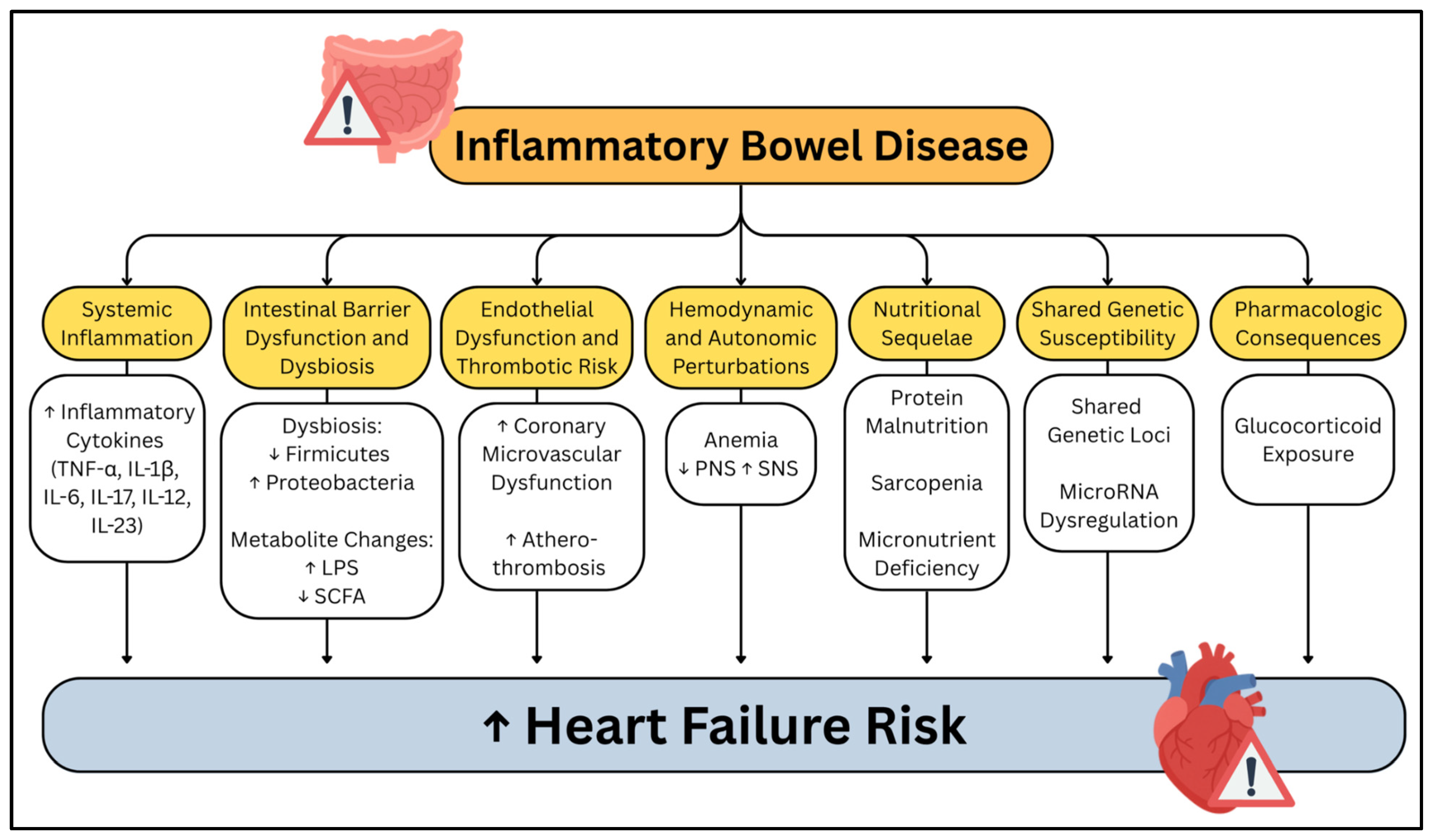
Figure 1. Summary of the mechanisms by which inflammatory bowel disease contributes to the development of heart failure. Yellow boxes represent major inflammatory bowel disease-related pathophysiologic domains, with white boxes provide examples of underlying mechanisms. Arrows indicate directionality, with upward (↑) and downward (↓) arrows denoting increases or decreases in relevant factors.
Cells 14 01124 g001
Figure 2. Summary of the effects of elevated cytokines in inflammatory bowel disease on the heart. Colored boxes represent individual cytokines and the associated mechanisms related to heart failure. Upward arrows (↑) denote increased cytokine levels in inflammatory bowel disease; rightward arrows (→) indicate downstream effects. The figure describes the influence of inflammatory bowel disease on subsequent elevation of inflammatory cytokines which drive progression to heart failure.
Cells 14 01124 g002
Figure 3. Summary of the effects of intestinal barrier dysfunction and dysbiosis in inflammatory bowel disease on the heart. Arrows (→) indicate mechanistic pathways linking microbial metabolites to cardiac dysfunction. Upward arrows (↑) denote increased cytokine levels in inflammatory bowel disease. Green (▲) and red (▼) arrows represent increased or decreased levels of key metabolites. Dysbiosis, characterized by reduced Firmicutes and increased Proteobacteria, leads to toxic metabolite accumulation and loss of protective metabolites. These changes promote inflammation, fibrosis, and thrombosis, contributing to heart failure.
Cells 14 01124 g003
Figure 4. Summary of the effect of elevated phenylacetylglutamine on the heart and thrombotic milieu. Phenylacetylglutamine is produced by Proteobacteria in the setting of dysbiosis, and contributes to cardiac dysfunction, pathologic cardiac remodeling, and prothrombotic milieu. Upward arrows (↑) indicate increased levels or activation of molecular or cellular processes.
Cells 14 01124 g004
Figure 5. Summary of the nutritional sequelae of inflammatory bowel disease and how they contribute to cardiac dysfunction. Circles highlight that intestinal pathology contributes to malabsorption and nutrient deficiency, including protein malnutrition, sarcopenia, iron deficiency, and vitamin D deficiency. Downward arrows () indicate stepwise progression from nutritional deficiencies to cardiac effects and ultimately heart failure.
Cells 14 01124 g005
Disclaimer/Publisher’s Note: The statements, opinions and data contained in all publications are solely those of the individual author(s) and contributor(s) and not of MDPI and/or the editor(s). MDPI and/or the editor(s) disclaim responsibility for any injury to people or property resulting from any ideas, methods, instructions or products referred to in the content.