Article

Transcriptomic Profile of Early Antral Follicles: Predictive Somatic Gene Markers of Oocyte Maturation Outcome

by
Alessia Peserico
1,†,
Barbara Barboni
1,†,
Chiara Camerano Spelta Rapini
1,
Chiara Di Berardino
1,*,
Giulia Capacchietti
1,
Angelo Canciello
1,
Fani Konstantinidou
2,3,
Marisa Donato
1,3,
Liborio Stuppia
2,3 and
Valentina Gatta
2,3
1
Department of Bioscience and Technology for Food, Agriculture and Environment, University of Teramo, 64100 Teramo, Italy
2
Department of Neuroscience, Imaging and Clinical Sciences, School of Medicine and Health Sciences, “G. d’Annunzio” University of Chieti-Pescara, 66100 Chieti, Italy
3
Unit of Molecular Genetics, Center for Advanced Studies and Technology (CAST), “G. d’Annunzio” University of Chieti-Pescara, 66100 Chieti, Italy
*
Author to whom correspondence should be addressed.
These authors contributed equally to the work.
Cells 2025, 14(10), 704; https://doi.org/10.3390/cells14100704
Submission received: 2 April 2025 / Revised: 2 May 2025 / Accepted: 9 May 2025 / Published: 12 May 2025

Abstract

:
Early antral follicles (EAfs) offer oocyte potential in Assisted Reproductive Technology (ART), but most fail to mature under current in vitro maturation (IVM) protocols. This study examines transcriptomic profiles of the follicular wall (FW) compartment during IVM in ovine EAfs using a 3D follicle-enclosed oocyte (FEO) culture to identify somatic gene markers predicting oocyte maturation success. Differentially expressed genes (DEGs) were identified across three comparisons: pre- vs. post-hCG in FW enclosing mature/fertilizable (1) or immature (2) oocytes, and post-hCG between FW supporting successful vs. failed maturation (3). Network analysis highlighted key modulated and HUB genes. Two DEG categories emerged: genes regulating meiosis resumption and genes defining follicular signatures linked to oocyte competence. Meiosis resumption involved ECM remodeling, hypoxia, and relaxin signaling activation, while proliferative and metabolic pathways were downregulated. MMP13 and EGFR regulated the ECM pathway, working for meiosis resumption, while TGFB1 predicted failure. Oocyte competence involves ECM activation and the suppression of stress and cell cycle pathways, with ITIH4 being conducive to central HUB tuning inflammation and angiogenesis-dependent maturation. This study reveals molecular mechanisms behind follicle maturation, identifying transcriptomic signatures for FW releasing mature/fertilizable and incompetent oocytes. It confirms known biomarkers and uncovers new regulators, offering tools to assess follicle quality, improve IVF–oocyte selection, and enhance fertility preservation.

1. Introduction

Assisted Reproductive Technologies (ARTs) have advanced significantly, addressing infertility in humans and animals. However, reliance on small and medium antral follicles limits oocyte availability. In this context, early antral follicles (EAfs), more abundant than antral follicles, emerge as an underused reserve that, with optimized maturation, could expand the pool of fertilizable gametes for reproductive applications [1,2,3,4,5].
EAfs, naturally present in ovaries, can also be generated in vitro as the final stage of folliculogenesis protocols across species (mouse: [6,7]; sheep: [2,4,8,9,10,11]; bovine: [12]; buffalo: [13]; goat: [14,15,16,17]). These follicles can be cultured from preantral stages (PAfs) or earlier follicular phases, expanding the gamete pool for embryo production. This approach enhances fertility preservation and reproductive engineering, offering innovative strategies to improve ART [18,19,20,21].
EAfs house oocytes undergo key developmental processes [4,22], including epigenetic remodeling [4,23], chromatin modifications [2,22,24], and telomere regulation [25], critical for oocyte quality and embryonic development [4]. The transition from late PAfs to EAfs marks a crucial phase where oocytes complete growth and acquire functional specializations. Advances in in vitro Folliculogenesis and animal studies highlight the promise of EAfs in ART, paving the way for clinical applications [1,2,19,26,27,28,29,30].
Despite their potential, oocytes from EAfs often show lower developmental competence compared to those from advanced-stage follicles, requiring specialized in vitro maturation (IVM) protocols. Traditional IVM targets cumulus–oocyte complexes (COCs) and has been extensively optimized in cattle [31,32,33] and sheep [9,26,34,35]. As an alternative, the follicle-enclosed oocyte (FEO) model exploits the intrinsic gonadotropin sensitivity of EAfs to achieve controlled maturation. This approach has been tested in hamsters [36], rats [37], rabbits [38], sheep [1,39,40], and pigs [41].
Recent studies show that bioinspired 3D culture systems markedly improve the maturation of oocytes derived from EAfs, outperforming conventional COC-IVM outcomes [1,2].
Beyond refining IVM techniques, advancements in ART are also focusing on ovarian stroma biomaterials and tissue culture methodologies to increase the availability of EAfs through in vitro Folliculogenesis [42,43,44,45,46,47]. These approaches hold great promise for optimizing the recruitment of fertilizable gametes, expanding the oocyte pool, and enhancing the efficiency of ART applications [2,48,49].
Notably, human chorionic gonadotropin (hCG) is crucial for oocyte maturation in EAfs because of its LH-like activity and minor residual FSH-like effects within the FEO system [1,2]. Experimental evidence shows that EAf-derived oocytes do not progress beyond meiotic arrest unless exposed to hCG [1]. Among the available hormonal stimuli, hCG provides the most effective and reliable maturation trigger in medium-sized mammalian models, supplying adequate gonadotropic support without supplemental FSH [1,2]. Nevertheless, only a small proportion of EAf-derived oocytes reach full competence, underscoring the need to clarify the factors determining success or failure in response to hCG [1].
Compelling evidence demonstrates that the follicular wall (FW)—mural granulosa plus theca—is both a sensor and effector of the preovulatory LH/hCG surge. Moreover, the gonadotrophin elicits a rapid, genome-wide transcriptional reset in these somatic layers that licenses meiotic resumption and, ultimately, shapes embryo potential [50,51,52,53,54,55,56,57]. Cross-species granulosa–oocyte co-profiling further shows that somatic gene signatures faithfully mirror oocyte competence, positioning the follicular transcriptome as a robust surrogate endpoint [58,59,60,61,62,63]. Exploiting this read-out is minimally invasive and promises novel biomarkers, although inter-species and culture-specific variability still demands systematic validation.
Against this backdrop, we employed the FEO-IVM model, which recreates physiological maturation (meiotic resumption occurs solely in response to a controlled LH/hCG stimulus), providing a clean link between somatic transcriptional dynamics and definitive oocyte fate. Using ovine early antral follicles (EAfs), we profiled the FW transcriptome pre- and post-hCG and contrasted EAfs that produced metaphase-II, fertilisable oocytes with those whose oocytes remained at the germinal vesicle stage. Differentially expressed genes (DEGs) associated with follicular activation and oocyte maturation were then prioritized as candidate biomarkers. By coupling this strategy to the FEO-IVM platform, the present study aims to refine IVM protocols, nominate non-invasive somatic markers of oocyte quality, and, ultimately, enhance ART approaches for fertility preservation and reproductive medicine.

2. Materials and Methods

2.1. Ovary Collection

The study involved collecting ovaries from Appenninica sheep lambs from discarded tissues from a local slaughterhouse. These ovaries, from prepubertal sheep around 5 months of age, intended for meat production, were obtained from a local slaughterhouse. They were then transported to the laboratory in a temperature-controlled container to ensure stability during the journey, which typically took less than an hour. Upon arrival, the ovaries were rinsed multiple times with a 0.9% NaCl solution, supplemented with 1 mg/mL Benzoxonium chloride (Bialcol #032186013, Vemedia Pharma, Nola, NA, Italy). After the medulla was removed, the ovaries were placed in a HEPES-buffered TCM199 medium (#M7528, Sigma Aldrich, St. Louis, MO, USA) and cut into uniform cortical fragments approximately 0.5 × 0.5 × 0.5 cm in size.

2.2. Ovarian Surface Epithelium (OSE) Cell Collection for Follicle-Enclosed Oocyte (FEO) Coculture System

Ovarian surface epithelial (OSE) cells were extracted from the ovarian cortex of prepubertal ovaries using a surgical scalpel. Prior to extraction, the ovaries were incubated in a 0.25% Trypsin/EDTA solution (200 mg/L) at 38.5 °C for 5 min. The resulting cell suspensions were transferred into a 6 cm petri dish containing DPBS solution with 30% fetal bovine serum (FBS; #10270-106, Gibco, Thermo Fisher Scientific, Waltham, MA, USA) to neutralize the Trypsin (#25200056, Gibco, Thermo Fisher Scientific, Waltham, MA, USA). After centrifugation, the supernatant was discarded. The OSE cells were then seeded into a growth medium consisting of alphaMEM (#BE02-002F, Lonza, Basel, CH, Switzerland), 20% fetal bovine serum (FBS: #10270-106, Gibco, Thermo Fisher Scientific, Waltham, MA, USA), 1% glutamine (#BE17-605E/U1, Lonza, Basel, CH, Switzerland), and antibiotics including 75 mg/L of penicillin-G and 50 mg/L of streptomycin sulfate (#DE17-602E, Lonza, Basel, CH, Switzerland) for one passage of cell expansion. Once expanded, the OSE cells were seeded into the bottom of the transwell system to create a feeder cell monolayer for FEO coculture.

2.3. FEO In Vitro Maturation from EAfs

Ovine EAfs were manually isolated from cortical fragments using sterile 32 G needles under a stereomicroscope in a laminar flow hood. The selection of EAfs was based on their size and morphology to ensure that the theca layer remained intact. For FEO maturation experiments, the selected EAfs follicles had an average diameter of 360 ± 31 μm. Before incubation, each EAfs was carefully examined using an inverted-phase microscope equipped with time-lapse imaging software (NIS-Elements Advanced Research, Version 4.51.00, Eclipse Ti Series, Nikon Europe BV, Amstelveen, The Netherlands) to measure the diameter and assess for any morphological signs of degeneration. This included checking for the darkening of somatic cells or oocytes, loss of compactness in the granulosa cell layer, or any disruption of the basal membrane. The selected EAfs were placed in transwell culture systems, each containing 96-well plates with U-shaped wells. The follicles were placed on scaffold holders made of PCL-patterned electrospun materials, as previously described [9], while the bottom of the transwell was lined with a monolayer of OSE cells.
Each transwell system was filled with 100 μL of maturation medium, composed of alphaMEM (Cat. No. BE02-002F Lonza, Basel, CH, Switzerland), 20% FBS (#10270-106, Gibco, Thermo Fisher Scientific, Waltham, MA, USA), 1% glutamine (#BE17-605E/U1, Lonza, Basel, CH, Switzerland), and antibiotics (75 mg/L penicillin-G and 50 mg/L streptomycin sulfate; #DE17-602E, Lonza, Basel, CH, Switzerland). The medium was also supplemented with 25 IU/mL human chorionic gonadotropin (hCG; equivalent to 6 µg/mL Chorulon®, MSD Animal Health, Segrate, MI, Italy) to promote in vitro maturation. The EAfs were incubated in this maturation medium for 24 h at 38.5 °C with 5% CO2. At the end of the FEO maturation process, the follicles were opened, and the FW were separated from the oocyte. These cells were then grouped according to the nuclear stage of the oocyte for further analysis and collection. Additionally, FW from EAfs were collected before the addition of hCG and separated from their respective oocytes, forming the control group of FW from EAfs prior to gonadotropic stimulation (FWs enclosing oocytes in the GV stage).

2.4. Oocyte Nuclear Stage Assessment and FW Collection

Oocytes obtained from FEO in vitro maturation were stripped of their surrounding cumulus cells, stained with DAPI, and visualized using the time-lapse imaging software NIS-Elements (Eclipse Ti Series, Nikon, Tokyo, Japan) to assess the nuclear stage. The oocytes were categorized into different meiotic nuclear stages, including germinal vesicle (GV) and Metaphase II (MII), as described previously [4].

2.5. In Vitro Embryo Production

The developmental potential of MII oocytes was assessed through in vitro fertilization (IVF). IVF was conducted using frozen-thawed semen obtained from rams with proven fertility, as described previously [4]. In summary, fertilization was performed in 50 µL drops with a sperm concentration of 1 × 10⁶ sperm/mL at 38.5 °C, in a humidified environment of 5% CO2 in air, for 20–22 h. After 2 and 8 days of culture, the uncleaved and cleaved embryos were examined under an inverted microscope with time-lapse imaging software (NIS-Elements Advanced Research, Version 4.51.00, NIS-Elements, Eclipse Ti Series, Nikon, Japan) to assess developmental stages by counting the number of blastomeres. The fertilization rate was determined by the number of cleaved embryos on day 2, and the blastocyst rate was calculated on day 8 based on the cleaved embryos recorded on day 2.

2.6. Microarray Transcriptomic Analysis

Total RNA was extracted from pools of five follicular walls for each experimental group—namely, GV-stage FWs before hCG stimulation, GV-stage FWs after hCG treatment (unsuccessful), and FWs from MII oocytes that developed to the blastocyst stage (successful) upon IVF—using the Single-Cell RNA Purification Kit (#51800, Norgen Biotek Corp., Thorold, ON, Canada) according to the manufacturer’s protocol. The transcriptomic profile analysis was carried out using the MicroArray GeneChip System (Applied Biosystems, Thermo Fisher Scientific, Waltham, MA, USA).
More specifically, microarray analysis was conducted on all FW samples using the GeneChip WT PLUS Reagent kit (Applied Biosystems, Thermo Fisher Scientific, Waltham, MA, USA). A concentration of 100 ng of total RNA from each sample was used as starting material to synthesize complementary DNA (cDNA). Complementary RNA (cRNA) was subsequently synthesized from in vitro transcription of cDNA and then purified and reverse transcribed. Finally, single-stranded cDNA (ss-cDNA) was synthesized, purified, fragmented, and labeled following the manufacturer’s instructions. Array Hybridization was performed using the GeneChip™ Hybridization, Wash, and Stain Kits and the OviGene 1.0 ST GeneChip Arrays (Ovine; Applied Biosystems) on the GeneChip Hybridization Oven 645 and GeneChip Fluidics Station (Applied Biosystems, Thermo Fisher Scientific, Waltham, MA, USA). Scanning was performed using the GeneChip Scanner 3000 7G and Affymetrix Command Console (AGCC) software, Version AGCC 7.0 (Applied Biosystems, Thermo Fisher Scientific, Waltham, MA, USA). The raw image data obtained after scanning were analyzed using the Transcriptome Analysis Console (TAC) software (ver. 4.0.2, Applied Biosystems). The raw data were normalized following the signal space transformation robust multi-chip analysis (SST-RMA) algorithm. The detected above-background (DABG) cutoff by default was set to 0.05. The positive versus negative area under the curve (AUC) value was set greater than or equal to 0.7. Finally, genes that passed the filter criteria of p value < 0.05 (one-way between-subjects ANOVA) and fold change > 2 for upregulation and <−2 for downregulation were considered as differentially expressed genes (DEGs).

2.7. Network Creation, Visualization, and Analysis

Three datasets (referred to, respectively, as pairwise 1, 2, and 3) were generated using the Transcriptome Analysis Console (TAC) 4.0 Software (Applied Biosystems, Thermo Fisher Scientific, Waltham, MA, USA). Each dataset was used as input to build a network using the Cytoscape 3.9.1 software (http://www.cytoscape.org, accessed on 2 May 2024). Each network was then analyzed with the dedicated plug-in Network Analyzer of Cytoscape according to a previous report [5,64] by computing the following topological parameters: number of nodes, number of edges, the average number of neighbors, network diameter, characteristic path length, clustering coefficient, and connected components (Supplementary Materials File S1), Additional analyses applied to networks and nodes have been listed in the following sub-sections.

2.8. Identification of Highly Interconnected Regions (Modules) Using the MCODE Algorithm

The Cytoscape plugin Molecular Complex Detection (MCODE) [65] was utilized to identify and analyze clusters of densely interconnected nodes (modules) within the network. Modules with k-core values greater than 5 and node degrees exceeding 5 were selected for further KEGG pathway analysis, which was conducted using the Cytoscape ClueGO plugin. The reference database used for network construction and pathway analysis was Ovis aries (Taxid:9940).

2.9. Identification of Drivers Within Network Modules

The study distinguished two types of drivers: highly modulated DEGs and highly interconnected DEGs (HUBs). Highly modulated DEGs, identified as outlier genes based on their expression levels, were detected by comparing gene expression values applying the interquartile range method [66,67] and visualized using a volcano plot (GraphPad Prism 10.1.1; https://www.graphpad.com/, accessed on 2 May 2024). Of note, a significance threshold of p < 0.05 was applied to determine whether a gene’s expression significantly deviated from the expected Gaussian distribution of non-outliers.
HUBs were identified using the CytoHubba plugin in Cytoscape [68] by following a four-step workflow according to recent literature methodological evidence [69,70,71,72,73,74,75].
First, 12 centrality coefficients supported by CytoHubba (Supplementary Materials File S2) were calculated for each DEG, providing each DEG with a score, and plotted using PCA (from GraphPad 10.1.1 Prism; https://www.graphpad.com/, accessed on 2 May 2024).
DEGs were then ranked based on the scores of these centrality coefficients (II).
The top 10 DEGs for each centrality coefficient (those with the highest scores) were selected (III), and DEGs appearing in the top 10 for at least 5 of the 12 algorithms were identified as hubs (IV).

2.10. Venn Diagram

A Venn diagram tool (https://bioinformatics.psb.ugent.be/webtools/Venn/, accessed on 2 May 2024) was used to illustrate the shared and signature DEGs across the networks.

2.11. Microarray Validation Through Real-Time qPCR

The total RNA was extracted with a Single-Cell RNA Purification Kit (#51800, Norgen Biotek Corp., Thorold, ON, Canada) following the manufacturer’s instructions. A total of 1 μg of total RNA was retrotranscribed using oligodT primers (Bioline, Memphis, TN, USA) and Tetro Reverse Transcriptase (Bioline, Bioline, Memphis, TN, USA), following the manufacturer’s instructions. The qPCRs were carried out in triplicate using the SensiFAST SYBR Lo-ROX kit (Bioline, Bioline, Memphis, TN, USA) on a QuantStudio3 System (Life Technologies, Thermo Fisher Scientific, Carlsbad, CA, USA), according to the manufacturer’s instructions. The following PCR conditions were used for all the experiments: 95 °C for 10 min, followed by 40 cycles at 95 °C for 10 s and 60 °C for 30 s. Relative quantification was performed by using the ∆∆Ct method. Glyceraldehyde 3-phosphate dehydrogenase (GAPDH) and Tyrosine 3-Monooxygenase/Tryptophan 5-Monooxygenase Activation Protein Zeta (YWHAZ) were selected from among the housekeeping genes for gene quantification. The expression profiles were similar for both reference genes. The primer sequences are reported in Supplementary Materials File S3.

2.12. Statistical Analysis

Three independent biological replicates for each experimental group (GV start-point, MII end-point, and GV end-point) were conducted. The statistical analysis was conducted as follows:
The Microarray GeneChip assays were performed through a One-Way ANOVA, followed by an empirical Bayes correction for differential expression using the Transcriptome Analysis Console (TAC software; version 4.0.1, Thermo Fisher Scientific, Waltham, MA, USA).
For the analyses related to oocyte maturation performance, embryonic developmental competence, and qPCR validation, One-Way ANOVA was performed with GraphPad Prism 10.1.1 (https://www.graphpad.com/, accessed on 2 May 2024), with p-values less than 0.05 considered statistically significant.
For the ClueGO analysis of the MCODE-defined modules, p-values < 0.05 were deemed significant and corrected with the Bonferroni test.

3. Results

3.1. Comparative Transcriptomic Analysis of FW Compartment from EAfs Enclosing Competent and Incompetent Oocytes

This study profiles, within EAfs, the transcriptome of the FW compartment—the combined mural granulosa and theca cell layers—to elucidate the hCG-driven signaling events that promote oocyte maturation.
EAfs were selected as a source of gonadotropin-sensitive follicles, which, through advanced IVM protocols, can yield an additional pool of fully grown oocytes for ARTs. The validated FEO-IVM method employs a 3D culture system using PCL-patterned scaffolds, significantly enhancing the number of fertilizable oocytes from EAfs.
As shown in Table 1A, FEO-IVM maintains meiotic arrest without hormonal stimulation (100% GV) and supports meiosis resumption in most oocytes (89% GVBD+MII) upon hCG exposure. Of the matured MII oocytes, 58.2% were fertilized, though only ~10% progressed to advanced embryonic stages (Table 1B). The small subset of EAf-enclosed oocytes that failed to initiate maturation (GV 11%) in response to hormonal stimulation provided the FWs used to analyze the transcriptomic profile of structures incapable of supporting oocyte maturation.
The experimental design (Figure 1) compared follicular cells (FWs) from EAfs under three conditions: (i) EAfs collected before hCG stimulation (GV start-point); (ii) EAfs whose oocytes progressed to the metaphase-II (MII) stage after hCG—designated successful EAfs (MII end-point); and (iii) EAfs whose oocytes failed to resume meiosis and remained at the germinal vesicle (GV) stage despite hCG—designated unsuccessful EAfs (GV end-point).
Each group was interrogated with an ovine whole-transcriptome microarray. For each pairwise comparison, differentially expressed genes (DEGs) were first delineated and subsequently subjected to network-based prioritization to extract putative driver genes. The pairwise analyses addressed two biological objectives: AIM 1—to elucidate follicular-cell driver genes governing the hCG-dependent activation of EAfs. AIM 2—to identify candidate drivers that discriminate follicular cells from EAfs capable of, versus refractory to, hCG-induced oocyte meiotic maturation. For AIM 1, two pairwise comparisons were conducted: first, FWs from successful EAfs releasing MII oocytes (MII end-point) versus unstimulated controls (GV start-point); second, FWs from unsuccessful EAfs releasing GV oocytes (GV end-point) versus GV start-point as control. AIM 2 involved a third pairwise comparison between FWs from successful and unsuccessful maturation outcomes (MII vs. GV end-point) to highlight transcriptomic differences associated with divergent oocyte fates following hCG treatment.
In Table 2, DEGs obtained from three pairwise comparisons after the analysis of 22,141 Ovis aries genes were summarized, adopting a fold change greater than 2 and a p-value less than 0.05.
In more detail, most DEGs were downregulated across the three pairwise comparisons—66%, 56%, and 90.7%, respectively.

3.2. FW Driver Genes Promoting Maturation in EAfs

3.2.1. Network 1MII-GV and Network 2GV-GV Signaling Modules

To identify the gene markers that characterized FW supporting hCG-induced meiotic resumption, the DEGs identified in pairwise 1 (end-point MII vs. start-point GV: MII-GV) and pairwise 2 (end-point GV vs. start-point GV: GV-GV) comparisons were processed using Cytoscape version 3.9.1. This procedure generated two networks representing, respectively, successful (Network 1MII-GV) and unsuccessful (Network 2GV-GV) EAfs.
Genes with uncharacterized function and long non-coding RNAs were excluded from the generation of the 2 Networks since they are not recognized by Cytoscape (457 of Network 1MII-GV and 437 of Network 2GV-GV were not recognized).
More in detail, Network 1MII-GV comprised 2.071 nodes (annotated ovine DEGs), 15.712 edges, and 92 connected components. Network 2GV-GV recognized fewer nodes and edges (1.562 and 6.546, respectively) yet retained a similar number of connected components (n = 95). Topological metrics confirmed the scale-free nature of both the networks (Supplementary Materials File S4).
The networks were analyzed using the MCODE clustering algorithm to extract highly interconnected clusters (modules) of DEGs (Supplementary Materials File S5). The resulting modules were functionally annotated in ClueGO and consolidated into the six broadest KEGG pathway categories. Specifically, Network1MII-GV recognized 23.3% (482/2071) of DEGs clustered into 10 functional modules belonging to five out of the seven largest KEGG categories (Scheme 1).
Network 1MII-GV denotes the network built from pairwise 1, whereas Network 2GV-GV corresponds to the network generated from pairwise 2. Modules were extracted with MCODE (k-core ≥ 5, node degree > 5; see Section 2.8) and functionally annotated in ClueGO; resulting terms were consolidated into the major KEGG pathway categories. Full statistics and thresholds are provided in Supplementary Materials File S5. The color code distinguishes (i) shared vs. unique DEGs and (ii) the different KEGG categories, while symbol style denotes up- or downregulation.
Three categories were distinctive of Network 1MII-GV: genetic information processing, environmental information processing, and the organismal system. The environmental information processing category recognized mainly upregulated DEGs (red signed genes) involved in ECM remodeling, relaxin, HIF signaling, and cytosolic DNA sensing pathways (modules 7–8). Conversely, the genetic information processing KEGG categories exclusively comprised downregulated DEGs (green signed genes) mapped to RNA and cell cycle pathways (modules 2 and 3).
Metabolism was the most populated KEGG path and the largest category of Network 1MII-GV. It consisted mainly of downregulated DEGs related to fatty acid and amino acid degradation (both in modules 3 and 6), terpenoid/steroid biosynthesis (module 3), and two distinctive metabolic pathways (purine metabolism in module 5 and pentose phosphate in module 7). Within cellular processes, additional downregulated DEGs involved in progesterone-mediated maturation (modules 2 and 3), oxidative phosphorylation (module 1), and lysosome and proteosome processes (module 1 and 5, respectively).
Network 2GV-GV recognized a lower percentage of clustered DEGs (13.6%: 213 out of 1562) that belonged to six diverse modules grouped in three of KEGG’s largest categories (Scheme 1). A distinctive feature was the immune signaling and response (modules 2, 6). It was composed of downregulated DEGs, all involved in the IL-17 pathway (module 2), and of genes with a mixed profile of expression (signed in black) related to viral protein interactions with cytokine and cytokine receptor pathways.
As in Network 1MII-GV, metabolism was also the most enriched KEGG category in Network 2GV-GV; however, the pathway composition differed. Upregulated genes mapped to glycolysis/gluconeogenesis (module 1) and carbon metabolism (module 3), whereas downregulated genes populated glutathione, pyrimidine, one-carbon-pool–by-folate (module 4), and various signaling-metabolism pathways (module 5).

3.2.2. Driver DEGs of Network 1MII-GV and Network 2GV-GV

The MCODE-derived clusters were further prioritized by applying two complementary filters. First, highly modulated DEGs—those exhibiting the greatest fold-changes and thus representing the most immediate, co-regulated response to hCG—were delineated. Second, highly interconnected DEGs (HUBs) were identified based on their central topological indices, indicating a pivotal role in organizing network behavior and governing the meiotic maturation program. This integrated approach distinguishes transcripts that respond most vigorously to hCG from those that structurally orchestrate the underlying signaling circuitry.

Highly Modulated DEGs of Network 1MII-GV and Network 2GV-GV

Highly modulated DEGs were visually selected using a volcano plot, which enabled their stratification based on gene modulation levels and significance in the two networks.
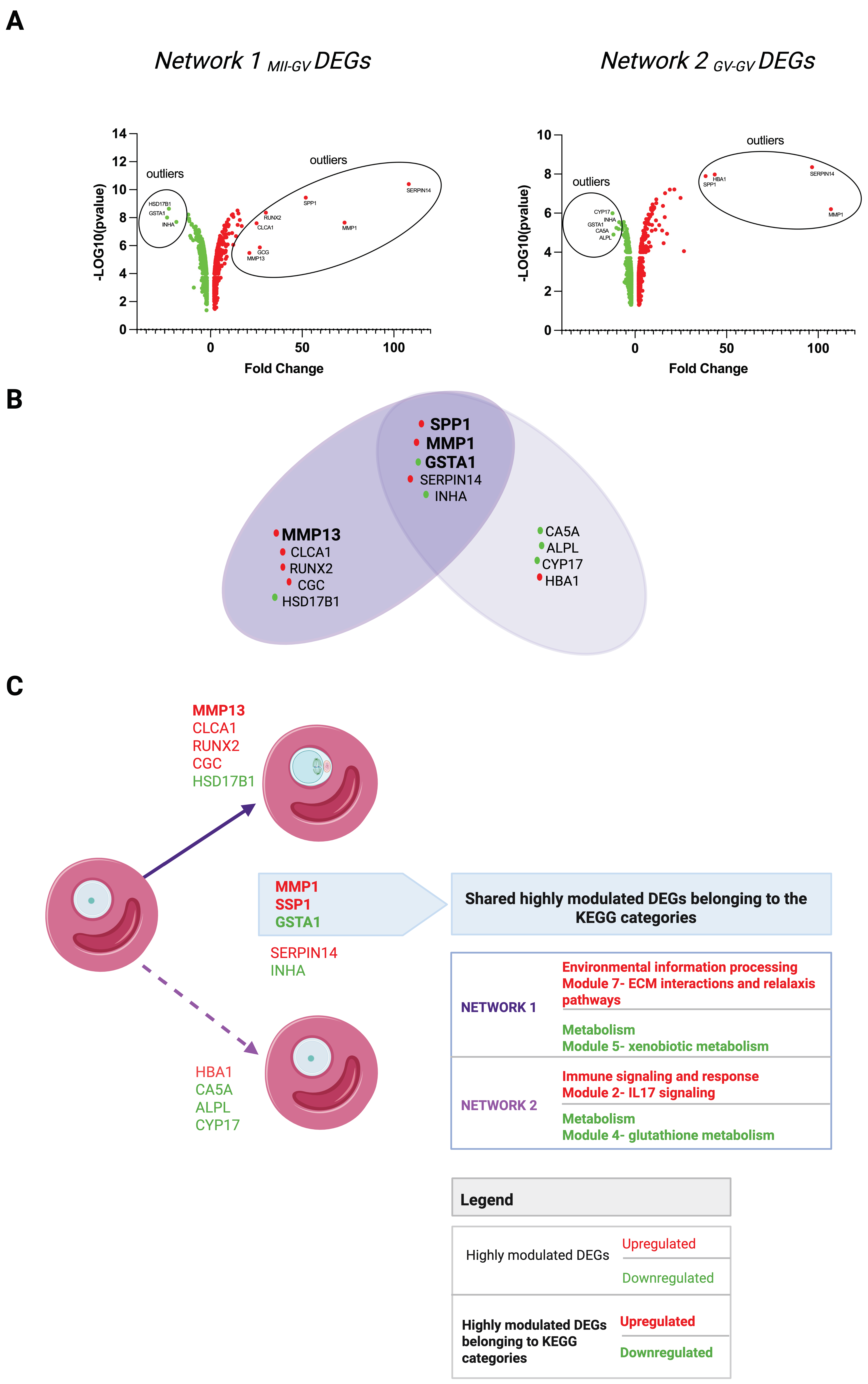
As summarized in Figure 2A, the highly modulated DEGs (outlier DEGs) are as follows:
Network 1MII-GV: Seven upregulated (MMP1, SPP1, CLCA1, MMP13, SERPIN14, GCG, RUNX2) and three downregulated (GSTA1, HSD17B, INHA);
Network 2GV-GV: Four upregulated (MMP1, SPP1, HBA1, SERPIN14) and five downregulated (GSTA1, CYP17, ALPL, CA5A, INHA).
The highly modulated DEGs identified using MCODE but positioned outside densely connected clusters (unclustered) were not regarded as critical nodes, even though they met the criteria outlined in point (I). Consequently, within Network 1MII-GV, only three out of seven upregulated (MMP1, SPP1, MMP13) and one out of three downregulated (GSTA1) outliers fell inside the MCODE modules (Scheme 1). All three upregulated genes resided in module 7 (environmental information processing), which encompasses the ECM–receptor interaction and relaxin-signaling pathways, whereas the single downregulated gene was located in module 5 (Metabolism), linked to xenobiotic metabolism.
Of note, MMP13 emerged as the only distinctive upregulated outlier with a key role in hCG-driven maturation within the FW (Network 1MII-GV). It displayed a great modulatory function, being linked with several other upregulated DEGs in the ECM remodeling pathway (module 7 of environmental information processing, the KEGG path’s largest category), such as BMP2, CD44, CXCR4, MMP1, MMP9, PLAU, SPP1, and TIMP3 (Supplementary Materials File S6).
In Network 2GV-GV, two out of four upregulated (MMP1, SPP1) and one out of five downregulated (GSTA1) DEGs were located within MCODE modules. Although Venn analysis flagged these DEGs as common outliers to both networks (Figure 2B), the MCODE approach revealed network-specific functions: in Network 2GV-GV, the upregulated outliers operated within the IL-17 signaling (module 2 of Immune signaling and response KEGG category), whereas the downregulated GSTA1, clustered into module 5 (metabolism KEGG category,) implicated glutathione metabolism signaling.
A comprehensive overview of the highly modulated genes unique to, or shared between, the two networks is provided in Figure 2C.

HUBs of Network 1MII-GV and Network 2GV-GV

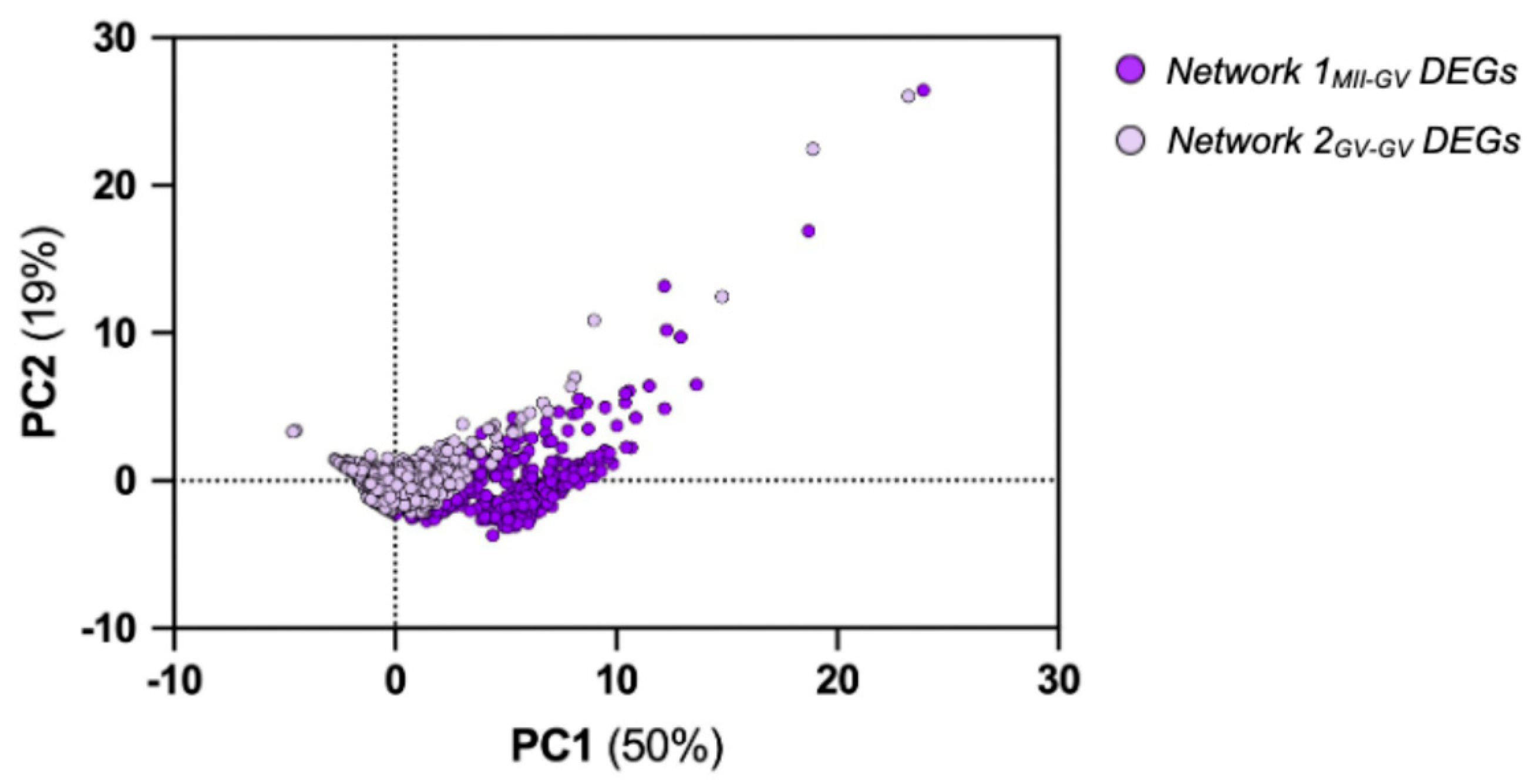
Finally, a computational workflow was applied to identify HUBs, DEGs that preserve network architecture and govern its functionality. In detail, HUBs were detected with the CytoHUBba plugin in Cytoscape via a four-step procedure. In more detail, through step I, each DEG was scored for each centrality categories (Closeness, Degree, Maximal Clique Centrality, Radiality, Stress, Maximum Neighborhood Component, Density of Maximum Neighborhood Component, Betweenness, Clustering Coefficient, EcCentricity, BottleNeck, and Edge Percolated Component) to measure the network influence through the relative coefficient. Once each coefficient of centrality was defined in step II, the gathered scores were defined through principal component analysis (PCA) using the multivariate approach. The analysis enabled the detection of the differences between the two networks and reduced the dataset’s complexity into two principal components. These components explained approximately 70% of the total variance (Figure 3; see also Supplementary Materials File S7).
DEGs of each network were then filtered for their priority, and the top 10 were ranked based on their centrality coefficient scores (step III). Finally, to obtain a reliable prediction of DEGs with a HUB role, only DEGs ranked as top 10 for at least 5 of the 12 centrality coefficient scores selected (step IV; Supplementary Materials File S8). To distinguish between shared or distinctive HUBs, Venn diagram analysis was conducted (Supplementary Materials File S8).
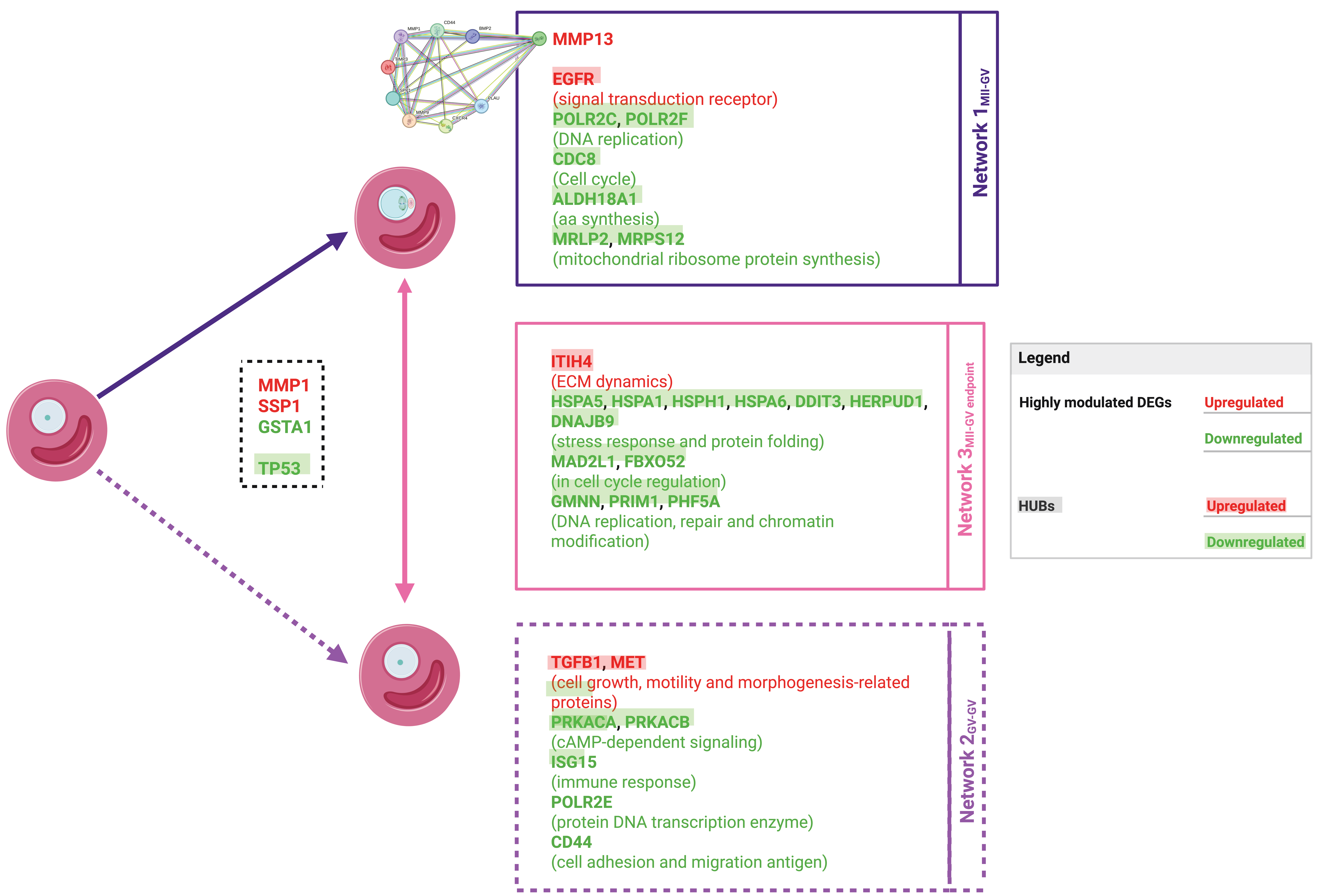
The computational analysis identified seven HUBs in Network 1MII-GV (Supplementary Materials File S8, sheet 3). EGFR was upregulated, whereas the others were downregulated and mapped to functions in DNA transcription (POLR2C, POLR2F), cell cycle regulation (CDCA8), amino acid biosynthesis (ALDH18A1), and mitochondrial protein synthesis (MRPL2, MRPS12).
Analogously, seven HUBs were detected in Network 2GV-GV (Supplementary Materials File S8, sheet 4). Only TGFB1 and MET were upregulated, both linked to cell growth, motility, and morphogenesis-related pathways. The downregulated HUBs were involved in cAMP-dependent signaling (PRKACA, PRKACB), immune response protein (ISG15), DNA transcription enzyme (POLR2E), and cell adhesion and migration antigen (CD44).
Venn diagram analysis revealed a unique shared HUB between the two networks: TP53, which was downregulated and is implicated in DNA damage repair.
Overall, the gene driver sets for each network are comprised of the following:
Network 1MII-GV: One highly modulated DEG (MMP13) and seven HUBs (POLR2C, POLR2F, CDCA8, ALDH18A1, MRPL2, MRPS12, and EGFR).
Network 2GV-GV: Seven HUBs (TGFB1, MET, PRKACA, PRKACB, ISG15, POLR2E, and CD44).

3.3. Signature Genes Distinguishing FWs from Successful Versus Unsuccessful EAfs at the End of the Maturation Phase

Following the identification of the key FW genes triggering oocyte maturation, the study then turned to those genes whose expression profiles distinguish follicular signatures predictive of oocyte competence at the maturation end-point (AIM 2 Figure 1).
To this end, the following workflow was applied to filter DEGs in the EA FW sampled 24 h after hCG stimulation.
(I) Analyze the transcriptomic landscape of FW enclosing MII (successful maturation) and GV (unsuccessful maturation) at the end-point of FEO in vitro maturation (pairwise 3 comparison).
(II) Build the Network 3MII-GV end-point using the DEGs identified from pairwise 3 as inputs.
(III) Classify DEGs according to their functional role.
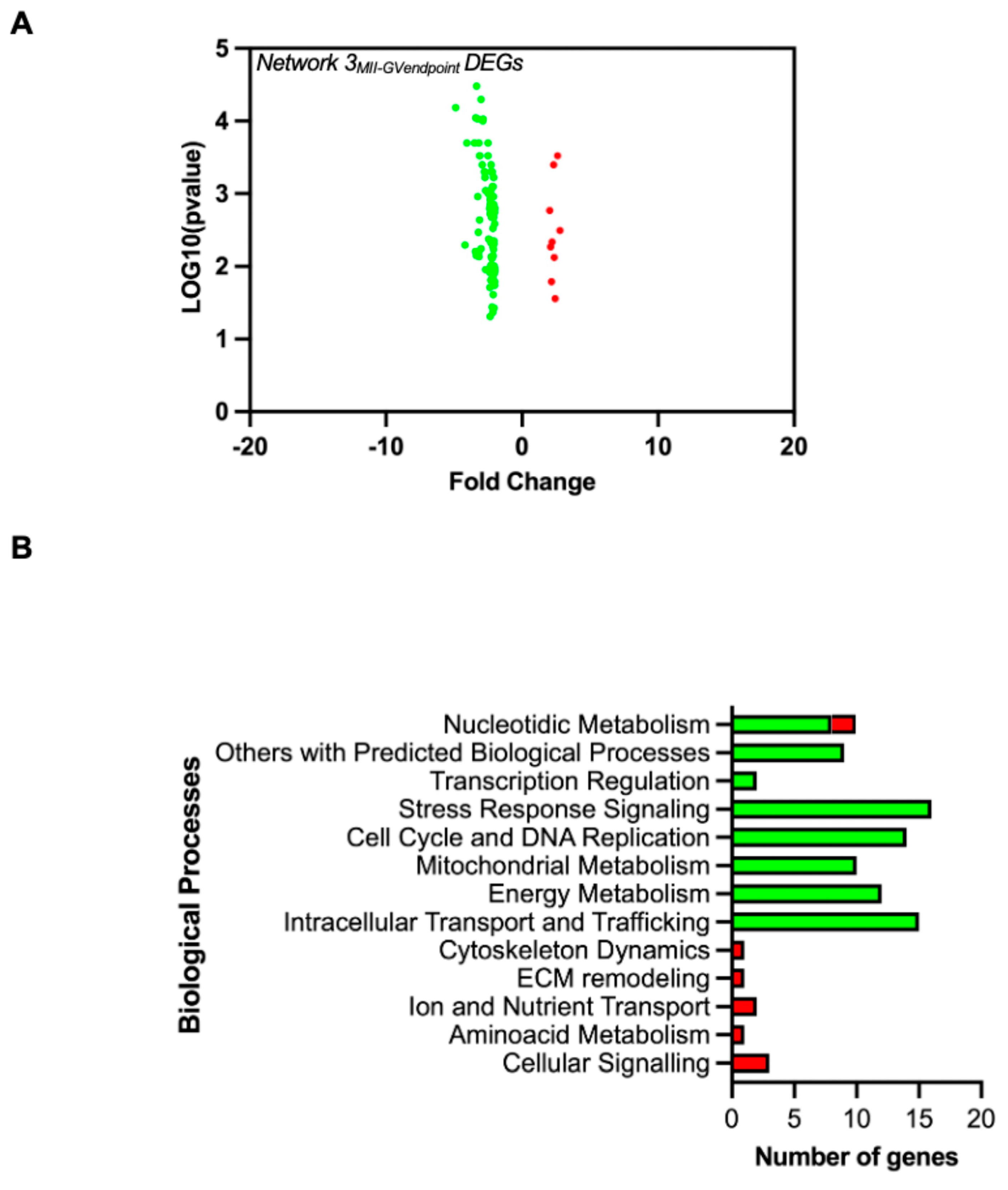
To delve into the details of the analysis, the maturation end-point transcriptome reveals 9 upregulated and 88 downregulated DEGs (Figure 4A and Table 2).
The Network 3MII-GVend-point (MII end-point vs. GV end-point), built on the selected DEGs, recognized 97 nodes, 44 edges, and 56 connected components. The topological parameters listed in Supplementary Materials File S9 confirmed its scale-free nature.
Given the small DEG set, MCODE clustering was not performed; instead, DEGs were categorized by their associated biological processes.
As depicted in Figure 4B and detailed in Supplementary Materials File S10, upregulated DEGs in Networks 3MII-GVend-point were predominantly enriched in metabolic and cellular homeostasis pathways—particularly amino acid metabolism, ion and nutrient transport, cellular signaling, ECM remodeling, and cytoskeleton dynamics—whereas downregulated DEGs mapped to energy, nucleotide, and mitochondrial metabolism, cell cycle control, transcriptional regulation, and DNA replication.
Driver genes were also selected in Network 3MII-GVend-point by distinguishing between highly modulated and HUBs. No highly modulated DEGs were identified; however, 13 HUBs were filtered (Supplementary Materials File S11).
Specifically, the HUBs including seven DEGs involved in stress response and protein folding (HSPA5, HSPA1A, HSPH1, HSPA6, DDIT3, HERPUD1, and DNAJB9), two in cell cycle regulation (MAD2L1, FBXO5), three in DNA replication, repair, and chromatin modification (GMNN, PRIM1, PHF5A) and, finally, one related to ECM dynamics (ITIH4).

3.4. Validation of Predicted Driver Genes in Oocyte Maturation: qRT-PCR Confirmation and Literature Review

Overall, the present transcriptomic and computational analysis identified genes with a potential regulatory role in the maturation process, also allowing the distinction of two different types of somatic biomarkers:
(I) Somatic markers differentiating between FWs before and after hCG-induced maturation (derived from Network 1MII-GV and Network 2GV-GV).
(II) Somatic biomarkers of follicle leading to oocyte competence (derived from Network 3MII-GV end-point). Figure 5 provides an illustrated overview of all the identified drivers along with their modulations.
The identified candidate drivers were subjected to a final validation regarding their gene modulations found through array analysis (I), as well as a review of any reported roles in meiotic maturation described in the literature (II).
In more detail, gene expression data from the ovine array were validated through a qRT-PCR analysis of selected DEGs from each of the three networks. Specifically, highly modulated DEGs such as MMP13 from Network 1MII-GV, the HUBs CDCA8 from Network 1MII-GV, TGFB1 and ISG15 for Network 2GV-GV, and HSPA6, GMNN, and ITIH4 for Network 3MII-GV end-point were selected (Supplementary Materials File S12).
Furthermore, the selected candidate genes were subjected to a comprehensive review of recent scientific literature to evaluate the latest insight into their biological roles in mediating the maturation process. The results of this literature review are summarized in Table 3. Notably, the biological reproductive role has been confirmed for some of the identified driver genes (15 out of 28), offering new insights for further in-depth investigations.

4. Discussion

This study provides new insights into the transcriptomic mechanisms regulating the FW of EAfs activated through hCG-mediated oocyte maturation. Using an FEO 3D culture system, somatic gene markers were identified that determine FW to be capable of supporting the maturation of high-quality oocytes from those that fail to trigger meiotic resumption.
A key merit of this study is the differentiation of molecular signatures governing meiosis resumption, alongside the identification of non-invasive somatic biomarkers predictive of oocyte competence. This is particularly critical for early antral follicles, the focus of our study. Although our FEO-IVM model yields high meiotic resumption rates (>65%) after hCG stimulation, the blastocyst formation rate remains low (~10%). Similar trends have been observed in other large mammals, where oocytes from smaller follicles show impaired cytoplasmic maturation and reduced cleavage and blastocyst yields versus those from larger follicles (ovine [94]; mouse [95]; bovine [96,97]). This limited developmental competence hinders the clinical use of early antral follicle-derived oocytes in ART and highlights the urgent need for non-invasive selection strategies. The somatic biomarkers identified here offer a means to enrich for oocytes from early follicles with true embryonic potential.
The identified DEGs fall into two major categories: (1) genes regulating meiosis resumption and (2) genes defining follicular signatures that support oocyte competence.
The first category—signing, signaling, and molecules required for meiosis resumption—was associated with the upregulation of ECM remodeling, relaxin, and hypoxia signaling pathways, while proliferative and metabolic pathways were downregulated.
Among the ECM-related genes, MMP13 was highly modulated, reinforcing its established role in ECM breakdown and ovulation supported in different species [79,98,99,100,101,102,103,104,105,106]. Also, the epidermal EGFR appeared as a critical HUB gene in the EDC category, highlighting its regulatory role in ECM remodeling, which requires the regulation of somatic germinal coupling during ovulation [78].
The structural remodeling of the follicle required for ovulation also depends on the activation of the relaxin signaling pathway [107,108]. Accordingly, this study identified this pathway as upregulated in follicular cells enclosing the mature oocyte. Relaxin has been shown to act as a paracrine factor during the preovulatory phase, regulating collagenase activity [109], and key molecules of this pathway have been detected in the follicular fluid of women [110], equines [111], and pigs [112].
Hypoxia signaling was also pivotal in follicular maturation. HIF-1α activation was evident in FW supporting competent oocytes, reflecting its well-established role in facilitating the transition from periovulation to early corpus luteum differentiation under low oxygen conditions [113,114]. The HIF-1α-mediated regulation of ECM remodeling and glucose metabolism supports oocyte maturation, with inhibition of HIF-1α in vivo preventing oocyte release [115]. These findings further highlight hypoxia as a key regulator of successful follicular remodeling and oocyte competence.
The suppression of proliferative and metabolic pathways additionally supports the transition from granulosa cell proliferation to differentiation, thus reinforcing the metabolic quiescence hypothesis, which posits that successful meiosis resumption requires a coordinated metabolic rearrangement [116,117,118,119].
Conversely, in FW, after enclosing oocytes that failed to resume meiosis, TGFB1 emerged as a critical HUB. TGFB1 is associated with meiotic arrest and follicular immaturity [120,121,122,123,124], suggesting that its upregulation may contribute to the failure of oocyte developmental progression.
The second category of DEGs was associated with follicular signatures supporting oocyte competence. Among these, ITIH4 emerged as an upregulated HUB gene involved in angiogenesis, ECM stability, and ovulation-dependent inflammatory pathways. ITIH4 was notably upregulated in follicular fluid from follicles yielding mature oocytes, alongside key complement activation molecules [92,93], highlighting its role in the inflammatory response driving ovulation. Additionally, its contribution to angiogenesis and ECM stability is supported by its interaction with Von Willebrand factor and hyaluronan [125,126,127].
Moreover, the downregulation of cell cycle, DNA replication, and stress-related genes in the second DEG category suggests a lower cellular stress burden and improved genomic integrity. Accordingly, downregulated HUBs involved in cell cycle regulation and DNA replication (MAD2L1, FBOX5, GMNN, PRIM1, PHF5A) were found to be switched off when the differentiation shift occurs in response to hCG/LH [53]. Regarding the identified HUBs related to stress response and protein folding (HSPA5, HSPA1A, HSPH1, HSPA6, DDIT3, HERPUD1, and DNAJB9), although their role in the somatic compartment remains unclear [128], knockout models from the MGI database (see Table 3) highlight their reproductive functions.

5. Conclusions

This research advances our understanding by identifying novel DEGs associated with reproductive processes. Among these, some may represent novel regulators of follicular maturation and oocyte competence, opening new avenues for understanding follicular biology. Analyzing the genomic signature of FW with defined maturation outcomes might allow the integration of multi-omics approaches like proteomics and epigenomics, further elucidating regulatory networks governing oocyte competence.
A panel of non-invasive predictive biomarkers has been delineated: MMP13, EGFR, and ITIH4 are indicative of a follicular environment conducive to successful maturation, whereas TGFB1 marks failure. Assessing these genes in FW could refine oocyte selection in ART, improving fertilization rates and embryo quality. Additionally, TGFB1 may be a therapeutic target, as modulating TGF-β signaling could enhance oocyte maturation from EAfs.
Also, crucial transcriptomic signatures distinguishing FWs from EAfs releasing competent and incompetent oocytes were defined, highlighting ECM remodeling genes as key regulators of oocyte maturation. The discovery of previously uncharacterized DEGs offers new insights into follicular biology and ART advancements. To fully leverage these findings, validation across species and clinical settings is necessary. Future research should investigate dynamic gene expression changes during IVM to elucidate follicular activation and characterize differential signaling pathways and molecules in EAfs enclosing oocytes that progress to the blastocyst stage versus those from MII oocytes that do not, thereby enabling a more refined and accurate selection of gametes with improved success rates.
Additionally, functional studies, such as knockdown experiments, are essential to confirm the roles of newly identified DEGs in follicular maturation and oocyte competence. These efforts will help refine IVM protocols and improve oocyte selection strategies, despite the challenges posed by studying low-competent oocytes.

Supplementary Materials

The following supporting information can be downloaded at: https://www.mdpi.com/article/10.3390/cells14100704/s1, File S1: Main Topological Parameters Assessed in the Present Study by Cytoscape Network Analyzer. File S2: List of centrality coefficients calculated in the present study by CytoHubba plugin of Cytoscape. File S3: Sequence of primers used for qPCR validation. File S4: Network 1MII-GV and Network 2GV-GV topological parameters. File S5: List of DEGs, clustering into MCODE modules and identification of the largest KEGG pathway categories in Network 1MII-GV and Network 2GV-GV. File S6: MMP13 in the MCODE cluster 7, connects with genes involved in ECM remodeling. The graphic depicts the PPI String computed interaction of MMP13 within genes of cluster 7 of Network 1. File S7: Network 1MII-GV and Network 2GV-GV centrality parameters. File S8: Network 1MII-GV and Network 2GV-GV top 10 DEGs defined on each centrality coefficient score. File S9: Network 3MII-GVend-point topological parameters. The table displays the computed topological parameters. File S10: Classification of DEGs in Network 3MII-GVend-point based on the biological processes in which they are involved. File S11: Centrality parameters computed by CytoHUBba for Network 3MII-GVend-point. Sheet 1 contains 12 centrality coefficients computed for each DEG of Network 3MII-GVend-point. File S12: Real time qPCR validation of GeneChip data, related to selected DEGs.

Author Contributions

Conceptualization B.B.; Methodology A.P. and B.B.; Validation A.P. and F.K.; Formal analysis A.P.; Investigation A.P., C.C.S.R., C.D.B., G.C., F.K. and A.C.; Resources B.B., L.S. and V.G.; Data Curation A.P., B.B. and F.K.; Writing—Original Draft A.P. and B.B.; Writing—Review and Editing A.P., B.B., C.D.B., F.K. and V.G.; Visualization A.P., C.D.B. and M.D.; Supervision B.B., L.S. and V.G.; Funding acquisition B.B. and A.P. All authors have read and agreed to the published version of the manuscript.

Funding

This research was funded by (I) PRIN PNRR 2022—Prot. P20222H7J7. Title: Profiling Redox, Epigenetic and Transgenerational Effects Induced by Micro-Nanoplastics: A Focus on Female Reprotoxicity and Protective Natural Antioxidants in Mammals; (II) European Union—NextGenerationEU, Mission 4, Component 1, under the Italian Ministry of University and Research (MUR) National Innovation Ecosystem grant ECS00000041—VITALITY—CUP: C43C22000380007.

Institutional Review Board Statement

Not applicable.

Informed Consent Statement

Not applicable.

Data Availability Statement

All data generated or analyzed during this study are included in this published article and its Supplementary Materials Files.

Acknowledgments

The authors would like to acknowledge the support provided by the European Union’s Next Generation EU initiative through the “Innovative Doctorates addressing the needs of industry” program, DM 117 3.3 (CUP: C46E23000130004), for the doctoral fellowships awarded to M.D., V.G., and B.B. Special thanks to Fabiana Verni for her invaluable assistance in the collection and transportation of biological materials. All individuals included in this section have consented to the acknowledgment.

Conflicts of Interest

The authors declare no conflicts of interest.

References

  1. Peserico, A.; Di Berardino, C.; Capacchietti, G.; Camerano Spelta Rapini, C.; Liverani, L.; Boccaccini, A.R.; Russo, V.; Mauro, A.; Barboni, B. IVM Advances for Early Antral Follicle-Enclosed Oocytes Coupling Reproductive Tissue Engineering to Inductive Influences of Human Chorionic Gonadotropin and Ovarian Surface Epithelium Coculture. Int. J. Mol. Sci. 2023, 24, 6626. [Google Scholar] [CrossRef]
  2. Di Berardino, C.; Peserico, A.; Camerano Spelta Rapini, C.; Liverani, L.; Capacchietti, G.; Russo, V.; Berardinelli, P.; Unalan, I.; Damian-Buda, A.I.; Boccaccini, A.R.; et al. Bioengineered 3D ovarian model for long-term multiple development of preantral follicle: Bridging the gap for poly(ε-caprolactone) (PCL)-based scaffold reproductive applications. Reprod. Biol. Endocrinol. 2024, 22, 95. [Google Scholar] [CrossRef]
  3. Chansaenroj, A.; Songsasen, N.; Chatdarong, K. Equine chorionic gonadotropin induces in vitro follicular growth from the multi-layered secondary developmental stage in cats. Theriogenology 2019, 123, 116–122. [Google Scholar] [CrossRef]
  4. Barboni, B.; Russo, V.; Cecconi, S.; Curini, V.; Colosimo, A.; Garofalo, M.L.A.; Capacchietti, G.; Di Giacinto, O.; Mattioli, M. In vitro grown sheep preantral follicles yield oocytes with normal nuclear-epigenetic maturation. PLoS ONE 2011, 6, e27550. [Google Scholar] [CrossRef]
  5. Bernabò, N.; Di Berardino, C.; Capacchietti, G.; Peserico, A.; Buoncuore, G.; Tosi, U.; Crociati, M.; Monaci, M.; Barboni, B. In Vitro Folliculogenesis in Mammalian Models: A Computational Biology Study. Front. Mol. Biosci. 2021, 8, 737912. [Google Scholar] [CrossRef]
  6. Cortvrindt, R.; Smitz, J.; Van Steirteghem, A.C. A morphological and functional study of the effect of slow freezing followed by complete in-vitro maturation of primary mouse ovarian follicles. Hum. Reprod. 1996, 11, 2648–2655. [Google Scholar] [CrossRef]
  7. Eppig, J.J.; O’Brien, M.J. Development in Vitro of Mouse Oocytes from Primordial Follicles. Biol. Reprod. 1996, 54, 197–207. [Google Scholar] [CrossRef]
  8. Cecconi, S.; Barboni, B.; Coccia, M.; Mattioli, M. In vitro development of sheep preantral follicles. Biol. Reprod. 1999, 60, 594–601. [Google Scholar] [CrossRef]
  9. Di Berardino, C.; Liverani, L.; Peserico, A.; Capacchietti, G.; Russo, V.; Bernabò, N.; Tosi, U.; Boccaccini, A.R.; Barboni, B. When Electrospun Fiber Support Matters: In Vitro Ovine Long-Term Folliculogenesis on Poly (Epsilon Caprolactone) (PCL)-Patterned Fibers. Cells 2022, 11, 1968. [Google Scholar] [CrossRef]
  10. Di Berardino, C.; Peserico, A.; Capacchietti, G.; Crociati, M.; Monaci, M.; Tosi, U.; Mauro, A.; Russo, V.; Bernabò, N.; Barboni, B. Equine Chorionic Gonadotropin as an Effective FSH Replacement for In Vitro Ovine Follicle and Oocyte Development. Int. J. Mol. Sci. 2021, 22, 12422. [Google Scholar] [CrossRef]
  11. Barros, V.R.P.; Monte, A.P.O.; Lins, T.L.B.G.; Santos, J.M.; Menezes, V.G.; Cavalcante, A.Y.P.; Arujo, V.R.; Gouveia, B.B.; Matos, M.H.T. In vitro survival, growth, and maturation of sheep oocytes from secondary follicles cultured in serum-free conditions: Impact of a constant or a sequential medium containing recombinant human FSH. Domest. Anim. Endocrinol. 2019, 67, 71–79. [Google Scholar] [CrossRef]
  12. Sun, J.; Li, X. Growth and antrum formation of bovine primary follicles in long-term culture in vitro. Reprod. Biol. 2013, 13, 221–228. [Google Scholar] [CrossRef] [PubMed]
  13. Gupta, P.S.P.; Ramesh, H.S.; Manjunatha, B.M.; Nandi, S.; Ravindra, J.P. Production of buffalo embryos using oocytes from in vitro grown preantral follicles. Zygote 2008, 16, 57–63. [Google Scholar] [CrossRef]
  14. Da Silva, G.M.; Rossetto, R.; Chaves, R.N.; Duarte, A.B.G.; Araújo, V.R.; Feltrin, C.; Bernuci, M.P.; Anselmo-Franci, J.A.; Xu, M.; Woodruff, T.K.; et al. In vitro development of secondary follicles from pre-pubertal and adult goats cultured in two-dimensional or three-dimensional systems. Zygote 2015, 23, 475–484. [Google Scholar] [CrossRef]
  15. Magalhães, D.M.; Duarte, A.B.G.; Araújo, V.R.; Brito, I.R.; Soares, T.G.; Lima, I.M.T.; Lopes, C.A.P.; Campello, C.C.; Rodrigues, A.P.R.; Figueiredo, J.R. In vitro production of a caprine embryo from a preantral follicle cultured in media supplemented with growth hormone. Theriogenology 2011, 75, 182–188. [Google Scholar] [CrossRef]
  16. Costa, S.L.; Costa, E.P.; Pereira, E.C.M.; Benjamin, L.A.; Rodrigues, M.T.; Mendes, V.R.A.; Silva, T.F. Influence of insulin-like growth factor I (IGF-I) on the survival and the in vitro development of caprine preantral follicles. Pesquisa Veterinaria Brasileira 2014, 34, 1037–1044. [Google Scholar] [CrossRef]
  17. Chaves, R.N.; Duarte, A.B.G.; Rodrigues, G.Q.; Celestino, J.J.H.; Silva, G.M.; Lopes, C.A.P.; Almeida, A.P.; Donato, M.A.M.; Peixoto, C.A.; Moura, A.A.A.; et al. The Effects of Insulin and Follicle-Simulating Hormone (FSH) During In Vitro Development of Ovarian Goat Preantral Follicles and the Relative mRNA Expression for Insulin and FSH Receptors and Cytochrome P450 Aromatase in Cultured Follicles. Biol. Reprod. 2012, 87, 69. [Google Scholar] [CrossRef]
  18. Ebrahimi, M.; Dattena, M.; Luciano, A.M.; Succu, S.; Gadau, S.D.; Mara, L.; Chessa, F.; Berlinguer, F. In vitro culture of sheep early-antral follicles: Milestones, challenges and future perspectives. Theriogenology 2024, 213, 114–123. [Google Scholar] [CrossRef]
  19. de Figueiredo, J.R.; Cadenas, J.; de Lima, L.F.; Santos, R.R. Advances in in vitro folliculogenesis in domestic ruminants. Anim. Reprod. 2020, 16, 52–65. [Google Scholar] [CrossRef]
  20. Xu, J.; Zelinski, M.B. Oocyte quality following in vitro follicle development†. Biol. Reprod. 2022, 106, 291–315. [Google Scholar] [CrossRef]
  21. Luciano, A.M.; Barros, R.G.; Soares, A.C.S.; Buratini, J.; Lodde, V.; Franciosi, F. Recreating the Follicular Environment: A Customized Approach for In Vitro Culture of Bovine Oocytes Based on the Origin and Differentiation State. Methods Mol. Biol. 2021, 2273, 1–15. [Google Scholar]
  22. Russo, V.; Martelli, A.; Berardinelli, P.; Di Giacinto, O.; Bernabò, N.; Fantasia, D.; Mattioli, M.; Barboni, B. Modifications in chromatin morphology and organization during sheep oogenesis. Microsc. Res. Tech. 2007, 70, 733–744. [Google Scholar] [CrossRef]
  23. Colosimo, A.; Di Rocco, G.; Curini, V.; Russo, V.; Capacchietti, G.; Berardinelli, P.; Mattioli, M.; Barboni, B. Characterization of the methylation status of five imprinted genes in sheep gametes. Anim. Genet. 2009, 40, 900–908. [Google Scholar] [CrossRef]
  24. Russo, V.; Bernabò, N.; Di Giacinto, O.; Martelli, A.; Mauro, A.; Berardinelli, P.; Curini, V.; Nardinocchi, D.; Mattioli, M.; Barboni, B. H3K9 trimethylation precedes DNA methylation during sheep oogenesis: HDAC1, SUV39H1, G9a, HP1, and Dnmts are involved in these epigenetic events. J. Histochem. Cytochem. 2013, 61, 75–89. [Google Scholar] [CrossRef]
  25. Russo, V.; Berardinelli, P.; Martelli, A.; Di Giacinto, O.; Nardinocchi, D.; Fantasia, D.; Barboni, B. Expression of telomerase reverse transcriptase subunit (TERT) and telomere sizing in pig ovarian follicles. J. Histochem. Cytochem. 2006, 54, 443–455. [Google Scholar] [CrossRef]
  26. Ebrahimi, M.; Dattena, M.; Mara, L.; Pasciu, V.; Sotgiu, F.D.; Chessa, F.; Luciano, A.M.; Berlinguer, F. In vitro production of meiotically competent oocytes from early antral follicles in sheep. Theriogenology 2024, 226, 253–262. [Google Scholar] [CrossRef]
  27. Alam, M.H.; Lee, J.; Miyano, T. Inhibition of PDE3A sustains meiotic arrest and gap junction of bovine growing oocytes in in vitro growth culture. Theriogenology 2018, 118, 110–118. [Google Scholar] [CrossRef]
  28. Garcia Barros, R.; Lodde, V.; Franciosi, F.; Luciano, A.M. A refined culture system of oocytes from early antral follicles promotes oocyte maturation and embryo development in cattle. Reproduction 2023, 165, 221–233. [Google Scholar] [CrossRef]
  29. Luciano, A.M.; Franciosi, F.; Modina, S.C.; Lodde, V. Gap junction-mediated communications regulate chromatin remodeling during bovine oocyte growth and differentiation through cAMP-dependent mechanism(s). Biol. Reprod. 2011, 85, 1252–1259. [Google Scholar] [CrossRef]
  30. de Sá, N.A.R.; Ferreira, A.C.A.; Sousa, F.G.C.; Duarte, A.B.G.; Paes, V.M.; Cadenas, J.; Anjos, J.C.; Fernandes, C.C.; Rosseto, R.; Cibin, F.W.; et al. First pregnancy after in vitro culture of early antral follicles in goats: Positive effects of anethole on follicle development and steroidogenesis. Mol. Reprod. Dev. 2020, 87, 966–977. [Google Scholar] [CrossRef]
  31. Buratini, J.; Soares, A.C.S.; Barros, R.G.; Dellaqua, T.T.; Lodde, V.; Franciosi, F.; Dal Canto, M.; Mignini Renzini, M.; Luciano, A.M. Physiological parameters related to oocyte nuclear differentiation for the improvement of IVM/IVF outcomes in women and cattle. Reprod. Fertil. Dev. 2021, 34, 27–35. [Google Scholar] [CrossRef]
  32. Luciano, A.M.; Franciosi, F.; Barros, R.G.; Dieci, C.; Lodde, V. The variable success of in vitro maturation: Can we do better? Anim. Reprod. 2018, 15, 727–736. [Google Scholar] [CrossRef]
  33. Dieci, C.; Lodde, V.; Labreque, R.; Dufort, I.; Tessaro, I.; Sirard, M.A.; Luciano, A.M. Differences in cumulus cell gene expression indicate the benefit of a pre-maturation step to improve in-vitro bovine embryo production. Mol. Hum. Reprod. 2016, 22, 882–897. [Google Scholar]
  34. Cecconi, S.; Mauro, A.; Capacchietti, G.; Berardinelli, P.; Bernabò, N.; Di Vincenzo, A.R.; Mattioli, M.; Barboni, B. Meiotic maturation of incompetent prepubertal sheep oocytes is induced by paracrine factor(s) released by gonadotropin-stimulated oocyte-cumulus cell complexes and involves mitogen-activated protein kinase activation. Endocrinology 2008, 149, 100–107. [Google Scholar] [CrossRef]
  35. Barros, R.G.; Lodde, V.; Franciosi, F.; Luciano, A.M. In Vitro Culture Strategy for Oocytes from Early Antral Follicle in Cattle. J. Vis. Exp. 2020, 161, e61625. [Google Scholar]
  36. Hubbard, C.J. The effects of forskolin and LH on cAMP changes and maturation in the follicle-enclosed oocytes of hamsters. Acta Endocrinol. 1985, 110, 413–420. [Google Scholar] [CrossRef]
  37. Tsafriri, A. Mammalian oocyte maturation: Model systems and their physiological relevance. Adv. Exp. Med. Biol. 1979, 112, 269–281. [Google Scholar]
  38. Yoshimura, Y.; Nakamura, Y.; Oda, T.; Yamada, H.; Nanno, T.; Ando, M.; Ubukata, Y.; Suzuki, M. Effects of Gonadotropin-Releasing Hormone Agonists on Meiotic Maturation of Follicle-Enclosed Oocytes in Rabbits. Biol. Reprod. 1990, 43, 1012–1018. [Google Scholar] [CrossRef]
  39. McIntosh, J.E.; Kaethner, M.; Stewart, F.; Allen, W.R.; Moor, R.M. Proceedings: FSH AND LH activites of PMSG fractions isolated from serum and from fetal trophoblast cells maintained in culture. J. Reprod. Fertil. 1976, 46, 521–522. [Google Scholar] [CrossRef]
  40. Moor, R.M.; Hay, M.F.; McIntosh, J.E.; Caldwell, B.V. Effect of gonadotrophins on the production of steroids by sheep ovarian follicles cultured in vitro. J. Endocrinol. 1973, 58, 599–611. [Google Scholar] [CrossRef]
  41. Mattioli, M.; Bacci, M.L.; Galeati, G.; Seren, E. Developmental competence of pig oocytes matured and fertilized in vitro. Theriogenology 1989, 31, 1201–1207. [Google Scholar] [CrossRef] [PubMed]
  42. Dadashzadeh, A.; Moghassemi, S.; Shavandi, A.; Amorim, C.A. A review on biomaterials for ovarian tissue engineering. Acta Biomater. 2021, 135, 48–63. [Google Scholar] [CrossRef]
  43. Tamadon, A.; Park, K.-H.; Kim, Y.Y.; Kang, B.-C.; Ku, S.-Y. Efficient biomaterials for tissue engineering of female reproductive organs. Tissue Eng. Regen. Med. 2016, 13, 447–454. [Google Scholar] [CrossRef]
  44. Peng, X.; Cheng, C.; Zhang, X.; He, X.; Liu, Y. Design and Application Strategies of Natural Polymer Biomaterials in Artificial Ovaries. Ann. Biomed. Eng. 2023, 51, 461–478. [Google Scholar] [CrossRef]
  45. Gargus, E.S.; Rogers, H.B.; McKinnon, K.E.; Edmonds, M.E.; Woodruff, T.K. Engineered reproductive tissues. Nat. Biomed. Eng. 2020, 4, 381–393. [Google Scholar]
  46. Raffel, N.; Dittrich, R.; Bäuerle, T.; Seyler, L.; Fattahi, A.; Hoffmann, I.; Leal-Egana, A.; Beckmann, M.W.; Boccaccini, A.R.; Liverani, L. Novel approach for the assessment of ovarian follicles infiltration in polymeric electrospun patterned scaffolds. PLoS ONE 2019, 14, e0215985. [Google Scholar] [CrossRef]
  47. Liverani, L.; Raffel, N.; Fattahi, A.; Preis, A.; Hoffmann, I.; Boccaccini, A.R.; Beckmann, M.W.; Dittrich, R. Electrospun patterned porous scaffolds for the support of ovarian follicles growth: A feasibility study. Sci. Rep. 2019, 9, 1150. [Google Scholar] [CrossRef]
  48. Khunmanee, S.; Park, H. Three-Dimensional Culture for In Vitro Folliculogenesis in the Aspect of Methods and Materials. Tissue Eng. Part. B Rev. 2022, 28, 1242–1257. [Google Scholar] [CrossRef]
  49. Ferronato Gde, A.; Vit, F.F.; da Silveira, J.C. 3D culture applied to reproduction in females: Possibilities and perspectives. Anim. Reprod. 2024, 21, e20230039. [Google Scholar] [CrossRef]
  50. Carletti, M.Z.; Christenson, L.K. Rapid effects of LH on gene expression in the mural granulosa cells of mouse periovulatory follicles. Reproduction 2009, 137, 843–855. [Google Scholar] [CrossRef]
  51. Kawamura, K.; Cheng, Y.; Kawamura, N.; Takae, S.; Okada, A.; Kawagoe, Y.; Mulders, S.; Terarda, Y.; Hsueh, A.J.W. Pre-ovulatory LH/hCG surge decreases C-type natriuretic peptide secretion by ovarian granulosa cells to promote meiotic resumption of pre-ovulatory oocytes. Hum. Reprod. 2011, 26, 3094–3101. [Google Scholar] [CrossRef]
  52. Jaffe, L.A.; Egbert, J.R. Regulation of Mammalian Oocyte Meiosis by Intercellular Communication Within the Ovarian Follicle. Annu. Rev. Physiol. 2017, 79, 237–260. [Google Scholar] [CrossRef] [PubMed]
  53. Christenson, L.K.; Gunewardena, S.; Hong, X.; Spitschak, M.; Baufeld, A.; Vanselow, J. Research resource: Preovulatory LH surge effects on follicular theca and granulosa transcriptomes. Mol. Endocrinol. 2013, 27, 1153–1171. [Google Scholar] [CrossRef]
  54. Borgbo, T.; Povlsen, B.B.; Andersen, C.Y.; Borup, R.; Humaidan, P.; Grøndahl, M.L. Comparison of gene expression profiles in granulosa and cumulus cells after ovulation induction with either human chorionic gonadotropin or a gonadotropin-releasing hormone agonist trigger. Fertil. Steril. 2013, 100, 994–1001. [Google Scholar] [CrossRef] [PubMed]
  55. Gilbert, I.; Robert, C.; Vigneault, C.; Blondin, P.; Sirard, M.A. Impact of the LH surge on granulosa cell transcript levels as markers of oocyte developmental competence in cattle. Reproduction 2012, 143, 735–747. [Google Scholar] [CrossRef] [PubMed]
  56. Zhang, J.; Deng, Y.; Chen, W.; Zi, Y.; Shi, D.; Lu, F. Theca cell-conditioned medium added to in vitro maturation enhances embryo developmental competence of buffalo (Bubalus bubalis) oocytes after parthenogenic activation. Reprod. Domest. Anim. 2020, 55, 1501–1510. [Google Scholar] [CrossRef]
  57. Richards, J.A.S.; Ren, Y.A.; Candelaria, N.; Adams, J.E.; Rajkovic, A. Ovarian Follicular Theca Cell Recruitment, Differentiation, and Impact on Fertility: 2017 Update. Endocr. Rev. 2018, 39, 1–20. [Google Scholar] [CrossRef]
  58. Fragouli, E.; Wells, D. Transcriptomic analysis of follicular cells provides information on the chromosomal status and competence of unfertilized oocytes. Expert. Rev. Mol. Diagn. 2012, 12, 1–4. [Google Scholar] [CrossRef]
  59. Zhang, Y.; Yan, Z.; Qin, Q.; Nisenblat, V.; Chang, H.-M.; Yu, Y.; Wang, T.; Lu, C.; Yang, M.; Yang, S.; et al. Transcriptome Landscape of Human Folliculogenesis Reveals Oocyte and Granulosa Cell Interactions. Mol. Cell 2018, 72, 1021–1034.e4. [Google Scholar] [CrossRef]
  60. D’Aurora, M.; Sperduti, S.; Di Emidio, G.; Stuppia, L.; Artini, P.G.; Gatta, V. Inside the granulosa transcriptome. Gynecol. Endocrinol. 2016, 32, 951–956. [Google Scholar] [CrossRef]
  61. Huang, Z.; Wells, D. The human oocyte and cumulus cells relationship: New insights from the cumulus cell transcriptome. Mol. Hum. Reprod. 2010, 16, 715–725. [Google Scholar] [CrossRef]
  62. Hu, Y.; Zhang, R.; Zhang, S.; Ji, Y.; Zhou, Q.; Leng, L.; Meng, F.; Gong, F.; Lu, G.; Lin, G.; et al. Transcriptomic profiles reveal the characteristics of oocytes and cumulus cells at GV, MI, and MII in follicles before ovulation. J. Ovarian Res. 2023, 16, 225. [Google Scholar] [CrossRef] [PubMed]
  63. Bonnet, A.; Cabau, C.; Bouchez, O.; Sarry, J.; Marsaud, N.; Foissac, S.; Woloszyn, F.; Mulsant, P.; Mandon-Pepin, B. An overview of gene expression dynamics during early ovarian folliculogenesis: Specificity of follicular compartments and bi-directional dialog. BMC Genom. 2013, 14, 904. [Google Scholar] [CrossRef] [PubMed]
  64. Peserico, A.; Barboni, B.; Russo, V.; Bernabò, N.; El Khatib, M.; Prencipe, G.; Cervero-Varona, A.; Haidar-Montes, A.A.; Faydaver, M.; Citeroni, M.R.; et al. Mammal comparative tendon biology: Advances in regulatory mechanisms through a computational modeling. Front. Vet. Sci. 2023, 10, 1175346. [Google Scholar] [CrossRef] [PubMed]
  65. Bader, G.D.; Hogue, C.W.V. An automated method for finding molecular complexes in large protein interaction networks. BMC Bioinform. 2003, 4, 2. [Google Scholar] [CrossRef]
  66. Han, J.; Kamber, M.; Pei, J. Getting to Know Your Data. In Data Mining; Elsevier: Amsterdam, The Netherlands, 2012; pp. 39–82. [Google Scholar]
  67. Irigoien, I.; Arenas, C. Identification of differentially expressed genes by means of outlier detection. BMC Bioinform. 2018, 19, 317. [Google Scholar] [CrossRef]
  68. Chin, C.H.; Chen, S.H.; Wu, H.H.; Ho, C.W.; Ko, M.T.; Lin, C.Y. cytoHubba: Identifying hub objects and sub-networks from complex interactome. BMC Syst. Biol. 2014, 8, S11. [Google Scholar] [CrossRef]
  69. Xie, Z.; Xia, T.; Wu, D.; Che, L.; Zhang, W.; Cai, X.; Liu, S. Identification of the key genes in chronic obstructive pulmonary disease by weighted gene co-expression network analysis. Ann. Transl. Med. 2022, 10, 665. [Google Scholar] [CrossRef]
  70. Qin, D.; Yue, R.; Deng, P.; Wang, X.; Zheng, Z.; Lv, M.; Zhang, Y.; Pu, J.; Xu, J.; Liang, Y.; et al. 8-Formylophiopogonanone B antagonizes doxorubicin-induced cardiotoxicity by suppressing heme oxygenase-1-dependent myocardial inflammation and fibrosis. Biomed. Pharmacother. 2021, 140, 111779. [Google Scholar] [CrossRef]
  71. Xue, J.; Chen, L.; Cheng, H.; Song, X.; Shi, Y.; Li, L.; Xu, R.; Qin, Q.; Ma, J.; Ge, J. The Identification and Validation of Hub Genes Associated with Acute Myocardial Infarction Using Weighted Gene Co-Expression Network Analysis. J. Cardiovasc. Dev. Dis. 2022, 9, 30. [Google Scholar] [CrossRef]
  72. Fang, X.; Duan, S.-F.; Gong, Y.-Z.; Wang, F.; Chen, X.-L. Identification of Key Genes Associated with Changes in the Host Response to Severe Burn Shock: A Bioinformatics Analysis with Data from the Gene Expression Omnibus (GEO) Database. J. Inflamm. Res. 2020, 13, 1029–1041. [Google Scholar] [CrossRef] [PubMed]
  73. Liu, Z.; Meng, J.; Li, X.; Zhu, F.; Liu, T.; Wu, G.; Zhang, L. Identification of Hub Genes and Key Pathways Associated with Two Subtypes of Diffuse Large B-Cell Lymphoma Based on Gene Expression Profiling via Integrated Bioinformatics. Biomed. Res. Int. 2018, 2018, 3574534. [Google Scholar] [CrossRef] [PubMed]
  74. Hasan, M.A.M.; Maniruzzaman, M.; Shin, J. Gene Expression and Metadata Based Identification of Key Genes for Hepatocellular Carcinoma Using Machine Learning and Statistical Models. IEEE/ACM Trans. Comput. Biol. Bioinform. 2023, 20, 3786–3799. [Google Scholar] [CrossRef]
  75. Wu, W.; Chen, A.; Lin, S.; Wang, Q.; Lian, G.; Luo, L.; Xie, L. The identification and verification of hub genes associated with pulmonary arterial hypertension using weighted gene co-expression network analysis. BMC Pulm. Med. 2022, 22, 474. [Google Scholar] [CrossRef] [PubMed]
  76. Zhang, C.; Zhao, L.; Leng, L.; Zhou, Q.; Zhang, S.; Gong, F.; Xie, P.; Lin, G. CDCA8 regulates meiotic spindle assembly and chromosome segregation during human oocyte meiosis. Gene 2020, 741, 144495. [Google Scholar] [CrossRef]
  77. Fang, L.; Yu, Y.; Zhang, R.; He, J.; Sun, Y.-P. Amphiregulin mediates hCG-induced StAR expression and progesterone production in human granulosa cells. Sci. Rep. 2016, 6, 24917. [Google Scholar] [CrossRef]
  78. Abbassi, L.; El-Hayek, S.; Carvalho, K.F.; Wang, W.; Yang, Q.; Granados-Aparici, S.; Mondadori, R.; Bordignon, V.; Clarke, H.J. Epidermal growth factor receptor signaling uncouples germ cells from the somatic follicular compartment at ovulation. Nat. Commun. 2021, 12, 1438. [Google Scholar] [CrossRef]
  79. Bakke, L.J.; Li, Q.; Cassar, C.A.; Dow, M.P.D.; Pursley, J.R.; Smith, G.W. Gonadotropin surge-induced differential upregulation of collagenase-1 (MMP-1) and collagenase-3 (MMP-13) mRNA and protein in bovine preovulatory follicles. Biol. Reprod. 2004, 71, 605–612. [Google Scholar] [CrossRef]
  80. Umehara, T.; Winstanley, Y.E.; Andreas, E.; Morimoto, A.; Williams, E.J.; Smith, K.M.; Carroll, J.; Febbraio, M.A.; Shimada, M.; Russell, D.L.; et al. Female reproductive life span is extended by targeted removal of fibrotic collagen from the mouse ovary. Sci. Adv. 2022, 8, eabn4564. [Google Scholar] [CrossRef]
  81. Chaffin, C.L.; Stouffer, R.L. Expression of matrix metalloproteinases and their tissue inhibitor messenger ribonucleic acids in macaque periovulatory granulosa cells: Time course and steroid regulation. Biol. Reprod. 1999, 61, 14–21. [Google Scholar] [CrossRef]
  82. Peluffo, M.C.; Murphy, M.J.; Baughman, S.T.; Stouffer, R.L.; Hennebold, J.D. Systematic analysis of protease gene expression in the rhesus macaque ovulatory follicle: Metalloproteinase involvement in follicle rupture. Endocrinology 2011, 152, 3963–3974. [Google Scholar] [CrossRef] [PubMed]
  83. Zhu, Y. Metalloproteases in gonad formation and ovulation. Gen. Comp. Endocrinol. 2021, 314, 113924. [Google Scholar] [CrossRef]
  84. Rosewell, K.L.; Al-Alem, L.; Zakerkish, F.; McCord, L.; Akin, J.W.; Chaffin, C.L.; Brannstrom, M.; Curry Jr, T.E. Induction of proteinases in the human preovulatory follicle of the menstrual cycle by human chorionic gonadotropin. Fertil. Steril. 2015, 103, 826–833. [Google Scholar]
  85. Foley, C.J.; Fanjul-Fernández, M.; Bohm, A.; Nguyen, N.; Agarwal, A.; Austin, K.; Koukos, G.; Covic, L.; Lopez-Otin, C.; Kuliopulos, A. Matrix metalloprotease 1a deficiency suppresses tumor growth and angiogenesis. Oncogene 2014, 33, 2264–2272. [Google Scholar] [CrossRef] [PubMed]
  86. Deady, L.D.; Shen, W.; Mosure, S.A.; Spradling, A.C.; Sun, J. Matrix Metalloproteinase 2 Is Required for Ovulation and Corpus Luteum Formation in Drosophila. PLoS Genet. 2015, 11, e1004989. [Google Scholar] [CrossRef] [PubMed]
  87. Chen, Y.; Zhou, J.; Wu, S.; Wang, L.; Chen, G.; Chen, D.; Peng, X.; Miao, Y.L.; Mei, S.; Li, F. ISG15 suppresses ovulation and female fertility by ISGylating ADAMTS1. Cell Biosci. 2023, 13, 84. [Google Scholar] [CrossRef]
  88. Kuwabara, Y.; Katayama, A.; Tomiyama, R.; Piao, H.; Kurihara, S.; Ono, S.; Mine, K.; Akira, S.; Orimo, H.; Takeshita, T. Gonadotropin regulation and role of ovarian osteopontin in the periovulatory period. J. Endocrinol. 2015, 224, 49–59. [Google Scholar] [CrossRef]
  89. Zhao, Y.; Huang, Z.; Gao, L.; Ma, H.; Chang, R. Osteopontin/SPP1, a potential mediator between immune cells and vascular calcification. Front. Immunol. Front. Media SA 2024, 15, 1395596. [Google Scholar]
  90. Skinner, M.K.; Schmidt, M.; Savenkova, M.I.; Sadler-Riggleman, I.; Nilsson, E.E. Regulation of granulosa and theca cell transcriptomes during ovarian antral follicle development. Mol. Reprod. Dev. 2008, 75, 1457–1472. [Google Scholar] [CrossRef]
  91. Homer, H. The APC/C in female mammalian meiosis I. Reproduction 2013, 146, R61–R71. [Google Scholar] [CrossRef]
  92. Pei, J.; Song, R.; Bao, P.; Yin, M.; Li, J.; Zhang, G.; Wu, F.; Luo, Z.; Wu, X.; Song, W.; et al. Differential proteomic analysis demonstrates follicle fluid participate immune reaction and protein translation in yak. BMC Vet. Res. 2022, 18, 34. [Google Scholar] [CrossRef] [PubMed]
  93. Jabbour, H.N.; Sales, K.J.; Catalano, R.D.; Norman, J.E. Inflammatory pathways in female reproductive health and disease. Reproduction 2009, 138, 903–919. [Google Scholar] [CrossRef] [PubMed]
  94. Ledda, S.; Bogliolo, L.; Leoni, G.; Naitana, S. Follicular size affects the meiotic competence of in vitro matured prepubertal and adult oocytes in sheep. Reprod. Nutr. Dev. 1999, 39, 503–508. [Google Scholar] [CrossRef] [PubMed]
  95. Eppig, J.J.; Schultz, R.M.; O’Brien, M.; Chesnel, F. Relationship between the developmental programs controlling nuclear and cytoplasmic maturation of mouse oocytes. Dev. Biol. 1994, 164, 1–9. [Google Scholar] [CrossRef]
  96. Raghu, H.M.; Nandi, S.; Reddy, S.M. Follicle size and oocyte diameter in relation to developmental competence of buffalo oocytes in vitro. Reprod. Fertil. Dev. 2002, 14, 55–61. [Google Scholar] [CrossRef]
  97. Lonergan, P.; Monaghan, P.; Rizos, D.; Boland, M.P.; Gordon, I. Effect of follicle size on bovine oocyte quality and developmental competence following maturation, fertilization, and culture in vitro. Mol. Reprod. Dev. 1994, 37, 48–53. [Google Scholar] [CrossRef]
  98. Reich, R.; Tsafriri, A.; Mechanic, G.L. The involvement of collagenolysis in ovulation in the rat. Endocrinology 1985, 116, 522–527. [Google Scholar] [CrossRef]
  99. Smith, G.W.; McCrone, S.; Petersen, S.L.; Smith, M.F. Expression of messenger ribonucleic acid encoding tissue inhibitor of metalloproteinases-2 within ovine follicles and corpora lutea. Endocrinology 1995, 136, 570–576. [Google Scholar] [CrossRef]
  100. McIntush, E.W.; Pletz, J.D.; Smith, G.D.; Long, D.K.; Sawyer, H.R.; Smith, M.F. Immunolocalization of tissue inhibitor of metalloproteinases-1 within ovine periovulatory follicular and luteal tissues. Biol. Reprod. 1996, 54, 871–878. [Google Scholar] [CrossRef]
  101. Smith, G.W.; Juengel, J.L.; Mclntush, E.W.; Youngquist, R.S.; Garverick, H.A.; Smith, M.F. Ontogenies of messenger RNA encoding tissue inhibitor of metalloproteinases 1 and 2 within bovine periovulatory follicles and luteal tissue. Domest. Anim. Endocrinol. 1996, 13, 151–160. [Google Scholar] [CrossRef]
  102. Curry, T.E.; Dean, D.D.; Sanders, S.L.; Pedigo, N.G.; Jones, P.B. The role of ovarian proteases and their inhibitors in ovulation. Steroids 1989, 54, 501–521. [Google Scholar] [CrossRef] [PubMed]
  103. Richards, J.S.; Russell, D.L.; Ochsner, S.; Espey, L.L. Ovulation: New dimensions and new regulators of the inflammatory-like response. Annu. Rev. Physiol. 2002, 64, 69–92. [Google Scholar] [CrossRef] [PubMed]
  104. Balbín, M.; Fueyo, A.; López, J.M.; Díez-Itza, I.; Velasco, G.; López-Otín, C. Expression of collagenase-3 in the rat ovary during the ovulatory process. J. Endocrinol. 1996, 149, 405–415. [Google Scholar] [CrossRef] [PubMed]
  105. Hrabia, A. Matrix Metalloproteinases (MMPs) and Inhibitors of MMPs in the Avian Reproductive System: An Overview. Int. J. Mol. Sci. 2021, 22, 8056. [Google Scholar] [CrossRef]
  106. Wolak, D.; Sechman, A.; Hrabia, A. Effect of eCG treatment on gene expression of selected matrix metalloproteinases (MMP-2, MMP-7, MMP-9, MMP-10, and MMP-13) and the tissue inhibitors of metalloproteinases (TIMP-2 and TIMP-3) in the chicken ovary. Anim. Reprod. Sci. 2021, 224, 106666. [Google Scholar] [CrossRef]
  107. Song, L.; Ryan, P.L.; Porter, D.G.; Coomber, B.L. Effects of relaxin on matrix remodeling enzyme activity of cultured equine ovarian stromal cells. Anim. Reprod. Sci. 2001, 66, 239–255. [Google Scholar] [CrossRef]
  108. Klein, C. The role of relaxin in mare reproductive physiology: A comparative review with other species. Theriogenology 2016, 86, 451–456. [Google Scholar] [CrossRef]
  109. Brännström, M.; MacLennan, A.H. Relaxin induces ovulations in the in-vitro perfused rat ovary. Hum. Reprod. 1993, 8, 1011–1014. [Google Scholar] [CrossRef]
  110. Wathes, D.C.; Wardle, P.G.; Rees, J.M.; Mitchell, J.D.; McLaughlin, E.A.; Hull, M.G.; Porter, D.G. Identification of relaxin immunoreactivity in human follicular fluid. Hum. Reprod. 1986, 1, 515–517. [Google Scholar] [CrossRef]
  111. Ryan, P.L.; Klonisch, T.; Yamashiro, S.; Renaud, R.L.; Wasnidge, C.; Porter, D.G. Expression and localization of relaxin in the ovary of the mare. J. Reprod. Fertil. 1997, 110, 329–338. [Google Scholar] [CrossRef]
  112. Ohleth, K.M.; Zhang, Q.; Bagnell, C.A. Relaxin protein and gene expression in ovarian follicles of immature pigs. J. Mol. Endocrinol. 1998, 21, 179–187. [Google Scholar] [CrossRef]
  113. Meidan, R.; Klipper, E.; Zalman, Y.; Yalu, R. The role of hypoxia-induced genes in ovarian angiogenesis. Reprod. Fertil. Dev. 2013, 25, 343–350. [Google Scholar] [CrossRef] [PubMed]
  114. Lim, M.; Thompson, J.G.; Dunning, K.R. HYPOXIA AND REPRODUCTIVE HEALTH: Hypoxia and ovarian function: Follicle development, ovulation, oocyte maturation. Reproduction 2021, 161, F33–F40. [Google Scholar] [CrossRef] [PubMed]
  115. Kim, J.; Bagchi, I.C.; Bagchi, M.K. Signaling by hypoxia-inducible factors is critical for ovulation in mice. Endocrinology 2009, 150, 3392–3400. [Google Scholar] [CrossRef] [PubMed]
  116. Arroyo, A.; Kim, B.; Yeh, J. Luteinizing Hormone Action in Human Oocyte Maturation and Quality: Signaling Pathways, Regulation, and Clinical Impact. Reprod. Sci. 2020, 27, 1223–1252. [Google Scholar]
  117. Turathum, B.; Gao, E.-M.; Chian, R.-C. The Function of Cumulus Cells in Oocyte Growth and Maturation and in Subsequent Ovulation and Fertilization. Cells 2021, 10, 2292. [Google Scholar] [CrossRef]
  118. Stocco, C.; Telleria, C.; Gibori, G. The molecular control of corpus luteum formation, function, and regression. Endocr. Rev. 2007, 28, 117–149. [Google Scholar]
  119. Wyse, B.A.; Fuchs Weizman, N.; Kadish, S.; Balakier, H.; Sangaralingam, M.; Librach, C.L. Transcriptomics of cumulus cells—A window into oocyte maturation in humans. J. Ovarian Res. 2020, 13, 93. [Google Scholar] [CrossRef]
  120. Yang, J.; Zhang, Y.; Xu, X.; Li, J.; Yuan, F.; Bo, S.; Qiao, J.; Xia, G.; Su, Y.; Zhang, M. Transforming growth factor-β is involved in maintaining oocyte meiotic arrest by promoting natriuretic peptide type C expression in mouse granulosa cells. Cell Death Dis. 2019, 10, 558. [Google Scholar] [CrossRef]
  121. Tsuji, T.; Kiyosu, C.; Akiyama, K.; Kunieda, T. CNP/NPR2 signaling maintains oocyte meiotic arrest in early antral follicles and is suppressed by EGFR-mediated signaling in preovulatory follicles. Mol. Reprod. Dev. 2012, 79, 795–802. [Google Scholar] [CrossRef]
  122. Zhang, M.; Su, Y.-Q.; Sugiura, K.; Wigglesworth, K.; Xia, G.; Eppig, J.J. Estradiol promotes and maintains cumulus cell expression of natriuretic peptide receptor 2 (NPR2) and meiotic arrest in mouse oocytes in vitro. Endocrinology 2011, 152, 4377–4385. [Google Scholar] [CrossRef] [PubMed]
  123. Patil, K.; Yelamanchi, S.; Kumar, M.; Hinduja, I.; Prasad, T.S.K.; Gowda, H.; Mukherjee, S. Quantitative mass spectrometric analysis to unravel glycoproteomic signature of follicular fluid in women with polycystic ovary syndrome. PLoS ONE 2019, 14, e0214742. [Google Scholar] [CrossRef] [PubMed]
  124. Rosairo, D.; Kuyznierewicz, I.; Findlay, J.; Drummond, A. Transforming growth factor-beta: Its role in ovarian follicle development. Reproduction 2008, 136, 799–809. [Google Scholar] [CrossRef] [PubMed]
  125. Pihl, R.; Jensen, R.K.; Poulsen, E.C.; Jensen, L.; Hansen, A.G.; Thøgersen, I.B.; Dobo, J.; Gal, P.; Andersen, G.R.; Enghild, J.J.; et al. ITIH4 acts as a protease inhibitor by a novel inhibitory mechanism. Sci Adv. 2021, 7, eaba7381. [Google Scholar] [CrossRef]
  126. Chandler, K.B.; Brnakova, Z.; Sanda, M.; Wang, S.; Stalnaker, S.H.; Bridger, R.; Zhao, P.; Wells, L.; Edwards, N.J.; Goldman, R. Site-specific glycan microheterogeneity of inter-alpha-trypsin inhibitor heavy chain H4. J. Proteome Res. 2014, 13, 3314–3329. [Google Scholar] [CrossRef]
  127. Hamm, A.; Veeck, J.; Bektas, N.; Wild, P.J.; Hartmann, A.; Heindrichs, U.; Kristiansen, G.; Werbowetski-Ogilvie, T.; Del Maestro, R.; Knuechel, R.; et al. Frequent expression loss of Inter-alpha-trypsin inhibitor heavy chain (ITIH) genes in multiple human solid tumors: A systematic expression analysis. BMC Cancer 2008, 8, 25. [Google Scholar] [CrossRef]
  128. Christians, E.; Boiani, M.; Garagna, S.; Dessy, C.; Redi, C.A.; Renard, J.P.; Zuccotti, M. Gene expression and chromatin organization during mouse oocyte growth. Dev. Biol. 1999, 207, 76–85. [Google Scholar] [CrossRef]
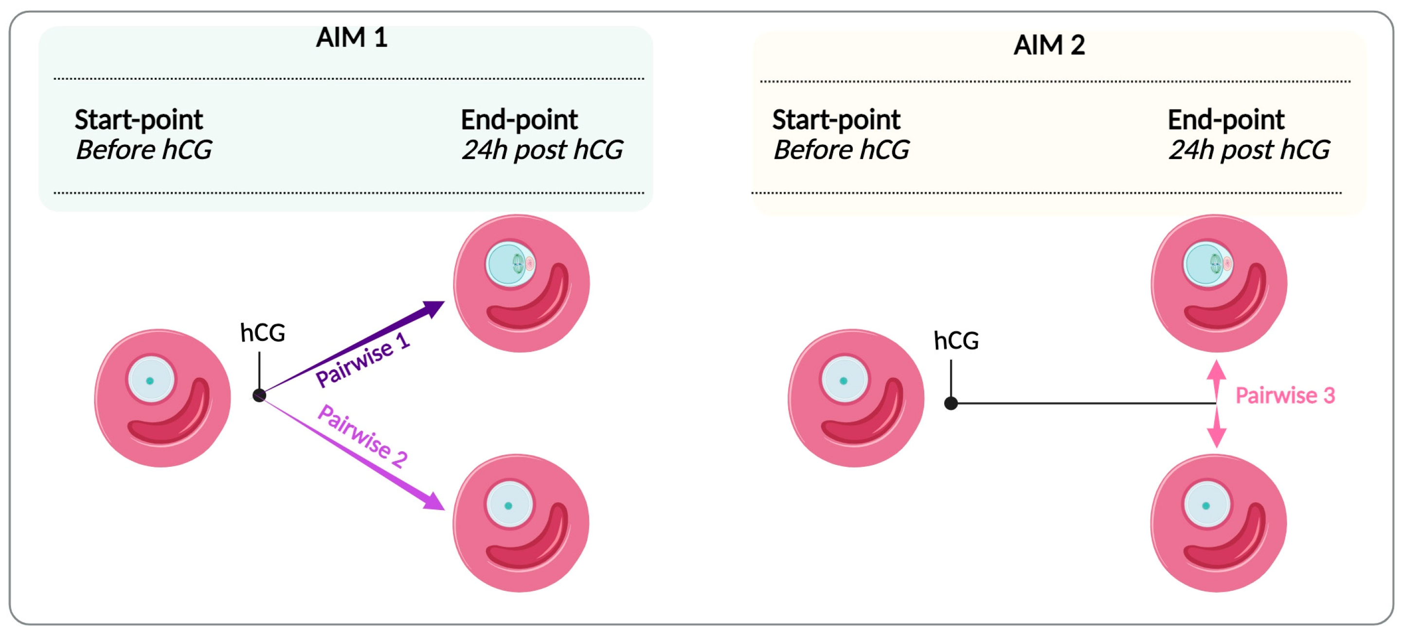
Figure 1. Experimental transcriptome plan. FEO IVM was induced by hCG to isolate FWs from EAfs associated with oocytes that reached MII or failed to resume meiosis, thus remaining at the GV stage. This setup supported two aims: AIM 1 (light green), comparing FW transcriptomes ±hCG with different maturation outcomes (MII or GV end-point vs. GV start-point); AIM 2 (light yellow), comparing FWs from successful and unsuccessful EAfs post-hCG (MII end-point vs. GV end-point). Created in BioRender., Peserico, A. (2025), https://www.biorender.com/.
Cells 14 00704 g001
Scheme 1. Network 1MII-GV and Network 2GV-GV DEGs modules identified with the MCODE Cytoscape plugin and annotated using ClueGO.
Cells 14 00704 sch001
Figure 2. Identification of highly modulated genes characterizing the FW of successful and unsuccessful EAfs: (A) Volcano plot showing the upregulated (red) and downregulated (green) DEGs in Network 1MII-GV (EAfs releasing competent oocytes) and Network 2GV-GV (EAfs releasing incompetent oocytes). DEGs exhibiting significantly high levels of modulation (outliers) are enclosed in a circle. Highly modulated DEGs were recorded through MCODE as unclustered, since, outside of the highly connected clusters, they were not selected as critical nodes, even if they met the criteria outlined in point (I). (B) Venn diagram of the outlier DEGs between the two networks, illustrating common DEGs as well as distinctive ones. Furthermore, the DEGs populating the highly interconnected modules in the respective networks are highlighted in bold. (C) Graphical representation of the highly modulated DEGs, highlighting the common and distinctive ones. DEGs within the highly interconnected KEGG modules in their respective networks are shown in bold, whereas highly modulated DEGs outside of the KEGG categories are displayed in regular font. Shared, highly modulated DEGs from KEGG categories are enclosed in the light blue box. The corresponding modules are specified in the legend. Created in Biorender. Peserico, A. (2025), https://www.biorender.com/.
Cells 14 00704 g002
Figure 3. Principal component analysis (PCA) demonstrates a sharp separation between the DEGs belonging to FW enclosing MII and those enclosing GV oocytes.
Cells 14 00704 g003
Figure 4. Network 3MII-GVend-point clustering DEGs and classification for biological processes: (A) Volcano plot showing the upregulated and downregulated DEGs in Network 3MII-GVend-point. Red dots indicate upregulated genes, and the green dots indicate downregulated genes. (B) DEGs were retrieved in each biological process category. Red bars indicate upregulated genes, and the green bars indicate downregulated genes.
Cells 14 00704 g004
Figure 5. Graphical representation of all identified network drivers. The driver genes are classified according to their respective network of origin and their transcriptional modulations impacting the meiotic maturation process. The three networks are represented by distinct colors (Network 1MII-GV in purple, Network 2GV-GV in light blue, and Network 3MII-GV end-point in orange). Transcriptional regulation levels are also color-coded, with red indicating upregulation and green indicating downregulation. Highly modulated DEGs belonging to KEGG categories are shown in bold. HUBs are underlined (red for upregulated, green for downregulated). Inside the dashed black rectangle, the drivers (highly modulated DEGs and HUBs) shared by both functional end-points (successful and unsuccessful maturation) are displayed, along with their respective transcriptional modulations. Created in Biorender. Peserico, A. (2025), https://www.biorender.com/.
Cells 14 00704 g005
Table 1. Comparative meiotic and developmental competence of FEO in vitro matured oocytes derived from early antral (EA) follicles.
A
Follicle CategoryHealthy Oocytes
(n°)
Oocyte Nuclear Stage
GV (%; SD)GVBD/MIMII
EAf − hCG 48100----
EAf + hCG 23611.0 ± 4.221.2 ± 5.567.8 ± 6.3
B
Healthy Oocytes
(n°)
Post-Fertilization Embryo Development
Uncleaved
(%; SD)
Fertilization Rate
(%; SD)
Blastocyst Rate
(%; SD)
EAf16032 ± 5.258.2 ± 5.59.9 ± 3.3
Comparison of meiotic and developmental competencies of EAf-enclosed oocytes. Table 1A FEO in vitro maturation was performed according to a validated method [1] by exposing EAfs (400 ± 26 µm diameter size) to hCG (25 IU/mL) in the presence of ovarian surface epithelial cells. Follicular cells (FW) from EAfs were collected at the end of the 24 h maturation window, according to the nuclear stage of the enclosed oocytes. The FWs of high-quality MII (only those reaching the blastocyst stage) were finally selected after IVF for the transcriptomic analysis. Table 1B shows the developmental competence of MII oocytes after IVF, tested to collect FWs for analysis. The fertilization rate was calculated based on the number of cleaved embryos at day 2. The blastocyst rate was calculated at day 8 on the cleaved embryo recorded on day 2. Data are expressed as the mean value of three independent biological replicates.
Table 2. Tree pairwise comparisons of DEGs.
Pairwise ComparisonGenerated NetworkTotal Number of GenesGenes passed Filter Criteria (DEG)Genes with No ChangesUpregulatedDownregulated
Pairwise 1Network 1MII-GV22,1412526 (11.4%)19,615 (88.6%)854 (33.8%)1672 (66.2%)
Pairwise 2Network 2GV-GV22,1411998 (9%)20,143 (91%)878 (43.9%)1120 (56%)
Pairwise 3Network 3MII-GV end-point22,14197 (0.44%)22,044 (99.6%)9 (9.3%)88 (90.7%)
The filtering criteria for identifying DEGs using GeneChip array analysis with Affimetrix include a fold change greater than 2 and a p-value less than 0.05. DEGs obtained from each pairwise comparison were used as input to build the corresponding gene-interaction network through systems biology workflows, as detailed in Section 3.2 and Section 3.3.
Table 3. Literature annotations on the female reproductive role of the identified candidate driver genes.
Driver Gene NameNetworkMcodeDEG Fold Changep-ValueFemale Fertility Effects
Knockout/Mutant MiceEffects Related to Maturation
AIM 1: Somatic Markers Distinguishing FW Pre- and Post-hCG MaturationCDCA814−2.090.0014N/AN/A any FWs role
Role in oocyte: it regulates meiotic spindle assembly and chromosome segregation during human oocyte meiosis [76]
EGFR182.630.0001Mutant mice (Egfrwa2/EgfrWa5) show infertility (J:92308 from the MGI database.EGFR activation is crucial for hCG-induced progesterone production in human granulosa cells, with amphiregulin (AREG) mediating this effect by upregulating StAR expression [77];
EGFR signaling in granulosa cells at ovulation triggers filopodia retraction, uncoupling germ and somatic cells [78]
MMP131721.183.37 × 10−6N/AGonadotropin Surge-Induced MMP-13 mRNA and protein in bovine preovulatory follicles [79].
Upregulation of MMP13, together with M2 macrophage polarization by the antifibrotic drug BGP-15, facilitated ovulation in old and obese mice [80]
MMP11; 28; 273.12; 106.962.24 × 10−8 6.21 × 10⁻⁷N/A Affected reproductive processes, including ovulation and folliculogenesis in chickens, drosophila, macaque, and humans [81,82,83,84,85,86]
ISG1521−2.970.0009Knockout in mice causes hyperfertility along with sensitive ovarian responses to gonadotropin, such as increases in cumulus expansion and ovulation rate [87].N/A
SPP11; 27; 251.92; 38.493.67 × 10−10
1.27 × 10⁻⁸
N/ASPP1/Opn upregulation in preovulatory granulosa cells, triggered by gonadotropin via EGFR signaling, boosts progesterone synthesis and VEGF expression during the early luteal phase [88].
In antral follicles, SPP1 is responsible for immune processes leading to ovarian follicular atresia [89].
SPP1 is upregulated in response to hormonal cues, particularly during the periovulatory phase, where it contributes to follicular rupture and corpus luteum formation [90]
TGFB1222.230.0009Knock out (Tgfb1tm1(Tgfb3)Kul/Tgfb1tm1(Tgfb3)Kul) in mice causes infertility (J:204892 from MGI
database).
N/A
AIM 2: Somatic Biomarkers of Follicular and Enclosed Oocyte CompetenceHSPA53/−3.370.0071Knock-out phenotype observed for the master regulator of HSP proteins, HSF1.
HSF1 knock out (Hsf1tm1Ijb/Hsf1tm1Ijb) in female mice causes infertility (J:58383, J:65267); abnormal female meiosis and abnormal meiotic spindle assembly checkpoint (J:175085).
N/A
HSPA1A3/−3.370.0071N/A
HSPH13/−2.340.0019N/A
HSPA63/−4.190.0051N/A
MAD2L13/−3.160.0074Mutant mice (Mad2l2repro22/Mad2l2repro22) show infertility (J:92463).N/A
GMNN3/−2.210.0078Oocyte-specific disruption of geminin (Gdf9-Cre Gmnn fl/fl) results in low fertility in mice.
Even though there was no evident anomaly of oogenesis, oocyte meiotic maturation, natural ovulation, or fertilization, early embryo development and implantation were impaired (MGI Database).
N/A
FBXO53/−2.10.0006No role in FWs
Role in oocyte: During oocyte maturation, it plays a role in meiosis through the inactivation of the APC-FZR1 complex. Inhibits APC through RPS6KA2 interaction that increases FBXO5 affinity for CDC20, leading to the metaphase arrest of the second meiotic division before fertilization [91].
N/A
ITIH43/2.090.0054N/AInvolved in the stabilization of the extracellular matrix and inflammatory processes. Found to be upregulated as a protein in FF from mature vs. immature oocytes (bovine) along with other proteins involved in complement activation (ITIH4, AHSG, FN1, HP) [92]. This inflammatory pathway may facilitate the local physiological and inflammatory reaction of ovulation [93].
Summary of annotated data for driver genes, for which literature information has been gathered regarding their roles in maturation. Other descriptive details are summarized, including gene name, associated network, MCODE cluster, DEG fold change, p-value, and role in maturation. These integrated annotations provide the mechanistic rationale underpinning the selection of every driver gene and pathway. N/A refers to “Not annotated”. Phenotypes from mutant mice were checked using the Mouse Genome Database (MGI; https://www.informatics.jax.org, accessed on 2 May 2024) and PubMed.
Disclaimer/Publisher’s Note: The statements, opinions and data contained in all publications are solely those of the individual author(s) and contributor(s) and not of MDPI and/or the editor(s). MDPI and/or the editor(s) disclaim responsibility for any injury to people or property resulting from any ideas, methods, instructions or products referred to in the content.