Review

Cooperative Role of Carbonic Anhydrase IX/XII in Driving Tumor Invasion and Metastasis: A Novel Targeted Therapeutic Strategy

1
School of Pharmacy, Southwest Medical University, Luzhou 646000, China
2
Department of Clinical Medicine, School of Clinical Medicine, Southwest Medical University, Luzhou 646000, China
3
Laboratory of Metabolism, Center for Cancer Research, National Institutes of Health, Bethesda, MD 20892, USA
*
Authors to whom correspondence should be addressed.
Cells 2025, 14(10), 693; https://doi.org/10.3390/cells14100693
Submission received: 13 April 2025 / Revised: 27 April 2025 / Accepted: 9 May 2025 / Published: 11 May 2025

Abstract

:
Cancer invasion and metastasis are critical factors that influence patient prognosis. Carbonic anhydrase IX (CA IX) and carbonic anhydrase XII (CA XII) are key regulators of hypoxia and pH homeostasis in the tumor microenvironment (TME). It has been verified that both CA IX and CA XII play significant roles in promoting tumor metastasis in recent years, but most of the literature tends to treat them as separate entities rather than exploring their synergistic effects. This review provides a comprehensive overview of the roles of CA IX and CA XII in tumor invasion and metastasis, along with their clinical applications, including their spatial distribution characteristics, molecular mechanisms that facilitate tumor metastasis, and their potential for clinical translation. Moreover, this review incorporates the classical tumor core–invasive front model to propose a metabolic coupling model of CA IX and CA XII, offering a fresh perspective on precision therapies that target tumor metabolism. By emphasizing the metabolic coupling between these two molecules, this review offers new insights distinct from previous studies and highlights the clinical therapeutic potential of simultaneously targeting both during treatment. It sheds new light on future research and clinical applications, aiming to enhance the prognosis of cancer patients through innovative therapeutic strategies.

1. Introduction

Cancer is a disease that remains difficult to cure, and its complexity and heterogeneity pose significant challenges to current therapeutic strategies. In recent years, the global cancer burden has been escalating, and it is projected that cancer will soon become the leading cause of premature mortality worldwide. According to GLOBOCAN statistics, the incidence of cancer is rising rapidly, with new global cases expected to reach 35 million by 2050—an increase of 77% compared to 2022. This trend is particularly pronounced in countries with a low Human Development Index (HDI), where the number of new cases is projected to double [1,2]. At present, the outlook for cancer treatment remains challenging, necessitating in-depth investigations into the mechanisms underlying tumor initiation and progression, as well as the continuous exploration of novel therapeutic approaches and strategies. Tumor invasion and metastasis are central topics in cancer research as they are directly linked to tumor malignancy and patient prognosis. Specifically, the invasive and metastatic potential of tumors serves as a critical criterion for prognostic evaluation. Highly invasive tumors are often associated with increased recurrence rates and a heightened risk of metastasis, ultimately affecting patient survival and quality of life. Therefore, a comprehensive discussion of tumor invasion and metastasis becomes a crucial aspect of cancer research [3,4].
CO2 is a simple molecule that is widely present in nature. For living organisms, CO2 serves as a stable and abundant carbon source. Under natural conditions, CO2 reacts with water to form unstable carbonic acid, which subsequently dissociates within biological systems into bicarbonate and hydrogen ions (CO2 + H2O ⇋ HCO3 + H+). Through this process, the carbon atoms in CO2 become bioavailable and enter metabolic cycles. However, the reaction between CO2 and water occurs at an inherently low efficiency under natural conditions, limiting the ability of organisms to utilize this abundant environmental carbon source. To overcome this challenge, organisms have evolved an enzyme that catalyzes the reversible hydration of CO2, known as carbonic anhydrase, which was first discovered in 1933 [5]. To date, several families of carbonic anhydrase enzymes have been identified, including α-, β-, γ-, δ-, ζ-, η-, θ-, and ι-CAs. These enzyme families are genetically independent of each other [6]. Most carbonic anhydrases (α-, β-, γ-, δ-, ζ-, η-, and θ-CAs) are metalloenzymes, and their cation properties are variable, exhibiting enhanced activity upon binding with Zn(II) [7].
In the human body, carbonic anhydrase is primarily of the α-CA type, with several isoforms present [8]. These isoforms vary in their distribution, quantity, and function across different cells, tissues, and organs in the human body. As a result, there are significant differences in the structure, pharmacological functions, and clinical effects of their inhibitors [9]. They have widespread applications both in traditional therapeutic fields and in the development of novel drug targets for conventional medications [10,11,12,13,14]. The existing literature reports that CA IX and CA XII have profound effects on various physiological activities of tumor cells, although the actions they produce are not entirely the same [15,16,17,18]. However, to date, discussions on the combined effects of these two enzymes on tumor physiological activities remain scarce. Only a few studies have suggested that CA IX and CA XII play a role in the invasion and metastasis of cancer cells [19]. It is noteworthy that the specific metabolic coupling relationship between CA IX and CA XII has been scarcely explored, although the metabolic compartmentalization characteristics of them have been preliminarily studied. Referring to the classical “core–invasive front” model in tumor research, CA IX exhibits high expression in the tumor core, while CA XII is highly expressed at the invasive front. They are not isolated within different compartments; rather, they are interconnected through a specific metabolic mechanism, which jointly influences tumor metabolism and invasive metastatic behavior, providing a new perspective for research on tumor metabolic metastasis.
This review provides a comprehensive summary of the roles of CA IX and CA XII in tumors, with a particular focus on the unique effects they exert on various physiological behaviors of tumor cells. CA IX and CA XII play crucial roles in regulating intracellular pH and promoting tumor cell invasion and metastasis, and the review delves deeply into the mechanisms through which these effects occur. Furthermore, the review discusses the clinical application prospects of CA IX and CA XII, including the current development status of enzyme inhibitors, antibody drugs, and combination therapies targeting these molecules. This review systematically summarizes the synergistic effects of CA IX and CA XII in tumor metastasis for the first time and proposes a metabolic coupling model based on the “core-invasive front” model. It provides new perspectives for understanding tumor metabolic adaptability and changes in the tumor microenvironment (TME), as well as opening new avenues for targeted therapeutic strategies.

2. CA IX and CA XII in Cancer Metastasis

Human carbonic anhydrases (h-CA) consist of sixteen α-CA isoenzymes, which exhibit differences in tissue distribution, kinetic properties, and physiological functions [20]. It is currently widely accepted that CA IX and CA XII (among the various isoforms of h-CA) are highly associated with the development of cancer. Specifically, the membrane-bound carbonic anhydrase isoform CA IX is significantly overexpressed in tumor cells compared to normal cells [21,22]. Furthermore, enzymatic assays indicate that CA IX exhibits higher catalytic activity in low pH environments, which is consistent with the hypoxic and acidic TME resulting from metabolic reprogramming in tumor cells [23]. In addition to its synergistic effect with CA IX, CA XII may also exhibit strong correlations with certain components of the TME, such as acidic and hypoxic conditions, as well as the ECM. By influencing these components, CA XII enables tumor cells to gain enhanced survival, increased invasiveness, and immune evasion. Growing evidence suggests that these two carbonic anhydrases may interact with each other and play crucial roles in regulating the TME and promoting tumor metastasis. Therefore, a deeper understanding of the roles of CA IX and CA XII in tumors can provide new insights and inspiration for research on the mechanisms of tumor metastasis and has gradually become a growing area of focus in recent years.

2.1. CA IX in Cancer Metastasis

The expression of CA IX is restricted in normal tissues, where it is only expressed in a few tissues, primarily in the gastrointestinal tract [24,25]. However, its expression is significantly upregulated in tumor cells, where it serves as a hypoxic marker in various solid tumors [26]. CA IX has multiple roles in tumor cells. It is widely believed that CA IX, as one of the key molecules in the HIF transcriptional regulation network, plays an important role in maintaining pH in tumor cells, which is significant for maintaining cellular metabolic activity and enhancing cell survival. It is currently believed that PKC is likely to play an important role in this process [27]. It is well known that changes in pH in the TME have an important impact on tumor metastasis, but the role of CA IX in this process has rarely been mentioned. It suggests that CA IX may have a currently underexplored mechanism that contributes to tumor metastasis in addition to its traditional role in regulating pH, providing a new perspective for research on tumor metastasis. Additionally, exosomes, as important mediators of intercellular communication, are closely related to tumor invasion and metastasis. Notably, the presence of CA IX alters certain characteristics of ordinary exosomes, causing cells to exhibit markedly different behaviors, which provides a new entry point for studying the role of CA IX in tumor metastasis. The pH-regulating function of CA IX is closely related to the hypoxic environment of tumor cells. When the oxygen concentration in the microenvironment decreases, the HIF-α subunit remains stable by escaping VHL-mediated ubiquitination degradation, leading to the accumulation of HIF-α in cells [28]. In the analysis of over 400 cases of clear cell renal cell carcinoma (ccRCC) from the TCGA database, approximately 80% of cases exhibit VHL gene mutations or promoter methylation. This alteration stabilizes HIF-α, resulting in the sustained high expression of downstream hypoxia-related genes, such as CA IX, one of the downstream genes regulated by HIF-1α [26,29,30,31]. Under hypoxic conditions, HIF-1α binds to the hypoxia response elements (HRE) in the promoter region of CA IX, activating its transcription. As one of the key molecules in the HIF transcriptional regulation network [32,33,34,35], CA IX responds to physiological stresses such as low oxygen concentration (hypoxia) and the accumulation of acidic products (pH) by catalyzing the CO₂ hydration reaction. Specifically, CA IX sequesters the excess CO₂ produced by aerobic glycolysis and participates in the process of proton (H+) extrusion, thereby maintaining the stability of intracellular pH [36]. The CO₂ generated in this process serves as a substrate for carboxylation reactions and enters fatty acid synthesis mediated by acetyl-CoA carboxylase (ACC), which contributes to replenishing oxaloacetate in the tricarboxylic acid (TCA) cycle and thus maintains aspartate metabolism, helping to resolve the inhibition of the electron transport chain caused by hypoxia [37]. Therefore, CA IX maintains the stability of the TME and metabolic balance through various mechanisms, serving as a crucial molecular foundation for cellular adaptation to harsh conditions such as hypoxia. On the other hand, some studies suggest that CA IX also plays an important role in promoting tumor cell metastasis. Research indicates that CA IX drives extracellular matrix (ECM) degradation and remodeling through its dual roles in acidifying the microenvironment and signal transduction, which enhances tumor invasion and metastasis through exosome-mediated angiogenesis, exhibiting organ-specific patterns during the metastasis process. Its influence on tumor metastasis is exerted through the combined actions of multiple molecules at multiple levels.
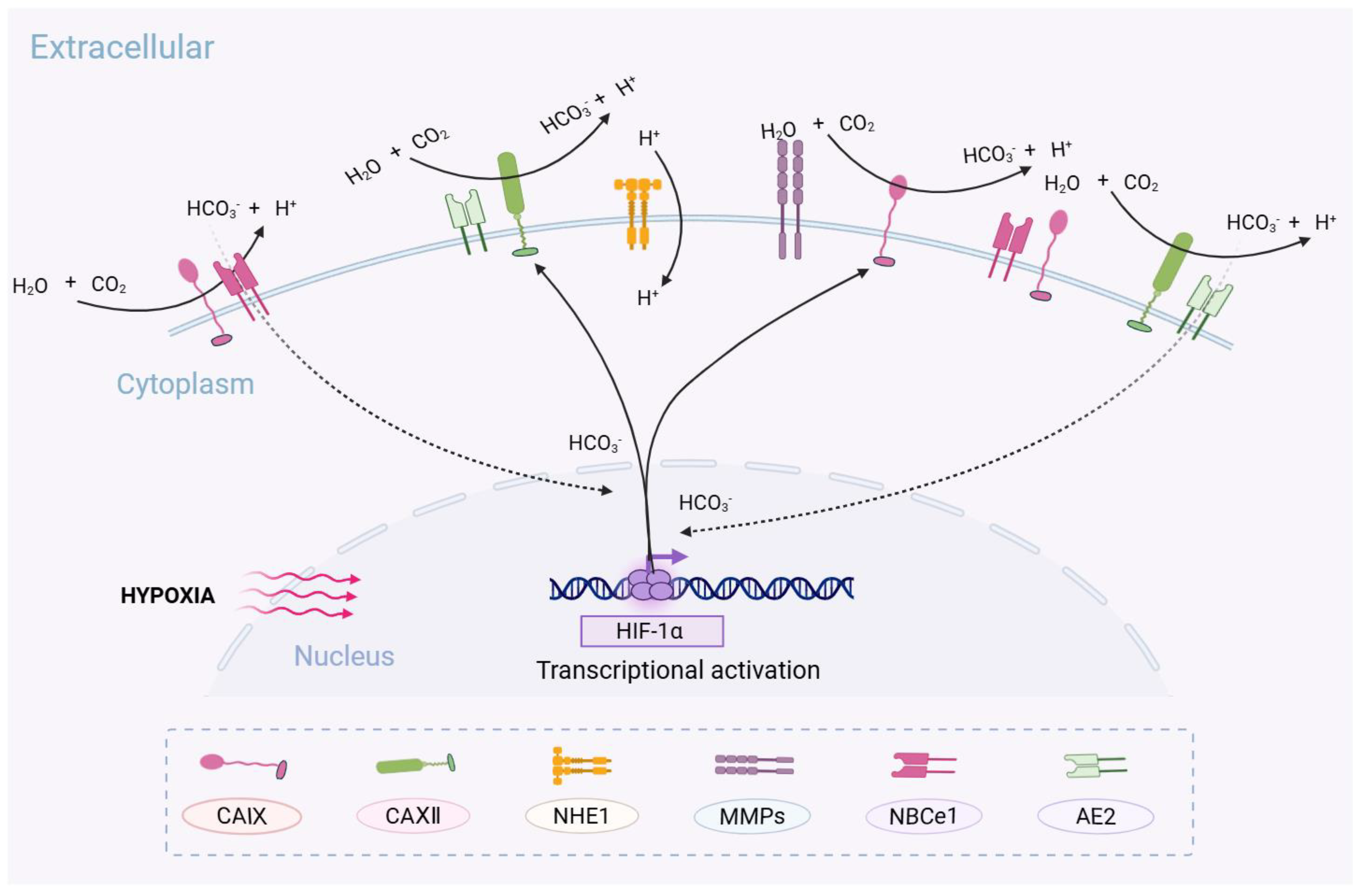
Changes in pH have a significant impact on tumor metastasis. Under acidic conditions, tumor cells may splice certain key proteins, and these protein alterations contribute to increased aggressiveness, enabling tumor cells to breach the surrounding ECM. It suggests that acidity may trigger the alternative splicing of specific genes and participate in ECM degradation, thereby facilitating tumor cell metastasis [38]. CA IX plays a role in regulating pH changes, suggesting that it is highly likely to influence cell invasion and metastasis through this mechanism. For instance, when the ECM becomes more acidic (pH 6.0–6.5), pro-matrix metalloproteinases (pro-MMPs) undergo conformational changes that expose their active sites, thereby disrupting intercellular adhesion, enhancing tumor cell invasiveness, and promoting tumor metastasis [39,40]. Specifically, membrane-type matrix metalloproteinase 1 (MT1-MMP) cleaves pro-MMP-2 through its catalytic domain, converting it into active MMP-2 (64 kDa), which degrades components of the ECM, including collagen and fibronectin, and participates in cytoskeletal remodeling, promoting the formation of pseudopodia, ultimately enhancing the invasiveness of tumor cells [41,42]. Additionally, CA IX can reduce cell adhesion and enhance invasiveness through multiple mechanisms, including interacting with β-catenin, influencing the expression of integrins, focal adhesion kinase (FAK), paxillin, cell adhesion-related proteins, as well as regulating intracellular metastasis-associated signaling pathways [43,44] (Figure 1).
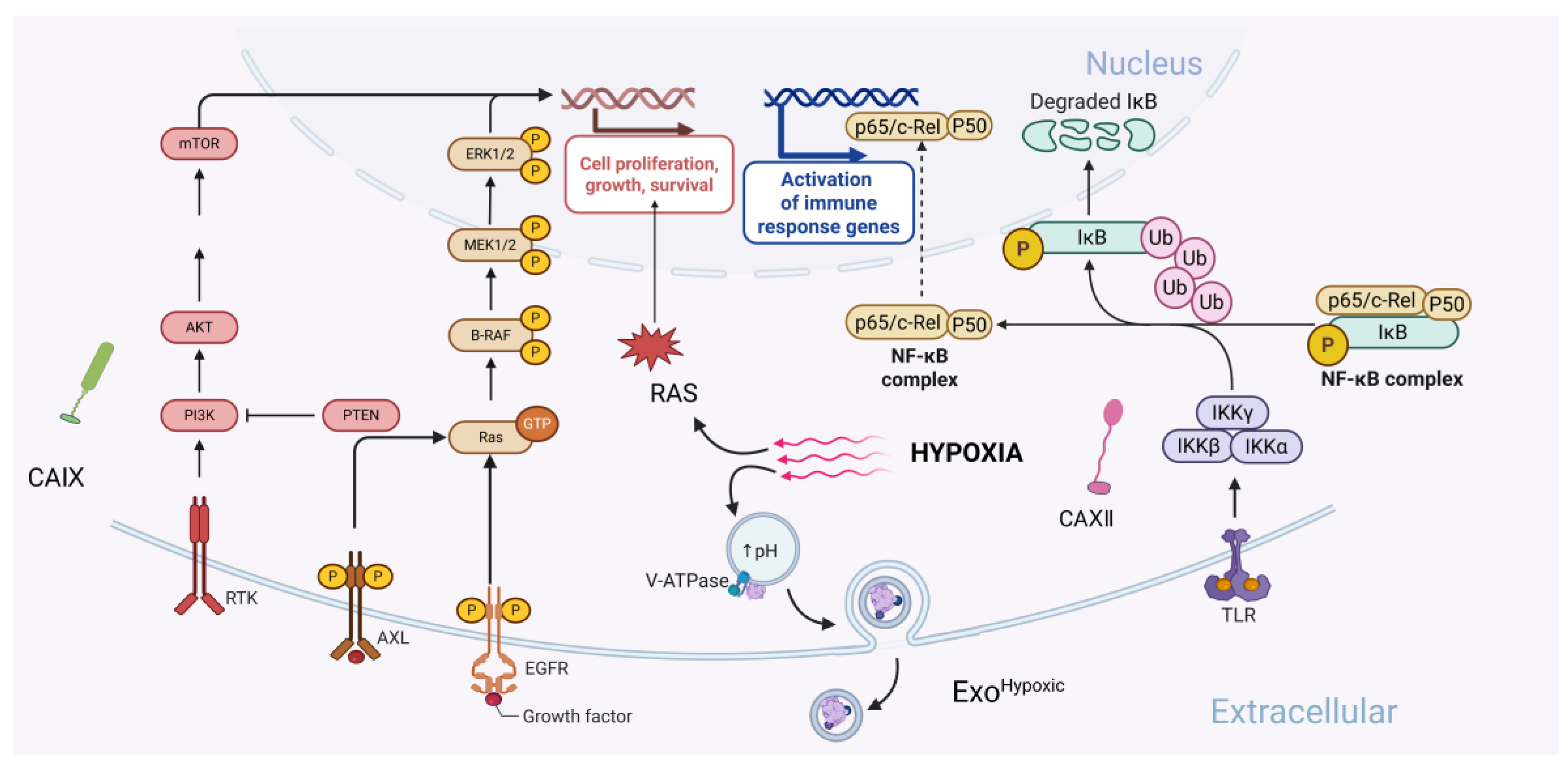
The expression and activity of CA IX also play a crucial role in maintaining the mesenchymal phenotype (EMT) of breast cancer stem cells (CSCs). Persistent high expression of CA IX promotes EMT, which is a process essential for tumor cells to acquire migratory and invasive capabilities and a prerequisite for metastasis, ultimately leading to the invasiveness of CSCs [45]. This regulation facilitates the further enhancement of aerobic glycolysis, exacerbating microenvironmental acidification and promoting resistance to cellular stress [34,39]. Previous research has suggested that CA IX may also directly participate in intercellular signaling through its extracellular domain (e.g., activation of the EGFR/PI3K pathway) or influence cell adhesion and differentiation, which is independent of its enzymatic activity contributing to tumor progression [46,47]. On the other hand, CA IX may also interact with other pH-regulating proteins on the cell membrane. NHE1 (Na+/H+ exchanger 1) is an ion transport protein that is crucial in tumor cell migration, invasion, proliferation, and chemoresistance. It can indirectly promote tumor cell migration by modulating cytoskeletal dynamics, acidifying the cell surface, and enhancing MMP activity [48]. There are currently no direct studies on the interaction between NHE1 and CA IX; however, both are directly involved in proton transfer in tumor cells, and protein expression analyses in various tumor samples suggest a potential yet unexplored connection between them. Finally, CA IX exhibits organ-specific patterns in tumor metastasis. During bone metastasis, CA IX promotes the degradation of type I collagen through MMP-13, resulting in the release of TGF-β and IGF-1, which in turn activate osteoclasts [49]. During brain metastasis, the CA IX/MMP-2 axis disrupts the tight junction proteins of the blood–brain barrier and the ECM, facilitating the intracranial migration of tumor cells [50] (Figure 2).
These findings indicate that the impact of CA IX on tumor metastasis relies on the cooperative action of multiple molecules and layers. The regulation of tumor metastasis by CA IX is subtype-specific and exhibits spatiotemporal dynamics. The multilayered ECM remodeling mechanism positions CA IX as a central hub linking metabolic adaptation with physical microenvironment remodeling.
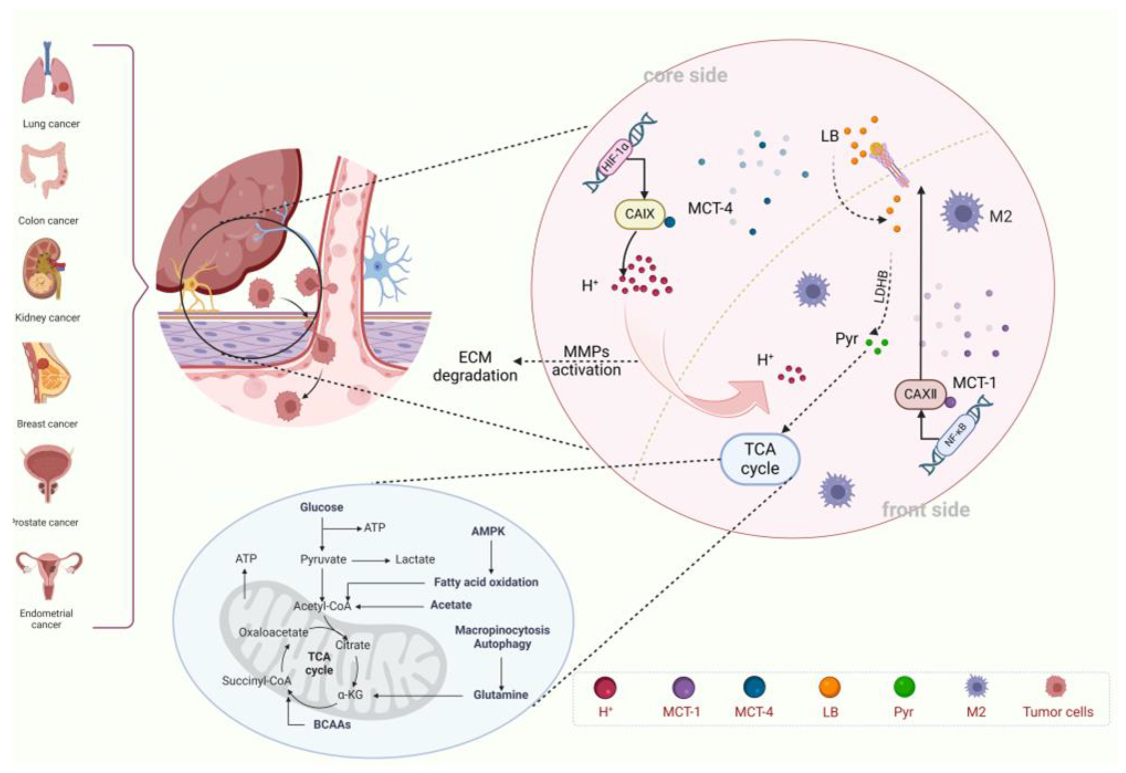
Additionally, as an important medium of intercellular communication, exosomes provide a novel perspective on the role of CA IX in tumor metastasis, distinct from traditional metabolic pathways. Exosomes, a type of extracellular vesicle (EV), are released into the extracellular space after the fusion of multivesicular bodies (MVBs) with the cell membrane. Exosomes have a lipid bilayer structure and are capable of carrying and transmitting a variety of bioactive molecules, including proteins, lipids, mRNA, and miRNA. Current research on exosomes focuses on their roles in cancer progression, metastasis, and immunity. Exosomes hold significant promise as cancer biomarkers and are being developed as targeted cancer therapeutics, showing promising clinical application potential [51,52,53]. Although research on CA IX exosomes is still in its early stages, significant progress has been made. In the acidic TME, the characteristics of exosomes undergo significant changes compared to other environments. Under acidic conditions in the LNCaP cell line (human prostate cancer cell line), the release of exosomes is higher than those in normal cells. Both the expression and activity of CA IX in the exosomes are significantly increased, with notable changes in the expression pattern [22]. Under hypoxic conditions, exosomes secreted by the LNCaP cell line (ExoHypoxic) have a smaller average size and contain higher levels of tetraspanin, heat shock proteins, and Annexin II. It has been shown that ExoHypoxic significantly enhances the invasive and migratory capacity of LNCaP cells, specifically manifested due to the following reasons: (i) ExoHypoxic promotes the formation of prostate cancer spheroids, enhancing their stem cell properties; (ii) ExoHypoxic increases the expression of α-SMA in prostate stromal cells and induces the conversion of these stromal cells into cancer-associated fibroblasts (CAFs); and (iii) ExoHypoxic exhibits higher metalloproteinase activity and targets E-cadherin and β-catenin, disrupting intercellular adhesion, and promoting the invasion and migration of the cells [54]. The expression of CA IX is significantly elevated in renal cell carcinoma (RCC) cells, which form exosomes containing CA IX protein through MVBs, which can be taken up by human umbilical vein endothelial cells (HUVECs). This process significantly enhances the migratory capacity and angiogenic ability of HUVECs, promoting angiogenesis and thereby increasing the tumor invasiveness and metastatic potential [55], which reveals its core role in the formation of the pre-metastatic niche during tumor invasion and metastasis [52,56,57]. Its unique dual regulation of enzyme activity and signal transduction, along with its organ-specific characteristics, strongly demonstrates its potential as a therapeutic target in the direction of precision treatment for tumors (Figure 3).

2.2. CA XII in CANCER Metastasis

CA XII is another member of the carbonic anhydrase family, and it is highly expressed in tumor tissues and tumor-associated immune cells, especially in tumor-infiltrating monocytes and macrophages. CA XII is associated with tumor progression and poor prognosis in patients through the production of chemokines and the regulation of intracellular signaling in tumor cells, and it has gradually gained attention in tumor research becoming a new research perspective [58].
Recent studies show that CA XII exhibits a unique expression pattern in cancers—it is not directly expressed in tumor cells but specifically accumulates in tumor-associated macrophages (TAMs), particularly in the CD206+/CD204+ M2-like macrophages in human hepatocellular carcinoma (HCC). It suggests that CA XII may play a crucial role in the survival and function of the M2-like macrophages in HCC tumor tissues [59]. Pathological studies have shown that the upregulation of CA XII expression is significantly correlated with tumor stage, metastasis rate, and poor prognosis, suggesting that it could serve as a novel biomarker for prognosis evaluation. Mechanistic studies indicate that CA XII helps maintain the pH within macrophages, protecting them from damage in acidic environments, thus enabling macrophages to survive and function under such unfavorable conditions. Furthermore, macrophages with high CA XII expression induce the production of C-C motif chemokine ligand 8 (CCL8) through the p38 signaling pathway. CCL8 is an important chemokine that promotes EMT in tumor cells, enhancing their migration and invasion capabilities. Animal experiments have demonstrated that the expression level of CA XII is positively correlated with tumor size and metastasis, and treatment with CA XII inhibitors resulted in reduced tumor volume and lower invasiveness in mouse models, confirming its potential as a therapeutic target [59,60,61].
CA XII is strongly associated with NF-κB, and it can influence tumor cell apoptosis and drug treatment efficacy by regulating the activity of NF-κB-related proteins. In pancreatic adenocarcinoma (PAAD), the expression of CA XII is significantly downregulated and associated with a poorer prognosis. Results show that overexpression of CA XII significantly inhibits the proliferation, invasion, and migration of PAAD cells, meanwhile, inducing cell cycle arrest at the phase of G0/G1 and apoptosis. In contrast, the downregulation of CA XII significantly suppresses these processes. Moreover, when CA XII is overexpressed, the level of NF-κB p65 protein decreases, and the IκBα protein increases, with both changes being significant. Conversely, when CA XII is knocked down, the opposite effects are observed. Given the close relationship between NF-κB, cell apoptosis, and invasion, CA XII is likely to influence apoptosis and metastasis by regulating NF-κB-related proteins, demonstrating its potential as a therapeutic target for cancer treatment [62]. Auranofin (AF) is a drug originally used to treat rheumatoid arthritis, due to its unique biological mechanisms, it has recently been investigated as an anti-cancer drug. Research has shown that AF exhibits good cytotoxicity against non-small cell lung cancer (NSCLC) cells and pancreatic ductal adenocarcinoma (PDAC) cells at concentrations below 1 μM, with minimal impact on normal cells, showing good selectivity and clinical application potential. Further studies have found that cancer cells with low CA XII expression are more sensitive to AF treatment, suggesting that CA XII could serve as a biomarker for predicting AF sensitivity. As mentioned above, CA XII could be closely associated with NF-κB, and it is reasonable to hypothesize that AF may exert its therapeutic effect by inhibiting NF-κB-related proteins. In cancer cells with low CA XII expression, the NF-κB signaling pathway is more active, which enhances the therapeutic effect of AF, highlighting the potential of CA XII as both a cancer treatment target and a biomarker [63]. In addition, other studies have shown that factors such as LncRNA and histamine regulate the expression of CA XII in tumors, inhibiting tumor survival, metastasis, and drug resistance [64,65]. These studies indicate that the role of CA XII in tumors is gradually gaining attention, and its potential as a therapeutic target for cancer treatment is becoming increasingly evident (Figure 3).

3. Synergistic Effect of CA IX and CA XII

CA IX and CA XII are involved in a closely coordinated metabolic network during tumor physiological processes; however, the evidence of direct molecular interactions between them still require further investigation. Multiple lines of evidence suggest that the interaction between CA IX and CA XII ultimately promotes tumor progression and invasion, leading to poor prognosis for patients. First, CA IX is widely overexpressed in tumor cells, catalyzing the conversion of CO2 to HCO3 in hypoxic regions, neutralizing intracellular acidity, and participating in proton efflux, thus helping tumor cells adapt to the hypoxic environment. CA XII is highly expressed in M2-like macrophages in certain tumors, aiding in the maintenance of the acidic TME and helping tumors resist acid-induced damage. In general, tumor cells recruit immune cells to the tumor periphery, a process that usually promotes tumor development, including enhancing tumor proliferation, weakening anti-tumor immune responses, promoting new tumor blood vessel formation, and assisting tumor immune evasion [66]. The high expression of CA XII in macrophages can accelerate this process, further affecting the acid–base balance and immune infiltration within the TME. CA IX and CA XII can synergistically maintain the tumor survival advantage through “intracellular alkalinization (CA IX)” and “extracellular acidification buffering (CA XII)”.
It is currently widely accepted that CA IX and CA XII regulate both intracellular and extracellular pH, while hypoxia-induced extracellular acidification can in turn affect the activity of CAs, forming a feedback regulatory loop. Under hypoxic conditions, the expression of CA IX and CA XII is regulated by HIF-1, which simultaneously enhances the catalytic activity of CA IX and CA XII [23,67]. Additionally, low pH levels upregulate CA XII expression in tumor-associated macrophages, activate NF-κB, and induce CCL8 secretion [59]. It constitutes a crucial positive feedback loop: hypoxia-induced acidosis enhances CA IX/CA XII activity, which further intensifies extracellular acidification, thereby promoting extracellular matrix degradation and tumor metastasis. Importantly, the effects vary among different cell types. In tumor cells, CA IX primarily maintains an alkaline intracellular environment to support cell survival; whereas in tumor-associated macrophages, CA XII mainly ensures macrophage survival under acidic conditions and facilitates immune evasion through the release of chemokines. The cell-specific functions of these enzymes highlight the complexity of targeting them therapeutically. Therefore, careful consideration must be given to their differential expression and function across various cellular populations in designing treatment strategies aimed at these two isoforms.
Deuterium-Depleted Water (DDW) is a type of water processed through specialized techniques to significantly reduce its deuterium (2H) content compared to that found in natural (ordinary) water. Although not yet widely applied in clinical cancer therapy, DDW has demonstrated considerable potential. Studies have shown that DDW can influence extracellular acidification of tumor cells through multiple mechanisms, including: (a) altering cancer cell metabolic strategies to reduce lactate production and secretion; and (b) affecting V-ATPase function, thereby decreasing proton efflux and markedly improving tumor cell drug sensitivity. These properties enable DDW to inhibit tumor cell proliferation, indirectly modulate immune responses in the TME, and enhance the cytotoxicity of immune cells against tumor cells, ultimately contributing to improved patient survival [68,69]. Moreover, studies have shown that in tumors with high expression of CA IX and CA XII, the tumor cells exhibit more invasiveness compared to other types of tumors [55,70,71]. It is due to the combined effects of CA IX and CA XII. On one hand, the high expression of CA IX in tumor cells leads to a downregulation of E-cadherin expression, while the expression of N-cadherin and Vimentin is upregulated, accelerating the process of EMT [72,73]. On the other hand, CA XII-positive macrophages directly induce EMT by secreting CCL8, and recruit immune-suppressive myeloid cells to form a pro-metastatic microenvironment [59]. CA IX promotes tumor angiogenesis by inducing VEGF and other pro-angiogenic factors [74], while CA XII+ macrophages secrete CCL8, which recruits endothelial progenitor cells and synergistically enhances angiogenesis [75,76]. From another perspective, this also suggests that both have a strong ability to enhance tumor metastasis and invasion. The original pro-metastatic effect is amplified under the multifaceted synergistic actions, which significantly enhance the invasiveness and metastatic potential of the tumor.
It is worth noting that tumor cells with high expression of CA IX and CA XII can cause drug resistance [77,78,79]. The acidic environment maintained by CA IX and CA XII through multiple pathways can affect the solubility and permeability of drugs, thereby reducing the efficacy of chemotherapy drugs. Furthermore, the high expression of CA IX and CA XII can activate drug efflux pumps, such as P-gp, which expel chemotherapy drugs from the cells, decreasing the intracellular drug concentration and ultimately leading to drug resistance. In neuroblastoma, the high expression of CA IX is significantly associated with poor prognosis and high risk, as it inhibits cisplatin-induced ferroptosis, promotes cell proliferation, and reduces drug efficacy [80]. When CA IX and CA XII inhibitors are used to suppress the activity, the sensitivity of tumor cells to cisplatin significantly increases, and the treatment effect improves significantly [21]. The expression level of CA XII shows a significant positive correlation with multidrug resistance in breast cancer. CA XII primarily enhances the drug efflux capacity of tumor cells by upregulating the expression of P-gp, resulting in drug resistance [81]. Currently, there has been development of combined inhibitors targeting both P-gp and CA XII. These inhibitors are expected to reverse multidrug resistance in tumors and improve the efficacy of chemotherapy [82].
The expression of HIF-1α is positively correlated with that of CA IX in cancer, and regions of high CA IX expression are typically accompanied by an enrichment of CA XII. It suggests that CA IX and CA XII may share hypoxia-driven metabolic pathways. Based on this, it can be speculated that the HCO3 produced by CA IX may be transmitted to TAMs through gap junctions or extracellular vesicles, serving as a substrate for the CA XII-catalyzed reaction, further regulating the extracellular pH gradient. Additionally, the H+ efflux mediated by CA XII may enhance the catalytic efficiency of CA IX, forming a “basic inside, acidic outside” feedback loop that promotes tumor cell invasiveness. These mechanisms collectively maintain the acidic microenvironment of tumor cells [83]. Although the correlation between HIF-1 and carbonic anhydrase expression is well-established, there is limited research on the relationship between other HIF family members and carbonic anhydrases. Under prolonged hypoxic conditions, the expression of HIF-2α in tumor cells significantly increases, and its role in tumor regulation cannot be ignored [84]. Multiple members of the HIF family may participate in the regulation of CA IX and CA XII at different time points, forming a time-dependent regulatory network, which needs further validation [35,85]. It is suggested that CA IX is associated with immune evasion in tumor cells [86]; however, there is little research on CA XII and its role in tumor immune evasion. It is worth exploring whether CA XII might contribute to immune evasion in a manner similar to CA IX, potentially promoting the tumor escape from immune system surveillance, which remains an important issue to explore further.

4. Clinical Significance of CA IX and CA XII

4.1. Compartmentalization of Metabolic Regions Coupled to Core-Invasion Fronts

In clinical practice, distinguishing between the tumor core and the invasive front of the tumor is important [87]. It primarily divides tumors into two functionally distinct regions that are significantly different at the transcriptomic and epigenetic levels: the relatively stable core region and the highly active invasive front region. Cells in the core region proliferate slowly, typically existing in a hypoxic and highly acidic microenvironment. In contrast, cells at the invasive front are more active, with stronger migratory and invasive abilities, exhibiting characteristics of EMT, and are more prone to metastasis [87,88].
Studies have found that the distribution of CA IX and CA XII aligns with the “core-invasive front” division of tumors (Table 1). In the core region, HIF-1α activates the expression of CA IX, acidifying the microenvironment. CA IX and monocarboxylate transporter 4 (MCT4) colocalize, pumping lactate produced by glycolysis out of the cells, causing lactate accumulation and forming a “lactate pool”, which enhances the Warburg effect [35,89,90]. Additionally, CA IX acidifies the lysosomes, promoting ECM degradation, facilitating tumor metastasis [91,92], and inhibiting immune surveillance [93,94]. At the invasive front, the TME is relatively oxygen rich. NF-κB signaling activates CA XII, maintaining a local weakly alkaline condition to support mitochondrial oxidative phosphorylation [62,63,95]. CA XII can also work in conjunction with MCT1 to transport lactate released from the core region to the invasive front cells. Under the action of lactate dehydrogenase B (LDHB), lactate is converted into pyruvate, which enters the TCA cycle, enabling efficient utilization of carbon sources [91]. On the other hand, lactate can promote the EMT of invasive front cells through epigenetic regulation [96]. The transfer of H+ generates a unique inter-regional proton concentration gradient in the exchange of substances between the core region and the invasive front [36,67], forming a “proton relay” model of metabolic coupling between CA IX and CA XII, demonstrating continuity in metabolic space.
The metabolic coupling between CA IX and CA XII has profound significance for tumor development. Firstly, this process promotes tumor invasion and metastasis. On one hand, the acidic microenvironment of the core region activates MMPs, facilitating ECM protein degradation, providing physical pathways for tumor invasion. On the other hand, the EMT occurring in the invasive front cells reduces cell adhesion, enhances metastatic ability, and paves the way for tumor metastasis. Furthermore, this metabolic coupling also leads to tumor resistance to treatment. Although directly inhibiting CA IX can reduce the acidity of the core region, the invasive front can resist this change through the CA XII-MCT1 axis, reducing drug efficacy. Drugs, such as 5-fluorouracil (5-FU), are protonated in the acidic core region, making it difficult to enter the cells. Meanwhile, cells in the invasive front using oxidative phosphorylation (OXPHOS) can upregulate ABC transporters to accelerate drug efflux [98,99], further promoting tumor drug resistance. Based on the above characteristics, the drugs currently being developed clinically are mostly dual-target drugs that co-inhibit CA IX and CA XII and have shown good therapeutic effects in cancers [100,101].
Although the CA IX/CA XII metabolic coupling model explains phenomena in the acidification mechanism of the TME, there are still many issues that need to be addressed. First, there is a significant discrepancy regarding the boundary of functional redundancy and compensatory effects between the two carbonic anhydrases. Current molecular mechanism studies on the association between CA IX and CA XII expression have not fully elucidated this relationship. Although a small number of preclinical model studies show that the knockout of CA IX can induce upregulation of CA XII expression [102], the universality of its compensatory effect remains unclarified. For example, the deletion of VHL leads to sustained activation of HIF-2α in ccRCC, which may serve as the molecular basis for inhibiting the compensatory role of CA XII [103,104]. However, the compensatory role of CA XII is not significant in breast cancer, suggesting that the compensatory mechanism may be dependent on the tumor type. Additionally, CA XII, as an independent enzyme, may have a pro-metastatic effect under hypoxic microenvironment that is independent of CA IX, a topic that has rarely been discussed and needs clarification regarding the functional boundary between CA IX and CA XII. Furthermore, the traditional “core-invasion front” spatial distribution model faces challenges. The conventional model suggests that CA IX is mainly enriched in the tumor core, while CA XII is localized at the invasion front. However, there may be a mixed cell population of CA IX+/CA XII+ at the invasion front in certain special tumors, such as gliomas [105]. It implies that the “core-invasion front” model of CA may have tissue-specific characteristics, and its spatial expression pattern needs to be validated in more tumor models (Figure 4).

4.2. Clinical Application of CA IX and CA XII

CA IX is considered a widely used marker in tumor therapy, due to its unique expression mechanism and its important role in tumor physiology, especially in evaluating the degree of tumor hypoxia and invasiveness. Similarly, CA XII, which is closely linked to CA IX, also holds prognostic significance for tumors. Evidence suggests that both CA IX and CA XII are highly expressed in cancers and are significantly associated with their invasiveness and poor prognosis [70,106,107,108]. For example, in ccRCC, CA IX is highly expressed, and its expression level is closely associated with tumor malignancy and patient prognosis, having already been established as a key diagnostic biomarker with significant clinical value [109]. Although CA IX is not currently used as a critical diagnostic marker in the clinical management of breast cancer, studies have confirmed that CA IX expression is associated with worse recurrence-free survival and overall survival in invasive breast cancer, suggesting important potential clinical significance and highlighting the need for further evaluation of its prognostic value in breast cancer [110]. Moreover, CA IX expression has also been observed in brain tumors (such as astrocytoma and ependymoma), lung cancer, ovarian cancer, and pancreatic cancer. Although research in these tumors is relatively limited, available evidence similarly indicates that CA IX expression is significantly correlated with poor prognosis and may contribute to tumor growth and metastasis [111,112,113,114].
Given the crucial roles of CA IX and CA XII in tumor progression, detecting their expression levels holds significant clinical diagnostic value. Currently, mainstream detection methods for their expression include RT-PCR, enzyme-linked immunosorbent assay (ELISA), immunohistochemistry, and Western blotting, however, these approaches present several limitations. For instance, RT-PCR relies on the concentration of mRNA in serum, and improper handling that leads to mRNA degradation may result in biased outcomes; ELISA is prone to interference from other proteins or antibodies in the serum; and immunohistochemistry is highly dependent on antibody specificity, and insufficient specificity may lead to false-positive or false-negative results. Most importantly, commonly used clinical detection methods fail to capture the dynamic changes in CA IX and CA XII protein expression and require the collection of serum or tissue samples from patients, which can negatively impact patient compliance. At present, specific molecular tracers targeting CA IX and CA XII have demonstrated potential in addressing this issue and may significantly improve their clinical detection.
In recent years, research on the clinical applications of CA IX has developed rapidly (Table 2). Girentuximab (Rencarex) is a monoclonal antibody drug targeting CA IX, primarily used for certain types of RCC, and has currently entered phase III clinical evaluation. Although some subgroups of patients may benefit from the treatment—such as those with a CA IX score ≥ 200 (high CA IX expression) and those under 65 years of age—where Girentuximab treatment was associated with improved disease-free survival (DFS), it did not significantly improve the overall DFS and overall survival (OS) of patients. As a result, the development of its therapeutic application has been halted [115]. Interestingly, even though the monotherapy studies on Girentuximab have been halted, its CA IX-targeting properties are still being actively explored. Currently, studies have found that when Girentuximab is combined with ferroptosis inducers or immune checkpoint inhibitors, it may have potential therapeutic efficacy. Therefore, Girentuximab still holds further research value [116,117]. Currently, the development of Girentuximab is mainly focused on disease diagnosis, and the diagnostic drug TLX250-CDx has progressed rapidly. Studies have shown that TLX250-CDx PET/CT exhibits high sensitivity and specificity in detecting ccRCC, especially for small tumors (≤4 cm or ≤2 cm), demonstrating its potential as a non-invasive diagnostic tool that can reduce unnecessary biopsies. Additionally, the drug has good tolerance, with a low incidence of adverse reactions, most of which are mild or related to postoperative conditions, and no severe toxicity [118,119]. Telix Pharmaceuticals submitted a Biologics License Application (BLA) to the U.S. FDA in December 2023. If it is approved, it will become the first targeted radioactive diagnostic drug for kidney cancer.
In 2022, a novel antibody–drug conjugate (ADC) targeting both CA IX and CA XII was first reported. This drug consists of monoclonal antibodies corresponding to CA IX and CA XII, along with a non-cleavable bifunctional linker, Sulfo-SMCC (sulfosuccinimidyl 4-(N-maleimidomethyl) cyclohexane-1-carboxylate). It is capable of simultaneously inhibiting multiple carbonic anhydrase isoenzymes that play a key role in tumor metabolism, providing a new tool for cancer treatment [120,121]. Small molecule–drug conjugates (SMDCs) are a new class of targeted therapeutic drugs that are formed by chemically linking small molecule ligands with cytotoxic drugs. These conjugates can specifically deliver the drug to tumor cells, thereby enhancing therapeutic efficacy while reducing toxicity to normal cells. AAZ+ is a specific ligand for CA IX, and the development of AAZ+ based on this ligand has already been underway, and their combination with certain drugs, such as those targeting interleukins, has shown promising therapeutic effects [122,123].
SLC-0111, a selective inhibitor targeting CA IX and CA XII, has demonstrated significant anti-tumor and anti-metastatic activity. Studies have shown that SLC-0111 exhibits notable inhibitory activity against human CA IX and CA XII in vitro, while having a weaker inhibitory effect on CA I and CA II widely distributed intracellular, indicating good selectivity. Furthermore, it has shown promising anti-tumor growth and metastasis effects in multiple studies and even enhances the sensitivity of tumor cells to traditional drugs such as cisplatin [21]. SLC-0111, as the first CA IX/XII inhibitor to enter clinical trials, not only demonstrates great application potential but also provides important insights for the development of new cancer therapies. It could play a significant role in the treatment of cancers in the future [124,125,126,127].
CA IX has also demonstrated its unique role among the new therapeutic approaches. chimeric antigen receptor T-cell therapy (CAR-T) is a novel immunotherapy method that involves genetically modifying a patient’s T cells to efficiently recognize and attack tumor cells in the patients’ body. Even though CAR-T cell therapy has achieved significant efficacy in hematologic malignancies, its application in solid tumors has been limited, and it faces the issue of “on-target” toxicity [128]. Studies have shown that targeting CA IX with CAR-T cells in metastatic RCC patients can result in “on-target” toxicity, and pre-treatment with CA IX monoclonal antibodies can effectively prevent hepatotoxicity and increase the tolerated dose of CAR-T cells. It provides a new perspective for optimizing CAR-T cell therapy in the future [129]. Moreover, oncolytic virotherapy (OAV-DEC), another novel therapy, can be combined with CA IX-targeted CAR-T therapy, leading to decreased tumor growth and the extended survival period of mice [130].
Recent studies have shown that researchers are developing novel applications for CA IX and CA XII beyond tumor-targeted therapy. Several newly developed monoclonal antibody drugs have characteristics beneficial for positron emission tomography (PET) imaging, suggesting the potential of CA IX and CA XII in imaging applications [47,121,131]. Traditional transcriptomic techniques struggle to analyze the spatial heterogeneity of CA IX and CA XII within the TME; however, spatial transcriptomics effectively solve this issue. Spatial transcriptomics capture and sequence mRNA on tissue sections while preserving its spatial location information. It allows for the identification of gene expression patterns within tissues or cells, helping us gain a deeper understanding of the potential relationships between CA IX and CA XII. Specifically, by using tools like Cottrazm, which can analyze the spatial interaction network between CA IX (tumor core distribution) and CA XII (invasive front distribution) at the tumor boundary. By combining single-cell sequencing with spatial transcriptomic data, subcellular-level gene expression profiles can be deconvoluted, exploring the co-regulation relationships between CA IX, CA XII, and non-coding RNAs, ultimately guiding patient stratification and the development of nano-targeted drugs [132,133,134,135].
Table 2. Drugs targeting CA IX/CA XII.
DrugsTargetsIndicationsDevelopment StageDrug TypeRefs.
GirentuximabCA IXClear Cell Renal Cell CarcinomaPhase III Clinical TrialMonoclonal Antibody[115,118]
SLC-0111CA IX/CA XIIPancreatic Cancer, Multiple Solid TumorsPhase I Clinical TrialSmall Molecule Inhibitor[124]
177Lu-cG250CA IXClear Cell Renal Cell Carcinoma, Triple-Negative Breast Cancer, etcPhase II Clinical TrialDiagnostic Radioisotope Conjugated Drug[136,137]
CA IX-PMTECA IXLocally advanced clear cell renal cell carcinomaPreclinicalBispecific T-cell engager[138]
ITM-31 (LuCaFab)CA XIIMalignant glioblastomaPhase I Clinical TrialAntibody-drug conjugate[139]
CA IXhu-1CA IXNon-small cell lung cancerPhase I Clinical TrialMonoclonal Antibody[47]
SLC-149CA IXVarious tumorsPreclinicalSmall Molecule Inhibitor[140]
JS-403CA XIICA IXMMP2Various tumorsPreclinicalSmall molecule chemical drugs[141]
Anti-CA XII antibodyCA XIILiver cancer, kidney cancer, etc.In developmentMonoclonal Antibody[108]
Bispecific antibody (CA IX + CA XII)CA IX, CA XIIVarious tumorsIn developmentBispecific antibody[138]
Antibody-drug conjugate (ADC)CA IX, CA XIIVarious tumorsIn developmentAntibody-drug conjugate[120]
Immune checkpoint inhibitor combination therapyCA IX, CA XIIVarious tumorsClinical trialImmunotherapy combination therapy[142]

5. Discussion and Conclusions

The review provides a comprehensive review of the molecular mechanisms of CA IX and CA XII in tumor survival, invasion, and metastasis, as well as their clinical application prospects. Although CA IX and CA XII exhibit different spatial expression patterns in tumors, they do not exist in isolation or act independently on tumor development. Studies have found that CA IX and CA XII can synergistically act in multiple aspects, such as regulating the pH inside and outside tumor cells, promoting EMT, influencing the release and function of exosomes, and affecting tumor cell survival and metastasis. The review also explores the current development status of inhibitors, antibodies, and combination therapy strategies targeting CA IX and CA XII for clinical use. Although this review provides a systematic overview of the roles of CA IX and CA XII in tumor invasion and metastasis, as well as their clinical applications, a deeper analysis reveals several shortcomings and limitations. Despite frequently mentioning the close metabolic synergistic network between CA IX and CA XII in tumor physiological processes, direct molecular interaction studies are scarce. Most conclusions are based on indirect evidence or correlation analysis, lacking direct evidence. It results in a superficial understanding of their synergistic mechanisms, making it difficult to gain an in-depth understanding of the metabolic synergy details. On the other hand, the traditional “core-invasion front” spatial distribution model faces significant challenges, as in certain specialized tumor cells, there may exist mixed cell subpopulations of CA IX+/CA XII+ [105]. It suggests that the “core-invasion front” model is likely to have tissue specificity. Currently, most studies are based on limited samples and lack validation from large sample sizes, making it difficult to determine the applicability of the model, which limits the construction of spatial models for tumor metabolism.
It has been confirmed that CA IX and CA XII play significant roles in promoting tumor progression; however, many issues remain unresolved. There is still limited knowledge about the direct interactions between them, such as co-localization or signal pathway crosstalk, which requires further validation through protein interaction analysis. Additionally, CA XII is generally considered oncogenic, a small number of studies have reported that CA XII may have tumor-suppressive effects [143]. These contradictory conclusions suggest that our current understanding of the molecular mechanisms related to the carbonic anhydrase family in tumor cells is still quite limited, and further in-depth studies are required. At present, the research outcomes related to carbonic anhydrases in tumors are still some distance from clinical application. This is especially true for the inhibitors currently under investigation, which still need to be further optimized due to potential off-target effects and low delivery efficiency. Interestingly, researchers proposed a novel perspective in 2018, suggesting that aside from CA IX and CA XII, other isoforms such as mitochondrial CA VA/VB and some cytosolic CAs may also play a role in tumors and have potential as targets for drug development [144]. This provides a completely new perspective for the study of the CA family, which is of significant importance. It suggests that other members of the CA family may be discovered to play roles in tumors, leading to the development of more drugs targeting CA family members in the future.

Author Contributions

H.Y. and Q.L. conceptualized the insights. X.Z., Y.L., F.K., R.C. and X.G. contributed to manuscript preparation. R.C., M.Y. and H.Y. drew figures. H.Y. wrote the manuscript. X.L. and Q.L. revised the manuscript. All authors have read and agreed to the published version of the manuscript.

Funding

This work was financially supported by the Luzhou Municipal People’s Government–Southwest Medical University Science and Technology Strategic Cooperation (2024LZXNYDJ074), Southwest Medical University (2024ZKZ008), and the Undergraduate Training Program for Innovation and Entrepreneurship (202410632054, 2024303).

Conflicts of Interest

The authors have declared that no competing interests exist.

Abbreviations

CA IXCarbonic Anhydrase IX.
CA XIICarbonic Anhydrase XII.
TMETumor Microenvironment.
HIFHypoxia-Inducible Factor.
VHLVon Hippel-Lindau (gene).
TCGAThe Cancer Genome Atlas.
EMTEpithelial-Mesenchymal Transition.
ECMExtracellular Matrix.
MMPMatrix Metalloproteinase.
MT1-MMPMembrane-Type Matrix Metalloproteinase 1.
FAKFocal Adhesion Kinase.
CSCsCancer Stem Cells.
EGFREpidermal Growth Factor Receptor.
PI3KPhosphatidylinositol 3-Kinase.
NHE1Na+/H+ Exchanger 1.
TGF-βTransforming Growth Factor Beta.
IGF-1Insulin-like Growth Factor 1.
EVExtracellular Vesicles.
MVBsMultivesicular Bodies.
HUVECsHuman Umbilical Vein Endothelial Cells.
TAMsTumor-Associated Macrophages.
CCL8C-C Motif Chemokine Ligand 8.
NF-κBNuclear Factor Kappa B.
PAADPancreatic Adenocarcinoma.
PDACPancreatic Ductal Adenocarcinoma.
AFAuranofin.
NSCLCNon-Small Cell Lung Cancer.
MCT1Monocarboxylate Transporter 1.
MCT4Monocarboxylate Transporter 4.
LDHALactate Dehydrogenase A.
LDHBLactate Dehydrogenase B.
OXPHOSOxidative Phosphorylation.
5-FU5-Fluorouracil.
CAR-TChimeric Antigen Receptor T-Cell Therapy.
BLABiologics License Application.
ADCAntibody-Drug Conjugate.
SMDCsSmall Molecule-Drug Conjugates.
PETPositron Emission Tomography.
P-gpP-glycoprotein.
VEGFVascular Endothelial Growth Factor.
RCCRenal Cell Carcinoma.
ccRCCClear Cell Renal Cell Carcinoma.
HCCHepatocellular Carcinoma.
HDIHuman Development Index.
HREHypoxia Response Element.
TCATricarboxylic Acid (Cycle).
ACCAcetyl-CoA Carboxylase.
CAFsCancer-Associated Fibroblasts.
DFSDisease-Free Survival.
OSOverall Survival.

References

  1. Soerjomataram, I.; Bray, F. Planning for tomorrow: Global cancer incidence and the role of prevention 2020–2070. Nat. Rev. Clin. Oncol. 2021, 18, 663–672. [Google Scholar] [CrossRef] [PubMed]
  2. Bray, F.; Laversanne, M.; Sung, H.; Ferlay, J.; Siegel, R.L.; Soerjomataram, I.; Jemal, A. Global Cancer Statistics 2022: GLOBOCAN estimates of incidence and mortality worldwide for 36 cancers in 185 countries. CA Cancer J. Clin. 2024, 74, 229–263. [Google Scholar] [CrossRef] [PubMed]
  3. Sleeboom, J.J.F.; van Tienderen, G.S.; Schenke-Layland, K.; van der Laan, L.J.W.; Khalil, A.A.; Verstegen, M.M.A. The extracellular matrix as hallmark of cancer and metastasis: From biomechanics to therapeutic targets. Sci. Transl. Med. 2024, 16, eadg3840. [Google Scholar] [CrossRef] [PubMed]
  4. Weiss, F.; Lauffenburger, D.; Friedl, P. Towards targeting of shared mechanisms of cancer metastasis and therapy resistance. Nat. Rev. Cancer 2022, 22, 157–173. [Google Scholar] [CrossRef]
  5. Meldrum, N.U.; Roughton, F.J.W. Carbonic anhydrase. Its preparation and properties. J. Physiol. 1933, 80, 113–142. [Google Scholar] [CrossRef]
  6. Supuran, C.T. Carbonic anhydrase inhibitors and their potential in a range of therapeutic areas. Expert Opin. Ther. Pat. 2018, 28, 709–712. [Google Scholar] [CrossRef]
  7. Supuran, C.T. Structure and function of carbonic anhydrases. Biochem. J. 2016, 473, 2023–2032. [Google Scholar] [CrossRef]
  8. Lindskog, S. Structure and mechanism of carbonic anhydrase. Pharmacol. Ther. 1997, 74, 1–20. [Google Scholar] [CrossRef]
  9. Supuran, C.T. Carbonic anhydrase activators. Future Med. Chem. 2018, 10, 561–573. [Google Scholar] [CrossRef]
  10. Pospelov, A.S.; Ala-Kurikka, T.; Kurki, S.; Voipio, J.; Kaila, K. Carbonic anhydrase inhibitors suppress seizures in a rat model of birth asphyxia. Epilepsia 2021, 62, 1971–1984. [Google Scholar] [CrossRef]
  11. Mullens, W.; Dauw, J.; Martens, P.; Verbrugge, F.H.; Nijst, P.; Meekers, E.; Tartaglia, K.; Chenot, F.; Moubayed, S.; Dierckx, R.; et al. Acetazolamide in Acute Decompensated Heart Failure with Volume Overload. N. Engl. J. Med. 2022, 387, 1185–1195. [Google Scholar] [CrossRef]
  12. Stoner, A.; Harris, A.; Oddone, F.; Belamkar, A.; Verticchio Vercellin, A.C.; Shin, J.; Januleviciene, I.; Siesky, B. Topical carbonic anhydrase inhibitors and glaucoma in 2021: Where do we stand? Br. J. Ophthalmol. 2022, 106, 1332–1337. [Google Scholar] [CrossRef]
  13. Supuran, C.T. Emerging role of carbonic anhydrase inhibitors. Clin. Sci. 2021, 135, 1233–1249. [Google Scholar] [CrossRef] [PubMed]
  14. Guttler, A.; Eiselt, Y.; Funtan, A.; Thiel, A.; Petrenko, M.; Kessler, J.; Thondorf, I.; Paschke, R.; Vordermark, D.; Bache, M. Betulin Sulfonamides as Carbonic Anhydrase Inhibitors and Anticancer Agents in Breast Cancer Cells. Int. J. Mol. Sci. 2021, 22, 8808. [Google Scholar] [CrossRef] [PubMed]
  15. Franke, C.M.; Gu, V.W.; Grimm, B.G.; Cassady, V.C.; White, J.R.; Weigel, R.J.; Kulak, M.V. TFAP2C regulates carbonic anhydrase XII in human breast cancer. Oncogene 2020, 39, 1290–1301. [Google Scholar] [CrossRef] [PubMed]
  16. Barnett, D.H.; Sheng, S.; Charn, T.H.; Waheed, A.; Sly, W.S.; Lin, C.Y.; Liu, E.T.; Katzenellenbogen, B.S. Estrogen receptor regulation of carbonic anhydrase XII through a distal enhancer in breast cancer. Cancer Res. 2008, 68, 3505–3515. [Google Scholar] [CrossRef]
  17. Ong, C.H.C.; Lee, D.Y.; Lee, B.; Li, H.; Lim, J.C.T.; Lim, J.X.; Yeong, J.P.S.; Lau, H.Y.; Thike, A.A.; Tan, P.H.; et al. Hypoxia-regulated carbonic anhydrase IX (CAIX) protein is an independent prognostic indicator in triple negative breast cancer. Breast Cancer Res. 2022, 24, 38. [Google Scholar] [CrossRef]
  18. Janoniene, A.; Mazutis, L.; Matulis, D.; Petrikaite, V. Inhibition of Carbonic Anhydrase IX Suppresses Breast Cancer Cell Motility at the Single-Cell Level. Int. J. Mol. Sci. 2021, 22, 11571. [Google Scholar] [CrossRef]
  19. Daunys, S.; Petrikaite, V. The roles of carbonic anhydrases IX and XII in cancer cell adhesion, migration, invasion and metastasis. Biol. Cell. 2020, 112, 383–397. [Google Scholar] [CrossRef]
  20. Neri, D.; Supuran, C.T. Interfering with pH regulation in tumours as a therapeutic strategy. Nat. Rev. Drug Discov. 2011, 10, 767–777. [Google Scholar] [CrossRef]
  21. Sarnella, A.; Ferrara, Y.; Auletta, L.; Albanese, S.; Cerchia, L.; Alterio, V.; De Simone, G.; Supuran, C.T.; Zannetti, A. Inhibition of carbonic anhydrases IX/XII by SLC-0111 boosts cisplatin effects in hampering head and neck squamous carcinoma cell growth and invasion. J. Exp. Clin. Cancer Res. 2022, 41, 122. [Google Scholar] [CrossRef]
  22. Logozzi, M.; Capasso, C.; Di Raimo, R.; Del Prete, S.; Mizzoni, D.; Falchi, M.; Supuran, C.T.; Fais, S. Prostate cancer cells and exosomes in acidic condition show increased carbonic anhydrase IX expression and activity. J. Enzym. Inhib. Med. Chem. 2019, 34, 272–278. [Google Scholar] [CrossRef]
  23. De Luca, L.; Mancuso, F.; Ferro, S.; Buemi, M.R.; Angeli, A.; Del Prete, S.; Capasso, C.; Supuran, C.T.; Gitto, R. Inhibitory effects and structural insights for a novel series of coumarin-based compounds that selectively target human CA IX and CA XII carbonic anhydrases. Eur. J. Med. Chem. 2018, 143, 276–282. [Google Scholar] [CrossRef] [PubMed]
  24. Supuran, C.T. Carbonic anhydrase inhibitors. Bioorg. Med. Chem. Lett. 2010, 20, 3467–3474. [Google Scholar] [CrossRef] [PubMed]
  25. Alterio, V.; Hilvo, M.; Di Fiore, A.; Supuran, C.T.; Pan, P.; Parkkila, S.; Scaloni, A.; Pastorek, J.; Parkkila, S.; Pedone, C.; et al. Crystal structure of the catalytic domain of the tumor-associated human carbonic anhydrase IX. Proc. Natl. Acad. Sci. USA 2009, 106, 16233–16238. [Google Scholar] [CrossRef] [PubMed]
  26. Supuran, C.T. Carbonic anhydrases: Novel therapeutic applications for inhibitors and activators. Nat. Rev. Drug Discov. 2008, 7, 168–181. [Google Scholar] [CrossRef]
  27. Vergara, D.; Ravaioli, S.; Fonzi, E.; Adamo, L.; Damato, M.; Bravaccini, S.; Pirini, F.; Gaballo, A.; Barbano, R.; Pasculli, B.; et al. Carbonic Anhydrase XII Expression Is Modulated during Epithelial Mesenchymal Transition and Regulated through Protein Kinase C Signaling. Int. J. Mol. Sci. 2020, 21, 715. [Google Scholar] [CrossRef]
  28. Kivela, A.J.; Parkkila, S.; Saarnio, J.; Karttunen, T.J.; Kivela, J.; Parkkila, A.K.; Bartosova, M.; Mucha, V.; Novak, M.; Waheed, A.; et al. Expression of von Hippel-Lindau tumor suppressor and tumor-associated carbonic anhydrases IX and XII in normal and neoplastic colorectal mucosa. World J. Gastroenterol. 2005, 11, 2616–2625. [Google Scholar] [CrossRef]
  29. Cancer Genome Atlas Research, N. Comprehensive molecular characterization of clear cell renal cell carcinoma. Nature 2013, 499, 43–49. [Google Scholar] [CrossRef]
  30. Linehan, W.M.; Ricketts, C.J. The metabolic basis of kidney cancer. Semin. Cancer Biol. 2013, 23, 46–55. [Google Scholar] [CrossRef]
  31. Potter, C.P.; Harris, A.L. Diagnostic, prognostic and therapeutic implications of carbonic anhydrases in cancer. Br. J. Cancer 2003, 89, 2–7. [Google Scholar] [CrossRef] [PubMed]
  32. Boedtkjer, E.; Pedersen, S.F. The Acidic Tumor Microenvironment as a Driver of Cancer. Annu. Rev. Physiol. 2020, 82, 103–126. [Google Scholar] [CrossRef] [PubMed]
  33. Chen, Z.; Han, F.; Du, Y.; Shi, H.; Zhou, W. Hypoxic microenvironment in cancer: Molecular mechanisms and therapeutic interventions. Signal Transduct. Target. Ther. 2023, 8, 70. [Google Scholar] [CrossRef]
  34. Pastorekova, S.; Zatovicova, M.; Pastorek, J. Cancer-associated carbonic anhydrases and their inhibition. Curr. Pharm. Des. 2008, 14, 685–698. [Google Scholar] [CrossRef]
  35. McDonald, P.C.; Chafe, S.C.; Brown, W.S.; Saberi, S.; Swayampakula, M.; Venkateswaran, G.; Nemirovsky, O.; Gillespie, J.A.; Karasinska, J.M.; Kalloger, S.E.; et al. Regulation of pH by Carbonic Anhydrase 9 Mediates Survival of Pancreatic Cancer Cells With Activated KRAS in Response to Hypoxia. Gastroenterology 2019, 157, 823–837. [Google Scholar] [CrossRef]
  36. Swietach, P.; Patiar, S.; Supuran, C.T.; Harris, A.L.; Vaughan-Jones, R.D. The role of carbonic anhydrase 9 in regulating extracellular and intracellular ph in three-dimensional tumor cell growths. J. Biol. Chem. 2009, 284, 20299–20310. [Google Scholar] [CrossRef]
  37. Swietach, P.; Vaughan-Jones, R.D.; Harris, A.L. Regulation of tumor pH and the role of carbonic anhydrase 9. Cancer Metastasis Rev. 2007, 26, 299–310. [Google Scholar] [CrossRef]
  38. Rohani, N.; Hao, L.; Alexis, M.S.; Joughin, B.A.; Krismer, K.; Moufarrej, M.N.; Soltis, A.R.; Lauffenburger, D.A.; Yaffe, M.B.; Burge, C.B.; et al. Acidification of Tumor at Stromal Boundaries Drives Transcriptome Alterations Associated with Aggressive Phenotypes. Cancer Res. 2019, 79, 1952–1966. [Google Scholar] [CrossRef] [PubMed]
  39. Yang, H.; Yang, J.; Zheng, X.; Chen, T.; Zhang, R.; Chen, R.; Cao, T.; Zeng, F.; Liu, Q. The Hippo Pathway in Breast Cancer: The Extracellular Matrix and Hypoxia. Int. J. Mol. Sci. 2024, 25, 12868. [Google Scholar] [CrossRef]
  40. Gatenby, R.A.; Gillies, R.J. Why do cancers have high aerobic glycolysis? Nat. Rev. Cancer 2004, 4, 891–899. [Google Scholar] [CrossRef]
  41. Tanaka, N.; Sakamoto, T. MT1-MMP as a Key Regulator of Metastasis. Cells 2023, 12, 2187. [Google Scholar] [CrossRef] [PubMed]
  42. Maybee, D.V.; Ink, N.L.; Ali, M.A.M. Novel Roles of MT1-MMP and MMP-2: Beyond the Extracellular Milieu. Int. J. Mol. Sci. 2022, 23, 9513. [Google Scholar] [CrossRef] [PubMed]
  43. Xu, J.; Zhu, S.; Xu, L.; Liu, X.; Ding, W.; Wang, Q.; Chen, Y.; Deng, H. CA9 Silencing Promotes Mitochondrial Biogenesis, Increases Putrescine Toxicity and Decreases Cell Motility to Suppress ccRCC Progression. Int. J. Mol. Sci. 2020, 21, 5939. [Google Scholar] [CrossRef] [PubMed]
  44. Švastová, E.; Žilka, N.; Zat’ovičová, M.; Gibadulinová, A.; Čiampor, F.; Pastorek, J.; Pastoreková, S. Carbonic anhydrase IX reduces E-cadherin-mediated adhesion of MDCK cells via interaction with β-catenin. Exp. Cell Res. 2003, 290, 332–345. [Google Scholar] [CrossRef]
  45. Lock, F.E.; McDonald, P.C.; Lou, Y.; Serrano, I.; Chafe, S.C.; Ostlund, C.; Aparicio, S.; Winum, J.-Y.; Supuran, C.T.; Dedhar, S. Targeting carbonic anhydrase IX depletes breast cancer stem cells within the hypoxic niche. Oncogene 2013, 32, 5210–5219. [Google Scholar] [CrossRef]
  46. Thiry, A.; Dogne, J.M.; Masereel, B.; Supuran, C.T. Targeting tumor-associated carbonic anhydrase IX in cancer therapy. Trends Pharmacol. Sci. 2006, 27, 566–573. [Google Scholar] [CrossRef]
  47. Zatovicova, M.; Kajanova, I.; Barathova, M.; Takacova, M.; Labudova, M.; Csaderova, L.; Jelenska, L.; Svastova, E.; Pastorekova, S.; Harris, A.L.; et al. Novel humanized monoclonal antibodies for targeting hypoxic human tumors via two distinct extracellular domains of carbonic anhydrase IX. Cancer Metab. 2022, 10, 3. [Google Scholar] [CrossRef]
  48. Stock, C.; Pedersen, S.F. Roles of pH and the Na(+)/H(+) exchanger NHE1 in cancer: From cell biology and animal models to an emerging translational perspective? Semin. Cancer Biol. 2017, 43, 5–16. [Google Scholar] [CrossRef]
  49. Xia, Y.; Inoue, K.; Du, Y.; Baker, S.J.; Reddy, E.P.; Greenblatt, M.B.; Zhao, B. TGFβ reprograms TNF stimulation of macrophages towards a non-canonical pathway driving inflammatory osteoclastogenesis. Nat. Commun. 2022, 13, 3920. [Google Scholar] [CrossRef]
  50. Izadi, N.; Solár, P.; Hašanová, K.; Zamani, A.; Akbar, M.S.; Mrázová, K.; Bartošík, M.; Kazda, T.; Hrstka, R.; Joukal, M. Breaking boundaries: Role of the brain barriers in metastatic process. Fluids Barriers CNS 2025, 22, 3. [Google Scholar] [CrossRef]
  51. Zhang, L.; Yu, D. Exosomes in cancer development, metastasis, and immunity. Biochim. Biophys. Acta Rev. Cancer 2019, 1871, 455–468. [Google Scholar] [CrossRef] [PubMed]
  52. Kok, V.C.; Yu, C.C. Cancer-Derived Exosomes: Their Role in Cancer Biology and Biomarker Development. Int. J. Nanomed. 2020, 15, 8019–8036. [Google Scholar] [CrossRef]
  53. Zhang, M.; Hu, S.; Liu, L.; Dang, P.; Liu, Y.; Sun, Z.; Qiao, B.; Wang, C. Engineered exosomes from different sources for cancer-targeted therapy. Signal Transduct. Target. Ther. 2023, 8, 124. [Google Scholar] [CrossRef] [PubMed]
  54. Ramteke, A.; Ting, H.; Agarwal, C.; Mateen, S.; Somasagara, R.; Hussain, A.; Graner, M.; Frederick, B.; Agarwal, R.; Deep, G. Exosomes secreted under hypoxia enhance invasiveness and stemness of prostate cancer cells by targeting adherens junction molecules. Mol. Carcinog. 2015, 54, 554–565. [Google Scholar] [CrossRef] [PubMed]
  55. Horie, K.; Kawakami, K.; Fujita, Y.; Sugaya, M.; Kameyama, K.; Mizutani, K.; Deguchi, T.; Ito, M. Exosomes expressing carbonic anhydrase 9 promote angiogenesis. Biochem. Biophys. Res. Commun. 2017, 492, 356–361. [Google Scholar] [CrossRef]
  56. Morrissey, S.M.; Zhang, F.; Ding, C.; Montoya-Durango, D.E.; Hu, X.; Yang, C.; Wang, Z.; Yuan, F.; Fox, M.; Zhang, H.-G.; et al. Tumor-derived exosomes drive immunosuppressive macrophages in a pre-metastatic niche through glycolytic dominant metabolic reprogramming. Cell Metab. 2021, 33, 2040–2058 e10. [Google Scholar] [CrossRef]
  57. Hoshino, A.; Costa-Silva, B.; Shen, T.L.; Rodrigues, G.; Hashimoto, A.; Tesic Mark, M.; Molina, H.; Kohsaka, S.; Di Giannatale, A.; Ceder, S.; et al. Tumour exosome integrins determine organotropic metastasis. Nature 2015, 527, 329–335. [Google Scholar] [CrossRef]
  58. Lee, S.; Toft, N.J.; Axelsen, T.V.; Espejo, M.S.; Pedersen, T.M.; Mele, M.; Pedersen, H.L.; Balling, E.; Johansen, T.; Burton, M.; et al. Carbonic anhydrases reduce the acidity of the tumor microenvironment, promote immune infiltration, decelerate tumor growth, and improve survival in ErbB2/HER2-enriched breast cancer. Breast Cancer Res. 2023, 25, 46. [Google Scholar] [CrossRef]
  59. Ning, W.R.; Jiang, D.; Liu, X.C.; Huang, Y.F.; Peng, Z.P.; Jiang, Z.Z.; Kang, T.; Zhuang, S.-M.; Wu, Y.; Zheng, L. Carbonic anhydrase XII mediates the survival and prometastatic functions of macrophages in human hepatocellular carcinoma. J. Clin. Investig. 2022, 132, 153110. [Google Scholar] [CrossRef]
  60. Cassetta, L.; Fragkogianni, S.; Sims, A.H.; Swierczak, A.; Forrester, L.M.; Zhang, H.; Soong, D.Y.H.; Cotechini, T.; Anur, P.; Lin, E.Y.; et al. Human Tumor-Associated Macrophage and Monocyte Transcriptional Landscapes Reveal Cancer-Specific Reprogramming, Biomarkers, and Therapeutic Targets. Cancer Cell 2019, 35, 588–602 e10. [Google Scholar] [CrossRef]
  61. Graham, N.; Pollard, J.W. An acid trip activates protumoral macrophages to promote hepatocellular carcinoma malignancy. J. Clin. Investig. 2022, 132, 158562. [Google Scholar]
  62. Du, Y.; Xin, Z.; Liu, T.; Xu, P.; Mao, F.; Yao, J. Overexpressed CA12 has prognostic value in pancreatic cancer and promotes tumor cell apoptosis via NF-kappaB signaling. J. Cancer Res. Clin. Oncol. 2021, 147, 1557–1564. [Google Scholar] [CrossRef] [PubMed]
  63. Deben, C.; Boullosa, L.F.; Fortes, F.R.; De La Hoz, E.C.; Le Compte, M.; Seghers, S.; Peeters, M.; Vanlanduit, S.; Lin, A.; Dijkstra, K.K.; et al. Auranofin repurposing for lung and pancreatic cancer: Low CA12 expression as a marker of sensitivity in patient-derived organoids, with potentiated efficacy by AKT inhibition. J. Exp. Clin. Cancer Res. 2024, 43, 88. [Google Scholar] [CrossRef]
  64. Chen, X.; Ding, J.C.; Hu, G.S.; Shu, X.Y.; Liu, Y.; Du, J.; Wen, Z.J.; Liu, J.Y.; Huang, H.H.; Tang, G.H.; et al. Estrogen-Induced LncRNA, LINC02568, Promotes Estrogen Receptor-Positive Breast Cancer Development and Drug Resistance Through Both In Trans and In Cis Mechanisms. Adv. Sci. 2023, 10, e2206663. [Google Scholar] [CrossRef] [PubMed]
  65. Kim, H.J.; Hong, J.H. Trafficking of carbonic anhydrase 12 and bicarbonate transporters by histamine stimulation mediates intracellular acidic scenario in lung cancer cells. J. Enzym. Inhib. Med. Chem. 2023, 38, 2247181. [Google Scholar] [CrossRef]
  66. Pena-Romero, A.C.; Orenes-Pinero, E. Dual Effect of Immune Cells within Tumour Microenvironment: Pro- and Anti-Tumour Effects and Their Triggers. Cancers 2022, 14, 1681. [Google Scholar] [CrossRef]
  67. Chiche, J.; Ilc, K.; Laferriere, J.; Trottier, E.; Dayan, F.; Mazure, N.M.; Brahimi-Horn, C.; Pouysségur, J. Hypoxia-inducible carbonic anhydrase IX and XII promote tumor cell growth by counteracting acidosis through the regulation of the intracellular pH. Cancer Res. 2009, 69, 358–368. [Google Scholar] [CrossRef]
  68. Kyriakopoulos, A.M.; Seneff, S. Explaining deuterium-depleted water as a cancer therapy: A narrative review. Eur. J. Cancer Prev. 2025, 10, 586069. [Google Scholar] [CrossRef]
  69. Somlyai, G.; Papp, A.; Somlyai, I.; Kovács, B.Z.; Debrődi, M. Real-World Data Confirm That the Integration of Deuterium Depletion into Conventional Cancer Therapy Multiplies the Survival Probability of Patients. Biomedicines 2025, 13, 876. [Google Scholar] [CrossRef]
  70. Kim, S.J.; Rabbani, Z.N.; Dewhirst, M.W.; Vujaskovic, Z.; Vollmer, R.T.; Schreiber, E.G.; Oosterwijk, E.; Kelley, M.J. Expression of HIF-1alpha, CA IX, VEGF, and MMP-9 in surgically resected non-small cell lung cancer. Lung Cancer 2005, 49, 325–335. [Google Scholar] [CrossRef]
  71. Wykoff, C.C.; Beasley, N.; Watson, P.H.; Campo, L.; Chia, S.K.; English, R.; Pastorek, J.; Sly, W.S.; Ratcliffe, P.; Harris, A.L. Expression of the hypoxia-inducible and tumor-associated carbonic anhydrases in ductal carcinoma in situ of the breast. Am. J. Pathol. 2001, 158, 1011–1019. [Google Scholar] [CrossRef] [PubMed]
  72. Hyuga, S.; Wada, H.; Eguchi, H.; Otsuru, T.; Iwgami, Y.; Yamada, D.; Noda, T.; Asaoka, T.; Kawamoto, K.; Gotoh, K.; et al. Expression of carbonic anhydrase IX is associated with poor prognosis through regulation of the epithelial-mesenchymal transition in hepatocellular carcinoma. Int. J. Oncol. 2017, 51, 1179–1190. [Google Scholar] [CrossRef] [PubMed]
  73. Srivastava, C.; Irshad, K.; Dikshit, B.; Chattopadhyay, P.; Sarkar, C.; Gupta, D.K.; Sinha, S.; Chosdol, K. FAT1 modulates EMT and stemness genes expression in hypoxic glioblastoma. Int. J. Cancer 2018, 142, 805–812. [Google Scholar] [CrossRef]
  74. da Motta, L.L.; Ledaki, I.; Purshouse, K.; Haider, S.; De Bastiani, M.A.; Baban, D.; Morotti, M.; Steers, G.; Wigfield, S.; Bridges, E.; et al. The BET inhibitor JQ1 selectively impairs tumour response to hypoxia and downregulates CA9 and angiogenesis in triple negative breast cancer. Oncogene 2016, 36, 122–132. [Google Scholar] [CrossRef]
  75. Kim, E.S.; Nam, S.M.; Song, H.K.; Lee, S.; Kim, K.; Lim, H.K.; Lee, H.; Kang, K.-T.; Kwon, Y.-J.; Chun, Y.-J.; et al. CCL8 mediates crosstalk between endothelial colony forming cells and triple-negative breast cancer cells through IL-8, aggravating invasion and tumorigenicity. Oncogene 2021, 40, 3245–3259. [Google Scholar] [CrossRef] [PubMed]
  76. Li, T.; Wang, J.; Guo, X.; Yu, Q.; Ding, S.; Xu, X.; Peng, Y.; Zhu, L.; Zou, G.; Zhang, X. Possible involvement of crosstalk between endometrial cells and mast cells in the development of endometriosis via CCL8/CCR1. Biomed. Pharmacother. 2020, 129, 110476. [Google Scholar] [CrossRef]
  77. Zhao, K.; Wang, X.; Jin, Y.; Zhu, X.; Zhou, T.; Yu, Y.; Ji, X.; Chang, Y.; Luo, J.; Ni, X.; et al. LncRNA ZNF674-AS1 drives cell growth and inhibits cisplatin-induced pyroptosis via up-regulating CA9 in neuroblastoma. Cell Death Dis. 2024, 15, 5. [Google Scholar] [CrossRef]
  78. Chen, H.Y.; Lin, C.E.; Wu, S.C.; Yang, Z.Y.; Chiang, Y.F.; Huang, K.C.; Wang, K.L.; Ali, M.; Shieh, T.M.; Chang, H.Y.; et al. Para-toluenesulfonamide, a novel potent carbonic anhydrase inhibitor, improves hypoxia-induced metastatic breast cancer cell viability and prevents resistance to alphaPD-1 therapy in triple-negative breast cancer. Biomed. Pharmacother. 2023, 167, 115533. [Google Scholar] [CrossRef]
  79. Teixeira, S.A.; Viapiano, M.S.; Andrade, A.F.; Nandhu, M.S.; Pezuk, J.A.; Bidinotto, L.T.; Suazo, V.K.; Neder, L.; Carlotti, C.G.; Becker, A.P.; et al. The Carbonic Anhydrase Inhibitor E7070 Sensitizes Glioblastoma Cells to Radio- and Chemotherapy and Reduces Tumor Growth. Mol. Neurobiol. 2021, 58, 4520–4534. [Google Scholar] [CrossRef]
  80. Zhao, Y.; Li, Z.X.; Zhu, Y.J.; Fu, J.; Zhao, X.F.; Zhang, Y.N.; Wang, S.; Wu, J.; Wang, K.; Wu, R.; et al. Single-Cell Transcriptome Analysis Uncovers Intratumoral Heterogeneity and Underlying Mechanisms for Drug Resistance in Hepatobiliary Tumor Organoids. Adv. Sci. 2021, 8, e2003897. [Google Scholar] [CrossRef]
  81. Huang, T.; Tang, L.; Wang, H.; Lin, L.; Fu, J. Carbonic anhydrase 12 gene silencing reverses the sensitivity of paclitaxel in drug-resistant breast cancer cells. Bioengineered 2021, 12, 9806–9818. [Google Scholar] [CrossRef] [PubMed]
  82. Teodori, E.; Braconi, L.; Bua, S.; Lapucci, A.; Bartolucci, G.; Manetti, D.; Romanelli, M.N.; Dei, S.; Supuran, C.T.; Coronnello, M. Dual P-Glycoprotein and CA XII Inhibitors: A New Strategy to Reverse the P-gp Mediated Multidrug Resistance (MDR) in Cancer Cells. Molecules 2020, 25, 1748. [Google Scholar] [CrossRef]
  83. Ronca, R.; Supuran, C.T. Carbonic anhydrase IX: An atypical target for innovative therapies in cancer. Biochim. Biophys. Acta (BBA)—Rev. Cancer 2024, 1879, 189120. [Google Scholar] [CrossRef] [PubMed]
  84. Jarman, E.J.; Ward, C.; Turnbull, A.K.; Martinez-Perez, C.; Meehan, J.; Xintaropoulou, C.; Sims, A.H.; Langdon, S.P. HER2 regulates HIF-2alpha and drives an increased hypoxic response in breast cancer. Breast Cancer Res. 2019, 21, 10. [Google Scholar] [CrossRef] [PubMed]
  85. Jiang, J.; Zhao, J.H.; Wang, X.L.; Guo, X.J.; Yang, J.; Bai, X.; Jin, S.Y.; Ge, R.L. Correlation between carbonic anhydrase IX (CA-9), XII (CA-12) and hypoxia inducible factor-2alpha (HIF-2alpha) in breast cancer. Neoplasma 2015, 62, 456–463. [Google Scholar] [CrossRef]
  86. Lin, C.; Lai, S.W.; Shen, C.K.; Chen, C.W.; Tsai, C.F.; Liu, Y.S.; Lu, D.Y.; Huang, B.R. Fenofibrate inhibits hypoxia-inducible factor-1 alpha and carbonic anhydrase expression through activation of AMP-activated protein kinase/HO-1/Sirt1 pathway in glioblastoma cells. Environ. Toxicol. 2021, 36, 2551–2561. [Google Scholar] [CrossRef]
  87. Wu, L.; Yan, J.; Bai, Y.; Chen, F.; Zou, X.; Xu, J.; Huang, A.; Hou, L.; Zhong, Y.; Jing, Z.; et al. An invasive zone in human liver cancer identified by Stereo-seq promotes hepatocyte-tumor cell crosstalk, local immunosuppression and tumor progression. Cell Res. 2023, 33, 585–603. [Google Scholar] [CrossRef]
  88. Maiques, O.; Sallan, M.C.; Laddach, R.; Pandya, P.; Varela, A.; Crosas-Molist, E.; Barcelo, J.; Courbot, O.; Liu, Y.; Graziani, V.; et al. Matrix mechano-sensing at the invasive front induces a cytoskeletal and transcriptional memory supporting metastasis. Nat. Commun. 2025, 16, 1394. [Google Scholar] [CrossRef]
  89. Rademakers, S.E.; Lok, J.; van der Kogel, A.J.; Bussink, J.; Kaanders, J.H.A.M. Metabolic markers in relation to hypoxia; staining patterns and colocalization of pimonidazole, HIF-1α, CAIX, LDH-5, GLUT-1, MCT1 and MCT4. BMC Cancer 2011, 11, 167. [Google Scholar] [CrossRef]
  90. Mboge, M.Y.; Chen, Z.; Khokhar, D.; Wolff, A.; Ai, L.; Heldermon, C.D.; Bozdag, M.; Carta, F.; Supuran, C.T.; Brown, K.D.; et al. A non-catalytic function of carbonic anhydrase IX contributes to the glycolytic phenotype and pH regulation in human breast cancer cells. Biochem. J. 2019, 476, 1497–1513. [Google Scholar] [CrossRef]
  91. Alfarouk, K.O. Tumor metabolism, cancer cell transporters, and microenvironmental resistance. J. Enzym. Inhib. Med. Chem. 2016, 31, 859–866. [Google Scholar] [CrossRef] [PubMed]
  92. Asgharzadeh, M.R.; Barar, J.; Pourseif, M.M.; Eskandani, M.; Jafari Niya, M.; Mashayekhi, M.R.; Omidi, Y. Molecular machineries of pH dysregulation in tumor microenvironment: Potential targets for cancer therapy. Bioimpacts 2017, 7, 115–133. [Google Scholar] [CrossRef] [PubMed]
  93. Babl, N.; Decking, S.M.; Voll, F.; Althammer, M.; Sala-Hojman, A.; Ferretti, R.; Korf, C.; Schmidl, C.; Schmidleithner, L.; Nerb, B.; et al. MCT4 blockade increases the efficacy of immune checkpoint blockade. J. Immunother. Cancer 2023, 11, e007349. [Google Scholar] [CrossRef]
  94. Brand, A.; Singer, K.; Koehl, G.E.; Kolitzus, M.; Schoenhammer, G.; Thiel, A.; Matos, C.; Bruss, C.; Klobuch, S.; Peter, K.; et al. LDHA-Associated Lactic Acid Production Blunts Tumor Immunosurveillance by T and NK Cells. Cell Metab. 2016, 24, 657–671. [Google Scholar] [CrossRef]
  95. Silagi, E.S.; Schipani, E.; Shapiro, I.M.; Risbud, M.V. The role of HIF proteins in maintaining the metabolic health of the intervertebral disc. Nat. Rev. Rheumatol. 2021, 17, 426–439. [Google Scholar] [CrossRef]
  96. Zhang, X.; Chen, J.; Lin, R.; Huang, Y.; Wang, Z.; Xu, S.; Wang, L.; Chen, F.; Zhang, J.; Pan, K.; et al. Lactate drives epithelial-mesenchymal transition in diabetic kidney disease via the H3K14la/KLF5 pathway. Redox. Biol. 2024, 75, 103246. [Google Scholar] [CrossRef]
  97. Darmanis, S.; Sloan, S.A.; Croote, D.; Mignardi, M.; Chernikova, S.; Samghababi, P.; Zhang, Y.; Neff, N.; Kowarsky, M.; Caneda, C.; et al. Single-Cell RNA-Seq Analysis of Infiltrating Neoplastic Cells at the Migrating Front of Human Glioblastoma. Cell Rep. 2017, 21, 1399–1410. [Google Scholar] [CrossRef] [PubMed]
  98. Hagen, J.T.; Montgomery, M.M.; Biagioni, E.M.; Krassovskaia, P.; Jevtovic, F.; Shookster, D.; Sharma, U.; Tung, K.; Broskey, N.T.; May, L.; et al. Intrinsic adaptations in OXPHOS power output and reduced tumorigenicity characterize doxorubicin resistant ovarian cancer cells. Biochim. Biophys. Acta Bioenerg. 2022, 1863, 148915. [Google Scholar] [CrossRef]
  99. Zhang, H.; Han, X.; Wang, Z.; Wang, Z.; Cui, Y.; Tian, R.; Zhu, Y.; Han, B.; Liu, H.; Zuo, X.; et al. Mitochondrial Breast Cancer Resistant Protein Sustains the Proliferation and Survival of Drug-Resistant Breast Cancer Cells by Regulating Intracellular Reactive Oxygen Species. Front Cell Dev. Biol. 2021, 9, 719209. [Google Scholar] [CrossRef]
  100. Hedlund, E.E.; McDonald, P.C.; Nemirovsky, O.; Awrey, S.; Jensen, L.D.E.; Dedhar, S. Harnessing Induced Essentiality: Targeting Carbonic Anhydrase IX and Angiogenesis Reduces Lung Metastasis of Triple Negative Breast Cancer Xenografts. Cancers 2019, 11, 1002. [Google Scholar] [CrossRef]
  101. Sarnella, A.; Ferrara, Y.; Albanese, S.; Omodei, D.; Cerchia, L.; De Simone, G.; Supuran, C.T.; Zannetti, A. Inhibition of Bone Marrow-Mesenchymal Stem Cell-Induced Carbonic Anhydrase IX Potentiates Chemotherapy Efficacy in Triple-Negative Breast Cancer Cells. Cells 2023, 12, 298. [Google Scholar] [CrossRef] [PubMed]
  102. Parks, S.K.; Cormerais, Y.; Durivault, J.; Pouyssegur, J. Genetic disruption of the pHi-regulating proteins Na+/H+ exchanger 1 (SLC9A1) and carbonic anhydrase 9 severely reduces growth of colon cancer cells. Oncotarget 2016, 8, 10225–10237. [Google Scholar] [CrossRef]
  103. Jonasch, E.; Donskov, F.; Iliopoulos, O.; Rathmell, W.K.; Narayan, V.K.; Maughan, B.L.; Oudard, S.; Else, T.; Maranchie, J.K.; Welsh, S.J.; et al. Belzutifan for Renal Cell Carcinoma in von Hippel-Lindau Disease. N. Engl. J. Med. 2021, 385, 2036–2046. [Google Scholar] [CrossRef]
  104. Schodel, J.; Grampp, S.; Maher, E.R.; Moch, H.; Ratcliffe, P.J.; Russo, P.; Mole, D.R. Hypoxia, Hypoxia-inducible Transcription Factors, and Renal Cancer. Eur. Urol. 2016, 69, 646–657. [Google Scholar] [CrossRef] [PubMed]
  105. Moncao, C.C.D.; Scrideli, C.A.; Andrade, A.F.; Viapiano, M.S.; Carlotti, C.G.; Moreno, D.A.; Baroni, M.; Tone, L.G.; Teixeira, S.A. Indisulam Reduces Viability and Regulates Apoptotic Gene Expression in Pediatric High-Grade Glioma Cells. Biomedicines 2022, 11, 68. [Google Scholar] [CrossRef] [PubMed]
  106. Brennan, D.J.; Jirstrom, K.; Kronblad, A.S.; Millikan, R.C.; Landberg, G.; Duffy, M.J.; Rydén, L.; Gallagher, W.M.; O’Brien, S.L. CA IX is an Independent Prognostic Marker in Premenopausal Breast Cancer Patients with One to Three Positive Lymph Nodes and a Putative Marker of Radiation Resistance. Clin. Cancer Res. 2006, 12, 6421–6431. [Google Scholar] [CrossRef]
  107. Chien, M.H.; Ying, T.H.; Hsieh, Y.H.; Lin, C.H.; Shih, C.H.; Wei, L.H.; Yang, S.F. Tumor-associated carbonic anhydrase XII is linked to the growth of primary oral squamous cell carcinoma and its poor prognosis. Oral. Oncol. 2012, 48, 417–423. [Google Scholar] [CrossRef]
  108. Haapasalo, J.; Hilvo, M.; Nordfors, K.; Haapasalo, H.; Parkkila, S.; Hyrskyluoto, A.; Rantala, I.; Waheed, A.; Sly, W.S.; Pastorekova, S.; et al. Identification of an alternatively spliced isoform of carbonic anhydrase XII in diffusely infiltrating astrocytic gliomas. Neuro. Oncol. 2008, 10, 131–138. [Google Scholar] [CrossRef]
  109. Stillebroer, A.B.; Mulders, P.F.; Boerman, O.C.; Oyen, W.J.; Oosterwijk, E. Carbonic anhydrase IX in renal cell carcinoma: Implications for prognosis, diagnosis, and therapy. Eur. Urol. 2010, 58, 75–83. [Google Scholar] [CrossRef]
  110. Chia, S.K.; Wykoff, C.C.; Watson, P.H.; Han, C.; Leek, R.D.; Pastorek, J.; Gatter, K.C.; Ratcliffe, P.; Harris, A.L. Prognostic significance of a novel hypoxia-regulated marker, carbonic anhydrase IX, in invasive breast carcinoma. J. Clin. Oncol. 2001, 19, 3660–3668. [Google Scholar] [CrossRef]
  111. Sathornsumetee, S.; Cao, Y.; Marcello, J.E.; Herndon, J.E.; McLendon, R.E.; Desjardins, A.; Friedman, H.S.; Dewhrist, M.W.; Vredenburgh, J.J.; Rich, J.N. Tumor Angiogenic and Hypoxic Profiles Predict Radiographic Response and Survival in Malignant Astrocytoma Patients Treated With Bevacizumab and Irinotecan. J. Clin. Oncol. 2008, 26, 271–278. [Google Scholar] [CrossRef] [PubMed]
  112. Preusser, M.; Wolfsberger, S.; Haberler, C.; Breitschopf, H.; Czech, T.; Slavc, I.; Harris, A.L.; Acker, T.; Budka, H.; Hainfellner, J.A. Vascularization and expression of hypoxia-related tissue factors in intracranial ependymoma and their impact on patient survival. Acta Neuropathol. 2005, 109, 211–216. [Google Scholar] [CrossRef] [PubMed]
  113. Swinson, D.E.; Jones, J.L.; Richardson, D.; Wykoff, C.; Turley, H.; Pastorek, J.; Taub, N.; Harris, L.; O’Byrne, K.J. Carbonic. anhydrase IX expression, a novel surrogate marker of tumor hypoxia, is associated with a poor prognosis in non-small-cell lung cancer. J. Clin. Oncol. 2003, 21, 473–482. [Google Scholar] [CrossRef] [PubMed]
  114. Song, X.; Zhu, S.; Xie, Y.; Liu, J.; Sun, L.; Zeng, D.; Wang, P.; Ma, X.; Kroemer, G.; Bartlett, D.L.; et al. JTC801 Induces pH-dependent Death Specifically in Cancer Cells and Slows Growth of Tumors in Mice. Gastroenterology 2018, 154, 1480–1493. [Google Scholar] [CrossRef]
  115. Chamie, K.; Donin, N.M.; Klopfer, P.; Bevan, P.; Fall, B.; Wilhelm, O.; Störkel, S.; Said, J.; Gambla, M.; Hawkins, R.E.; et al. Adjuvant Weekly Girentuximab Following Nephrectomy for High-Risk Renal Cell Carcinoma: The ARISER Randomized Clinical Trial. JAMA Oncol. 2017, 3, 913–920. [Google Scholar] [CrossRef]
  116. Zhang, S.; Wang, X.; Gao, X.; Chen, X.; Li, L.; Li, G.; Liu, C.; Miao, Y.; Wang, R.; Hu, K. Radiopharmaceuticals and their applications in medicine. Signal Transduct. Target. Ther. 2025, 10, 1. [Google Scholar] [CrossRef]
  117. McDonald, P.C.; Chafe, S.C.; Supuran, C.T.; Dedhar, S. Cancer Therapeutic Targeting of Hypoxia Induced Carbonic Anhydrase IX: From Bench to Bedside. Cancers 2022, 14, 3297. [Google Scholar] [CrossRef]
  118. Shuch, B.; Pantuck, A.J.; Bernhard, J.C.; Morris, M.A.; Master, V.; Scott, A.M.; van Praet, C.; Bailly, C.; Önal, B.; Aksoy, T.; et al. [(89)Zr]Zr-girentuximab for PET-CT imaging of clear-cell renal cell carcinoma: A prospective, open-label, multicentre, phase 3 trial. Lancet Oncol. 2024, 25, 1277–1287. [Google Scholar] [CrossRef]
  119. Shuch, B.M.; Pantuck, A.J.; Bernhard, J.-C.; Morris, M.A.; Master, V.A.; Scott, A.M.; Van Praet, C.; Bailly, C.; Aksoy, T.; Merkx, R.; et al. Results from phase 3 study of 89Zr-DFO-girentuximab for PET/CT imaging of clear cell renal cell carcinoma (ZIRCON). J. Clin. Oncol. 2023, 41, LBA602. [Google Scholar] [CrossRef]
  120. Testa, C.; Papini, A.M.; Zeidler, R.; Vullo, D.; Carta, F.; Supuran, C.T.; Rovero, P. First studies on tumor associated carbonic anhydrases IX and XII monoclonal antibodies conjugated to small molecule inhibitors. J. Enzym. Inhib. Med. Chem. 2022, 37, 592–596. [Google Scholar] [CrossRef]
  121. Lenferink, A.E.G.; McDonald, P.C.; Cantin, C.; Grothe, S.; Gosselin, M.; Baardsnes, J.; Banville, M.; Lachance, P.; Robert, A.; Cepero-Donates, Y.; et al. Isolation and characterization of monoclonal antibodies against human carbonic anhydrase-IX. MAbs 2021, 13, 1999194. [Google Scholar] [CrossRef] [PubMed]
  122. Cazzamalli, S.; Ziffels, B.; Widmayer, F.; Murer, P.; Pellegrini, G.; Pretto, F.; Wulhfard, S.; Neri, D. Enhanced Therapeutic Activity of Non-Internalizing Small-Molecule-Drug Conjugates Targeting Carbonic Anhydrase IX in Combination with Targeted Interleukin-2. Clin. Cancer Res. 2018, 24, 3656–3667. [Google Scholar] [CrossRef] [PubMed]
  123. Millul, J.; Krudewig, C.; Zana, A.; Dakhel Plaza, S.; Puca, E.; Villa, A.; Neri, D.; Cazzamalli, S. Immunotherapy with Immunocytokines and PD-1 Blockade Enhances the Anticancer Activity of Small Molecule-Drug Conjugates Targeting Carbonic Anhydrase IX. Mol. Cancer Ther. 2021, 20, 512–522. [Google Scholar] [CrossRef]
  124. Supuran, C.T. Carbonic Anhydrase Inhibition and the Management of Hypoxic Tumors. Metabolites 2017, 7, 48. [Google Scholar] [CrossRef]
  125. Andreucci, E.; Peppicelli, S.; Carta, F.; Brisotto, G.; Biscontin, E.; Ruzzolini, J.; Bianchini, F.; Biagioni, A.; Supuran, C.T.; Calorini, L. Carbonic anhydrase IX inhibition affects viability of cancer cells adapted to extracellular acidosis. J. Mol. Med. 2017, 95, 1341–1353. [Google Scholar] [CrossRef] [PubMed]
  126. McDonald, P.C.; Chia, S.; Bedard, P.L.; Chu, Q.; Lyle, M.; Tang, L.; Singh, M.; Zhang, Z.; Supuran, C.T.; Renouf, D.J.; et al. A Phase 1 Study of SLC-0111, a Novel Inhibitor of Carbonic Anhydrase IX, in Patients With Advanced Solid Tumors. Am. J. Clin. Oncol. 2020, 43, 484–490. [Google Scholar] [CrossRef]
  127. Pacchiano, F.; Carta, F.; McDonald, P.C.; Lou, Y.; Vullo, D.; Scozzafava, A.; Dedhar, S.; Supuran, C.T. Ureido-substituted benzenesulfonamides potently inhibit carbonic anhydrase IX and show antimetastatic activity in a model of breast cancer metastasis. J. Med. Chem. 2011, 54, 1896–1902. [Google Scholar] [CrossRef]
  128. Wang, Y.; Buck, A.; Grimaud, M.; Culhane, A.C.; Kodangattil, S.; Razimbaud, C.; Bonal, D.M.; Nguyen, Q.-D.; Zhu, Z.; Wei, K.; et al. Anti-CAIX BBzeta CAR4/8 T cells exhibit superior efficacy in a ccRCC mouse model. Mol. Ther. Oncolytics 2022, 24, 385–399. [Google Scholar] [CrossRef]
  129. Lamers, C.H.; Sleijfer, S.; van Steenbergen, S.; van Elzakker, P.; van Krimpen, B.; Groot, C.; Vulto, A.; den Bakker, M.; Oosterwijk, E.; Debets, R.; et al. Treatment of metastatic renal cell carcinoma with CAIX CAR-engineered T cells: Clinical evaluation and management of on-target toxicity. Mol. Ther. 2013, 21, 904–912. [Google Scholar] [CrossRef]
  130. Zhang, C.; Fang, L.; Wang, X.; Yuan, S.; Li, W.; Tian, W.; Chen, J.; Zhang, Q.; Zhang, Y.; Zhang, Q.; et al. Oncolytic adenovirus-mediated expression of decorin facilitates CAIX-targeting CAR-T therapy against renal cell carcinoma. Mol. Ther. Oncolytics 2022, 24, 14–25. [Google Scholar] [CrossRef]
  131. Sheff, J.G.; Kelly, J.F.; Robotham, A.; Sulea, T.; Malenfant, F.; L’Abbe, D.; Duchesne, M.; Pelletier, A.; Lefebvre, J.; Acel, A.; et al. Hydrogen-deuterium exchange mass spectrometry reveals three unique binding responses of mAbs directed to the catalytic domain of hCAIX. MAbs 2021, 13, 1997072. [Google Scholar] [CrossRef]
  132. Xun, Z.; Ding, X.; Zhang, Y.; Zhang, B.; Lai, S.; Zou, D.; Zheng, J.; Chen, G.; Su, B.; Han, L.; et al. Reconstruction of the tumor spatial microenvironment along the malignant-boundary-nonmalignant axis. Nat. Commun. 2023, 14, 933. [Google Scholar] [CrossRef]
  133. Moncada, R.; Barkley, D.; Wagner, F.; Chiodin, M.; Devlin, J.C.; Baron, M.; Hajdu, C.H.; Simeone, D.M.; Yanai, I. Integrating microarray-based spatial transcriptomics and single-cell RNA-seq reveals tissue architecture in pancreatic ductal adenocarcinomas. Nat. Biotechnol. 2020, 38, 333–342. [Google Scholar] [CrossRef] [PubMed]
  134. Hunter, M.V.; Moncada, R.; Weiss, J.M.; Yanai, I.; White, R.M. Spatially resolved transcriptomics reveals the architecture of the tumor-microenvironment interface. Nat. Commun. 2021, 12, 6278. [Google Scholar] [CrossRef] [PubMed]
  135. Hirz, T.; Mei, S.; Sarkar, H.; Kfoury, Y.; Wu, S.; Verhoeven, B.M.; Subtelny, A.O.; Zlatev, D.V.; Wszolek, M.W.; Salari, K.; et al. Dissecting the immune suppressive human prostate tumor microenvironment via integrated single-cell and spatial transcriptomic analyses. Nat. Commun. 2023, 14, 663. [Google Scholar] [CrossRef] [PubMed]
  136. Stillebroer, A.B.; Zegers, C.M.L.; Boerman, O.C.; Oosterwijk, E.; Mulders, P.F.A.; O’Donoghue, J.A.; Visser, E.P.; Oyen, W.J. Dosimetric Analysis of 177Lu-cG250 Radioimmunotherapy in Renal Cell Carcinoma Patients: Correlation with Myelotoxicity and Pretherapeutic Absorbed Dose Predictions Based on 111In-cG250 Imaging. J. Nucl. Med. 2012, 53, 82–89. [Google Scholar] [CrossRef]
  137. Oosterwijk-Wakka, J.C.; de Weijert, M.C.A.; Franssen, G.M.; Kolev, D.R.; de Haan, T.A.F.J.; Boerman, O.C.; Mulders, P.F.; Oosterwijk, E. Combination of sunitinib and 177Lu-labeled antibody cG250 targeted radioimmunotherapy: A promising new therapeutic strategy for patients with advanced renal cell cancer. Neoplasia 2022, 32, 100826. [Google Scholar] [CrossRef]
  138. O’Connell, R.P.; Liaw, K.; Wellhausen, N.; Chuckran, C.A.; Bhojnagarwala, P.S.; Bordoloi, D.; Park, D.; Shupin, N.; Kulp, D.; June, C.H.; et al. Format-tuning of in vivo-launched bispecific T cell engager enhances efficacy against renal cell carcinoma. J. Immunother. Cancer 2024, 12, 8733. [Google Scholar] [CrossRef]
  139. Roll, W.; Müther, M.; Böning, G.; Delker, A.; Warneke, N.; Gildehaus, F.-J.; Schäfers, M.; Stummer, W.; Zeidler, R.; Reulen, H.-J.; et al. First clinical experience with fractionated intracavitary radioimmunotherapy using [177Lu]Lu-6A10-Fab fragments in patients with glioblastoma: A pilot study. EJNMMI Res. 2023, 13, 78. [Google Scholar] [CrossRef]
  140. Mboge, M.Y.; Combs, J.; Singh, S.; Andring, J.; Wolff, A.; Tu, C.; Zhang, Z.; McKenna, R.; Frost, S.C. Inhibition of Carbonic Anhydrase Using SLC-149: Support for a Noncatalytic Function of CAIX in Breast Cancer. J. Med. Chem. 2021, 64, 1713–1724. [Google Scholar] [CrossRef]
  141. Hoffman, A.; Qadri, B.; Frant, J.; Katz, Y.; Bhusare, S.R.; Breuer, E.; Hadar, R.; Reich, R. Carbamoylphosphonate matrix metalloproteinase inhibitors 6: Cis-2-aminocyclohexylcarbamoylphosphonic acid, a novel orally active antimetastatic matrix metalloproteinase-2 selective inhibitor--synthesis and pharmacodynamic and pharmacokinetic analysis. J. Med. Chem. 2008, 51, 1406–1414. [Google Scholar] [CrossRef] [PubMed]
  142. Chafe, S.C.; McDonald, P.C.; Saberi, S.; Nemirovsky, O.; Venkateswaran, G.; Burugu, S.; Gao, D.; Delaidelli, A.; Kyle, A.H.; Baker, J.H.E.; et al. Targeting Hypoxia-Induced Carbonic Anhydrase IX Enhances Immune-Checkpoint Blockade Locally and Systemically. Cancer Immunol. Res. 2019, 7, 1064–1078. [Google Scholar] [CrossRef] [PubMed]
  143. Guerrini, G.; Durivault, J.; Filippi, I.; Criscuoli, M.; Monaci, S.; Pouyssegur, J.; Naldini, A.; Carraro, F.; Parks, S. Carbonic anhydrase XII expression is linked to suppression of Sonic hedgehog ligand expression in triple negative breast cancer cells. Biochem. Biophys. Res. Commun. 2019, 516, 408–413. [Google Scholar] [CrossRef] [PubMed]
  144. Mboge, M.Y.; Mahon, B.P.; McKenna, R.; Frost, S.C. Carbonic Anhydrases: Role in pH Control and Cancer. Metabolites 2018, 8, 19. [Google Scholar] [CrossRef]
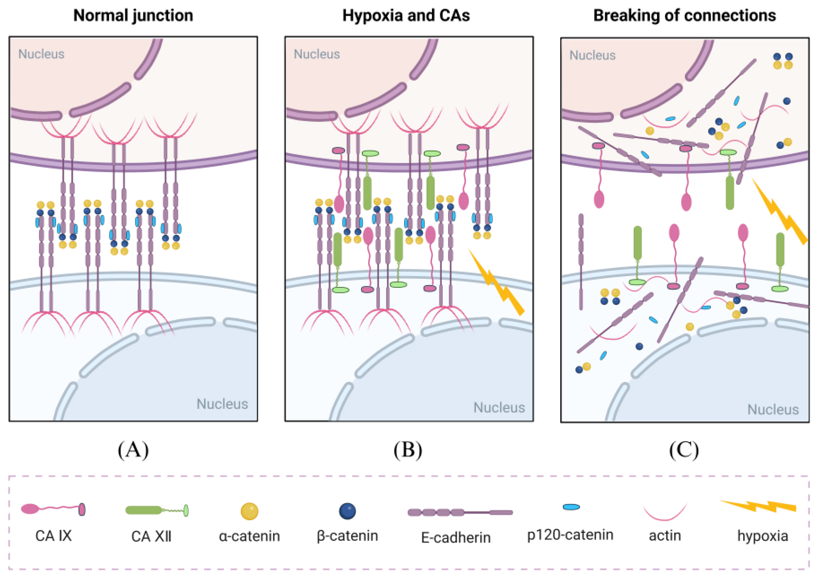
Figure 1. Disruptive effects of CA IX and CA XII on cell adhesion. (A) Under normal conditions, cell adhesion-related proteins (such as E-cadherin and β-catenin) are normally expressed, forming tight intercellular junctions that inhibit cell migration and motility. (B) Under hypoxic conditions, the expression of CA IX and CA XII is upregulated in cells, and these proteins can interact with E-cadherin. (C) Under the influence of CAs, intercellular adhesion is disrupted, greatly promoting the invasion and metastasis of tumor cells. Created with BioRender.com.
Cells 14 00693 g001
Figure 2. CA IX and CA XII are closely associated with specific proteins in hypoxia-induced cell invasion. During hypoxia-induced invasion, CA IX and CA XII can be coupled with specific proteins to exert synergistic effects. CA IX and CA XII can associate with membrane ion transporters NBCe1 and AE2, facilitating the influx of HCO3 produced by CA-catalyzed hydration of CO2, thereby maintaining intracellular pH homeostasis and influencing gene expression in concert with hypoxia. CA IX can also colocalize with matrix metalloproteinases (MMPs) and sustain their activity through H⁺ generated by CO2 hydration. It enhances the activity of other MMPs (such as MMP2, MMP3, and MMP9), ultimately affecting the composition of the extracellular matrix. Created with BioRender.com.
Cells 14 00693 g002
Figure 3. Overview of carbonic anhydrases-associated signaling pathways in tumor cells. CA IX can influence signal transduction through the EGFR/PI3K pathway, enhancing pathway activity and thereby regulating the expression of proteins related to tumor cell growth and survival. CA XII modulates the activity of NF-κB-related proteins, primarily by promoting the ubiquitin-mediated degradation of IκB, allowing the p65/cRel–P50 complex to translocate into the nucleus and exert its effects, thereby impacting tumor-associated immune responses and therapeutic outcomes. Acidic and hypoxic conditions regulate tumor physiology mainly through RAS and Exo pathways. RAS signaling enhances the expression of genes associated with cell growth and survival, while exosomes under hypoxia and acidic conditions exhibit distinct features compared to those in other environments, including smaller size, higher quantity, elevated protein expression, and greater functional activity. These combined signals promote tumor cell growth, survival, and immune evasion. Created with BioRender.com.
Cells 14 00693 g003
Figure 4. Model of metabolic regions coupled to core-invasion fronts. Tumors comprise two metabolically distinct compartments: the tumor core and the invasive front, with CA IX and CA XII expression patterns closely aligning with this spatial division. CA IX is highly expressed in the tumor core, facilitating lactate export via MCT4, forming a “lactate pool” that promotes the Warburg effect. In contrast, CA XII is highly expressed at the invasive front, working in concert with MCT1 to convert lactate into pyruvate, which enters the TCA cycle to support oxidative phosphorylation. A “proton relay” is established through lactate shuttling and proton gradient formation between these zones, promoting tumor invasion, metastasis, and resistance to therapy. Created with BioRender.com.
Cells 14 00693 g004
Table 1. The core differences between metabolic compartments.
Metabolic CompartmentsCore FeaturesKey TargetsIntervention StrategiesRefs.
Tumor Core ZoneHypoxia, High Expression of CA IX, GlycolysisCA IX/MCT4/LDHACA IX Inhibitor + LDHA Inhibitor[97]
Invasion Front ZoneRich in Oxygen, High Expression of CA XII, OXPHOSCA XII/MCT1/PDHK1MCT1 Inhibitor + PDHK1 Inhibitor[87]
Metabolic Coupling InterfaceLactate Shuttle, Exosome CommunicationExosomal CA IX/ANXA2ANXA2 Antibody + Exosome Capture Agent[22,36]
Disclaimer/Publisher’s Note: The statements, opinions and data contained in all publications are solely those of the individual author(s) and contributor(s) and not of MDPI and/or the editor(s). MDPI and/or the editor(s) disclaim responsibility for any injury to people or property resulting from any ideas, methods, instructions or products referred to in the content.