Article

Genome Instability and Senescence Are Markers of Cornelia de Lange Syndrome Cells

by
Maddalena Di Nardo
1,
Ian D. Krantz
2 and
Antonio Musio
1,*
1
Institute for Biomedical Technologies, National Research Council, 56124 Pisa, Italy
2
Roberts Individualized Medical Genetics Center, Division of Human Genetics, The Department of Pediatrics, The Children’s Hospital of Philadelphia and the Perelman School of Medicine at the University of Pennsylvania, Philadelphia, PA 19104-4318, USA
*
Author to whom correspondence should be addressed.
Cells 2024, 13(23), 2025; https://doi.org/10.3390/cells13232025
Submission received: 29 October 2024 / Revised: 5 December 2024 / Accepted: 6 December 2024 / Published: 7 December 2024
(This article belongs to the Special Issue The Role of Cellular Senescence in Health, Disease, and Aging)

Abstract

:
Cornelia de Lange syndrome (CdLS) is a rare, dominantly inherited multisystem developmental disorder. Pathogenic variants in genes encoding the structural subunits and regulatory proteins of the cohesin complex (NIPBL, SMC1A, SMC3, HDAC8, and RAD21) are the primary contributors to the pathogenesis of CdLS. Pathogenic variations in these genes disrupt normal cohesin function, leading to the syndrome’s diverse and complex clinical presentation. In this study, we discovered that cells harboring variants in the NIPBL, SMC1A and HDAC8 genes exhibit spontaneous genome instability, elevated oxidative stress and premature cellular aging. These findings suggest that cohesin plays a critical role in maintaining proper cellular function and highlight its contribution to the pathophysiology seen in the related diagnoses.

Graphical Abstract

1. Introduction

Cohesin forms a ring-shaped complex composed of four core structural members, SMC1A, SMC3, RAD21 and STAG1/2. SMC1A and SMC3, belonging to the SMC protein family, are long, flexible coiled-coil proteins forming a V-shaped heterodimer that dimerizes via the SMC hinge domains on one side and are connected by the kleisin RAD21 subunit at the globular ATP-head domains on the other side. In addition, RAD21 interacts via its middle region with STAG1/2 proteins, ensuring a stable association between chromatin and cohesin. The sister chromatid cohesion mediated by cohesin is indispensable for proper chromosome segregation during the cell cycle [1,2]. Cohesin also modulates chromatin organization to mediate long-range interactions, controls gene expression regulation and repairs damaged DNA by ensuring proximity of sister chromatids [3,4,5]. Pathogenic variants within or dysregulation of the genes that encode structural cohesin components or regulators have been associated with chromosome aneuploidy, chromosomal instability and DNA damage repair errors, which are common features of cancer [4]. In fact, somatic variants and altered expression have been identified in Ewing’s sarcoma, glioblastoma, colorectal carcinoma, breast cancer, lung carcinoma, urothelial bladder carcinoma, myeloid neoplasms and melanoma [6,7,8,9,10,11,12,13,14,15]. Germline pathogenic variants of cohesin genes result in a group of human developmental diagnoses collectively called “disorders of transcriptional regulation (DTRs)” [16]. Of the DTRs, Cornelia de Lange syndrome (CdLS, OMIM #122470, #300590, #610759, #30882 and #614701) occurs most frequently, with an incidence of between 1:10,000 and 1:30,000 live births. CdLS is a sporadic and genetically heterogeneous autosomal or X-linked disease affecting multiple organs and systems. Affected individuals are characterized by pre- and post-natal growth delay, intellectual disability, limb differences and characteristic facial features [17]. CdLS is caused by pathogenic variants in the NIPBL, SMC1A, SMC3, RAD21 and HDAC8 genes [18,19,20,21]. The molecular mechanisms of CdLS are not well understood. CdLS cell lines and developmental models of CdLS, such as mice and zebrafish, show modest, wide-ranging and conserved perturbations in gene expression [22,23,24,25,26]. It is thought that defective loop extrusion mediated by cohesin provokes an alteration in the chromatin structure, leading to gene dysregulation [27]. However, gene expression dysregulation is not the only marker of CdLS cells. Mutations in cohesin also impair the cell’s ability to repair damaged DNA [28,29]. Consequently, cells from individuals with CdLS can exhibit spontaneous chromosome anomalies [30]. These cells also show increased sensitivity to the interstrand cross-linking agent mitomycin C (MMC). Moreover, when exposed to X-rays during the G2 phase of the cell cycle, where repair processes rely on the establishment of sister chromatid cohesion, there is a marked dose-dependent increase in the formation of sister chromatid exchanges (SCEs) and double-strand breaks (DSBs). This finding indicates that CdLS cells manifest as defective in homologous recombination (HR) DNA repair, leading to delayed and/or faulty repair of damaged DNA [29,31,32,33]. We showed that two CdLS cell lines carrying variants in the SMC1A gene are characterized by high levels of oxidative stress, genome instability and reduced cell lifespan [34]. Until now, no systematic study had been performed to investigate whether genome instability and senescence occurrs in CdLS cells, beyond SMC1A-mutated cells. To gain insight into this topic, we cultured CdLS cell lines harboring pathogenic variants of the HDAC8 and NIPBL genes. Here, we found that CdLS-derived cell lines exhibited increased oxidative stress, spontaneous genome instability, and premature cellular aging, features that can be considered as biomarkers for CdLS. These observations underscore the crucial role of cohesin in preserving cellular integrity and highlight its significant impact on CdLS pathogenesis.

2. Materials and Methods

2.1. Cell Culture

Primary human fibroblasts were grown in RPMI 1640 medium supplemented with 10% fetal bovine serum, 1% L-glutamine and antibiotics (100 U/mL penicillin, 0.1 mg/mL streptomycin) in a humidified 5% CO2 atmosphere. We used six CdLS fibroblast cell lines derived from patients carrying variants in the HDAC8 and NIPBL genes. In addition, a previously analyzed SMC1A-mutated cell line [34] was used as a positive control. Two cell lines purchased from Coriell Cell Repositories and a homegrown cell line called NIG served as control samples. All cell lines were cultured in duplicate. During the early passages, cells were subcultured on a weekly basis once the monolayer became confluent. If the cells did not reach confluency within one week, the culture was classified as “senescent”, and fresh medium was replenished weekly. After a period of four weeks, if confluency was still not achieved, the culture was deemed to have fully “senesced”.

2.2. Ethics Statement

This study was conducted according to the principles expressed in the Declaration of Helsinki. All patients were enrolled under an IRB-approved protocol of informed consent at The Children’s Hospital of Philadelphia and by the Children Ethics Committee (CEP, protocol number 130/2016).

2.3. Treatments

X-ray sensitivity was assayed by exposing cells to 2 Gy using a linear accelerator with a 6 MV photon energy source. For the drug sensitivity assay, fibroblast cell lines were seeded 24 h prior to treatment with 1, 2, 3 and 4 μM MMC for 1 h, followed by washing and recovery for 6 days.

2.4. Cytogenetic Analysis

KaryoMax Colcemid (ThermoFisher, Waltham, MA, USA) was introduced to the cell cultures for 90 min, after which the cells were incubated for 30 min in a hypotonic solution (0.075 M KCl) at 37 °C. The cells then underwent several washes with Carnoy’s fixative (methanol:acid, 3:1). Following fixation, 100 metaphases were stained with Giemsa and directly examined using a Leica DM2500 microscope. Gaps and breaks were scored according to the criteria laid down by ISCN 1985 as follows: a gap is a non-staining (achromatic lesion) region (of a single chromatid or at the same locus in both chromatids of a chromosome in cases of a chromatid gap or chromosome gap, respectively) and a break is a discontinuity in which there is a clear misalignment (of one of the chromatids or at the same locus in both chromatids in cases of a chromatid gap or chromosome gap, respectively) [35].

2.5. Immunofluorescence Labeling and Microscopy

To examine γ-H2AX foci formation following irradiation, cells were fixed in 2% paraformaldehyde for 10 min, permeabilized in 0.2% Triton X-100 and blocked in PBS with 1% BSA. Thereafter, cells were incubated with a primary antibody against γ-H2AX (Trevigen, Minneapolis, MN, USA) for 1 h at 37 °C in a humidified atmosphere and then incubated with Alexa Fluor 488-conjugated goat anti-rabbit secondary antibody (Molecular Probes, Eugene, OR, USA). After three washes with PBS/0.2% Triton X-100, the cells were counterstained with 4′,6-diamino-2-phenylindole (DAPI) in mounting medium (Vector Laboratories, Newark, CA, USA). γ-H2AX foci were scored manually from 300 cells from three independent experiments using a Leica DM2500 microscope.

2.6. Senescence-Associated β-Galactosidase (SA-β-gal) Staining

SA-β-Gal staining was performed using the Senescence Cell Histochemical Staining Kit (Sigma Aldrich, St. Louis, MO, USA) following the manufacturer’s protocol. Briefly, fibroblasts were washed three times with PBS and fixed with paraformaldehyde for 10 min. After washing, fixed fibroblasts were incubated in SA-β-Gal staining solution at 37 °C. Stained cells were imaged using a Leica DM2500 microscope. Total fibroblasts and SA-β-gal positive fibroblasts were counted in three random fields per dish.

2.7. Oxidative Stress

The level of oxidative stress was measured by protein carbonyl content with the enzyme-linked immunosorbent assay (ELISA) using a Protein Carbonyl ELISA Kit (Cell Biolabs, San Diego, CA, USA) as previously described [34].

2.8. RNA Purification and Quantitative Real-Time PCR (qRT-PCR) Analysis

cDNAs were prepared with SuperScript II reverse transcriptase using oligo dT (Invitrogen; Waltham, MA, USA) from total RNA (RNAeasy Mini-kit, Qiagen, Hilde, Germany). qRT-PCR analyses were performed in triplicate using the Rotor Gene 3000 (Corbett, Manchester, UK). Expression was normalized with respect to the mean level of expression of the β-Actin housekeeping gene. Since no difference was found in control cell lines, data were pooled. The primers used for mRNA expression analysis are listed in Supplementary Table S1.

2.9. Statistics

Data were analyzed by Student’s t-test. p-values of <0.05 were considered statistically significant.

3. Results

3.1. CdLS Cells Display Reduced In Vitro Lifespan

Patients with CdLS exhibit signs of premature aging, including symptoms such as rumination, cutis verticis gyrata, and early-onset osteoporosis, these often appearing during their teenage years. Additionally, these individuals may experience premature graying of hair and noticeable changes in the skin, particularly on the face, leading to an appearance that is more aged than expected for their chronological age [36]. This notion was further supported by the observation that SMC1A-mutated cell lines showed a reduced in vitro lifespan, suggesting that premature aging and a shortened cell life are intrinsically linked [34]. To determine whether this is a phenomenon common in CdLS independent of causative genes, we analyzed six CdLS primary fibroblast cell lines carrying variants in the HDAC8 and NIPBL genes. The HDAC8-mutated cells harbor two different pathogenic missense variants (539A>G and 1001A>G, leading to H180R and H334R amino acid changes, respectively). These variants severely affect the HDAC8 deacetylase activity. Of the NIPBL-mutated cells, three are characterized by premature stop codons caused by nonsense or frameshift variants (1372C>T, 2479_2480delAG leading to Q458X and R827gfsX2 amino acid changes, respectively) and one carries a missense variant (6893G>A leading to R2298H change). This latter variant maps to the HEAT domain and likely alters the interaction of NIPBL with other proteins. In addition, a cell line with a missense variant (c.3146G>A leading to p.R1049Q change) in the SMC1A gene was used as a positive control (Table 1). This variant is located at a coiled-coil domain. It is thought that coiled-coil interactions are important for the correct folding of an SMC monomer and are thus crucial for the formation of the head domain.
Cell culture senescence was defined as the number of cell population doublings at which the cell culture failed to reach confluence at 1 week after subcultivation. By this definition, CdLS cell cultures reached their senescent phase at lower in vitro passages than control cells. CdLS cells became senescent between the 25th and the 34.5th passage, with a considerable decrease (about 45% in NIPBL cells carrying premature stop codon) in their in vitro lifespan when compared with control cells (Figure 1A and Supplementary Table S2).
β-Galactoside staining, a measure of cell senescence, showed that the senescence ratio of CdLS fibroblasts had increased considerably. The results demonstrated that the percentage of the SA-β-gal-positive senescent cells increased significantly during in vitro cell culture progression, ranging from 25% to 88%. The RT-qPCR results showed elevated mRNA expression levels of the senescence markers, CDKN2A p16 (cyclin-dependent kinase inhibitor 2A) and CDKN1A p21 (cyclin-dependent kinase inhibitor 1A) as compared to control cells (Figure 1B, p < 0.05, Supplementary Figure S1). Next, we analyzed the level of carbonyl derivatives of proline, lysine, arginine and threonine residues as a consequence of protein oxidation during cell progression. These results indicated that the levels of protein carbonyl remained comparatively low in control cells but increased in CdLS cells (Figure 1C). Overall, these findings suggest that CdLS cells exhibit a shortened lifespan in vitro that is associated with high levels of oxidative stress.

3.2. CdLS-Causative Genes Are Associated with Increased Sensitivity to Genotoxic Agents

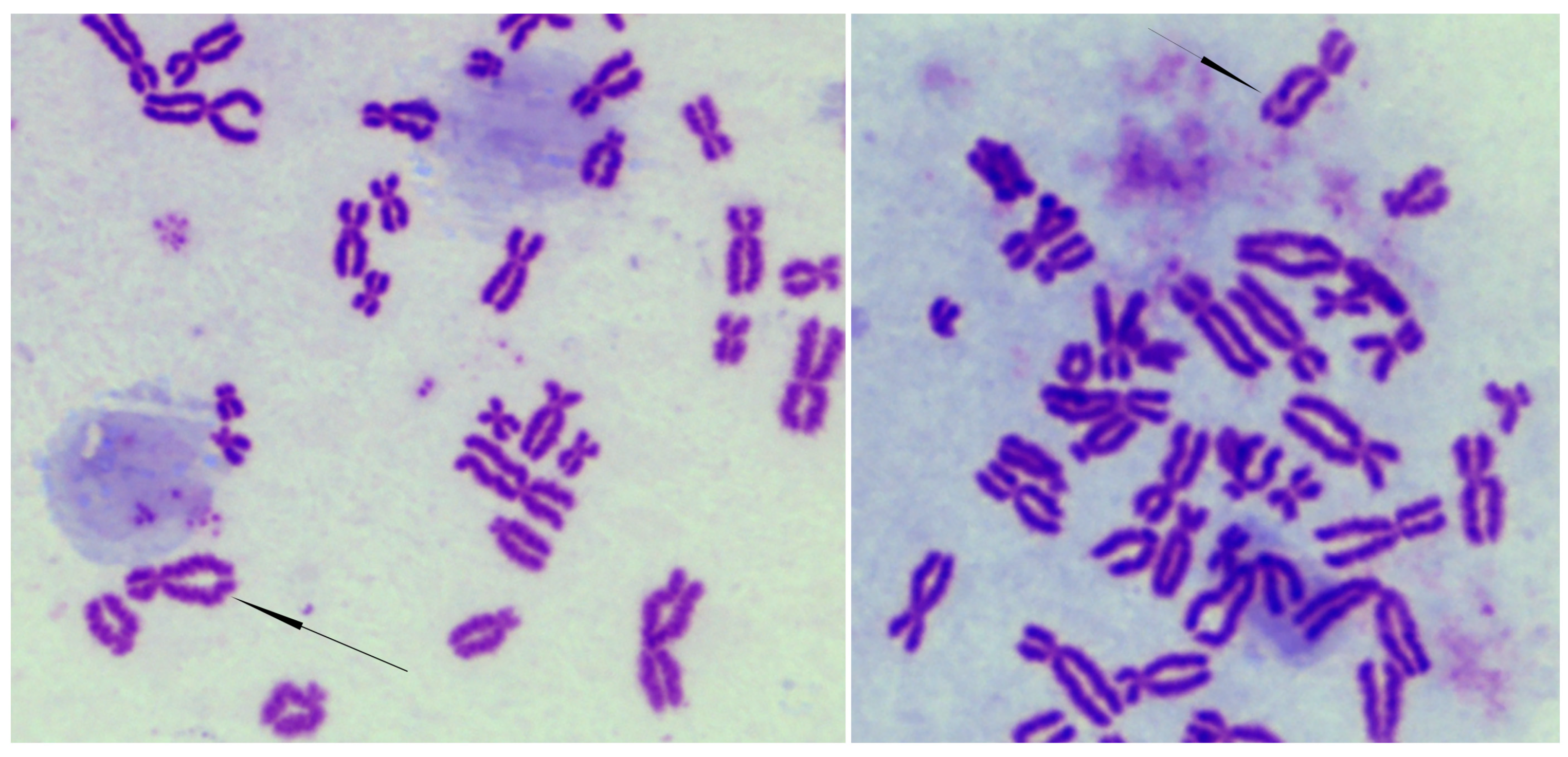
Since cohesin is involved in DNA repair and maintaining genome stability [4], we investigated the occurrence of spontaneous genomic instability and whether CdLS cells are sensitive to DNA-damaging agents. At passage 13, the incidence of spontaneous chromosome abnormalities was observed to be significantly higher in CdLS cell lines as compared to control cell lines (Figure 2 and Table 2).
In particular, cells carrying a NIPBL-frameshift variant showed more aberrations (ranging from 11 to 15 in one hundred metaphases) than cells with the NIPBL-missense (7–9 in one hundred metaphases) or HDAC8-missense variants (ranging from 6 to 7 in one hundred metaphases). However, the SMC1A-mutated cell line displayed the highest frequency of chromosome aberrations, 23-25 in one hundred metaphases.
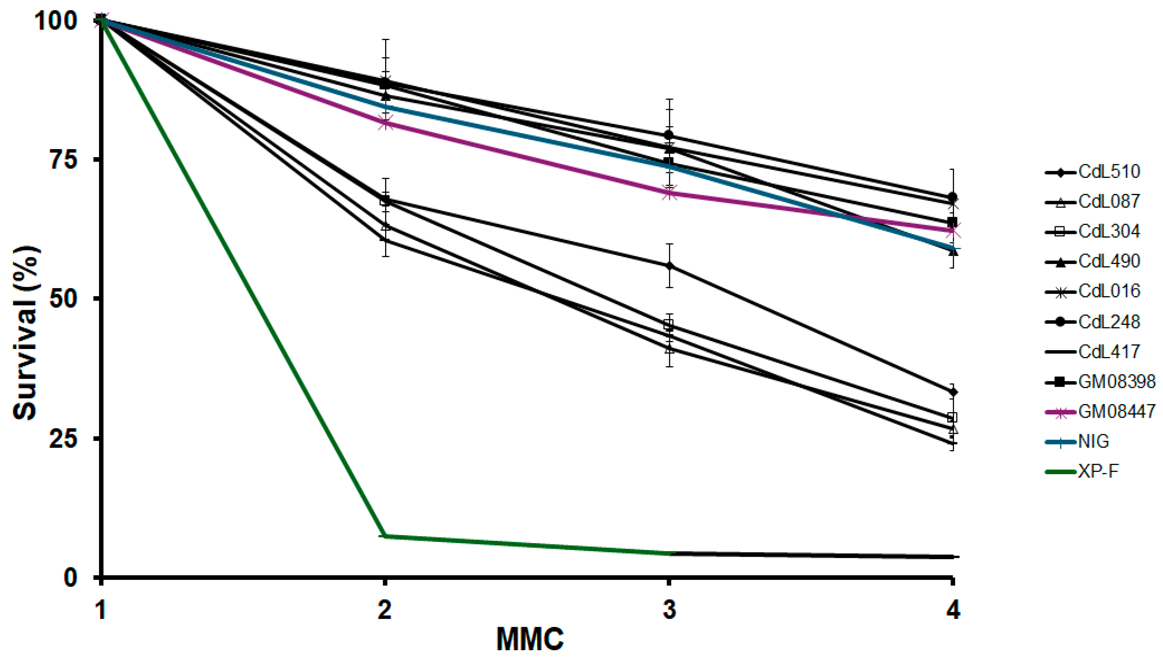
Then, we exposed cohesin-mutated cell lines to MMC, a well-characterized DNA crosslinking agent. In this analysis, a consistent response was observed, with SMC1A-mutated cells and all cell lines carrying nonsense or frameshift NIPBL variants displaying heightened sensitivity to MMC compared to the control cells. However, their sensitivity can be categorized as moderate, as it did not reach the sensitivity levels observed in a Xeroderma Pigmentosum (XP-F) cell line. Instead, all cell lines with missense variants in the NIPBL and HDAC8 genes showed similar sensitivity to control cells (Figure 3).
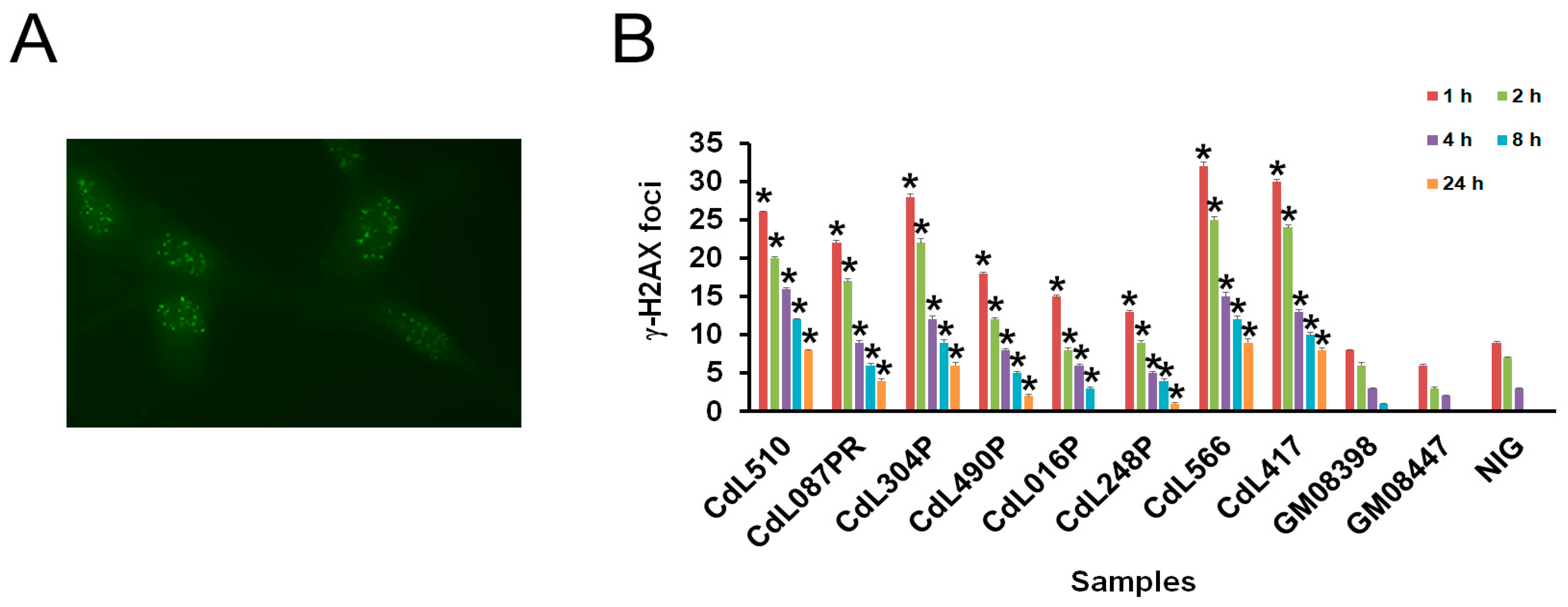
To further assess the occurrence of chromosomal instability, we examined γ-H2AX foci formation. Our results showed that CdLS cells exhibited a higher number of γ-H2AX foci 30 min after exposure to 2 Gy of irradiation compared to control cells. The average number of γ-H2AX foci per cell ranged from 46 in NIPBL-mutated cells to 28 in cells harboring HDAC8 variants (Figure 4A and Supplementary Figure S2).
Once again, cells with NIPBL variants resulting in a premature stop codon and those with a missense variant in the SMC1A gene showed a considerable increase in the number of foci. In particular, cells with the SMC1A variant demonstrated the highest level of γ-H2AX foci, an average of 51/cell. Time course experiments revealed that 4 h after irradiation, about 50% of DSBs were repaired in both the control and CdLS cells. Thereafter, it declined almost to the control level. For repair times of 8 h and 24 h, both cells carrying NIPBL frameshift variants and cells with the SMC1A variant showed more foci than cells with the HDAC8- and NIPBL-missense variants (Figure 4B). These data suggest that genome instability and an impaired DNA repair pathway may be a common mechanism in CdLS-derived cell lines.

4. Discussion

CdLS is a genetic multisystem developmental disorder. Variants in the NIPBL gene account for approximately 60% of CdLS cases, while a smaller proportion of affected individuals, comprising 5–7% of cases, have pathogenic variants in the HDAC8, RAD21, SMC1A and SMC3 genes [37]. CdLS is characterized by a wide range of phenotypic effects, including small size, craniofacial and limb differences, multiorgan defects, intellectual disability and signs of premature aging relative to their chronological age [17]. Although the functional consequences of cohesin disruption in CdLS remain incompletely understood, we have previously shown that variants in both SMC1A and SMC3 genes make these cells more susceptible to protein oxidation, DNA damage and cellular senescence [34,38]. Here, we show that cells carrying pathogenic variants in the HDAC8 and NIPBL genes also display genomic instability, sensitivity to genotoxic agents and premature in vitro senescence, suggesting that they are markers of CdLS cells irrespective of the causative genes.
The mean of spontaneous aberrations per cell ranges from 6 to 7 and from 7 to 15 in HDAC8 and NIPBL-mutated cells, respectively. It is interesting to note that cells with NIPBL variants leading to a premature stop codon have approximately twice the number of aberrations compared to those with missense variants in the NIPBL and HDAC8 genes. However, these values are significantly lower than the number of aberrations present in cells with the SMC1A variant. This could reflect the different roles of NIPBL, HDAC8 and SMC1A in preserving genome stability through cohesin functions. NIPBL, in association with its molecular partner MAU2, is involved in cohesin deposition onto chromatin. The silencing of NIPBL increases cellular sensitivity to genotoxic agents, and the repair of DSBs caused by endonuclease cleavage and laser microirradiation requires the cohesin complex to accumulate at the sites of damage, a process reliant on the NIPBL–MAU2 loading complex [39,40,41]. Conversely, the deacetylation of cohesin by the deacetylase HDAC8 is essential for the recycling of cohesin complexes, enabling them to participate in subsequent rounds of sister chromatid cohesion [20,42]. The loss of HDAC8 activity leads to the elevated acetylation of SMC3, which is reloaded onto chromatin, leading to a reduced occupancy of cohesin at its localization sites [42]. It is likely that this results in a distinct DNA repair pattern alteration to be observed in CdLS cell lines harboring mutations in either NIPBL or HDAC8. SMC1A is a member of the cohesin core. Following DNA damage, SMC1A is phosphorylated by ATM or ATR kinases on the serine 957 and serine 966 residues. Cells expressing mutated SMC1A that cannot undergo this phosphorylation display impaired DNA repair mechanisms and reduced cell viability [42,43,44,45]. This indicates that SMC1A is directly involved in preserving the genome stability of human cells, ensuring an effective and coordinated response to repair DNA damage. This is supported by the finding that SMC1A mutations have been identified in human cancers characterized by genome instability, such as colorectal cancer [15,46,47,48,49].
Though CdLS cells exhibit genome instability, it has been shown there is no increased risk of cancer in CdLS patients, although NIPBL variants may genetically predispose to early Barrett’s esophagus development [50]. Since tumorigenesis develops over many years because of the accumulation of specific variants, it is likely that additional genetic alterations are required for a fully malignant transformation beyond the initial cohesin variants.
Different pathogenic variants in the HDAC8, NIPBL and SMC1A genes are associated with varying frequencies of chromosomal aberrations. In fact, cells with a mutation in the SMC1A gene exhibited the highest level of chromosomal aberrations, followed by cells with nonsense or frameshift mutations in NIPBL and, finally, cells with missense mutations in NIPBL and HDAC8. In addition, a clonogenic survival assay revealed that SMC1A-mutated cells (CdL417) and all cell lines (CdL510, CdL087 and CdL304) harboring NIPBL variants leading to premature stop codon displayed an increased sensitivity to MMC. These observations suggest that both SMC1A and NIPBL may play a role in the resolution of interstrand cross-linking. Therefore, we propose that these cytogenetic assays can be used to differentiate between CdLS patients with variants in different causative genes. Chromosomal breakage assays are used as a diagnostic tool for other genetic diagnoses. For example, Fanconi anemia (FA), an autosomal recessive diagnosis marked by pancytopenia, diverse congenital anomalies, heightened cancer susceptibility and chromosomal instability in cultured cells with an increased vulnerability to chromosomal breakage when exposed to a non-toxic concentration of the bifunctional alkylating agent diepoxybutane [51]. The ability to distinguish between the different CdLS-causative genes in an in vitro system that measures the occurrence of spontaneous chromosome aberrations may represent a test that can help differentiate genetic subtypes of CdLS and could help in stratifying patients in which a known molecular etiology has not been identified. Additionally, this assay may be able to serve as a biomarker to assess the efficacy of various therapeutic modalities and drug screening in cellular assays.
By employing γ-H2AX foci analysis to assess DSB repair following irradiation, we show that mutations in SMC1A, NIPBL and, to a lesser extent, HDAC8 cause defects in DNA repair in primary fibroblasts. Although SMC1A- and NIPBL-mutated cells repair most DSBs in a manner similar to HDAC8-mutated and control cells, a small subset of irradiation-induced breaks remains unresolved. It is reasonable to infer that DNA repair efficacy may depend on the complexity of the damage at individual sites and the specific variants that disrupt these repair mechanisms, leading to persistent γ-H2AX foci indicative of permanent DNA damage.
Cellular senescence is a biological process in which cells permanently stop dividing but do not die. This state is often a response to various forms of stress, such as DNA damage, oxidative stress or telomere shortening, and plays a crucial role in aging, tumor suppression and tissue repair.
We observed that CdLS cells with NIPBL frameshift variants entered senescence at the 26th or 27th passage, showing a significant decrease in their lifespan compared to three control cell lines. This senescence was further validated using a β-galactosidase assay, where the percentage of positive cells ranged from 78% to 82% in control cells and was approximately 90% in CdLS cells. Results supportive of this included RT-qPCR data, which indicated that the mRNA expression levels of the senescence markers p16 and p21 were elevated. It is probable that both persistent DNA damage and premature aging contribute significantly to the pathogenesis of CdLS. These factors may intersect in influencing the development and progression of the syndrome, highlighting the complex interplay between genomic instability and accelerated cellular aging observed in individuals with CdLS. This notion is further supported by the observations that senescence is exacerbated by persistent DNA damage and the high levels of γ-H2AX foci that are observed in the Nipbl- and Hdac8-mutant mice [52].

5. Conclusions

In conclusion, our data indicate that genome instability and senescence serve as distinctive markers of CdLS cells. These cellular characteristics underscore the pathophysiological complexities of CdLS, providing crucial insights into the molecular consequences of cohesin disruption and the impact that this has on the phenotype and outcomes in related diagnoses. The presence of genome instability and premature senescence highlights the multifaceted nature of CdLS, where abnormalities in the cellular processes contribute to the diverse clinical manifestations observed in affected individuals.

Supplementary Materials

The following supporting information can be downloaded at: https://www.mdpi.com/article/10.3390/cells13232025/s1, Table S1: Primer sequences used in this study. Table S2: Cellular passage at which cells become senescent in vitro. Figure S1: RT-qPCR analysis of p16 at three different passages in vitro. Data represent the average from three independent experiments. Figure S2: γ-H2AX foci 30 min after exposure to 2 Gy.

Author Contributions

A.M. conceived the study, designed experiments and interpreted data. M.D.N. and A.M. performed experiments. I.D.K. provided reagents. A.M. wrote the manuscript, with input from M.D.N. and I.D.K. All authors have read and agreed to the published version of the manuscript.

Funding

This work was supported by grants from Associazione Italiana per la Ricerca sul Cancro (AIRC, IG23284) and CNR project FOE-2021 DBA.AD005.225 to A.M.

Institutional Review Board Statement

All patients were enrolled under an IRB-approved protocol of informed consent at The Children’s Hospital of Philadelphia and by the Children Ethics Committee (CEP, protocol number 130/2016).

Informed Consent Statement

Informed consent was obtained from all subjects involved in the study.

Data Availability Statement

The relevant data supporting the findings of this study are available in this article and its Supplementary Information files.

Conflicts of Interest

The authors declare no conflicts of interest. The funders had no role in the design of the study; in the collection, analyses, or interpretation of data; in the writing of the manuscript; or in the decision to publish the results.

References

  1. Yatskevich, S.; Rhodes, J.; Nasmyth, K. Organization of Chromosomal DNA by SMC Complexes. Annu. Rev. Genet. 2019, 53, 445–482. [Google Scholar] [CrossRef] [PubMed]
  2. Nishiyama, T. Cohesion and cohesin-dependent chromatin organization. Curr. Opin. Cell Biol. 2019, 58, 8–14. [Google Scholar] [CrossRef] [PubMed]
  3. Horsfield, J.A. Full circle: A brief history of cohesin and the regulation of gene expression. FEBS J. 2022, 290, 1670–1687. [Google Scholar] [CrossRef] [PubMed]
  4. Di Nardo, M.; Pallotta, M.M.; Musio, A. The multifaceted roles of cohesin in cancer. J. Exp. Clin. Cancer Res. 2022, 41, 96. [Google Scholar] [CrossRef] [PubMed]
  5. van Ruiten, M.S.; Rowland, B.D. SMC Complexes: Universal DNA Looping Machines with Distinct Regulators. Trends Genet. 2018, 34, 477–487. [Google Scholar] [CrossRef]
  6. Kandoth, C.; McLellan, M.D.; Vandin, F.; Ye, K.; Niu, B.; Lu, C.; Xie, M.; Zhang, Q.; McMichael, J.F.; Wyczalkowski, M.A.; et al. Mutational landscape and significance across 12 major cancer types. Nature 2013, 502, 333–339. [Google Scholar] [CrossRef]
  7. Lawrence, M.S.; Stojanov, P.; Mermel, C.H.; Robinson, J.T.; Garraway, L.A.; Golub, T.R.; Meyerson, M.; Gabriel, S.B.; Lander, E.S.; Getz, G. Discovery and saturation analysis of cancer genes across 21 tumour types. Nature 2014, 505, 495–501. [Google Scholar] [CrossRef]
  8. Leiserson, M.D.; Vandin, F.; Wu, H.T.; Dobson, J.R.; Eldridge, J.V.; Thomas, J.L.; Papoutsaki, A.; Kim, Y.; Niu, B.; McLellan, M.; et al. Pan-cancer network analysis identifies combinations of rare somatic mutations across pathways and protein complexes. Nat. Genet. 2015, 47, 106–114. [Google Scholar] [CrossRef]
  9. Zhu, H.E.; Li, T.; Shi, S.; Chen, D.X.; Chen, W.; Chen, H. ESCO2 promotes lung adenocarcinoma progression by regulating hnRNPA1 acetylation. J. Exp. Clin. Cancer Res. 2021, 40, 64. [Google Scholar] [CrossRef]
  10. Solomon, D.A.; Kim, J.S.; Bondaruk, J.; Shariat, S.F.; Wang, Z.F.; Elkahloun, A.G.; Ozawa, T.; Gerard, J.; Zhuang, D.; Zhang, S.; et al. Frequent truncating mutations of STAG2 in bladder cancer. Nat. Genet. 2013, 45, 1428–1430. [Google Scholar] [CrossRef]
  11. Crompton, B.D.; Stewart, C.; Taylor-Weiner, A.; Alexe, G.; Kurek, K.C.; Calicchio, M.L.; Kiezun, A.; Carter, S.L.; Shukla, S.A.; Mehta, S.S.; et al. The genomic landscape of pediatric Ewing sarcoma. Cancer Discov. 2014, 4, 1326–1341. [Google Scholar] [CrossRef] [PubMed]
  12. Tirode, F.; Surdez, D.; Ma, X.; Parker, M.; Le Deley, M.C.; Bahrami, A.; Zhang, Z.; Lapouble, E.; Grossetete-Lalami, S.; Rusch, M.; et al. Genomic landscape of Ewing sarcoma defines an aggressive subtype with co-association of STAG2 and TP53 mutations. Cancer Discov. 2014, 4, 1342–1353. [Google Scholar] [CrossRef]
  13. Brennan, C.W.; Verhaak, R.G.; McKenna, A.; Campos, B.; Noushmehr, H.; Salama, S.R.; Zheng, S.; Chakravarty, D.; Sanborn, J.Z.; Berman, S.H.; et al. The somatic genomic landscape of glioblastoma. Cell 2013, 155, 462–477. [Google Scholar] [CrossRef]
  14. Kon, A.; Shih, L.Y.; Minamino, M.; Sanada, M.; Shiraishi, Y.; Nagata, Y.; Yoshida, K.; Okuno, Y.; Bando, M.; Nakato, R.; et al. Recurrent mutations in multiple components of the cohesin complex in myeloid neoplasms. Nat. Genet. 2013, 45, 1232–1237. [Google Scholar] [CrossRef] [PubMed]
  15. Di Nardo, M.; Astigiano, S.; Baldari, S.; Pallotta, M.M.; Porta, G.; Pigozzi, S.; Antonini, A.; Emionite, L.; Frattini, A.; Valli, R.; et al. The synergism of SMC1A cohesin gene silencing and bevacizumab against colorectal cancer. J. Exp. Clin. Cancer Res. 2024, 43, 49. [Google Scholar] [CrossRef] [PubMed]
  16. Izumi, K. Disorders of Transcriptional Regulation: An Emerging Category of Multiple Malformation Syndromes. Mol. Syndr. Syndromol. 2016, 7, 262–273. [Google Scholar] [CrossRef]
  17. Kline, A.D.; Moss, J.F.; Selicorni, A.; Bisgaard, A.M.; Deardorff, M.A.; Gillett, P.M.; Ishman, S.L.; Kerr, L.M.; Levin, A.V.; Mulder, P.A.; et al. Diagnosis and management of Cornelia de Lange syndrome: First international consensus statement. Nat. Rev. Genet. 2018, 19, 649–666. [Google Scholar] [CrossRef] [PubMed]
  18. Deardorff, M.A.; Wilde, J.J.; Albrecht, M.; Dickinson, E.; Tennstedt, S.; Braunholz, D.; Monnich, M.; Yan, Y.; Xu, W.; Gil-Rodriguez, M.C.; et al. RAD21 mutations cause a human cohesinopathy. Am. J. Hum. Genet. 2012, 90, 1014–1027. [Google Scholar] [CrossRef]
  19. Deardorff, M.A.; Kaur, M.; Yaeger, D.; Rampuria, A.; Korolev, S.; Pie, J.; Gil-Rodriguez, C.; Arnedo, M.; Loeys, B.; Kline, A.D.; et al. Mutations in cohesin complex members SMC3 and SMC1A cause a mild variant of cornelia de Lange syndrome with predominant mental retardation. Am. J. Hum. Genet. 2007, 80, 485–494. [Google Scholar] [CrossRef]
  20. Deardorff, M.A.; Bando, M.; Nakato, R.; Watrin, E.; Itoh, T.; Minamino, M.; Saitoh, K.; Komata, M.; Katou, Y.; Clark, D.; et al. HDAC8 mutations in Cornelia de Lange syndrome affect the cohesin acetylation cycle. Nature 2012, 489, 313–317. [Google Scholar] [CrossRef]
  21. Tonkin, E.T.; Wang, T.J.; Lisgo, S.; Bamshad, M.J.; Strachan, T. NIPBL, encoding a homolog of fungal Scc2-type sister chromatid cohesion proteins and fly Nipped-B, is mutated in Cornelia de Lange syndrome. Nat. Genet. 2004, 36, 636–641. [Google Scholar] [CrossRef] [PubMed]
  22. Chea, S.; Kreger, J.; Lopez-Burks, M.E.; MacLean, A.L.; Lander, A.D.; Calof, A.L. Gastrulation-stage gene expression in Nipbl (+/−) mouse embryos foreshadows the development of syndromic birth defects. Sci. Adv. 2024, 10, eadl4239. [Google Scholar] [CrossRef] [PubMed]
  23. Muto, A.; Calof, A.L.; Lander, A.D.; Schilling, T.F. Multifactorial Origins of Heart and Gut Defects in nipbl-Deficient Zebrafish, a Model of Cornelia de Lange Syndrome. PLoS Biol. 2011, 9, e1001181. [Google Scholar] [CrossRef] [PubMed]
  24. Kawauchi, S.; Calof, A.L.; Santos, R.; Lopez-Burks, M.E.; Young, C.M.; Hoang, M.P.; Chua, A.; Lao, T.; Lechner, M.S.; Daniel, J.A.; et al. Multiple organ system defects and transcriptional dysregulation in the Nipbl(+/-) mouse, a model of Cornelia de Lange Syndrome. PLoS Genet. 2009, 5, e1000650. [Google Scholar] [CrossRef] [PubMed]
  25. Liu, J.; Zhang, Z.; Bando, M.; Itoh, T.; Deardorff, M.A.; Clark, D.; Kaur, M.; Tandy, S.; Kondoh, T.; Rappaport, E.; et al. Transcriptional dysregulation in NIPBL and cohesin mutant human cells. PLoS Biol. 2009, 7, e1000119. [Google Scholar] [CrossRef] [PubMed]
  26. Gimenez, G.; Kalev-Zylinska, M.L.; Morison, I.; Bohlander, S.K.; Horsfield, J.A.; Antony, J. Cohesin rad21 mutation dysregulates erythropoiesis and granulopoiesis output within the whole kidney marrow of adult zebrafish. Am. J. Physiol. Cell Physiol. 2024. [Google Scholar] [CrossRef]
  27. Garcia, P.; Fernandez-Hernandez, R.; Cuadrado, A.; Coca, I.; Gomez, A.; Maqueda, M.; Latorre-Pellicer, A.; Puisac, B.; Ramos, F.J.; Sandoval, J.; et al. Disruption of NIPBL/Scc2 in Cornelia de Lange Syndrome provokes cohesin genome-wide redistribution with an impact in the transcriptome. Nat. Commun. 2021, 12, 4551. [Google Scholar] [CrossRef]
  28. Olley, G.; Pradeepa, M.M.; Grimes, G.R.; Piquet, S.; Polo, S.E.; FitzPatrick, D.R.; Bickmore, W.A.; Boumendil, C. Cornelia de Lange syndrome-associated mutations cause a DNA damage signalling and repair defect. Nat. Commun. 2021, 12, 3127. [Google Scholar] [CrossRef]
  29. Vrouwe, M.G.; Elghalbzouri-Maghrani, E.; Meijers, M.; Schouten, P.; Godthelp, B.C.; Bhuiyan, Z.A.; Redeker, E.J.; Mannens, M.M.; Mullenders, L.H.; Pastink, A.; et al. Increased DNA damage sensitivity of Cornelia de Lange syndrome cells: Evidence for impaired recombinational repair. Hum. Mol. Genet. 2007, 16, 1478–1487. [Google Scholar] [CrossRef]
  30. Di Nardo, M.; Musio, A. Cohesin—Bridging the gap among gene transcription, genome stability, and human diseases. FEBS Lett. 2024, 598. [Google Scholar] [CrossRef]
  31. Revenkova, E.; Focarelli, M.L.; Susani, L.; Paulis, M.; Bassi, M.T.; Mannini, L.; Frattini, A.; Delia, D.; Krantz, I.; Vezzoni, P.; et al. Cornelia de Lange syndrome mutations in SMC1A or SMC3 affect binding to DNA. Hum. Mol. Genet. 2009, 18, 418–427. [Google Scholar] [CrossRef] [PubMed]
  32. Plesser Duvdevani, M.; Pettersson, M.; Eisfeldt, J.; Avraham, O.; Dagan, J.; Frumkin, A.; Lupski, J.R.; Lindstrand, A.; Harel, T. Whole-genome sequencing reveals complex chromosome rearrangement disrupting NIPBL in infant with Cornelia de Lange syndrome. Am. J. Med. Genet. A 2020, 182, 1143–1151. [Google Scholar] [CrossRef] [PubMed]
  33. Gervasini, C.; Picinelli, C.; Azzollini, J.; Rusconi, D.; Masciadri, M.; Cereda, A.; Marzocchi, C.; Zampino, G.; Selicorni, A.; Tenconi, R.; et al. Genomic imbalances in patients with a clinical presentation in the spectrum of Cornelia de Lange syndrome. BMC Med. Genet. 2013, 14, 41. [Google Scholar] [CrossRef] [PubMed]
  34. Cukrov, D.; Newman, T.A.C.; Leask, M.; Leeke, B.; Sarogni, P.; Patimo, A.; Kline, A.D.; Krantz, I.D.; Horsfield, J.A.; Musio, A. Antioxidant treatment ameliorates phenotypic features of SMC1A-mutated Cornelia de Lange syndrome in vitro and in vivo. Hum. Mol. Genet. 2018, 27, 3002–3011. [Google Scholar] [CrossRef]
  35. Harnden, D.G.; Klinger, H.P. An International System for Human Cytogenetic Nomenclature (1985) ISCN 1985. Report of the Standing Committee on Human Cytogenetic Nomenclature. Birth Defects Orig. Artic. Ser. 1985, 21, 1–117. [Google Scholar]
  36. Kline, A.D.; Grados, M.; Sponseller, P.; Levy, H.P.; Blagowidow, N.; Schoedel, C.; Rampolla, J.; Clemens, D.K.; Krantz, I.; Kimball, A.; et al. Natural history of aging in Cornelia de Lange syndrome. Am. J. Med. Genet. C Semin. Med. Genet. 2007, 145, 248–260. [Google Scholar] [CrossRef]
  37. Kaur, M.; Blair, J.; Devkota, B.; Fortunato, S.; Clark, D.; Lawrence, A.; Kim, J.; Do, W.; Semeo, B.; Katz, O.; et al. Genomic analyses in Cornelia de Lange Syndrome and related diagnoses: Novel candidate genes, genotype-phenotype correlations and common mechanisms. Am. J. Med. Genet. A 2023, 191, 2113–2131. [Google Scholar] [CrossRef]
  38. Gimigliano, A.; Mannini, L.; Bianchi, L.; Puglia, M.; Deardorff, M.A.; Menga, S.; Krantz, I.D.; Musio, A.; Bini, L. Proteomic profile identifies dysregulated pathways in Cornelia de Lange syndrome cells with distinct mutations in SMC1A and SMC3 genes. J. Proteome Res. 2012, 11, 6111–6123. [Google Scholar] [CrossRef]
  39. Strom, L.; Lindroos, H.B.; Shirahige, K.; Sjogren, C. Postreplicative recruitment of cohesin to double-strand breaks is required for DNA repair. Mol. Cell 2004, 16, 1003–1015. [Google Scholar] [CrossRef]
  40. Unal, E.; Arbel-Eden, A.; Sattler, U.; Shroff, R.; Lichten, M.; Haber, J.E.; Koshland, D. DNA damage response pathway uses histone modification to assemble a double-strand break-specific cohesin domain. Mol. Cell 2004, 16, 991–1002. [Google Scholar] [CrossRef]
  41. Xu, W.; Ying, Y.; Shan, L.; Feng, J.; Zhang, S.; Gao, Y.; Xu, X.; Yao, Y.; Zhu, C.; Mao, W. Enhanced expression of cohesin loading factor NIPBL confers poor prognosis and chemotherapy resistance in non-small cell lung cancer. J. Transl. Med. 2015, 13, 153. [Google Scholar] [CrossRef] [PubMed]
  42. Beckouet, F.; Hu, B.; Roig, M.B.; Sutani, T.; Komata, M.; Uluocak, P.; Katis, V.L.; Shirahige, K.; Nasmyth, K. An Smc3 acetylation cycle is essential for establishment of sister chromatid cohesion. Mol. Cell 2010, 39, 689–699. [Google Scholar] [CrossRef] [PubMed]
  43. Kitagawa, R.; Bakkenist, C.J.; McKinnon, P.J.; Kastan, M.B. Phosphorylation of SMC1 is a critical downstream event in the ATM-NBS1-BRCA1 pathway. Genes. Dev. 2004, 18, 1423–1438. [Google Scholar] [CrossRef]
  44. Yazdi, P.T.; Wang, Y.; Zhao, S.; Patel, N.; Lee, E.Y.; Qin, J. SMC1 is a downstream effector in the ATM/NBS1 branch of the human S-phase checkpoint. Genes. Dev. 2002, 16, 571–582. [Google Scholar] [CrossRef]
  45. Kim, S.T.; Xu, B.; Kastan, M.B. Involvement of the cohesin protein, Smc1, in Atm-dependent and independent responses to DNA damage. Genes. Dev. 2002, 16, 560–570. [Google Scholar] [CrossRef]
  46. Barber, T.D.; McManus, K.; Yuen, K.W.; Reis, M.; Parmigiani, G.; Shen, D.; Barrett, I.; Nouhi, Y.; Spencer, F.; Markowitz, S.; et al. Chromatid cohesion defects may underlie chromosome instability in human colorectal cancers. Proc. Natl. Acad. Sci. USA 2008, 105, 3443–3448. [Google Scholar] [CrossRef]
  47. Cucco, F.; Servadio, A.; Gatti, V.; Bianchi, P.; Mannini, L.; Prodosmo, A.; De Vitis, E.; Basso, G.; Friuli, A.; Laghi, L.; et al. Mutant cohesin drives chromosomal instability in early colorectal adenomas. Hum. Mol. Genet. 2014, 23, 6. [Google Scholar] [CrossRef]
  48. Pallotta, M.M.; Di Nardo, M.; Musio, A. Synthetic Lethality between Cohesin and WNT Signaling Pathways in Diverse Cancer Contexts. Cells 2024, 13, 608. [Google Scholar] [CrossRef] [PubMed]
  49. Sarogni, P.; Palumbo, O.; Servadio, A.; Astigiano, S.; D’Alessio, B.; Gatti, V.; Cukrov, D.; Baldari, S.; Pallotta, M.M.; Aretini, P.; et al. Overexpression of the cohesin-core subunit SMC1A contributes to colorectal cancer development. J. Exp. Clin. Cancer Res. 2019, 38, 108. [Google Scholar] [CrossRef]
  50. Pallotta, M.M.; Di Nardo, M.; Hennekam, R.C.M.; Kaiser, F.J.; Parenti, I.; Pie, J.; Ramos, F.J.; Kline, A.D.; Musio, A. Cornelia de Lange syndrome and cancer: An open question. Am. J. Med. Genet. A 2023, 191, 292–295. [Google Scholar] [CrossRef]
  51. Mehta, P.A.; Ebens, C. Fanconi Anemia. In GeneReviews((R)); Adam, M.P., Feldman, J., Mirzaa, G.M., Pagon, R.A., Wallace, S.E., Bean, L.J.H., Gripp, K.W., Amemiya, A., Eds.; University of Washington: Seattle, WA, USA, 1993. [Google Scholar]
  52. Singh, V.P.; McKinney, S.; Gerton, J.L. Persistent DNA Damage and Senescence in the Placenta Impacts Developmental Outcomes of Embryos. Dev. Cell 2020, 54, 333–347. [Google Scholar] [CrossRef] [PubMed]
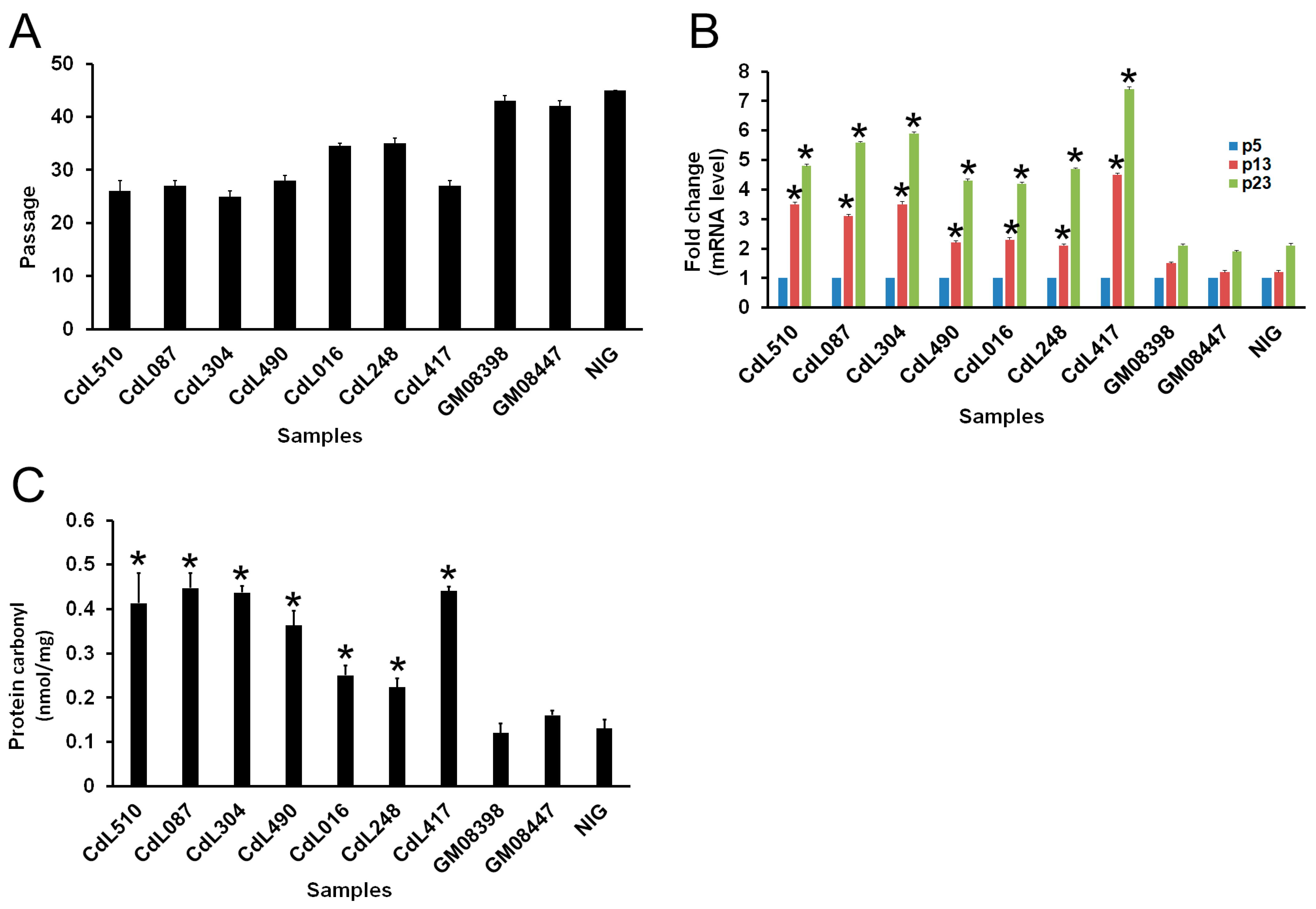
Figure 1. CdLS cell markers during in vitro cell culture progression. (A) NIPBL-, HDAC8- and SMC1A-mutated cells are characterized by replicative senescence. (B) RT-qPCR analysis of p16 at three different passages in vitro. Data represented the average ± SE from three independent experiments. (C) Protein carbonyl content, as a marker of oxidative stress, was measured in NIPBL-, HDAC8- and SMC1A-mutated cells. * p < 0.05.
Cells 13 02025 g001
Figure 2. CdLS and damaged DNA repair during in vitro cell culture progression. Partial Giemsa-stained metaphases showing a chromatid break (indicated by an arrow) and a chromatid gap (indicated by an arrow) during in vitro cell culture progression of CdL510 (left) and CdL248 (right) cells, respectively. According to ISCN 1985, the break is clearly visible as region in which there is a misalignment of one of the chromatids.
Cells 13 02025 g002
Figure 3. Sensitivity of cohesin-mutated cell lines to MMC at passage 13 of in vitro cell culture. Control fibroblasts, a Xeroderma Pigmentosum cell line (XP-F) and cell lines carrying variants in the HDAC8, NIPBL and SMC1A genes were plated the day before the exposure. Cells were treated with different doses of MMC (1, 2, 3 and 4 μM) for 1 h. The survival was evaluated after 6 days of recovery. The data represent the average of three independent experiments.
Cells 13 02025 g003
Figure 4. (A) γ-H2AX foci after 30 min following 2 Gy irradiation in NIPBL-mutated cells (CdL510 cell line). (B) Time course of γ-H2AX foci disappearance following 2 Gy irradiation. Error bars represent the SE from the analysis of 300 cells from three independent experiments. * p < 0.05.
Cells 13 02025 g004
Table 1. Human primary fibroblast cell lines used in this work.
Cell LineGeneGene VariationAmino Acid Change
CdL510NIPBLc.1372 C>Tp.Q458X
CdL087NIPBLc.2479_2480del AGp.R827gfsX2
CdL304NIPBLc.2479_2480del AGp.R827gfsX2
CdL490NIPBLc.6893G>Ap.R2298H
CdL016HDAC8c.539A>Gp.H180R
CdL248HDAC8c.1001A>Gp.H334R
CdL417SMC1Ac.3146G>Ap.R1049Q
GM08398Control
GM08447Control
NIGControl
Table 2. Spontaneous chromosome aberrations at passage 13 of in vitro cell culture.
Cell LineChromosome AberrationsMetaphases
CdL510a15100
CdL510b13100
CdL087a12100
CdL087b14100
CdL304a13100
CdL304b11100
CdL490a9100
CdL490b7100
CdL016a7100
CdL016b6100
CdL248a6100
CdL248b6100
CdL417a 23100
CdL417b25100
GM08398a3100
GM08398b4100
GM08447a2100
GM08447b4100
NIGa2100
NIGb3100
Disclaimer/Publisher’s Note: The statements, opinions and data contained in all publications are solely those of the individual author(s) and contributor(s) and not of MDPI and/or the editor(s). MDPI and/or the editor(s) disclaim responsibility for any injury to people or property resulting from any ideas, methods, instructions or products referred to in the content.