Modelling Functional Thyroid Follicular Structures Using P19 Embryonal Carcinoma Cells
Abstract
1. Introduction
2. Materials and Methods
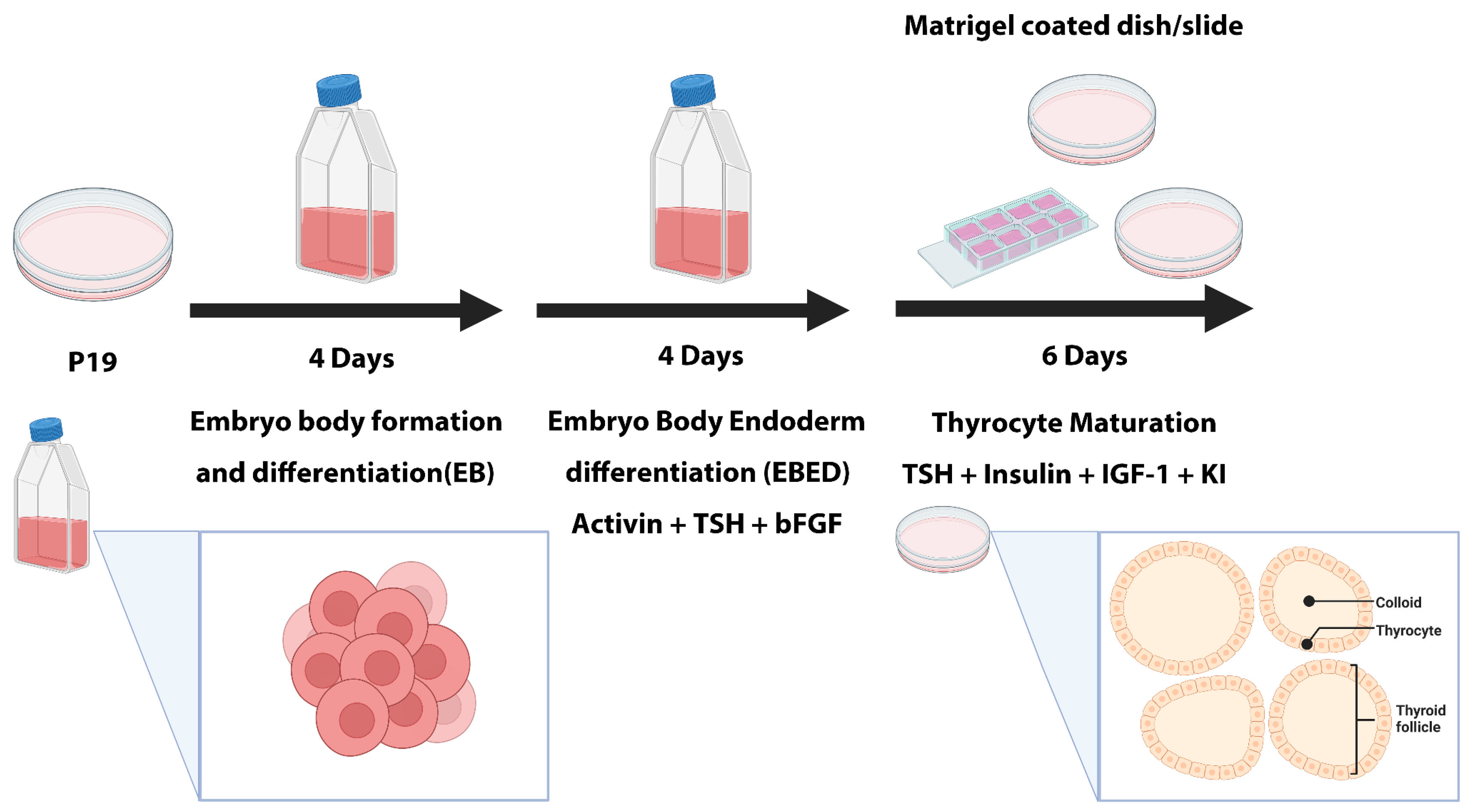
2.1. Cell Culture and Thyrocyte Differentiation
2.2. Generating Silencing RNA Control P19 Stable Cell Line
2.3. Primary Follicular Thyrocytes Culture
2.4. Quantitative Real-Time PCR
2.5. Immunostaining
2.6. ELISA
2.7. Statistical Analysis
3. Results
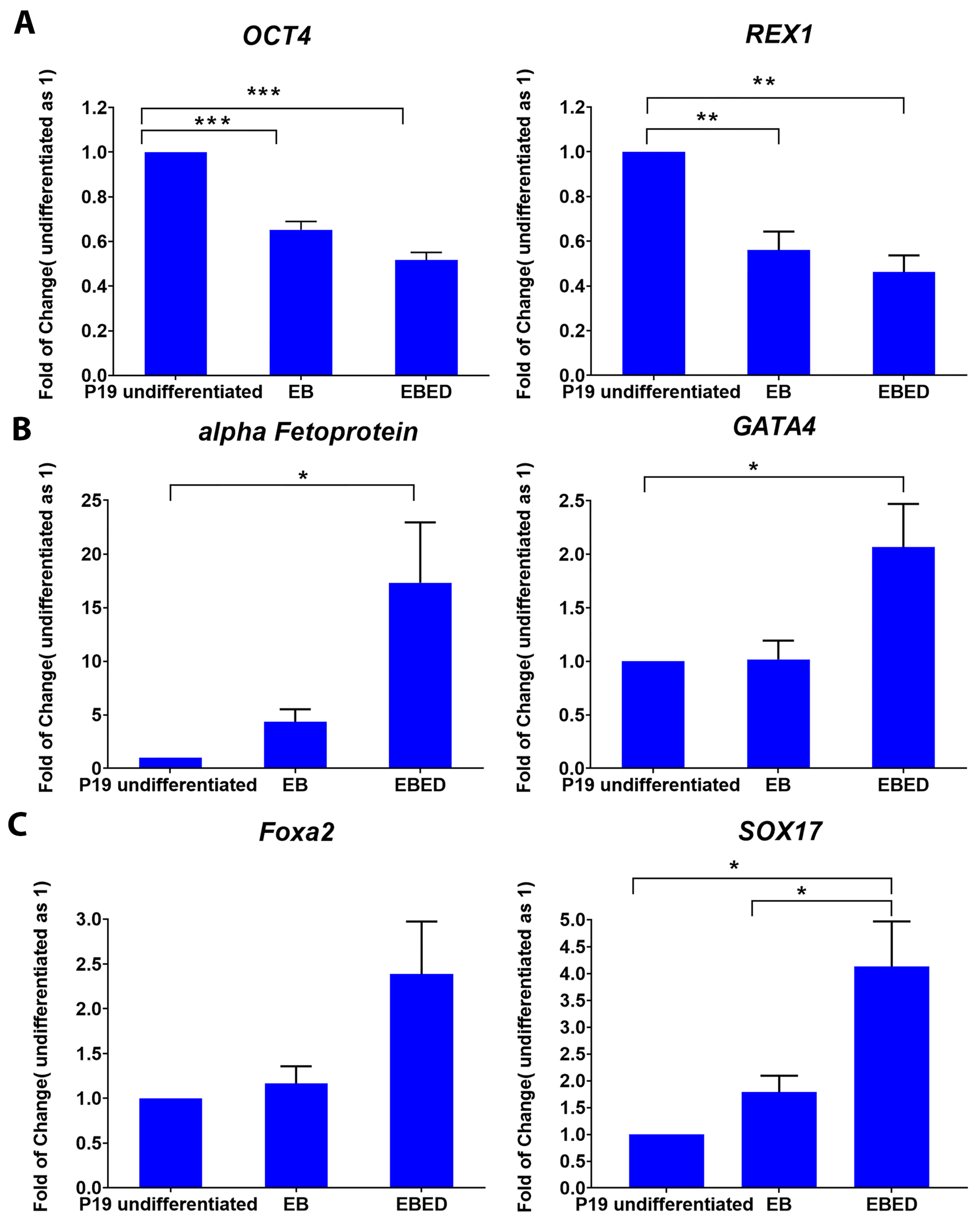
3.1. Generation and Characterization of P19 Differentiated Thyrocyte
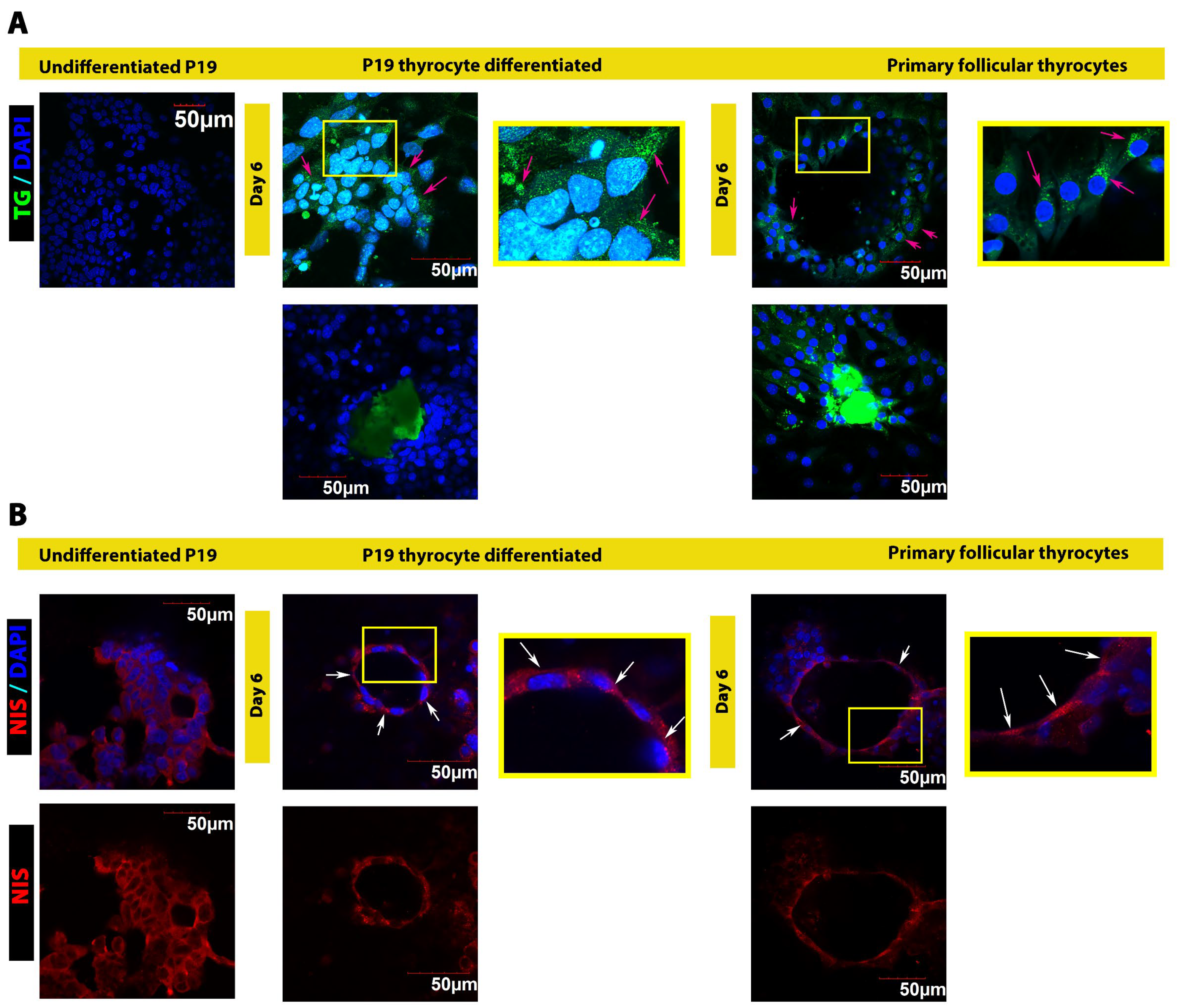
3.2. P19 Differentiated Thyrocytes Form Follicle-like Structures Resembling Primary Follicles
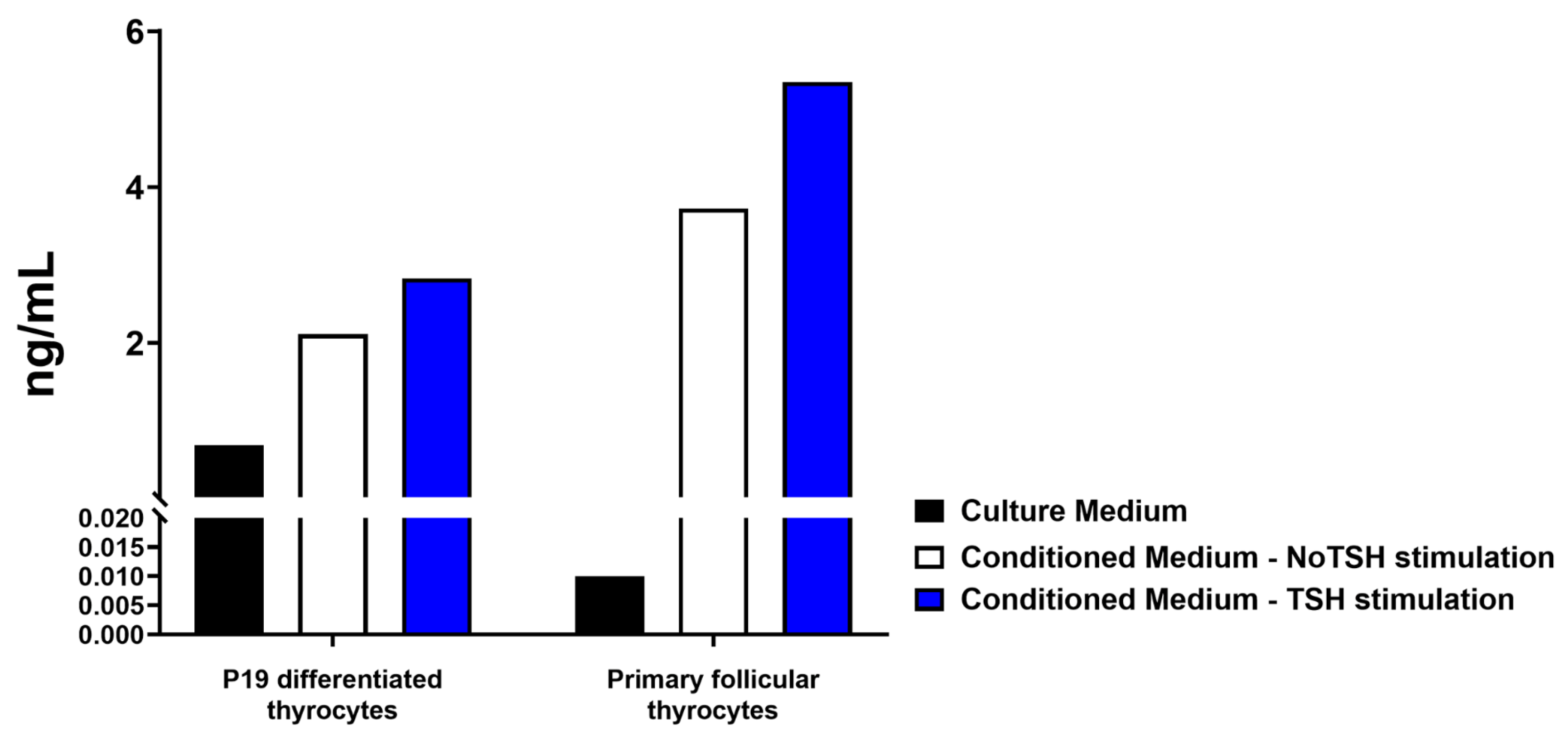
3.3. P19 Differentiated Thyrocytes Respond to TSH Stimulation
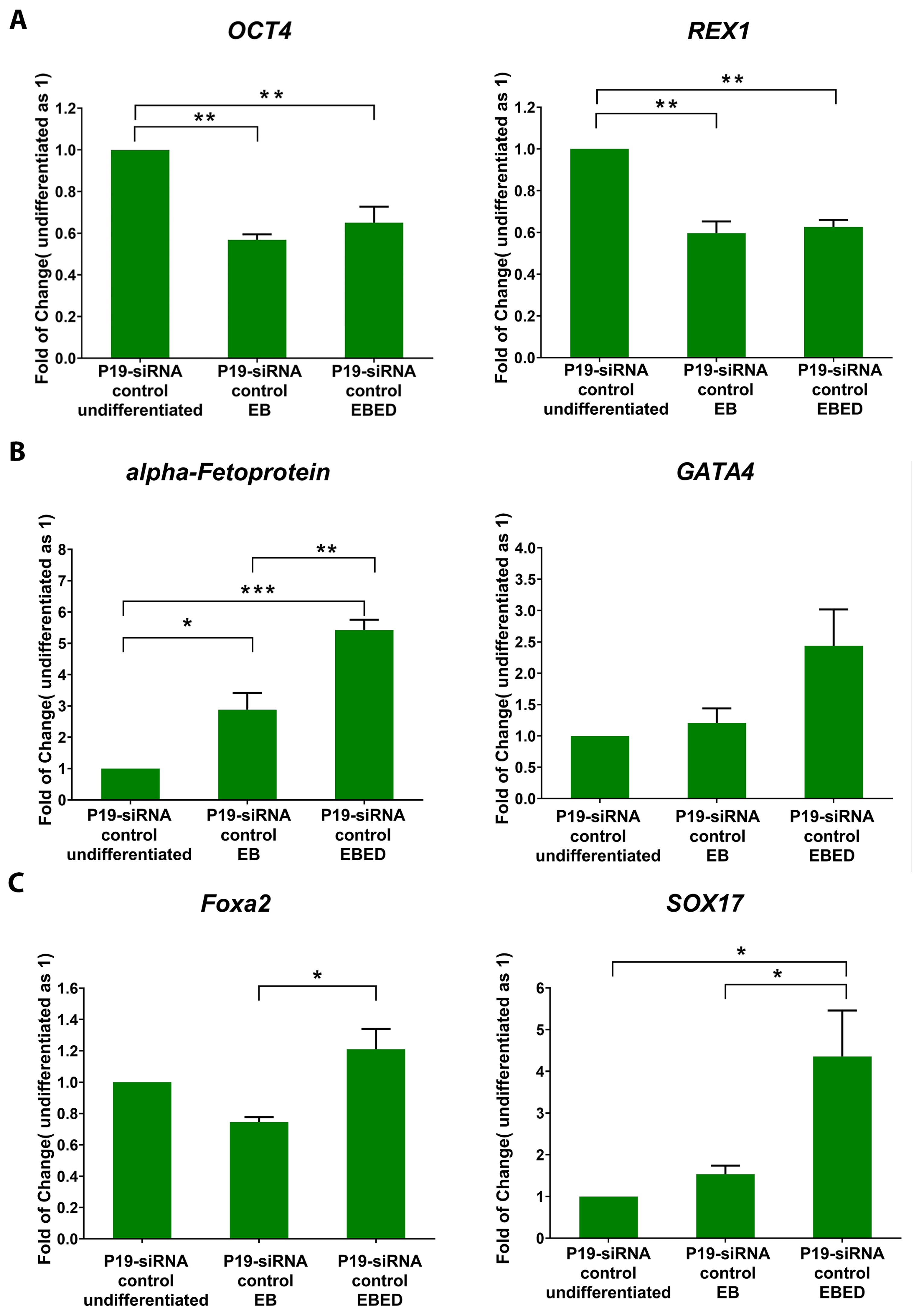
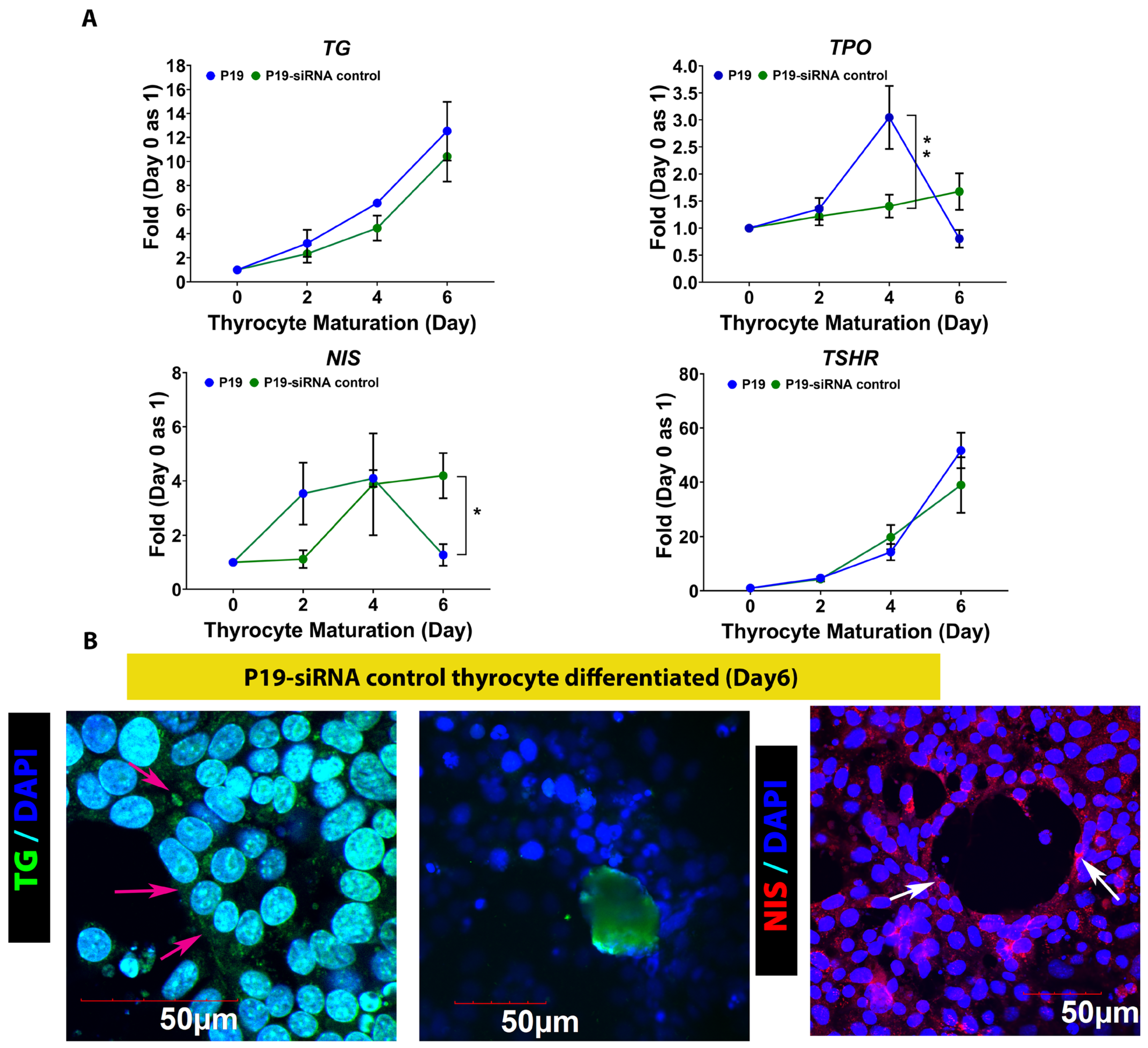
3.4. P19-siRNA Control Can Differentiate into Mature Thyrocytes
4. Discussion
5. Conclusions
Supplementary Materials
Author Contributions
Funding
Institutional Review Board Statement
Informed Consent Statement
Data Availability Statement
Conflicts of Interest
References
- Toda, S.; Koike, N.; Sugihara, H. Thyrocyte integration, and thyroid folliculogenesis and tissue regeneration: Perspective for thyroid tissue engineering. Pathol. Int. 2001, 51, 403–417. [Google Scholar] [CrossRef] [PubMed]
- Toda, S.; Aoki, S.; Uchihashi, K.; Matsunobu, A.; Yamamoto, M.; Ootani, A.; Yamasaki, F.; Koike, E.; Sugihara, H. Culture models for studying thyroid biology and disorders. ISRN Endocrinol. 2011, 2011, 275782. [Google Scholar] [CrossRef] [PubMed]
- Tomás, G.; Tarabichi, M.; Gacquer, D.; Hébrant, A.; Dom, G.; Dumont, J.E.; Keutgen, X.; Fahey, T.J.; Maenhaut, C.; Detours, V. A general method to derive robust organ-specific gene expression-based differentiation indices: Application to thyroid cancer diagnostic. Oncogene 2012, 31, 4490–4498. [Google Scholar] [CrossRef] [PubMed]
- Najjar, F.; Nhieu, J.; Wei, C.-W.; Milbauer, L.; Burmeister, L.; Seelig, D.; Wei, L.-N. Deleting Cellular Retinoic-Acid-Binding Protein-1 (Crabp1) Gene Causes Adult-Onset Primary Hypothyroidism in Mice. Endocrines 2023, 4, 138–150. [Google Scholar] [CrossRef]
- Nilsson, M.; Fagman, H. Development of the thyroid gland. Development 2017, 144, 2123–2140. [Google Scholar] [CrossRef]
- Carvalho, D.P.; Dupuy, C. Thyroid hormone biosynthesis and release. Mol. Cell. Endocrinol. 2017, 458, 6–15. [Google Scholar] [CrossRef]
- Mullur, R.; Liu, Y.-Y.; Brent, G.A. Thyroid hormone regulation of metabolism. Physiol. Rev. 2014, 94, 355–382. [Google Scholar] [CrossRef]
- Fagman, H.; Nilsson, M. Morphogenesis of the thyroid gland. Mol. Cell. Endocrinol. 2010, 323, 35–54. [Google Scholar] [CrossRef]
- Lin, R.-Y.; Kubo, A.; Keller, G.M.; Davies, T.F. Committing Embryonic Stem Cells to Differentiate into Thyrocyte-Like Cells in Vitro. Endocrinology 2003, 144, 2644–2649. [Google Scholar] [CrossRef]
- Bizhanova, A.; Kopp, P. Minireview: The sodium-iodide symporter NIS and pendrin in iodide homeostasis of the thyroid. Endocrinology 2009, 150, 1084–1090. [Google Scholar] [CrossRef]
- Pirahanchi, Y.; Toro, F.; Jialal, I. Physiology, Thyroid Stimulating Hormone; StatPearls Publishing: Treasure Island, FL, USA, 2022. Available online: https://www.ncbi.nlm.nih.gov/books/NBK499850/ (accessed on 12 February 2023).
- Di Jeso, B.; Arvan, P. Thyroglobulin From Molecular and Cellular Biology to Clinical Endocrinology. Endocr. Rev. 2016, 37, 2–36. [Google Scholar] [CrossRef] [PubMed]
- Gaitonde, D.Y.; Rowley, K.D.; Sweeney, L.B. Hypothyroidism: An update. Am. Fam. Phys. 2012, 86, 244–251. [Google Scholar] [CrossRef]
- Chiovato, L.; Magri, F.; Carlé, A. Hypothyroidism in Context: Where We’ve Been and Where We’re Going. Adv. Ther. 2019, 36, 47–58. [Google Scholar] [CrossRef] [PubMed]
- Morshed, S.A.; Davies, T.F. Understanding Thyroid Cell Stress. J. Clin. Endocrinol. Metab. 2020, 105, e66–e69. [Google Scholar] [CrossRef]
- Lorenz, S.; Eszlinger, M.; Paschke, R.; Aust, G.; Weick, M.; Führer, D.; Krohn, K. Calcium signaling of thyrocytes is modulated by TSH through calcium binding protein expression. Biochim. Biophys. Acta 2010, 1803, 352–360. [Google Scholar] [CrossRef]
- Feng, F.; Wang, H.; Hou, S.; Fu, H. Re-induction of cell differentiation and (131)I uptake in dedifferentiated FTC-133 cell line by TSHR gene transfection. Nucl. Med. Biol. 2012, 39, 1261–1265. [Google Scholar] [CrossRef]
- Mauchamp, J.; Mirrione, A.; Alquier, C.; André, F. Follicle-like structure and polarized monolayer: Role of the extracellular matrix on thyroid cell organization in primary culture. Biol. Cell 1998, 90, 369–380. [Google Scholar] [CrossRef]
- Khoruzhenko, A.; Miot, F.; Massart, C.; Van Sande, J.; Dumont, J.E.; Beauwens, R.; Boom, A. Functional model of rat thyroid follicles cultured in Matrigel. Endocr. Connect. 2021, 10, 570–578. [Google Scholar] [CrossRef]
- Denef, J.F.; Björkman, U.; Ekholm, R. Structural and functional characteristics of isolated thyroid follicles. J. Ultrastruct. Res. 1980, 71, 185–202. [Google Scholar] [CrossRef]
- Jeker, L.T.; Hejazi, M.; Burek, C.L.; Rose, N.R.; Caturegli, P. Mouse thyroid primary culture. Biochem. Biophys. Res. Commun. 1999, 257, 511–515. [Google Scholar] [CrossRef]
- Li, Y.-S.; Kanamoto, N.; Hataya, Y.; Moriyama, K.; Hiratani, H.; Nakao, K.; Akamizu, T. Transgenic mice producing major histocompatibility complex class II molecules on thyroid cells do not develop apparent autoimmune thyroid diseases. Endocrinology 2004, 145, 2524–2530. [Google Scholar] [CrossRef] [PubMed][Green Version]
- Arufe, M.C.; Lu, M.; Lin, R.-Y. Differentiation of murine embryonic stem cells to thyrocytes requires insulin and insulin-like growth factor-1. Biochem. Biophys. Res. Commun. 2009, 381, 264–270. [Google Scholar] [CrossRef] [PubMed]
- Ma, R.; Latif, R.; Davies, T.F. Human embryonic stem cells form functional thyroid follicles. Thyroid 2015, 25, 455–461. [Google Scholar] [CrossRef] [PubMed]
- Antonica, F.; Kasprzyk, D.F.; Opitz, R.; Iacovino, M.; Liao, X.-H.; Dumitrescu, A.M.; Refetoff, S.; Peremans, K.; Manto, M.; Kyba, M.; et al. Generation of functional thyroid from embryonic stem cells. Nature 2012, 491, 66–71. [Google Scholar] [CrossRef]
- Li, L.; Sheng, Q.; Zeng, H.; Li, W.; Wang, Q.; Ma, G.; Qiu, M.; Zhang, W.; Shan, C. Engineering a functional thyroid as a potential therapeutic substitute for hypothyroidism treatment: A systematic review. Front. Endocrinol. 2022, 13, 1065410. [Google Scholar] [CrossRef]
- McBurney, M.W. P19 embryonal carcinoma cells. Int. J. Dev. Biol. 1993, 37, 135–140. Available online: https://www.ncbi.nlm.nih.gov/pubmed/8507558 (accessed on 2 May 2024).
- Bain, G.; Ray, W.J.; Yao, M.; Gottlieb, D.I. From embryonal carcinoma cells to neurons: The P19 pathway. Bioessays 1994, 16, 343–348. [Google Scholar] [CrossRef]
- Nhieu, J.; Milbauer, L.; Lerdall, T.; Najjar, F.; Wei, C.-W.; Ishida, R.; Ma, Y.; Kagechika, H.; Wei, L.-N. Targeting Cellular Retinoic Acid Binding Protein 1 with Retinoic Acid-like Compounds to Mitigate Motor Neuron Degeneration. Int. J. Mol. Sci. 2023, 24, 4980. [Google Scholar] [CrossRef]
- Jasmin Spray, D.C.; Campos de Carvalho, A.C.; Mendez-Otero, R. Chemical induction of cardiac differentiation in p19 embryonal carcinoma stem cells. Stem Cells Dev. 2010, 19, 403–412. [Google Scholar] [CrossRef]
- Jang, D.; Eliseeva, E.; Klubo-Gwiezdzinska, J.; Neumann, S.; Gershengorn, M.C. TSH stimulation of human thyroglobulin and thyroid peroxidase gene transcription is partially dependent on internalization. Cell Signal. 2022, 90, 110212. [Google Scholar] [CrossRef]
- Sellitti, D.F.; Suzuki, K. Intrinsic regulation of thyroid function by thyroglobulin. Thyroid 2014, 24, 625–638. [Google Scholar] [CrossRef] [PubMed]
- Dentice, M.; Luongo, C.; Elefante, A.; Ambrosio, R.; Salzano, S.; Zannini, M.; Nitsch, R.; Di Lauro, R.; Rossi, G.; Fenzi, G.; et al. Pendrin is a novel in vivo downstream target gene of the TTF-1/Nkx-2.1 homeodomain transcription factor in differentiated thyroid cells. Mol. Cell Biol. 2005, 25, 10171–10182. [Google Scholar] [CrossRef] [PubMed]
- Puppin, C.; D’Elia, A.V.; Pellizzari, L.; Russo, D.; Arturi, F.; Presta, I.; Filetti, S.; Bogue, C.W.; Denson, L.A.; Damante, G. Thyroid-specific transcription factors control Hex promoter activity. Nucleic Acids Res. 2003, 31, 1845–1852. [Google Scholar] [CrossRef] [PubMed][Green Version]
- Francis-Lang, H.; Price, M.; Polycarpou-Schwarz, M.; Di Lauro, R. Cell-type-specific expression of the rat thyroperoxidase promoter indicates common mechanisms for thyroid-specific gene expression. Mol. Cell. Biol. 1992, 12, 576–588. [Google Scholar] [CrossRef]
- Zannini, M.; Francis-Lang, H.; Plachov, D.; Di Lauro, R. Pax-8, a paired domain-containing protein, binds to a sequence overlapping the recognition site of a homeodomain and activates transcription from two thyroid-specific promoters. Mol. Cell. Biol. 1992, 12, 4230–4241. [Google Scholar] [CrossRef]
- Ohno, M.; Zannini, M.; Levy, O.; Carrasco, N.; di Lauro, R. The paired-domain transcription factor Pax8 binds to the upstream enhancer of the rat sodium/iodide symporter gene and participates in both thyroid-specific and cyclic-AMP-dependent transcription. Mol. Cell. Biol. 1999, 19, 2051–2060. [Google Scholar] [CrossRef]
- D’Andrea, B.; Iacone, R.; Di Palma, T.; Nitsch, R.; Baratta, M.G.; Nitsch, L.; Di Lauro, R.; Zannini, M. Functional inactivation of the transcription factor Pax8 through oligomerization chain reaction. Mol. Endocrinol. 2006, 20, 1810–1824. [Google Scholar] [CrossRef]
- Christophe-Hobertus, C.; Christophe, D. Human Thyroid Oxidases genes promoter activity in thyrocytes does not appear to be functionally dependent on Thyroid Transcription Factor-1 or Pax8. Mol. Cell. Endocrinol. 2007, 264, 157–163. [Google Scholar] [CrossRef]
- Ruiz-Llorente, S.; Carrillo Santa de Pau, E.; Sastre-Perona, A.; Montero-Conde, C.; Gómez-López, G.; Fagin, J.A.; Valencia, A.; Pisano, D.G.; Santisteban, P. Genome-wide analysis of Pax8 binding provides new insights into thyroid functions. BMC Genom. 2012, 13, 147. [Google Scholar] [CrossRef]
- Kurmann, A.A.; Serra, M.; Hawkins, F.; Rankin, S.A.; Mori, M.; Astapova, I.; Ullas, S.; Lin, S.; Bilodeau, M.; Rossant, J.; et al. Regeneration of Thyroid Function by Transplantation of Differentiated Pluripotent Stem Cells. Cell Stem Cell 2015, 17, 527–542. [Google Scholar] [CrossRef]

| Gene Name | Primer Sequence |
|---|---|
| OCT-4 | Forward: 5′-CAGCAGATCACTCACATCGCCA-3′; Reverse: 5′-GCCTCATACTCTTCTCGTTGGG-3′ |
| REX1 | Forward: 5′- GAGAAGAGGAGGATTGCTCACG-3′; Reverse: 5′-CAATGTGCGTGTCTTCAGTGGC-3′ |
| a-Fetoprotein | Forward: 5′-GCTCACATCCACGAGGAGTGTT-3′; Reverse: 5′-CAGAAGCCTAGTTGGATCATGGG-3′ |
| GATA-4 | Forward: 5′-GCCTCTATCACAAGATGAACGGC-3′; Reverse: 5′-TACAGGCTCACCCTCGGCATTA-3′ |
| Foxa2 | Forward: 5′-CGAGCACCATTACGCCTTCAAC-3′; Reverse: 5′-AGTGCATGACCTGTTCGTAGGC-3′ |
| SOX17 | Forward: 5′-GCCGATGAACGCCTTTATGGTG-3′; Reverse: 5′-TCTCTGCCAAGGTCAACGCCTT-3′ |
| TSHR | Forward: 5′-GCTGTCGTTGAGTTTCCTCCAC-3′; Reverse: 5′-CTGCTCTCATTACACATCAAAGAC-3′ |
| TPO | Forward: 5′-GAGAGGCTCTTCGTGCTGTCTA-3′; Reverse: 5′-AGGCGTGACAAGCCACAGAACT-3′ |
| TG | Forward: 5′-TTGTAGCCTGGAGAGTCAGCAC-3′; Reverse: 5′-CACTGCACATCTTTCCTGGTGG-3′ |
| NIS | Forward: 5′-CATGCCATTGCTCGTGTTGGAC-3′; Reverse: 5′-GCCATAGCGTTGATACTGGTGG-3′ |
| TTF1 | Forward: 5′-CAGGACACCATGCGGAACAGC-3′; Reverse: 5′-GCCATGTTCTTGCTCACGTCCC-3′ |
| Pax8 | Forward: 5′-TGCTCAGCCTGGCAATGACAAC-3′; Reverse: 5′-ACGAAGGTGCTTTCGAGGACCA-3′ |
| TTF2 | Forward: 5′-AACAGCATCCGCCACAACCTCA-3′; Reverse: 5′-AGGAAGCTGCCGCTTTCGAACA-3′ |
| Hhex | Forward: 5′-CGGTCAAGTGAGGTTCTCCAAC-3′; Reverse: 5′-CTCGGCGATTCTGAAACCAGGT-3′ |
Disclaimer/Publisher’s Note: The statements, opinions and data contained in all publications are solely those of the individual author(s) and contributor(s) and not of MDPI and/or the editor(s). MDPI and/or the editor(s) disclaim responsibility for any injury to people or property resulting from any ideas, methods, instructions or products referred to in the content. |
© 2024 by the authors. Licensee MDPI, Basel, Switzerland. This article is an open access article distributed under the terms and conditions of the Creative Commons Attribution (CC BY) license (https://creativecommons.org/licenses/by/4.0/).
Share and Cite
Najjar, F.; Milbauer, L.; Wei, C.-W.; Lerdall, T.; Wei, L.-N. Modelling Functional Thyroid Follicular Structures Using P19 Embryonal Carcinoma Cells. Cells 2024, 13, 1844. https://doi.org/10.3390/cells13221844
Najjar F, Milbauer L, Wei C-W, Lerdall T, Wei L-N. Modelling Functional Thyroid Follicular Structures Using P19 Embryonal Carcinoma Cells. Cells. 2024; 13(22):1844. https://doi.org/10.3390/cells13221844
Chicago/Turabian StyleNajjar, Fatimah, Liming Milbauer, Chin-Wen Wei, Thomas Lerdall, and Li-Na Wei. 2024. "Modelling Functional Thyroid Follicular Structures Using P19 Embryonal Carcinoma Cells" Cells 13, no. 22: 1844. https://doi.org/10.3390/cells13221844
APA StyleNajjar, F., Milbauer, L., Wei, C.-W., Lerdall, T., & Wei, L.-N. (2024). Modelling Functional Thyroid Follicular Structures Using P19 Embryonal Carcinoma Cells. Cells, 13(22), 1844. https://doi.org/10.3390/cells13221844


