Review

“All for One and One for All”: The Secreted Heat Shock Protein gp96-Ig Based Vaccines

by
Laura Padula
,
Eva Fisher
and
Natasa Strbo
*
Department of Microbiology and Immunology, Miller School of Medicine, University of Miami, Miami, FL 33136, USA
*
Author to whom correspondence should be addressed.
Cells 2024, 13(1), 72; https://doi.org/10.3390/cells13010072
Submission received: 21 November 2023 / Revised: 21 December 2023 / Accepted: 22 December 2023 / Published: 29 December 2023

Abstract

:
It has been 50 years since Peter Charles Doherty and Rolf M Zinkernagel proposed the principle of “simultaneous dual recognition”, according to which adaptive immune cells recognized “self” and “non-self” simultaneously to establish immunological efficacy. These two scientists shared the 1996 Nobel Prize in Physiology or Medicine for this discovery. Their basic immunological principle became the foundation for the development of numerous vaccine approaches against infectious diseases and tumors, including promising strategies grounded on the use of recombinant gp96-Ig developed by our lab over the last two decades. In this review, we will highlight three major principles of the gp96-Ig vaccine strategy: (1) presentation of pathogenic antigens to T cells (specificity); (2) activation of innate immune responses (adjuvanticity); (3) priming of T cells to home to the epithelial compartments (mucosal immunity). In summary, we provide a paradigm for a vaccine approach that can be rapidly engineered and customized for any future pathogens that require induction of effective tissue-resident memory responses in epithelial tissues.

1. Introduction

Antigen presentation is an essential immune process that is responsible for T cell immune responses. Peter Charles Doherty and Rolf M Zinkernagel were the first to present the evidence that “the interaction of cytotoxic T cells with other somatic cells budding lymphocytic choriomeningitis (LCM) virus is restricted by H-2 gene complex” [1]. This discovery “of the specificity of the cell-mediated immune defense” had an immediate and widespread impact on immunological research. The Nobel Prize in Physiology or Medicine was awarded to Peter Charles Doherty and Rolf M Zinkernagel 20 years after their original discovery, recognizing their findings as one of the pillars of basic immunological principles. Major histocompatibility complex (MHC)-restricted T-cell recognition has initiated a considerable number of subsequent studies resulting in molecular understanding of T-cell recognition of virally infected target cells. Moreover, as addressed in the Zinkernagel Nature Lecture from December 1996, “recognition that peptides derived from viruses, bacteria or parasites are presented to T cells via MHC class I or class II molecules” suggested that instead of live, and therefore potentially harmful infectious agents, peptides could be used as vaccines to induce T-cell responses” [2]. In 1986, the first recombinant vaccine against hepatitis B virus (HBV) was approved by the FDA, which marked the beginning of the era of recombinant protein-based and peptide-based vaccines. However, subunit vaccines that consist primarily of peptides or proteins, in contrast to inactivated or attenuated pathogen or vector vaccines, can face significant limitations concerning immunogenicity, thus, they require carriers and adjuvants to counterbalance the low vaccine efficiency.
Here, we present a unique vaccine strategy based on the heat shock protein gp96, that provides antigen specificity and adjuvanticity within one molecule. Dr. Podack’s inspiring work on the development of an anti-tumor gp96 vaccine twenty years ago was successfully translated into numerous pre-clinical and clinical studies, including Phase 1b/2 clinical trial (NCT02439450) for patients with non-small cell lung cancer (NSCLC). Over the last decade, our lab has been extensively involved in the development of vaccines against human immunodeficiency virus/simian immunodeficiency virus (HIV/SIV), malaria, Zika virus (ZIKV), cytomegalovirus (CMV) and COVID-19 [3,4,5,6,7,8,9].
We propose that the recombinant protein gp96-Ig vaccine represents an “All for one and One for all” vaccine approach because different pathogens could be targeted by the gp96 vaccine, and gp96 could be engineered as a universal vaccine targeting numerous conserved antigens derived from pathogens or tumors, at the same time.

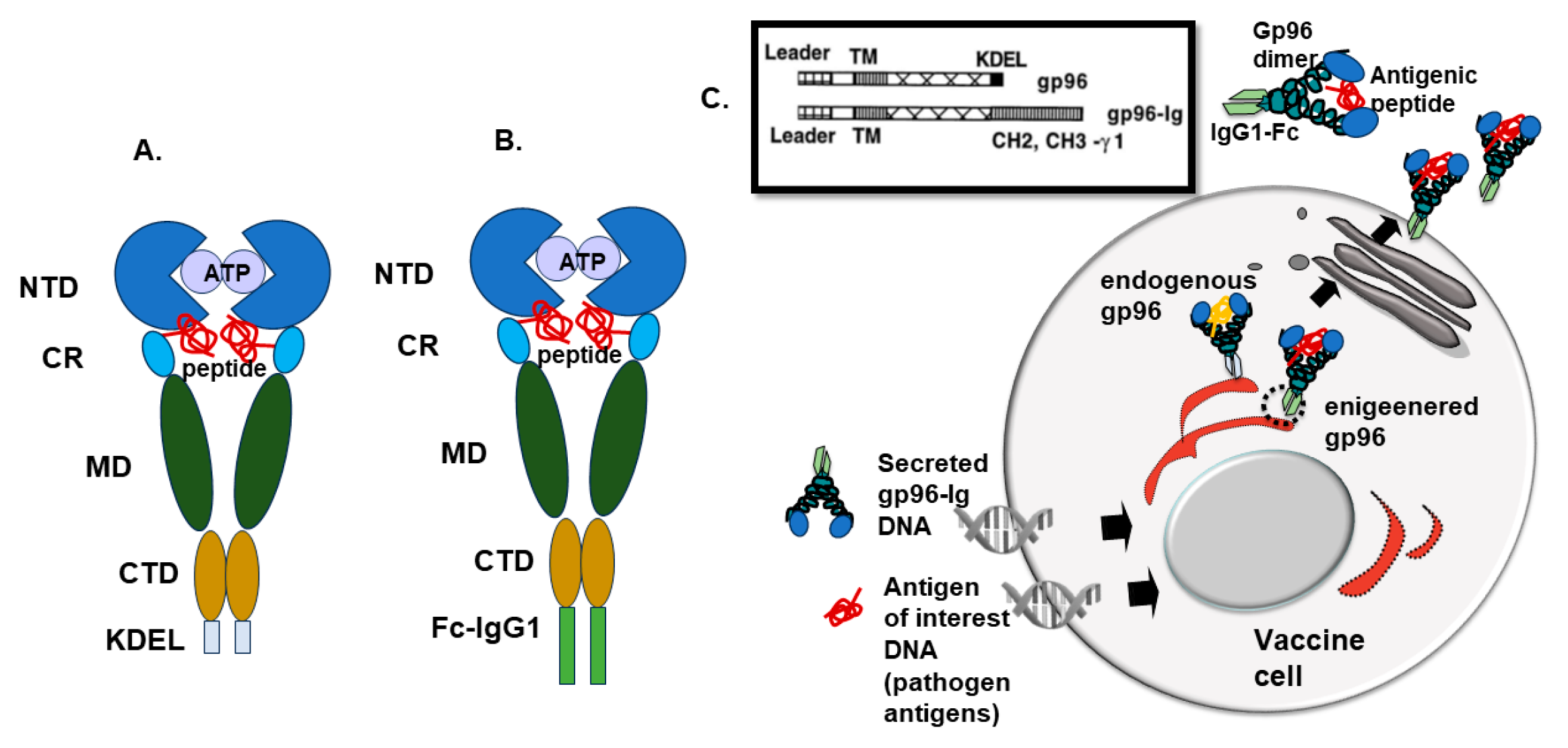
2. Gp96 as Molecular Chaperon

It was over 60 years ago that one of the students in Dr. Ritossa’s lab raised the temperature in the incubator in which he had some Drosophila salivary gland cells [10]. The next day, Dr. Ritossa discovered that heating induced puffs at the various regions of the polytene chromosomes. These puffs were areas of localized transcription that correlated with an increase in several families of heat shock proteins (HSPs). This extraordinary combination of serendipity, curiosity, and knowledge led to the identification of HSPs, a group of intracellular proteins that are induced when a cell undergoes several types of environmental stresses (heat, anoxia, glucose starvation) [10,11,12]. HSPs are ubiquitous, occurring in all organisms, from bacteria and yeast to humans, in various intracellular compartments [13]. Proteins within the HSPs family are categorized into families based on their molecular weights ranging from 10-150kDa (HSP27, HSP70, HSP90, HSP96) [13,14].
Gp96 (Grp94 or ERp99) is a member of the beta subfamily of HSPs 90 protein chaperon, a paralog of HSP90, found in the endoplasmic reticulum (ER) [15,16,17]. Under stress conditions (glucose starvation, oxidative stress, ER calcium depletion, and accumulation of misfolded proteins), gp96 is strongly upregulated and accelerates its function as a molecular chaperone [17,18,19]. Gp96 plays a critical role in folding proteins in the secretory pathway, such as Toll-like receptors [20], glycoprotein IX subunit [21], insulin-like growth factors (IGFs) [22,23], proinsulin [24], and integrins [25]. These interactions are associated with macrophage host defense, activation of platelets in blood clotting, cell growth, and cell adhesion [26]. Gp96 is also one of the major calcium-binding proteins in the ER regulating calcium homeostasis [26,27]. Gp96 protein consists of signal peptide (SP), N-terminal domain (NTD), C-terminal domain (CTD), middle domain (MD), and KDEL retention signal (Figure 1) [26,28,29]. NTD has ATPase activity and contains a nucleotide binding site and peptide binding site [30], while MD contains a catalytic loop for ATP hydrolysis [31]. In the CTD, there is a dimerization domain and client protein binding site [32,33]. Gp96 is a dimer that can change its state from closed to open depending on the ATP binding and ATP hydrolysis [26,28].
The peptide-binding channel in Gp96 supports to the proposal that Gp96 interacts with the peptide repertoire of the ER as part of the MHC peptide presentation machinery [16,34] and, therefore, has been implicated as an essential immune chaperone to regulate both innate and adaptive immunity.

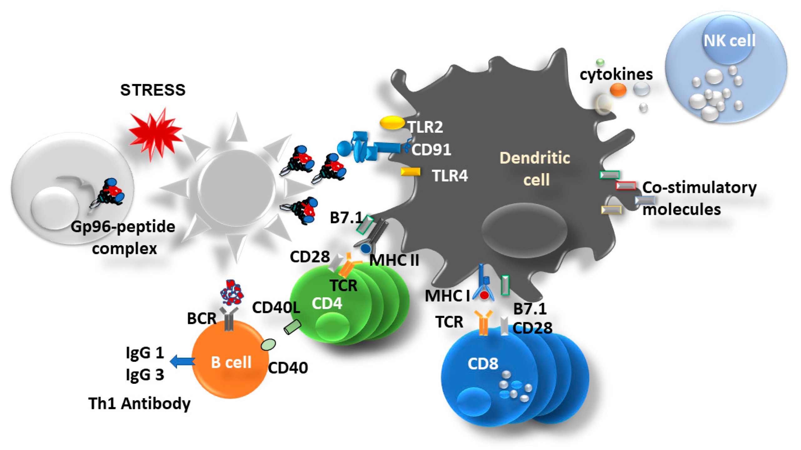
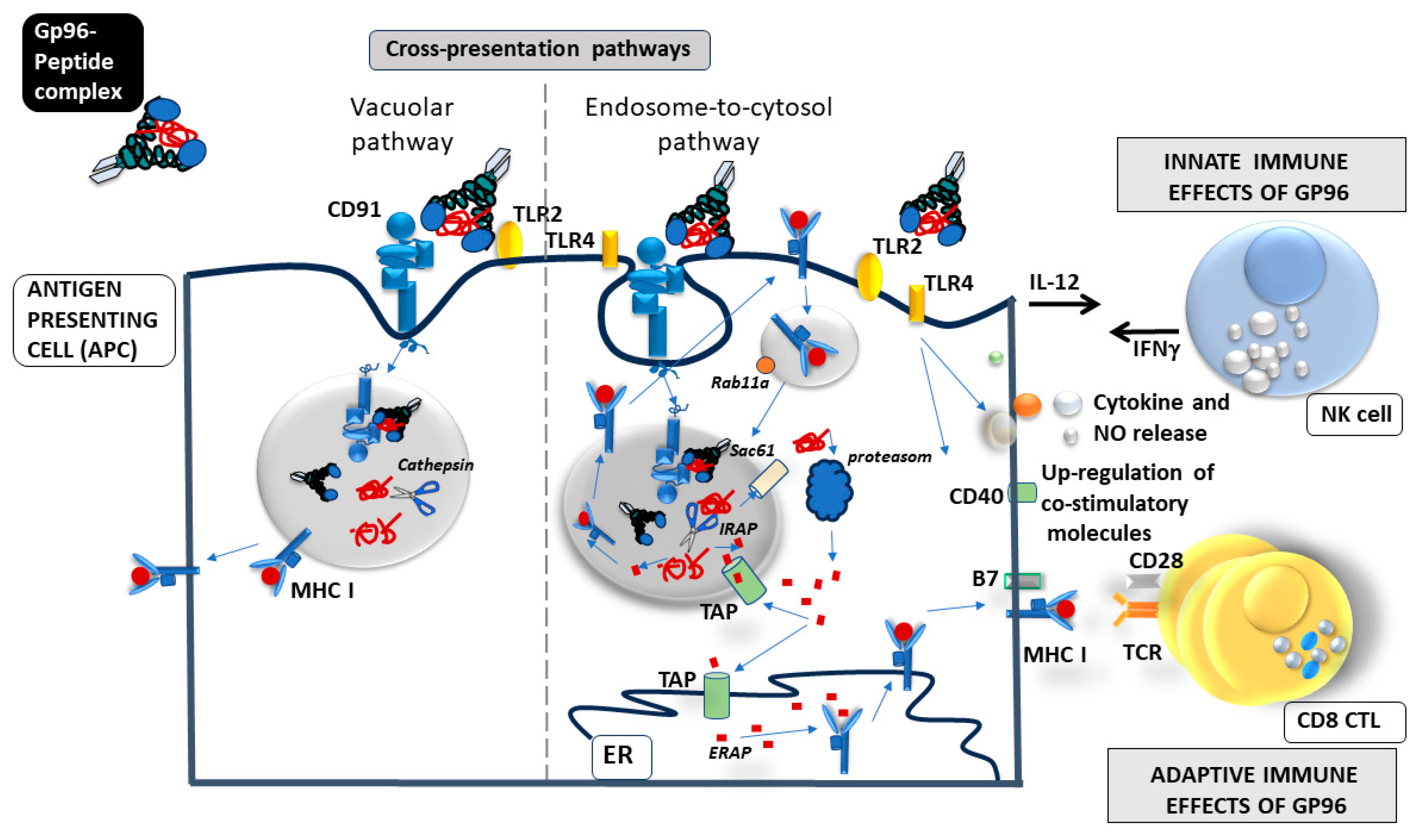
3. Gp96 as an Immune Modulator

Once cells become infected by a pathogen or upon apoptotic stimuli or any other stressor, cells will eventually die, and the intracellular content, including HSPs, will become a part of the extracellular danger-associated molecular patterns (DAMPs) [35,36]. Extracellular gp96 will activate antigen-presenting cells (APCs) and will further induce both arms of immune responses, innate and adaptive immune responses (Figure 2).
Gp96 and other HSPs have been described as ideal cross-priming vehicles because of their property to bind intracellular and extracellular peptides and shuttle them between different antigen processing compartments inside the cell and between cells [38]. Pioneering work by Dr. Srivastava demonstrated that several HSPs (HSP7, gp96, and HSP90) can induce immunity against autologous tumor preparations from which these HSPs had been isolated [39,40]. Furthermore, it was well established that gp96 binds to several different receptors on APCs (Toll-like receptor (TLR) 2, TLR4, SRA, LOX1) [41,42,43] including the endocytic receptor, CD91 responsible for the internalization of gp96-peptide complex and simultaneous activation/maturation of APC (up-regulation of co-stimulatory molecules, pro-inflammatory cytokines, and NO release) and further activation of NK cells [44,45,46,47,48,49] (Figure 2). Endocytosed gp96 chaperoned peptides are presented within the MHC class I molecules and recognized by CD8 T cells. Specific activation of CD8 T cells is independent of the haplotype of the cells from which the HSP-peptide complexes originate [48,50,51]. The process of presentation of exogenous peptides within MHC class I molecules is known as cross-presentation, and gp96 was recognized as a very potent activator of the cross-presentation pathway in dendritic cells [48] (Figure 3). Cross-presentation pathways have two different pathways of delivering exogenous antigens to MHC class I molecules: vacuolar and endosome-to-cytosol pathways and gp96 engages in both [52] (Figure 3). In the endosome, gp96-peptide complex gets dissociated, and peptides escape to cytosol where they reach TAP which transports peptides to nascent MHC class I molecule in ER, and finally the MHC I-peptide complex is shuttled towards the cell surface [44,52,53] (Figure 3). However, some of the gp96-peptide complexes can also be processed in the recycling endosomes that carry MHC I molecule, this seems to be particularly important pathway for non-classical MHC I molecules, such an HLA-E [52,54,55,56] (Figure 3).
Immunogenic gp96 binds a variety of endogenous/exogenous peptides and engages conventional (cDC) and plasmacytoid (pDC) dendritic cells, monocytes/macrophages, and all other cells expressing CD91 [52,58]. In addition to the immunostimulatory properties of HSPs, it has been confirmed that HSPs also support an inhibitory role in the activation of autoimmunity, allograft rejection, and tumor immunity [59,60,61,62,63]. It has been reported that gp96 drives dichotomous T-cell response [58] depending on the dose: a low dose of gp96 will primarily engage with cDC and will result in the Th1 responses and expansion of CTLs, while a high dose of gp96 will activate pDC and prime CD4+T regulatory cells [60,61,62,63,64,65,66].

4. Recombinant Fusion Protein gp96-Ig

Eckhard R. Podack (1943–2015) was fascinated by the immune roles of gp96, and in the late 1990s, he developed a gp96-Ig-secreted vaccine approach [67]. To be immunogenic, gp96 must be outside of the cells, therefore Eckhard engineered a secreted form of gp96 simply by replacing the KDEL retention signal of gp96 with the Fc portion of the IgG1 molecule (Figure 1) [67]. In this way, gp96-Ig gets into the secretory pathway and is released from the cell in a complex with endogenous/exogenous peptides (Figure 1C).
At first, this engineered form of gp96-Ig was expressed in numerous tumor cell lines and tumor-secreted gp96-induced tumor-specific CD8 T cells that were required for tumor rejection [67]. Molecular and cellular gp96-Ig requirements for enhanced cross-antigen presentation to CD8 T cells have been described [47]. Secreted heat shock protein, gp96-Ig-chaperoned peptides enhance the efficiency of Ag cross-priming of CD8 cytotoxic T lymphocytes (CTL) by several million-fold over the cross-priming activity of unchaperoned protein alone [47]. Gp96 also acts as an adjuvant for cross-priming by unchaperoned proteins, but in this capacity, gp96 is 1000-fold less active than as a peptide chaperone [47]. Gp96-mediated cross-priming of CD8 T cells requires B7.1/2 co-stimulation but proceeds unimpeded in lymph node-deficient mice in the absence of NKT and CD4 cells and without CD40L [47]. Gp96-driven MHC I cross-priming of CD8 CTL in the absence of lymph nodes provides a novel mechanism for local, tissue-based CTL generation at the site of gp96 release. Podack and Strbo proposed that the gp96-Ig vaccine induced CD8 T cell responses and may constitute a critically important, early detection, and rapid response mechanism that is operative in parenchymal tissues for effective defense against tissue-damaging antigenic agents [37,47].
Srivastava and colleagues were the first to recognize the potential role of HSPs in the immune response to cancer [38,43]. They demonstrated that HSPs complexed with antigenic peptides obtained from lysed tumor cells (or virus-infected cells) provide an optimal source of tumor antigens, generating DC with improved cross-presentation capacity. Tumor-derived gp96 is taken up by APCs and subsequently cross-presented to stimulate potent CD8-mediated anti-tumor immunity [21,37,38,43,68] (Figure 3). To take advantage of this unique adjuvant effect and the ability to transport relevant peptides, we set up a model system that imitates necrotic cell death regarding the release of HSP (cell-secreted gp96-Ig). This system allowed us to analyze the immunological effects of HSP in vivo independent of tumor/infectious agents and cell death. We found that parenteral administration of gp96-Ig-secreting tumors triggers robust, antigen-specific CD8 cytotoxic T lymphocyte expansion combined with activation of the innate immune system [47,67,69]. Tumor-secreted gp96 causes the recruitment of dendritic cells (DCs) and natural killer (NK) cells to the site of gp96 secretion and mediates DC activation via binding to CD91 and Toll-like receptor-2 and Toll-like receptor-4 [47,49,70] (Figure 3). The endocytic uptake of gp96 and its chaperoned peptides triggers peptide cross-presentation via major histocompatibility complex (MHC) class I and strong, cognate CD8 activation independent of CD4 cells [37,47] (Figure 3). In this model system, CD8 CTL expansion can be precisely quantitated within 4 to 5 days of vaccination by the use of adoptively transferred, T-cell receptor (TCR) transgenic, green fluorescent protein (GFP)-marked CD8 T cells [47,49,70]. Using this test system, we showed that in our model system, tumor-induced immune suppression is antigen nonspecific and can be overcome by frequent immunization or by the absence of B cells [69].
In addition to the anti-tumor cell-based secreted gp96-Ig vaccine approach, which was successfully used in Phase 1b/2 clinical trial against non-small cell lung cancer (NSCLC) (NCT02439450 [71]), we have also generated an anti-infectious cell-based secreted gp96-Ig vaccine approach [37] (Figure 1). The major difference between these two platforms is that in the anti-tumor approach, we use tumor cells as a source of antigens by transfecting gp96-Ig construct in individual tumor cell lines, while in the anti-infectious approach, we have chosen the ATCC human embryonal kidney (HEK)-293 cell line, as a vehicle for gp96-Ig delivery in combination with different antigenic peptides that are co-transfected in the cell where the final product is gp96-Ig complexed with array of different antigenic peptides [37] (Figure 1C).
The reason why the secreted gp96-Ig vaccine approach is unique and has an advantage compared to other recombinant protein vaccine approaches is that gp96-Ig-induced antigen-specific CD8 T cells can migrate to mucosal surfaces and provide immediate and enhanced protection at the most likely entry site of invading pathogens [70] (Figure 4). We found that antigen-specific CD8 T cells are generated not only systemically (in blood and spleen) but also in the epithelia/mucosal compartments such as the gut, reproductive tract, and the lungs [3,4,7,8,9,37,70]. This was the main reason why we decided to adapt this anti-tumor vaccine approach to emerging infectious diseases. Expanding the analysis to mucosal compartments, we found a dramatic increase in simian immunodeficiency virus/human immunodeficiency virus (SIV/HIV)- specific CD8 T cells in the rectal mucosa of SIV vaccinated macaques (up to 30% of all CD8 T cells in the rectal mucosa are SIV-specific) (Figure 4) [3,4]. Plasmodium falciparum CSP and AMA1-positive CD8 T cells were found in the liver of malaria-vaccinated non-human primates (NHPs), while zika virus (ZIKV)- and cytomegalovirus (CMV)-specific CD8 T cells were found in the placenta and decidua of vaccinated mice and humanized mice, and finally, melanoma- and SARS-CoV-2- protein S- specific CD8 T cells were found in the lungs and bronchioalveolar lavage of gp96-Ig-melanoma and gp96-Ig COVID-19 vaccinated mice (Table 1 and Figure 4) [5,6,7,8,9].

5. How Does gp96 Induce Mucosal Immune Response?
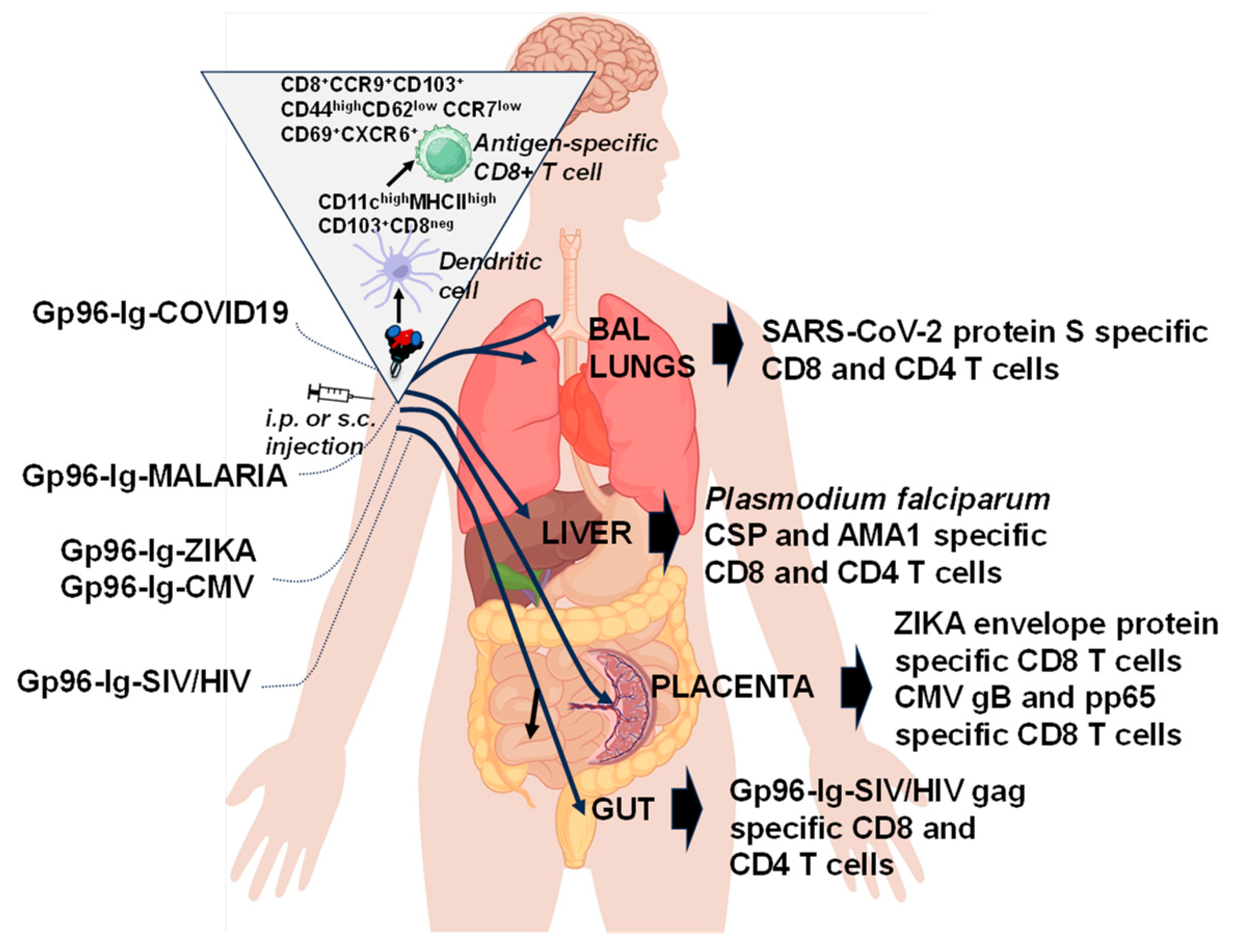
We observed that gp96-Ig immunization increases the frequency of the subset of DC, CD11chigh MHC class IIhigh CD103+ cells at the site of vaccination [70] (Figure 4). Phenotypic analysis of CD11chigh MHC class IIhigh CD103+ cells revealed that these cells are also CD8neg and express CD11b, CD40, CD80, CCR7, B220, and low levels of CD86 and Gr-1. CD103+ CD11b+ and CD103+ CD8neg DC populations are more prominent in mesenteric lymph nodes and colonic lamina propria. Recently, CD8neg CD11bhigh MHC class IIhigh CD11chigh CD103+ DC population was found within the omental tissue of normal healthy mice. These cells cross-present exogenous antigen for MHC class I-restricted T-cell responses. In light of this finding and the findings of Bedoui et al. [72] that CD103+ DCs are the main migratory subtype with dominant cross-presenting ability, induction of CD103+ DCs by gp96 represents an ideal vaccination strategy for priming effective immunity. In addition, we have also confirmed that CD103+ DCs are responsible for the activation of CCR9+ CD8 T cells. Immigrating cells were clearly identifiable by the expression of high levels of CD44, CD103, CCR9, and α4β7 and downregulation of CD62L and CCR7 [70] that allow these cells to cross endothelial cells and migrate to the gut tissue. The use of this immunological mechanism seems ideally suited for vaccine purposes, including stimulation of cellular mucosal immunity against SIV/HIV (Figure 4).
Tissue-resident memory (TRM) T cells have been recognized as a distinct population of memory cells that are capable of rapidly responding to infection in the tissue without requiring priming in the lymph nodes [73,74,75,76]. In our recent work [7,9] we confirmed that gp96-Ig, secreted from allogeneic cells expressing full-length SARS-CoV-2 protein S and Plasmodium falciparum CSP and AMA1 proteins generates powerful, protein S polyepitope-specific CD4+ and CD8+ T cell responses in both lung interstitium and airways as well as CSP- and AMA-1-specific CD8+ T cells in the liver. Importantly, antigen-specific cells were characterized as tissue memory-specific cells (CXCR6+, CD69+, and CXCR3+) (Figure 4). These findings were further strengthened by the observation that protein-S -specific CD8+ T cells were induced in human leukocyte antigen HLA-A2.1 transgenic mice and Plasmodium falciparum-specific CD8 T cells in non-human primates, thus providing encouraging translational data that the vaccine is likely to work in humans. Therefore, vaccination strategies targeting the generation of TRM and their persistence may provide enhanced immunity compared with vaccines that rely on circulating responses [77].
The placenta acts as a barrier against infections due to multiple unique structural, cellular, and immune properties. The detrimental effects of congenital viruses, including ZIKV and HCMV, on pregnancy and fetal outcomes occur in part because of impaired placental function. Targeting the congenital infection with vaccines offers the best opportunity to generate protective immunity in the most vulnerable population of child-bearing women before viruses have a chance to cross the placental barrier and before infection takes hold in the fetus. We have succeeded in generating a vaccine that expresses gp96-Ig and ZIKV envelope protein and HCMV gB and pp65 protein. We confirmed that vaccination with secreted gp96-Ig is safe and induces CD8 T cell responses in maternal decidua (Figure 4) [5,6].

6. “All for One”

In the last decade, we developed the following emerging disease gp96-Ig vaccines, combining secreted gp96-Ig protein with the following antigens (Table 1 and Figure 4):
We have tested the immunogenicity of these vaccines in different animal models: mice, NHPs, and humanized mice; however, vaccine efficacy was only tested in an SIV infection model [3]. A combination of cell-secreted gp96-SIV-Ig and gp120 protein as immunogens achieved a significant reduction (73%) in the risk for SIV acquisition in NHPs [3]. Protection from mucosal SIVmac251 infection was associated with strong mucosal CTL and non-neutralizing antibody responses. The antibody isotype suggested Th1 polarization. Our control vaccines had no protective effect even though they generated either SIV-specific CTL or SIV-specific antibodies, suggesting that both SIV-specific CTL and antibodies are required for protection. 73% immunization efficiency is an encouraging starting point for further development of the SIV immunization strategy.
To this day, the cell-based secreted gp96-Ig vaccine approach was tested and completed in a Phase 1b/2 study of Viagenpumatucel-L (HS-110) in combination with multiple treatment regimens in patients with non-small cell lung cancer (NSCLC) (The “DURGA” Trial)(NCT02439450) [71,78]. HS-110 was well tolerated when administered in combination with nivolumab, an anti-programmed cell death receptor-1 (PD-1) monoclonal antibody. Significantly longer progression-free survival (PFS) and median overall survival (OS) in both checkpoint inhibitors (CPI) naïve and CPI progressor cohorts were observed [78]. In addition, a trend of improved OS in baseline blood tumor mutational burden (bTMB) patients in the CPI progressor cohort was also reported. Further clinical evaluation of HS-110 is warranted in both CPI naïve and CPI progressor NSCLC patients [78].
We are currently testing the efficacy of all other vaccines in different pre-clinical emerging disease models.

7. “One for All”

The recent pandemic highlighted the need to be prepared for the next emerging disease, in particular, the idea of having a vaccine that will give people a baseline level of immune memory to diverse pathogen strains with pandemic potential (flu, COVID variants) so that there will be far less disease and death after encountering a new emerging pathogen. Unlike current mono- or polyvalent vaccines, which confer immunity to one or several antigenic variants of a pathogen, universal vaccines do this by targeting an element of the pathogen that remains the same across all variants. Given the gp96 propensity to carry all peptides of a cell that will be selected in the recipient/vaccinee for MHC I loading, including transfected antigens, gp96 has the broadest, theoretically possible antigenic epitope-spectrum for cross-priming of CD8 T cells by any MHC I type. More so, it has been recently shown that tumor-derived gp96 can induce protection against lymphocytic choriomeningitis virus (LCMV) infection and against tumors, thereby emphasizing that gp96 fits in the “one for all” vaccine category [79,80]. Given the antigenic similarity between cancer and embryonic tissues [81] and the capability of gp96 to bind the entire peptide repertoire generated within the cell [82], there is a rational basis for the use of placenta-derived gp96 as a multivalent prophylactic cancer vaccine. [83,84]. In addition, gp96-activated DC can take up antigenic proteins and, after processing, present their epitopes via MHC II, thereby promoting antibody production by B cells [3,37]. Gp96 is thus a powerful Th1 adjuvant for CTL priming and for stimulation of Th1-type antibodies [3,8,37]. Current vaccines are all made against unique pathogen antigens rather than antigens that are shared by different pathogens. Universal preventative vaccines, such as recombinant gp96 vaccines, could activate the immune system based on the predicted changes in infected cells or transformed tumor cells and efficiently recognize and control emerging pathogens and tumors.
However, cell-based delivery of recombinant protein gp96-Ig by an irradiated human cell line (HEK-293) represents a major hurdle for further clinical development. Cell-based technology is impractical for large manufacturing, and the gp96-Ig vaccine requires cold-chain storage and distribution. Other types of vaccine delivery (recombinant DNA and RNA) are currently under investigation.

8. Conclusions

Secreted recombinant gp96-Ig vaccines act by initiating the innate immune response and activating APCs, thereby inducing a protective adaptive immune response to either a tumor or pathogen antigens. Gp96 is efficient in antigen cross-presentation at physiologic concentrations of antigen (picogram range), so endogenous amounts of gp96-antigenic peptide complexes secreted by vaccine cells are sufficient to induce robust memory CTL responses in epithelial tissues without the addition of other adjuvants. Furthermore, this vaccine platform is proof of concept for the prototype vaccine approach for all emerging infectious pathogens that require induction of effective tissue-resident memory responses in epithelial tissues.

Author Contributions

Conceptualization, visualization, supervision N.S.; writing—original draft preparation, project administration L.P.; writing—review and editing, project administration E.F. All authors have read and agreed to the published version of the manuscript.

Funding

This research received no external funding.

Acknowledgments

We would like to acknowledge visionary and inspiring work of late Eckhard Podack in the development of the secreted gp96-Ig vaccine approach.

Conflicts of Interest

N.S. is the inventor of SIV/HIV, malaria, Zika, CMV, and COVID-19 gp96-Ig vaccines. Other co-authors declare no conflict of interest. The funders had no role in the writing of the manuscript.

References

  1. Zinkernagel, R.M.; Doherty, P.C. Restriction of in Vitro T Cell-Mediated Cytotoxicity in Lymphocytic Choriomeningitis within a Syngeneic or Semiallogeneic System. Nature 1974, 248, 701–702. [Google Scholar] [CrossRef]
  2. Deres, K.; Schild, H.; Wiesmuller, K.H.; Jung, G.; Rammensee, H.G. In vivo Priming of Virus-Specific Cytotoxic T Lymphocytes with Synthetic Lipopeptide Vaccine. Nature 1989, 342, 561–564. [Google Scholar] [CrossRef]
  3. Strbo, N.; Vaccari, M.; Pahwa, S.; Kolber, M.A.; Doster, M.N.; Fisher, E.; Gonzalez, L.; Stablein, D.; Franchini, G.; Podack, E.R. Cutting Edge: Novel Vaccination Modality Provides Significant Protection against Mucosal Infection by Highly Pathogenic Simian Immunodeficiency Virus. J. Immunol. 2013, 190, 2495–2499. [Google Scholar] [CrossRef]
  4. Strbo, N.; Vaccari, M.; Pahwa, S.; Kolber, M.A.; Fisher, E.; Gonzalez, L.; Doster, M.N.; Hryniewicz, A.; Felber, B.K.; Pavlakis, G.N.; et al. Gp96 Siv Ig Immunization Induces Potent Polyepitope Specific, Multifunctional Memory Responses in Rectal and Vaginal Mucosa. Vaccine 2011, 29, 2619–2625. [Google Scholar] [CrossRef]
  5. Strbo, N.; Romero, L.; Fisher, E.; Garcia, D. Induction of Zika Specific Cd8 T Cell Responses in the Placenta after Heat Shock Protein Gp96-Ig Vaccination. J. Immunol. 2018, 200, 180. [Google Scholar]
  6. Strbo, N.; Romero, L.; Garcia, D. Heat Shock Protein Gp96-Ig Vaccine Induces Antigen Specific Cd8 T Cell Response in the Placenta. J. Immunol. 2017, 198, 73. [Google Scholar]
  7. Fisher, E.; Padula, L.; Podack, K.; O’Neill, K.; Seavey, M.M.; Jayaraman, P.; Jasuja, R.; Strbo, N. Induction of SARS-CoV-2 Protein S-Specific CD8+ T Cells in the Lungs of Gp96-Ig-S Vaccinated Mice. Front. Immunol. 2020, 11, 602254. [Google Scholar] [CrossRef]
  8. Padula, L.; Fisher, E.; Rivas, K.; Podack, K.; Frasca, D.; Kupritz, J.; Seavey, M.M.; Jayaraman, P.; Dixon, E.; Jasuja, R.; et al. Secreted Heat Shock Protein Gp96-Ig and Ox40l-Fc Combination Vaccine Enhances SARS-CoV-2 Spike (S) Protein-Specific B and T Cell Immune Responses. Vaccine X 2022, 12, 100202. [Google Scholar] [CrossRef]
  9. Padula, L.; Fisher, E.; Wijayalath, W.; Patterson, N.B.; Huang, J.; Ganeshan, H.; Robinson, T.; Bates, F.A.; Hanson, M.A.; Martin, M.L.; et al. Induction of Antigen Specific Intrahepatic Cd8+ T Cell Responses by a Secreted Heat Shock Protein based Gp96-Ig-PfCA Malaria Vaccine. Front. Immunol. 2023, 14, 1130054. [Google Scholar] [CrossRef]
  10. Ritossa, F. Discovery of the Heat Shock Response. Cell Stress Chaperones 1996, 1, 97–98. [Google Scholar] [CrossRef]
  11. Ritossa, F.M. Experimental Activation of Specific Loci in Polytene Chromosomes of Drosophila. Exp. Cell Res. 1964, 35, 601–607. [Google Scholar] [CrossRef]
  12. Ritossa, F.M.; Vonborstel, R.C. Chromosome Puffs in Drosophila Induced by Ribonuclease. Science 1964, 145, 513–514. [Google Scholar] [CrossRef]
  13. Lindquist, S.; Craig, E.A. The Heat-Shock Proteins. Annu. Rev. Genet. 1988, 22, 631–677. [Google Scholar] [CrossRef]
  14. Kampinga, H.H.; Hageman, J.; Vos, M.J.; Kubota, H.; Tanguay, R.M.; Bruford, E.A.; Cheetham, M.E.; Chen, B.; Hightower, L.E. Guidelines for the Nomenclature of the Human Heat Shock Proteins. Cell Stress Chaperones 2009, 14, 105–111. [Google Scholar] [CrossRef]
  15. Picard, D. Heat-Shock Protein 90, a Chaperone for Folding and Regulation. Cell Mol. Life Sci. 2002, 59, 1640–1648. [Google Scholar] [CrossRef]
  16. Srivastava, P. Interaction of Heat Shock Proteins with Peptides and Antigen Presenting Cells: Chaperoning of the Innate and Adaptive Immune Responses. Annu. Rev. Immunol. 2002, 20, 395–425. [Google Scholar] [CrossRef]
  17. Joslin, G.; Hafeez, W.; Perlmutter, D.H. Expression of Stress Proteins in Human Mononuclear Phagocytes. J. Immunol. 1991, 147, 1614–1620. [Google Scholar]
  18. Lee, A.S. Mammalian Stress Response: Induction of the Glucose-Regulated Protein Family. Curr. Opin. Cell Biol. 1992, 4, 267–273. [Google Scholar] [CrossRef]
  19. Yang, Y.; Li, Z. Roles of Heat Shock Protein Gp96 in the Er Quality Control: Redundant or Unique Function? Mol. Cells 2005, 20, 173–182. [Google Scholar]
  20. Yang, Y.; Liu, B.; Dai, J.; Srivastava, P.K.; Zammit, D.J.; Lefrancois, L.; Li, Z. Heat Shock Protein Gp96 is a Master Chaperone for Toll-like Receptors and is Important in the Innate Function of Macrophages. Immunity 2007, 26, 215–226. [Google Scholar] [CrossRef]
  21. Staron, M.; Wu, S.; Hong, F.; Stojanovic, A.; Du, X.; Bona, R.; Liu, B.; Li, Z. Heat-Shock Protein Gp96/Grp94 is an Essential Chaperone for the Platelet Glycoprotein Ib-IX-V Complex. Blood 2011, 117, 7136–7144. [Google Scholar] [CrossRef]
  22. Barton, E.R.; Park, S.; James, J.K.; Makarewich, C.A.; Philippou, A.; Eletto, D.; Lei, H.; Brisson, B.; Ostrovsky, O.; Li, Z.; et al. Deletion of Muscle Grp94 Impairs Both Muscle and Body Growth by Inhibiting Local Igf Production. FASEB J. 2012, 26, 3691–3702. [Google Scholar] [CrossRef]
  23. Ostrovsky, O.; Ahmed, N.T.; Argon, Y. The Chaperone Activity of Grp94 Toward Insulin-like Growth Factor II is Necessary for the Stress Response to Serum Deprivation. Mol. Biol. Cell 2009, 20, 1855–1864. [Google Scholar] [CrossRef]
  24. Ghiasi, S.M.; Dahlby, T.; Hede Andersen, C.; Haataja, L.; Petersen, S.; Omar-Hmeadi, M.; Yang, M.; Pihl, C.; Bresson, S.E.; Khilji, M.S.; et al. Endoplasmic Reticulum Chaperone Glucose-Regulated Protein 94 is Essential for Proinsulin Handling. Diabetes 2019, 68, 747–760. [Google Scholar] [CrossRef]
  25. Wu, S.; Hong, F.; Gewirth, D.; Guo, B.; Liu, B.; Li, Z. The Molecular Chaperone Gp96/Grp94 Interacts with Toll-like Receptors and Integrins via Its C-Terminal Hydrophobic Domain. J. Biol. Chem. 2012, 287, 6735–6742. [Google Scholar] [CrossRef]
  26. Kim, J.W.; Cho, Y.B.; Lee, S. Cell Surface Grp94 as a Novel Emerging Therapeutic Target for Monoclonal Antibody Cancer Therapy. Cells 2021, 10, 670. [Google Scholar] [CrossRef]
  27. Van, P.N.; Peter, F.; Soling, H.D. Four Intracisternal Calcium-Binding Glycoproteins from Rat Liver Microsomes with High Affinity for Calcium. No Indication for Calsequestrin-Like Proteins in Inositol 1,4,5-Trisphosphate-Sensitive Calcium Sequestering Rat Liver Vesicles. J. Biol. Chem. 1989, 264, 17494–17501. [Google Scholar]
  28. Dollins, D.E.; Warren, J.J.; Immormino, R.M.; Gewirth, D.T. Structures of Grp94-Nucleotide Complexes Reveal Mechanistic Differences between the Hsp90 Chaperones. Mol. Cell 2007, 28, 41–56. [Google Scholar] [CrossRef]
  29. Munro, S.; Pelham, H.R. A C-Terminal Signal Prevents Secretion of Luminal Er Proteins. Cell 1987, 48, 899–907. [Google Scholar] [CrossRef]
  30. Frey, S.; Leskovar, A.; Reinstein, J.; Buchner, J. The Atpase Cycle of the Endoplasmic Chaperone Grp94. J. Biol. Chem. 2007, 282, 35612–35620. [Google Scholar] [CrossRef]
  31. Vogen, S.; Gidalevitz, T.; Biswas, C.; Simen, B.B.; Stein, E.; Gulmen, F.; Argon, Y. Radicicol-Sensitive Peptide Binding to the N-Terminal Portion of GRP94. J. Biol. Chem. 2002, 277, 40742–40750. [Google Scholar] [CrossRef]
  32. Dutta, R.; Inouye, M. GHKL, an Emergent Atpase/Kinase Superfamily. Trends Biochem. Sci. 2000, 25, 24–28. [Google Scholar] [CrossRef]
  33. Yamada, S.; Ono, T.; Mizuno, A.; Nemoto, T.K. A Hydrophobic Segment within the C-Terminal Domain is Essential for Both Client-Binding and Dimer Formation of the Hsp90-Family Molecular Chaperone. Eur. J. Biochem. 2003, 270, 146–154. [Google Scholar] [CrossRef]
  34. Li, Z.; Srivastava, P.K. Tumor Rejection Antigen Gp96/Grp94 is an Atpase: Implications for Protein Folding and Antigen Presentation. EMBO J. 1993, 12, 3143–3151. [Google Scholar] [CrossRef]
  35. Chen, G.Y.; Nunez, G. Sterile Inflammation: Sensing and Reacting to Damage. Nat. Rev. Immunol. 2010, 10, 826–837. [Google Scholar] [CrossRef]
  36. Wallin, R.P.; Lundqvist, A.; More, S.H.; von Bonin, A.; Kiessling, R.; Ljunggren, H.G. Heat-Shock Proteins as Activators of the Innate Immune System. Trends Immunol. 2002, 23, 130–135. [Google Scholar] [CrossRef]
  37. Strbo, N.; Garcia-Soto, A.; Schreiber, T.H.; Podack, E.R. Secreted Heat Shock Protein Gp96-Ig: Next-Generation Vaccines for Cancer and Infectious Diseases. Immunol. Res. 2013, 57, 311–325. [Google Scholar] [CrossRef]
  38. Srivastava, P.K.; Udono, H.; Blachere, N.E.; Li, Z. Heat Shock Proteins Transfer Peptides during Antigen Processing and Ctl Priming. Immunogenetics 1994, 39, 93–98. [Google Scholar] [CrossRef]
  39. Schild, H.; Arnold-Schild, D.; Lammert, E.; Rammensee, H.G. Stress Proteins and Immunity Mediated by Cytotoxic T Lymphocytes. Curr. Opin. Immunol. 1999, 11, 109–113. [Google Scholar] [CrossRef]
  40. Srivastava, P.K.; Menoret, A.; Basu, S.; Binder, R.J.; McQuade, K.L. Heat Shock Proteins Come of Age: Primitive Functions Acquire New Roles in an Adaptive World. Immunity 1998, 8, 657–665. [Google Scholar] [CrossRef]
  41. Vabulas, R.M.; Braedel, S.; Hilf, N.; Singh-Jasuja, H.; Herter, S.; Ahmad-Nejad, P.; Kirschning, C.J.; Da Costa, C.; Rammensee, H.G.; Wagner, H.; et al. The Endoplasmic Reticulum-Resident Heat Shock Protein Gp96 Activates Dendritic Cells via the Toll-like Receptor 2/4 Pathway. J. Biol. Chem. 2002, 277, 20847–20853. [Google Scholar] [CrossRef]
  42. Berwin, B.; Delneste, Y.; Lovingood, R.V.; Post, S.R.; Pizzo, S.V. SREC-I, a Type F Scavenger Receptor, is an Endocytic Receptor for Calreticulin. J. Biol. Chem. 2004, 279, 51250–51257. [Google Scholar] [CrossRef]
  43. Calderwood, S.K.; Mambula, S.S.; Gray, P.J., Jr. Extracellular Heat Shock Proteins in Cell Signaling and Immunity. Ann. N. Y. Acad. Sci. 2007, 1113, 28–39. [Google Scholar] [CrossRef]
  44. Basu, S.; Binder, R.J.; Ramalingam, T.; Srivastava, P.K. Cd91 is a Common Receptor for Heat Shock Proteins Gp96, Hsp90, Hsp70, and Calreticulin. Immunity 2001, 14, 303–313. [Google Scholar] [CrossRef]
  45. Binder, R.J.; Han, D.K.; Srivastava, P.K. Cd91: A Receptor for Heat Shock Protein Gp96. Nat. Immunol. 2000, 1, 151–155. [Google Scholar] [CrossRef]
  46. Binder, R.J.; Srivastava, P.K. Essential Role of Cd91 in Re-Presentation of gp96-Chaperoned Peptides. Proc. Natl. Acad. Sci. USA 2004, 101, 6128–6133. [Google Scholar] [CrossRef]
  47. Oizumi, S.; Strbo, N.; Pahwa, S.; Deyev, V.; Podack, E.R. Molecular and Cellular Requirements for Enhanced Antigen Cross-Presentation to Cd8 Cytotoxic T lymphocytes. J. Immunol. 2007, 179, 2310–2317. [Google Scholar] [CrossRef]
  48. Singh-Jasuja, H.; Toes, R.E.; Spee, P.; Munz, C.; Hilf, N.; Schoenberger, S.P.; Ricciardi-Castagnoli, P.; Neefjes, J.; Rammensee, H.G.; Arnold-Schild, D.; et al. Cross-Presentation of Glycoprotein 96-Associated Antigens on Major Histocompatibility Complex Class I Molecules Requires Receptor-Mediated Endocytosis. J. Exp. Med. 2000, 191, 1965–1974. [Google Scholar] [CrossRef]
  49. Strbo, N.; Oizumi, S.; Sotosek-Tokmadzic, V.; Podack, E.R. Perforin is Required for Innate and Adaptive Immunity Induced by Heat Shock Protein Gp96. Immunity 2003, 18, 381–390. [Google Scholar] [CrossRef]
  50. Arnold, D.; Faath, S.; Rammensee, H.; Schild, H. Cross-Priming of Minor Histocompatibility Antigen-Specific Cytotoxic T Cells Upon Immunization with the Heat Shock Protein Gp96. J. Exp. Med. 1995, 182, 885–889. [Google Scholar] [CrossRef]
  51. Singh-Jasuja, H.; Scherer, H.U.; Hilf, N.; Arnold-Schild, D.; Rammensee, H.G.; Toes, R.E.; Schild, H. The Heat Shock Protein Gp96 Induces Maturation of Dendritic Cells and Down-Regulation of Its Receptor. Eur. J. Immunol. 2000, 30, 2211–2215. [Google Scholar] [CrossRef]
  52. Tobian, A.A.; Canaday, D.H.; Boom, W.H.; Harding, C.V. Bacterial Heat Shock Proteins Promote Cd91-Dependent Class I MHC Cross-Presentation of Chaperoned Peptide to Cd8+ T Cells by Cytosolic Mechanisms in Dendritic Cells Versus Vacuolar Mechanisms in Macrophages. J. Immunol. 2004, 172, 5277–5286. [Google Scholar] [CrossRef]
  53. Suto, R.; Srivastava, P.K. A Mechanism for the Specific Immunogenicity of Heat Shock Protein-Chaperoned Peptides. Science 1995, 269, 1585–1588. [Google Scholar] [CrossRef]
  54. Schirmbeck, R.; Bohm, W.; Reimann, J. Stress Protein (Hsp73)-Mediated, TAP-Independent Processing of Endogenous, Truncated Sv40 Large T Antigen for Db-Restricted Peptide Presentation. Eur. J. Immunol. 1997, 27, 2016–2023. [Google Scholar] [CrossRef]
  55. Schirmbeck, R.; Reimann, J. Peptide Transporter-Independent, Stress Protein-Mediated Endosomal Processing of Endogenous Protein Antigens for Major Histocompatibility Complex Class I Presentation. Eur. J. Immunol. 1994, 24, 1478–1486. [Google Scholar] [CrossRef]
  56. He, W.; Gea-Mallorqui, E.; Colin-York, H.; Fritzsche, M.; Gillespie, G.M.; Brackenridge, S.; Borrow, P.; McMichael, A.J. Intracellular Trafficking of Hla-E and ITS Regulation. J. Exp. Med. 2023, 220, e20221941. [Google Scholar] [CrossRef]
  57. Embgenbroich, M.; Burgdorf, S. Current concepts of antigen cross-presentation. Front. Immunol. 2018, 9, 1643. [Google Scholar]
  58. Kinner-Bibeau, L.B.; Sedlacek, A.L.; Messmer, M.N.; Watkins, S.C.; Binder, R.J. Hsps Drive Dichotomous T-Cell Immune Responses via DNA Methylome Remodelling in Antigen Presenting Cells. Nat. Commun. 2017, 8, 15648. [Google Scholar] [CrossRef]
  59. Broere, F.; van der Zee, R.; van Eden, W. Heat Shock Proteins are no Damps, Rather ‘DAMPERs’. Nat. Rev. Immunol. 2011, 11, 565, author reply 565. [Google Scholar] [CrossRef]
  60. Chandawarkar, R.Y.; Wagh, M.S.; Kovalchin, J.T.; Srivastava, P. Immune Modulation with High-Dose Heat-Shock Protein Gp96: Therapy of Murine Autoimmune Diabetes and Encephalomyelitis. Int. Immunol. 2004, 16, 615–624. [Google Scholar] [CrossRef]
  61. Chandawarkar, R.Y.; Wagh, M.S.; Srivastava, P.K. The Dual Nature of Specific Immunological Activity of Tumor-Derived Gp96 Preparations. J. Exp. Med. 1999, 189, 1437–1442. [Google Scholar] [CrossRef]
  62. Kovalchin, J.T.; Mendonca, C.; Wagh, M.S.; Wang, R.; Chandawarkar, R.Y. In Vivo Treatment of Mice with heat Shock Protein, Gp 96, Improves Survival of Skin Grafts with Minor and Major Antigenic Disparity. Transpl. Immunol. 2006, 15, 179–185. [Google Scholar] [CrossRef]
  63. Kovalchin, J.T.; Murthy, A.S.; Horattas, M.C.; Guyton, D.P.; Chandawarkar, R.Y. Determinants of Efficacy of Immunotherapy with Tumor-Derived Heat Shock Protein Gp96. Cancer Immun. 2001, 1, 7. [Google Scholar]
  64. Li, X.; Liu, Z.; Yan, X.; Zhang, X.; Li, Y.; Zhao, B.; Wang, S.; Zhou, X.; Gao, G.F.; Meng, S. Induction of Regulatory T Cells by High-Dose Gp96 Suppresses Murine Liver Immune Hyperactivation. PLoS ONE 2013, 8, e68997. [Google Scholar] [CrossRef]
  65. Liu, Z.; Li, X.; Qiu, L.; Zhang, X.; Chen, L.; Cao, S.; Wang, F.; Meng, S. Treg Suppress Ctl Responses upon Immunization with HSP Gp96. Eur. J. Immunol. 2009, 39, 3110–3120. [Google Scholar] [CrossRef]
  66. Srivastava, P.K.; DeLeo, A.B.; Old, L.J. Tumor Rejection Antigens of Chemically Induced Sarcomas of Inbred Mice. Proc. Natl. Acad. Sci. USA 1986, 83, 3407–3411. [Google Scholar] [CrossRef]
  67. Yamazaki, K.; Nguyen, T.; Podack, E.R. Cutting Edge: Tumor Secreted Heat Shock-Fusion Protein Elicits Cd8 Cells for Rejection. J. Immunol. 1999, 163, 5178–5182. [Google Scholar]
  68. Aguilera, R.; Saffie, C.; Tittarelli, A.; Gonzalez, F.E.; Ramirez, M.; Reyes, D.; Pereda, C.; Hevia, D.; Garcia, T.; Salazar, L.; et al. Heat-Shock Induction of Tumor-Derived Danger Signals Mediates Rapid Monocyte Differentiation into Clinically Effective Dendritic Cells. Clin. Cancer Res. 2011, 17, 2474–2483. [Google Scholar] [CrossRef]
  69. Oizumi, S.; Deyev, V.; Yamazaki, K.; Schreiber, T.; Strbo, N.; Rosenblatt, J.; Podack, E.R. Surmounting Tumor-Induced Immune Suppression by Frequent Vaccination or Immunization in the Absence of B cells. J. Immunother. 2008, 31, 394–401. [Google Scholar] [CrossRef]
  70. Strbo, N.; Pahwa, S.; Kolber, M.A.; Gonzalez, L.; Fisher, E.; Podack, E.R. Cell-Secreted Gp96-Ig-Peptide Complexes Induce Lamina Propria and Intraepithelial Cd8+ Cytotoxic T Lymphocytes in the Intestinal Mucosa. Mucosal. Immunol. 2010, 3, 182–192. [Google Scholar] [CrossRef]
  71. Morgensztern, D.; Bazhenova, L.; Waqar, S.N.; McDermott, L.; Hutchins, J.; Harb, W.; Pennell, N.; Cohen, R.B. Viagenpumatucel-L (HS-110) Plus Nivolumab in Previously Treated Patients with Advanced Non-Small Cell Lung Cancer (NSCLC). Cancer Immunol. Res. 2020, 8, 101. [Google Scholar]
  72. Bedoui, S.; Whitney, P.G.; Waithman, J.; Eidsmo, L.; Wakim, L.; Caminschi, I.; Allan, R.S.; Wojtasiak, M.; Shortman, K.; Carbone, F.R.; et al. Cross-Presentation of Viral and Self Antigens by Skin-Derived Cd103+ Dendritic Cells. Nat. Immunol. 2009, 10, 488–495. [Google Scholar] [CrossRef]
  73. Beura, L.K.; Mitchell, J.S.; Thompson, E.A.; Schenkel, J.M.; Mohammed, J.; Wijeyesinghe, S.; Fonseca, R.; Burbach, B.J.; Hickman, H.D.; Vezys, V.; et al. Intravital Mucosal Imaging of Cd8(+) Resident Memory T Cells Shows Tissue-Autonomous Recall Responses That Amplify Secondary Memory. Nat. Immunol. 2018, 19, 173–182. [Google Scholar] [CrossRef]
  74. Park, S.L.; Zaid, A.; Hor, J.L.; Christo, S.N.; Prier, J.E.; Davies, B.; Alexandre, Y.O.; Gregory, J.L.; Russell, T.A.; Gebhardt, T.; et al. Local Proliferation Maintains a Stable Pool of Tissue-Resident Memory T Cells after Antiviral Recall Responses. Nat. Immunol. 2018, 19, 183–191. [Google Scholar] [CrossRef]
  75. Wakim, L.M.; Waithman, J.; van Rooijen, N.; Heath, W.R.; Carbone, F.R. Dendritic Cell-Induced Memory T Cell Activation in Nonlymphoid Tissues. Science 2008, 319, 198–202. [Google Scholar] [CrossRef]
  76. Wein, A.N.; McMaster, S.R.; Takamura, S.; Dunbar, P.R.; Cartwright, E.K.; Hayward, S.L.; McManus, D.T.; Shimaoka, T.; Ueha, S.; Tsukui, T.; et al. Cxcr6 Regulates Localization of Tissue-Resident Memory Cd8 T Cells to the Airways. J. Exp. Med. 2019, 216, 2748–2762. [Google Scholar] [CrossRef]
  77. Zens, K.D.; Chen, J.K.; Farber, D.L. Vaccine-Generated Lung Tissue-Resident Memory T Cells Provide Heterosubtypic Protection to Influenza Infection. JCI Insight 2016, 1, e85832. [Google Scholar] [CrossRef]
  78. Cohen, R.B.; Peoples, G.E.; Kawashima, T.; Arana, B.; Cui, X.; Bazhenova, L.; Sanborn, R.E.; Harb, W.A.; Pennell, N.A.; Morgensztern, D. Interim Results of Viagenpumatucel-L (HS-110) Plus Nivolumab in Previously Treated Patients (pts) with Advanced Non-Small Cell Lung Cancer (NSCLC) in Two Treatment Settings. J. Clin. Oncol. 2021, 39, 9100. [Google Scholar] [CrossRef]
  79. Jacqueline, C.; Dracz, M.; Xue, J.; Binder, R.J.; Minden, J.; Finn, O. LCVM Infection Generates Tumor Antigen-Specific Immunity and Inhibits Growth of Nonviral Tumors. Oncoimmunology 2022, 11, 2029083. [Google Scholar] [CrossRef]
  80. Iheagwara, U.K.; Beatty, P.L.; Van, P.T.; Ross, T.M.; Minden, J.S.; Finn, O.J. Influenza Virus Infection Elicits Protective Antibodies and T Cells Specific for Host Cell Antigens Also Expressed as Tumor-Associated Antigens: A New View of Cancer Immunosurveillance. Cancer Immunol. Res. 2014, 2, 263–273. [Google Scholar] [CrossRef]
  81. Brewer, B.G.; Mitchell, R.A.; Harandi, A.; Eaton, J.W. Embryonic Vaccines against Cancer: An Early History. Exp. Mol. Pathol. 2009, 86, 192–197. [Google Scholar] [CrossRef]
  82. Kropp, L.E.; Garg, M.; Binder, R.J. Ovalbumin-Derived Precursor Peptides are Transferred Sequentially from Gp96 and Calreticulin to MHC Class I in the Endoplasmic Reticulum. J. Immunol. 2010, 184, 5619–5627. [Google Scholar] [CrossRef]
  83. Zhao, B.; Wang, Y.; Wu, B.; Liu, S.; Wu, E.; Fan, H.; Gui, M.; Chen, L.; Li, C.; Ju, Y.; et al. Placenta-Derived Gp96 as a Multivalent Prophylactic Cancer Vaccine. Sci. Rep. 2013, 3, 1947. [Google Scholar] [CrossRef]
  84. Zheng, H.; Liu, L.; Zhang, H.; Kan, F.; Wang, S.; Li, Y.; Tian, H.; Meng, S. Dendritic Cells Pulsed with Placental Gp96 Promote Tumor-Reactive Immune Responses. PLoS ONE 2019, 14, e0211490. [Google Scholar] [CrossRef]
Figure 1. Schematic representation of gp96 and gp96-Ig structure and cellular distribution. N-terminal domain (NTD); the charged linker region (CR); the middle domain (MD). The C-terminal domain (CTD), and KDEL, the endoplasmic reticulum (ER) retention/retrieval ligand for the KDEL receptor (modified from [26]) (A) and Fc-IgG1 in gp96-Ig fusion protein (B). Gp96-Ig fusion protein co-expressed in the vaccine cells together with antigens of interest (pathogen antigens). Engineered gp96-Ig in complex with the transfected peptides is secreted outside the cells, while endogenous gp96 is retained in the ER (C).
Cells 13 00072 g001
Figure 2. Gp96 is an immune modulator. Different environmental and endogenous stress signals will lead to cell death. Extracellular gp96 stimulates dendritic cells via different cell receptors (TLR2/TLR4, CD91), which results in the activation/maturation of dendritic cells (up-regulation of co-stimulatory molecules), secretion of pro-inflammatory cytokines (IL-12, IL-6), and activation of NK cells. At the same time, gp96 chaperoned peptides will be cross-presented to CD8 and CD4 T cells within MHC class I and MHC class II molecules, respectively. Activated CD4 T cells will further activate B cells that will produce Th1-type antibodies (modified from [37]).
Cells 13 00072 g002
Figure 3. Schematic overview of gp96-Ig-peptide/antigen complex endocytosis and antigen cross-presentation. Internalized gp96-Ig chaperoned peptides/antigens can be presented via two cross-presentation pathways: vacuolar or endosome-to-cytosol pathway. In the vacuolar pathway, peptides/antigens are degraded in endosomes by cathepsin S and subsequently loaded onto major histocompatibility complex (MHC) I. In the endosome-to-cytosol pathway, peptides/antigens are transported into the cytosol for proteasomal degradation via Sec61 translocon. Afterward, antigen-derived peptides are transported back into the endosomes or the ER via a Transporter associated with antigen processing (TAP). There, they are trimmed by Insulin responsive aminopeptidase (IRAP) (endosomes) or Endoplasmic reticulum (ER) aminopeptidases (ERAP) and loaded onto MHC I. Recycling MHC I (Rab11a+ endosomes) are loaded with peptides within the endosomes. Gp96-Ig activates APCs through upregulation of co-stimulatory molecules (B71/2 and CD40) and secretion of pro-inflammatory cytokines (IL-12, IL-6) that activate NK cells (activation and IFN gamma production) (innate immune effects) while peptides presented within MHC I molecules are recognized by antigen-specific CD8+ T cells and further induce their activation/proliferation (adaptive immune effects). (Modified from [57]).
Cells 13 00072 g003
Figure 4. Proposed pathway for antigen-specific CD8+ T cells induction in different epithelial compartments by secreted gp96-Ig vaccine (based on the pre-clinical models). Recombinant gp96-Ig delivered by subcutaneous (s.c.) or intraperitoneal (i.p.) route of vaccination will induce activation/migration of CD103+ DCs that are crucial for induction of CD8+ cells expressing CCR9 and CXCR6 that will allow these cells to cross endothelial cells and migrate to the gut, reproductive (placenta) and lung tissue and airways.
Cells 13 00072 g004
Table 1. Secreted gp96-Ig vaccines against different emerging pathogens.
VaccineGp96-Ig Concentration *Co-Expressed Antigens
(Full-Length Proteins)
Vaccine Induced Immune Responses
SIV/HIV vaccine1 μg/mLSIV/HIV gagBlood
SIV/HIV ReTaNefRectal and vaginal lamina propria
SIV/HIV gp120Rectal and Vaginal Epithelial [3,4]
Malaria vaccine2 μg/mLPlasmodium falciparum CSPBlood
Liver [9]
Plasmodium falciparum AMA1
Zika vaccine0.5 μg/mLZika Pre-membrane EnvelopBlood
Spleen
Placenta
Decidua [5,6]
CMV vaccine0.5–1 μg/mLHuman cytomegalo virus (HCMV) gBpp65 Blood
Spleen
Placenta
Decidua [6]
1 μg/mLSARS-CoV-2- protein SBlood
Spleen
Lungs
Air ducts [7,8]
COVID-19
* Gp96-Ig concentration measured by ELISA (1 million vaccine cells/1 mL media after 24 h hours culture).
Disclaimer/Publisher’s Note: The statements, opinions and data contained in all publications are solely those of the individual author(s) and contributor(s) and not of MDPI and/or the editor(s). MDPI and/or the editor(s) disclaim responsibility for any injury to people or property resulting from any ideas, methods, instructions or products referred to in the content.

