Article

Activin and BMP Signalling in Human Testicular Cancer Cell Lines, and a Role for the Nucleocytoplasmic Transport Protein Importin-5 in Their Crosstalk

by
Karthika Radhakrishnan
1,*,
Michael Luu
1,
Josie Iaria
2,
Jessie M. Sutherland
3,4,
Eileen A. McLaughlin
3,4,5,
Hong-Jian Zhu
2 and
Kate L. Loveland
1,6,*
1
Centre for Reproductive Health, Hudson Institute of Medical Research, 27-31 Kanooka Grove, Clayton, VIC 3168, Australia
2
Department of Surgery, Royal Melbourne Hospital, University of Melbourne, Parkville, VIC 3050, Australia
3
Priority Research Centre for Reproductive Science, Schools of Biomedical Science & Pharmacy and Environmental & Life Sciences, University of Newcastle, Callaghan, NSW 2305, Australia
4
Hunter Medical Research Institute (HMRI), New Lambton Heights, NSW 2305, Australia
5
Faculty of Science, Medicine and Health, University of Wollongong, Gwynneville, NSW 2500, Australia
6
Department of Molecular and Translational Sciences, Monash University, Clayton, VIC 3800, Australia
*
Authors to whom correspondence should be addressed.
Cells 2023, 12(7), 1000; https://doi.org/10.3390/cells12071000
Submission received: 2 February 2023 / Revised: 5 March 2023 / Accepted: 9 March 2023 / Published: 24 March 2023
(This article belongs to the Special Issue Mechanisms of TGF-β Signaling in Disease Progression)

Abstract

:
Testicular germ cell tumours (TGCTs) are the most common malignancy in young men. Originating from foetal testicular germ cells that fail to differentiate correctly, TGCTs appear after puberty as germ cell neoplasia in situ cells that transform through unknown mechanisms into distinct seminoma and non-seminoma tumour types. A balance between activin and BMP signalling may influence TGCT emergence and progression, and we investigated this using human cell line models of seminoma (TCam-2) and non-seminoma (NT2/D1). Activin A- and BMP4-regulated transcripts measured at 6 h post-treatment by RNA-sequencing revealed fewer altered transcripts in TCam-2 cells but a greater responsiveness to activin A, while BMP4 altered more transcripts in NT2/D1 cells. Activin significantly elevated transcripts linked to pluripotency, cancer, TGF-β, Notch, p53, and Hippo signalling in both lines, whereas BMP4 altered TGF-β, pluripotency, Hippo and Wnt signalling components. Dose-dependent antagonism of BMP4 signalling by activin A in TCam-2 cells demonstrated signalling crosstalk between these two TGF-β superfamily arms. Levels of the nuclear transport protein, IPO5, implicated in BMP4 and WNT signalling, are highly regulated in the foetal mouse germline. IPO5 knockdown in TCam-2 cells using siRNA blunted BMP4-induced transcript changes, indicating that IPO5 levels could determine TGF-β signalling pathway outcomes in TGCTs.

1. Introduction

The incidence of type II testicular germ cell cancers is increasing worldwide and accounts for roughly 1% of the malignancies affecting young men aged between 15–40 [1,2]. These are understood to arise from foetal germ cells that failed to differentiate into spermatogonia but did not undergo apoptosis as would occur normally in foetal life; instead they are retained in the adult testis as germ cell neoplasia in situ (GCNIS) cells which share gene expression and epigenetic signatures with primordial germ cells and foetal male germline cells (gonocytes) [1,3,4,5,6,7]. Although genetic factors contribute to testicular germ cell tumour (TGCT) risk, inappropriate signals from the somatic environment are understood to be key to their aetiology and progression [8].
TGCTs are generally diagnosed after puberty, when GCNIS cells have transformed into either a seminoma or non-seminoma subtype [1]. Seminoma cells appear homogeneous, resembling primitive germ cells and GCNIS. In contrast, the stem components of non-seminomas, known as embryonal carcinomas (EC), are pluripotent; they are sometimes described as malignant embryonic stem cells (ESCs) because they can differentiate into all three germ layers and extra embryonic tissue, presenting as teratomas, yolk sac tumours, and choriocarcinomas [1]. It has been proposed that the formation of seminomas from GCNIS is a default pathway, so that the formation of non-seminomas requires the reprograming or reactivation of pluripotency factors [9,10]. Seminomas can eventually differentiate into non-seminomas with or without the formation of an EC intermediate cell type [11]. Based on the finding that the KIT gene is mutated in some seminomas but appears to be wild type in non-seminomas, it has been proposed that only seminoma cells with wild type KIT can transform into non-seminoma [12]. These findings collectively suggest that the emergence of a particular TGCT subtype may reflect both how and at what stage germ cell development is disrupted.
The conditions contributing to foetal germ cell transformation into GCNIS and their further progression into seminomas or non-seminomas are not well understood. Several TGF-β (transforming growth factor-β) superfamily ligands influence the function of developing somatic cells in mice during the equivalent developmental window of relevance to GCNIS emergence in the foetal human testis. In particular, activin A levels are known to determine steroid production, affect testis cord and vasculature formation, and influence foetal germ cell differentiation [13,14,15,16]. Due to the varied roles of TGF-β superfamily cytokines during testicular germline and somatic cell differentiation [17,18], their potential roles in testicular germ cell tumour aetiology have been investigated [18,19,20,21,22,23]. In particular, mutations in an ortholog of the human BMPR1B gene resulted in the formation of seminoma-like tumour in zebrafish [23,24]. In humans, single nucleotide polymorphisms in TGFB1 gene are associated with an increased risk of developing testicular cancer [25]. Gene expression profiling studies identified a reduction in BMP signalling activity in undifferentiated tumours, such as seminomas and dysgerminomas, as compared to more differentiated tumours, such as yolk sac tumours [22]. Overall, these studies have implicated both arms of the TGF-β signalling pathway in the pathogenesis of testicular cancer and indicate that BMP signalling activities and factors that govern them are of clinical importance.
The TCam-2 seminoma-like cell line has been an important model for investigations of TGCT biology, and it shares many features with gonocytes, as would be expected [26]. TCam-2 cells can be reprogrammed by a somatic microenvironment into embryonal carcinoma-like cells following long-term culture and transplantation [27,28]. Importantly, inhibition of BMP signalling and of the BMP target gene, SOX17, enable the upregulation of Nodal signalling [28,29]. This may be fundamental to the early events that reprogram TCam-2 cells into an EC-like cell and by extension reflect processes in the foetal testis that lead to TGCT emergence. Similarly, activin A exposure promotes a less differentiated phenotype in short-term cultures of TCam-2 cells [30]. Thus, understanding the complex network of concurrent TGF-β superfamily signals and its regulation may reveal how cells with gonocyte characteristics survive into adulthood in young men and transform into TGCTs.
The initiation of TGF-β signalling by the binding of a TGF-β ligand to cell membrane receptors results in the recruitment and phosphorylation of rSMADs (receptor activated SMADs (Suppressor of Mothers Against Decapentaplegic)), typically either SMAD2 and/or SMAD3 in response to TGF-βs/activins/Nodal or SMAD1, SMAD 5, and/or SMAD9 in response to the BMPs (reviewed in [31]). The trimeric complex formed when two phosphorylated SMADs combine with a SMAD4 molecule is transported by importin proteins into the nucleus where SMADs effect changes in gene transcription by acting in concert with various nuclear co-factors [32]. A key feature of this pathway is the shared engagement of a relatively small number of receptors and intracellular signal transduction molecules by a large number of different ligands. The importance of ligand competition for receptor access has been recently reviewed, with the relevance of this to diverse normal and pathological conditions addressed [33]. Here, it is also important to note that in addition to transducing signals from TGF-β signalling pathway, SMADs also participate in extensive crosstalk with other signalling pathways, such as Notch, Wnt, and fibroblast growth factor receptors (FGFR). In addition, mitogen-activated protein (MAP), phosphoinositide 3-kinase (PI3K)-Akt, nuclear factor κB (NF-κB), and Janus kinase/signal transducers and activators of transcription (JAK/STAT) pathways [34,35,36], many of which also play a crucial role during germ cell development [37] are also implicated.
Importins are of central importance to many cellular functions because they selectively bind cargo; best studied for their roles in facilitating protein transport into the nucleus through nuclear pores, importins also anchor binding partners in particular subcellular domains to regulate developmental switches and cellular phenotypes [38]. Amongst the seven different IMPα proteins in humans and 20 IMPβs in mammals, the tightly regulated expression of several has been documented during development, including in embryonic stem cells and muscle cells, as well as in the germ and somatic cells of the testis [39,40,41]. Importin 5, IPO5, (previously named RANBP5 and KPNB3), is a highly conserved member of IMPβ family that is strikingly abundant in foetal mouse germ cells and has a sex-specific subcellular localization pattern after sex determination [42]. Known to transport cargo linked to Wnt and Notch signalling, IPO5 was also shown to selectively transport the BMP signalling mediators, SMADs 1/5/9, but not the TGFβ/activin/Nodal SMADs, SMAD2/3, into the nucleus [43]. This is particularly important in the context of recent studies which have revealed the importance of the balance between TGF-β-Smad2/3 and BMP-Smad1/5 activities in determining the outcome of tumour metastasis in several models [44]. We previously reported IPO5 expression in GCNIS and seminomas and in the seminoma-derived TCam-2 cell line [45]. The ablation of IPO5 using siRNA reduced BMP4-induced SNAI2 expression in TCam-2 cells [45], revealing a link between IPO5 levels and BMP4 signalling outcomes that may regulate the levels of an important transcription factor of known general importance to tumour metastasis. However, the role of IPO5 in TGCT progression and metastasis is otherwise unknown and may relate to other processes, such as proteasome formation and localization [46]. An inspection of the GeneCards database revealed IPO5 to be highly expressed in the NT2/D1 embryonic carcinoma cell line; this provides an additional point of interest, as IPO5 is transiently upregulated in differentiating embryonic stem cells [47] and may therefore be involved in the transition between developmental fates. On this basis, we hypothesized that IPO5 levels determine the outcomes of TGF-β superfamily signals that are of key importance to developmental switches in male germ cells in normal and pathological states.
The accumulating evidence for the importance of TGF-β superfamily signalling activities in testicular cancer provides impetus for understanding potential cross-talk between the two arms of this pathway that could determine TGCT pathologies. With the ultimate goal of understanding how TGF-β superfamily ligands influence testicular cancer progression, this study examines activin A and BMP4 signalling in the seminoma-derived TCam-2 cell line and in the NT2/D1 cell line representing non-seminoma TGCTs. RNA sequencing, bioinformatic analyses and qRT-PCR were used to identify and validate target genes and to assess the major signalling pathways they affected. The potential for other factors to impact the activities of activin A and BMP4 is addressed, with points of overlap and distinction indicated. A dose-dependent suppression of BMP signalling activation by activin A was documented in both TCam-2 and NT2/D1 cells, and the impact of IPO5 knockdown on BMP4 signalling in TCam-2 cells was demonstrated for selected target genes. These findings advance our knowledge of potential mechanisms by which input from both arms of the TGF-β signalling pathway may influence the establishment and outcomes of TGCTs.

2. Materials and Methods:

2.1. Cell Lines and Growth Factor Treatments

The TCam-2 human testicular seminoma cell line (originally provided by Prof. Sohei Kitazawa) [26] was maintained in RPMI 1640 medium (Gibco, Thermo Fisher Scientific, Waltham, MA, USA) supplemented with 10% foetal bovine serum (FBS, Bovogen, Keilor East, Australia) and 0.5% penicillin/streptomycin (Gibco, Thermo Fisher Scientific, Waltham, MA, USA) at 37 °C in 5% CO2. The NT2/D1 human embryonal carcinoma cell line (provided by Prof. Andrew Sinclair) [48] was maintained in DMEM GlutaMAXTM medium (Gibco, Thermo Fisher Scientific, Waltham, MA, USA) supplemented with 10% FBS and 0.5% penicillin/streptomycin at 37 °C in 5% CO2. The culture medium was replaced every two days and the cells were passaged when 80–85% confluent.
For growth factor exposure experiments, TCam-2 and NT2/D1 cells were serum starved overnight (12–14 h), followed by stimulation with either activin A (R & D Systems, Minneapolis, MN, USA) or BMP4 at 5 ng/mL (low dose) or at 25 ng/mL (high dose) in serum-free medium for 6 or 24 h. To investigate potential synergy or antagonism, cells were stimulated with a combination of activin A and BMP4 at low and high doses. An equivalent volume of diluent (0.4% BSA in 4 mM HCl) was used as a vehicle control in every experiment. Experimental replicates are indicated in figure legends.

2.2. RNA Sequencing and Bioinformatic Analysis

Following cell line exposure to growth factors, RNA was extracted using TRIzol (Sigma-Aldrich, Merck, St. Louis, MO, USA) and treated with DNase-free kit (Invitrogen, Thermo Fisher Scientific, Waltham, MA, USA) as per the manufacturer’s specifications. Duplicate samples were prepared for each cell line and treatment, and this was repeated once in order to yield a total of 4 samples for each condition. RNA sequencing was performed on the DNBSEQ-G400 with PE100 at Beijing Genomics Institute (BGI, Shenzhen, China). Initial bioinformatic analyses was performed by staff at the Monash University Bioinformatics Platform. Adaptor sequence and low read contamination were removed and filtered results were provided as a FASTAQ file format. Files were analysed through the RNAsik (v1.5.4) pipeline [49] using STAR aligner [50] in conjunction with the human reference genome (GRCh38). Raw counts were analysed using Degust [51], a web interface where differential expression data were analysed with respect to limma–voom normalisation. Differentially expressed transcriptomic data sets across both technical and independent experiments were identified using a false-discovery rate (FDR) of <0.05 and a >3-fold change (log2FC   ± 1.5) in expression level. The genes identified as differentially expressed were functionally annotated using the Database for Annotation, Visualization, and Integrated Discovery (DAVID) online tool. Ingenuity pathway analysis (IPA) software (Qiagen, Thermo Fisher Scientific, Waltham, MA, USA) was used to identify potential upstream regulators. Z-scoring was used to determine the predicted activation score or inhibition score; Z score cut off of ±1.8 was employed.

2.3. cDNA Synthesis and qRT-PCR

First-strand cDNA synthesis was performed with 500 ng of DNAse-treated RNA, 50 μM random hexamers (Promega, Madison, WI, USA), 200 units/μL of Superscript III Reverse Transcriptase (Invitrogen, Thermo Fisher Scientific, Waltham, MA, USA), and 10 μM of dNTPs (Sigma-Aldrich, Merck, St. Louis, MO, USA). RNA denaturation was carried out at 65 °C for 5 min, primer extension at 50 °C for 1 h, and enzyme inactivation at 70 °C for 15 min. Negative control reactions lacking Superscript III were included for each sample.
Quantitative real-time PCR (qRT-PCR) reactions were performed using diluted cDNA template (1:20), Power SYBR-Green PCR master mix (Thermo Fisher Scientific, Waltham, MA, USA), and specific primer pairs (Table 1; Integrated DNA Technologies, Coralville, IA, USA). The qPCR primers were designed using Primer-BLAST software (https://www.ncbi.nlm.nih.gov/tools/primer-blast/, accessed on 31 September 2022). qPCR was performed on the Applied Biosystems 7900HT Sequencing Detection machine (Applied Biosystems, Thermo Fisher Scientific, Waltham, MA, USA) at that Medical Genomics Facility, Monash Health Translation Precinct. The qPCR was carried out at 95 °C for 10 min, with 45 cycles of amplification (95 °C for 15 s) and extension (62 °C for 30 s). Results were analysed using the SDS Automatic Controller 2.3 (Applied Biosystems, Thermo Fisher Scientific, Waltham, MA, USA). Three technical replicates were performed for each sample with each primer pair, and CT values averaged to yield a single data point. The target gene values were normalized to that of the housekeeper, RPLP0 (ribosomal protein lateral stalk subunit P0). Vehicle control values were set to one, and growth factor-treated values are presented as the fold-change relative to values obtained in the vehicle control.

2.4. Dual Promoter Luciferase Assay

The BMP-SMAD1/5 signalling activity reporter, Adenoviral (Ad)-Bre-Firefly (F)-luciferase (Luc) (multiplicity of infection, MOI: 2000), and the activin A-SMAD3 reporter, Ad-CAGA-Gaussia (G)-Luc (MOI: 1000), were produced as previously described [44,52]. To measure the two signalling activities in the same cell, TCam-2 and NT2/D1 cells were simultaneously infected with both adenoviral reporter constructs in a 96-well plate and cultured overnight (12–14 h) with 5000 cells per well. The following morning, cells were exposed to either growth factor(s) or the vehicle in serum-free medium. Luciferase activity was assessed 6 and 24 h after stimulation using the Luciferase Reporter Assay Kit (Promega, Madison, WI, USA). All treatments were carried out in triplicate. Luciferase activity outcomes are presented as relative luciferase units normalized to the basal reporter level. Vehicle control readings were set to 1, and all other readings were normalized to this value. Representative results of at least three independent experiments are shown.

2.5. Transient IPO5 Silencing

Cells seeded in 12-well plates at 75,000 cells per well were incubated overnight until 50–60% confluent, then transfected with 12.5 pmol of scramble siRNA (SCRAM) or IPO5 siRNA constructs (4390771, Thermo Fisher Scientific, Waltham, MA, USA), according to the Life Technologies RNA iMAX lipofection protocol (Thermo Fisher Scientific, Waltham, MA, USA), and incubated for 24 h at 37 °C, 5% CO2. To determine the silencing efficiency, RNA was extracted from the transfected cells and qPCR performed using IPO5 primers (Table 1). A silencing efficiency of 70% or higher was consistently achieved. To examine the impact of IPO5 silencing on activin A and BMP4 treatment outcomes, transfected cells were stimulated with growth factors for 48 h as described above. All treatments were performed in triplicate. The SCRAM siRNA construct and vehicle were used as transfection and treatment controls, respectively.

2.6. Protein Extraction and Immunoblotting

IPO5 levels in TCam-2 and NT2/D1 cell lines were assessed by Western blotting. Cells were washed in cold PBS and lysed in RIPA lysis buffer (107 cells per ml of buffer; 150 mM NaCl, 1% Trition X-100, 0.5% sodium deoxycholate, 0.1% SDS, 50 mM Tris, pH 8) containing Halt™ Protease Inhibitor Cocktail (Thermo Fisher Scientific, Waltham, MA, USA). Proteins were quantitated using the DC Protein Assay Kit (Bio-Rad Laboratories, Hercules, CA, USA). A total of 5 µg of protein was diluted 1:1 in 2X sample buffer (0.125 m Tris-HCl pH 6.8, 4% SDS, 20% glycerol, 0.02% bromophenol blue, 10% β-mercaptoethanol), then denatured for 10 min at 95 °C. Electrophoresis was performed using 10% Mini-PROTEAN® TGX™ Precast Protein Gels (Bio-Rad Laboratories) followed by transfer onto nitrocellulose Hybond C-Extra membrane (GERPN303D, Merck, Thermo Fisher Scientific, Waltham, MA, USA). For protein detection, the membrane was blocked with 5% non-fat instant milk in Tris-buffered saline, 0.1% Tween (TBST), followed by overnight incubation with 4 ug/mL of anti-Importin-5 antibody (GTX32684; GeneTex, Irvine, CA, USA) at 4 °C with rocking. The antibody to α-tubulin at 1:5000 (T5168; Sigma-Aldrich) was used as the loading control, and 800 nm anti-rabbit (926-32211, LI-COR, Lincoln, NE, USA) and 700 nm anti-mouse secondary antibodies (A-21057, Thermo Fisher Scientific, Waltham, MA, USA) were added (1:10000 dilution, 1 h at room temperature) to detect the primary antibodies. All primary and secondary antibodies were diluted in 3% milk in TBST. Images were processed using Li-Cor Odyssey Infrared Imaging System (LI-COR Biosciences, model #9120, Lincoln, NE, USA), and densitometric quantification was performed using ImageJ software [53]. Quantitative metrics extracted from sample bands in each lane were first normalised to the α-tubulin loading control value; the IPO5 signal for each sample is expressed as a fold-change relative to the respective SCRAM control sample.

2.7. Statistical Analyses

All graphs were plotted using GraphPad Prism version 9 (San Diego, CA, USA). Values from control versus treated samples from 3 or 4 independent experimental results presented as mean ± SE, unless stated otherwise, are described in each figure legend. A two-tailed t-test and ordinary one-way ANOVA followed by Sidak’s multiple comparison test was performed, with p < 0.05 determining significance.

3. Results

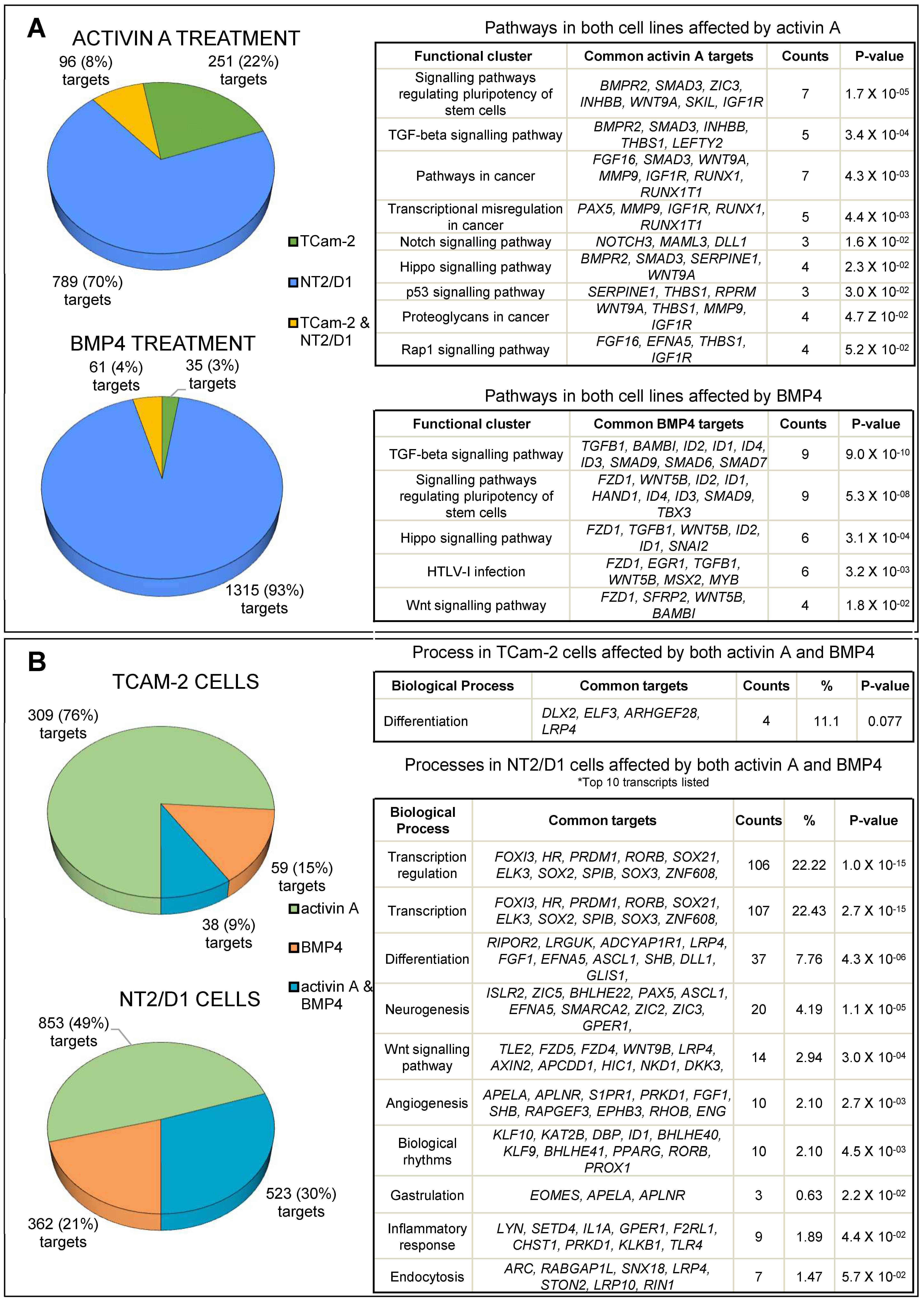
3.1. Transcriptome Profiling and Bioinformatic Analyses of Activin A- and BMP4-Treated TCam-2 and NT2/D1 Cells

RNA sequencing was performed to identify and compare the gene expression profiles of TCam-2 and NT2/D1 cells in response to 6 h of activin A or BMP4 treatment. As a starting point, the analysis of untreated TCam-2 and NT2/D1 cells illustrates the presence of signalling molecules (receptors, SMADs) in each (Figure 1).
A gene was considered as differentially expressed if transcript levels were >3-fold different following growth factor exposure, with an FDR value ≤0.05. Functional analyses of differentially expressed genes used the DAVID online tool (in Figure 2, and Supplementary Tables S1 and S2), while potential upstream regulators were identified using Ingenuity Pathway Analysis (Table 2, and Supplementary Table S3). Selected differentially expressed transcripts were confirmed using real time PCR on independent samples (Figure 3).

3.2. Activin A Treatment

Activin A exposure altered levels of 347 (1.7%) and 885 (4.4%) transcripts in TCam-2 and NT2/D1 cells, respectively; amongst these, 96 were altered in both cell lines (Figure 2A). The pathway analyses of activin A targets common to both cell lines revealed that activin A treatment significantly upregulated pluripotency, cancer, TGF-β, Notch, p53, and Hippo signalling pathways (Figure 2A). Detailed DAVID pathway analyses are provided as in the, Table S1. A closer look at individual signalling pathways identified key affected modulators, effectors, and target genes involved (Table S2). Ras/Rap signalling components, including guanine nucleotide exchange factors (GEFs; RASGRF1, RASGRF2, RASGRP1, RASA3, and RAPGEF), GTPase-activating proteins (GAPs; SIPAIL2), and effectors involved in endocytosis and cytoskeletal remodelling (RGL1, RIN1, and TIAM1), as well as cell adhesion, polarity, and migration (TIAM1, PRKD1, and ARAP3), were upregulated following activin A stimulation. PI3K-Akt signalling effectors and target genes involved in cell survival (NR4A1, CREB5, BCL2L11, and MYB) and cell cycle progression (CCND1, CCNE2, and MYC) were modulated primarily in NT2/D1 cells by activin A treatment. The transcript encoding a key PI3K Akt pathway component, serine/threonine kinase AKT3, which was upregulated in activin A-treated TCam-2 cells. Activin A altered P53 signalling pathway components in both lines, including several involved in cell cycle and DNA repair (RPRM, GADD45B, GADD45G, and SESN3) and metastasis (SERPINE1, ADGBR1, and THBS1). The elevation of NODAL, LEFTY1, and LEFTY2 in NT2/D1 cells and LEFTY2 in TCam-2 indicates Nodal signalling is active 6 h post-activin A exposure. Notch signalling pathway components, including the receptor NOTCH3 and the coactivator MAML3, were elevated by activin A in both NT2/D1 and TCam-2 cell lines, while readouts of Notch activity, HEY1 and HES1, were only elevated in NT2/D1 cells. Activin A affected transcripts relating to activin signalling, increasing SMAD3 and decreasing INHBB and INHBA. It also elevated those involved in BMP signalling inhibition, SMAD6, SMAD7, and BAMBI, in NT2/D1 cells, and lowered BMP4 target gene transcripts, ID3 and ID4, in TCam-2 cells.

3.3. BMP4 Treatment

Only 96 transcripts (0.48%) were significantly altered in TCam-2 cells by 6 h of exposure to BMP4, compared with 1376 (6.9%) in NT2/D1 cells; 61 of these were common to both lines (Figure 2A). These shared BMP4 targets were associated with TGF-β, pluripotency, Hippo, HTLV-I infection, and Wnt pathways. Reflecting the synexpression of BMP signalling components [55], common transcripts also included BMP signalling inhibitors, SMAD6, SMAD7, and BAMBI, and known downstream targets inhibitors of DNA binding (ID1/2/3/4), MSX2 [56], SNAI2 [45], and EVX1 [57]. BMP4 elevated SMAD9 in both cell lines and BMPR2 in NT2/D1 cells. Additional interesting transcripts modulated by BMP4 in both cells included TGFΒ1, EGR1 (early growth Response 1), KDM7A (lysine demethylase 7A), and HAND1 (heart and neural crest derivatives expressed 1). In BMP4-treated NT2/D1 cells, Wnt, MAPK, and PI3K-Akt signalling pathways were prominent amongst those affected. Downstream gene targets also encoded transcriptional effectors (NFKBIA, CREB5, and TIAM1) as well as proteins involved in cell survival (BDNF, FOS, and JUN) and cytoskeletal remodelling (RAC2) (Table S2). Strikingly, BMP4 treatment elevated transcripts encoding several MAPK signalling pathway components only in NT2/D1 cells, including MAPKK kinases (MAP3K4 and MAP3K14) and phosphatases (DUSP2, DUSP5, DUSP6, and DUSP9).

3.4. Common Targets of Activin A and BMP4

Both activin A and BMP4 elevated many transcripts in NT2/D1 cells encoding components linked with Hippo signalling, including transcription factors TEAD1 and FRMD6 (FERM domain-containing protein 6), the transcriptional repressor (DLG2), and target genes involved in anti-apoptotic or proliferative functions FGF1, SOX2, and AXIN2 (Figure 2A and Supplementary Materials, Table S2).
Levels of 38 transcripts in TCam-2 cells and 523 in NT2/D1 cells were altered by both activin A and BMP4 exposures, indicating the potential for cross-talk between activin A and BMP4 pathways in testicular cancer cells (Figure 2B). Importantly, however, the transcriptional outcomes demonstrate that their activities would provoke distinct responses in each cell line. DAVID functional analyses showed that activin A and BMP4 modulate several common genes in TCam-2 cells that are primarily involved in differentiation, whereas in NT2/D1 cells, the potentially affected functions relate to transcript regulation, differentiation, neurogenesis, Wnt signalling, and angiogenesis (Figure 2B).
A fundamental feature of TGF-β superfamily signalling is the shared use of downstream signalling machinery. However, because there is an imperative to understand how germ cell tumours are affected by extrinsic factors, we also used Ingenuity Pathway software to explore the potential for upstream regulators to influence the outcomes of activin A and BMP4 signalling in these cells (Table 2 and Table S3). We identified several key TGF-β superfamily components linked to common transcriptional outcomes, with TGFB1 and SMAD4 consistently identified. As stated earlier, signalling by multiple BMPs using SMAD4 would be expected to strongly influence BMP4 target expression, and this was robustly indicated in NT2/D1 cells. Environmental factors and competing signalling pathways were also identified. Common pharmaceuticals identified as potential upstream regulators of these signalling outcomes included aspirin, medroxyprogesterone acetate, estradiol, simvastatin, candesartan, curcumin, and tretinoin. In NT2/D1 cells, lipopolysaccharides, inflammatory signalling components, and cigarette smoke were notable potential upstream modulators. Several of these signalling molecules are of unequivocal importance for early germline development, including FGF2, KITLG, EGF, NRG1, and CXCL12.

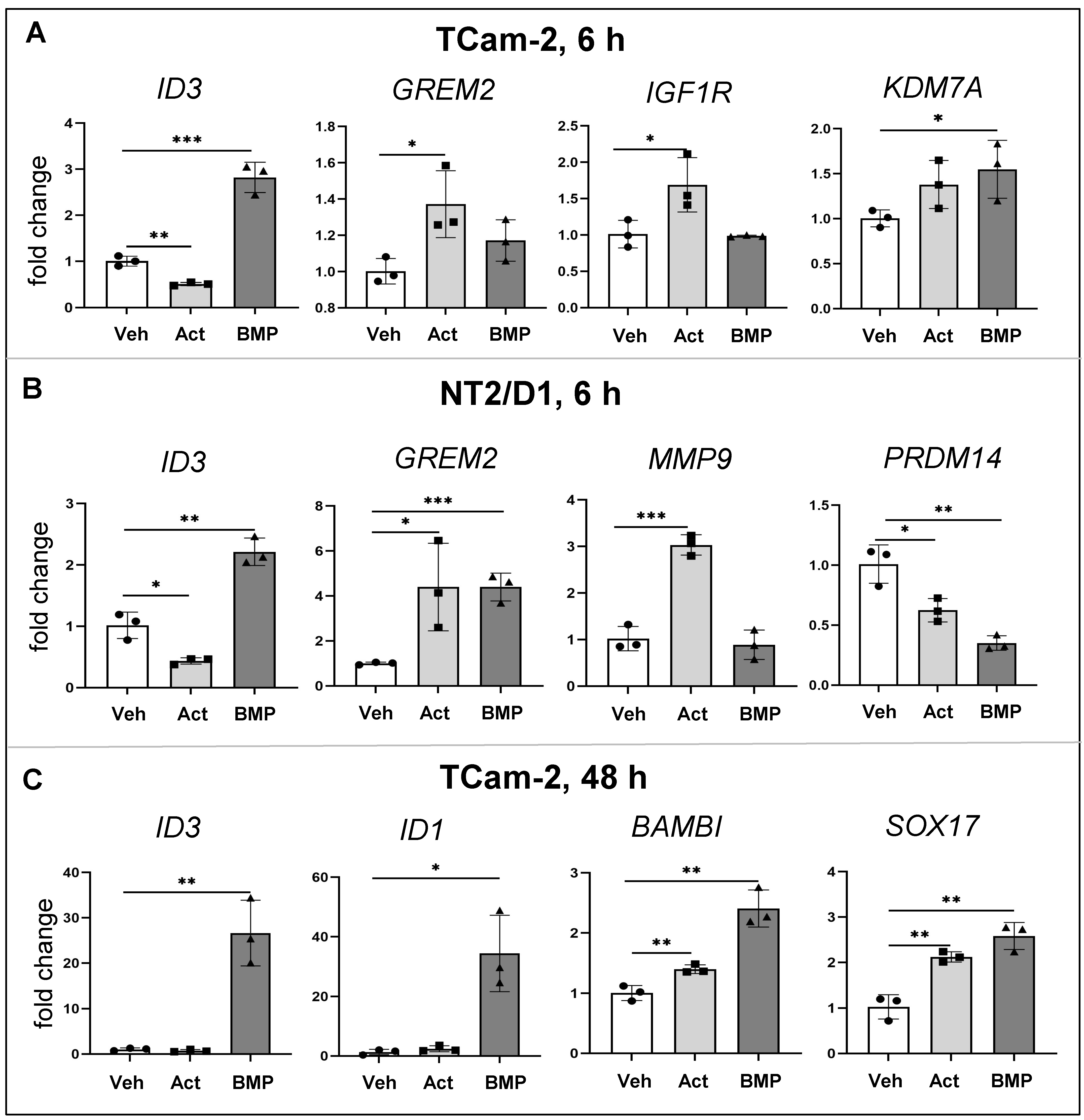
3.5. Common and Distinct Regulation of Specific Genes by Activin A and BMP4

The validation of RNA-seq data using qRT-PCR is presented to illustrate the common and distinct responses of these two cell lines to activin A and BMP4 following 6 h of growth factor exposure (Figure 3A,B). ID3 elevation by BMP4 and reduction by activin A was observed in both cell lines at 6 h. IGFR1 and MMP9 were significantly elevated in activin A-treated cells, whereas KDM7A levels were significantly higher following BMP4 treatment. GREM2 was significantly elevated in activin A- treated TCam-2 cells, while in NT2/D1 cells it was higher following both activin A and BMP4 exposures. PRDM14 was significantly decreased by both activin A and BMP4 in NT2/D1. Subsequent experiments over a longer time interval also used qRT-PCR analyses to document BMP4 target transcript levels following 48 h of growth factor exposure. ID1 and ID3 levels were robustly elevated in TCam-2 cells following 48 h of BMP4 treatment. Intriguingly, SOX17 and BAMBI, implicated in yolk sac tumours [58], were increased in TCam-2 cells by both activin A and BMP4 exposure at 48 h.

3.6. Effects of Activin A on BMP Signalling Activation

To further investigate signalling cross-talk between activin A and BMP4 in TCam-2 and NT2/D1 cells, dual promoter luciferase assays were used to examine their signalling activities simultaneously [59]. Cells were infected with adenoviral reporter constructs, then stimulated with these factors. Activin A antagonized the BMP4-induced activation of BMP response element (BRE) in a dose-dependent manner in TCam-2 and in NT2/D1 cells, at both 6 h and 24 h (Figure 4A,B). In contrast, BMP4 did not impede the activin A-induced activation of the activin response element (CAGA) in TCam-2 cells but did result in the inhibition of NT2/D1 cells at the 24 hr timepoint at the higher dose (Figure 4C,D). Activin A exposure also impeded the BMP4-induced upregulation of BMP4 target genes ID3 and PRDM1 in TCam-2 cells in a dose-dependent manner, but it did not affect the levels of two other BMP4 target gene transcripts, ID2 and SOX17 (Figure 4E).

3.7. Importins in TCam-2 and NT2/D1 Cells: A Role for IPO5 in BMP4 Responses

RNA sequencing analyses revealed that several importin-β family members were highly expressed in TCam-2 and NT2/D1 cell lines, most notably IPO5, previously shown to selectively transport BMP SMADs (SMAD 1/5/9) but not the activin SMADs (SMAD 2/3) [43] (Figure 5A).
To test whether IPO5 might enable BMP signalling in testicular cancer cells, siRNA was used to achieve transient IPO5-silencing in TCam-2 cells; silencing efficiency was determined by qPCR and Western blot. While IPO5 mRNA levels were significantly reduced in both lines at 48 h and 72 h, the profile of IPO5 protein level reduction differed between the two. In TCam-2 cells, IPO5 was unaffected at 48 h but was significantly reduced at 72 h post-transfection; in contrast, IPO5 levels were lower in NT2/D1 cells at 48 h (p = 0.053) but not at 72 h (Figure 5B).
BMP4 target gene transcripts levels measured in IPO5-silenced and scram-transfected TCam-2 cells showed that ID3, PRDM1, and SOX17 were significantly lower at 48 h post-BMP4 treatment in TCam-2 cells with siRNA treatment. NT2/D1 cell viability was severely reduced in the serum-free culture conditions required for this experiment, so the results of longer-term effects could not be obtained from this cell line under conditions used for the TCam-2 cells.

4. Discussion

Seminomas and embryonal carcinomas represent the vast majority of testicular germ cell tumours (TGCTs). This study reports the effects of activin A and BMP4 on the gene expression profiles of TCam-2 (representing seminoma cells) and NT2/D1 (representing non-seminoma cell type) cell lines and provides evidence that IPO5 may be important to selectively mediate BMP signals. RNA sequencing, bioinformatic analyses, and qRT-PCR were used to identify target genes and major signalling pathways altered by activin A and BMP4 in TCam-2 and NT2/D1 cell lines. Strikingly, activin A and BMP4 upregulated many growth factors, tyrosine kinase receptors (RTKs), and G protein-coupled receptors in both the cell lines, many of which are associated with the development of cancer. Notably, the insulin-like growth factor 1 receptor (IGF1R) was upregulated in both TCam-2 and NT2/D1 cells following activin A treatment. The IGFR is important for normal functioning of germ cells [60,61,62,63,64,65,66] and is highly upregulated in many TGCTs [67]. IGFR activation has been shown to increase the expression of the CXCR4 chemokine receptor, which is associated with cell survival and migration, including in the male germline [68,69,70] and matrix metalloproteins MMP2 and MMP9, associated with an invasive phenotype [71]. Interestingly, our data showed that transcripts encoding CCXR4 and MMP9 were upregulated in activin A-treated NT2/D1, whereas MMP2 and MMP9 were upregulated in activin A-treated TCam-2 cells, suggesting the potential for activin A to induce more active IGF1R signalling in both cell lines.
Activin A treatment upregulated several key modulators of pathways that signal through RTKs, such as Ras/Raf/MAPK and PI3 kinase. The upregulation of RAS [72], PI3 kinase [73], and MAP kinase [74,75] pathways has been demonstrated in both seminomas and non-seminomas. Interestingly, AKT3, a key PI3K pathway modulator shown here to be upregulated by activin A in TCam-2 cells, was identified to be frequently overexpressed in TGCTs and associated with poor survival outcomes [34]. Our data also indicate that activin A and BMP4 treatment modulates the expression of transcription factors, effectors, and targets genes involved in the Ras/Raf/MAPK and PI3K pathways. While activin A modulated several genes involved in MAPK signalling pathway in both cell lines, the impact of BMP4 treatment upregulating transcripts for several MAPK signalling pathway components was primarily documented in NT2/D1 cells. These influence processes affecting cell migration, including cytoskeletal remodelling, endocytosis, and cell mobility, in alignment with our observation that these factors increased TCam-2 cell migration [45]. p53 signalling pathway components, involved in mediating responses to DNA damage induced by agents such as cisplatin, were also altered in both cell lines by activin A. The activation of the p53 signalling pathway in TGCTs was previously shown to lead to apoptosis rather than cell cycle arrest [76]. This accords with the upregulation of the transcript encoding the executioner caspase, CASP3, in activin A-treated NT2/D1 cells. In both cell lines, activin A also upregulated the core components of the Notch signalling pathway, which, when constitutively active, results in migration defects and premature differentiation, and results in germ cell loss when active in Sertoli cells [77,78].
Amongst the novel BMP4 targets identified were the transcript encoding the dual histone demethylase, KDM7A, previously shown to promote tumour growth and mediate androgen receptor activity, and FGF4 [79,80]; both were upregulated in both cell lines following BMP4 stimulation. FGF4 supports germ cell survival/proliferation [81,82], and its expression in testicular cancer has been reported [83,84,85]. Activin A and BMP4 treatments elevated transcripts encoding several important immune-related molecules in TCam-2 and NT2/D1 cells, notably the proinflammatory cytokine IL6 and its receptor, IL6R. Elevated IL6 levels are documented in seminomas [86,87], while dysregulated IL6 expression features in many cancers and is associated with tumour growth and invasiveness [88]. In this context, it is interesting to note that compounds with anti-inflammatory properties, such as aspirin and curcumin, were predicted to have inhibitory effects on activin A signalling in both cell lines. Aspirin has been shown to inhibit cell proliferation and its effects can be mediated by TGFB1 [89,90]. The TGFB1 transcript was upregulated in both cell lines following BMP4 exposure and in NT2/D1 cells following activin A exposure. TGF-β1 was also predicted as a common upstream regulator of both activin A and BMP4 signalling. The higher expression of TGFB1 has been reported in several cancers [91], including testis cancer [87]. TGF-β1 acts both as a tumour suppressor in initial cancer stages and as an oncogene in later stages, driving epithelial to mesenchymal transition (EMT) and tumour metastasis [92]. In NT2/D1 cells, both activin A and BMP4 treatments significantly altered Hippo signalling pathway, an important pathway that controls cell growth and differentiation, often dysregulated in a number human cancers [93]. The key transcriptional factor and readout of the Hippo signalling pathway, TEAD1, was significantly upregulated following activin A and BMP4 stimulation, where as its coactivator TAZ/WWTR1 was upregulated following activin A treatment in NT2/D1 cells. Both TEAD and TAZ play a central role in Hippo signalling-mediated tumorigenesis and the overexpression or activation of factors are linked to tumour initiation and metastasis [94,95]. These transcriptional changes, following short term exposures in testicular cancer cell lines, demonstrate how both activin A and BMP4 can influence multiple signalling pathways that modify cell migration, survival, and cell cycle progression behaviours in TGCTs.
The identification of several transcripts and signalling pathways altered by both activin A and BMP4 treatments highlights points where both synergistic and antagonistic outcomes could emerge from cells exposed to inputs from the two arms of TGF-β signalling. These investigations using a dual luciferase reporter assay demonstrated that activin A dose-dependently inhibited BMP4-mediated BMP response element (BRE) activation in TCam-2 and NT2/D1 cell lines. In contrast, BMP4 had limited effects on activin A-induced promoter (CAGA) activation, although there was some degree of inhibition in NT2/D1 cells. Validating qPCR analysis showed that activin A impeded the upregulation of established BMP4 target genes ID3 and PRDM1 following BMP4 stimulation. This may be of particular relevance in the context of evidence that BMP signalling inhibition and decreased levels of its targets (e.g., SOX17) are key to the early steps in reprogramming TCam-2 cells into an EC-like cell type [11]. However, not all BMP4 target genes tested in TCam-2 cells in this study, including SOX17, were affected by activin A treatment. Here, it is important to note that, unlike the aforementioned studies carried out over several weeks, our goal was to identify the early targets of activin A and BMP4 in testicular cancer cells to investigate the initial stages of pathway crosstalk, and thus the treatments were short term (≤48 h). It will be of interest to ascertain the time course over which activin A exerts inhibitory effects on BMP signalling; we predict this could reveal stages of tumour development that are sensitive to manipulation or vulnerable to environmental exposures relating to changes in TGF-β pathway signalling.
Our data showed that Activin A altered many more transcripts in TCam-2 cells than in NT2/D1 cells, while BMP4 altered expression of many more genes in NT2/D1 cells relative to TCam-2 cells. This could be due to intrinsic differences in genomic or proteomic landscape between the two cell lines [96,97]. In this study, we further explored the importance of the high levels of IPO5 transcripts in NT2/D1 cells, as IPO5 selectively transports BMP-specific SMADs, SMADs 1/5/9, but not those generally involved in activin/TGF-β/nodal signalling, SMADs 2/3 [43]. In recent years, IPO5 has attracted much attention in the fields of cancer and virology. IPO5 is elevated in colorectal cancers and oesophageal cancer, and this has been associated with increased tumorigenicity and metastasis [98,99]. Elevated IPO5 levels in the serum of oesophageal and cervical cancer patients has been proposed as a cancer biomarker [100]. We had previously demonstrated that IPO5 transcript and protein are abundant in foetal germ cells [40,42], GCNIS, seminomas, and in TCam-2 cells [45]. Further, the siRNA reduction of IPO5 in TCam-2 cells lowered levels of SNAI2, a BMP4-regulated gene expressed in TGCTs known to function in cancer metastasis [45]. The present results demonstrate that IPO5 knockdown in TCam-2 cells impeded the expression of additional BMP targets, such as ID3, SOX17, and PRDM1 (PR domain containing 1, previously called BLIMP1). These findings illustrate that IPO5 can selectively modulate BMP signalling in TCam-2 cells and thus may determine the fate of testicular germ cell tumours. Although NT2/D1 cells exhibit high levels of IPO5, the experimental conditions used in our study did not support the analysis of IPO5 in mediating BMP signalling in these cells. Given the differences in how each cell responds to activin A and BMP4 stimulation, we propose that the persistent or induced elevation of IPO5 may be a feature enabling the emergence of pluripotent embryonal carcinoma cells from testicular germ cells.
In summary, this study identified signalling pathways and novel signalling components that were altered in testicular cancer cell lines stimulated with activin A and BMP4, demonstrating the potential for antagonism and synergy between the two arms of TGF-β signalling in testicular cancer cells. Furthermore, our study reinforced the involvement of IPO5 in mediating BMP signalling in seminoma cells. These outcomes (summarized in Figure 6) support the accumulating evidence that local changes in TGF-β signalling components, including ligands, receptors, and inhibitors, will have implications for testicular germ cell tumour aetiology and progression.

Supplementary Materials

The following supporting information can be downloaded at: https://www.mdpi.com/article/10.3390/cells12071000/s1. Table S1: DAVID pathway analysis of differentially expressed genes in activin A and BMP4 treated cells. Table S2: List of signalling pathways enriched in the differentially expressed genes. Table S3: Predicted upstream regulators of activin A and BMP signalling targets using Ingenuity Pathway Analysis.

Author Contributions

K.R. was involved in study design, experimental data acquisition, data analysis and interpretation, manuscript writing, manuscript revision, and final manuscript approval. M.L. was involved in experimental data acquisition for RNA sequencing and Western blots, data analysis and interpretation, manuscript writing, and final manuscript approval. J.I. was involved in experimental data acquisition for luciferase assays, analysis and data interpretation, and final manuscript approval. J.M.S. was involved in data analysis and interpretation and final manuscript approval. E.A.M. was involved in data interpretation, manuscript revision, and final manuscript approval. H.-J.Z. was involved in study design, data analysis and interpretation, manuscript revision, final manuscript approval, and the acquisition of resources for luciferase assays. K.L.L. was involved in study conception and design, data analysis and interpretation, manuscript writing, manuscript revision, manuscript approval, and funding acquisition. All authors have read and agreed to the published version of the manuscript.

Funding

This work was supported by the Australian Research Council (DP200103463 to KL) and the Victorian State Government Operational Infrastructure Scheme.

Institutional Review Board Statement

Not applicable.

Informed Consent Statement

Not applicable.

Data Availability Statement

Contact corresponding authors.

Acknowledgments

The authors would like to thank Diana Micati for technical support and the Monash University Bioinformatics Platform staff for help with RNA-seq analysis.

Conflicts of Interest

The authors declare no conflict of interest.

References

  1. Oosterhuis, J.W.; Looijenga, L.H. Testicular germ-cell tumours in a broader perspective. Nat. Rev. Cancer 2005, 5, 210–222. [Google Scholar] [CrossRef] [PubMed]
  2. Shanmugalingam, T.; Soultati, A.; Chowdhury, S.; Rudman, S.; Van Hemelrijck, M. Global incidence and outcome of testicular cancer. Clin. Epidemiol. 2013, 5, 417–427. [Google Scholar] [CrossRef] [Green Version]
  3. Berney, D.M.; Looijenga, L.H.; Idrees, M.; Oosterhuis, J.W.; Rajpert-De Meyts, E.; Ulbright, T.M.; Skakkebaek, N.E. Germ cell neoplasia in situ (GCNIS): Evolution of the current nomenclature for testicular pre-invasive germ cell malignancy. Histopathology 2016, 69, 7–10. [Google Scholar] [CrossRef]
  4. Biermann, K.; Heukamp, L.C.; Steger, K.; Zhou, H.; Franke, F.E.; Sonnack, V.; Brehm, R.; Berg, J.; Bastian, P.J.; Muller, S.C.; et al. Genome-wide expression profiling reveals new insights into pathogenesis and progression of testicular germ cell tumors. Cancer Genom. Proteom. 2007, 4, 359–367. [Google Scholar]
  5. Hoei-Hansen, C.E.; Nielsen, J.E.; Almstrup, K.; Hansen, M.A.; Skakkebaek, N.E.; Rajpert-DeMeyts, E.; Leffers, H. Identification of genes differentially expressed in testes containing carcinoma in situ. Mol. Hum. Reprod. 2004, 10, 423–431. [Google Scholar] [CrossRef]
  6. Skakkebaek, N.E. Possible carcinoma-in-situ of the testis. Lancet 1972, 2, 516–517. [Google Scholar] [CrossRef] [PubMed]
  7. Sonne, S.B.; Almstrup, K.; Dalgaard, M.; Juncker, A.S.; Edsgard, D.; Ruban, L.; Harrison, N.J.; Schwager, C.; Abdollahi, A.; Huber, P.E.; et al. Analysis of gene expression profiles of microdissected cell populations indicates that testicular carcinoma in situ is an arrested gonocyte. Cancer Res. 2009, 69, 5241–5250. [Google Scholar] [CrossRef] [Green Version]
  8. Skakkebaek, N.E.; Rajpert-De Meyts, E.; Buck Louis, G.M.; Toppari, J.; Andersson, A.M.; Eisenberg, M.L.; Jensen, T.K.; Jørgensen, N.; Swan, S.H.; Sapra, K.J.; et al. Male Reproductive Disorders and Fertility Trends: Influences of Environment and Genetic Susceptibility. Physiol. Rev. 2016, 96, 55–97. [Google Scholar] [CrossRef] [Green Version]
  9. El-Naggar, A.K.; Ro, J.Y.; McLemore, D.; Ayala, A.G.; Batsakis, J.G. DNA ploidy in testicular germ cell neoplasms. Histogenetic and clinical implications. Am. J. Surg. Pathol. 1992, 16, 611–618. [Google Scholar] [CrossRef]
  10. Oosterhuis, J.W.; Looijenga, L.H. Current views on the pathogenesis of testicular germ cell tumours and perspectives for future research: Highlights of the 5th Copenhagen Workshop on Carcinoma in situ and Cancer of the Testis. Apmis 2003, 111, 280–289. [Google Scholar] [CrossRef]
  11. Nettersheim, D.; Schorle, H. The plasticity of germ cell cancers and its dependence on the cellular microenvironment. J. Cell. Mol. Med. 2017, 21, 1463–1467. [Google Scholar] [CrossRef] [PubMed] [Green Version]
  12. Shen, H.; Shih, J.; Hollern, D.P.; Wang, L.; Bowlby, R.; Tickoo, S.K.; Thorsson, V.; Mungall, A.J.; Newton, Y.; Hegde, A.M.; et al. Integrated Molecular Characterization of Testicular Germ Cell Tumors. Cell Rep. 2018, 23, 3392–3406. [Google Scholar] [CrossRef] [PubMed]
  13. Mendis, S.H.; Meachem, S.J.; Sarraj, M.A.; Loveland, K.L. Activin A balances Sertoli and germ cell proliferation in the fetal mouse testis. Biol. Reprod. 2011, 84, 379–391. [Google Scholar] [CrossRef] [PubMed] [Green Version]
  14. Whiley, P.A.F.; O’Donnell, L.; Moody, S.C.; Handelsman, D.J.; Young, J.C.; Richards, E.A.; Almstrup, K.; Western, P.S.; Loveland, K.L. Activin A Determines Steroid Levels and Composition in the Fetal Testis. Endocrinology 2020, 161, bqaa058. [Google Scholar] [CrossRef] [PubMed]
  15. Archambeault, D.R.; Yao, H.H. Activin A, a product of fetal Leydig cells, is a unique paracrine regulator of Sertoli cell proliferation and fetal testis cord expansion. Proc. Natl. Acad. Sci. USA 2010, 107, 10526–10531. [Google Scholar] [CrossRef] [Green Version]
  16. Moody, S.C.; Whiley, P.A.F.; Western, P.S.; Loveland, K.L. The Impact of Activin A on Fetal Gonocytes: Chronic Versus Acute Exposure Outcomes. Front. Endocrinol. 2022, 13, 896747. [Google Scholar] [CrossRef]
  17. Young, J.C.; Wakitani, S.; Loveland, K.L. TGF-β superfamily signaling in testis formation and early male germline development. Semin. Cell Dev. Biol. 2015, 45, 94–103. [Google Scholar] [CrossRef]
  18. Dias, V.L.; Rajpert-De Meyts, E.; McLachlan, R.; Loveland, K.L. Analysis of activin/TGFB-signaling modulators within the normal and dysfunctional adult human testis reveals evidence of altered signaling capacity in a subset of seminomas. Reproduction 2009, 138, 801–811. [Google Scholar] [CrossRef] [Green Version]
  19. Spiller, C.M.; Bowles, J.; Koopman, P. Nodal/Cripto signaling in fetal male germ cell development: Implications for testicular germ cell tumors. Int. J. Dev. Biol. 2013, 57, 211–219. [Google Scholar] [CrossRef] [Green Version]
  20. Spiller, C.M.; Gillis, A.J.; Burnet, G.; Stoop, H.; Koopman, P.; Bowles, J.; Looijenga, L.H. Cripto: Expression, epigenetic regulation and potential diagnostic use in testicular germ cell tumors. Mol. Oncol. 2016, 10, 526–537. [Google Scholar] [CrossRef] [Green Version]
  21. Dias, V.; Meachem, S.; Rajpert-De Meyts, E.; McLachlan, R.; Manuelpillai, U.; Loveland, K.L. Activin receptor subunits in normal and dysfunctional adult human testis. Hum. Reprod. 2008, 23, 412–420. [Google Scholar] [CrossRef]
  22. Fustino, N.; Rakheja, D.; Ateek, C.S.; Neumann, J.C.; Amatruda, J.F. Bone morphogenetic protein signalling activity distinguishes histological subsets of paediatric germ cell tumours. Int. J. Androl. 2011, 34, e218–e233. [Google Scholar] [CrossRef] [PubMed] [Green Version]
  23. Neumann, J.C.; Chandler, G.L.; Damoulis, V.A.; Fustino, N.J.; Lillard, K.; Looijenga, L.; Margraf, L.; Rakheja, D.; Amatruda, J.F. Mutation in the type IB bone morphogenetic protein receptor Alk6b impairs germ-cell differentiation and causes germ-cell tumors in zebrafish. Proc. Natl. Acad. Sci. USA 2011, 108, 13153–13158. [Google Scholar] [CrossRef] [PubMed] [Green Version]
  24. Neumann, J.C.; Dovey, J.S.; Chandler, G.L.; Carbajal, L.; Amatruda, J.F. Identification of a heritable model of testicular germ cell tumor in the zebrafish. Zebrafish 2009, 6, 319–327. [Google Scholar] [CrossRef]
  25. Purdue, M.P.; Sakoda, L.C.; Graubard, B.I.; Welch, R.; Chanock, S.J.; Sesterhenn, I.A.; Rubertone, M.V.; Erickson, R.L.; McGlynn, K.A. A case-control investigation of immune function gene polymorphisms and risk of testicular germ cell tumors. Cancer Epidemiol. Biomarkers Prev. 2007, 16, 77–83. [Google Scholar] [CrossRef] [Green Version]
  26. De Jong, J.; Stoop, H.; Gillis, A.J.; Hersmus, R.; van Gurp, R.J.; van de Geijn, G.J.; van Drunen, E.; Beverloo, H.B.; Schneider, D.T.; Sherlock, J.K.; et al. Further characterization of the first seminoma cell line TCam-2. Genes Chromosomes Cancer 2008, 47, 185–196. [Google Scholar] [CrossRef]
  27. Nettersheim, D.; Gillis, A.J.; Looijenga, L.H.; Schorle, H. TGF-β1, EGF and FGF4 synergistically induce differentiation of the seminoma cell line TCam-2 into a cell type resembling mixed non-seminoma. Int. J. Androl. 2011, 34, e189–e203. [Google Scholar] [CrossRef]
  28. Nettersheim, D.; Jostes, S.; Sharma, R.; Schneider, S.; Hofmann, A.; Ferreira, H.J.; Hoffmann, P.; Kristiansen, G.; Esteller, M.B.; Schorle, H. BMP Inhibition in Seminomas Initiates Acquisition of Pluripotency via NODAL Signaling Resulting in Reprogramming to an Embryonal Carcinoma. PLoS Genet. 2015, 11, e1005415. [Google Scholar] [CrossRef] [PubMed] [Green Version]
  29. Nettersheim, D.; Heimsoeth, A.; Jostes, S.; Schneider, S.; Fellermeyer, M.; Hofmann, A.; Schorle, H. SOX2 is essential for in vivo reprogramming of seminoma-like TCam-2 cells to an embryonal carcinoma-like fate. Oncotarget 2016, 7, 47095–47110. [Google Scholar] [CrossRef] [Green Version]
  30. Moody, S.C.; Wakitani, S.; Young, J.C.; Western, P.S.; Loveland, K.L. Evidence that activin A directly modulates early human male germline differentiation status. Reproduction 2020, 160, 141–154. [Google Scholar] [CrossRef]
  31. Morikawa, M.; Derynck, R.; Miyazono, K. TGF-β and the TGF-β Family: Context-Dependent Roles in Cell and Tissue Physiology. Cold Spring Harb. Perspect Biol. 2016, 8, a021873. [Google Scholar] [CrossRef] [Green Version]
  32. Coda, D.M.; Patel, H.; Gori, I.; Gaarenstroom, T.E.; Song, O.R.; Howell, M.; Hill, C.S. A network of transcription factors governs the dynamics of NODAL/Activin transcriptional responses. J. Cell Sci. 2022, 135, jcs259972. [Google Scholar] [CrossRef] [PubMed]
  33. Martinez-Hackert, E.; Sundan, A.; Holien, T. Receptor binding competition: A paradigm for regulating TGF-β family action. Cytokine Growth Factor Rev. 2021, 57, 39–54. [Google Scholar] [CrossRef] [PubMed]
  34. Luo, K. Signaling Cross Talk between TGF-β/Smad and Other Signaling Pathways. Cold Spring Harb. Perspect. Biol. 2017, 9, a022137. [Google Scholar] [CrossRef] [Green Version]
  35. Mu, Y.; Gudey, S.K.; Landström, M. Non-Smad signaling pathways. Cell Tissue Res. 2012, 347, 11–20. [Google Scholar] [CrossRef] [PubMed]
  36. Hill, C.S. Transcriptional Control by the SMADs. Cold Spring Harb. Perspect. Biol. 2016, 8, a022079. [Google Scholar] [CrossRef] [PubMed] [Green Version]
  37. Windley, S.P.; Wilhelm, D. Signaling Pathways Involved in Mammalian Sex Determination and Gonad Development. Sex. Dev. 2015, 9, 297–315. [Google Scholar] [CrossRef]
  38. Miyamoto, Y.; Yamada, K.; Yoneda, Y. Importin α: A key molecule in nuclear transport and non-transport functions. J. Biochem. 2016, 160, 69–75. [Google Scholar] [CrossRef] [Green Version]
  39. Loveland, K.L.; Major, A.T.; Butler, R.; Young, J.C.; Jans, D.A.; Miyamoto, Y. Putting things in place for fertilization: Discovering roles for importin proteins in cell fate and spermatogenesis. Asian J. Androl. 2015, 17, 537–544. [Google Scholar] [CrossRef]
  40. Nathaniel, B.; Whiley, P.A.F.; Miyamoto, Y.; Loveland, K.L. Importins: Diverse roles in male fertility. Semin. Cell. Dev. Biol. 2022, 121, 82–98. [Google Scholar] [CrossRef]
  41. Yasuhara, N.; Yoneda, Y. Importins in the maintenance and lineage commitment of ES cells. Neurochem. Int. 2017, 105, 32–41. [Google Scholar] [CrossRef] [PubMed]
  42. Hogarth, C.A.; Jans, D.A.; Loveland, K.L. Subcellular distribution of importins correlates with germ cell maturation. Dev. Dyn. 2007, 236, 2311–2320. [Google Scholar] [CrossRef] [PubMed]
  43. Baas, R.; Sijm, A.; van Teeffelen, H.A.; van Es, R.; Vos, H.R.; Marc Timmers, H.T. Quantitative Proteomics of the SMAD (Suppressor of Mothers against Decapentaplegic) Transcription Factor Family Identifies Importin 5 as a Bone Morphogenic Protein Receptor SMAD-specific Importin. J. Biol. Chem. 2016, 291, 24121–24132. [Google Scholar] [CrossRef] [Green Version]
  44. Ren, J.; Wang, Y.; Ware, T.; Iaria, J.; Ten Dijke, P.; Zhu, H.J. Reactivation of BMP signaling by suboptimal concentrations of MEK inhibitor and FK506 reduces organ-specific breast cancer metastasis. Cancer Lett. 2020, 493, 41–54. [Google Scholar] [CrossRef]
  45. Micati, D.J.; Radhakrishnan, K.; Young, J.C.; Rajpert-De Meyts, E.; Hime, G.R.; Abud, H.E.; Loveland, K.L. ‘Snail factors in testicular germ cell tumours and their regulation by the BMP4 signalling pathway’. Andrology 2020, 8, 1456–1470. [Google Scholar] [CrossRef] [PubMed]
  46. Spits, M.; Janssen, L.J.; Voortman, L.M.; Kooij, R.; Neefjes, A.C.M.; Ovaa, H.; Neefjes, J. Homeostasis of soluble proteins and the proteasome post nuclear envelope reformation in mitosis. J. Cell Sci. 2019, 132, jcs225524. [Google Scholar] [CrossRef] [Green Version]
  47. Young, J.C.; Major, A.T.; Miyamoto, Y.; Loveland, K.L.; Jans, D.A. Distinct effects of importin α2 and α4 on Oct3/4 localization and expression in mouse embryonic stem cells. FASEB J. 2011, 25, 3958–3965. [Google Scholar] [CrossRef]
  48. Andrews, P.W.; Damjanov, I.; Simon, D.; Banting, G.S.; Carlin, C.; Dracopoli, N.C.; Føgh, J. Pluripotent embryonal carcinoma clones derived from the human teratocarcinoma cell line Tera-2. Differentiation in vivo and in vitro. Lab. Investig. 1984, 50, 147–162. [Google Scholar]
  49. Tsyganov, K.; James Perry, A.; Kenneth Archer, S.; Powell, D. RNAsik: A Pipeline for complete and reproducible RNA-seq analysis that runs anywhere with speed and ease. J. Open Source Softw. 2018, 3, 583. [Google Scholar] [CrossRef]
  50. Dobin, A.; Davis, C.A.; Schlesinger, F.; Drenkow, J.; Zaleski, C.; Jha, S.; Batut, P.; Chaisson, M.; Gingeras, T.R. STAR: Ultrafast universal RNA-seq aligner. Bioinformatics 2013, 29, 15–21. [Google Scholar] [CrossRef]
  51. Degust—Monash University. Available online: https://degust.erc.monash.edu/ (accessed on 1 February 2023).
  52. Luwor, R.B.; Wang, B.; Nheu, T.V.; Iaria, J.; Tsantikos, E.; Hibbs, M.L.; Sieber, O.M.; Zhu, H.J. New reagents for improved in vitro and in vivo examination of TGF-β signalling. Growth Factors 2011, 29, 211–218. [Google Scholar] [CrossRef] [PubMed]
  53. Schneider, C.A.; Rasband, W.S.; Eliceiri, K.W. NIH Image to ImageJ: 25 years of image analysis. Nat. Methods 2012, 9, 671–675. [Google Scholar] [CrossRef] [PubMed]
  54. Morianos, I.; Papadopoulou, G.; Semitekolou, M.; Xanthou, G. Activin-A in the regulation of immunity in health and disease. J. Autoimmun. 2019, 104, 102314. [Google Scholar] [CrossRef] [PubMed]
  55. Paulsen, M.; Legewie, S.; Eils, R.; Karaulanov, E.; Niehrs, C. Negative feedback in the bone morphogenetic protein 4 (BMP4) synexpression group governs its dynamic signaling range and canalizes development. Proc. Natl. Acad. Sci. USA 2011, 108, 10202–10207. [Google Scholar] [CrossRef] [Green Version]
  56. Childs, A.J.; Kinnell, H.L.; Collins, C.S.; Hogg, K.; Bayne, R.A.; Green, S.J.; McNeilly, A.S.; Anderson, R.A. BMP signaling in the human fetal ovary is developmentally regulated and promotes primordial germ cell apoptosis. Stem Cells 2010, 28, 1368–1378. [Google Scholar] [CrossRef] [Green Version]
  57. Bell, C.C.; Amaral, P.P.; Kalsbeek, A.; Magor, G.W.; Gillinder, K.R.; Tangermann, P.; di Lisio, L.; Cheetham, S.W.; Gruhl, F.; Frith, J.; et al. The Evx1/Evx1as gene locus regulates anterior-posterior patterning during gastrulation. Sci. Rep. 2016, 6, 26657. [Google Scholar] [CrossRef] [Green Version]
  58. Wruck, W.; Bremmer, F.; Kotthoff, M.; Fichtner, A.; Skowron, M.A.; Schönberger, S.; Calaminus, G.; Vokuhl, C.; Pfister, D.; Heidenreich, A.; et al. The pioneer and differentiation factor FOXA2 is a key driver of yolk-sac tumour formation and a new biomarker for paediatric and adult yolk-sac tumours. J. Cell. Mol. Med. 2021, 25, 1394–1405. [Google Scholar] [CrossRef]
  59. Fonseca Teixeira, A.; Iaria, J.; Zhu, H.J. Fast Quantitation of TGF-β Signaling Using Adenoviral Reporter. Methods Mol. Biol. 2022, 2488, 13–22. [Google Scholar] [CrossRef]
  60. Bingol-Kologlu, M.; Bahadir, G.B.; Vargun, R.; Ilkay, H.; Bagriacik, E.U.; Yolbakan, S.; Guven, C.; Endogan, T.; Hasirci, N.; Dindar, H. Effects of local and sustained release of FGF, IGF, and GH on germ cells in unilateral undescended testis in rats. Urology 2010, 75, 223–228. [Google Scholar] [CrossRef]
  61. Castilla-Cortazar, I.; Garcia, M.; Quiroga, J.; Diez, N.; Diez-Caballero, F.; Calvo, A.; Diaz, M.; Prieto, J. Insulin-like growth factor-I reverts testicular atrophy in rats with advanced cirrhosis. Hepatology 2000, 31, 592–600. [Google Scholar] [CrossRef]
  62. Froment, P.; Staub, C.; Hembert, S.; Pisselet, C.; Magistrini, M.; Delaleu, B.; Seurin, D.; Levine, J.E.; Johnson, L.; Binoux, M.; et al. Reproductive abnormalities in human insulin-like growth factor-binding protein-1 transgenic male mice. Endocrinology 2004, 145, 2080–2091. [Google Scholar] [CrossRef] [Green Version]
  63. Huang, Y.H.; Chin, C.C.; Ho, H.N.; Chou, C.K.; Shen, C.N.; Kuo, H.C.; Wu, T.J.; Wu, Y.C.; Hung, Y.C.; Chang, C.C.; et al. Pluripotency of mouse spermatogonial stem cells maintained by IGF-1- dependent pathway. FASEB J. 2009, 23, 2076–2087. [Google Scholar] [CrossRef] [Green Version]
  64. Yao, J.; Zuo, H.; Gao, J.; Wang, M.; Wang, D.; Li, X. The effects of IGF-1 on mouse spermatogenesis using an organ culture method. Biochem. Biophys. Res. Commun. 2017, 491, 840–847. [Google Scholar] [CrossRef]
  65. Sang, X.; Curran, M.S.; Wood, A.W. Paracrine insulin-like growth factor signaling influences primordial germ cell migration: In vivo evidence from the zebrafish model. Endocrinology 2008, 149, 5035–5042. [Google Scholar] [CrossRef] [Green Version]
  66. Nef, S.; Verma-Kurvari, S.; Merenmies, J.; Vassalli, J.D.; Efstratiadis, A.; Accili, D.; Parada, L.F. Testis determination requires insulin receptor family function in mice. Nature 2003, 426, 291–295. [Google Scholar] [CrossRef]
  67. Selfe, J.; Goddard, N.C.; McIntyre, A.; Taylor, K.R.; Renshaw, J.; Popov, S.D.; Thway, K.; Summersgill, B.; Huddart, R.A.; Gilbert, D.C.; et al. IGF1R signalling in testicular germ cell tumour cells impacts on cell survival and acquired cisplatin resistance. J. Pathol. 2018, 244, 242–253. [Google Scholar] [CrossRef]
  68. Kuo, Y.C.; Au, H.K.; Hsu, J.L.; Wang, H.F.; Lee, C.J.; Peng, S.W.; Lai, S.C.; Wu, Y.C.; Ho, H.N.; Huang, Y.H. IGF-1R Promotes Symmetric Self-Renewal and Migration of Alkaline Phosphatase(+) Germ Stem Cells through HIF-2α-OCT4/CXCR4 Loop underHypoxia. Stem Cell Rep. 2018, 10, 524–537. [Google Scholar] [CrossRef] [Green Version]
  69. Li, Y.; Yu, X.; Lin, S.; Li, X.; Zhang, S.; Song, Y.H. Insulin-like growth factor 1 enhances the migratory capacity of mesenchymal stem cells. Biochem. Biophys. Res. Commun. 2007, 356, 780–784. [Google Scholar] [CrossRef]
  70. Yang, Q.E.; Kim, D.; Kaucher, A.; Oatley, M.J.; Oatley, J.M. CXCL12-CXCR4 signaling is required for the maintenance of mouse spermatogonial stem cells. J. Cell Sci. 2013, 126, 1009–1020. [Google Scholar] [CrossRef]
  71. Das, S.K.; Pradhan, A.K.; Bhoopathi, P.; Talukdar, S.; Shen, X.N.; Sarkar, D.; Emdad, L.; Fisher, P.B. The MDA-9/Syntenin/IGF1R/STAT3 Axis Directs Prostate Cancer Invasion. Cancer Res. 2018, 78, 2852–2863. [Google Scholar] [CrossRef] [Green Version]
  72. Gilbert, D.C.; McIntyre, A.; Summersgill, B.; Missiaglia, E.; Goddard, N.C.; Chandler, I.; Huddart, R.A.; Shipley, J. Minimum regions of genomic imbalance in stage I testicular embryonal carcinoma and association of 22q loss with relapse. Genes Chromosomes Cancer 2011, 50, 186–195. [Google Scholar] [CrossRef]
  73. Di Vizio, D.; Cito, L.; Boccia, A.; Chieffi, P.; Insabato, L.; Pettinato, G.; Motti, M.L.; Schepis, F.; D’Amico, W.; Fabiani, F.; et al. Loss of the tumor suppressor gene PTEN marks the transition from intratubular germ cell neoplasias (ITGCN) to invasive germ cell tumors. Oncogene 2005, 24, 1882–1894. [Google Scholar] [CrossRef] [PubMed] [Green Version]
  74. Kemmer, K.; Corless, C.L.; Fletcher, J.A.; McGreevey, L.; Haley, A.; Griffith, D.; Cummings, O.W.; Wait, C.; Town, A.; Heinrich, M.C. KIT mutations are common in testicular seminomas. Am. J. Pathol. 2004, 164, 305–313. [Google Scholar] [CrossRef] [PubMed] [Green Version]
  75. Sommerer, F.; Hengge, U.R.; Markwarth, A.; Vomschloss, S.; Stolzenburg, J.U.; Wittekind, C.; Tannapfel, A. Mutations of BRAF and RAS are rare events in germ cell tumours. Int. J. Cancer 2005, 113, 329–335. [Google Scholar] [CrossRef] [PubMed]
  76. Spierings, D.C.; de Vries, E.G.; Stel, A.J.; te Rietstap, N.; Vellenga, E.; de Jong, S. Low p21Waf1/Cip1 protein level sensitizes testicular germ cell tumor cells to Fas-mediated apoptosis. Oncogene 2004, 23, 4862–4872. [Google Scholar] [CrossRef] [Green Version]
  77. Garcia, T.X.; DeFalco, T.; Capel, B.; Hofmann, M.C. Constitutive activation of NOTCH1 signaling in Sertoli cells causes gonocyte exit from quiescence. Dev. Biol. 2013, 377, 188–201. [Google Scholar] [CrossRef] [PubMed] [Green Version]
  78. Huang, Z.; Rivas, B.; Agoulnik, A.I. NOTCH1 gain of function in germ cells causes failure of spermatogenesis in male mice. PLoS ONE 2013, 8, e71213. [Google Scholar] [CrossRef] [Green Version]
  79. Huang, C.; Xiang, Y.; Wang, Y.; Li, X.; Xu, L.; Zhu, Z.; Zhang, T.; Zhu, Q.; Zhang, K.; Jing, N.; et al. Dual-specificity histone demethylase KIAA1718 (KDM7A) regulates neural differentiation through FGF4. Cell Res. 2010, 20, 154–165. [Google Scholar] [CrossRef] [Green Version]
  80. Lee, K.H.; Hong, S.; Kang, M.; Jeong, C.W.; Ku, J.H.; Kim, H.H.; Kwak, C. Histone demethylase KDM7A controls androgen receptor activity and tumor growth in prostate cancer. Int. J. Cancer 2018, 143, 2849–2861. [Google Scholar] [CrossRef] [Green Version]
  81. Yamamoto, H.; Ochiya, T.; Tamamushi, S.; Toriyama-Baba, H.; Takahama, Y.; Hirai, K.; Sasaki, H.; Sakamoto, H.; Saito, I.; Iwamoto, T.; et al. HST-1/FGF-4 gene activation induces spermatogenesis and prevents adriamycin-induced testicular toxicity. Oncogene 2002, 21, 899–908. [Google Scholar] [CrossRef] [Green Version]
  82. Ohta, H.; Yabuta, Y.; Kurimoto, K.; Nakamura, T.; Murase, Y.; Yamamoto, T.; Saitou, M. Cyclosporin A and FGF signaling support the proliferation/survival of mouse primordial germ cell-like cells in vitro†. Biol. Reprod. 2021, 104, 344–360. [Google Scholar] [CrossRef] [PubMed]
  83. Strohmeyer, T.; Peter, S.; Hartmann, M.; Munemitsu, S.; Ackermann, R.; Ullrich, A.; Slamon, D.J. Expression of the hst-1 and c-kit protooncogenes in human testicular germ cell tumors. Cancer Res. 1991, 51, 1811–1816. [Google Scholar]
  84. Suzuki, K.; Tokue, A.; Kamiakito, T.; Kuriki, K.; Saito, K.; Tanaka, A. Predominant expression of fibroblast growth factor (FGF) 8, FGF4, and FGF receptor 1 in nonseminomatous and highly proliferative components of testicular germ cell tumors. Virchows Arch. 2001, 439, 616–621. [Google Scholar] [CrossRef] [PubMed]
  85. Yoshida, T.; Tsutsumi, M.; Sakamoto, H.; Miyagawa, K.; Teshima, S.; Sugimura, T.; Terada, M. Expression of the HST1 oncogene in human germ cell tumors. Biochem. Biophys. Res. Commun. 1988, 155, 1324–1329. [Google Scholar] [CrossRef]
  86. Białas, M.; Fiszer, D.; Rozwadowska, N.; Kosicki, W.; Jedrzejczak, P.; Kurpisz, M. The role of IL-6, IL-10, TNF-alpha and its receptors TNFR1 and TNFR2 in the local regulatory system of normal and impaired human spermatogenesis. Am. J. Reprod. Immunol. 2009, 62, 51–59. [Google Scholar] [CrossRef] [PubMed]
  87. Klein, B.; Haggeney, T.; Fietz, D.; Indumathy, S.; Loveland, K.L.; Hedger, M.; Kliesch, S.; Weidner, W.; Bergmann, M.; Schuppe, H.C. Specific immune cell and cytokine characteristics of human testicular germ cell neoplasia. Hum. Reprod. 2016, 31, 2192–2202. [Google Scholar] [CrossRef] [Green Version]
  88. Guo, Y.; Xu, F.; Lu, T.; Duan, Z.; Zhang, Z. Interleukin-6 signaling pathway in targeted therapy for cancer. Cancer Treat. Rev. 2012, 38, 904–910. [Google Scholar] [CrossRef] [PubMed]
  89. Redondo, S.; Santos-Gallego, C.G.; Ganado, P.; García, M.; Rico, L.; Del Rio, M.; Tejerina, T. Acetylsalicylic acid inhibits cell proliferation by involving transforming growth factor-beta. Circulation 2003, 107, 626–629. [Google Scholar] [CrossRef] [Green Version]
  90. Wang, Y.; Du, C.; Zhang, N.; Li, M.; Liu, Y.; Zhao, M.; Wang, F.; Luo, F. TGF-β1 mediates the effects of aspirin on colonic tumor cell proliferation and apoptosis. Oncol. Lett. 2018, 15, 5903–5909. [Google Scholar] [CrossRef] [Green Version]
  91. Massagué, J. TGFbeta in Cancer. Cell 2008, 134, 215–230. [Google Scholar] [CrossRef] [Green Version]
  92. Kubiczkova, L.; Sedlarikova, L.; Hajek, R.; Sevcikova, S. TGF-β—An excellent servant but a bad master. J. Transl. Med. 2012, 10, 183. [Google Scholar] [CrossRef] [Green Version]
  93. Cunningham, R.; Hansen, C.G. The Hippo pathway in cancer: YAP/TAZ and TEAD as therapeutic targets in cancer. Clin. Sci. 2022, 136, 197–222. [Google Scholar] [CrossRef]
  94. Luo, M.; Xu, Y.; Chen, H.; Wu, Y.; Pang, A.; Hu, J.; Dong, X.; Che, J.; Yang, H. Advances of targeting the YAP/TAZ-TEAD complex in the hippo pathway for the treatment of cancers. Eur. J. Med. Chem. 2022, 244, 114847. [Google Scholar] [CrossRef]
  95. Howard, A.; Bojko, J.; Flynn, B.; Bowen, S.; Jungwirth, U.; Walko, G. Targeting the Hippo/YAP/TAZ signalling pathway: Novel opportunities for therapeutic interventions into skin cancers. Exp. Dermatol. 2022, 31, 1477–1499. [Google Scholar] [CrossRef]
  96. Van der Zwan, Y.G.; Rijlaarsdam, M.A.; Rossello, F.J.; Notini, A.J.; de Boer, S.; Watkins, D.N.; Gillis, A.J.; Dorssers, L.C.; White, S.J.; Looijenga, L.H. Seminoma and embryonal carcinoma footprints identified by analysis of integrated genome-wide epigenetic and expression profiles of germ cell cancer cell lines. PLoS ONE 2014, 9, e98330. [Google Scholar] [CrossRef] [Green Version]
  97. Bremmer, F.; Bohnenberger, H.; Küffer, S.; Oellerich, T.; Serve, H.; Urlaub, H.; Strauss, A.; Maatoug, Y.; Behnes, C.L.; Oing, C.; et al. Proteomic Comparison of Malignant Human Germ Cell Tumor Cell Lines. Dis. Markers 2019, 2019, 8298524. [Google Scholar] [CrossRef]
  98. Li, X.F.; Aierken, A.L.; Shen, L. IPO5 promotes malignant progression of esophageal cancer through activating MMP7. Eur. Rev. Med. Pharmacol. Sci. 2020, 24, 4246–4254. [Google Scholar] [CrossRef]
  99. Zhang, W.; Lu, Y.; Li, X.; Zhang, J.; Lin, W.; Zhang, W.; Zheng, L.; Li, X. IPO5 promotes the proliferation and tumourigenicity of colorectal cancer cells by mediating RASAL2 nuclear transportation. J. Exp. Clin. Cancer Res. 2019, 38, 296. [Google Scholar] [CrossRef] [Green Version]
  100. Van der Watt, P.J.; Okpara, M.O.; Wishart, A.; Parker, M.I.; Soares, N.C.; Blackburn, J.M.; Leaner, V.D. Nuclear transport proteins are secreted by cancer cells and identified as potential novel cancer biomarkers. Int. J. Cancer 2022, 150, 347–361. [Google Scholar] [CrossRef]
Figure 1. TGF-β superfamily signalling pathway transcript levels. Transcript levels encoding activin and BMP signaling components identified in [54] differ between the two testicular cancer cell lines. Data are the mean of counts per million (cpm) in control cells (cultured in vehicle only) from four independent RNA-seq experiments (n = 4); error bars indicate standard error of the mean (SEM).
Cells 12 01000 g001
Figure 2. RNA-seq analyses of activin A- and BMP4-treated TCam-2 and NT2/D1 cells. (A): Transcript levels changes in TCam-2 cells (347 significantly different) and NT2/D1 cells (885) resulting from 6 h of 5 ng/mL activin A exposure identified in RNA-seq data, with 96 transcripts altered in both cell lines. Significant transcript level changes in TCam-2 cells (96) and NT2/D1 cells (1376) were identified after 6 h of 5 ng/mL BMP4 exposure, with 35 transcripts altered in both. (B): Common transcripts affected by both activin A and BMP4 in TCam-2 cells (38) and NT2/D1 cells (523). The KEGG pathway analysis of these common differentially expressed genes used the DAVID online tool, with FDR cut-off set at 0.1. Supplementary Tables S1 and S2 contain the full DAVID analysis of the differentially expressed genes and the signalling pathways affected, respectively.
Cells 12 01000 g002
Figure 3. Validation of selected transcripts in TCam-2 cells and NT2 cells. Independent cultures of TCam-2 (A,C) and NT2/D1 (B) cells were stimulated with vehicle, 5 ng/mL activin A or 5 ng/mL BMP4 (indicated on the Y-axis) in serum-free medium for the indicated time and transcript levels measured by qRT-PCR. Ct values were normalized to RPLP0 and treatment outcomes expressed as fold-change relative to the vehicle control, which was set to one. Individual data points show outcomes from three independent experiments (n = 3); error bars indicate SEM. Statistical significance determined by unpaired t test; *** indicates p ≥ 0.001; ** indicates p ≥ 0.01; * indicates p ≥ 0.05.
Cells 12 01000 g003
Figure 4. Transcript levels and signalling outcomes show activin A affects BMP signalling activation. TCam-2 and NT2/D1 cells infected with the adenoviral BMP response element reporter construct, Ad-Bre-F-Luc (BRE), and adenoviral activin response element reporter construct, Ad-CAGA-G-Luc (CAGA), were stimulated with activin A (Act) or BMP4 (BMP) at 5 ng/mL (low dose, denoted by ‘+’) and 25 ng/mL (high dose, ‘++’) or by both factors at the indicated doses. Luciferase activity was measured at 6 and 24 h after factor addition. All treatments were carried out in triplicate. Vehicle control readings were normalized to 1; all other readings were normalized to vehicle controls. (AD): representative outcomes from one experiment following BRE and CAGA luciferase measurements at 6 and 24 h after growth factor addition are presented as mean ± SD. (E): After the stimulation of TCam-2 cells with activin A or BMP4 at 5 ng/mL (‘+’) and 25 ng/mL (‘++’) or a combination at indicated dose for 48 h, samples were collected to measure BMP4 target gene transcripts. Ct values normalized to RPLP0 are expressed as fold-change relative to vehicle control. Individual data points show outcomes from three independent experiments (n = 3); error bars indicate SEM. Statistical significance determined by ordinary one-way ANOVA; **** p ≥ 0.0001; *** p ≥ 0.001; ** p ≥ 0.01.
Cells 12 01000 g004
Figure 5. Importins in TCam-2 and NT2/D1 cells, and a role for IPO5 in BMP4 responses. (A): Transcripts encoding importin proteins were present in TCam-2 and NT2/D1 cells at relatively equivalent levels; however, IPO5 (dotted box) was >2-fold higher in NT2/D1 compared to TCam-2 cells. Data are the mean cpm in vehicle-treated (control) cells from four independent experiments (n = 4); error bars indicate SEM. (B,C): The qRT-PCR and Western blotting measurements of IPO5 mRNA and protein levels at 48- and 72 h post-transfection with IPO5 siRNA or scram siRNA. Data are from three independent experiments (n = 3). Protein band values normalized to loading control and IPO5 siRNA outcomes are shown as fold-change relative to the scram siRNA value set to 1. (D): Protocol used to assess contribution of IPO5 to BMP4 signalling in TCam-2 cells. (E): IPO5 knockdown reduced the BMP4-induced upregulation of BMP target genes ID1, ID3, PRDM1, and SOX17 in TCam-2 cells. All values are normalised to RPLP0. 2^ (-delta CT) values were compared between scram siRNA and IPO5 siRNA transfected cells following activin A and BMP4 treatment. Data are mean of three independent experiments (n = 3); error bars indicate SEM. Significance was determined by a paired two-tailed t test; ** p ≥ 0.01; * p ≥ 0.05.
Cells 12 01000 g005
Figure 6. Summary: Both Activin A and BMP4 modulate a number of different signalling pathways that can alter migration, cell proliferation, survival, and differentiation in germ cells; ultimately determining the tumorigenicity of the cells. The elevation of activin A can dampen the BMP signalling response, presumably by competing for access to receptors and intracellular signalling molecules. Decreasing nuclear protein, IPO5 levels can reduce BMP4 responsiveness. Thus, based on the existing data, we propose that BMP4 signalling, which is integral to maintenance of the seminoma cell characteristics of TCam-2 cells, can be disrupted by a local increase in activin A bioactivity or by a stimulus, as yet undefined, which reduces the levels of IPO5 in these cells. Alternatively, the elevation of IPO5 expression, as seen in a number of different cancers, may be a contributing factor to tumorigenesis by increasing the BMP signalling activity and BMP responsiveness in these cells.
Cells 12 01000 g006
Table 1. Primer sequences used for qRT-PCR.
GeneAccession No.Forward Primer (5′ to 3′)Reverse Primer (5′ to 3′)
IPO5NM_178310AGGTCCTTCCACACTGGTTGAATTGCCTCGTGCATTTCTC
GREM2NM_022469.4GCTGATGTGTTCCTGACCGATGATCCACCGCCTGGTTTAG
NOTCH3NM_000435.3ATGGTATCTGCACCAACCTGGGATGTCCTGATCGCAGGAAGG
MMP9NM_004994.3CAGTCCACCCTTGTGCTCTTCGACTCTCCACGCATCTGTG
ID1NM_002165.4CGAGGCGGCATGCGTTACGTAATTCCTCTTGCCCCC
ID2NM_002166.5CCGTGAGGTCCGTTAGGAAAAGCTTGGAGTAGCAGTCGTT
ID3NM_002167.5AGCGCGTCATCGACTACATTTGACAAGTTCCGGAGTGAGC
ID4NM_001546.4GTGCGATATGAACGACTGCTTGCTGACTTTCTTGTTGGGC
PRDM1NM_001198.4TACATACCAAAGGGCACACGTGAAGCTCCCCTCTGGAATA
PRDM14NM_024504.4CAGAGGGAGCCTCTCTACGATGGACGTGGGGAATTGGGTA
SOX17NM_022454.4GGACCGCACGGAATTTGAACGGACACCACCGAGGAAATGG
RPLP0NM_001002.3CTATCATCAACGGGTACAAACGAGCAGATGGATCAGCCAAGAAGG
Table 2. Potential upstream regulators of outcomes from activin A and BMP4 signalling identified using Ingenuity Pathway Analysis. The Z score cut off of ±1.8 was employed, with a Z-score ≤ -1.8 indicating inhibition and a Z-score ≥1.8 indicating activation. The predicted upstream regulators were categorized as: TGF-ß signalling components, other signalling components, or pharmaceuticals. (A): Key upstream regulators predicted to affect either activin A or BMP4 targets in each cell line. (B): Key upstream regulators predicted to affect both activin A and BMP4 signalling targets in either the TCam-2 or NT2/D1 cell line. The bold font denotes Z score differences >1 between the groups. Table S3 contains the full list of identified upstream regulators.
A B
Upstream Regulators Affecting Activin A Targets in Both Cell Lines Upstream Regulators Affecting BMP4 Targets in Both Cell Lines Upstream Regulators Affecting Activin A and BPM4 Targets in NT2 CellsUpstream Regulators Affecting BMP4 and Activin A Targets in TCam-2 Cells
Z-Score in NT2/D1Z-Score in TCam-2 Z-Score in NT2/D1Z-Score in TCam-2 Z-Score Activin AZ-Score in BMP4 Z-Score in Activin AZ-Score in BMP4
TGF BETA SIGNALLING COMPONENTSTGF BETA SIGNALLING COMPONENTS
SB-431542−2.521−2.207BMP72.2142.359BMP41.9415.063TGFB13.3921.984
SMAD21.9531.98BMP152.4432BMP22.1923.885SMAD41.8441.972
SMAD42.3471.844BMP102.5831.976BMP62.2163.254
TGFB32.3982.565BMP63.2541.974SMAD42.3473.89
SMAD32.5782.422BMP23.8852.478TGFB32.3982.214
ACVR1C2.631.982SMAD43.891.972SMAD32.5782.325
Tgf beta3.2472.378TGFB13.9341.984Tgf beta3.2472.624
TGFB14.4353.392BMP45.0632.767TGFB14.4353.934
OTHER SIGNALLING MOLECULESOTHER SIGNALLING MOLECULES
NR3C21.9531.953IGF11.9112.155LIF1.8111.803EGF2.52.563
IL1B2.7252.064Insulin2.0192.158NRG12.0352.498
IL1A2.7481.925IL62.0922.578CXCL122.1042.468
NFKBIA3.0151.8CXCL122.4681.934OSM2.1651.952
EGF3.5862.5GDF92.5742.236EGR12.5871.818
TNF4.3953.184IFNG2.6711.988KITLG2.5952.599
FGF22.7242.378IL1B2.7252.122
ESR22.7891.912IL1A2.7481.843
PDGF BB3.2741.943EGF3.5864.007
GDF23.632.376RELA3.9943.257
EGF4.0072.563TNF4.3954.034
PHARMACEUTICALSPHARMACEUTICALS
aspirin−3.323−1.953beta-estradiol2.6972.632aspirin−3.323−3.035beta-estradiol2.152.632
curcumin−1.99−1.951MPA3.0972.449triptolide−2.538−2.111MPA1.9472.449
tretinoin2.7963.35 simvastatin−2.412−1.937
MPA3.4461.947 candesartan−1.948−1.948
beta-estradiol1.9572.15 TSH1.9011.901
beta-estradiol1.9572.697
ciprofibrate1.981.98
GnRH-A1.9872.216
deferoxamin2.1372.9
tretinoin2.7964.597
MPA3.4463.097
Disclaimer/Publisher’s Note: The statements, opinions and data contained in all publications are solely those of the individual author(s) and contributor(s) and not of MDPI and/or the editor(s). MDPI and/or the editor(s) disclaim responsibility for any injury to people or property resulting from any ideas, methods, instructions or products referred to in the content.

